
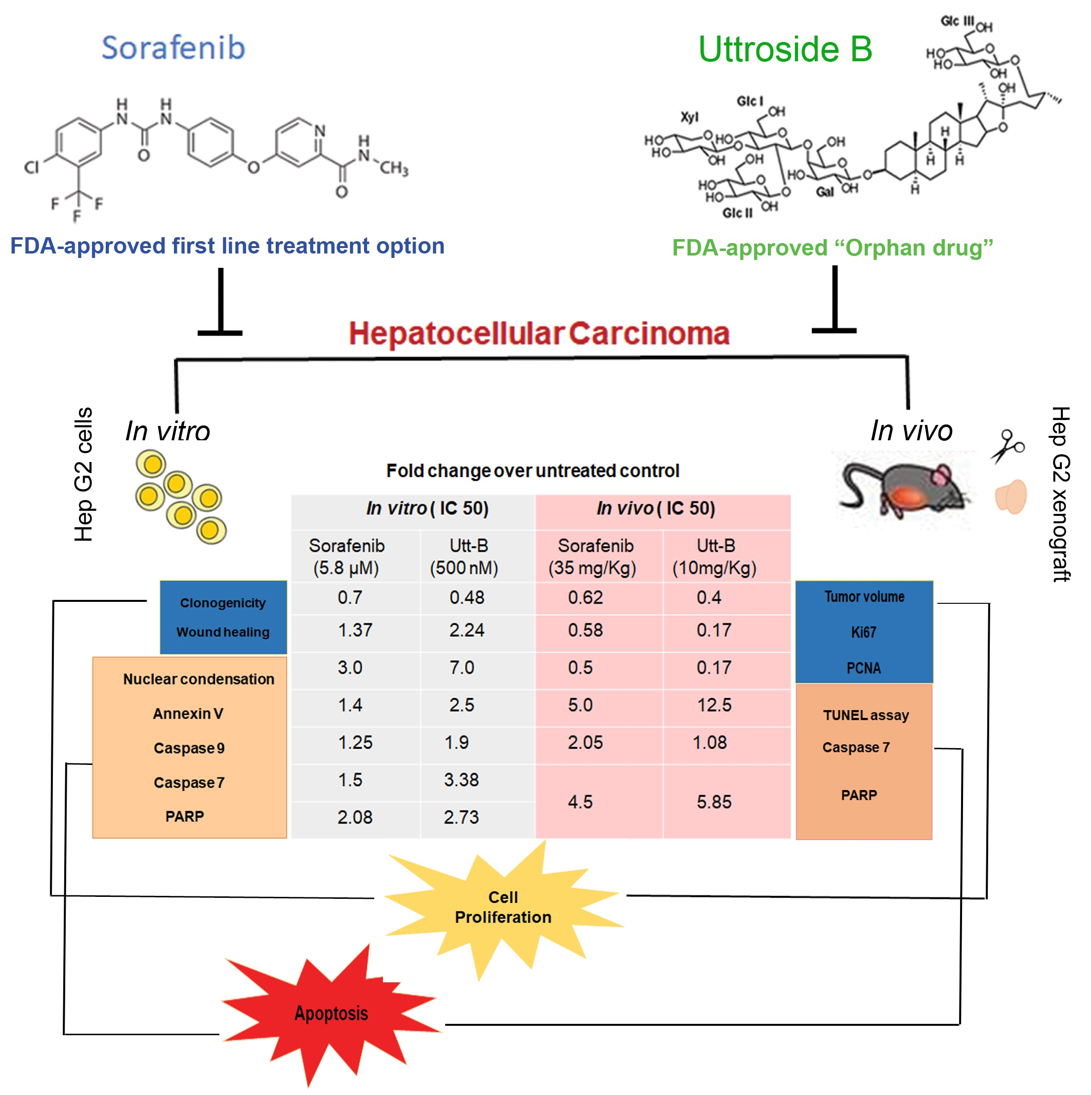
Augmented Efficacy of Uttroside B over Sorafenib in a Murine Model of Human Hepatocellular Carcinoma

 ,
,  , , , ,
  , and
, and
Abstract
:
1. Introduction
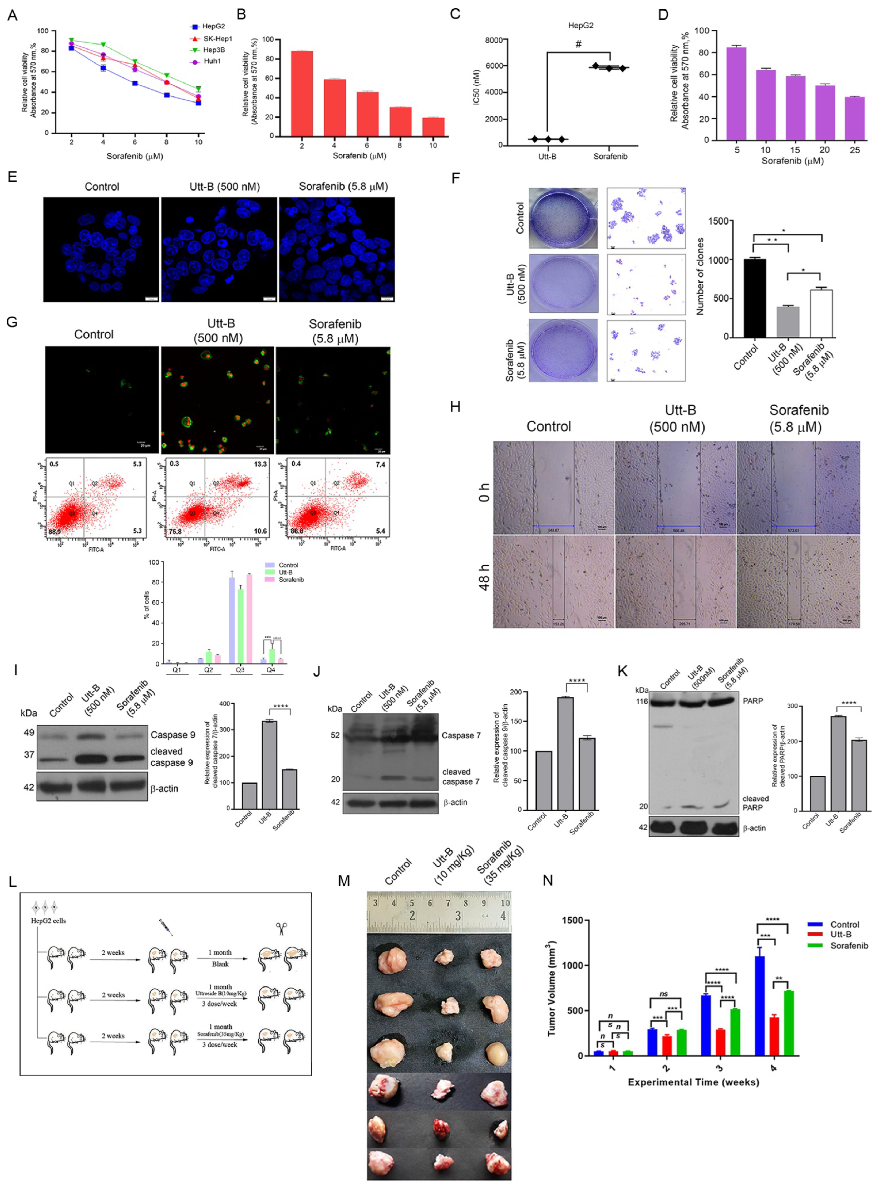
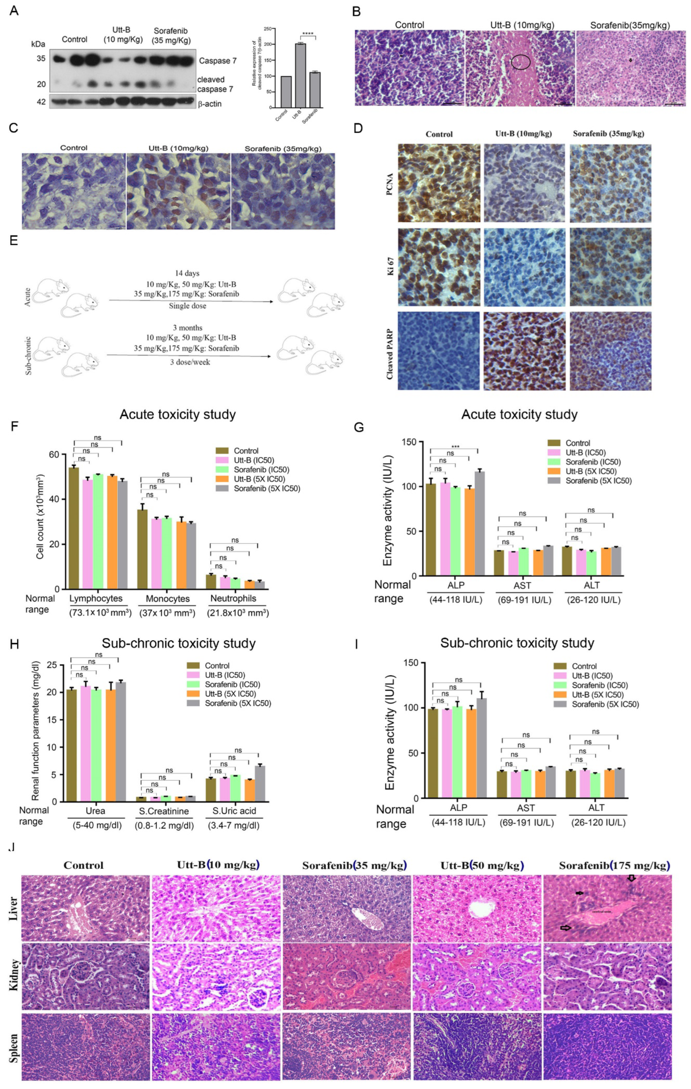
2. Results
3. Discussion
4. Materials and Methods
4.1. Chemicals
4.2. Cell Culture
4.3. In Vitro Assays
4.4. Animals
4.5. Flow Cytometry
4.6. Statistical Analysis
5. Conclusions
6. Patents
Author Contributions
Funding
Institutional Review Board Statement
Informed Consent Statement
Data Availability Statement
Acknowledgments
Conflicts of Interest
Abbreviations
| DAPI | 4′,6-diamidino-2-phenylindole |
| GAPDH | Glyceraldehyde-3-Phosphate Dehydrogenase |
| HCC | Hepatocellular Carcinoma |
| IC50 | Inhibitory Concentration 50 |
| MTT | 3-[4,5-dimethylthiazole-2-yl]-2,5-diphenyltetrazolium bromide |
| PARP | Poly Adenosine Diphosphate-Ribose Polymerase |
| PCNA | Proliferating Cell Nuclear Antigen |
| PDGFR | Platelet-Derived Growth Factor Receptor |
| RAF | Rapidly Accelerating Fibrosarcoma |
| Utt-B | Uttroside B |
| VEGF | Vascular Endothelial Growth Factor |
References
- Sung, H.; Ferlay, J.; Siegel, R.L.; Laversanne, M.; Soerjomataram, I.; Jemal, A.; Bray, F. Global cancer statistics 2020: GLOBOCAN estimates of incidence and mortality worldwide for 36 cancers in 185 countries. CA Cancer J. Clin. 2021, 71, 209–249. [Google Scholar] [CrossRef] [PubMed]
- Llovet, J.M.; Ricci, S.; Mazzaferro, V.; Hilgard, P.; Gane, E.; Blanc, J.-F.; De Oliveira, A.C.; Santoro, A.; Raoul, J.-L.; Forner, A. Sorafenib in advanced hepatocellular carcinoma. N. Engl. J. Med. 2008, 359, 378–390. [Google Scholar] [CrossRef] [PubMed] [Green Version]
- Woo, H.Y.; Heo, J. Sorafenib in liver cancer. Expert Opin. Pharmacother. 2012, 13, 1059–1067. [Google Scholar] [CrossRef] [PubMed]
- Rimassa, L.; Santoro, A. Sorafenib therapy in advanced hepatocellular carcinoma: The SHARP trial. Expert Rev. Anticancer Ther. 2009, 9, 739–745. [Google Scholar] [PubMed]
- Ibrahim, N.; Yu, Y.; Walsh, W.R.; Yang, J.-L. Molecular targeted therapies for cancer: Sorafenib monotherapy and its combination with other therapies. Oncol. Rep. 2012, 27, 1303–1311. [Google Scholar]
- Tang, W.; Chen, Z.; Zhang, W.; Cheng, Y.; Zhang, B.; Wu, F.; Wang, Q.; Wang, S.; Rong, D.; Reiter, F. The mechanisms of sorafenib resistance in hepatocellular carcinoma: Theoretical basis and therapeutic aspects. Signal Transduct. Target. Ther. 2020, 5, 1–15. [Google Scholar] [CrossRef]
- Cheng, A.-L.; Kang, Y.-K.; Chen, Z.; Tsao, C.-J.; Qin, S.; Kim, J.S.; Luo, R.; Feng, J.; Ye, S.; Yang, T.-S. Efficacy and safety of sorafenib in patients in the Asia-Pacific region with advanced hepatocellular carcinoma: A phase III randomised, double-blind, placebo-controlled trial. Lancet Oncol. 2009, 10, 25–34. [Google Scholar] [CrossRef]
- Basak, D.; Arrighi, S.; Darwiche, Y.; Deb, S. Comparison of Anticancer Drug Toxicities: Paradigm Shift in Adverse Effect Profile. Life 2021, 12, 48. [Google Scholar] [CrossRef]
- Nath, L.R.; Swetha, M.; Vijayakurup, V.; Thangarasu, A.K.; Haritha, N.H.; Shabna, A.; Aiswarya, S.U.; Rayginia, T.P.; Keerthana, C.K.; Kalimuthu, K.; et al. Blockade of Uttroside B-Induced Autophagic Pro-Survival Signals Augments Its Chemotherapeutic Efficacy Against Hepatocellular Carcinoma. Front. Oncol. 2022, 12, 1–16. [Google Scholar] [CrossRef]
- Guo, S.; Tian, Y.; Jian, L. Optimization of ethanol extraction process of Solanum nigrum Linn. and structural confirmation of its compounds. Asian J. Chem. 2014, 26, 4615–4618. [Google Scholar] [CrossRef]
- Wang, L.; Gao, G.; Bai, Y.; Luo, W.; Lin, C.; Jia, Q. Fingerprint quality detection of Solanum nigrum using high-performance liquid chromatography–evaporative light scattering detection. Pharm. Biol. 2011, 49, 595–601. [Google Scholar] [CrossRef] [PubMed]
- Heinig, U.; Aharoni, A. Analysis of steroidal alkaloids and saponins in Solanaceae plant extracts using UPLC-qTOF mass spectrometry. In Plant Isoprenoids; Humana Press: New York, NY, USA, 2014; pp. 171–185. [Google Scholar]
- Nath, L.R.; Gorantla, J.N.; Thulasidasan, A.K.T.; Vijayakurup, V.; Shah, S.; Anwer, S.; Joseph, S.M.; Antony, J.; Veena, K.S.; Sundaram, S. Evaluation of uttroside B, a saponin from Solanum nigrum Linn, as a promising chemotherapeutic agent against hepatocellular carcinoma. Sci. Rep. 2016, 6, 36318. [Google Scholar] [CrossRef] [PubMed] [Green Version]
- Strasser, A.; Cory, S.; Adams, J.M. Deciphering the rules of programmed cell death to improve therapy of cancer and other diseases. EMBO J. 2011, 30, 3667–3683. [Google Scholar] [CrossRef]
- Kuczynski, E.A.; Lee, C.R.; Man, S.; Chen, E.; Kerbel, R.S. Effects of sorafenib dose on acquired reversible resistance and toxicity in hepatocellular carcinoma. Cancer Res. 2015, 75, 2510–2519. [Google Scholar] [CrossRef] [PubMed] [Green Version]
- Ahmad, R. Steroidal glycoalkaloids from Solanum nigrum target cytoskeletal proteins: An in silico analysis. PeerJ 2019, 7, e6012. [Google Scholar] [CrossRef] [PubMed] [Green Version]
- Ahmad, R.; Gupta, A.; Fatima, A.; Husain, I.; Srivastava, A.N. The evaluation of biological activity of methanolic extracts of Solanum nigrum and molecular docking analysis of selected phytoconstituents against Vimentin. J. Intercult. Ethnopharmacol. 2017, 6, 391–400. [Google Scholar] [CrossRef]
- Bengala, C.; Bertolini, F.; Malavasi, N.; Boni, C.; Aitini, E.; Dealis, C.; Zironi, S.; Depenni, R.; Fontana, A.; Del Giovane, C. Sorafenib in patients with advanced biliary tract carcinoma: A phase II trial. Br. J. Cancer 2010, 102, 68–72. [Google Scholar] [CrossRef] [Green Version]
- Hussaarts, K.G.; van Doorn, L.; Bins, S.; Sprengers, D.; de Bruijn, P.; van Leeuwen, R.W.; Koolen, S.L.; van Gelder, T.; Mathijssen, R.H. Combining sorafenib and immunosuppression in liver transplant recipients with hepatocellular carcinoma. Pharmaceuticals 2021, 14, 46. [Google Scholar] [CrossRef]
- Nasser, H.M.; El-Naggar, S.A.; El-Sayed Rizk, M.E.-S.R.; Elmetwalli, A.; Salama, A.F. Effect of sorafenib on liver biochemistry prior to vitamin b17 coadministration in ehrlich ascites carcinoma mice model: Preliminary phase study. Biochem. Lett. 2021, 17, 40–49. [Google Scholar] [CrossRef]
- Staufer, K.; Fischer, L.; Seegers, B.; Vettorazzi, E.; Nashan, B.; Sterneck, M. High toxicity of sorafenib for recurrent hepatocellular carcinoma after liver transplantation. Transpl. Int. 2012, 25, 1158–1164. [Google Scholar] [CrossRef]
- Antonelli, G.; Gigante, E.; Iavarone, M.; Begini, P.; Sangiovanni, A.; Iannicelli, E.; Biondetti, P.; Pellicelli, A.M.; Miglioresi, L.; Marchetti, P. Sarcopenia is associated with reduced survival in patients with advanced hepatocellular carcinoma undergoing sorafenib treatment. United Eur. Gastroenterol. J. 2018, 6, 1039–1048. [Google Scholar] [CrossRef] [PubMed]
- Zhao, W.; Ma, B.; Tian, Z.; Han, H.; Tang, J.; Dong, B.; An, G.; Cao, B.; Wang, B. Inhibiting CBX4 efficiently protects hepatocellular carcinoma cells against sorafenib resistance. Br. J. Cancer 2021, 124, 1237–1248. [Google Scholar] [CrossRef] [PubMed]
- Childs, A.; Zakeri, N.; Ma, Y.T.; O’Rourke, J.; Ross, P.; Hashem, E.; Hubner, R.A.; Hockenhull, K.; Iwuji, C.; Khan, S. Biopsy for advanced hepatocellular carcinoma: Results of a multicentre UK audit. Br. J. Cancer 2021, 125, 1350–1355. [Google Scholar] [CrossRef] [PubMed]
- Regan-Fendt, K.; Li, D.; Reyes, R.; Yu, L.; Wani, N.A.; Hu, P.; Jacob, S.T.; Ghoshal, K.; Payne, P.R.; Motiwala, T. Transcriptomics-based drug repurposing approach identifies novel drugs against sorafenib-resistant hepatocellular carcinoma. Cancers 2020, 12, 2730. [Google Scholar] [CrossRef]


Publisher’s Note: MDPI stays neutral with regard to jurisdictional claims in published maps and institutional affiliations. |
© 2022 by the authors. Licensee MDPI, Basel, Switzerland. This article is an open access article distributed under the terms and conditions of the Creative Commons Attribution (CC BY) license (https://creativecommons.org/licenses/by/4.0/).
Share and Cite
Swetha, M.; Keerthana, C.K.; Rayginia, T.P.; Nath, L.R.; Haritha, N.H.; Shabna, A.; Kalimuthu, K.; Thangarasu, A.K.; Aiswarya, S.U.; Jannet, S.; et al. Augmented Efficacy of Uttroside B over Sorafenib in a Murine Model of Human Hepatocellular Carcinoma. Pharmaceuticals 2022, 15, 636. https://doi.org/10.3390/ph15050636
Swetha M, Keerthana CK, Rayginia TP, Nath LR, Haritha NH, Shabna A, Kalimuthu K, Thangarasu AK, Aiswarya SU, Jannet S, et al. Augmented Efficacy of Uttroside B over Sorafenib in a Murine Model of Human Hepatocellular Carcinoma. Pharmaceuticals. 2022; 15(5):636. https://doi.org/10.3390/ph15050636
Chicago/Turabian StyleSwetha, Mundanattu, Chenicheri K. Keerthana, Tennyson P. Rayginia, Lekshmi R. Nath, Nair Hariprasad Haritha, Anwar Shabna, Kalishwaralal Kalimuthu, Arun K. Thangarasu, Sreekumar U. Aiswarya, Somaraj Jannet, and et al. 2022. "Augmented Efficacy of Uttroside B over Sorafenib in a Murine Model of Human Hepatocellular Carcinoma" Pharmaceuticals 15, no. 5: 636. https://doi.org/10.3390/ph15050636
APA StyleSwetha, M., Keerthana, C. K., Rayginia, T. P., Nath, L. R., Haritha, N. H., Shabna, A., Kalimuthu, K., Thangarasu, A. K., Aiswarya, S. U., Jannet, S., Pillai, S., Harikumar, K. B., Sundaram, S., Anto, N. P., Wu, D. H., Lankalapalli, R. S., Towner, R., Isakov, N., Deepa, S. S., & Anto, R. J. (2022). Augmented Efficacy of Uttroside B over Sorafenib in a Murine Model of Human Hepatocellular Carcinoma. Pharmaceuticals, 15(5), 636. https://doi.org/10.3390/ph15050636