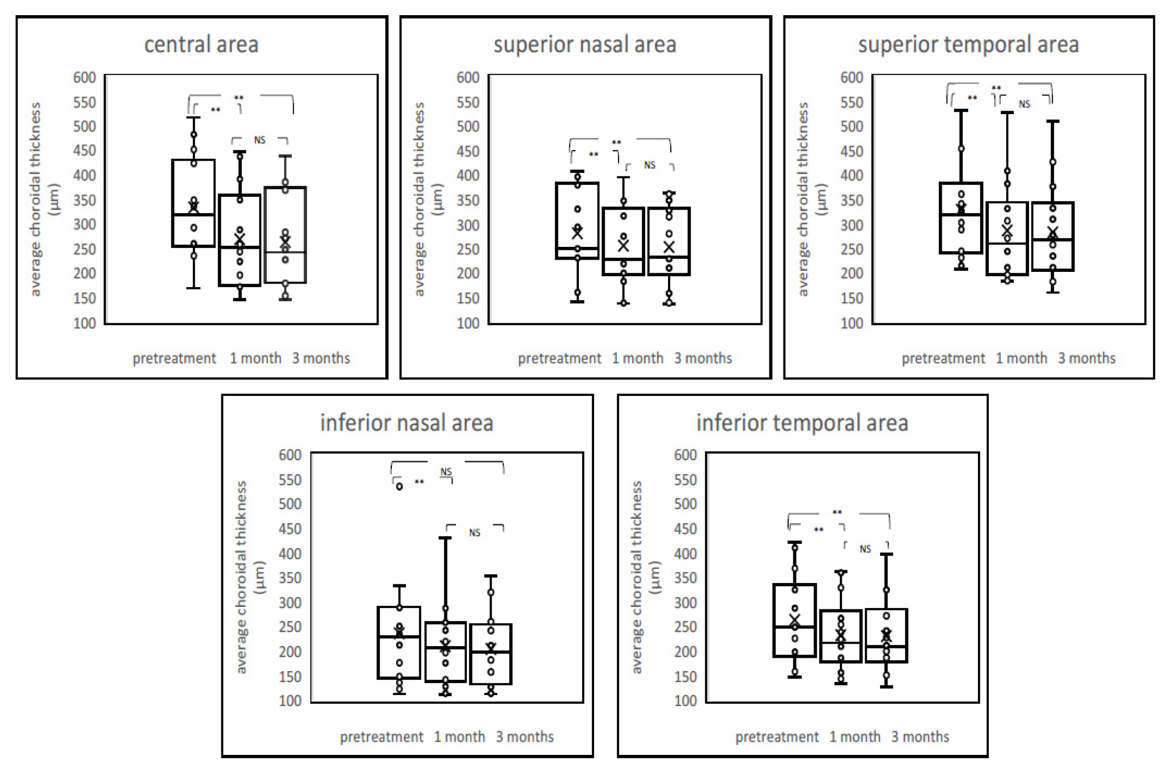
Correction: Sato-Akushichi et al. Choroidal Volume Evaluation after Photodynamic Therapy Using New Optical Coherence Tomography Imaging Algorithm. Pharmaceuticals 2021, 14, 1140
Reference
- Sato-Akushichi, M.; Ono, S.; Klose, G.; Song, Y. Choroidal Volume Evaluation after Photodynamic Therapy Using New Optical Coherence Tomography Imaging Algorithm. Pharmaceuticals 2021, 14, 1140. [Google Scholar] [CrossRef]

Publisher’s Note: MDPI stays neutral with regard to jurisdictional claims in published maps and institutional affiliations. |
© 2022 by the authors. Licensee MDPI, Basel, Switzerland. This article is an open access article distributed under the terms and conditions of the Creative Commons Attribution (CC BY) license (https://creativecommons.org/licenses/by/4.0/).
Share and Cite
Sato-Akushichi, M.; Ono, S.; Klose, G.; Song, Y. Correction: Sato-Akushichi et al. Choroidal Volume Evaluation after Photodynamic Therapy Using New Optical Coherence Tomography Imaging Algorithm. Pharmaceuticals 2021, 14, 1140. Pharmaceuticals 2022, 15, 349. https://doi.org/10.3390/ph15030349
Sato-Akushichi M, Ono S, Klose G, Song Y. Correction: Sato-Akushichi et al. Choroidal Volume Evaluation after Photodynamic Therapy Using New Optical Coherence Tomography Imaging Algorithm. Pharmaceuticals 2021, 14, 1140. Pharmaceuticals. 2022; 15(3):349. https://doi.org/10.3390/ph15030349
Chicago/Turabian StyleSato-Akushichi, Miki, Shinji Ono, Gerd Klose, and Youngseok Song. 2022. "Correction: Sato-Akushichi et al. Choroidal Volume Evaluation after Photodynamic Therapy Using New Optical Coherence Tomography Imaging Algorithm. Pharmaceuticals 2021, 14, 1140" Pharmaceuticals 15, no. 3: 349. https://doi.org/10.3390/ph15030349
APA StyleSato-Akushichi, M., Ono, S., Klose, G., & Song, Y. (2022). Correction: Sato-Akushichi et al. Choroidal Volume Evaluation after Photodynamic Therapy Using New Optical Coherence Tomography Imaging Algorithm. Pharmaceuticals 2021, 14, 1140. Pharmaceuticals, 15(3), 349. https://doi.org/10.3390/ph15030349


