Review

Protein-Protein Interaction Inhibitors Targeting the Eph-Ephrin System with a Focus on Amino Acid Conjugates of Bile Acids

Department of Food and Drug, University of Parma, I-43124 Parma, Italy
*
Authors to whom correspondence should be addressed.
Pharmaceuticals 2022, 15(2), 137; https://doi.org/10.3390/ph15020137
Submission received: 23 December 2021 / Revised: 19 January 2022 / Accepted: 20 January 2022 / Published: 24 January 2022
(This article belongs to the Section Medicinal Chemistry)

Abstract

:
The role of the Eph-ephrin system in the etiology of pathological conditions has been consolidated throughout the years. In this context, approaches directed against this signaling system, intended to modulate its activity, can be strategic therapeutic opportunities. Currently, the most promising class of compounds able to interfere with the Eph receptor-ephrin protein interaction is composed of synthetic derivatives of bile acids. In the present review, we summarize the progresses achieved, in terms of chemical expansions and structure-activity relationships, both in the steroidal core and the terminal carboxylic acid group, along with the pharmacological characterization for the most promising Eph-ephrin antagonists in in vivo settings.

Graphical Abstract

1. Introduction

Erythropoietin-producing human hepatocellular (Eph) receptors form the largest subfamily of receptor tyrosine-kinases (RTKs), with several known members [1]. They are classified into two subclasses, A (EphA1-EphA8, EphA10) and B (EphB1-EphB4, EphB6) based on their amino acid sequences, as well as for their affinity for a class of membrane proteins, known as ephrins [2]. Eph receptors are composed by an extracellular region containing the ligand-binding domain (LBD) responsible for the interaction with the ephrins, a transmembrane portion, and a cytoplasmatic domain, which includes (i) a catalytically competent tyrosine-kinase domain; (ii) a sterile alpha motif (SAM); and (iii) a PDZ-binding motif [3], all dealing with signal transductions in Eph receptor expressing cells [4].
The ephrins are membrane proteins classified in two groups upon the sequence homology. The distinctive element between ephrin-A and -B is the presence in the latter one of a cytoplasmatic portion where a tyrosine-rich domain and a PDZ binding domain are present, while ephrin-As are exposed on the membrane by a glycosylphosphatidylinositol (GPI) anchor [4].
Due to their organization and localization, Eph-ephrin association occurs when contiguous cells have physical contact with one another [5]. Once a first Eph-ephrin heterodimer is formed, this event triggers the formation of heterotetramers; these structures associate further with generate oligomers [6]. It is now well understood that, upon Eph-ephrin association, Eph receptors form large polymeric clusters, and the phenotypic cellular response will depend on the size, composition, and spatiotemporal assembly [7,8].
A unique feature of the Eph-ephrin system is the ability to generate bidirectional signals [9]. The formation of an Eph-ephrin cluster generates forward signals in receptor expressing cells and reverse signals in ligand expressing cells [10,11,12]. The Eph-ephrin system plays a crucial role during embryogenesis as it is essential for development processes including cell migration and differentiation [10,13,14,15]. Conversely, in adults, the expression of this system is limited to those tissues where cell renewal is frequent, i.e., skin [16], breast [17], and intestinal tissues [18].
Compelling data gathered in the last two decades evidenced that the Eph-ephrin system is involved in the insurgence of different pathological conditions, including cancer diseases [19,20,21,22]. Its involvement within different facets of tumorigenesis and cancer progression, such as tumor angiogenesis, vasculomimicry, metastasis development, and tumor stem cell propagation, were reported and exhaustively described in the literature [20,21]. The Eph-ephrin system has emerged as an oncogenic pathway driving the insurgence of solid and liquid tumors. For instance, high expression of EphA2 and EphA3 has been linked to poor survival identified in glioblastoma multiforme (GBM) [23,24,25]. Similarly, EphB4, and ephrin-B2 have recently been found to concur to proliferation and in the invasive potential of GBM [26]. These and other findings indicate that multiple Eph receptors and ephrin ligand subtypes synergistically act in the insurgence of this type of cancer [27].
This scenario has therefore prompted the search of molecular agents targeting the Eph-ephrin system with a two-fold aim: (i) confirming the relevance of clinical data reported in the literature; and (ii) designing an efficient strategy based on biologics and small molecules to interact with targets of this elusive system in cancer therapy.

2. Agents Targeting the Eph-Eph System

2.1. Biologics

First attempts to target the Eph-ephrin system involved monoclonal antibodies (mAbs) and soluble forms of the Eph receptors or ephrin proteins. Monoclonal antibodies, acting in most cases as agonist agents, disrupt the Eph receptor ligand-independent activities mimicking the binding of the natural ligand at the extracellular LBD level. However, mAb agonists promote EphA2 degradation after induction of the phosphorylation activity of the receptor leading to an efficient blocking of forward signal transduction [28,29,30].
One of the most investigated mAb is MEDI-547, a fully human monoclonal antibody, identified after the phage display selection, which targets the LBD of EphA2 [31]. Its CDR-H3 loop mimics the interaction between EphA2 receptor–ephrin-A1, inducing the phosphorylation and the internalization of the receptor [32,33]. MEDI-547 reached the phase I clinical trial in patients with relapsed and refractory solid tumors (NCT00796055), but its safety and tolerability were not confirmed due to treatment-related bleeding and coagulation events [33]. Other recent mAbs that reached clinical phases are DS-8895, an anti-EphA2 monoclonal antibody developed by Daichii-Sankyo, and KB004, a non-fucosylated IgG1k antibody developed by Kalobios, and known by the name ifabotuzumab. The former, after a favorable phase I, was discontinued [34], while the latter reached phase II in patients with EphA3-expressing hematologic malignancies but discontinued for myelofibrosis (MF) (NCT01211691). KB004 is now under investigation in clinical phase I for glioblastoma multiforme (GBM) (NCT03374943) [35,36,37].
Soluble fragments of Eph receptors are alternative approaches to modulate Eph forward signaling. Receptors engineered with Fc domain of IgGs to increase their stability, can be used to compete with the endogenous Eph-ephrin system through ligand binding. The most advanced biological agent is a recombinant protein in which the ectodomain of EphB4 is fused with human serum albumin. A phase II trial study has been testing the combination of pembrolizumab and sEphB4-HSA in patients with previously treated urothelial carcinoma (NCT02717156) [38]. Another pilot study is focused on the administration of sEphB4-HSA before surgery in patients with bladder, prostate, or kidney cancer, with the aim to shrink the tumor and increase the amount of healthy tissue to be preserved with the surgery (NCT02767921) [39].

2.2. Peptides and Peptidomimetics

Another strategy to block Eph-ephrin signaling is to harness peptides of medium size. The mechanism of action of this class of agents is represented by the occupation of the LBD of Eph receptor, preventing the binding of the ephrin ligands.
A first generation of peptides was identified by Pasquale and colleagues through a phage display screening. The approach led to the discovery of YSA and SWL (Figure 1i), two 12-mer peptides, showing high potency and selectivity for the EphA2 receptor. YSA and SWL display an agonist behavior, inducing receptor phosphorylation and signal transduction [40]. This and other studies allowed drawing a robust structure–activity relationship around peptides binding to Eph. For instance, fragments that preferentially bind class-A Eph receptors possess in their sequence a conserved Ω-X-X-Ω motif, where Ω represents an aromatic amino acid residue and X is a non-conserved amino acid. This binding motif is also present in the G-H loop of nearly all ephrin ligands, confirming the importance of Ω-X-X-Ω motif for Eph receptor binding [41]. APY, KYL, and VTM peptides were later reported as selective antagonists of EphA4 receptor able to prevent receptor activation [42]. While showing promising good pharmacodynamic properties, most of these peptides suffer from an extensive and rapid degradation in plasma. Modifications of these linear peptides, intended to overcome metabolic stability issues, were based on cyclization (i.e., through cysteine-cysteine disulfide bridges), PEGylation, dimerization, insertion of unnatural amino acids, or inclusion into nanoparticles [43]. Peptides targeting Eph receptors can be employed for the selective delivery of chemotherapy in cancer cells overexpressing Eph receptors. Recently, Pasquale and co-workers showed that YSA PEGylated with lipid nanoparticles loaded with docetaxel showed improvements in terms of efficacy and toxicity when compared to the administration of the free drug, in a rat model of choroidal neovascularization [44].
The development of a peptidic ligand targeting Eph receptor is an active area of research and prompted the search for peptidomimetic agents. YSA was also the starting point for an extensive work made by Pellecchia and coworkers that led to the identification of novel peptidomimetic agonists with a nanomolar affinity for the EphA2 receptor and improved physicochemical properties. This was the case of dYNH peptide (Figure 1ii), in which replacement of two unnatural amino acids at the C-terminal portion and the substitution of L-Tyr with D-Tyr at the N-terminus allowed to achieve good plasma stability (10 times higher for dYNH than YSA), with a moderate loss of affinity for EphA2 [45]. This finding steered synthetic efforts toward a deep exploration of the role played by the N-terminal amino acid of EphA2 ligands. Therefore, employing an NMR-based binding assay using recombinant EphA2-LBD [46], researchers identified 123B9 (Figure 1iii), a peptidomimetic agent with an affinity comparable to the parent compound and an improved metabolic profile both in vitro and in vivo [45]. Subsequently, optimization of the C-terminal region of 123B9 structure identified two other peptidomimetics, 135E2 and 135G3, which resulted significantly more potent than the parent agents [47]. Moreover, the X-ray structure of 135E2 in complex with the EphA2 ligand-binding domain helped the structure-based optimization and subsequent identification of 135H11 (Figure 1iv), a peptidomimetic with a IC50 of 0.15 μM against EphA2 [47].
Phage display screenings were also performed to identify peptides selectively targeting EphB receptors, leading to SNEW (Figure 2i), a dodecameric peptide, able to bind EphB2 (KD = 6 μM) while inhibiting ephrinB2 recruitment by EphB2 with an IC50 value of 15 μM [48]. Visual inspection of the SNEW-EphB2 crystallographic complex showed that SNEW adopts a different binding mode with respect to ephrinB2 ligand [49].
Another dodecameric peptide (TNYL—Figure 2ii) was identified as a weak inhibitor of the ephrinB2–EphB4 interaction (IC50 = 50–150 μM) [48]. Modifications of the C-terminal region TNYL led to TNYL-RAW (Figure 2iii) peptide, which displayed an inhibitory potency in the nanomolar range. The substantial increase in potency was ascribed to the ability of TNYL-RAW of undertaking additional productive interactions with EphB4-LBD. This hypothesis was validated by the resolution of the crystal structure of TNYL-RAW bounded to the EphB4 [50,51].
TNYL-RAW was characterized by short half-life in plasma and in cells due to its susceptibility to proteolytic degradation, thereby advancing attempts to increase its chemical stability were performed. Cyclization of TNYL-RAW provided cTNYL-RAW (Figure 2iv) which exhibits greatly increased stability in mouse plasma, without affecting the affinity for EphB4 [52]. Similar results were obtained through PEGylation or inclusion in gold nanosphere [52,53].

2.3. Bile Acids Derivatives: An Emerging Class of Small-Molecules Disrupting Eph-ephrin Interaction

An alternative strategy to interfere with signals transduced by the Eph-ephrin system is represented by small molecules targeting the protein-protein interaction (PPI) surface, and specifically by agents able to recognize the extracellular LBD of Eph receptors [54]. Small molecules able to interfere with the formation of a Eph-ephrin heterodimer can block both forward and reverse signals in cells with potential benefits in term of efficacy [10]. The presence in the LBD of Eph receptors of a large and solvent exposed cavity deputed to the recognition of ephrin ligands has discouraged drug discovery efforts in this area for many years. This negative thinking was reinforced by the finding that the activity displayed by the first class of small molecules reported to be active on the EphA2 receptor, i.e., 4-(2,5-dimethyl-CH-pyrrol-1-yl)-2-hydroxybenzoic acids, was actually due to an uncharacterized mixture of polymers of high molecular weight, accidentally generated after the exposure of these pyrrolyl-based compounds to air and light [55].
The resolution of X-ray structures of the LBD of many other Eph receptors in complex with ephrin ligands and the commitment of academic research groups favored the discovery of small molecules targeting this system. Most of the available complexes (i.e., EphA2-ephrin-A1 or EphB2-ephrin-A5) confirmed that the formation of the Eph-ephrin heterodimer was driven by the accommodation of the G-H loop present in ephrin ligands within a well-defined channel present in the LBD of the Eph receptors. Crucial for drug design was the identification of polar hot spots in the LBD of Eph receptors (such as Arg103 and Arg156 in EphA2), which later steered screening campaigns versus specific classes of compounds [41].
The presence of accessible basic residues in the EphA2 LBD suggested that acid compounds might display some affinity for this receptor. A screening campaign of a chemical collection of acid compounds at the University of Parma allowed to identify lithocholic acid (LCA) as weak competitive antagonist of the EphA2, able to displace ephrin-A1 from EphA2 with a Ki = 49 µM [56]. Other bile acids, such as cholic (CA), deoxycholic (DCA), and chenodeoxycholic (CDCA), were unable to interfere with ephrin-A1 binding to EphA2. These preliminary results underlined the presence of tight structural requirements (summarized in Figure 3) for engaging the lipophilic channel present in the LBD of EphA2 [56].
A following investigation by Tognolini et al. in 2012 allowed expanding structure–activity relationships (SARs) around LCA [57]. In this work, it was confirmed that the introduction in position 6, 7, and 12 of the gonane core of LCA of polar substituents were not tolerated by EphA2, as shown by the lack of activity displayed by compounds 26 (Table 1). Conversely, modification of 3α-hydroxyl group appeared promising for the identification of more potent antagonists. Inversion of the absolute configuration of position 3 or removal of the 3α-hydroxyl group led to isolithocholic acid (7) or cholanic acid (8), which displayed Ki values of 25 and 5 μM, respectively. Other important information was inferred from the modification of the terminal carboxylic group of LCA. Compounds 911, characterized by the presence of functional groups of lower acidity (9 and 10) or by an ester functionality (11), were inactive. Removal of an ionizable group (negatively charged at physiological pH) resulted in a detrimental effect on compound potency, suggesting that a terminal carboxylate was a fundamental pharmacophoric element for EphA2 binding [57].
Molecular modeling investigations suggested that LCA could bind the LBD of the EphA2 receptor occupying the same space of the ephrin-A1 G-H loop, by inserting its gonane scaffold into the hydrophobic channel present in the EphA2 receptor. According to this model, the 2-methylbutanoic acid chain of LCA could form a salt bridge with Arg103, mimicking the interaction of ephrin-A1 Glu119 [57]. The 3α-hydroxy group weakly interacted with Arg159 of EphA2 usually engaged in a hydrogen bond (H-bond) with Asp86 of ephrin-A1. Although other arrangements of LCA can be identified (i.e., with the gonane ring superposed on the G-H loop region but with the terminal carboxyl group of LCA forming salt bridge with Arg159), the binding mode reported in Figure 4 accounts to some extent for SAR data reported in Table 1, and more importantly, could be exploited for the design of novel Eph-ephrin antagonists.
Visual inspection of the EphA2-LCA complex generated by docking suggested that coupling of LCA with α-amino acids could give antagonists able to form a salt bridge with Arg103, while potentially forming additional productive interactions within the LBD of EphA2 [58]. The fair activity of glycolithocholic acid 12 (Table 2) suggested that conjugation was a promising strategy to identify better EphA2 ligands.
An experimental design, aimed at independently explore the impact of lipophilicity and steric hindrance on the inhibitory potency, identified a set of four α-amino acids (Ala, Asn, Ser, Val of both L- and D-series) to be conjugated with LCA for SAR evaluation. The resulting compounds (1421, listed in the Table 2) were tested for their ability to displace biotinylated ephrin-A1 from EphA2 [59]. Compounds with a hydrophobic sidechain (Ala or Val, 1417) resulted more potent than those with polar chains (Ser or Asn, 1821). Other α-amino acids were conjugated to LCA giving compounds 22–31, to further cover the space of lipophilic and steric properties. The introduction of amino acids with lipophilic side chains always led to active compounds. Notably, the introduction of aromatic substituents had a positive impact on the inhibitory potency. Phenylalanine conjugates 26 and 27 were ten times more potent than LCA. This trend was confirmed by the activity of tryptophan conjugates 30 and 31, which were significantly more potent than LCA. With an IC50 value of 2.0 μM, the L-Trp conjugate (30, UniPR126) was the most potent antagonist of the series [59]. Noteworthy, the potency of UniPR126 did depend on the stereochemistry of the α-amino acid, as D-Trp conjugate (31) was 10 times less potent than UniPR126 [59]. This could be due to the accommodation of the indole moiety in a region of the EphA2 receptor which specifically recognizes aromatic substituents [41]. Docking of UniPR126 within EphA2 receptor (Figure 5) supported this hypothesis as the indole ring of this antagonist occupied the same space of Phe111 of ephrin-A1 forming π–π interactions with Phe108 of EphA2 [59,60].
Crucially, visual inspection of the EphA2−UniPR126 complex also suggested that affinity for the EphA2 receptor could be increased by bringing the carboxylate group of UniPR126 closer to the guanidine group of Arg103 [61]. This working hypothesis prompted the synthesis of the L-β-Homo-Trp conjugate of LCA, UniPR129 (Figure 6).
Pharmacological evaluation of UniPR129 supported the improvement sought with the help of molecular modeling. This compound showed an IC50 of 0.9 μM for inhibition of ephrin-A1 binding to EphA2, a value two-fold lower than that reported for UniPR126 (IC50 = 2 μM) [59,61]. In analogy to what was observed for UniPR126, UniPR129 was able to interfere with the binding of ephrin ligands to all the cloned Eph receptors (i.e., EphA1-A8; EphB1-B4, EphB6) with similar potency, suggesting that this compound likely interacted with a conserved region of the LBD of the Eph receptors [61]. UniPR129 emerged as useful pharmacological tool for in vitro experiments. UniPR129 inhibited EphA2 autophosphorylation in human prostate cancer (PC3) cells, and it was able to modify prototypical phenotypes controlled by ephrin-A1 stimulation, including cell-rounding and migration. Additionally, at low micromolar concentration, UniPR129 inhibited the ability of human umbilical vein endothelial cells (HUVECs) to form capillary-like tubes in vitro, proposing this compound as potential antiangiogenetic agents to be evaluated in vivo [61].
Nevertheless, UniPR129 suffered from poor physicochemical properties which heavily hampered its bioavailability when orally administered to mice [62], and prompted investigators to further explore the SAR to yield antagonist suitable for in vivo administration.
In 2017, Incerti et al. reported an extended investigation on the SAR around UniPR129. The impact of the length of the linker connecting the terminal carboxylic acid to the amino group of the amino acid moiety on inhibitory potency was investigated (Table 3).
This analysis suggested that the β-alanine conjugate (32) had the optimal length as a further lengthening of the linker gave inactive compounds as observed in the cases of 33 and 34 [63]. Replacement of the carboxylic acid of 32 with a sulfonic acid (as for 35) or its esterification (as for 36) led to a significant loss of potency [63]. The presence of a negatively charged group in the lateral side chain of the amino acid moiety was beneficial for activity only when specific geometric requirements were satisfied.
Given these results, researchers focused their subsequent efforts on the synthesis and pharmacological evaluation of β-alanine conjugates of LCA bearing in β-position lipophilic sidechains of varying steric requirements (Table 4) [63].
Compounds possessing aliphatic chains (3739) were generally less potent than the parent compound 32. The introduction of a phenyl group with different spacers in the side chain resulted in an increase of the inhibitory potency, as in the case of compounds 40 and 41. Only compound 42, with a phenylethyl substituent, resulted inactive.
Important improvements were obtained with the introduction of larger aryl groups as side chain substituents. When the lateral chain was featured by a (i) indol-3-ylmethyl (UniPR129); (ii) α-naphthylmethyl (43); or (iii) benzo[b]thiophen-3-ylmethyl (44) substituent, a significant improvement in the inhibitory potency was observed, with the resulting compounds active in the low μM. UniPR129 was the best compound of the series, and its higher potency compared to 43 and 44 could be ascribed to the presence of an indole N-H group, potentially able to form polar interactions within EphA2. This was somehow supported by the significant drop of activity displayed by diastereomer of UniPR129 (compound 45), in which the absolute configuration of the β carbon was inverted [63]. To better understand the possible interactions formed by UniPR129 within EphA2, molecular dynamics (MD) simulations were performed. These simulations supported the formation of a H-bond occurring between the N-H of the indole ring of UniPR129 and the carboxylate group of Asp53, giving a model-based explanation to the observed SAR [63].
The same MD simulations suggested that the 3α-hydroxyl group of UniPR129 was an important anchor point for EphA2 binding, being able to form a H-bond with the side chain of Asn57 and/or with Ile58 backbone [63]. This computational suggestion opened a new route for further investigations. In this scenario, a SAR evaluation directed towards position 3 of UniPR129 (Table 5) was performed with the two-fold aim of improving inhibitory potency and modulate physicochemical properties.
A focused set of modifications at position 3 of UniPR129 was planned to confirm if the presence of a substituent able form a H-bond with EphA2 was critical for the activity. To this end, the 3α-hydroxyl of UniPR129 was replaced by a 3β-hydroxyl (46), by a hydrogen (47), or oxidized to a 3-keto functional group (48). A significant loss in the inhibitory potency was observed for these three analogues [63].
These results prompted the insertion of other groups able to form productive polar interactions, including H-bonds, within EphA2. Replacement of the 3α-hydroxyl of UniPR129 with a 3-hydroxyimino led to compound 49, which resulted rather potent at preventing ephrin-A1 binding to EphA2. Crucially, compound 51 (also known as UniPR502), a 3α-carbamoyloxy derivative, resulted slightly more potent than UniPR129 showing an IC50 value of 0.8 μM. Methylation of 49 and inversion of the C3 configuration of UniPR502 gave compounds 50 and 52, respectively which were less potent than their cognate analogues by nearly five-fold [64].
The slightly improvement of the inhibitory potency obtained with UniPR502, led to a follow-up exploration. Efforts were thus taken to exploit the carbamoyloxy functionality and to introduce a point for chemical diversification into the 5β-cholan-24-oic acid scaffold of UniPR502, to search for better EphA2 antagonists.
The nitrogen atom of the 3α-carbamoyloxy was functionalized (Table 6) with different substituents [64], while preserving the possibility to form a H-bond with EphA2 receptor. The ethyl, butyl and hexyl carbamate derivatives, 53 (UniPR505), 54 and 55, showed IC50 values in line with that of UniPR502, suggesting that alkyl chains of moderate size were tolerated by EphA2 receptor. The further lengthening of the alkyl chain was detrimental for activity as in the case of octyl derivative 56 that displayed a marked decrease of the inhibitory potency (IC50 = 15 µM) [64]. Finally, the introduction of branched or cyclic substituents gave compounds (i.e., 57, 58, 59) fair potency but always lower than that displayed by the reference EphA2 antagonist UniPR502.
Compound 53 (UniPR505) emerged as one of the most interesting compounds of this series and it was the subject of an intense biochemical characterization. UniPR505 demonstrated to act as a competitive and reversible antagonist of the EphA2 receptor, displaying a Ki of 0.3 μM in ELISA binding assay. When tested on PC3 cells, UniPR505 dose-dependently inhibited EphA2 phosphorylation in PC3 cells with an IC50 of 1.5 μM, very close to the inhibitory activity showed in the cell-free displacement assay (IC50 of 0.95 μM) [64]. When tested on endothelial cells (HUVECs), UniPR505 was able to dose-dependently inhibit angiogenesis with an IC50 value lower 3 μM, resulting a better antagonist than UniPR129. This prompted the use of UniPR505 in an in vivo model of neovascularization such as the chick chorioallantoic membrane (CAM) system. In the CAM model, UniPR505 dramatically reduced the growth of blood vessels in the presence of angiogenic growth factors [64]. Finally, differently from what reported for 3α-hydroxy-5β-cholan-24-oyl amino acid conjugates (including UniPR126 and UniPR129, able to engage all the Eph receptors with comparable inhibitory potency), UniPR505 demonstrated a preference for the EphA2, since it spared some Eph receptor subtypes i.e., EphA1, EphA7, EphB1, EphB4, and EphB6. UniPR505 emerged as one of the most interesting Eph-ephrin antagonist with a moderate selectivity for the EphA2 receptor, and physicochemical properties compatible with its use in vivo [64].

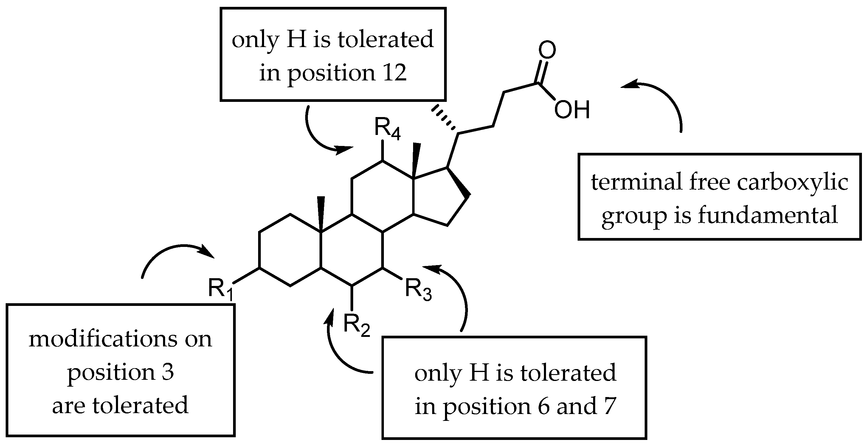
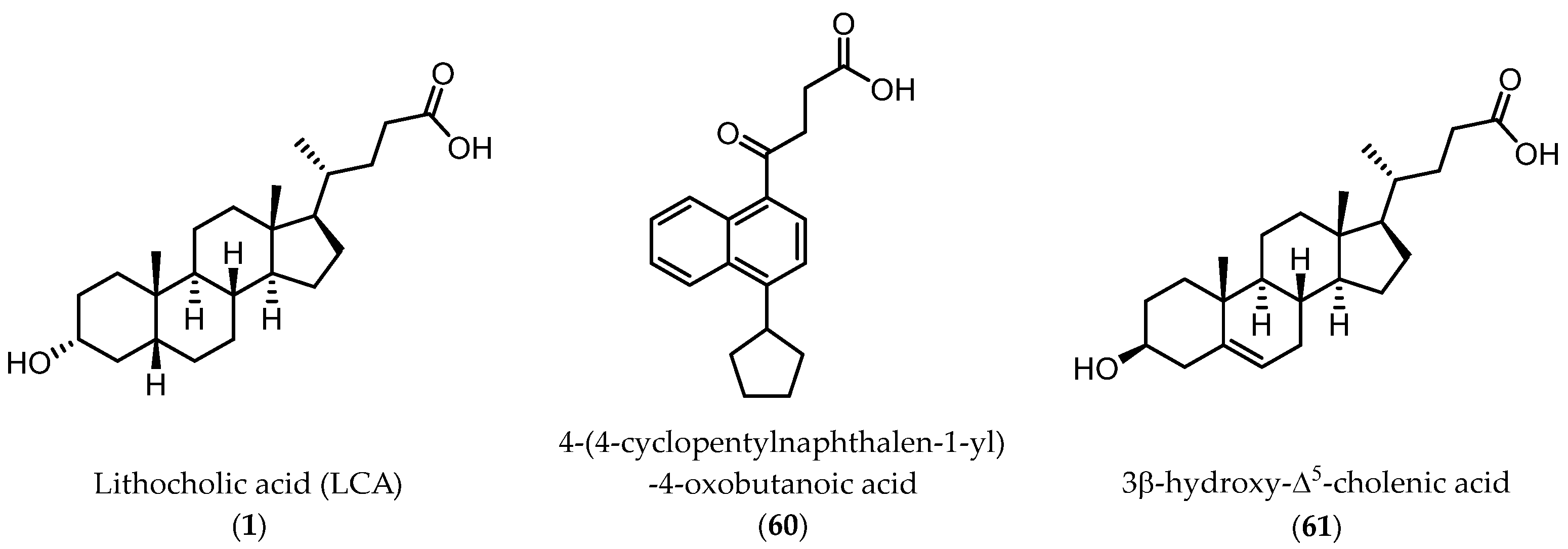
2.3.1. A New Scaffold for EphA2 Receptor Antagonists: Discovery of 3β-Hydroxy-Δ5-Cholenic Acid

Despite the positive results and the improvements with amino acid conjugates of LCA, other strategies were taken to discover new chemotypes working on the EphA2 receptor. A virtual screening campaign performed at University of Parma identified, 4-(4-cyclopentylnaphthalen-1-yl)-4-oxobutanoic acid (60) and 3β-hydroxy-Δ5-cholenic acid (61) (Figure 7) as inhibitors of the EphA2−ephrin-A1 interaction [65].
While the 3β-hydroxy-Δ5-cholenic acid (61) showed an inhibitory activity comparable to that of LCA, the geometry of its steroidal scaffold, resembling that of a 5α-gonane nucleus in contrast to the 5β-gonane of LCA, had the potential to ensure to 61 itself a better selectivity profile. Intriguingly, compound 61 did not engage classical targets of bile acids, including nuclear receptors (FXR, PXR) and G-protein coupled receptors [65], proposing it as new scaffold for the generation of new Eph-ephrin antagonists.
Conjugation of 60 with L-Trp led to an inactive compound, while conjugation of 61 with the same amino acid generated a new EphA2 antagonist named, UniPR1331 (Figure 8), featured by low micromolar affinity (IC50 value of 3.3 μM, vide infra) for this receptor. This result paralleled what observed for LCA, in which conjugation with L-Trp guaranteed a significant improvement in the inhibitory potency on EphA2-ephrin-A1 system, with the resulting compound (UniPR126, compound 30) displaying an IC50 value of 2.0 μM.
Even though UniPR126 and UniPR1331 share a high 2D similarity, these two compounds significantly differ in their 3D geometry. The bile acid moiety of UniPR126 has a bent shape due to a cis-junction connecting ring A and B of the 5β-gonane scaffold, while Δ5-cholenic acid moiety of UniPR1331 has an extended structure due to a trans-like junction between A and B ring of the gonane nucleus arising from the presence of a double bond between C5 and C6 atoms.
Despite this structural difference, both compounds adopted a similar binding mode at the level of the L-Trp moiety (Figure 9), suggesting that the SAR at the level of amino acidic portion should be somehow transferable from LCA conjugates to 3β-hydroxy-Δ5-cholenic conjugates [66].

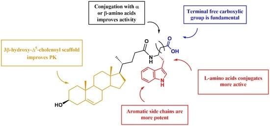
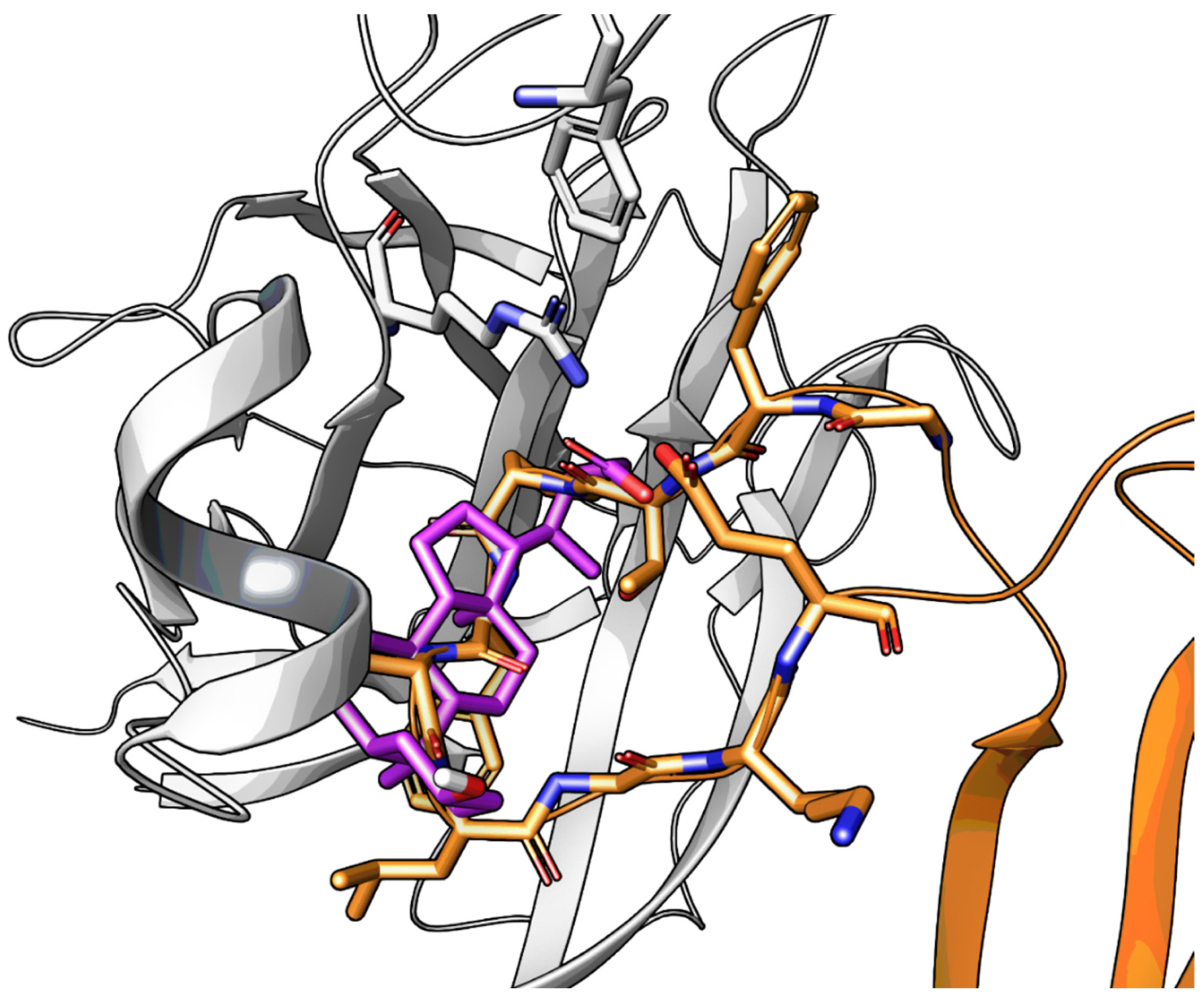
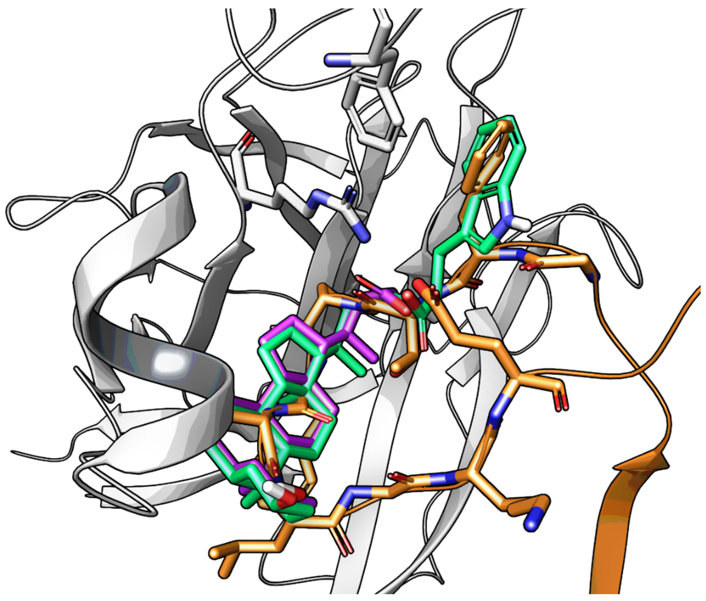
2.3.2. Amino Acid Conjugates of 3β-Hydroxy-Δ5-Cholenic Acid as EphA2 Antagonists

The synthesis of a new set of compounds based on the 3β-hydroxy-Δ5-cholenic acid structure was thus performed. The 3β-hydroxy-Δ5-cholenic acid nucleus was conjugated to α-amino acids of small and large sizes, as reported in Table 7.
Activity data obtained from the EphA2-ephrin-A1 displacement assays were somehow in line with the SAR drawn for LCA-derivatives. When 61 was conjugated with amino acids bearing aliphatic side chains, the resulting compounds 6264 were inactive. Conjugation with aromatic amino acids gave inactive (L-Phe 65) or weakly active (L-O-phenylserine 66) derivatives [66]. However, when L-Trp was coupled with 3β-hydroxy-Δ5-cholenic acid 61, the resulting conjugate UniPR1331 emerged as a fair EphA2 antagonist able to displace ephrin-A1 from EphA2 at low micromolar concentration (IC50 value of 3.5 μM). Conjugation of cholenic acid with D-Trp gave compound 67, which lacked activity on the EphA2 receptor [66]. This suggested that the indole ring of the L-Trp of UniPR1331 was involved in specific interactions with EphA2 and it could represent a starting point for an informative SAR exploration. Modification of the indole N-H group by methylation (compound 68) or replacement of the indole itself with a naphthalene group (compound 69), led to antagonists with potency comparable to that of UniPR1331 (IC50 of 5.0 and 4.1 μM for 68 and 69, respectively), indicating that the indole N-H group of UniPR1331 was not involved in crucial polar interactions with EphA2.
The indole ring of UniPR1331 was further exploited for SAR investigations by inserting in position 5 a set of substituents with different stereoelectronic properties (see compounds 7073). Regardless of the electronic properties, only substituents featuring a low steric hindrance (-F, -OH) were tolerated by EphA2. Only compounds 70 and 72 displayed a potency value close to that of UniPR1331.
Finally, the importance of the two polar ends of UniPR1331 for EphA2 inhibition was investigated with the synthesis of additional compounds. When the terminal carboxylic acid group of UniPR1331 was methylated, the resulting methyl ester 74 was inactive indicating that a free carboxylic group was essential for activity also in the case of the 3β-hydroxy-Δ5-cholenic acid series. Removal of a H-bond donor group by acetylation of the 3β-hydroxyl group had a detrimental effect on the inhibitory potency as indicated by lack of potency of compound 76.
On the base of SAR data and thanks to docking simulations alternative binding modes for UniPR1331 within EphA2 were proposed. These differed only for the orientation of the L-Trp moiety in the LBD of EphA2. To identify the most-likely binding pose for UniPR1331, a conformationally restricted analogue, in which position 2 of the indole ring was connected to the nitrogen of the amino acid by a methylene linker, was synthetized and tested. The resulting compound 77 displayed an IC50 value of 6.3 μM [66], close to that of UniPR1331. The orientation assumed by the indole ring of compound 77 matched only one of the proposed docking poses of UniPR1331 (Figure 10), in which the indole ring was able to form a π-π interaction with Phe108 of EphA2. Overall, this confirmed that L-Trp improved inhibitory potency over other conjugates thanks to specific interactions with EphA2 [66].

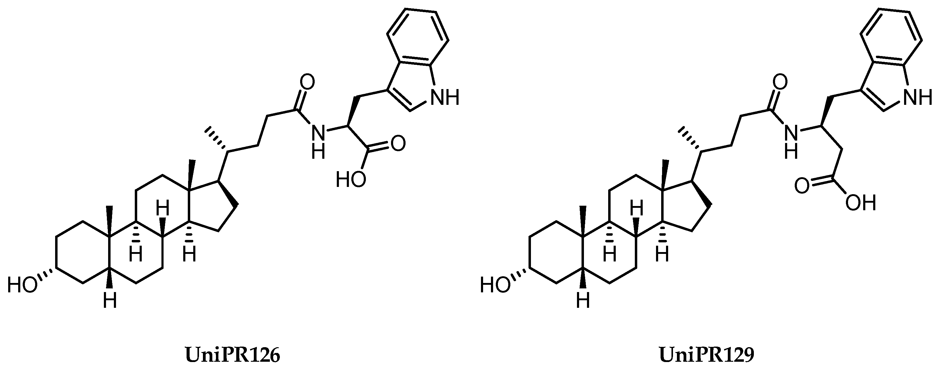
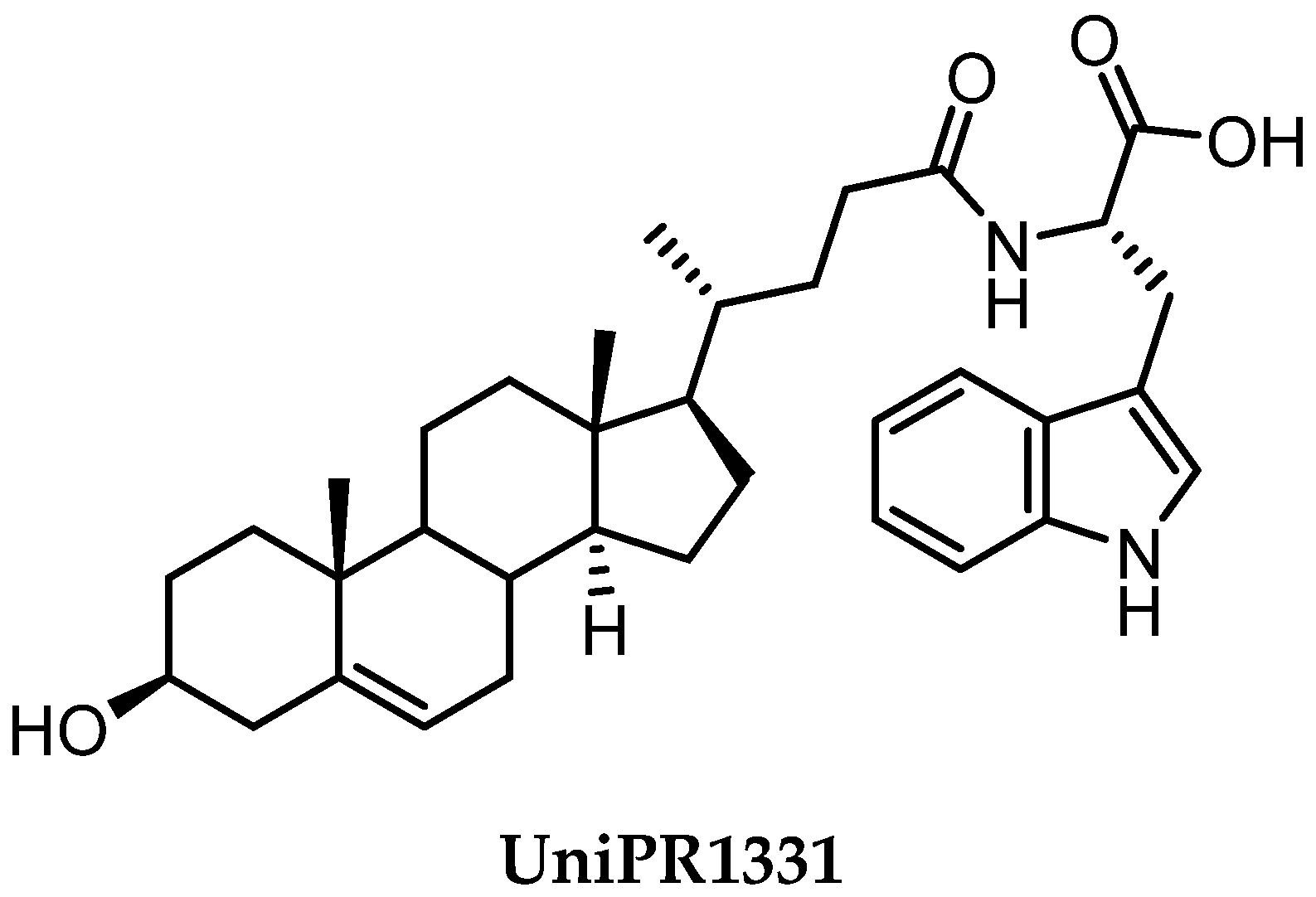
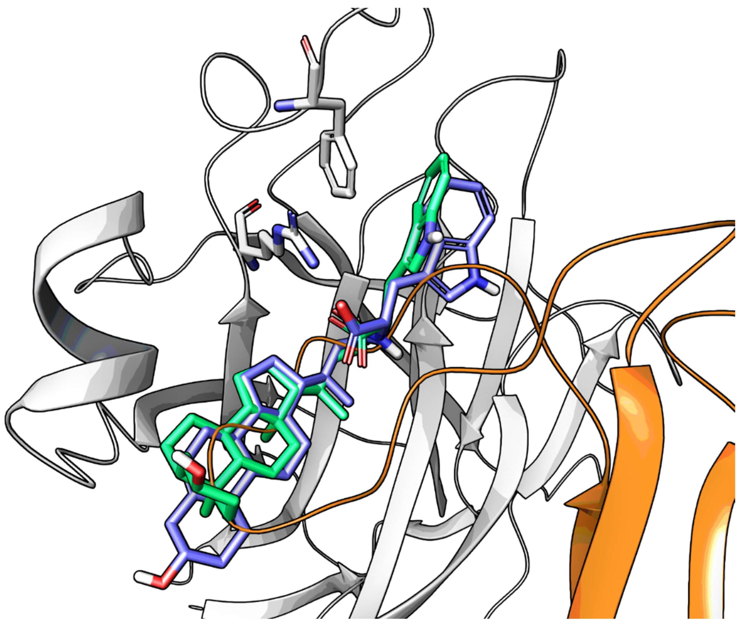
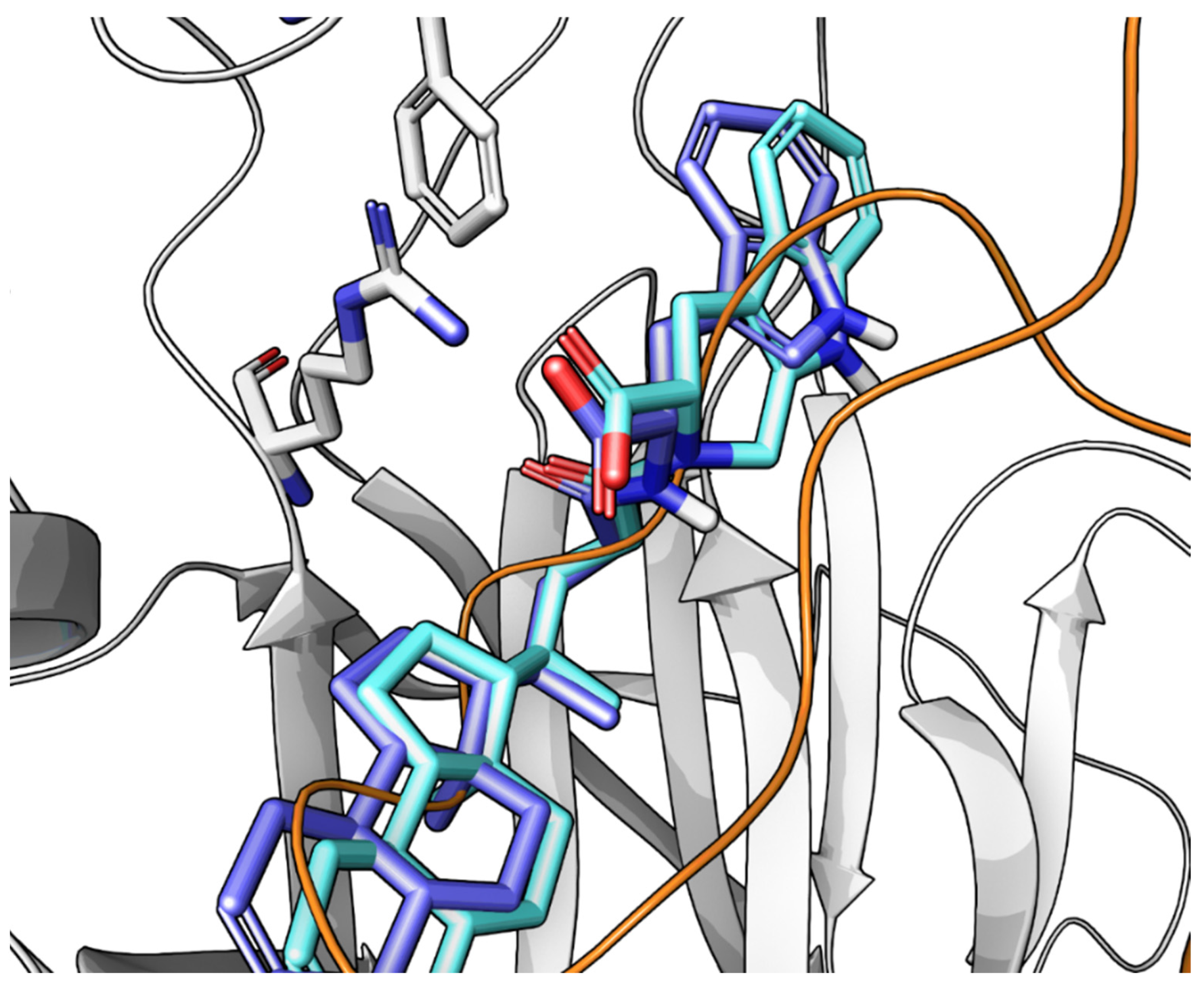
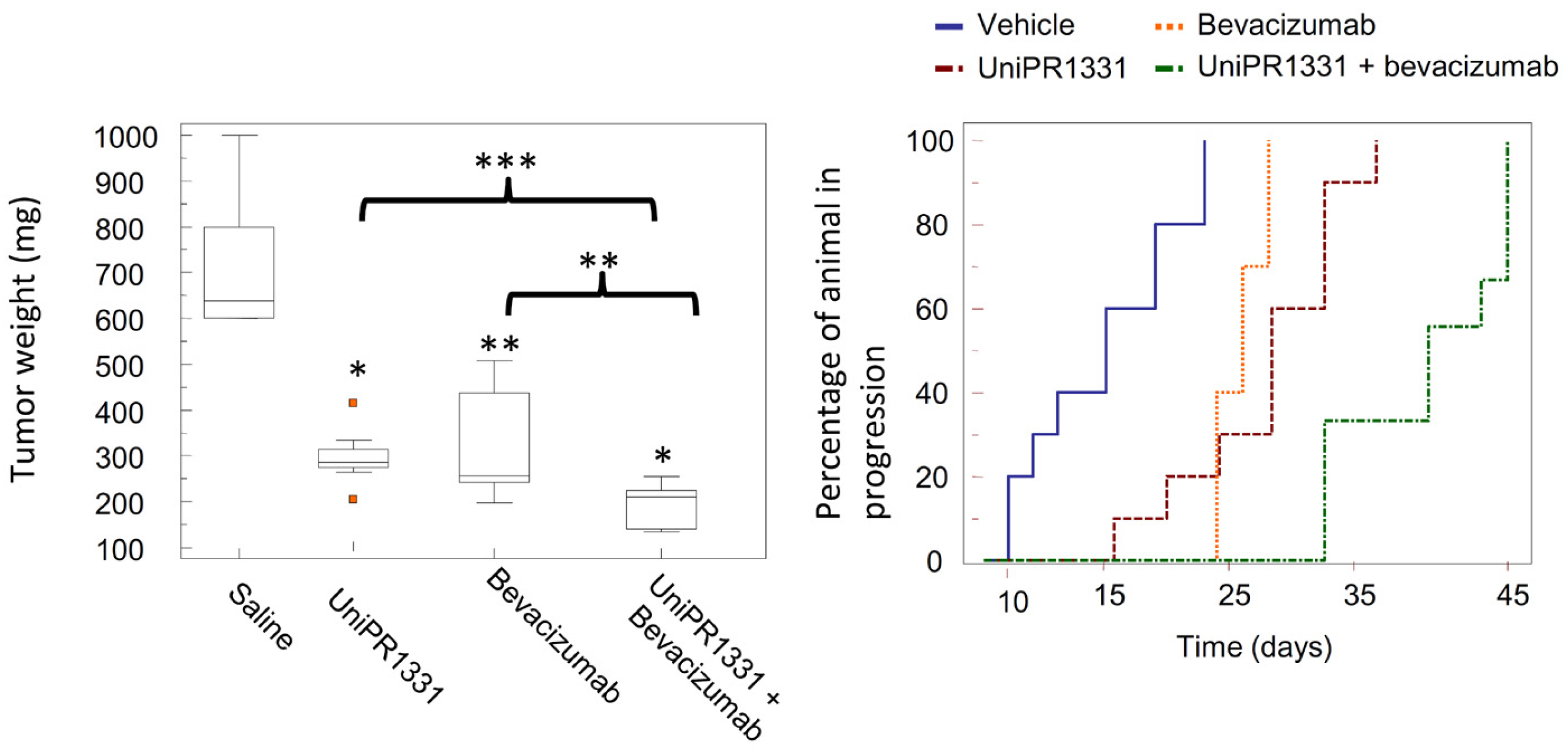
2.3.3. UniPR1331: An Emerging Eph-ephrin Antagonist for In Vivo Study

UniPR1331 became the subject of an intensive biochemical characterization aimed at clarifying its mechanism of action, its selectivity versus other Eph receptors and versus biological targets modulated by bile acids.
Surface plasmon resonance investigations showed that UniPR1331 binds EphA2 in a concentration-dependent and saturable manner, with 1:1 stoichiometry, and an affinity constant (KD) value of 3.3 μM [66]. This confirmed that the ability of this compound to competitively displaces ephrin-A1 from EphA2 relies on the ability of UniPR1331 to bind the same site on EphA2 recognized by ephrin-A1.
By strict analogy to what was observed in the case of bile acid derivatives UniPR126 and UniPR129, UniPR1331 also inhibited ephrin binding to all members of the Eph receptor family with low micromolar potency. UniPR1331 displayed excellent selectivity with respect to molecular targets activated by bile acids as it did not interfere with the activity of TGR5, FXR, and PXR receptors [66].
Experiments in cells showed that UniPR1331 inhibited phosphorylation of EphA2 in prostate cancer (PC3) cells without directly interfering with the kinase domain. In endothelial cells (HUVEC model), UniPR1331 blocked angiogenesis in a concentration-dependent manner with an IC50 value of 2.9 μM [66]. These promising in vitro data proposed UniPR1331 as reference Eph-ephrin antagonist and prompted researchers to evaluate if UniPR1331 could have also better pharmacokinetics (PK) properties than the bile-acid ligand UniPR129.
Further investigations showed that UniPR1331 had a fair PK profile. In fact, UniPR1331 presented a Cmax, after a single administration (30 mg/kg, os), at 30 min of 1.4 μM, a hundred-fold higher than what observed for UniPR129 [66]. The improvement was also significant in the systemic exposure, represented by the area under the curve (AUC0-t) with a value of 573.1 ng/mL h, compared to 21.2 ng/mL h displayed by UniPR129. The improvement was likely due to a different steroidal core that guaranteed a better oral bioavailability, with UniPR1331 less susceptible to liver metabolism than UniPR129 [66].
Despite the improvement of the PK profile, UniPR1331 showed a rapid decline in plasma after oral administration. To better understand the in vivo behavior of this compound, metabolic stability assays were performed to evaluate phase I and phase II biotransformations [67]. Characterization of UniPR1331 metabolites was performed using high performance liquid chromatography coupled to tandem mass spectrometry (HPLC-ESI-MS/MS) and high-resolution mass spectrometry (HPLC-ESI-HR-MS). In vitro metabolism data did not account for the short biological half-life observed in mice following oral administration of UniPR1331. Moreover, the in vitro metabolic profiling did not match with the in vivo profile of UniPR1331 metabolites, except for a keto analogue at C3 position. A different metabolite was detected after oral administration, which was never identified in vitro. Intriguingly, exposure in vitro to mice fecal material of UniPR1331 produced the very same main metabolite observed in vivo, supporting a major role of gut microbiota for the metabolism of UniPR1331 [67].
Despite a suboptimal PK profile, UniPR1331 displayed promising antitumoral activity when tested in vivo by the oral route. Extended work performed in collaboration with the group of Festuccia at the University of L’Aquila allowed to investigate the antitumoral activity of UniPR1331 on in vivo models of glioblastoma multiforme (GBM). UniPR1331 (administered per os at 30 mg/kg 5 days per week) dramatically reduced GBM growth in two distinct xenograft models (based on U87MG and U251MG cells), leading to a significant delay in the time to progression of the disease [68].
Biochemical analysis of the GBM indicated that this marked antitumoral response was due to a dramatic blockage of angiogenesis and vasculomimicry obtained through the inhibition of the activity of the Eph-ephrin system and, to some extent, to the inhibition of the VEGF–VEGFR axis, as recently suggested by throughout work of Rusnati and collaborators [69].
Combination of UniPR1331 with the anti-VEGF monoclonal antibody bevacizumab dramatically reduced vasculomimicry with appearance of large avascular tumor areas in U87MG tumors with beneficial effects for treated mice (Figure 11) [68]. The activity of UniPR1331 (alone or in combination with bevacizumab) was also evaluated on orthotopic models generated from patient-derived GBM stem cells injected in the brains of nude mice. In this set of experiments, a significant increase of the overall survival was observed in treated animals, confirming the ability of UniPR1331 to act as an effective anti-GBM agent.

2.4. Kinase Inhibitors

Another approach to interfere with the Eph-ephrin exploits the clinically validated approach of tyrosine-kinase inhibitors (TKI), directed towards the ATP binding site of the kinase domain of Eph receptors with small molecules. Advances in this field have been recently discussed in the literature [70]. These inhibitors were further exploited as biological tools for the development of radiotracers, to investigate the role and impact of the Eph-ephrin system in cancer, as recently described by Neuber et al. [71].

3. Conclusions and Perspective

Clinical evidence has demonstrated the involvement of the Eph-ephrin system in pathological conditions, especially in cancer insurgence and progression. Targeting this signaling pathway has emerged as a possible strategy to tackle cancers in which the activity or expression of Eph receptors or ephrin ligands is abnormal compared to healthy tissues. Several pharmacological approaches have been proposed and attempted in last 20 years. Kinase inhibitors have been used to target Eph-ephrin system, but while being able to block Eph receptor phosphorylation (forward signal), they are unable to inhibit the activation of ephrins (reverse signal). Moreover, their limited selectivity, due to interaction with several members of the kinome, has been observed [72].
A promising strategy, represented by small molecules able to interfere with the formation of the Eph-ephrin signaling system through the selective targeting of the extracellular LBD of the Eph receptors, has recently emerged. Compounds with such a mechanism of action (protein-protein interaction inhibition, PPI-i) can indeed simultaneously inhibit the forward and the reverse signaling in contiguous cells with potential therapeutic benefits, surpassing in terms of selectivity Eph kinase inhibitors.
Different classes of small molecules have been reported in the literature in the last decade, most of them designed around the structure of the secondary bile acid, lithocholic acid (LCA), previously reported to be a physiological agonist of TGR5 and FXR receptors. By a combined effort merging structural data, computer-aided drug design and pharmacological assays, researchers have exploited the weak activity of LCA on EphA2 receptor to devise potent compounds, culminating in the L-Trp and L-β-Homo-Trp conjugates of LCA, known as UniPR126 and UniPR129. These compounds displayed fair potency in vitro and SAR investigations around their structures allowed to uncover key information: (a) the importance of their terminal carboxylic group, needed to bind Arg103 of the EphA2; (b) the impossibility of replacing the steroid portion with less hydrophobic scaffolds; (c) the importance of a polar group in position 3, capable of forming H-bonds with the LBD of the Eph receptor.
To further advance this class of compounds, aiming to eliminate potential off-targets, the search for chemotype alternatives to LCA led to the identification of 3β-hydroxy-Δ5-cholenic acid as a novel scaffold to be exploited for the synthesis of new Eph-ephrin antagonists. A medicinal chemistry optimization prompted the discovery of UniPR1331, the L-Trp conjugate of 3β-hydroxy-Δ5-cholenic acid, which overcame most of the specific limitations observed with LCA-based compounds, such as selectivity over TGR5 and FXR receptors, and poor bioavailability [66].
UniPR1331 was reported to have significant in vivo antiangiogenic properties and a remarkable anti-tumor activity in animal models against GBM. UniPR1331 increased the disease-free survival in both xenograft and orthotopic mice models of GBM [68]. Thus, UniPR1331 has emerged as a promising tool compound to be considered as a starting point for the generation of Eph-ephrin antagonists, featuring higher potency and improved metabolic stability.

Author Contributions

Conceptualization, L.G, A.L.; Writing—Original Draft Preparation, L.G, A.L. and R.C.; Writing—Review & Editing, L.G., R.C., L.S., F.F., M.C., C.G., M.T. and A.L.; Visualization, A.L., M.T and R.C.; Supervision, A.L. and M.T. All authors have read and agreed to the published version of the manuscript.

Funding

This research received no external funding.

Institutional Review Board Statement

Not applicable.

Informed Consent Statement

Not applicable.

Data Availability Statement

Date sharing is not applicable.

Acknowledgments

Associazione Italiana Ricerca sul Cancro, Italy, is kindly acknowledged for supporting L.G. via a Ph.D. scholarship.

Conflicts of Interest

The authors declare that they have no known competing financial interests or personal relationships that could have influenced the work reported in this paper.

References

  1. Zisch, A.H.; Pasquale, E.B. The Eph Family: A Multitude of Receptors That Mediate Cell Recognition Signals. Cell Tissue Res. 1997, 290, 217–226. [Google Scholar] [CrossRef] [PubMed]
  2. Eph Nomenclature Committee. Unified Nomenclature for Eph Family Receptors and Their Ligands, the Ephrins. Cell 1997, 90, 403–404. [Google Scholar] [CrossRef] [Green Version]
  3. Romero, G.; von Zastrow, M.; Friedman, P.A. Role of PDZ Proteins in Regulating Trafficking, Signaling, and Function of GPCRs: Means, Motif, and Opportunity. Adv. Pharmacol. 2011, 62, 279–314. [Google Scholar] [CrossRef] [PubMed] [Green Version]
  4. Himanen, J.P.; Nikolov, D.B. Eph Receptors and Ephrins. Int. J. Biochem. Cell Biol. 2003, 35, 130–134. [Google Scholar] [CrossRef]
  5. Himanen, J.P.; Saha, N.; Nikolov, D.B. Cell-Cell Signaling via Eph Receptors and Ephrins. Curr. Opin. Cell Biol. 2007, 19, 534–542. [Google Scholar] [CrossRef] [Green Version]
  6. Janes, P.W.; Nievergall, E.; Lackmann, M. Concepts and Consequences of Eph Receptor Clustering. Semin. Cell Dev. Biol. 2012, 23, 43–50. [Google Scholar] [CrossRef]
  7. Ojosnegros, S.; Cutrale, F.; Rodríguez, D.; Otterstrom, J.J.; Chiu, C.L.; Hortigüela, V.; Tarantino, C.; Seriola, A.; Mieruszynski, S.; Martínez, E.; et al. Eph-Ephrin Signaling Modulated by Polymerization and Condensation of Receptors. Proc. Natl. Acad. Sci. USA 2017, 114, 13188–13193. [Google Scholar] [CrossRef] [Green Version]
  8. Nikolov, D.B.; Xu, K.; Himanen, J.P. Homotypic Receptor-Receptor Interactions Regulating Eph Signaling. Cell Adh. Migr. 2014, 8, 360–365. [Google Scholar] [CrossRef]
  9. Himanen, J.P.; Nikolov, D.B. Eph Signaling: A Structural View. Trends Neurosci. 2003, 26, 46–51. [Google Scholar] [CrossRef]
  10. Pasquale, E.B. Eph-Ephrin Bidirectional Signaling in Physiology and Disease. Cell 2008, 133, 38–52. [Google Scholar] [CrossRef] [Green Version]
  11. Dravis, C.; Yokoyama, N.; Chumley, M.J.; Cowan, C.A.; Silvany, R.E.; Shay, J.; Baker, L.A.; Henkemeyer, M. Bidirectional Signaling Mediated by Ephrin-B2 and EphB2 Controls Urorectal Development. Dev. Biol. 2004, 15, 272–290. [Google Scholar] [CrossRef] [PubMed] [Green Version]
  12. Egea, J.; Diger Klein, R. Bidirectional Eph-Ephrin Signaling during Axon Guidance. Trends Cell Biol. 2007, 17, 230–238. [Google Scholar] [CrossRef]
  13. Groppa, E.; Brkic, S.; Uccelli, A.; Wirth, G.; Korpisalo-Pirinen, P.; Filippova, M.; Dasen, B.; Sacchi, V.; Muraro, M.G.; Trani, M.; et al. EphrinB2/EphB4 Signaling Regulates Non-sprouting Angiogenesis by VEGF. EMBO Rep. 2018, 19, e45054. [Google Scholar] [CrossRef]
  14. Mosch, B.; Reissenweber, B.; Neuber, C.; Pietzsch, J. Eph Receptors and Ephrin Ligands: Important Players in Angiogenesis and Tumor Angiogenesis. J. Oncol. 2010, 2010, 135285. [Google Scholar] [CrossRef] [PubMed]
  15. Kania, A.; Klein, R. Mechanisms of Ephrin-Eph Signalling in Development, Physiology and Disease. Nat. Rev. Mol. Cell Biol. 2016, 17, 240–256. [Google Scholar] [CrossRef] [PubMed]
  16. Genander, M.; Holmberg, J.; Frisén, J. Ephrins Negatively Regulate Cell Proliferation in the Epidermis and Hair Follicle. Stem Cells 2010, 28, 1196–1205. [Google Scholar] [CrossRef]
  17. Ji, H.; Goode, R.J.A.; Vaillant, F.; Mathivanan, S.; Kapp, E.A.; Mathias, R.A.; Lindeman, G.J.; Visvader, J.E.; Simpson, R.J. Proteomic Profiling of Secretome and Adherent Plasma Membranes from Distinct Mammary Epithelial Cell Subpopulations. Proteomics 2011, 11, 4029–4039. [Google Scholar] [CrossRef]
  18. Kosinski, C.; Li, V.S.W.; Chan, A.S.Y.; Zhang, J.; Ho, C.; Wai, Y.T.; Tsun, L.C.; Mifflin, R.C.; Powell, D.W.; Siu, T.Y.; et al. Gene Expression Patterns of Human Colon Tops and Basal Crypts and BMP Antagonists as Intestinal Stem Cell Niche Factors. Proc. Natl. Acad. Sci. USA 2007, 104, 15418–15423. [Google Scholar] [CrossRef] [Green Version]
  19. Huang, Y.C.; Lin, S.J.; Lin, K.M.; Chou, Y.C.; Lin, C.W.; Yu, S.C.; Chen, C.L.; Shen, T.L.; Chen, C.K.; Lu, J.; et al. Regulation of EBV LMP1-Triggered EphA4 Downregulation in EBV-Associated B Lymphoma and Its Impact on Patients’ Survival. Blood 2016, 128, 1578–1589. [Google Scholar] [CrossRef]
  20. Pasquale, E.B. Eph Receptors and Ephrins in Cancer: Bidirectional Signalling and Beyond. Nat. Rev. Cancer 2010, 10, 165–180. [Google Scholar] [CrossRef] [Green Version]
  21. Barquilla, A.; Pasquale, E.B. Eph Receptors and Ephrins: Therapeutic Opportunities. Annu. Rev. Pharmacol. Toxicol. 2015, 55, 467–487. [Google Scholar] [CrossRef] [PubMed] [Green Version]
  22. Kaushansky, A.; Douglass, A.N.; Arang, N.; Vigdorovich, V.; Dambrauskas, N.; Kain, H.S.; Austin, L.S.; Sather, D.N.; Kappe, S.H.I. Malaria Parasites Target the Hepatocyte Receptor EphA2 for Successful Host Infection. Science 2015, 350, 1089–1092. [Google Scholar] [CrossRef] [PubMed] [Green Version]
  23. Binda, E.; Visioli, A.; Giani, F.; Lamorte, G.; Copetti, M.; Pitter, K.L.; Huse, J.T.; Cajola, L.; Zanetti, N.; DiMeco, F.; et al. The EphA2 Receptor Drives Self-Renewal and Tumorigenicity in Stem-like Tumor-Propagating Cells from Human Glioblastomas. Cancer Cell 2012, 22, 765–780. [Google Scholar] [CrossRef] [Green Version]
  24. Miao, H.; Li, D.Q.; Mukherjee, A.; Guo, H.; Petty, A.; Cutter, J.; Basilion, J.P.; Sedor, J.; Wu, J.; Danielpour, D.; et al. EphA2 Mediates Ligand-Dependent Inhibition and Ligand-Independent Promotion of Cell Migration and Invasion via a Reciprocal Regulatory Loop with Akt. Cancer Cell 2009, 16, 9–20. [Google Scholar] [CrossRef] [Green Version]
  25. Day, B.W.; Stringer, B.W.; Al-Ejeh, F.; Ting, M.J.; Wilson, J.; Ensbey, K.S.; Jamieson, P.R.; Bruce, Z.C.; Lim, Y.C.; Offenhäuser, C.; et al. EphA3 Maintains Tumorigenicity and Is a Therapeutic Target in Glioblastoma Multiforme. Cancer Cell 2013, 23, 238–248. [Google Scholar] [CrossRef] [Green Version]
  26. Tu, Y.; He, S.; Fu, J.; Li, G.; Xu, R.; Lu, H.; Deng, J. Expression of EphrinB2 and EphB4 in Glioma Tissues Correlated to the Progression of Glioma and the Prognosis of Glioblastoma Patients. Clin. Transl. Oncol. 2012, 14, 214–220. [Google Scholar] [CrossRef] [PubMed]
  27. Ferluga, S.; Debinski, W. Ephs and Ephrins in Malignant Gliomas. Growth Factors 2014, 32, 190. [Google Scholar] [CrossRef] [PubMed] [Green Version]
  28. Carles-Kinch, K.; Kilpatrick, K.E.; Stewart, J.C.; Kinch, M.S. Antibody Targeting of the EphA2 Tyrosine Kinase Inhibits Malignant Cell Behavior. Cancer Res. 2002, 62, 2840–2847. [Google Scholar] [PubMed]
  29. Hammond, S.A.; Lutterbuese, R.; Roff, S.; Lutterbuese, P.; Schlereth, B.; Bruckheimer, E.; Kinch, M.S.; Coats, S.; Baeuerle, P.A.; Kufer, P.; et al. Selective Targeting and Potent Control of Tumor Growth Using an EphA2/CD3-Bispecific Single-Chain Antibody Construct. Cancer Res. 2007, 67, 3927–3935. [Google Scholar] [CrossRef] [Green Version]
  30. Dall’Acqua, W.F.; Damschroder, M.M.; Zhang, J.; Woods, R.M.; Widjaja, L.; Yu, J.; Wu, H. Antibody Humanization by Framework Shuffling. Methods 2005, 36, 43–60. [Google Scholar] [CrossRef]
  31. Peng, L.; Oganesyan, V.; Damschroder, M.M.; Wu, H.; Dall’Acqua, W.F. Structural and Functional Characterization of an Agonistic Anti-Human EphA2 Monoclonal Antibody. J. Mol. Biol. 2011, 413, 390–405. [Google Scholar] [CrossRef] [PubMed]
  32. Lee, J.W.; Stone, R.L.; Lee, S.J.; Nam, E.J.; Roh, J.W.; Nick, A.M.; Han, H.D.; Shahzad, M.M.K.; Kim, H.S.; Mangala, L.S.; et al. EphA2 Targeted Chemotherapy Using an Antibody Drug Conjugate in Endometrial Carcinoma. Clin. Cancer Res. 2010, 16, 2562–2570. [Google Scholar] [CrossRef] [PubMed] [Green Version]
  33. Annunziata, C.M.; Kohn, E.C.; LoRusso, P.; Houston, N.D.; Coleman, R.L.; Buzoianu, M.; Robbie, G.; Lechleider, R. Phase 1, Open-Label Study of MEDI-547 in Patients with Relapsed or Refractory Solid Tumors. Investig. New Drugs 2012, 31, 77–84. [Google Scholar] [CrossRef] [PubMed] [Green Version]
  34. Shitara, K.; Satoh, T.; Iwasa, S.; Yamaguchi, K.; Muro, K.; Komatsu, Y.; Nishina, T.; Esaki, T.; Hasegawa, J.; Kakurai, Y.; et al. Safety, Tolerability, Pharmacokinetics, and Pharmacodynamics of the Afucosylated, Humanized Anti-EPHA2 Antibody DS-8895a: A First-in-Human Phase i Dose Escalation and Dose Expansion Study in Patients with Advanced Solid Tumors. J. Immunother. Cancer 2019, 7, 219. [Google Scholar] [CrossRef] [PubMed]
  35. Swords, R.T.; Greenberg, P.L.; Wei, A.H.; Durrant, S.; Advani, A.S.; Hertzberg, M.S.; Lewis, I.D.; Rivera, G.; Gratzinger, D.; Fan, A.C.; et al. KB004, a First in Class Monoclonal Antibody Targeting the Receptor Tyrosine Kinase EphA3, in Patients with Advanced Hematologic Malignancies: Results from a Phase 1 Study. Leuk. Res. 2016, 50, 123–131. [Google Scholar] [CrossRef] [PubMed]
  36. Gan, H.; Cher, L.; Inglis, P.; Lwin, Z.; Lau, E.; Ackermann, U.; Coombs, N.; Remen, K.; Guo, N.; Ting Lee, S.; et al. Preliminary Findings of a Phase I Safety and Bioimaging Trial of Kb004 (Ifabotuzumab) In Patients with Glioblastoma. Neuro. Oncol. 2019, 21, vi6. [Google Scholar] [CrossRef]
  37. Hughes, A.; Clarson, J.; Gargett, T.; Yu, W.; Brown, M.P.; Lopez, A.F.; Hughes, T.P.; Yong, A.S.M. Comment on “KB004, a First in Class Monoclonal Antibody Targeting the Receptor Tyrosine Kinase EphA3, in Patients with Advanced Hematologic Malignancies: Results from a Phase 1 Study”. Leuk. Res. 2017, 55, 55–57. [Google Scholar] [CrossRef]
  38. Combination Therapy With Pembrolizumab and SEphB4-HSA in Previously Treated Urothelial Carcinoma. Available online: https://clinicaltrials.gov/ct2/show/NCT02717156 (accessed on 19 January 2022).
  39. SEphB4-HSA in Treating Patients With Kaposi Sarcoma. Available online: https://clinicaltrials.gov/ct2/show/NCT02799485?term=ephB4&draw=2&rank=2 (accessed on 19 January 2022).
  40. Koolpe, M.; Dail, M.; Pasquale, E.B. An Ephrin Mimetic Peptide That Selectively Targets the EphA2 Receptor. J. Biol. Chem. 2002, 277, 46974–46979. [Google Scholar] [CrossRef] [Green Version]
  41. Himanen, J.P.; Rajashankar, K.R.; Lackmann, M.; Cowan, C.A.; Henkemeyer, M.; Nikolov, D.B. Crystal Structure of an Eph Receptor-Ephrin Complex. Nature 2001, 414, 933–938. [Google Scholar] [CrossRef]
  42. Murai, K.K.; Nguyen, L.N.; Koolpe, M.; McLennan, R.; Krull, C.E.; Pasquale, E.B. Targeting the EphA4 Receptor in the Nervous System with Biologically Active Peptides. Mol. Cell. Neurosci. 2003, 24, 1000–1011. [Google Scholar] [CrossRef]
  43. Riedl, S.; Pasquale, E. Targeting the Eph System with Peptides and Peptide Conjugates. Curr. Drug Targets 2015, 16, 1031–1047. [Google Scholar] [CrossRef] [PubMed]
  44. Wang, J.; Liu, Y.; Li, Y.; Dai, W.; Guo, Z.; Wang, Z. EphA2 Targeted Doxorubicin Stealth Liposomes as a Therapy System for Choroidal Neovascularization in Rats. Investig. Ophthalmol. Vis. Sci. 2012, 53, 7348–7357. [Google Scholar] [CrossRef] [PubMed] [Green Version]
  45. Wu, B.; Wang, S.; De, S.K.; Barile, E.; Quinn, B.A.; Zharkikh, I.; Purves, A.; Stebbins, J.L.; Oshima, R.G.; Fisher, P.B.; et al. Design and Characterization of Novel EphA2 Agonists for Targeted Delivery of Chemotherapy to Cancer Cells. Chem. Biol. 2015, 22, 876–887. [Google Scholar] [CrossRef] [PubMed] [Green Version]
  46. Barile, E.; Pellecchia, M. NMR-Based for the Identification and Optimization of Inhibitors of Protein–Protein Interactions. Chem. Rev. 2014, 114, 4749–4763. [Google Scholar] [CrossRef] [PubMed]
  47. Gambini, L.; Salem, A.F.; Udompholkul, P.; Tan, X.-F.; Baggio, C.; Shah, N.; Aronson, A.; Song, J.; Pellecchia, M. Structure-Based Design of Novel EphA2 Agonistic Agents with Nanomolar Affinity in Vitro and in Cell. ACS Chem. Biol. 2018, 13, 2633–2644. [Google Scholar] [CrossRef]
  48. Koolpe, M.; Burgess, R.; Dail, M.; Pasquale, E.B. EphB Receptor-Binding Peptides Identified by Phage Display Enable Design of an Antagonist with Ephrin-like Affinity. J. Biol. Chem. 2005, 280, 17301–17311. [Google Scholar] [CrossRef] [Green Version]
  49. Chrencik, J.E.; Brooun, A.; Recht, M.I.; Nicola, G.; Davis, L.K.; Abagyan, R.; Widmer, H.; Pasquale, E.B.; Kuhn, P. Three-Dimensional Structure of the EphB2 Receptor in Complex with an Antagonistic Peptide Reveals a Novel Mode of Inhibition. J. Biol. Chem. 2007, 282, 36505–36513. [Google Scholar] [CrossRef] [Green Version]
  50. Chrencik, J.E.; Brooun, A.; Kraus, M.L.; Recht, M.I.; Kolatkar, A.R.; Gye, W.H.; Seifert, J.M.; Widmer, H.; Auer, M.; Kuhn, P. Structural and Biophysical Characterization of the EphB4*ephrinB2 Protein-Protein Interaction and Receptor Specificity. J. Biol. Chem. 2006, 281, 28185–28192. [Google Scholar] [CrossRef] [Green Version]
  51. Chrencik, J.E.; Brooun, A.; Recht, M.I.; Kraus, M.L.; Koolpe, M.; Kolatkar, A.R.; Bruce, R.H.; Martiny-Baron, G.; Widmer, H.; Pasquale, E.B.; et al. Structure and Thermodynamic Characterization of the EphB4/Ephrin-B2 Antagonist Peptide Complex Reveals the Determinants for Receptor Specificity. Structure 2006, 14, 321–330. [Google Scholar] [CrossRef] [Green Version]
  52. You, J.; Wang, Z.; Du, Y.; Yuan, H.; Zhang, P.; Zhou, J.; Liu, F.; Li, C.; Hu, F. Specific Tumor Delivery of Paclitaxel Using Glycolipid-like Polymer Micelles Containing Gold Nanospheres. Biomaterials 2013, 34, 4510–4519. [Google Scholar] [CrossRef] [Green Version]
  53. Noberini, R.; Mitra, S.; Salvucci, O.; Valencia, F.; Duggineni, S.; Prigozhina, N.; Wei, K.; Tosato, G.; Huang, Z.; Pasquale, E.B. PEGylation Potentiates the Effectiveness of an Antagonistic Peptide That Targets the EphB4 Receptor with Nanomolar Affinity. PloS ONE 2011, 6, e28611. [Google Scholar] [CrossRef] [PubMed] [Green Version]
  54. Tognolini, M.; Lodola, A. Targeting the Eph-Ephrin System with Protein-Protein Interaction (PPI) Inhibitors. Curr. Drug Targets 2015, 16, 1048–1056. [Google Scholar] [CrossRef] [PubMed]
  55. Lodola, A.; Incerti, M.; Tognolini, M. On the Use of 2,5-Dimethyl-Pyrrol-1-Yl-Benzoic Acid Derivatives as EPH-Ephrin Antagonists. J. Virol. 2014, 88, 12173. [Google Scholar] [CrossRef] [PubMed] [Green Version]
  56. Giorgio, C.; Mohamed, I.H.; Flammini, L.; Barocelli, E.; Incerti, M.; Lodola, A.; Tognolini, M. Lithocholic Acid Is an Eph-Ephrin Ligand Interfering with Eph-Kinase Activation. PLoS ONE 2011, 6, e18128. [Google Scholar] [CrossRef]
  57. Tognolini, M.; Incerti, M.; Hassan-Mohamed, I.; Giorgio, C.; Russo, S.; Bruni, R.; Lelli, B.; Bracci, L.; Noberini, R.; Pasquale, E.B.; et al. Structure-Activity Relationships and Mechanism of Action of Eph-Ephrin Antagonists: Interaction of Cholanic Acid with the EphA2 Receptor. ChemMedChem 2012, 7, 1071–1083. [Google Scholar] [CrossRef] [Green Version]
  58. Himanen, J.P.; Goldgur, Y.; Miao, H.; Myshkin, E.; Guo, H.; Buck, M.; Nguyen, M.; Rajashankar, K.R.; Wang, B.; Nikolov, D.B. Ligand Recognition by A-Class Eph Receptors: Crystal Structures of the EphA2 Ligand-Binding Domain and the EphA2/Ephrin-A1 Complex. EMBO Rep. 2009, 10, 722–728. [Google Scholar] [CrossRef]
  59. Incerti, M.; Tognolini, M.; Russo, S.; Pala, D.; Giorgio, C.; Hassan-Mohamed, I.; Noberini, R.; Pasquale, E.B.; Vicini, P.; Piersanti, S.; et al. Amino Acid Conjugates of Lithocholic Acid as Antagonists of the EphA2 Receptor. J. Med. Chem. 2013, 56, 2936–2947. [Google Scholar] [CrossRef]
  60. Lema Tome, C.M.; Palma, E.; Ferluga, S.; Lowther, W.T.; Hantgan, R.; Wykosky, J.; Debinski, W. Structural and Functional Characterization of Monomeric EphrinA1 Binding Site to EphA2 Receptor. J. Biol. Chem. 2012, 287, 14012–14022. [Google Scholar] [CrossRef] [Green Version]
  61. Hassan-Mohamed, I.; Giorgio, C.; Incerti, M.; Russo, S.; Pala, D.; Pasquale, E.B.; Zanotti, I.; Vicini, P.; Barocelli, E.; Rivara, S.; et al. UniPR129 Is a Competitive Small Molecule Eph-Ephrin Antagonist Blocking in Vitro Angiogenesis at Low Micromolar Concentrations. Br. J. Pharmacol. 2014, 171, 5195–5208. [Google Scholar] [CrossRef] [Green Version]
  62. Tognolini, M.; Incerti, M.; Lodola, A. Are We Using the Right Pharmacological Tools to Target EphA4? ACS Chem. Neurosci. 2014, 5, 1146–1147. [Google Scholar] [CrossRef] [Green Version]
  63. Incerti, M.; Russo, S.; Callegari, D.; Pala, D.; Giorgio, C.; Zanotti, I.; Barocelli, E.; Vicini, P.; Vacondio, F.; Rivara, S.; et al. Metadynamics for Perspective Drug Design: Computationally Driven Synthesis of New Protein-Protein Interaction Inhibitors Targeting the EphA2 Receptor. J. Med. Chem. 2017, 60, 787–796. [Google Scholar] [CrossRef] [PubMed]
  64. Incerti, M.; Russo, S.; Corrado, M.; Giorgio, C.; Ballabeni, V.; Chiodelli, P.; Rusnati, M.; Scalvini, L.; Callegari, D.; Castelli, R.; et al. Optimization of EphA2 Antagonists Based on a Lithocholic Acid Core Led to the Identification of UniPR505, a New 3α-Carbamoyloxy Derivative with Antiangiogenetic Properties. Eur. J. Med. Chem. 2020, 189, 112083. [Google Scholar] [CrossRef] [PubMed]
  65. Pala, D.; Castelli, R.; Incerti, M.; Russo, S.; Tognolini, M.; Giorgio, C.; Hassan-Mohamed, I.; Zanotti, I.; Vacondio, F.; Rivara, S.; et al. Combining Ligand- and Structure-Based Approaches for the Discovery of New Inhibitors of the EPHA2-Ephrin-A1 Interaction. J. Chem. Inf. Model. 2014, 54, 2621–2626. [Google Scholar] [CrossRef] [PubMed]
  66. Castelli, R.; Tognolini, M.; Vacondio, F.; Incerti, M.; Pala, D.; Callegari, D.; Bertoni, S.; Giorgio, C.; Hassan-Mohamed, I.; Zanotti, I.; et al. Δ5-Cholenoyl-Amino Acids as Selective and Orally Available Antagonists of the Epheephrin System. Eur. J. Med. Chem. 2015, 103, 312–324. [Google Scholar] [CrossRef]
  67. Ferlenghi, F.; Castelli, R.; Scalvini, L.; Giorgio, C.; Corrado, M.; Tognolini, M.; Mor, M.; Lodola, A.; Vacondio, F. Drug-Gut Microbiota Metabolic Interactions: The Case of UniPR1331, Selective Antagonist of the Eph-Ephrin System, in Mice. J. Pharm. Biomed. Anal. 2020, 180, 113067. [Google Scholar] [CrossRef] [PubMed]
  68. Festuccia, C.; Gravina, G.L.; Giorgio, C.; Mancini, A.; Pellegrini, C.; Colapietro, A.; Monache, S.D.; Maturo, M.G.; Sferra, R.; Chiodelli, P.; et al. UniPR1331, a Small Molecule Targeting Eph/Ephrin Interaction, Prolongs Survival in Glioblastoma and Potentiates the Effect of Antiangiogenic Therapy in Mice. Oncotarget 2018, 9, 24347–24363. [Google Scholar] [CrossRef] [Green Version]
  69. Rusnati, M.; Paiardi, G.; Tobia, C.; Urbinati, C.; Lodola, A.; D’Ursi, P.; Corrado, M.; Castelli, R.; Wade, R.C.; Tognolini, M.; et al. Cholenic Acid Derivative UniPR1331 Impairs Tumor Angiogenesis via Blockade of VEGF/VEGFR2 in Addition to Eph/Ephrin. Cancer Gene Ther. 2021, 1–10. [Google Scholar] [CrossRef]
  70. Unzue, A.; Lafleur, K.; Zhao, H.; Zhou, T.; Dong, J.; Kolb, P.; Liebl, J.; Zahler, S.; Caflisch, A.; Nevado, C. Three Stories on Eph Kinase Inhibitors: From in Silico Discovery to in Vivo Validation. Eur. J. Med. Chem. 2016, 112, 347–366. [Google Scholar] [CrossRef] [Green Version]
  71. Neuber, C.; Belter, B.; Mamat, C.; Pietzsch, J. Radiopharmacologist’s and Radiochemist’s View on Targeting the Eph/Ephrin Receptor Tyrosine Kinase System. ACS Omega 2020, 5, 16318–16331. [Google Scholar] [CrossRef]
  72. Davis, M.I.; Hunt, J.P.; Herrgard, S.; Ciceri, P.; Wodicka, L.M.; Pallares, G.; Hocker, M.; Treiber, D.K.; Zarrinkar, P.P. Comprehensive Analysis of Kinase Inhibitor Selectivity. Nat. Biotechnol. 2011, 29, 1046–1051. [Google Scholar] [CrossRef]
Figure 1. Structure of peptides and peptidomimetics antagonist of EphA2 receptor. i. YSA; ii. dNYH; iii. 123B9; iv. 135H11.
Pharmaceuticals 15 00137 g001
Figure 2. Structure of peptides and peptidomimetics antagonist of EphB2 and EphB4 receptors. i. SNEW; ii. TNYL; iii. TNYL-RAW; iv. cTNYL-RAW.
Pharmaceuticals 15 00137 g002
Figure 3. Graphical abstract of the general structure of bile acids antagonist of the EphA2 receptor and the identification of hot spots relevant for the activity.
Pharmaceuticals 15 00137 g003
Figure 4. Visualization of the ephrin-A1 G-H loop (in orange) within the EphA2 receptor (white cartoon) and the docking pose of LCA (in purple). Arg103 and Phe108 of EphA2 are also displayed with a white stick representation.
Pharmaceuticals 15 00137 g004
Figure 5. Docking of UniPR126 (in green) within the EphA2 receptor (white cartoons, with Arg103 and Phe108 displayed by a stick representation). The indole moiety mimics the benzyl group of the Phe111 residue of the natural ligand ephrin-A1 (in orange). The free carboxylic group of UniPR126 undertakes interactions with Arg103.
Pharmaceuticals 15 00137 g005
Figure 6. Structural representation of UniPR126 and UniPR129.
Pharmaceuticals 15 00137 g006
Figure 7. Chemical structures of compound 60 and 61 identified as analogues of 1 by virtual screening of a library of commercially available compounds.
Pharmaceuticals 15 00137 g007
Figure 8. Structure of UniPR1331.
Pharmaceuticals 15 00137 g008
Figure 9. Comparison between the docking poses of UniPR126 (in green) and UniPR1331 (in blue) within EphA2 receptor (white cartoons). The A ring of the steroid portion of UniPR1331 assumes a different orientation within the receptor due to the presence of a double bond between C5 and C6 of the gonane scaffold.
Pharmaceuticals 15 00137 g009
Figure 10. Magnification of the terminal portion of the docking poses of UniPR1331 (blue) and compound 77 (light blue). The indole ring assumes the same position and orientation inside the receptor.
Pharmaceuticals 15 00137 g010
Figure 11. Effect of UniPR1331 on GBM growth (U87MG cells). Effect on tumor weight in GBM xenograft mice model. Kaplan-Meier curves for UniPR1331 (red dotted line), bevacizumab (orange dotted line) or a combination of UniPR1331 with bevacizumab (green dotted line). Blue line represents the vehicle. p values <0.05 were considered statistically significant. * <0.05; ** <0.01; *** <0.001.
Pharmaceuticals 15 00137 g011
Table 1. Ki values of LCA derivatives measured by an ELISA binding assay.
Pharmaceuticals 15 00137 i001
Cpd.R1R2R3R4RSKi (μM)
1 (LCA) Pharmaceuticals 15 00137 i002 Pharmaceuticals 15 00137 i003 Pharmaceuticals 15 00137 i004 Pharmaceuticals 15 00137 i005 Pharmaceuticals 15 00137 i00649
2 (CA) Pharmaceuticals 15 00137 i007 Pharmaceuticals 15 00137 i008 Pharmaceuticals 15 00137 i009 Pharmaceuticals 15 00137 i010 Pharmaceuticals 15 00137 i011>100
3 (DCA) Pharmaceuticals 15 00137 i012 Pharmaceuticals 15 00137 i013 Pharmaceuticals 15 00137 i014 Pharmaceuticals 15 00137 i015 Pharmaceuticals 15 00137 i016>100
4 (CDCA) Pharmaceuticals 15 00137 i017 Pharmaceuticals 15 00137 i018 Pharmaceuticals 15 00137 i019 Pharmaceuticals 15 00137 i020 Pharmaceuticals 15 00137 i021>100
5 Pharmaceuticals 15 00137 i022 Pharmaceuticals 15 00137 i023 Pharmaceuticals 15 00137 i024 Pharmaceuticals 15 00137 i025 Pharmaceuticals 15 00137 i026>100
6 Pharmaceuticals 15 00137 i027 Pharmaceuticals 15 00137 i028 Pharmaceuticals 15 00137 i029 Pharmaceuticals 15 00137 i030 Pharmaceuticals 15 00137 i031>100
7 Pharmaceuticals 15 00137 i032 Pharmaceuticals 15 00137 i033 Pharmaceuticals 15 00137 i034 Pharmaceuticals 15 00137 i035 Pharmaceuticals 15 00137 i03625
8 Pharmaceuticals 15 00137 i037 Pharmaceuticals 15 00137 i038 Pharmaceuticals 15 00137 i039 Pharmaceuticals 15 00137 i040 Pharmaceuticals 15 00137 i0415.1
9 Pharmaceuticals 15 00137 i042 Pharmaceuticals 15 00137 i043 Pharmaceuticals 15 00137 i044 Pharmaceuticals 15 00137 i045 Pharmaceuticals 15 00137 i046>100
10 Pharmaceuticals 15 00137 i047 Pharmaceuticals 15 00137 i048 Pharmaceuticals 15 00137 i049 Pharmaceuticals 15 00137 i050 Pharmaceuticals 15 00137 i051>100
11 Pharmaceuticals 15 00137 i052 Pharmaceuticals 15 00137 i053 Pharmaceuticals 15 00137 i054 Pharmaceuticals 15 00137 i055 Pharmaceuticals 15 00137 i056>100
Table 2. IC50 values for amino acid conjugates of LCA measured by ELISA binding assay.
Pharmaceuticals 15 00137 i057
Cpd.RIC50 (μM)Cpd.RIC50 (μM)
1 (LCA) Pharmaceuticals 15 00137 i0585722 Pharmaceuticals 15 00137 i059>100
12 Pharmaceuticals 15 00137 i0604923 Pharmaceuticals 15 00137 i061>100
13 Pharmaceuticals 15 00137 i062< 30024 Pharmaceuticals 15 00137 i06328
14 Pharmaceuticals 15 00137 i0642025 Pharmaceuticals 15 00137 i06528
15 Pharmaceuticals 15 00137 i0663126 Pharmaceuticals 15 00137 i0676.6
16 Pharmaceuticals 15 00137 i0682427 Pharmaceuticals 15 00137 i0697.5
17 Pharmaceuticals 15 00137 i0701728 Pharmaceuticals 15 00137 i07150
18 Pharmaceuticals 15 00137 i0723329 Pharmaceuticals 15 00137 i073100
19 Pharmaceuticals 15 00137 i0746030 Pharmaceuticals 15 00137 i0752.0
20 Pharmaceuticals 15 00137 i076>10031 Pharmaceuticals 15 00137 i07720
21 Pharmaceuticals 15 00137 i078>100
Table 3. IC50 values of LCA derivatives coupled with linear natural and unnatural amino acids.
Pharmaceuticals 15 00137 i079
Cpd.RIC50 (μM)
LCA Pharmaceuticals 15 00137 i08057
12 Pharmaceuticals 15 00137 i08149
32 Pharmaceuticals 15 00137 i08229
33 Pharmaceuticals 15 00137 i083inactive
34 Pharmaceuticals 15 00137 i084Inactive
35 Pharmaceuticals 15 00137 i08572
36 Pharmaceuticals 15 00137 i086inactive
Table 4. IC50 Values of β-homo-amino acid conjugates of LCA.
Pharmaceuticals 15 00137 i087
Cpd.RIC50 (μM)
32 Pharmaceuticals 15 00137 i08829
37 Pharmaceuticals 15 00137 i08983
38 Pharmaceuticals 15 00137 i09010
39 Pharmaceuticals 15 00137 i091Inactive
40 Pharmaceuticals 15 00137 i09214
41 Pharmaceuticals 15 00137 i09318
42 Pharmaceuticals 15 00137 i094Inactive
43 Pharmaceuticals 15 00137 i0953.9
44 Pharmaceuticals 15 00137 i0961.8
UniPR129 Pharmaceuticals 15 00137 i0970.95
45 Pharmaceuticals 15 00137 i09826
Table 5. IC50 of 3-Substituted 5β-Cholan-24-oyl-L-β-homo-tryptophan derivatives.
Pharmaceuticals 15 00137 i099
Cpd.RIC50 (μM)
UniPR129 Pharmaceuticals 15 00137 i1000.95
46 Pharmaceuticals 15 00137 i10117
47 Pharmaceuticals 15 00137 i10228
48 Pharmaceuticals 15 00137 i10328
49 Pharmaceuticals 15 00137 i1043.1
50 Pharmaceuticals 15 00137 i10513
51 Pharmaceuticals 15 00137 i1060.8
52 Pharmaceuticals 15 00137 i1075.2
Table 6. IC50 values for 5β-cholan-24-oyl-L-β-homo-tryptophan derivatives.
Pharmaceuticals 15 00137 i108
Cpd.RIC50 (μM)
UniPR129 Pharmaceuticals 15 00137 i1090.91
UniPR502 Pharmaceuticals 15 00137 i1100.8
53
(UniPR505)
Pharmaceuticals 15 00137 i1110.95
54 Pharmaceuticals 15 00137 i1121.1
55 Pharmaceuticals 15 00137 i1131.5
56 Pharmaceuticals 15 00137 i11415
57 Pharmaceuticals 15 00137 i1157.1
58 Pharmaceuticals 15 00137 i1161.2
59 Pharmaceuticals 15 00137 i1175.5
Table 7. IC50 values for compounds 6277 tested in the EphA2-ephrin-A1 ELISA assay.
Pharmaceuticals 15 00137 i118
Cpd.RIC50 (μM)Cpd.RIC50 (μM)
61 Pharmaceuticals 15 00137 i1194069 Pharmaceuticals 15 00137 i1204.1
62 Pharmaceuticals 15 00137 i121inactive 70 Pharmaceuticals 15 00137 i1228.9
63 Pharmaceuticals 15 00137 i123>100 71 Pharmaceuticals 15 00137 i12422
64 Pharmaceuticals 15 00137 i125inactive 72 Pharmaceuticals 15 00137 i1266.0
65 Pharmaceuticals 15 00137 i127>100 73 Pharmaceuticals 15 00137 i12847
66 Pharmaceuticals 15 00137 i1295074 Pharmaceuticals 15 00137 i130inactive
UniPR
1331
Pharmaceuticals 15 00137 i1313.5 75 Pharmaceuticals 15 00137 i1324.2
67 Pharmaceuticals 15 00137 i133>100 76 Pharmaceuticals 15 00137 i13489
68 Pharmaceuticals 15 00137 i1355.077 Pharmaceuticals 15 00137 i1366.3
Publisher’s Note: MDPI stays neutral with regard to jurisdictional claims in published maps and institutional affiliations.

