Article

Design and Synthesis of 2,6-Disubstituted-4′-Selenoadenosine-5′-N,N-Dimethyluronamide Derivatives as Human A3 Adenosine Receptor Antagonists

1
Research Institute of Pharmaceutical Sciences, College of Pharmacy, Seoul National University, Seoul 08826, Korea
2
Molecular Recognition Section, Laboratory of Bioorganic Chemistry, National Institute of Diabetes and Digestive and Kidney Disease, National Institutes of Health, Bethesda, MD 20892, USA
3
Chemical Kinomics Research Center, Korea Institute of Science and Technology (KIST), Seoul 02792, Korea
*
Authors to whom correspondence should be addressed.
Pharmaceuticals 2021, 14(4), 363; https://doi.org/10.3390/ph14040363
Submission received: 23 March 2021 / Revised: 7 April 2021 / Accepted: 9 April 2021 / Published: 14 April 2021
(This article belongs to the Special Issue Medicinal Chemistry of Adenosine Receptor Antagonists)

Abstract

:
A new series of 4′-selenoadenosine-5′-N,N-dimethyluronamide derivatives as highly potent and selective human A3 adenosine receptor (hA3AR) antagonists, is described. The highly selective A3AR agonists, 4′-selenoadenosine-5′-N-methyluronamides were successfully converted into selective antagonists by adding a second N-methyl group to the 5′-uronamide position. All the synthesized compounds showed medium to high binding affinity at the hA3AR. Among the synthesized compounds, 2-H-N6-3-iodobenzylamine derivative 9f exhibited the highest binding affinity at hA3AR. (Ki = 22.7 nM). The 2-H analogues generally showed better binding affinity than the 2-Cl analogues. The cAMP functional assay with 2-Cl-N6-3-iodobenzylamine derivative 9l demonstrated hA3AR antagonist activity. A molecular modelling study suggests an important role of the hydrogen of 5′-uronamide as an essential hydrogen bonding donor for hA3AR activation.

Graphical Abstract

1. Introduction

Adenosine, which is the endogenous ligand of the adenosine receptors (ARs), is an important neuromodulator and mediates through activation of its four receptors, consisting of A1, A2A, A2B, and A3 subtypes. These receptors are widely distributed in tissues and involved in various physiological activities [1]. Each subtype couples to a preferred type of G protein; A1 and A3ARs primarily couple to the Gi/o proteins, and A2A and A2BARs couple to Gs proteins. A2B and A3ARs are also known to be coupled to Gq proteins. AR signaling and their physiological roles have been extensively studied [2,3]. Among them, the A3AR is an important receptor to regulate cardioprotection in cardiac ischemia [4], degranulation of neutrophils [5], and cell proliferation [6]. These results led to the development of A3AR agonists as anticancer agents [7]. Selective A3AR antagonists are also promising ligands to modulate inflammation [8] and cerebroprotection [9,10]. Some studies showed that A3AR antagonists could enhance cancer treatment via the inhibition of HIF-1α and VEGF protein accumulation in hypoxia and in tumors [11] and are potential anti-glaucoma therapeutics as they reduce intraocular pressure in mouse and monkey [12].
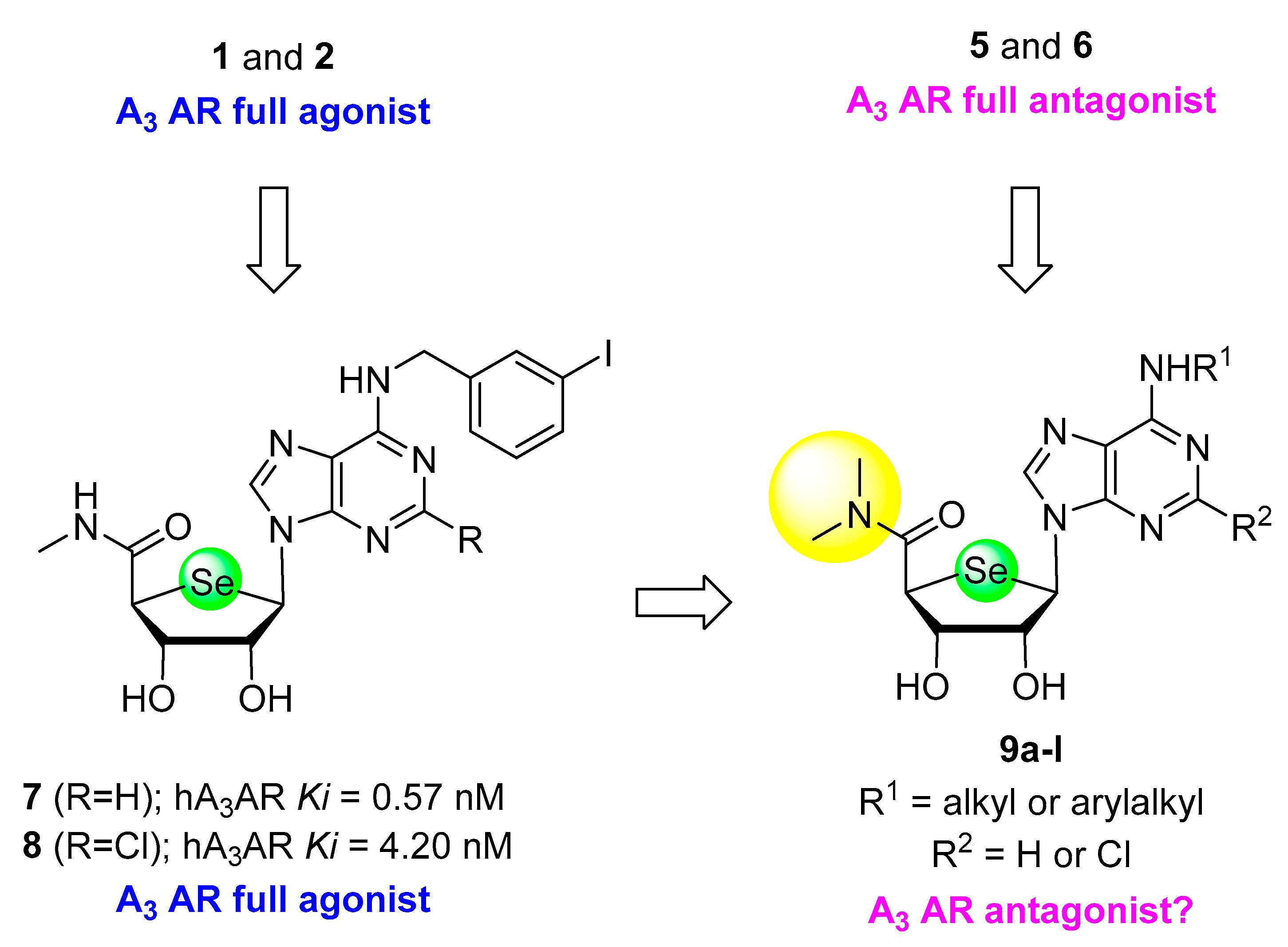
In the past decades, a variety of approaches have been followed to discover novel drug candidates targeting A3AR. 2-Chloro-N6-(3-iodobenzyl)adenosine-5′-N-methyluronamide (Cl-IB-MECA, 1) and its 4′-thio analogue 2 were discovered as potent and selective A3AR agonists from the extensive structure-activity relationships based on the structure of adenosine [13] (Figure 1). The 5′-uronamide hydrogen of 1, required for full agonism, forms a putative hydrogen bond with T94 (3.36) at hA3AR as modeled, suggesting that this interaction is essential for receptor activation by adenosine agonists [14]. Consistent with these findings, the 4′-truncated analogues, such as 3 and 4, lacking this hydrogen bond donor were discovered to act as A3AR antagonists or low-efficacy agonists, demonstrating that a hydrogen bond donating ability of the 5′-uronamide promotes A3AR activation [15,16]. The removal of the hydrogen bond donor ability by appending another methyl group to the 5′-uronamide, e.g., 5′-N,N-dimethyluronamide derivatives 5 and 6, similarly reduced A3AR efficacy. These compounds were characterized as potent and selective A3AR antagonists [17,18].
On the basis of a bioisosteric rationale, we recently reported that 4′-seleno analogues of 1 and 2, i.e., 7 and 8, were discovered as potent and selective hA3AR agonists [19] (Figure 2). They exhibited comparable A3AR binding affinity as the corresponding 4′-oxo- and 5′-thio nucleosides 1 and 2. However, X-ray analysis indicated that in the pure crystalline state they preferred a syn nucleobase orientation and a South sugar conformation, unlike 1 and 2. As mentioned above, removal of the amide hydrogen of the 5′-uronamide of 1 and 2 by N-methylation, resulting in 5 and 6 successfully converted A3AR agonists into A3AR antagonists. Based on these findings, we hypothesized that the 4′-seleno analogue of 5 or 6, bearing a 5′-N,N-dimethyluronamide moiety might be an A3AR antagonist (Figure 2). Thus, we analyzed the structure-activity relationship as A3AR ligands of this series by modifying N6- and C2 positions, by synthesizing novel 4′-selenonucleosides 9a–l. Herein, we report the synthesis and biological evaluation of 2,6-disubstituted-4′-selenoadenosine-5′-N,N-dimethyluronamide derivatives 9a–l as potent and selective A3AR a3ntagonists.

2. Results

2.1. Chemistry

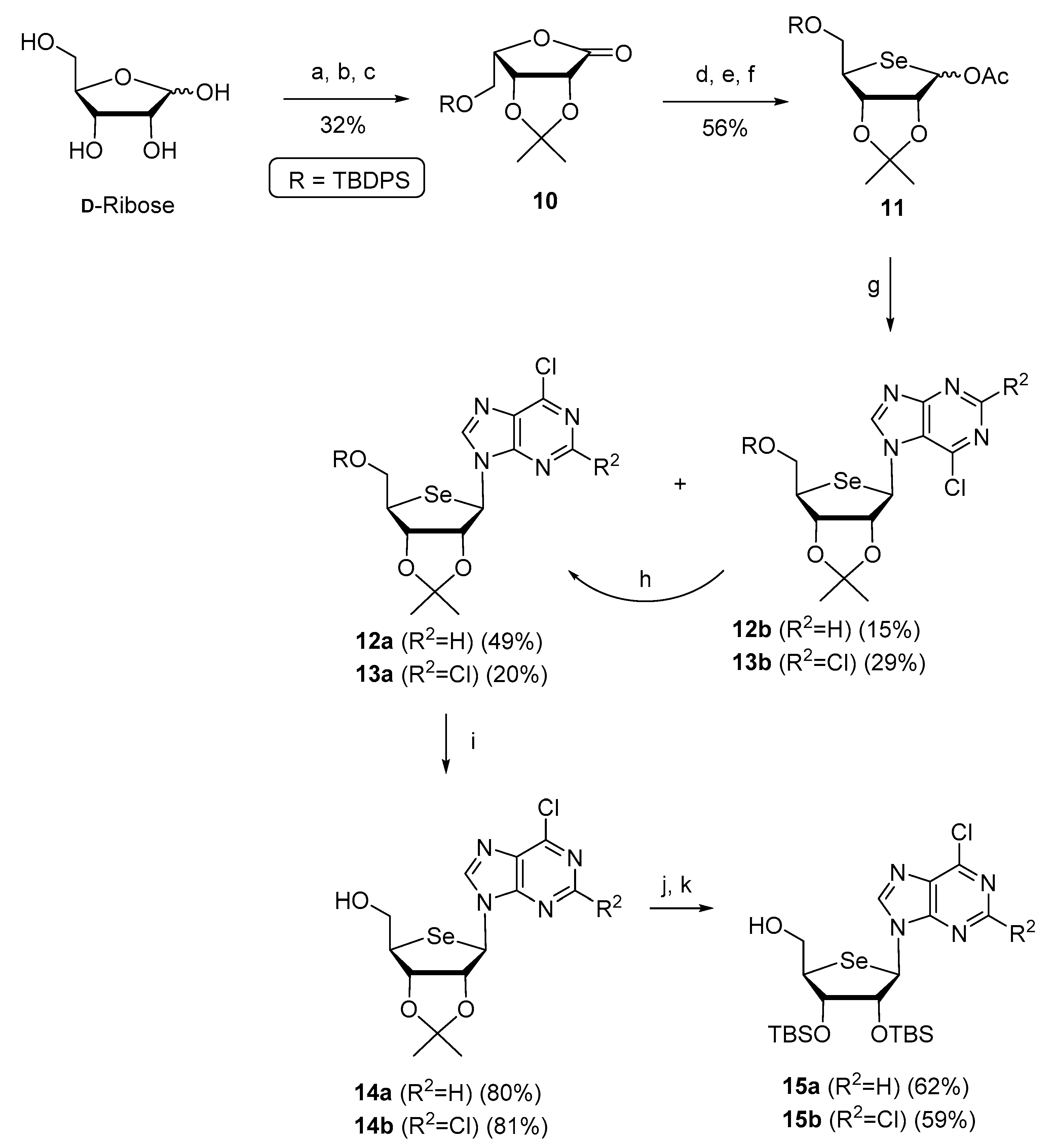
For the synthesis of final compounds 9a–l, key intermediates, 4′-seleno purine nucleosides 15a–b were synthesized from D-ribose following the previously reported procedures [18,19] (Scheme 1). Briefly, D-ribose was converted to L-lyxonolactone derivative 10 in three steps (oxidation to lactone, conversion of D-ribo configuration to L-lyxo configuration, and tert-butyldiphenylsililyl (TBDPS) protection). Reduction of 10 with NaBH4, ring cyclization of resulting diol with selenide ion, and a Pummerer rearrangement of 4-seleno sugar afforded the glycosyl donor 11. A Vorbrüggen condensation of 11 with 6-chloropurine and 2,6-dichloropurine produced the N9-β-anomers 12a and 13a with concomitant formation of their corresponding N7-β-anomers 12b and 13b, respectively. Conversion of N7 isomers 12b and 13b to their corresponding N9 isomers 12a and 13a was achieved by using TMSOTf at high temperature. Removal of the TBDPS group of 12a and 13a yielded the 5′-CH2OH derivatives 14a and 14b, respectively. Conversion of the 5′-hydroxymethyl group of 14a and 14b into a 5′-N,N-dimethyluronamide was successfully achieved, but the final deprotection of the acetonide group under strongly acidic conditions resulted in decomposition, instead of giving the desired final products. Thus, we exchanged the acetonide protecting group of 14a and 14b with a TBS group in four steps, giving 15a and 15b, respectively. Firstly, a PNB protecting group was attached to the 5′-position of 14a and 14b, followed by acetonide group deprotection with 50% aqueous TFA to give diols. The diols were then protected with a TBS group using TBSOTf followed by deprotection of the PNB group with sodium hydroxide in 1,4-dioxane to give 15a and 15b, respectively. The final deprotection with sodium hydroxide required mild reaction conditions (room temperature, overnight) because of the possible hydrolytic conversion of 6-chloropurine to hypoxanthine.
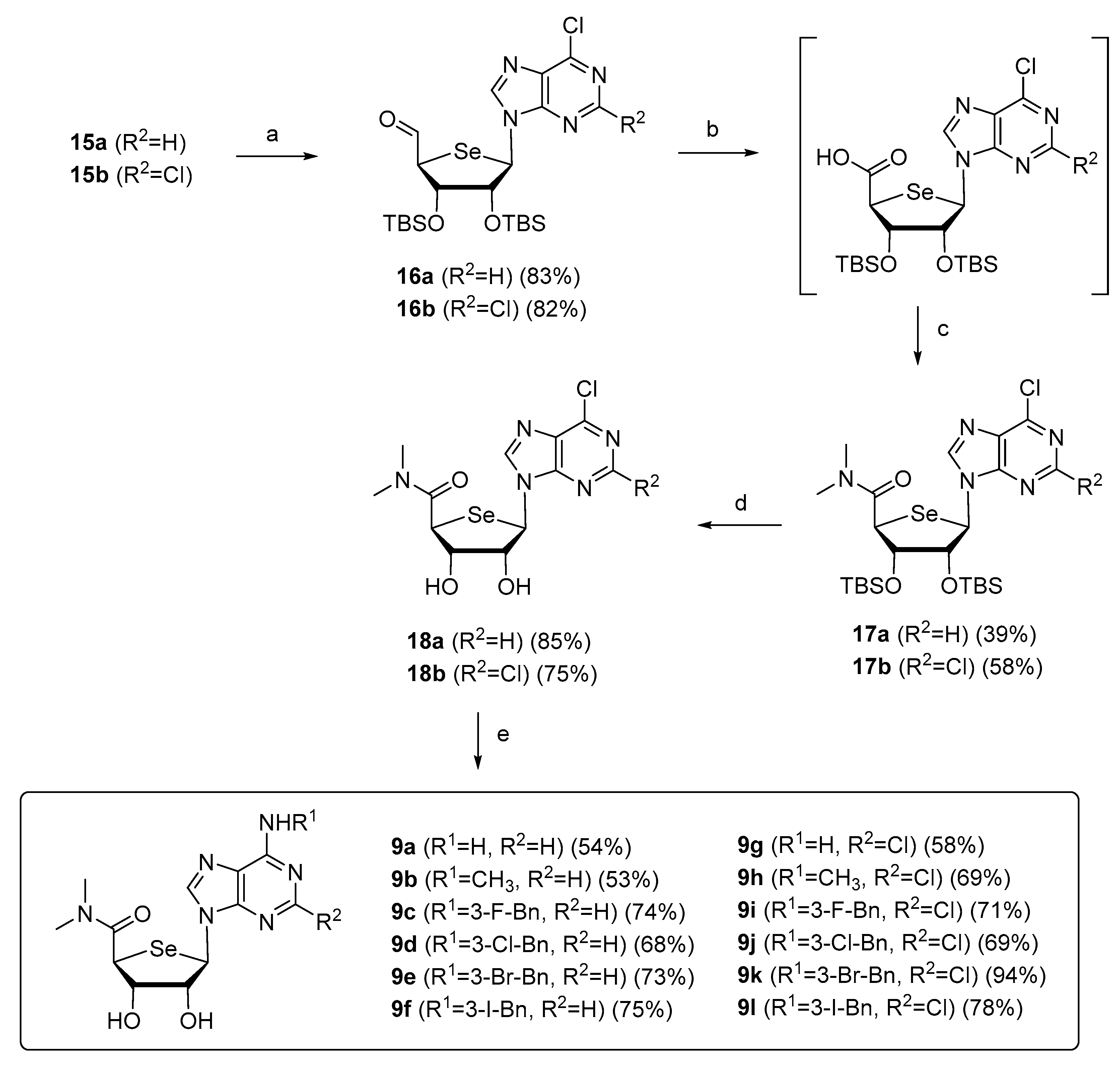
Synthesis of the final nucleosides, 9al from the key intermediates, 15a and 15b is shown in Scheme 2. The direct oxidation of the alcohols of 15a and 15b to the carboxylic acids have been tried with many oxidizing reagents, but none of them could give the desired acid. Thus, we employed a sequential oxidation method via aldehyde instead of direct oxidation to the carboxylic acid. Albright–Goldman oxidations of 15a and 15b, using DMSO as an oxidizing agent under mild condition afforded the aldehydes 16a and 16b, respectively. Tollens′ oxidation converted the aldehydes 16a and 16b to the corresponding carboxylic acids smoothly, which without purification underwent the amide coupling reaction with dimethylamine in the presence of DIPEA, and HATU to yield the 5′-N,N-dimethyluronamides 17a and 17b, respectively. The TBS deprotection of 17a and 17b with TBAF and acetic acid gave the diols 18a and 18b, respectively. The key intermediates 18a and 18b were treated with various amines such as ammonia, alkylamines and 3-halobenzylamines to yield 2-H derivatives 9af and 2-Cl derivatives 9gl, respectively.

2.2. Biology

2.2.1. Binding Affinity

The binding affinities of all the final compounds 9a–l were evaluated, using radioligand binding assays at four human AR subtypes (Table 1), by reported methods [20]. All of the final compounds 9al exhibited medium to high binding affinity at the hA3AR, while no binding affinity at other subtypes such as hA1, hA2A, and hA2BARs was observed. Among the tested compounds, compound 9f exhibited the highest affinity (Ki = 22.7 nM) at hA3AR, which is comparable to the corresponding 4′-oxo- and 4′-thio analogues 5 (Ki = 29.0 nM) and 6 (Ki = 15.5 nM). The introduction of a 3-halobenzyl group at the N6 position increased the hA3AR binding affinity when compared to the N6-unsubstituted adenine compounds 9a or 9g, indicating that a favorable hydrophobic interaction exits at the hA3AR binding site. In the 2-H series, the binding affinity of 3-halobenzyl derivatives 9c–f was decreased in the following order: 3-I-benzyl 9f > 3-Br-benzyl 9e > 3-Cl-benzyl 9d > 3-F-benzyl 9c. The halogen size correlated with hA3AR binding affinity, whereas in the 2-Cl series, the binding affinity of 3-halobenzyl derivatives 9il was almost same within the range of 180–250 nM. In general, the 4′-seleno-5′-N,N-dimethyluronamide derivatives 9a-l exhibited lower binding affinity than the 4′-seleno-5′-N-methyluronamide derivatives 7 and 8. It is interesting to note that 2-Cl-N6-3-iodobenzyl analogue 9l exhibited much weaker binding affinity than the corresponding 2-H analogue 9f. This tendency was also found in the 5′-N-methyluronamide 4′-seleno derivatives 7 and 8.

2.2.2. CAMP Functional Assay

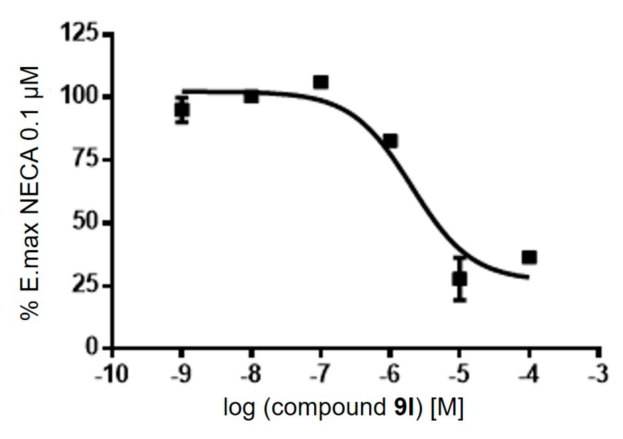
In a cAMP functional assay at hA3AR expressed in CHO cells, compound 9l behaved as an antagonist, like compounds 5 and 6, with KB value of 114.5 nM (Figure 3). Like 5 and 6, an additional methyl group on the 5′-N-methyluronamide converted an agonist into an antagonist, indicating that amide hydrogen is essential for receptor activation in this series, as well. However, the fact that the 5′-N,N-dimethyluronamide derivatives exhibited weaker binding affinity than the corresponding 5′-N-methyluronamide derivatives demonstrates that steric effects induced by 5′-N,N-dimethyluronamide reduce the binding affinity at the A3AR.

2.3. Molecular Modelling Studies

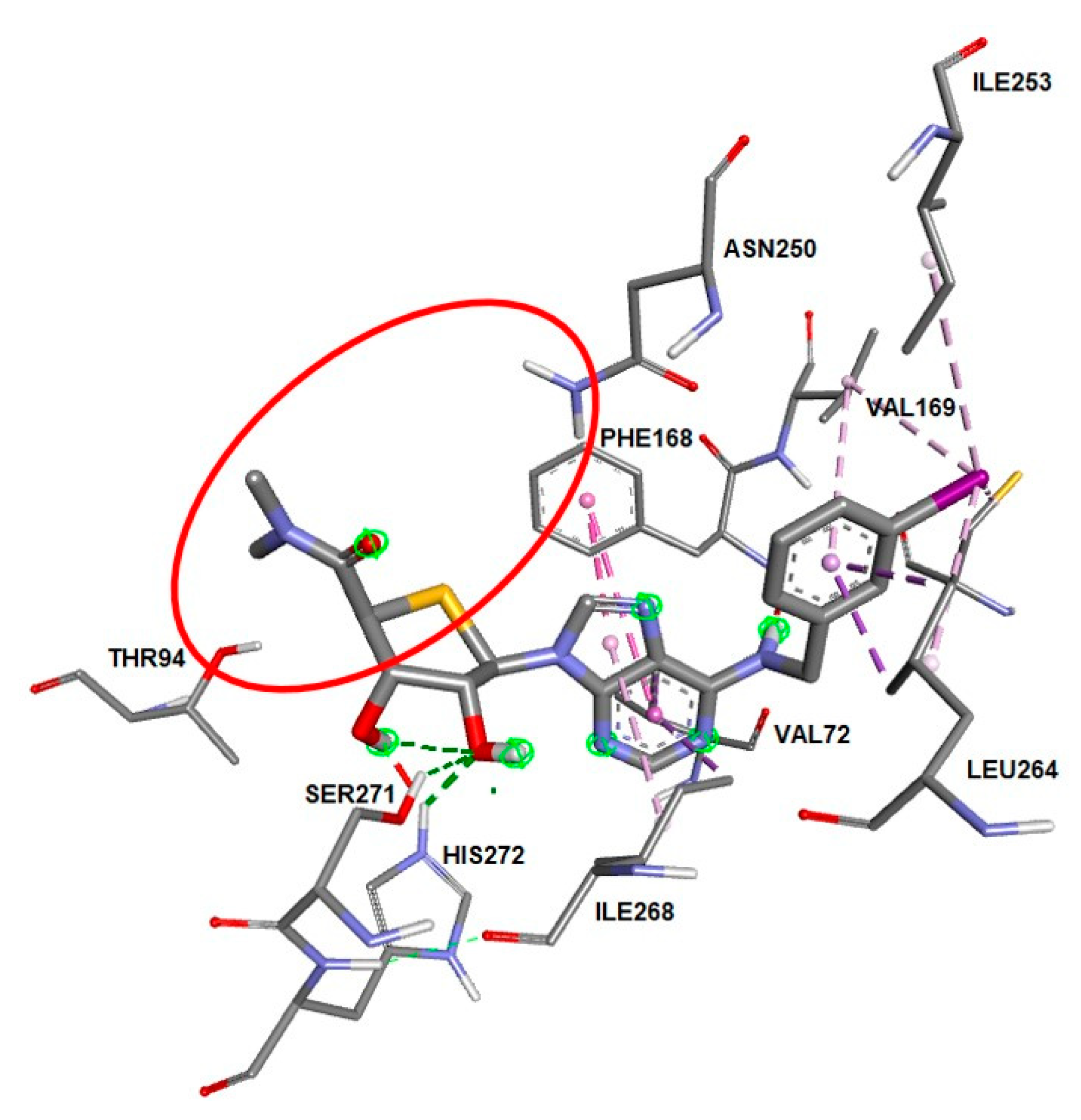
To investigate how 5′-N,N-dimethyluronamide 4′-selenonucleoside derivatives bind at hA3AR, we docked our compounds into the reported homology model of hA3AR [21] using Autodock Vina [22]. The most potent compound 9f bound well at the orthosteric binding site with a South ring conformation (2′-endo/3′-exo), displaying H-bonds with Ser271 and His272 (Figure 4). Compared to the 5′-N-methyluronamide derivative 5, the adenine ring still maintained π−π interaction with Phe168 and the iodobenzene ring had interactions with Val169, Ile253 and Leu264 [19]. The glycosidic bond was in an anti conformation. However, either H-bonding of 5′-N-uronamide with Thr94 or the adenine with Asn250 was not observed (marked as a red circle in Figure 4), suggesting that this H-bonding plays a key role in discriminating an agonist from an antagonist.

3. Materials and Methods

3.1. Chemical Synthesis

Proton (1H) and carbon (13C) NMR spectra were obtained on a Jeol JNM-ECA 300 (JEOL Ltd. Tokyo, Japan; 300/75 MHz), Bruker AV 400 (Bruker, Billerica, MA, USA; 400/100 MHz), and AMX 500 (Bruker, Billerica, MA, USA; 500/125 MHz) spectrometer. The 1H NMR data were reported as peak multiplicities: s for singlet; d for doublet; dd for doublet of doublets; t for triplet; td for triplet of doublet; q for quartet; quin for quintet; bs for broad singlet and m for multiplet. Coupling constants were reported in hertz. The chemical shifts were reported as ppm (δ) relative to the solvent peak. All reactions were routinely carried out under an inert atmosphere of dry nitrogen. IKA RCT basic type heating mantle was used to provide a constant heat source. Microwave-assisted reactions were carried out in sealed vessels using a Biotage Initiator + US/JPN (Biotage, Uppsala, Sweden; part no. 356007) microwave reactor, and the reaction temperatures were monitored by an external surface IR sensor. High-resolution mass spectra were measured with electrospray-ionization quadrupole time-of-flight (ESI-Q-TOF) techniques. Melting points were recorded on a Barnstead electrothermal 9100 instrument and are uncorrected. Reactions were checked by thin layer chromatography (Kieselgel 60 F254, Merck, Kenilworth, NJ, US). Spots were detected by viewing under a UV light, and by colorizing with charring after dipping in a p-anisaldehyde solution. The crude compounds were purified by column chromatography on a silica gel (Kieselgel 60, 70−230 mesh, Merck). All the anhydrous solvents were redistilled over CaH2, or P2O5, or sodium/benzophenone prior to the reaction.
(2S,3S,4R,5R)-3,4-Bis((tert-butyldimethylsilyl)oxy)-5-(6-chloro-9H-purin-9-yl)tetrahydroselenophene-2-carbaldehyde (16a). To a solution of 15a [19] (348 mg, 0.60 mmol) in dimethyl sulfoxide (5 mL) was added acetic anhydride (0.11 mL, 1.2 mmol), and the reaction mixture was stirred at 100 °C for 1 h, cooled to room temperature, and diluted with dichloromethane (20 mL). The mixture was washed with water (10 mL × 2), and the aqueous layer was further extracted with dichloromethane (20 mL × 2). The combined organic layers were washed with brine (5 mL), dried (MgSO4), filtered, and concentrated under reduced pressure. The residue was purified by silica gel column chromatography (hexane–ethyl acetate = 4:1) to give 16a (288 mg, 83%) as a white foam: 1H NMR (400 MHz, CDCl3) δ 9.76 (d, 1 H, J = 2.4 Hz), 8.78 (s, 1 H), 8.32 (s, 1 H), 6.24 (d, 1 H, J = 7.2 Hz), 4.86 (dd, 1 H, J = 2.6, 7.0 Hz), 4.68 (t, 1 H, J = 3.0 Hz), 4.12 (dd, 1 H, J = 2.4, 2.8 Hz), 0.95 (s, 9 H), 0.71 (s, 9 H), 0.12 (s, 6 H), −0.05 (s, 3 H), −0.4 (s, 3 H); 13C NMR (100 MHz, CDCl3) δ 194.6, 152.3, 152.1, 151.7, 145.3, 132.5, 81.0, 77.4, 74.6, 56.9, 56.8, 53.0, 25.9, 25.7, 18.3, 17.9, 0.21, −4.2, −4.45, −4.55, −5.3.
(2S,3S,4R,5R)-3,4-Bis((tert-butyldimethylsilyl)oxy)-5-(2,6-dichloro-9H-purin-9-yl)tetrahydroselenophene-2-carbaldehyde (16b). Compound 15b [19] (1.49 g, 2.43 mmol) was converted to 16b (1.22 g, 82%) as a yellow foam, using a procedure similar to that used in the preparation of 16a: 1H NMR (500 MHz, CDCl3) δ 9.73 (d, J = 2.1 Hz, 1 H), 8.40 (s, 1 H), 6.13 (d, J = 6.8 Hz, 1 H), 4.74−4.73 (m, 1 H), 4.63 (t, J = 2.9 Hz, 1 H), 4.11 (s, 1 H), 0.91 (s, 9 H), 0.71 (s, 9 H), 0.090 (s, 6 H), −0.043 (s, 3 H), −0.38 (s, 3 H); 13C NMR (75 MHz, CDCl3) δ 194.3, 153.2, 153.0, 152.2, 145.7, 131.3, 81.0, 74.4, 56.7, 52.9, 25.69, 25.66, 25.6, 18.0, 17.6, −4.42, −4.70, −4.81, −4.86, −5.55.
(2S,3S,4R,5R)-3,4-Bis((tert-butyldimethylsilyl)oxy)-5-(6-chloro-9H-purin-9-yl)-N-methyltetrahydroselenophene-2-carboxamide (17a). To a solution of AgNO3 (170 mg, 1.00 mmol) was added 1 N NaOH (1.0 mL, 1.00 mmol) at 0 °C. After Ag2O was precipitated, 24% ammonia−water (0.3 mL) was added to the reaction mixture until the mixture was clear. The solution of 16a (288 mg, 0.50 mmol) in THF (8 mL) was added to the above Tollens’ reagent at 0 °C, and the reaction mixture was stirred at the same temperature for 30 min and diluted with water (10 mL). The aqueous layer was extracted with ethyl acetate (10 mL × 2), and the aqueous layer was acidified with 1 N HCl solution (3 mL) and extracted with ethyl acetate (10 mL × 2). The combined organic layers were washed with brine, dried (MgSO4), filtered, and evaporated under reduced pressure to give the crude acid. To a solution of the crude acid in THF (5 mL) were added 1-[bis(dimethylamino)methylene]-1H-1,2,3-triazolo[4,5-b]-pyridinium 3-oxide hexafluorophosphate (HATU) (190 mg, 0.50 mmol), dimethylamine hydrochloride (82 mg, 1.00 mmol), and diisopropylamine (0.19 mL, 1.10 mmol) at room temperature. The reaction mixture was stirred at same temperature for 1 h and diluted with ethyl acetate (10 mL). The organic layer was washed with water (5 mL × 2), and the aqueous layer was further extracted with ethyl acetate (10 mL × 2). The combined organic layers were washed with brine (5 mL), dried (MgSO4), filtered, and concentrated under reduced pressure. The residue was purified by silica gel column chromatography (hexane–ethyl acetate = 3:1) to give 17a (120 mg, 39%) as a white foam; 1H NMR (400 MHz, CDCl3) δ 8.76 (s, 1 H), 8.67 (bs, 1 H), 6.32 (d, J = 6.8 Hz, 1 H), 4.9 (bs, 1 H), 4.63 (m, 1 H), 4.21 (m, 1 H), 3.04 (s, 3 H), 2.99 (s, 3 H), 0.92 (s, 9 H), 0.71 (s, 9 H), 0.08 (d, J = 3.6 Hz, 6 H), 0.01 (s, 3 H)
(2S,3S,4R,5R)-3,4-Bis((tert-butyldimethylsilyl)oxy)-5-(2,6-dichloro-9H-purin-9-yl)-N-methyltetrahydroselenophene-2-carboxamide (17b). Compound 16b (623 mg, 1.02 mmol) was converted to 17b (260 mg, 58%) as a yellow foam, using a procedure similar to that used in the preparation of 17a: 1H NMR (400 MHz, MeOD) δ 8.69 (s, 1 H), 6.20 (d, J = 6.4 Hz, 1 H), 4.75–4.85 (bs, 1 H), 4.60 (m, 1 H), 4.22 (m, 1 H), 3.02 (s, 3 H), 2.98 (s, 3 H), 0.90 (s, 9 H), 0.74 (s, 9 H), 0.06 (s, 6 H), 0.02 (s, 3 H)
(2S,3S,4R,5R)-5-(6-Chloro-9H-purin-9-yl)-3,4-dihydroxy-N-methyltetrahydro selenophene-2-carboxamide (18a). To a stirred solution of 17a (80 mg, 0.13 mmol) in THF (3 mL) was added 1 M tetra-n-butylammonium fluoride in THF solution (0.13 mL, 0.13 mmol) and acetic acid (7 μL, 0.13 mmol), and the reaction mixture was stirred at room temperature for 15 h. The solvent was evaporated, and the resulting residue was purified by silica gel column chromatography (dichloromethane–methanol = 100:1−10:1) to give 18a (43 mg, 85%) as a white solid; 1H NMR (400 MHz, CDCl3) δ 8.93 (s, 1 H), 8.73 (s, 1 H), 6.37 (d, J = 6.4 Hz, 1 H), 4.95 (dd, J = 6 Hz, 3.2 Hz, 1 H), 4.67 (t, J = 3.8 Hz, 1 H), 4.50 (d, J = 4 Hz, 1 H), 3.03 (s, 3 H), 2.99 (s, 3 H).
(2S,3S,4R,5R)-5-(2,6-Dichloro-9H-purin-9-yl)-3,4-dihydroxy-N-methyltetra hydroselenophene-2-carboxamide (18b). Compound 17b (83 mg, 0.127 mmol) was converted to 18b (41 mg, 75%) as a white solid, using a procedure similar to that used in the preparation of 18a; 1H NMR (400 MHz, MeOD) δ 8.93 (s, 1 H), 6.27 (d, J = 6 Hz, 1 H), 4.88 (m, 1 H), 4.65 (t, J = 4 Hz, 1 H), 4.50 (d, J = 4.4 Hz, 1 H), 3.04 (s, 3 H), 2.99 (s, 3 H).

General Procedure for the Synthesis of 9al

To a stirred solution of 18a−b (1 equiv.) in ethanol were added amine (3 equiv.) and triethylamine (6 equiv.), and the reaction mixture was stirred at 95 °C for 15−30 h. All volatiles were evaporated, and the residue was purified by silica gel column chromatography (dichloromethane–methanol = 100:1−10:1) to give 9a-l as a white solid.
(2S,3S,4R,5R)-5-(6-amino-9H-purin-9-yl)-3,4-dihydroxy-N,N-dimethyltetra hydroselenophene-2-carboxamide (9a). White solid; yield: 54%; 1H NMR (400 MHz, MeOD) δ 8.50 (s, 1 H), 8.19 (s, 1 H), 6.24 (d, J = 5.6 Hz, 1 H), 4.68 (t, J = 4 Hz. 1 H), 4.49 (d, J = 4.8 Hz, 3.03 (s, 3 H), 2.98 (s, 3 H); 13C NMR (100 MHz, MeOD) δ 173.9, 157.5, 154.1, 151.3, 142.0, 120.4, 82.8, 77.7, 56.8, 42.4, 38.3, 36.5; HRMS (FAB) found 373.0537 (calculated for C12H17N6O3Se (M + H)+ 373.0527).
(2S,3S,4R,5R)-3,4-dihydroxy-N,N-dimethyl-5-(6-(methylamino)-9H-purin-9-yl)tetrahydroselenophene-2-carboxamide (9b). White solid; yield: 53%; 1H NMR (400 MHz, MeOD) δ 8.43 (s, 1 H), 8.23 (s, 1 H), 6.23 (d, J = 6 Hz, 1 H), 4.67 (t, J = 4 Hz, 1 H), 4.48 (d, J = 4.4 Hz, 1 H), 3.02 (s, 3 H), 2.97 (s, 3 H); 13C NMR (100 MHz, MeOD) δ 173.89, 157.0, 154.1, 149.6, 141.3, 120.9, 82.0, 77.6, 62.4, 56.8, 42.4, 38.3, 36.4; HRMS (FAB) found 387.0675 (calculated for C13H19N6O3Se (M + H)+ 387.0684).
(2S,3S,4R,5R)-5-(6-((3-fluorobenzyl)amino)-9H-purin-9-yl)-3,4-dihydroxy-N,N-dimethyltetrahydroselenophene-2-carboxamide (9c). White solid; yield: 74%; 1H NMR (400 MHz, MeOD) δ 8.46 (s, 1 H) 8.23 (s, 1 H), 7.28 (q, J = 6 Hz, 1 H) 7.15 (d, J = 7.6 Hz, 1 H), 7.07 (d, J = 9.6 Hz, 1 H), 6.92 (td, J = 8, 2 Hz, 1 H), 6.24 (d, J = 5.6 Hz, 1 H), 4.68 (dd, J = 4.4, 4 Hz, 1 H) 4.48 (d, J = 4.8 Hz, 1 H) 3.02 (s, 3 H), 2.97 (s, 3H); 13C NMR (100 MHz, MeOD) δ 173.9, 165.8, 163.3, 156.2, 154.2, 150.7, 143.5, 141.6, 131.4, 124.3, 120.8, 115.1, 82.1, 77.6, 56.8, 42.4, 38.3, 36.4; HRMS (FAB) found 481.0894 (calculated for C19H22FN6O3Se (M + H)+ 481.0903).
(2S,3S,4R,5R)-5-(6-((3-chlorobenzyl)amino)-9H-purin-9-yl)-3,4-dihydroxy-N,N-dimethyltetrahydroselenophene-2-carboxamide (9d). White solid; yield: 68%; 1H NMR (400 MHz, MeOD) δ 8.46 (s, 1 H), 8.24 (s, 1 H), 7.36 (s, 1 H), 7.18–7.28 (m, 4 H), 6.24 (d, J = 5.2 Hz, 1 H), 4.68 (dd, J = 4.4 Hz, 4 Hz, 1 H), 4.48 (d, J = 4.8 Hz, 1 H), 3.02 (s, 3 H), 2.97 (s, 3 H); 13C NMR (100 MHz, MeOD) δ 173.8, 156.1, 154.2, 143.1, 141.7, 135.5, 131.2, 128.6, 128.3, 128.3, 127.0, 120.8, 82.0, 77.6, 56.9, 42.4, 38.2, 36.4; HRMS (FAB) found 497.0599 (calculated for C19H22ClN6O3Se (M + H)+ 497.0607).
(2S,3S,4R,5R)-5-(6-((3-bromobenzyl)amino)-9H-purin-9-yl)-3,4-dihydroxy-N,N-dimethyltetrahydroselenophene-2-carboxamide (9e). White solid; yield: 75%, 1H NMR (400 MHz, MeOD + CDCl3 = 1:3); δ 8.42 (s, 1 H), 8.29 (s, 1 H), 7.50 (s, 1 H), 7.36 (d, J = 7.6 Hz, 1 H), 7.28 (d, J = 7.6 Hz, 1 H), 7.17 (t, J = 8 Hz, 1 H), 6.17 (d, J = 4.8 Hz, 1 H), 4.76 (bs, 2 H), 4.73–4.69 (m, 1 H), 4.44 (d, 4.8 Hz, 1 H), 3.02 (s, 3 H), 2.98 (s, 3 H); 13C NMR (100 MHz, MeOD + CDCl3 = 1:3) δ 172.1, 155.0, 153.3, 141.2, 140.6, 130.8, 130.7, 130.5, 126.5, 122.9, 119.8, 81.3, 77.8, 76.6, 55.9, 41.5, 38.0, 36.3; HRMS (FAB) found 541.0106 (calculated for C19H21BrN6O3Se (M + H)+ 541.0102).
(2S,3S,4R,5R)-3,4-dihydroxy-5-(6-((3-iodobenzyl)amino)-9H-purin-9-yl)-N,N-dimethyltetrahydroselenophene-2-carboxamide (9f). White solid; yield: 73%, 1H NMR (400 MHz, MeOD + CDCl3 =1:3) δ 8.45 (s, 1 H), 8.27 (s, 1 H), 7.71 (s, 1 H), 7.56 (d, J = 7.4 Hz, 1 H), 7.33 (d, J = 7.7 Hz, 1 H), 7.04 (t, J = 7.8 Hz, 1 H), 6.21 (d, J = 5.4 Hz, 1 H), 4.78–4.76 (m, 1 H), 4.74 (bs, 2 H), 4,71–4.69 (m, 1 H), 4.46 (d, J = 4.9 Hz, 1 H), 3.03 (s, 3 H), 2.99 (s, 3 H); 13C NMR (100 MHz, DMSO-d6) δ 173.5, 156.1, 154.3, 142.5, 141.7, 137.8, 137.7, 131.6, 128.1, 120.9, 95.6, 82.3, 79.2, 78.8, 77.7, 57.0, 42.5, 38.9, 37.2; HRMS (FAB) found 588.9971 (calculated for C19H21IN6O3Se (M + H)+ 588.9963).
(2S,3S,4R,5R)-5-(2-chloro-6-amino-9H-purin-9-yl)-3,4-dihydroxy-N,N-dimethyltetrahydroselenophene-2-carboxamide (9g). White solid; yield: 58%; 1H NMR (400 MHz, DMSO-d6) δ 8.33 (s, 1 H), 5.98 (d, J = 4.8 Hz, 1 H), 5.78 (d, J = 4 Hz, 1 H), 5.48 (d, J = 3.2 Hz, 1 H), 4.74 (bs, 1 H), 4.50 (bs, 1 H), 4.32 (d, J = 2.8 Hz, 1 H), 2.91 (s, 3 H), 2.87 (s, 3 H); 13C NMR (100 MHz, DMSO-d6) δ 173.7, 157.5, 154.1, 151.3, 141.9, 120.3, 81.9, 77.5, 49.6, 42.2, 38.1, 36.3; HRMS (FAB) found 407.0132 (calculated for C12H16ClN6O3Se (M + H)+ 407.0138).
(2S,3S,4R,5R)-5-(2-chloro-6-(methylamino)-9H-purin-9-yl)-3,4-dihydroxy-N,N-dimethyltetrahydroselenophene-2-carboxamide (9h). White solid; yield: 69%; 1H NMR (400 MHz, DMSO-d6) δ 8.33 (s, 1 H), 5.98 (d, J = 4.8 Hz, 1 H), 5.78 (d, J = 4 Hz, 1 H), 5.48 (d, J = 3.2 Hz, 1 H), 4.74 (bs, 1 H), 4.50 (bs, 1 H), 4.32 (d, J = 2.8 Hz, 1 H), 2.91 (s, 3 H), 2.87 (s, 3 H); 13C NMR (100 MHz, DMSO-d6) δ 171.0, 155.5, 153.4, 149.8, 139.6, 118.3, 79.5, 75.4, 64.9, 55.1, 41.1, 37.1, 35.3; HRMS (FAB) found 421.0293 (calculated for C13H18ClN6O3Se (M + H)+ 421.0294).
(2S,3S,4R,5R)-5-(2-chloro-6-((3-fluorobenzyl)amino)-9H-purin-9-yl)-3,4-dihydroxy-N,N-dimethyltetrahydroselenophene-2-carboxamide (9i). White solid; yield: 71%; 1H NMR (400 MHz, MeOD) δ 8.43 (s, 1 H), 7.29 (m, 1 H), 7.17 (d, J = 6.8 Hz, 1 H), 7.09 (d, J = 10 Hz, 1 H), 6.94 (m, 1 H), 6.15 (d, J = 5.2 Hz, 1 H), 4.76 (m, 1 H), 4.66 (t, J = 4 Hz, 1 H), 4.47 (d, J = 5.2 Hz, 1 H); 13C NMR (100 MHz, MeOD) δ 173.8, 165.7, 163.3, 156.5, 152.0, 143.5, 142.0, 131.4, 129.7, 126.5, 124.6, 115.3, 82.1, 77.6, 57.0, 42.4, 38.3, 36.5; HRMS (FAB) found 515.0518 (calculated for C19H21ClFN6O3Se (M + H)+ 515.0513).
(2S,3S,4R,5R)-5-(2-chloro-6-((3-chlorobenzyl)amino)-9H-purin-9-yl)-3,4-dihydroxy-N,N-dimethyltetrahydroselenophene-2-carboxamide (9j). White solid; yield: 69%; 1H NMR (400 MHz, MeOD) δ 8.43 (s 1 H), 7.38 (s, 1 H), 7.21–7.30 (m, 3 H), 6.15 (d, J = 5.6 Hz, 1 H), 4.76 (dd, J = 5.6 Hz, 3.2 Hz, 1 H), 4.71 (bs, 1 H), 4.65 (dd, J = 4.8 Hz, 3.2 Hz, 1 H), 4.47 (d, 5.2 Hz, 1 H), 3.03 (s, 3 H), 2.98 (s, 3 H); 13C NMR (100 MHz, MeOD) δ 173.8, 156.6, 155.7, 152.0, 142.7, 142.0, 135.5, 131.2, 128.9, 128.4, 127.3, 119.3, 82.1, 77.6, 57.0, 44.7, 42.4, 38.3, 36.5; HRMS (FAB) found 531.0211 (calculated for C19H21Cl2N6O3Se (M + H)+ 531.0217).
(2S,3S,4R,5R)-5-(6-((3-bromobenzyl)amino)-2-chloro-9H-purin-9-yl)-3,4-dihydroxy-N,N-dimethyltetrahydroselenophene-2-carboxamide (9k). White solid; yield: 94%; 1H NMR (400 MHz, MeOD) δ 8.43 (s, 1 H), 7.54 (s, 1 H), 7.36 (m, 2 H), 7.33–7.39 (t, J = 7.6 Hz, 1 H), 6.15 (d, J = 5.6 Hz, 1 H), 4.76 (dd, J = 6.4 Hz, 3.2 Hz, 1 H), 4.71 (bs, 1 H), 4.65 (dd, J = 3.2 Hz, 1 H), 4.48 (d, J = 4.8 Hz, 1 H), 3.03 (s, 3 H), 2.98 (s, 3 H); 13C NMR (100 MHz, MeOD) δ 174.5, 157.3, 156.5, 152.7, 143.6, 142.7, 132.6, 132.1, 128.4, 124.2, 82.8, 78.3, 57.6, 45.3, 43.0, 38.9, 37.1; HRMS (FAB) found 574.9708 (calculated for C19H21BrClN6O3Se (M + H)+ 574.9712).
(2S,3S,4R,5R)-5-(2-chloro-6-((3-iodobenzyl)amino)-9H-purin-9-yl)-3,4-dihydroxy-N,N-dimethyltetrahydroselenophene-2-carboxamide (9l). White solid; yield: 78%; 1H NMR (400 MHz, MeOD) δ 8.43 (s, 1 H), 7.75 (s, 1 H), 7.57 (d, J = 4 Hz, 1 H), 7.36 (d, J = 3.6 Hz, 1 H), 7.07 (t, J = 8 Hz, 1 H), 6.15 (d, J = 5.6 Hz, 1 H), 4.76 (dd, J = 5.2 Hz, 3.2 Hz, 1 H), 4.64–4.68 (m, 2 H), 4.48 (d, J = 4.8 Hz, 1 H), 3.03 (s, 3 H), 2.98 (s, 3 H); 13C NMR (100 MHz, MeOD) δ 173.8, 156.8, 155.9, 152.0, 142.8, 142.0, 138.0, 137.6, 131.5, 128.3, 95.1, 82.1, 77.6, 57.0, 44.5, 42.4, 38.3, 36.5.; HRMS (FAB) found 622.9584 (calculated for C19H21ClIN6O3Se (M + H)+ 622.9574).

3.2. Biological Evaluation

3.2.1. Binding Assay at hA1AR

Adenosine A1 receptor competition binding experiments were carried out in membranes from CHO-A1 cells (Euroscreen, Gosselies, Belgium). On the day of assay, membranes were defrosted and re-suspended in incubation buffer 20 mM Hepes, 100 mM NaCl, 10 mM MgCl2, 2 UI/mL adenosine deaminase (pH = 7.4). Each reaction well of a GF/C multiscreen plate (Millipore, Madrid, Spain), prepared in duplicate, contained 15 μg of protein, 2 nM [3H]DPCPX and test compound. Nonspecific binding was determined in the presence of 10 μM (R)-PIA. The reaction mixture was incubated at 25 °C for 60 min, after which samples were filtered and measured in a microplate beta scintillation counter (Microbeta Trilux, Perkin Elmer, Madrid, Spain).

3.2.2. Binding Assay at hA2AAR

Adenosine A2A receptor competition binding experiments were carried out in membranes from HeLa-A2A cells. On the day of assay, membranes were defrosted and re-suspended in incubation buffer 50 mM Tris-HCl, 1 mM EDTA, 10 mM MgCl2 and 2 UI/mL adenosine deaminase (pH = 7.4). Each reaction well of a GF/C multiscreen plate (Millipore, Madrid, Spain), prepared in duplicate, contained 10 μg of protein, 3 nM [3H]ZM241385 and test compound. Nonspecific binding was determined in the presence of 50 μM NECA. The reaction mixture was incubated at 25 °C for 30 min, after which samples were filtered and measured in a microplate beta scintillation counter (Microbeta Trilux, Perkin Elmer, Madrid, Spain).

3.2.3. Binding Assay at hA2BAR

Adenosine A2B receptor competition binding experiments were carried out in membranes from HEK-293-A2B cells (Euroscreen, Gosselies, Belgium) prepared following the provider’s protocol. On the day of assay, membranes were defrosted and re-suspended in incubation buffer 50 mM Tris-HCl, 1 mM EDTA, 10 mM MgCl2, 0.1 mM benzamidine, 10 μg/mL bacitracine and 2 UI/mL adenosine deaminase (pH = 6.5). Each reaction well prepared in duplicate contained 18 μg of protein, 35 nM [3H]DPCPX and test compound. Nonspecific binding was determined in the presence of 400 μM NECA. The reaction mixture was incubated at 25 °C for 30 min, after which samples were filtered through a multiscreen GF/C microplate and measured in a microplate beta scintillation counter (Microbeta Trilux, Perkin Elmer, Madrid, Spain).

3.2.4. Binding Assay at hA3AR

Adenosine A3 receptor competition binding experiments were carried out in a multiscreen GF/B 96-well plate (Millipore, Madrid, Spain) pretreated with binding buffer (Tris-HCl 50 mM, EDTA 1 mM, MgCl2 5 mM, 2 U/mL adenosine deaminase, pH = 7.4). In each well was incubated 30 μg of membranes from Hela-A3 cell line and prepared in laboratory (Lot: A005/05-07-2019, protein concentration = 3925 μg/mL), 10 nM [3H]-NECA (26.3 Ci/mmol, 1 mCi/mL, Perkin Elmer NET811250UC) and compounds studied in standard methods. Nonspecific binding was determined in the presence of R-PIA 100μM (Sigma P4532, Sigma-Aldrich, St. Louis, MO, USA). The reaction mixture (Vt: 200 μL/well) was incubated at 25 °C for 180 min, after filtered and washed six times with 250 μL wash buffer (Tris-HCl 50mM pH = 7.4), before measuring in a microplate beta scintillation counter (Microbeta Trilux, PerkinElmer, Madrid, Spain).

3.2.5. Cyclic AMP Accumulation Assay

Human adenosine A3 receptor functional experiments were carried out in CHO-A3#18 cell line. The day before the assay, the cells were seeded on the 96-well culture plate (Falcon 353072, Corning, Glendale, AZ, USA). The cells are washed with wash buffer (Dulbecco’s modified eagle’s medium nutrient mixture F-12 ham (Sigma D8062), 25 mM Hepes; pH = 7.4). Wash buffer is replaced by incubation buffer (Dulbecco’s modified eagle’s medium nutrient mixture F-12 ham (Sigma D8062), 25 mM Hepes, 30 μM Rolipram (Sigma R6520); pH = 7.4). Test compounds and MRS1220 as reference compound (Sigma M228) are added and the cells incubated at 37 °C for 15 min. After, 0.1 μM of 5′-N-ethylcarboxamido-adenosine (NECA) (Sigma E2387) is added and the cells incubated at 37 °C for 10 min. Forskolin (Sigma F3917, Sigma-Aldrich, St. Louis, MO, USA) is added and incubated at 37 °C for 5 min. After incubation, the amount of cAMP is determined using a cAMP Biotrak Enzymeimmunoassay (EIA) System Kit (GE Healthcare RPN225, GE Healthcare, Chicago, IL, USA).

3.3. Molecular Modelling

The hA3AR homology model was obtained from reference 20. Autodock Vina 4 (The Scripps Research Institute, La Jolla, CA, USA) was used as the docking tool to generate ligand-protein complex using the following settings: center_x = −7.288, center_y = −8.071, center_z = 51.576, size_x = 40, size_y = 40, size_z = 40, energy_range = 4, exhaustiveness = 8. The ligand–protein complex with the best IFD score were selected and analyzed. The molecular graphic figures were generated by Biovia Discovery Studio Visualizer software (https://3dsbiovia.com/) (accessed on 13 April 2021.).

4. Conclusions

On the basis of potent and selective antagonist 5 and 6 at the human A3AR, N6-substituted-5′-N,N-dimethylcarbamoyl-4′-selenonuceloside derivatives (9a–l) were synthesized from D-ribose and evaluated for their binding affinity toward hARs. All final compounds exhibited medium to high binding affinity toward A3AR with high selectivity compared to other subtypes. Among these derivatives, compound 9f was found to show the highest binding affinity (Ki = 22.7 nM) at hA3AR, comparable to the corresponding 4′-oxo- and 4′-thio analogues 5 (Ki = 29.0 nM) and 6 (Ki = 15.5 nM). As in the case of 4′-oxo- and 4′-thio analogues 5 and 6, addition of another methyl group to the 5′-N-methyluronamide converted an A3AR agonist into an A3AR antagonist, demonstrating the importance of amide hydrogen for receptor activation, which was supported by the molecular modelling study.
We believe that this study helps to define the pharmacophore needed for receptor activation or inactivation and will aid in the design of selective A3AR ligands by medicinal chemists.

Author Contributions

Conceptualization, L.S.J.; methodology, J.Y.; software, J.Y.; validation, L.S.J. and J.Y.; formal analysis, H.C.; investigation, H.C. and K.A.J.; data curation, H.C.; writing—original draft preparation, J.Y.; writing—review and editing, L.S.J.; supervision, L.S.J.; project administration, L.S.J.; funding acquisition, L.S.J. All authors have read and agreed to the published version of the manuscript.

Funding

This research was funded by the National Research Foundation (NRF) of Korea (NRF-2018R1D1A1A02085459 and 2021R1A2B5B02001544) and the Korea Institute of Science and Technology (KIST); ZIADK31117 (NIDDK).

Institutional Review Board Statement

Not applicable.

Informed Consent Statement

Not applicable.

Data Availability Statement

The data presented in this study are available in the article.

Conflicts of Interest

The authors declare no conflict of interest.

References

  1. Borea, P.A.; Varani, K.; Vincenzi, F.; Baraldi, P.G.; Tabrizi, M.A.; Merighi, S.; Gessi, S. The A3 adenosine receptor: History and perspectives. Pharmacol. Rev. 2015, 67, 74–102. [Google Scholar] [CrossRef] [PubMed] [Green Version]
  2. Jacobson, K.A.; Gao, Z.G. Adenosine receptors as therapeutic targets. Nat. Rev. Drug Discov. 2006, 5, 247–264. [Google Scholar] [CrossRef] [Green Version]
  3. Fishman, P.; Bar-Yehuda, S.; Varani, K.; Gessi, S.; Merighi, S.; Borea, P.A. Agonists and Antagonists: Molecular Mechanisms and Therapeutic Applications. In A3 Adenosine Receptors from Cell Biology to Pharmacology and Therapeutics; Borea, P., Ed.; Springer: Dordrecht, The Netherlands, 2010; pp. 301–317. [Google Scholar] [CrossRef]
  4. Ge, Z.D.; Peart, J.N.; Kreckler, L.M.; Wan, T.C.; Jacobson, M.A.; Gross, G.J.; Auchampach, J.A. Cl-IB-MECA [2-chloro-N6-(3-iodobenzyl)adenosine-5′-N-methylcarboxamide] reduces ischemia/reperfusion injury in mice by activating the A3 adenosine receptor. J. Pharmacol. Exp. Ther. 2006, 319, 1200–1210. [Google Scholar] [CrossRef] [PubMed]
  5. Chen, Y.; Corriden, R.; Inoue, Y.; Yip, L.; Hashiguchi, N.; Zinkernagel, A.; Nizet, V.; Insel, P.A.; Junger, W.G. ATP release guides neutrophil chemotaxis via P2Y2 and A3 receptors. Science 2006, 314, 1792–1795. [Google Scholar] [CrossRef] [Green Version]
  6. Wang, X.; Chen, D. Purinergic Regulation of Neutrophil Function. Front. Immunol. 2018, 9, 399. [Google Scholar] [CrossRef] [PubMed] [Green Version]
  7. Marucci, G.; Santinelli, C.; Buccioni, M.; Navia, A.M.; Lambertucci, C.; Zhurina, A.; Yli-Harja, O.; Volpini, R.; Kandhavelu, M. Anticancer activity study of A3 adenosine receptor agonists. Life Sci. 2018, 205, 155–163. [Google Scholar] [CrossRef] [PubMed]
  8. Cohen, S.; Stemmer, S.M.; Zozulya, G.; Ochaion, A.; Patoka, R.; Barer, F.; Bar-Yehuda, S.; Rath-Wolfson, L.; Jacobson, K.A.; Fishman, P. CF102 an A3 adenosine receptor agonist mediates anti-tumor and anti-inflammatory effects in the liver. J. Cell. Physiol. 2011, 226, 2438–2447. [Google Scholar] [CrossRef] [Green Version]
  9. Jacobson, K.A.; Merighi, S.; Varani, K.; Borea, P.A.; Baraldi, S.; Tabrizi, M.A.; Romagnoli, R.; Baraldi, P.G.; Ciancetta, A.; Tosh, D.K.; et al. A3 adenosine receptors as modulators of inflammation: From medicinal chemistry to therapy. Med. Res. Rev. 2018, 38, 1031–1072. [Google Scholar] [CrossRef] [PubMed]
  10. Von Lubitz, D.K.J.E.; Carter, M.F.; Deutsch, S.I.; Lin, R.C.S.; Mastropaolo, J.; Meshulam, Y.; Jacobson, K.A. The effects of adenosine A3 receptor stimulation on seizures in mice. Eur. J. Pharmacol. 1995, 275, 23–29. [Google Scholar] [CrossRef]
  11. Merighi, S.; Benini, A.; Mirandola, P.; Gessi, S.; Varani, K.; Leung, E.; Maclennan, S.; Borea, P.A. Adenosine modulates vascular endothelial growth factor expression via hypoxia-inducible factor-1 in human glioblastoma cells. Biochem. Pharmacol. 2006, 72, 19–31. [Google Scholar] [CrossRef]
  12. Fishman, P.; Cohen, S.; Bar-Yehuda, S. Targeting the A3 adenosine receptor for glaucoma treatment (Review). Mol. Med. Rep. 2013, 7, 1723–1725. [Google Scholar] [CrossRef] [Green Version]
  13. Kim, H.O.; Ji, X.; Siddiqi, S.M.; Olah, M.E.; Stiles, G.L.; Jacobson, K.A. 2-Substitution of N6-Benzyladenosine-5′-uronamides Enhances Selectivity for A3 Adenosine Receptors. J. Med. Chem. 1994, 37, 3614–3621. [Google Scholar] [CrossRef]
  14. Gao, Z.-G.; Kim, S.-K.; Biadatti, T.; Chen, W.; Lee, K.; Barak, D.; Kim, S.G.; Johnson, C.R.; Jacobson, K.A. Structural Determinants of A3 Adenosine Receptor Activation:  Nucleoside Ligands at the Agonist/Antagonist Boundary. J. Med. Chem. 2002, 45, 4471–4484. [Google Scholar] [CrossRef] [PubMed]
  15. Jeong, L.S.; Pal, S.; Choe, S.A.; Choi, W.J.; Jacobson, K.A.; Gao, Z.-G.; Klutz, A.M.; Hou, X.; Kim, H.O.; Lee, H.W.; et al. Structure−Activity Relationships of Truncated d- and l-4′-Thioadenosine Derivatives as Species-Independent A3 Adenosine Receptor Antagonists. J. Med. Chem. 2008, 51, 6609–6613. [Google Scholar] [CrossRef] [PubMed] [Green Version]
  16. Tosh, D.K.; Salmaso, V.; Rao, H.; Bitant, A.; Fisher, C.L.; Lieberman, D.I.; Vorbrüggen, H.; Reitman, M.L.; Gavrilova, O.; Gao, Z.-G.; et al. Truncated (N)-Methanocarba Nucleosides as Partial Agonists at Mouse and Human A3 Adenosine Receptors: Affinity Enhancement by N6-(2-Phenylethyl) Substitution. J. Med. Chem. 2020, 63, 4334–4348. [Google Scholar] [CrossRef] [PubMed]
  17. Gao, Z.-G.; Joshi, B.V.; Klutz, A.M.; Kim, S.-K.; Lee, H.W.; Kim, H.O.; Jeong, L.S.; Jacobson, K.A. Conversion of A3 adenosine receptor agonists into selective antagonists by modification of the 5′-ribofuran-uronamide moiety. Bioorg. Med. Chem. Lett. 2006, 16, 596–601. [Google Scholar] [CrossRef] [Green Version]
  18. Jeong, L.S.; Lee, H.W.; Kim, H.O.; Tosh, D.K.; Pal, S.; Choi, W.J.; Gao, Z.G.; Patel, A.R.; Williams, W.; Jacobson, K.A.; et al. Structure-activity relationships of 2-chloro-N6-substituted-4′-thioadenosine-5′-N,N-dialkyluronamides as human A3 adenosine receptor antagonists. Bioorg. Med. Chem. Lett. 2008, 18, 1612–1616. [Google Scholar] [CrossRef]
  19. Yu, J.; Zhao, L.X.; Park, J.; Lee, H.W.; Sahu, P.K.; Cui, M.; Moss, S.M.; Hammes, E.; Warnick, E.; Gao, Z.-G.; et al. N6-Substituted 5′-N-Methylcarbamoyl-4′-selenoadenosines as Potent and Selective A3 Adenosine Receptor Agonists with Unusual Sugar Puckering and Nucleobase Orientation. J. Med. Chem. 2017, 60, 3422–3437. [Google Scholar] [CrossRef] [PubMed] [Green Version]
  20. Yu, J.; Mannes, P.; Jung, Y.-H.; Ciancetta, A.; Bitant, A.; Lieberman, D.I.; Khaznadar, S.; Auchampach, J.A.; Gao, Z.-G.; Jacobson, K.A. Structure activity relationship of 2-arylalkynyl-adenine derivatives as human A3 adenosine receptor antagonists. Med. Chem. Comm. 2018, 9, 1920–1932. [Google Scholar] [CrossRef] [PubMed]
  21. Petrelli, R.; Torquati, I.; Kachler, S.; Luongo, L.; Maione, S.; Franchetti, P.; Grifantini, M.; Novellino, E.; Lavecchia, A.; Klotz, K.-N.; et al. 5′-C-Ethyl-tetrazolyl-N6-Substituted Adenosine and 2-Chloro-adenosine Derivatives as Highly Potent Dual Acting A1 Adenosine Receptor Agonists and A3 Adenosine Receptor Antagonists. J. Med. Chem. 2015, 58, 2560–2566. [Google Scholar] [CrossRef]
  22. Trott, O.; Olson, A.J. AutoDock Vina: Improving the speed and accuracy of docking with a new scoring function, efficient optimization, and multithreading. J. Comput. Chem. 2010, 31, 455–461. [Google Scholar] [CrossRef] [PubMed] [Green Version]
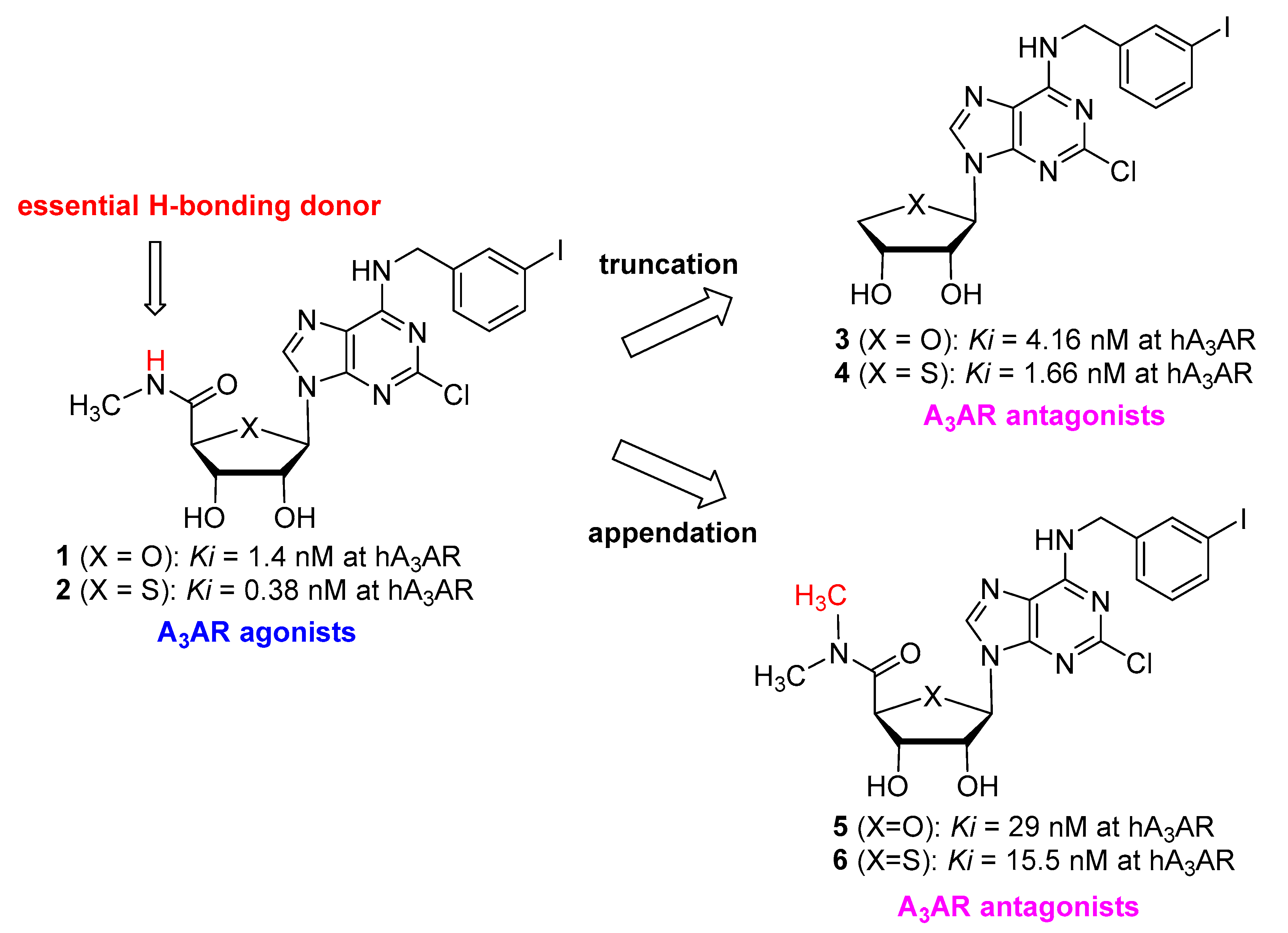
Figure 1. Conversion of A3AR agonists to A3AR antagonists.
Pharmaceuticals 14 00363 g001
Figure 2. The rationale for the design of the target nucleosides 9a–l.
Pharmaceuticals 14 00363 g002
Scheme 1. Synthesis of intermediates 15a and 15b from D-ribose (a) i. Br2, H2O, K2CO3; ii. Acetone, H2SO4; (b) i. MsCl, Et3N, CH2Cl2, 0 °C, 2 h; ii. KOH, H2O, rt, 15 h; (c) TBDPSCl, Et3N, DMAP, CH2Cl2, rt, 4 h; (d) NaBH4, MeOH, rt, 1 h; (e) i. MsCl, Et3N, CH2Cl2, rt, 1 h; ii. Se, NaBH4, EtOH, THF, 60 °C, 15 h; (f) i. mCPBA, CH2Cl2, −78 °C, 1 h; ii. Ac2O, 100 °C, 4 h; (g) 6-chloropurine, BSA, TMSOTf, PhCH3, 95 °C, 15 h or 2,6-dichloropurine, BSA, TMSOTf, CH3CN, 60 °C, 2 h; (h) TMSOTf, PhCH3, 95 °C, 30 min; (i) TBAF, THF, rt, 1 h; (j) i. PNBCl, Et3N, CH2Cl2, rt, 1 h; ii. TFA/H2O, THF, rt, 2 h; (k) i. TBSOTf, Et3N, DMAP, CH2Cl2, rt, 1 h; ii. NaOH, 1,4-dioxane, rt, 15 h.
Pharmaceuticals 14 00363 sch001
Scheme 2. Synthesis of 5′-N,N-dimethyluronamide 4′-selenonucleoside analogues 9a–l. (a) DMSO, Ac2O, 100 °C, 1 h; (b) AgNO3, NaOH, NH4OH, THF, 0 °C, 30 min; (c) (CH3)2NH∙HCl, DIPEA, HATU, THF, rt, 1 h; (d) TBAF, AcOH, THF, rt, 15 h; (e) RNH2, Et3N, EtOH, reflux, 15–30 h.
Pharmaceuticals 14 00363 sch002
Figure 3. Concentration−response curve of 9l in a functional assay at human A3AR measuring inhibition of 10 μM NECA-induced cAMP accumulation. Points represent mean ± SD (vertical bars) of duplicate experiments.
Pharmaceuticals 14 00363 g003
Figure 4. Predicted binding mode of compound 9f in the homology model of hA3AR. Hydrogen bonds are depicted as green dashed line. Hydrophobic interactions are marked with a purple dashed line and π-π interactions are marked with a pink dashed line.
Pharmaceuticals 14 00363 g004
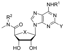
Table 1. Binding affinities of known A3AR ligands and 5′-N,N-dimethyluronamide-4′-selenonucleoside derivatives (9a–l) at human A1, A2A, A2B, and A3ARs.
Pharmaceuticals 14 00363 i001
CompoundAffinity, Ki, nM ± SEM a,b (or % Inhibition at 10 uM)
XYR1R2hA1ARhA2AARhA2BARhA3AR
5 cOCl3-I-BnCH35870 ± 930>10,000>10,00029.0 ± 4.9
6 cSCl3-I-BnCH36220 ± 640>10,000>10,00015.5 ± 3.1
7 dSeH3-I-BnH480 ± 941080 ± 140ND0.57 ± 0.10
8 dSeCl3-I-BnH311 ± 471200 ± 70ND4.20 ± 0.73
9aSeHHCH316% ± 422% ± 317% ± 13710 ± 600
9bSeHCH3CH39% ± 23% ± 29% ± 4609 ± 47
9cSeH3-F-BnCH313% ± 212% ± 119% ± 52020 ± 170
9dSeH3-Cl-BnCH323% ± 54% ± 315% ± 11190 ± 160
9eSeH3-Br-BnCH343% ± 737% ± 8ND36.3 ± 12.7
9fSeH3-I-BnCH336% ± 632% ± 1ND22.7 ± 8.9
9gSeClHCH39% ± 13% ± 325% ± 53250 ± 370
9hSeClCH3CH358% ± 75% ± 219% ± 51060 ± 140
9iSeCl3-F-BnCH334% ± 29% ± 414% ± 2238 ± 37
9jSeCl3-Cl-BnCH363% ± 513% ± 127% ± 4195 ± 40
9kSeCl3-Br-BnCH358% ± 210% ± 427% ± 6180 ± 12
9lSeCl3-I-BnCH368% ± 210% ± 123% ± 3253 ± 29
a All binding experiments were performed using adherent mammalian cells stably transfected with cDNA encoding the appropriate hAR (A1AR and A3AR in CHO cells, A2AAR in Hela cells and A2BAR in HEK-293 cells). Binding was carried out using 2 nM [3H]DPCPX, 3 nM [3H]ZM241385, 25 nM [3H]DPCPX or 0.5 nM [3H]NECA as radioligands for A1, A2A, A2B, and A3ARs, respectively. Values are expressed as mean ± SEM (n = 2). b When a value is expressed as a percentage, it refers to the percent inhibition of a specific radioligand binding at 10 μM, with nonspecific binding defined using 10 μM NECA. c Ref [16]. d Ref [19].
Publisher’s Note: MDPI stays neutral with regard to jurisdictional claims in published maps and institutional affiliations.