
Adipose Tissue-Derived Mesenchymal Stem Cells as a Potential Restorative Treatment for Cartilage Defects: A PRISMA Review and Meta-Analysis


Abstract
:
1. Introduction
2. Methods
2.1. Search Algorithm
2.2. Inclusion and Exclusion Criteria
2.3. Data Extraction
- Study characteristics, such as study design, level of evidence, outcome measures and duration of follow-up.
- Subject information such as subject model, mean age, mean BMI, percentage of female subjects and ethnicity.
- Intervention information, including method of AMSC administration, AMSC cell count.
- Results, including any complications arising during follow-up.
- Outcome measures that were pertinent to cartilage regeneration would also be extracted in detail. This would include clinical scores related to cartilage damage, imaging scores and histological cartilage repair scores (Numerical data were extracted corrected to 3 significant figures).
2.4. Data Analysis
2.5. Assessing Risk of Bias
3. Results
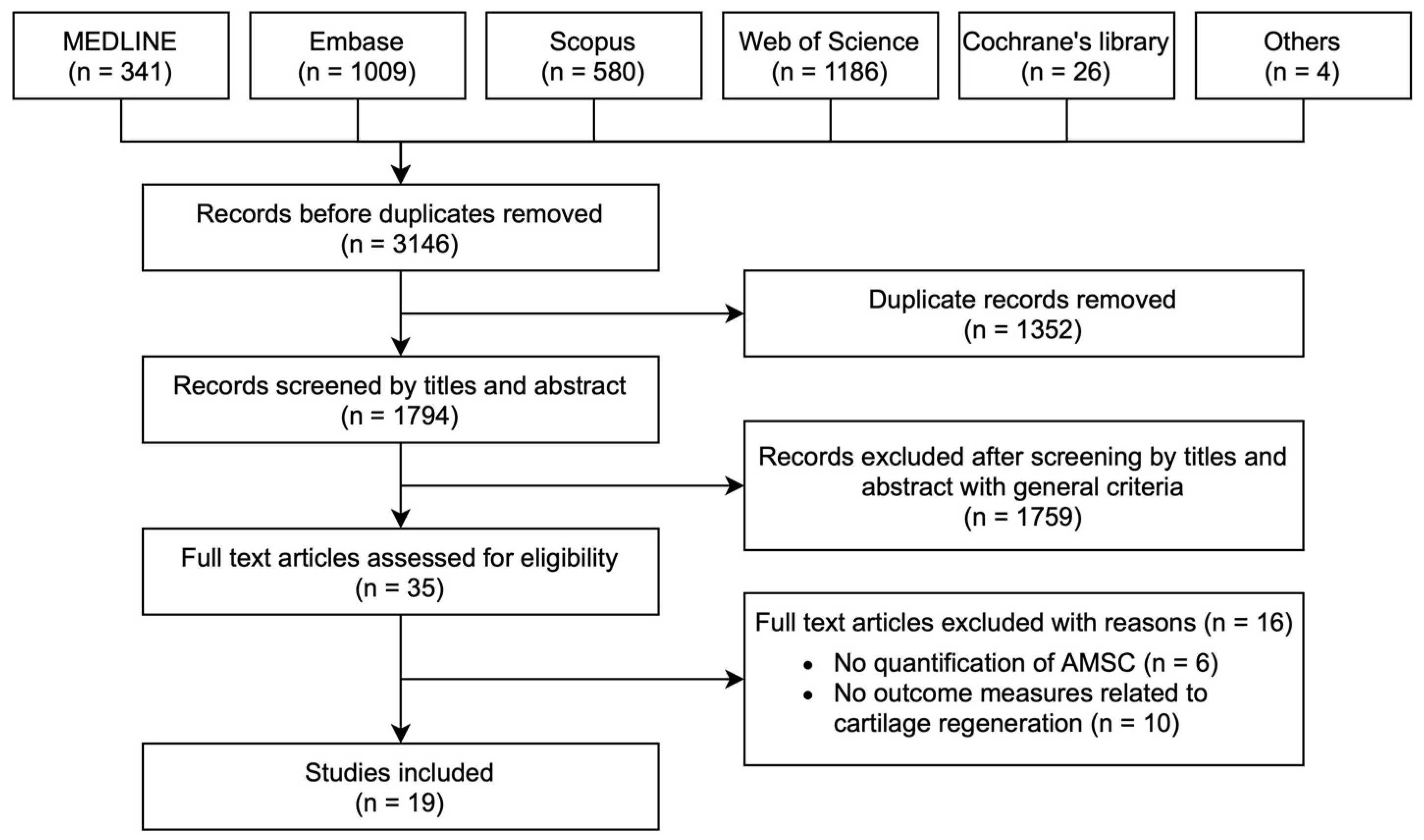
3.1. Search Results
3.2. Characteristics of Selected Studies
3.3. Administration of AMSCs Leads to Improvement of Cartilage Defects
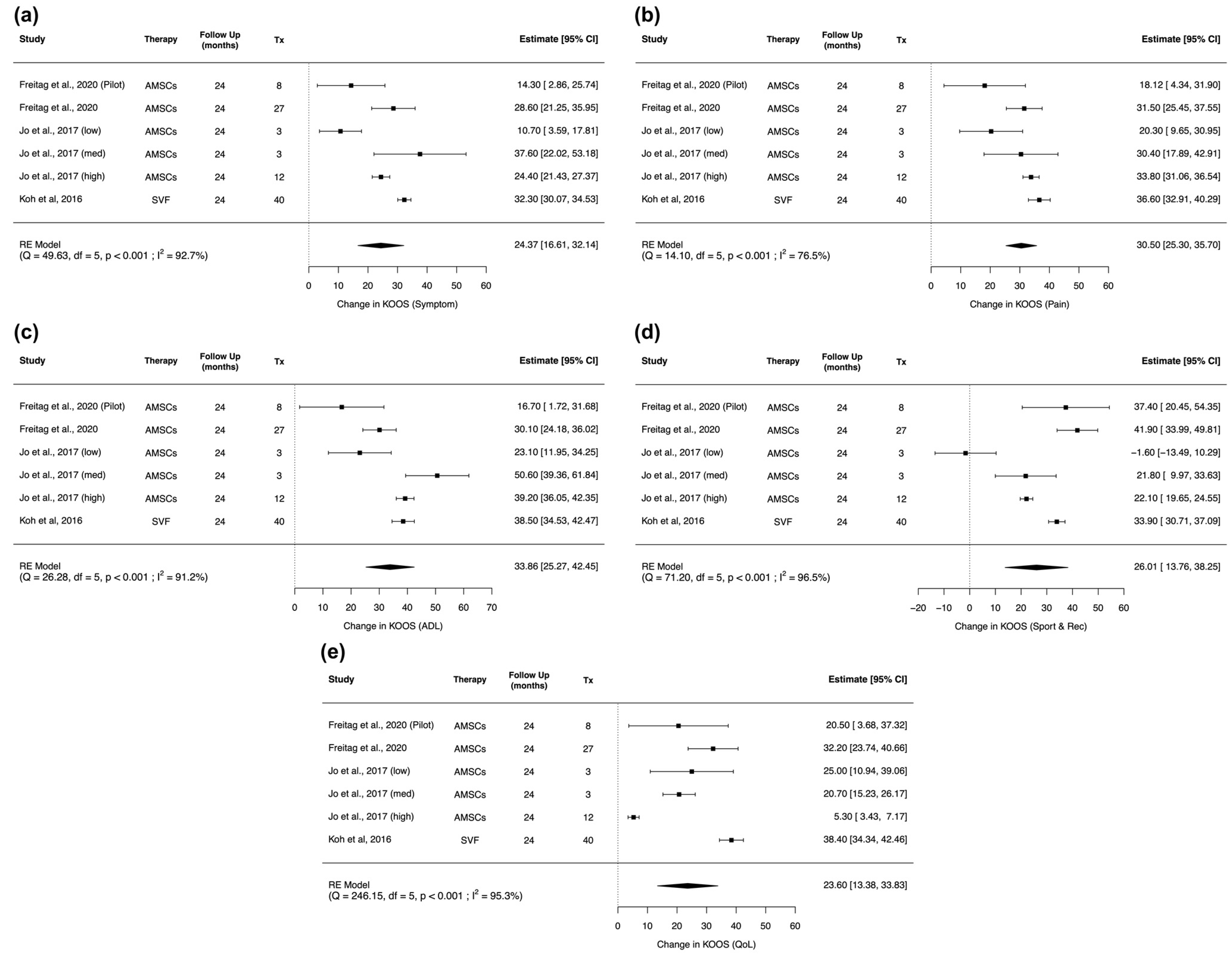
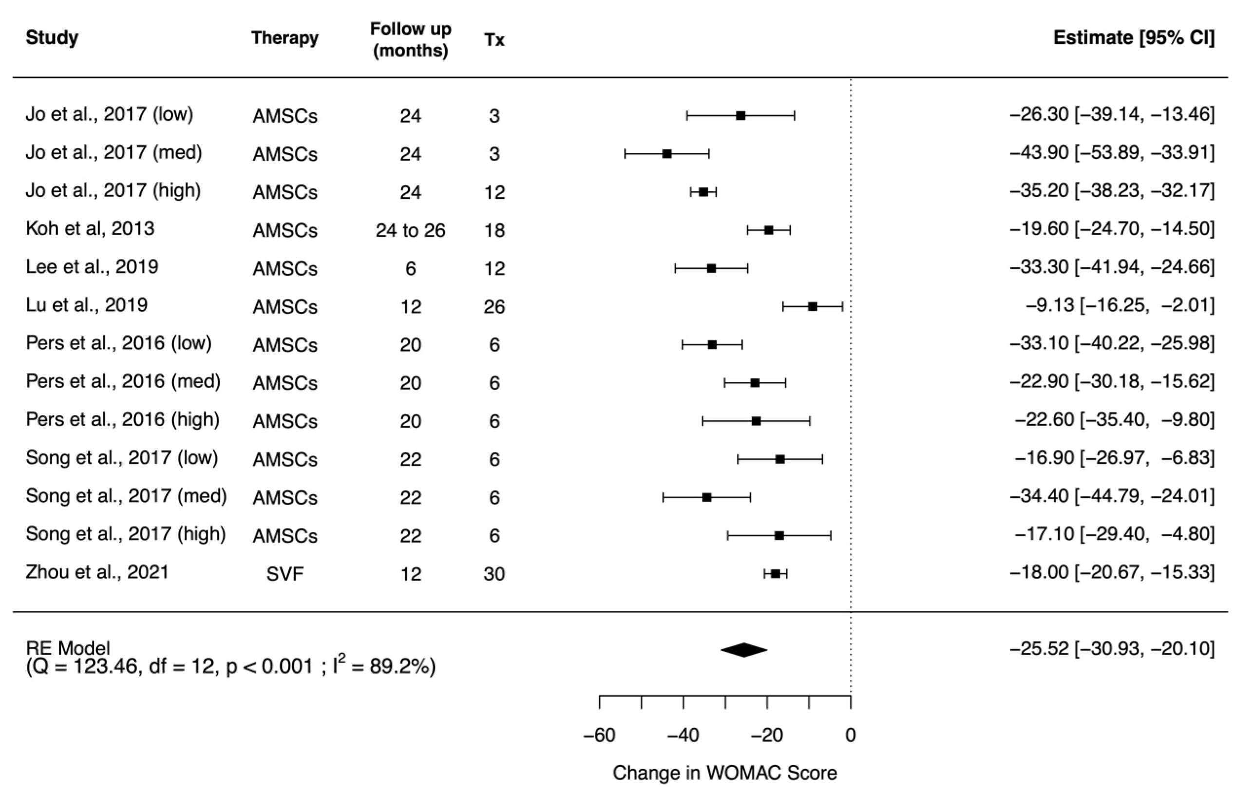
3.3.1. Improvement of Clinical Outcomes
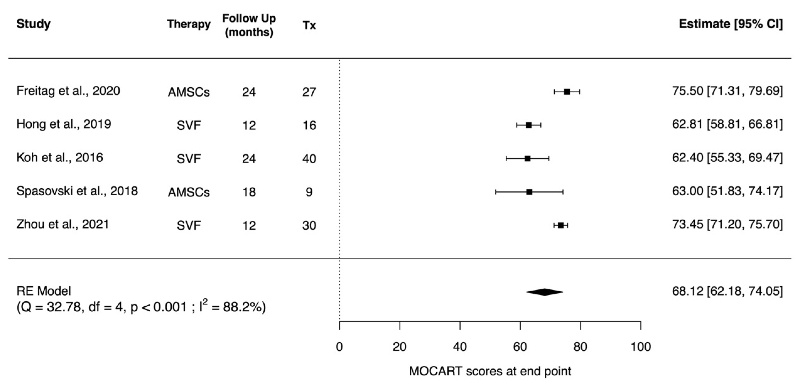
3.3.2. Improvement of Imaging Outcomes
3.3.3. Improvement of Histological Outcomes
3.3.4. Other Improvements
3.4. Assessment of Methodological Bias
4. Discussion
4.1. Optimal Source of Stem Cell
4.2. Augmenting the Function of AMSCs
4.3. Methods of AMSC Administration
4.4. Improved Clinical Outcomes
4.5. Hyaline-Like Cartilage Regeneration
4.6. Strengths and Limitations
5. Conclusions
Supplementary Materials
Author Contributions
Funding
Institutional Review Board Statement
Informed Consent Statement
Data Availability Statement
Conflicts of Interest
References
- Sophia Fox, A.J.; Bedi, A.; Rodeo, S.A. The basic science of articular cartilage: Structure, composition, and function. Sports Health 2009, 1, 461–468. [Google Scholar] [CrossRef] [PubMed]
- Sherwood, J.; Bertrand, J.; Nalesso, G.; Poulet, B.; Pitsillides, A.; Brandolini, L.; Karystinou, A.; De Bari, C.; Luyten, F.P.; Pitzalis, C. A homeostatic function of CXCR2 signalling in articular cartilage. Ann. Rheum. Dis. 2015, 74, 2207–2215. [Google Scholar] [CrossRef] [PubMed] [Green Version]
- Chuang, C.Y.; Lord, M.S.; Melrose, J.; Rees, M.D.; Knox, S.M.; Freeman, C.; Iozzo, R.V.; Whitelock, J.M. Heparan sulfate-dependent signaling of fibroblast growth factor 18 by chondrocyte-derived perlecan. Biochemistry 2010, 49, 5524–5532. [Google Scholar] [CrossRef] [Green Version]
- Pap, T.; Korb-Pap, A. Cartilage damage in osteoarthritis and rheumatoid arthritis—Two unequal siblings. Nat. Rev. Rheumatol. 2015, 11, 606–615. [Google Scholar] [CrossRef] [PubMed]
- Turkiewicz, A.; Petersson, I.F.; Björk, J.; Hawker, G.; Dahlberg, L.E.; Lohmander, L.S.; Englund, M. Current and future impact of osteoarthritis on health care: A population-based study with projections to year 2032. Osteoarthr. Cartil. 2014, 22, 1826–1832. [Google Scholar] [CrossRef] [PubMed] [Green Version]
- WHO. Chronic Rheumatic Conditions; WHO: Geneva, Switzerland, 2016. [Google Scholar]
- Veronese, N.; Stubbs, B.; Solmi, M.; Smith, T.O.; Noale, M.; Cooper, C.; Maggi, S. Association between lower limb osteoarthritis and incidence of depressive symptoms: Data from the osteoarthritis initiative. Age Ageing 2017, 46, 470–476. [Google Scholar] [CrossRef] [Green Version]
- Kye, S.-Y.; Park, K. Suicidal ideation and suicidal attempts among adults with chronic diseases: A cross-sectional study. Compr. Psychiatry 2017, 73, 160–167. [Google Scholar] [CrossRef]
- Widuchowski, W.; Widuchowski, J.; Trzaska, T. Articular cartilage defects: Study of 25,124 knee arthroscopies. Knee 2007, 14, 177–182. [Google Scholar] [CrossRef] [PubMed]
- Nelson, A.E.; Allen, K.D.; Golightly, Y.M.; Goode, A.P.; Jordan, J.M. A systematic review of recommendations and guidelines for the management of osteoarthritis: The chronic osteoarthritis management initiative of the US bone and joint initiative. Semin. Arthritis Rheum. 2014, 43, 701–712. [Google Scholar] [CrossRef]
- Anandacoomarasamy, A.; March, L. Current evidence for osteoarthritis treatments. Ther. Adv. Musculoskelet. Dis. 2010, 2, 17–28. [Google Scholar] [CrossRef]
- Detterline, A.J.; Goldberg, S.; Bach Jr, B.R.; Cole, B.J. Treatment options for articular cartilage defects of the knee. Orthop. Nurs. 2005, 24, 361–366. [Google Scholar] [CrossRef]
- Willers, C.; Wood, D.J.; Zheng, M.H. A current review on the biology and treatment of articular cartilage defects (part I & part II). J. Musculoskelet. Res. 2003, 7, 157–181. [Google Scholar]
- Berstock, J.R.; Beswick, A.D.; Lenguerrand, E.; Whitehouse, M.R.; Blom, A.W. Mortality after total hip replacement surgery: A systematic review. Bone Jt. Res. 2014, 3, 175–182. [Google Scholar] [CrossRef]
- Weber, A.E.; Locker, P.H.; Mayer, E.N.; Cvetanovich, G.L.; Tilton, A.K.; Erickson, B.J.; Yanke, A.B.; Cole, B.J. Clinical outcomes after microfracture of the knee: Midterm follow-up. Orthop. J. Sports Med. 2018, 6, 2325967117753572. [Google Scholar] [CrossRef] [PubMed]
- Matrix-Applied Characterised Autologous Cultured Chondrocytes (Maci). Available online: https://www.ema.europa.eu/en/medicines/human/EPAR/maci (accessed on 6 November 2021).
- MACI (Autologous Cultured Chondrocytes on a Porcine Collagen Membrane). Available online: https://www.fda.gov/vaccines-blood-biologics/cellular-gene-therapy-products/maci-autologous-cultured-chondrocytes-porcine-collagen-membrane (accessed on 6 November 2021).
- Saris, D.B.; Vanlauwe, J.; Victor, J.; Almqvist, K.F.; Verdonk, R.; Bellemans, J.; Luyten, F.P. Treatment of symptomatic cartilage defects of the knee: Characterized chondrocyte implantation results in better clinical outcome at 36 months in a randomized trial compared to microfracture. Am. J. Sports Med. 2009, 37, 10–19. [Google Scholar] [CrossRef]
- Assenmacher, J.A.; Kelikian, A.S.; Gottlob, C.; Kodros, S. Arthroscopically assisted autologous osteochondral transplantation for osteochondral lesions of the talar dome: An MRI and clinical follow-up study. Foot Ankle Int. 2001, 22, 544–551. [Google Scholar] [CrossRef]
- Mendicino, R.W.; Catanzariti, A.R.; Hallivis, R. Mosaicplasty for the treatment of osteochondral defects of the ankle joint. Clin. Podiatr. Med. Surg. 2001, 18, 495–513. [Google Scholar]
- LaPrade, R.F.; Botker, J.C. Donor-site morbidity after osteochondral autograft transfer procedures. Arthrosc. J. Arthrosc. Relat. Surg. 2004, 20, e69–e73. [Google Scholar] [CrossRef]
- Reddy, S.; Pedowitz, D.I.; Parekh, S.G.; Sennett, B.J.; Okereke, E. The morbidity associated with osteochondral harvest from asymptomatic knees for the treatment of osteochondral lesions of the talus. Am. J. Sports Med. 2007, 35, 80–85. [Google Scholar] [CrossRef] [PubMed]
- Bexkens, R.; Ogink, P.T.; Doornberg, J.N.; Kerkhoffs, G.M.; Eygendaal, D.; Oh, L.S.; van den Bekerom, M.P. Donor-site morbidity after osteochondral autologous transplantation for osteochondritis dissecans of the capitellum: A systematic review and meta-analysis. Knee Surg. Sports Traumatol. Arthrosc. 2017, 25, 2237–2246. [Google Scholar] [CrossRef] [PubMed] [Green Version]
- Schnabel, M.; Marlovits, S.; Eckhoff, G.; Fichtel, I.; Gotzen, L.; Vecsei, V.; Schlegel, J. Dedifferentiation-associated changes in morphology and gene expression in primary human articular chondrocytes in cell culture. Osteoarthr. Cartil. 2002, 10, 62–70. [Google Scholar] [CrossRef] [Green Version]
- Niemeyer, P.; Salzmann, G.; Feucht, M.; Pestka, J.; Porichis, S.; Ogon, P.; Südkamp, N.; Schmal, H. First-generation versus second-generation autologous chondrocyte implantation for treatment of cartilage defects of the knee: A matched-pair analysis on long-term clinical outcome. Int. Orthop. 2014, 38, 2065–2070. [Google Scholar] [CrossRef]
- Fernandes, T.L.; Kimura, H.A.; Pinheiro, C.C.G.; Shimomura, K.; Nakamura, N.; Ferreira, J.R.; Gomoll, A.H.; Hernandez, A.J.; Bueno, D.F. Human synovial mesenchymal stem cells good manufacturing practices for articular cartilage regeneration. Tissue Eng. Part C Methods 2018, 24, 709–716. [Google Scholar] [CrossRef] [Green Version]
- Garcia, J.; Wright, K.; Roberts, S.; Kuiper, J.H.; Mangham, C.; Richardson, J.; Mennan, C. Characterisation of synovial fluid and infrapatellar fat pad derived mesenchymal stromal cells: The influence of tissue source and inflammatory stimulus. Sci. Rep. 2016, 6, 24295. [Google Scholar] [CrossRef] [Green Version]
- Xu, L.; Liu, Y.; Sun, Y.; Wang, B.; Xiong, Y.; Lin, W.; Wei, Q.; Wang, H.; He, W.; Li, G. Tissue source determines the differentiation potentials of mesenchymal stem cells: A comparative study of human mesenchymal stem cells from bone marrow and adipose tissue. Stem Cell Res. Ther. 2017, 8, 275. [Google Scholar] [CrossRef] [PubMed] [Green Version]
- Hocking, A.M.; Gibran, N.S. Mesenchymal stem cells: Paracrine signaling and differentiation during cutaneous wound repair. Exp. Cell Res. 2010, 316, 2213–2219. [Google Scholar] [CrossRef] [Green Version]
- Linero, I.; Chaparro, O. Paracrine effect of mesenchymal stem cells derived from human adipose tissue in bone regeneration. PLoS ONE 2014, 9, e107001. [Google Scholar]
- Chen, L.; Tredget, E.E.; Wu, P.Y.; Wu, Y. Paracrine factors of mesenchymal stem cells recruit macrophages and endothelial lineage cells and enhance wound healing. PLoS ONE 2008, 3, e1886. [Google Scholar] [CrossRef] [PubMed] [Green Version]
- Maxson, S.; Lopez, E.A.; Yoo, D.; Danilkovitch-Miagkova, A.; LeRoux, M.A. Concise review: Role of mesenchymal stem cells in wound repair. Stem Cells Transl. Med. 2012, 1, 142–149. [Google Scholar] [CrossRef]
- Gupta, P.K.; Das, A.K.; Chullikana, A.; Majumdar, A.S. Mesenchymal stem cells for cartilage repair in osteoarthritis. Stem Cell Res. Ther. 2012, 3, 25. [Google Scholar] [CrossRef] [Green Version]
- Lim, W.L.; Liau, L.L.; Ng, M.H.; Chowdhury, S.R.; Law, J.X. Current progress in tendon and ligament tissue engineering. Tissue Eng. Regen. Med. 2019, 16, 549–571. [Google Scholar] [CrossRef] [PubMed]
- Beckermann, B.; Kallifatidis, G.; Groth, A.; Frommhold, D.; Apel, A.; Mattern, J.; Salnikov, A.; Moldenhauer, G.; Wagner, W.; Diehlmann, A. VEGF expression by mesenchymal stem cells contributes to angiogenesis in pancreatic carcinoma. Br. J. Cancer 2008, 99, 622–631. [Google Scholar] [CrossRef] [PubMed] [Green Version]
- Karnoub, A.E.; Dash, A.B.; Vo, A.P.; Sullivan, A.; Brooks, M.W.; Bell, G.W.; Richardson, A.L.; Polyak, K.; Tubo, R.; Weinberg, R.A. Mesenchymal stem cells within tumour stroma promote breast cancer metastasis. Nature 2007, 449, 557–563. [Google Scholar] [CrossRef]
- Cho, P.S.; Messina, D.J.; Hirsh, E.L.; Chi, N.; Goldman, S.N.; Lo, D.P.; Harris, I.R.; Popma, S.H.; Sachs, D.H.; Huang, C.A. Immunogenicity of umbilical cord tissue–derived cells. Blood J. Am. Soc. Hematol. 2008, 111, 430–438. [Google Scholar] [CrossRef] [PubMed] [Green Version]
- Li, X.; Wang, M.; Jing, X.; Guo, W.; Hao, C.; Zhang, Y.; Gao, S.; Chen, M.; Zhang, Z.; Zhang, X. Bone marrow-and adipose tissue-derived mesenchymal stem cells: Characterization, differentiation, and applications in cartilage tissue engineering. Crit. Rev. Eukaryot. Gene Expr. 2018, 28, 285–310. [Google Scholar] [CrossRef] [PubMed]
- Peng, L.; Jia, Z.; Yin, X.; Zhang, X.; Liu, Y.; Chen, P.; Ma, K.; Zhou, C. Comparative analysis of mesenchymal stem cells from bone marrow, cartilage, and adipose tissue. Stem Cells Dev. 2008, 17, 761–774. [Google Scholar] [CrossRef]
- Strioga, M.; Viswanathan, S.; Darinskas, A.; Slaby, O.; Michalek, J. Same or not the same? Comparison of adipose tissue-derived versus bone marrow-derived mesenchymal stem and stromal cells. Stem Cells Dev. 2012, 21, 2724–2752. [Google Scholar] [CrossRef]
- Bourin, P.; Bunnell, B.A.; Casteilla, L.; Dominici, M.; Katz, A.J.; March, K.L.; Redl, H.; Rubin, J.P.; Yoshimura, K.; Gimble, J.M. Stromal cells from the adipose tissue-derived stromal vascular fraction and culture expanded adipose tissue-derived stromal/stem cells: A joint statement of the International Federation for Adipose Therapeutics and Science (IFATS) and the International Society for Cellular Therapy (ISCT). Cytotherapy 2013, 15, 641–648. [Google Scholar]
- Dominici, M.; Le Blanc, K.; Mueller, I.; Slaper-Cortenbach, I.; Marini, F.; Krause, D.; Deans, R.; Keating, A.; Prockop, D.; Horwitz, E. Minimal criteria for defining multipotent mesenchymal stromal cells. The International Society for Cellular Therapy position statement. Cytotherapy 2006, 8, 315–317. [Google Scholar] [CrossRef]
- Page, M.J.; McKenzie, J.E.; Bossuyt, P.M.; Boutron, I.; Hoffmann, T.C.; Mulrow, C.D.; Shamseer, L.; Tetzlaff, J.M.; Akl, E.A.; Brennan, S.E. The PRISMA 2020 statement: An updated guideline for reporting systematic reviews. BMJ 2021, 372, n71. [Google Scholar] [CrossRef]
- Methley, A.M.; Campbell, S.; Chew-Graham, C.; McNally, R.; Cheraghi-Sohi, S. PICO, PICOS and SPIDER: A comparison study of specificity and sensitivity in three search tools for qualitative systematic reviews. BMC Health Serv. Res. 2014, 14, 579. [Google Scholar] [CrossRef] [PubMed] [Green Version]
- Myles, P.S.; Troedel, S.; Boquest, M.; Reeves, M. The Pain Visual Analog Scale: Is It Linear or Nonlinear? Anesth. Analg. 1999, 87, 1517. [Google Scholar] [CrossRef] [Green Version]
- Roos, E.M.; Lohmander, L.S. The Knee injury and Osteoarthritis Outcome Score (KOOS): From joint injury to osteoarthritis. Health Qual. Life Outcomes 2003, 1, 64. [Google Scholar] [CrossRef] [PubMed] [Green Version]
- Bellamy, N.; Buchanan, W.W.; Goldsmith, C.H.; Campbell, J.; Stitt, L.W. Validation study of WOMAC: A health status instrument for measuring clinically important patient relevant outcomes to antirheumatic drug therapy in patients with osteoarthritis of the hip or knee. J. Rheumatol. 1988, 15, 1833–1840. [Google Scholar] [PubMed]
- Marlovits, S.; Striessnig, G.; Resinger, C.T.; Aldrian, S.M.; Vecsei, V.; Imhof, H.; Trattnig, S. Definition of pertinent parameters for the evaluation of articular cartilage repair tissue with high-resolution magnetic resonance imaging. Eur. J. Radiol. 2004, 52, 310–319. [Google Scholar] [CrossRef]
- Schreiner, M.M.; Raudner, M.; Röhrich, S.; Zalaudek, M.; Weber, M.; Kaiser, G.; Aldrian, S.; Chiari, C.; Windhager, R.; Trattnig, S. Reliability of the MOCART (Magnetic Resonance Observation of Cartilage Repair Tissue) 2.0 knee score for different cartilage repair techniques—A retrospective observational study. Eur. Radiol. 2021, 31, 5734–5745. [Google Scholar] [CrossRef]
- Viechtbauer, W. Conducting meta-analyses in R with the metafor package. J. Stat. Softw. 2010, 36, 1–48. [Google Scholar] [CrossRef] [Green Version]
- Sterne, J.A.; Savović, J.; Page, M.J.; Elbers, R.G.; Blencowe, N.S.; Boutron, I.; Cates, C.J.; Cheng, H.-Y.; Corbett, M.S.; Eldridge, S.M. RoB 2: A revised tool for assessing risk of bias in randomised trials. BMJ 2019, 366, l4898. [Google Scholar] [CrossRef] [Green Version]
- Sterne, J.A.; Hernán, M.A.; Reeves, B.C.; Savović, J.; Berkman, N.D.; Viswanathan, M.; Henry, D.; Altman, D.G.; Ansari, M.T.; Boutron, I. ROBINS-I: A tool for assessing risk of bias in non-randomised studies of interventions. BMJ 2016, 355, i4919. [Google Scholar] [CrossRef] [Green Version]
- McGuinness, L.A.; Higgins, J.P. Risk-of-bias VISualization (robvis): An R package and Shiny web app for visualizing risk-of-bias assessments. Res. Synth. Methods 2021, 12, 55–61. [Google Scholar] [CrossRef]
- Jo, C.H.; Chai, J.W.; Jeong, E.C.; Oh, S.; Shin, J.S.; Shim, H.; Yoon, K.S. Intra-articular injection of mesenchymal stem cells for the treatment of osteoarthritis of the knee: A 2-year follow-up study. Am. J. Sports Med. 2017, 45, 2774–2783. [Google Scholar] [CrossRef] [PubMed]
- Jo, C.H.; Lee, Y.G.; Shin, W.H.; Kim, H.; Chai, J.W.; Jeong, E.C.; Kim, J.E.; Shim, H.; Shin, J.S.; Shin, I.S. Intra-articular injection of mesenchymal stem cells for the treatment of osteoarthritis of the knee: A proof-of-concept clinical trial. Stem Cells 2014, 32, 1254–1266. [Google Scholar] [CrossRef] [PubMed]
- Freitag, J.; Shah, K.; Wickham, J.; Li, D.; Norsworthy, C.; Tenen, A. Evaluation of autologous adipose-derived mesenchymal stem cell therapy in focal chondral defects of the knee: A pilot case series. Regen. Med. 2020, 15, 1703–1717. [Google Scholar] [CrossRef] [PubMed]
- Freitag, J.; Wickham, J.; Shah, K.; Li, D.; Norsworthy, C.; Tenen, A. Mesenchymal stem cell therapy combined with arthroscopic abrasion arthroplasty regenerates cartilage in patients with severe knee osteoarthritis: A case series. Regen. Med. 2020, 15, 1957–1977. [Google Scholar] [CrossRef] [PubMed]
- Lu, L.; Dai, C.; Zhang, Z.; Du, H.; Li, S.; Ye, P.; Fu, Q.; Zhang, L.; Wu, X.; Dong, Y. Treatment of knee osteoarthritis with intra-articular injection of autologous adipose-derived mesenchymal progenitor cells: A prospective, randomized, double-blind, active-controlled, phase Iib clinical trial. Stem Cell Res. Ther. 2019, 10, 143. [Google Scholar] [CrossRef] [Green Version]
- Hong, Z.; Chen, J.; Zhang, S.; Zhao, C.; Bi, M.; Chen, X.; Bi, Q. Intra-articular injection of autologous adipose-derived stromal vascular fractions for knee osteoarthritis: A double-blind randomized self-controlled trial. Int. Orthop. 2019, 43, 1123–1134. [Google Scholar] [CrossRef]
- Kim, Y.S.; Kwon, O.R.; Choi, Y.J.; Suh, D.S.; Heo, D.B.; Koh, Y.G. Comparative matched-pair analysis of the injection versus implantation of mesenchymal stem cells for knee osteoarthritis. Am. J. Sports Med. 2015, 43, 2738–2746. [Google Scholar] [CrossRef]
- Koh, Y.-G.; Jo, S.-B.; Kwon, O.-R.; Suh, D.-S.; Lee, S.-W.; Park, S.-H.; Choi, Y.-J. Mesenchymal stem cell injections improve symptoms of knee osteoarthritis. Arthrosc. J. Arthrosc. Relat. Surg. 2013, 29, 748–755. [Google Scholar] [CrossRef] [PubMed]
- Koh, Y.G.; Choi, Y.J.; Kwon, O.R.; Kim, Y.S. Second-look arthroscopic evaluation of cartilage lesions after mesenchymal stem cell implantation in osteoarthritic knees. Am. J. Sports Med. 2014, 42, 1628–1637. [Google Scholar] [CrossRef]
- Koh, Y.-G.; Kwon, O.-R.; Kim, Y.-S.; Choi, Y.-J.; Tak, D.-H. Adipose-derived mesenchymal stem cells with microfracture versus microfracture alone: 2-year follow-up of a prospective randomized trial. Arthrosc. J. Arthrosc. Relat. Surg. 2016, 32, 97–109. [Google Scholar] [CrossRef]
- Kyriakidis, T.; Iosifidis, M.; Michalopoulos, E.; Melas, I.; Stavropoulos-Giokas, C.; Verdonk, R. Good mid-term outcomes after adipose-derived culture-expanded mesenchymal stem cells implantation in knee focal cartilage defects. Knee Surg. Sports Traumatol. Arthrosc. 2020, 28, 502–508. [Google Scholar] [CrossRef]
- Lapuente, J.P.; Dos-Anjos, S.; Blázquez-Martínez, A. Intra-articular infiltration of adipose-derived stromal vascular fraction cells slows the clinical progression of moderate-severe knee osteoarthritis: Hypothesis on the regulatory role of intra-articular adipose tissue. J. Orthop. Surg. Res. 2020, 15, 137. [Google Scholar] [CrossRef] [Green Version]
- Lee, W.S.; Kim, H.J.; Kim, K.I.; Kim, G.B.; Jin, W. Intra-articular injection of autologous adipose tissue-derived mesenchymal stem cells for the treatment of knee osteoarthritis: A phase Iib, randomized, placebo-controlled clinical trial. Stem Cells Transl. Med. 2019, 8, 504–511. [Google Scholar] [CrossRef] [PubMed] [Green Version]
- Pers, Y.-M.; Rackwitz, L.; Ferreira, R.; Pullig, O.; Delfour, C.; Barry, F.; Sensebe, L.; Casteilla, L.; Fleury, S.; Bourin, P. Adipose mesenchymal stromal cell-based therapy for severe osteoarthritis of the knee: A phase I dose-escalation trial. Stem Cells Transl. Med. 2016, 5, 847–856. [Google Scholar] [CrossRef] [PubMed] [Green Version]
- Simunec, D.; Salari, H.; Meyer, J. Treatment of grade 3 and 4 osteoarthritis with intraoperatively separated adipose tissue-derived stromal vascular fraction: A comparative case series. Cells 2020, 9, 2096. [Google Scholar] [CrossRef] [PubMed]
- Spasovski, D.; Spasovski, V.; Baščarević, Z.; Stojiljković, M.; Vreća, M.; Anđelković, M.; Pavlović, S. Intra-articular injection of autologous adipose-derived mesenchymal stem cells in the treatment of knee osteoarthritis. J. Gene Med. 2018, 20, e3002. [Google Scholar] [CrossRef] [PubMed]
- Song, Y.; Du, H.; Dai, C.; Zhang, L.; Li, S.; Hunter, D.J.; Lu, L.; Bao, C. Human adipose-derived mesenchymal stem cells for osteoarthritis: A pilot study with long-term follow-up and repeated injections. Regen. Med. 2018, 13, 295–307. [Google Scholar] [CrossRef]
- Zhao, X.; Ruan, J.; Tang, H.; Li, J.; Shi, Y.; Li, M.; Li, S.; Xu, C.; Lu, Q.; Dai, C. Multi-compositional MRI evaluation of repair cartilage in knee osteoarthritis with treatment of allogeneic human adipose-derived mesenchymal progenitor cells. Stem Cell Res. Ther. 2019, 10, 308. [Google Scholar] [CrossRef] [Green Version]
- Zhou, Y.; Li, H.; Xiang, D.; Shao, J.; Fu, Q.; Han, Y.; Zhu, J.; Chen, Y.; Qian, Q. The clinical efficacy of arthroscopic therapy with knee infrapatellar fat pad cell concentrates in treating knee cartilage lesion: A prospective, randomized, and controlled study. J. Orthop. Surg. Res. 2021, 16, 87. [Google Scholar] [CrossRef] [PubMed]
- Seo, Y.; Shin, T.-H.; Kim, H.-S. Current Strategies to Enhance Adipose Stem Cell Function: An Update. Int. J. Mol. Sci. 2019, 20, 3827. [Google Scholar] [CrossRef] [PubMed] [Green Version]
- Baer, P.C.; Geiger, H. Adipose-derived mesenchymal stromal/stem cells: Tissue localization, characterization, and heterogeneity. Stem Cells Int. 2012, 2012, 812693. [Google Scholar] [CrossRef] [PubMed] [Green Version]
- Beane, O.S.; Fonseca, V.C.; Cooper, L.L.; Koren, G.; Darling, E.M. Impact of aging on the regenerative properties of bone marrow-, muscle-, and adipose-derived mesenchymal stem/stromal cells. PLoS ONE 2014, 9, e115963. [Google Scholar]
- Ronzière, M.C.; Perrier, E.; Mallein-Gerin, F.; Freyria, A.M. Chondrogenic potential of bone marrow- and adipose tissue-derived adult human mesenchymal stem cells. Biomed. Mater. Eng. 2010, 20, 145–158. [Google Scholar] [CrossRef]
- Zhong, Y.-C.; Wang, S.-C.; Han, Y.-H.; Wen, Y. Recent advance in source, property, differentiation, and applications of infrapatellar fat pad adipose-derived stem cells. Stem Cells Int. 2020, 2020, 2560174. [Google Scholar] [CrossRef] [PubMed]
- Ding, D.C.; Wu, K.C.; Chou, H.L.; Hung, W.T.; Liu, H.W.; Chu, T.Y. Human Infrapatellar Fat Pad-Derived Stromal Cells Have More Potent Differentiation Capacity Than Other Mesenchymal Cells and Can Be Enhanced by Hyaluronan. Cell Transpl. 2015, 24, 1221–1232. [Google Scholar] [CrossRef] [Green Version]
- Domergue, S.; Bony, C.; Maumus, M.; Toupet, K.; Frouin, E.; Rigau, V.; Vozenin, M.-C.; Magalon, G.; Jorgensen, C.; Noël, D. Comparison between Stromal Vascular Fraction and Adipose Mesenchymal Stem Cells in Remodeling Hypertrophic Scars. PLoS ONE 2016, 11, e0156161. [Google Scholar] [CrossRef] [Green Version]
- Pak, J.; Lee, J.H.; Pak, N.; Pak, Y.; Park, K.S.; Jeon, J.H.; Jeong, B.C.; Lee, S.H. Cartilage regeneration in humans with adipose tissue-derived stem cells and adipose stromal vascular fraction cells: Updated status. Int. J. Mol. Sci. 2018, 19, 2146. [Google Scholar] [CrossRef] [PubMed] [Green Version]
- Lv, X.; He, J.; Zhang, X.; Luo, X.; He, N.; Sun, Z.; Xia, H.; Liu, V.; Zhang, L.; Lin, X.; et al. Comparative Efficacy of Autologous Stromal Vascular Fraction and Autologous Adipose-Derived Mesenchymal Stem Cells Combined with Hyaluronic Acid for the Treatment of Sheep Osteoarthritis. Cell Transpl. 2018, 27, 1111–1125. [Google Scholar] [CrossRef]
- Sohni, A.; Verfaillie, C.M. Mesenchymal stem cells migration homing and tracking. Stem Cells Int. 2013, 2013, 130763. [Google Scholar] [CrossRef] [PubMed]
- Perdisa, F.; Gostyńska, N.; Roffi, A.; Filardo, G.; Marcacci, M.; Kon, E. Adipose-derived mesenchymal stem cells for the treatment of articular cartilage: A systematic review on preclinical and clinical evidence. Stem Cells Int. 2015, 2015, 597652. [Google Scholar] [CrossRef] [PubMed] [Green Version]
- de Windt, T.S.; Vonk, L.A.; Slaper-Cortenbach, I.C.; Nizak, R.; van Rijen, M.H.; Saris, D.B. Allogeneic MSCs and recycled autologous chondrons mixed in a one-stage cartilage cell transplantion: A first-in-man trial in 35 patients. Stem Cells 2017, 35, 1984–1993. [Google Scholar] [CrossRef] [Green Version]
- Dhollander, A.A.; Verdonk, P.C.; Lambrecht, S.; Verdonk, R.; Elewaut, D.; Verbruggen, G.; Almqvist, K.F. Midterm results of the treatment of cartilage defects in the knee using alginate beads containing human mature allogenic chondrocytes. Am. J. Sports Med. 2012, 40, 75–82. [Google Scholar] [CrossRef] [PubMed]
- Jiang, P.; Mao, L.; Qiao, L.; Lei, X.; Zheng, Q.; Li, D. Efficacy and safety of mesenchymal stem cell injections for patients with osteoarthritis: A meta-analysis and review of RCTs. Arch. Orthop. Trauma Surg. 2021, 141, 1241–1251. [Google Scholar] [CrossRef] [PubMed]
- Galipeau, J.; Sensébé, L. Mesenchymal Stromal Cells: Clinical Challenges and Therapeutic Opportunities. Cell Stem Cell 2018, 22, 824–833. [Google Scholar] [CrossRef] [Green Version]
- McLeod, C.M.; Mauck, R.L. On the origin and impact of mesenchymal stem cell heterogeneity: New insights and emerging tools for single cell analysis. Eur. Cell Mater. 2017, 34, 217–231. [Google Scholar] [CrossRef]
- Eggenhofer, E.; Benseler, V.; Kroemer, A.; Popp, F.C.; Geissler, E.K.; Schlitt, H.J.; Baan, C.C.; Dahlke, M.H.; Hoogduijn, M.J. Mesenchymal stem cells are short-lived and do not migrate beyond the lungs after intravenous infusion. Front Immunol. 2012, 3, 297. [Google Scholar] [CrossRef] [Green Version]
- Fahy, N.; Farrell, E.; Ritter, T.; Ryan, A.E.; Murphy, J.M. Immune modulation to improve tissue engineering outcomes for cartilage repair in the osteoarthritic joint. Tissue Eng. Part B Rev. 2015, 21, 55–66. [Google Scholar] [CrossRef]
- Lu, V.; Tennyson, M.; Zhang, J.; Khan, W. Mesenchymal Stem Cell-Derived Extracellular Vesicles in Tendon and Ligament Repair—A Systematic Review of In Vivo Studies. Cells 2021, 10, 2553. [Google Scholar] [CrossRef]
- Waterman, R.S.; Tomchuck, S.L.; Henkle, S.L.; Betancourt, A.M. A new mesenchymal stem cell (MSC) paradigm: Polarization into a pro-inflammatory MSC1 or an Immunosuppressive MSC2 phenotype. PLoS ONE 2010, 5, e10088. [Google Scholar] [CrossRef] [PubMed]
- Raicevic, G.; Najar, M.; Pieters, K.; De Bruyn, C.; Meuleman, N.; Bron, D.; Toungouz, M.; Lagneaux, L. Inflammation and Toll-like receptor ligation differentially affect the osteogenic potential of human mesenchymal stromal cells depending on their tissue origin. Tissue Eng. Part A 2012, 18, 1410–1418. [Google Scholar] [CrossRef]
- Hossain, M.M.; Murali, M.R.; Kamarul, T. Genetically modified mesenchymal stem/stromal cells transfected with adiponectin gene can stably secrete adiponectin. Life Sci. 2017, 182, 50–56. [Google Scholar] [CrossRef]
- Chen, S.; Tang, Y.; Liu, Y.; Zhang, P.; Lv, L.; Zhang, X.; Jia, L.; Zhou, Y. Exosomes derived from miR-375-overexpressing human adipose mesenchymal stem cells promote bone regeneration. Cell Prolif. 2019, 52, e12669. [Google Scholar] [CrossRef] [PubMed] [Green Version]
- Lou, G.; Yang, Y.; Liu, F.; Ye, B.; Chen, Z.; Zheng, M.; Liu, Y. MiR-122 modification enhances the therapeutic efficacy of adipose tissue-derived mesenchymal stem cells against liver fibrosis. J. Cell Mol. Med. 2017, 21, 2963–2973. [Google Scholar] [CrossRef]
- Kim, J.Y.; Shin, K.K.; Lee, A.L.; Kim, Y.S.; Park, H.J.; Park, Y.K.; Bae, Y.C.; Jung, J.S. MicroRNA-302 induces proliferation and inhibits oxidant-induced cell death in human adipose tissue-derived mesenchymal stem cells. Cell Death Dis. 2014, 5, e1385. [Google Scholar] [CrossRef] [Green Version]
- Schönitzer, V.; Wirtz, R.; Ulrich, V.; Berger, T.; Karl, A.; Mutschler, W.; Schieker, M.; Böcker, W. Sox2 is a potent inhibitor of osteogenic and adipogenic differentiation in human mesenchymal stem cells. Cell Reprogram 2014, 16, 355–365. [Google Scholar] [CrossRef] [PubMed]
- Choi, E.W.; Shin, I.S.; Song, J.W.; Lee, M.; Yun, T.W.; Yang, J.; Choi, K.S.; Kim, S.J. Effects of Transplantation of CTLA4Ig-Overexpressing Adipose Tissue-Derived Mesenchymal Stem Cells in Mice with Sustained Severe Rheumatoid Arthritis. Cell Transpl. 2016, 25, 243–259. [Google Scholar] [CrossRef] [Green Version]
- Boulaiz, H.; Marchal, J.A.; Prados, J.; Melguizo, C.; Aránega, A. Non-viral and viral vectors for gene therapy. Cell Mol. Biol. 2005, 51, 3–22. [Google Scholar] [PubMed]
- Ema, H.; Suda, T. Two anatomically distinct niches regulate stem cell activity. Blood 2012, 120, 2174–2181. [Google Scholar] [CrossRef] [Green Version]
- Yoon, H.H.; Bhang, S.H.; Shin, J.-Y.; Shin, J.; Kim, B.-S. Enhanced cartilage formation via three-dimensional cell engineering of human adipose-derived stem cells. Tissue Eng. Part A 2012, 18, 1949–1956. [Google Scholar] [CrossRef] [Green Version]
- Bartosh, T.J.; Ylöstalo, J.H.; Mohammadipoor, A.; Bazhanov, N.; Coble, K.; Claypool, K.; Lee, R.H.; Choi, H.; Prockop, D.J. Aggregation of human mesenchymal stromal cells (MSCs) into 3D spheroids enhances their antiinflammatory properties. Proc. Natl. Acad. Sci. USA 2010, 107, 13724–13729. [Google Scholar] [CrossRef] [Green Version]
- Edmondson, R.; Broglie, J.J.; Adcock, A.F.; Yang, L. Three-dimensional cell culture systems and their applications in drug discovery and cell-based biosensors. Assay Drug Dev. Technol. 2014, 12, 207–218. [Google Scholar] [CrossRef] [PubMed] [Green Version]
- Xu, Y.; Malladi, P.; Chiou, M.; Bekerman, E.; Giaccia, A.J.; Longaker, M.T. In vitro expansion of adipose-derived adult stromal cells in hypoxia enhances early chondrogenesis. Tissue Eng. 2007, 13, 2981–2993. [Google Scholar] [CrossRef] [PubMed]
- Rubio-Azpeitia, E.; Andia, I. Partnership between platelet-rich plasma and mesenchymal stem cells: In vitro experience. Muscles Ligaments Tendons J. 2014, 4, 52–62. [Google Scholar] [CrossRef] [PubMed]
- Mishra, A.; Tummala, P.; King, A.; Lee, B.; Kraus, M.; Tse, V.; Jacobs, C.R. Buffered platelet-rich plasma enhances mesenchymal stem cell proliferation and chondrogenic differentiation. Tissue Eng. Part C Methods 2009, 15, 431–435. [Google Scholar] [CrossRef]
- Yokota, N.; Hattori, M.; Ohtsuru, T.; Otsuji, M.; Lyman, S.; Shimomura, K.; Nakamura, N. Comparative clinical outcomes after intra-articular injection with adipose-derived cultured stem cells or noncultured stromal vascular fraction for the treatment of knee osteoarthritis. Am. J. Sports Med. 2019, 47, 2577–2583. [Google Scholar] [CrossRef] [PubMed]
- To, K.; Zhang, B.; Romain, K.; Mak, C.; Khan, W. Synovium-derived mesenchymal stem cell transplantation in cartilage regeneration: A PRISMA review of in vivo studies. Front. Bioeng. Biotechnol. 2019, 7, 314. [Google Scholar] [CrossRef]
- Agarwal, N.; Mak, C.; Bojanic, C.; To, K.; Khan, W. Meta-Analysis of Adipose Tissue Derived Cell-Based Therapy for the Treatment of Knee Osteoarthritis. Cells 2021, 10, 1365. [Google Scholar] [CrossRef]
- Goebel, L.; Orth, P.; Müller, A.; Zurakowski, D.; Bücker, A.; Cucchiarini, M.; Pape, D.; Madry, H. Experimental scoring systems for macroscopic articular cartilage repair correlate with the MOCART score assessed by a high-field MRI at 9.4 T--comparative evaluation of five macroscopic scoring systems in a large animal cartilage defect model. Osteoarthr. Cartil. 2012, 20, 1046–1055. [Google Scholar] [CrossRef] [Green Version]
- Nguyen, P.D.; Tran, T.D.; Nguyen, H.T.; Vu, H.T.; Le, P.T.; Phan, N.L.; Vu, N.B.; Phan, N.K.; Van Pham, P. Comparative Clinical Observation of Arthroscopic Microfracture in the Presence and Absence of a Stromal Vascular Fraction Injection for Osteoarthritis. Stem Cells Transl. Med. 2017, 6, 187–195. [Google Scholar] [CrossRef]
- Tran, T.D.X.; Wu, C.M.; Dubey, N.K.; Deng, Y.H.; Su, C.W.; Pham, T.T.; Thi Le, P.B.; Sestili, P.; Deng, W.P. Time- and Kellgren-awrence Grade-Dependent Changes in Intra-Articularly Transplanted Stromal Vascular Fraction in Osteoarthritic Patients. Cells 2019, 8, 308. [Google Scholar] [CrossRef] [PubMed] [Green Version]
- Marędziak, M.; Marycz, K.; Tomaszewski, K.A.; Kornicka, K.; Henry, B.M. The Influence of Aging on the Regenerative Potential of Human Adipose Derived Mesenchymal Stem Cells. Stem Cells Int. 2016, 2016, 2152435. [Google Scholar] [CrossRef]
- Urban, H.; Little, C.B. The role of fat and inflammation in the pathogenesis and management of osteoarthritis. Rheumatology 2018, 57, iv10–iv21. [Google Scholar] [CrossRef] [Green Version]
- Hass, R.; Kasper, C.; Böhm, S.; Jacobs, R. Different populations and sources of human mesenchymal stem cells (MSC): A comparison of adult and neonatal tissue-derived MSC. Cell Commun. Signal. 2011, 9, 12. [Google Scholar] [CrossRef] [Green Version]
- Vinardell, T.; Thorpe, S.; Buckley, C.; Kelly, D. Chondrogenesis and integration of mesenchymal stem cells within an in vitro cartilage defect repair model. Ann. Biomed. Eng. 2009, 37, 2556. [Google Scholar] [CrossRef] [PubMed]
- de Vries-van Melle, M.L.; Narcisi, R.; Kops, N.; Koevoet, W.J.; Bos, P.K.; Murphy, J.M.; Verhaar, J.A.; van der Kraan, P.M.; van Osch, G.J. Chondrogenesis of mesenchymal stem cells in an osteochondral environment is mediated by the subchondral bone. Tissue Eng. Part A 2014, 20, 23–33. [Google Scholar] [CrossRef]
- Bornes, T.D.; Adesida, A.B.; Jomha, N.M. Mesenchymal stem cells in the treatment of traumatic articular cartilage defects: A comprehensive review. Arthritis Res. Ther. 2014, 16, 432. [Google Scholar] [CrossRef] [Green Version]
- Pak, J. Regeneration of human bones in hip osteonecrosis and human cartilage in knee osteoarthritis with autologous adipose-tissue-derived stem cells: A case series. J. Med. Case Rep. 2011, 5, 296. [Google Scholar] [CrossRef] [PubMed] [Green Version]
- Park, Y.B.; Ha, C.W.; Lee, C.H.; Yoon, Y.C.; Park, Y.G. Cartilage Regeneration in Osteoarthritic Patients by a Composite of Allogeneic Umbilical Cord Blood-Derived Mesenchymal Stem Cells and Hyaluronate Hydrogel: Results from a Clinical Trial for Safety and Proof-of-Concept with 7 Years of Extended Follow-Up. Stem Cells Transl. Med. 2017, 6, 613–621. [Google Scholar] [CrossRef] [PubMed]


| Domain | Inclusion Criteria | Exclusion Criteria |
|---|---|---|
| Population | Clinical study conducted on human populations regardless of age, gender, ethnicity, demography, geography, presenting complaints or history with cartilage damage | Studies conducted on animals. Ex vivo studies and in vitro studies |
| Intervention | Studies that evaluate the use of adipose tissue-derived mesenchymal stem cell transplant as a method to induce cartilage regeneration | Studies which use AMSCs in an unquantifiable manner |
| Comparison | Adipose tissue-derived mesenchymal stem cells on cartilage regeneration that are compared with non-exposed control groups and or mesenchymal stem cells derived from other sources | Studies that treat cartilage loss not related to cartilage regeneration, such as for pain relief |
| Outcome | Any outcomes pertaining to the regeneration of cartilage will be included, such as macroscopic, radiological and histological scores | Studies where no outcome measures are directly related to cartilage regeneration |
| Study Type | English with full text available | Case reports |
| Author | Study Design | AMSC-Based Therapy | Administration Method | Control | Cell Count |
|---|---|---|---|---|---|
| Freitag et al., 2020 (Pilot) [56] | Pilot case series | Autologous abdominal AMSCs | Intra-articular injection at 1 week at 6 months (after arthroscopic abrasion arthroplasty) | N/A | 5.21 × 10⁶ |
| Freitag et al., 2020 [57] | Prospective case series | Autologous abdominal AMSCs | Intra-articular injection at 1 week at 6 months (after arthroscopic abrasion arthroplasty) | N/A | 5.21 × 10⁶ |
| Hong et al., 2019 [59] | Double blind randomized self-controlled trial | Autologous abdominal SVF | Intra-articular injection at cartilage lesion site (with arthroscopic debridement) | HA injection | SVF cell density (7.45 ± 3.73 × 10⁶/mL) |
| Jo et al., 2014 [55], 2017 [54] | Dose-escalation Cohort study | Autologous abdominal AMSCs | Intra-articular injection | Comparison between high, med, low dose | Low: 1.00 × 10⁷ Medium: 5.00 × 10⁷ High: 1.00 × 10⁸ |
| Kim et al., 2015 [60] | Single cohort | Autologous gluteal AMSCs | Implantation onto fibrin glue scaffold over lesion site (with debridement) | N/A | 4.01 × 10⁶ |
| Koh et al., 2013 [61] | Therapeutic case series | Autologous infrapatellar fat pad AMSCs | Intra-articular injection (with PRP) | N/A | 1.18 × 10⁶ |
| Koh et al., 2014 [62] | Retrospective Case Series | Autologous gluteal AMSCs | Implantation (with debridement) | N/A | 3.80 × 10⁶ |
| Koh et al., 2016 [63] | Randomized prospective comparative study | Autologous gluteal SVF | Intra-articular injection (with microfracture) | Microfracture | 4.97 × 10⁶ |
| Kyriakidis et al., 2020 [64] | Case series | Autologous hypogastric derived AMSCs | Implantation onto three-dimensional matrix | N/A | Cells identified as AMSCs but not counted |
| Lapuente et al., 2020 [65] | Retrospective non-controlled study | Autologous abdominal SVF | Intra-articular injection | N/A | 3.21 × 10⁶ |
| Lee et al., 2019 [66] | Randomized double blind placebo controlled trial | Autologous abdominal AMSCs | Intra-articular injection | Normal saline injection | 1.00 × 10⁸ |
| Lu et al., 2019 [58] | Randomized double blind active-controlled controlled trial | Autologous abdominal AMSCs | Intra-articular injection at week 0 and 3 | HA injection | 5.00 × 10⁷ |
| Pers et al., 2016 [67] | Single-arm, open-label, dose-escalating | Autologous AMSCs | Intra-articular injection | N/A | Low: 2.00 × 10⁶ Medium: 1.00 × 10⁷ High: 5.00 × 10⁷ |
| Simunec et al., 2020 [68] | Comparative case series | Autologous SVF | Intra-articular injection (with PRP) | N/A | 4.24–10.2 × 10⁶ |
| Spasovski et al., 2018 [69] | Single-arm, open-label | Autologous abdominal AMSCs | Intra-articular injection | N/A | 0.500–1.00 × 10⁷ |
| Song et al., 2018 [70] | Randomized double-blinded dose-escalating Phase I trial with Open Phase IIa trial | Autologous AMSCs | Intra-articular injection | N/A | Low: 1.00 × 10⁷ Medium: 2.00 × 10⁷ High: 5.00 × 10⁷ |
| Zhao et al., 2019 [71] | Randomized double blind placebo controlled trial | Allogeneic abdominal AMSCs | Intra-articular injection | Comparison between high, med, low dose | Low: 1.00 × 10⁷ Medium: 2.00 × 10⁷ High: 5.00 × 10⁷ |
| Zhou et al., 2021 [72] | Randomized double blind placebo controlled trial | Autologous infrapatellar fat pad SVF | Intra-articular injection | Knee arthroscopic therapy | 3.91 × 10⁶ |
| Author | Model | Subjects | Control | Mean Age of Subjects | Mean Age of Control | BMI of Subject | BMI of Control | Sex Ratio of Subject (M–F) | Sex Ratio of Control (M–F) | Participant Ethnicity |
|---|---|---|---|---|---|---|---|---|---|---|
| Freitag et al., 2020 (Pilot) [56] | Patients with a focal full thickness chondral defect of the knee | 8 | N/A | 23–52 | N/A | 25.2 | N/A | 4:4 | N/A | Australian |
| Freitag et al., 2020 [57] | Knee OA patients (K–L grade IV) | 27 | N/A | 53.6 ± 6.7 | N/A | 27.7 ± 5.3 | N/A | 18:9 | N/A | Australian |
| Hong et al., 2019 [59] | Bilateral Knee OA patients (K–L grade II–III) | 16 | 16 (16 knee) | 52 ± 8.4 | N/A | 26.4 ± 1.8 | N/A | 3:13 | N/A | Chinese |
| Jo et al., 2014 [55], 2017 [54] | Knee OA patients | 18 (divided into low, medium, high dose) | N/A | N/A | N/A | N/A | N/A | N/A | N/A | Korean |
| Kim et al., 2015 [60] | Knee OA patients (K–L grade I–II) | Injection: 20Implantation: 20 | N/A | 59.3 ± 3.3 | N/A | 26.8 ± 2.4 | N/A | 14:26 | N/A | Korean |
| Koh et al., 2013 [61] | Knee OA patients (K–L grade III–IV) | 18 | N/A | N/A | N/A | N/A | N/A | 6:12 | N/A | Korean |
| Koh et al., 2014 [62] | Knee OA patients (K–L grade I–II) | 56 (60 knees) | N/A | 56.6 ± 5.2 | N/A | 26.5 ± 3.0 | N/A | 22:34 | N/A | Korean |
| Koh et al., 2016 [63] | Patients with ICRS grade III–IV cartilage defect on femoral condyle | 40 | 40 | 38.4 ± 6.4 | 39.1 ± 7.1 | 26.8 ± 3.5 | 27.1 ± 3.1 | 14:26 | 16:24 | Korean |
| Kyriakidis et al., 2020 [64] | Patients with ICRS grade III–IV cartilage defects of the knee | 25 | N/A | 30.5 | N/A | 23.6 | N/A | 10:15 | N/A | Greek |
| Lapuente et al., 2020 [65] | Knee OA patients (K–L grade III–IV) | 50 | N/A | 50 to 89 | N/A | N/A | N/A | N/A | N/A | Spanish |
| Lee et al., 2019 [66] | Knee OA patients (K–L grade II–IV) | 12 | 12 | 62.2-± 6.5 | 63.2 ± 4.2 | 25.3 ± 4.9 | 25.4 ± 3.0 | 3:9 | 3:9 | Korean |
| Lu et al., 2019 [58] | Knee OA patients (K–L grade I–III) | 26 | 26 | 55.0 ± 9.2 | 59.6 ± 6.0 | 24.3 ± 3.0 | 24.3 ± 2.6 | 3:23 | 3:23 | Chinese |
| Pers et al., 2016 [67] | Patients with Knee OA | 18 (divided into low, medium, high dose) | N/A | 64.6 ± 4.8 | N/A | 27.6 ± 5.6 | N/A | 8:10 | N/A | French and German |
| Simunec et al., 2020 [68] | Knee OA patients (K–L grade III–IV) | 12 | N/A | 61.0 | N/A | 26.4 | N/A | 7:5 | N/A | German |
| Spasovski et al., 2018 [69] | Knee OA patients | 9 | N/A | 63.0 ± 10.4 | N/A | 29.5 ± 4.0 | N/A | 3:6 | N/A | Serbian |
| Song et al., 2018 [70] | Knee OA patients (K–L grade >II) | 18 (divided into low, medium, high dose) | N/A | N/A | N/A | N/A | N/A | N/A | N/A | Chinese |
| Zhao et al., 2019 [71] | Knee OA patients | 18 (divided into low, medium, high dose) | N/A | 54.8 ± 10.2 | N/A | 24.5 ± 2.1 | N/A | 5:13 | N/A | Chinese |
| Zhou et al., 2021 [72] | Knee OA patients | 30 | 30 | 52.3 ± 1.2 | 51.8 ± 7.6 | 24.2 ± 2.5 | 23.6 ± 3.0 | 5:25 | 8:22 | Chinese |
| Author | Outcome Measures | Follow-Up (Months) | Main Results | Complications | Level of Evidence |
|---|---|---|---|---|---|
| Freitag et al., 2020 (Pilot) [56] | WOMAC, KOOS, NPRS, MRI (ICRS score, MOCART score, T2 cartilage mapping) | 24 | Improvement in pain and function observed, corresponding with MRI imaging analysis showing cartilage regeneration | No serious adverse event occurred | IV |
| Freitag et al., 2020 [57] | WOMAC, KOOS, NPRS, Patient global impression of change; MRI (MOCART, T2 cartilage mapping) | 24 | Significant improvements in pain and function were observed, as well as hyaline-like cartilage regeneration | Treatment tolerated with no serious adverse events | IV |
| Hong et al., 2019 [59] | WOMAC, VAS, ROM, WORMS, MRI (MOCART) | 12 | VAS, WOMAC and ROM improved in patients with injection of SVF, MOCART revealed improvement of articular improvement compared with hyaluronic acid-treated knees | No severe adverse events observed | II |
| Jo et al., 2014 [55], 2017 [54] | WOMAC, KOOS, VAS, KSS, Knee pain, MRI (depth of cartilage) | 24 | Knee function improved (WOMAC, KSS, KOOS), reduced knee pain (VAS), Corresponding with MRI results, showing significant decreased cartilage defect depth at 2 years, as well as significant increased regenerated cartilage volume initially but non-significant from baseline after 2 years. Higher dose cohort has improved clinical outcomes compared with lower doses cohort. | No treatment-related adverse events | III |
| Kim et al., 2015 [60] | IKDC, Tegner, ICRS | 24 | Improved IKDC, Tegner activity scores, in both injection and implantation groups. Implantation group has better ICRS outcome compared with injection groups | N/A | III |
| Koh et al., 2013 [61] | WOMAC, VAS, Lysholm, MRI (cartilage whole-organ MRI score) | 24–26 | WOMAC, Lysholm, VAS scores improved. Radiography shows improved cartilage whole-organ MRI scores and was related to amount of stem cell injection | N/A | IV |
| Koh et al., 2014 [62] | IKDC, Tegner, ICRS | 24–30 | IKDC and Tegner scale scores were significantly improved as well as ICRS overall repair grades | No severe adverse events observed | IV |
| Koh et al., 2016 [63] | KOOS, symptom subscores, MOCART, ICRS histologic outcomes | 24 | MRI showed better cartilage coverage of lesion in patients receiving AMSCs with microfracture. Improvement in KOOS scores is significantly greater than in control | N/A | II |
| Kyriakidis et al., 2020 [64] | KOOS, VAS, IKDC, MRI (MOCART) | 36 | KOOS, IKDC subjective, Tegner activity, VAS for pain scores were improved. MRI findings showed filling of defect and integration to border zone in 65% of patients. Two patient underwent post-operative biopsies and histological analysis, showing presence of hyaline-tissue | No complications nor treatment-related adverse events were observed | IV |
| Lapuente et al., 2020 [65] | WOMAC, VAS, Synovial fluid profile, Articular ultrasound; Femoral articular cartilage sonographic evaluation (clarity, integrity, thickness in millimetre) | 12 | WOMAC, VAS scales improved, correlated with ultrasound observations. Biomarker analysis shows decrease in catabolic and pro-inflammatory pathways and increase in anabolic and anti-inflammatory pathways | No serious adverse event occurred | IV |
| Lee et al., 2019 [66] | WOMAC, KOOS, VAS, MRI (Modified Noyes grading system, with cartilage defect size) | 6 | WOMAC Score improvement at 6 months, MRI showed no significant change of defect size in injection group but increased defect in control | No serious adverse event occurred | II |
| Lu et al., 2019 [58] | WOMAC, VAS, SF-36, MRI (Knee cartilage volume) | 12 | More patients achieved 50% improvement in WOMAC and more increase in articular cartilage volume of both knees in the injection group than HA group as measured by MRI. Adverse events were comparable | No serious adverse event in injection group, mild moderates Adverse event. Serious Adverse Event of right knee joint infection in the HA group | II |
| Pers et al., 2016 [67] | WOMAC, KOOS, MRI (dGEMRIC), Histologic analysis, OARSI grading | 20 | WOMAC improved, dGEMRIC performed on 7 patients, which improved over time for 3 patients but opposite effect in the other 3 patients. | No serious adverse event occurred | IV |
| Simunec et al., 2020 [68] | KOOS, MRI | 12 | KOOS scores improved, MRI revealed widening of joint space, restructuring of cartilage and alleviation of effusions in treated joints | No serious adverse event occurred | IV |
| Spasovski et al., 2018 [69] | VAS, KSS, HSS-KS, Tegner-Lysholm, plain radiography, MRI (MOCART) | 18 | Improvement of KSS, HSS-KS, T-L scores, and significant improvement in MOCART score; Radiography shows neither improvement nor further joint degeneration | No serious adverse event occurred | IV |
| Song et al., 2018 [70] | WOMAC, NRS-11, SF-36, MRI (cartilage volume) | 22 (96 weeks) | AMSCs improved pain, function and cartilage volume in the knee joint, with a dose-dependent effect | No serious adverse event occurred | IV |
| Zhao et al., 2019 [71] | WORMS, MRI | 11 (48 weeks) | Improvement in WOMAC and SF-36 scores; Changes in T1rho, T2, T2star, R2star and ADC measurements suggests possible compositional changes in cartilage | No serious adverse event occurred | II |
| Zhou et al., 2021 [72] | WOMAC, VAS, MRI (MOCART) | 12 | WOMAC, VAS rest, VAS motion scores were found significantly lower than control group; MOCART scores of experiment groups were found significantly higher than experiment group; No significant difference in WOMAC stiffness scores | No serious adverse event occurred | II |
Publisher’s Note: MDPI stays neutral with regard to jurisdictional claims in published maps and institutional affiliations. |
© 2021 by the authors. Licensee MDPI, Basel, Switzerland. This article is an open access article distributed under the terms and conditions of the Creative Commons Attribution (CC BY) license (https://creativecommons.org/licenses/by/4.0/).
Share and Cite
Meng, H.Y.-H.; Lu, V.; Khan, W. Adipose Tissue-Derived Mesenchymal Stem Cells as a Potential Restorative Treatment for Cartilage Defects: A PRISMA Review and Meta-Analysis. Pharmaceuticals 2021, 14, 1280. https://doi.org/10.3390/ph14121280
Meng HY-H, Lu V, Khan W. Adipose Tissue-Derived Mesenchymal Stem Cells as a Potential Restorative Treatment for Cartilage Defects: A PRISMA Review and Meta-Analysis. Pharmaceuticals. 2021; 14(12):1280. https://doi.org/10.3390/ph14121280
Chicago/Turabian StyleMeng, Henry Yue-Hong, Victor Lu, and Wasim Khan. 2021. "Adipose Tissue-Derived Mesenchymal Stem Cells as a Potential Restorative Treatment for Cartilage Defects: A PRISMA Review and Meta-Analysis" Pharmaceuticals 14, no. 12: 1280. https://doi.org/10.3390/ph14121280
APA StyleMeng, H. Y.-H., Lu, V., & Khan, W. (2021). Adipose Tissue-Derived Mesenchymal Stem Cells as a Potential Restorative Treatment for Cartilage Defects: A PRISMA Review and Meta-Analysis. Pharmaceuticals, 14(12), 1280. https://doi.org/10.3390/ph14121280