Measurement Technologies for Ankle-Dorsiflexion Function After Stroke: A Systematic Review and Meta-Analysis of Sensing Approaches and Their Relationships with Gait Performance


Highlights
- Sensor-based assessments after stroke mainly use handheld dynamometers, load cells, or isokinetic dynamometers. Ankle-dorsiflexor strength generally correlates positively with gait speed and endurance.
- Across studies, the strength of the association between ankle-dorsiflexion indices and walking speed varied widely depending on the sensing technology, dorsiflexion metric, and patient characteristics. Quantitative pooling was therefore treated as exploratory and used only to complement the qualitative synthesis of these heterogeneous patterns.
- Handheld dynamometry is a practical and reasonably valid tool for the routine assessment of ankle-dorsiflexor strength in stroke rehabilitation.
- Standardized dorsiflexion protocols and scalable sensor systems are required to better predict gait outcomes and guide ankle-focused interventions.
Abstract
1. Introduction
2. Materials and Methods
2.1. Protocol and Registration
2.2. Search Strategy
2.3. Eligibility Criteria
2.4. Study Selection
2.5. Data Extraction
2.6. Quality Assessment
2.7. Data Synthesis
3. Results
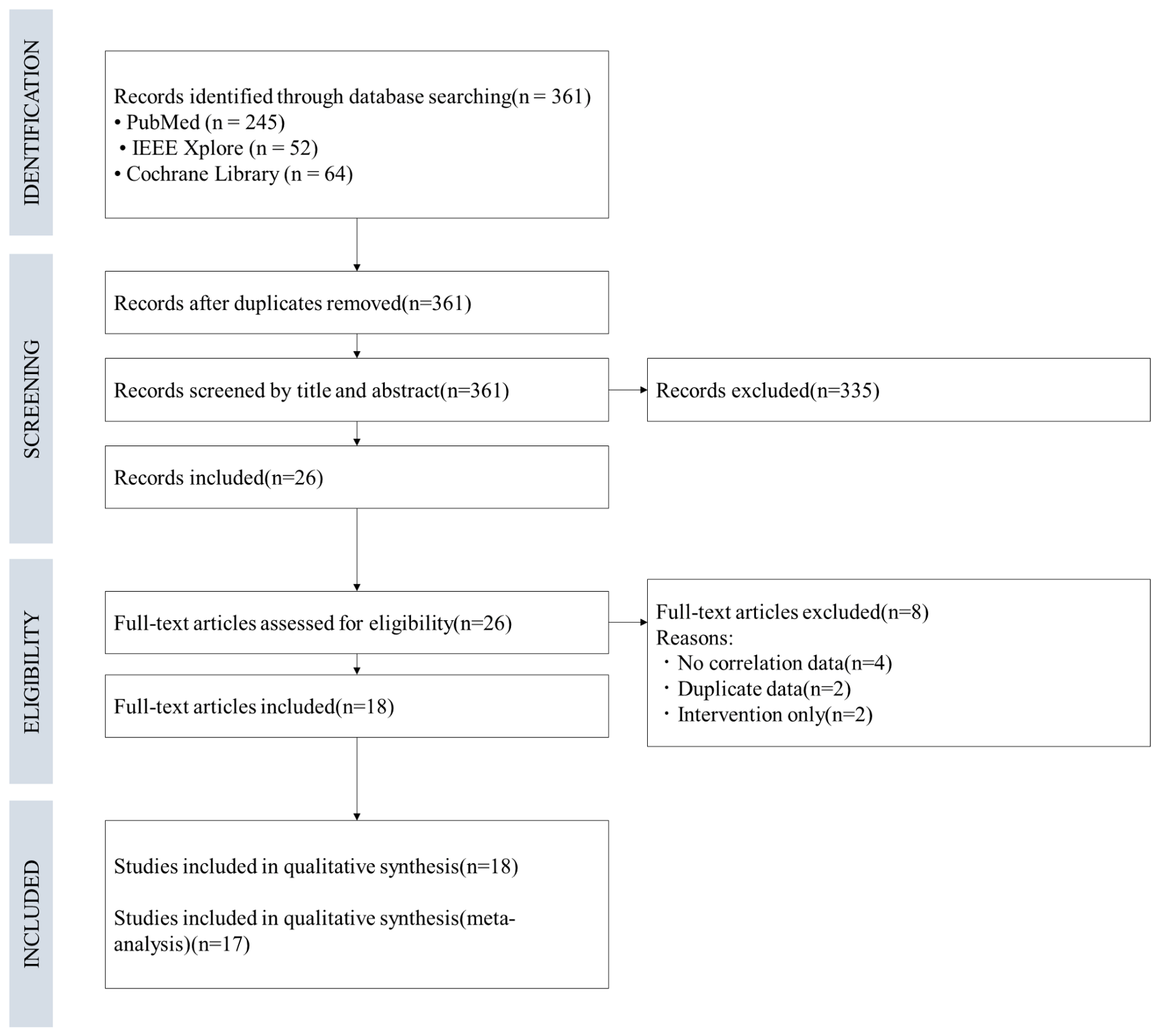
3.1. Study Selection
3.2. Study Characteristics
3.3. Quality Assessment
3.4. Correlation Between Ankle-Dorsiflexion Measures and Gait Performance
3.4.1. Individual Study Results
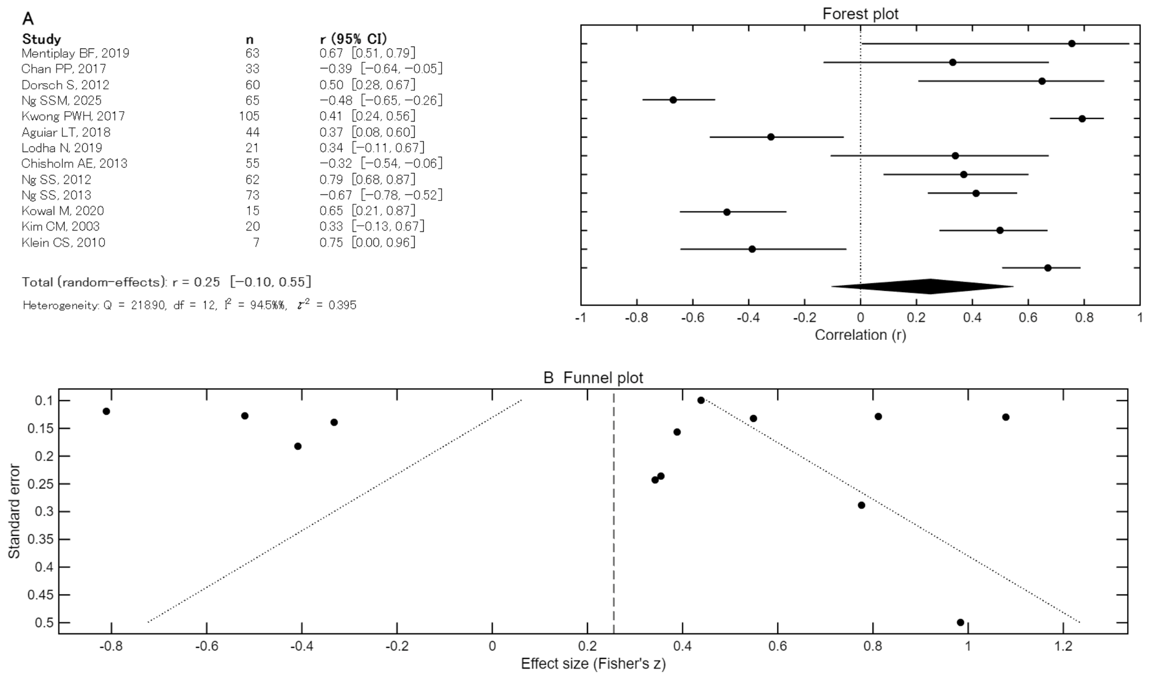
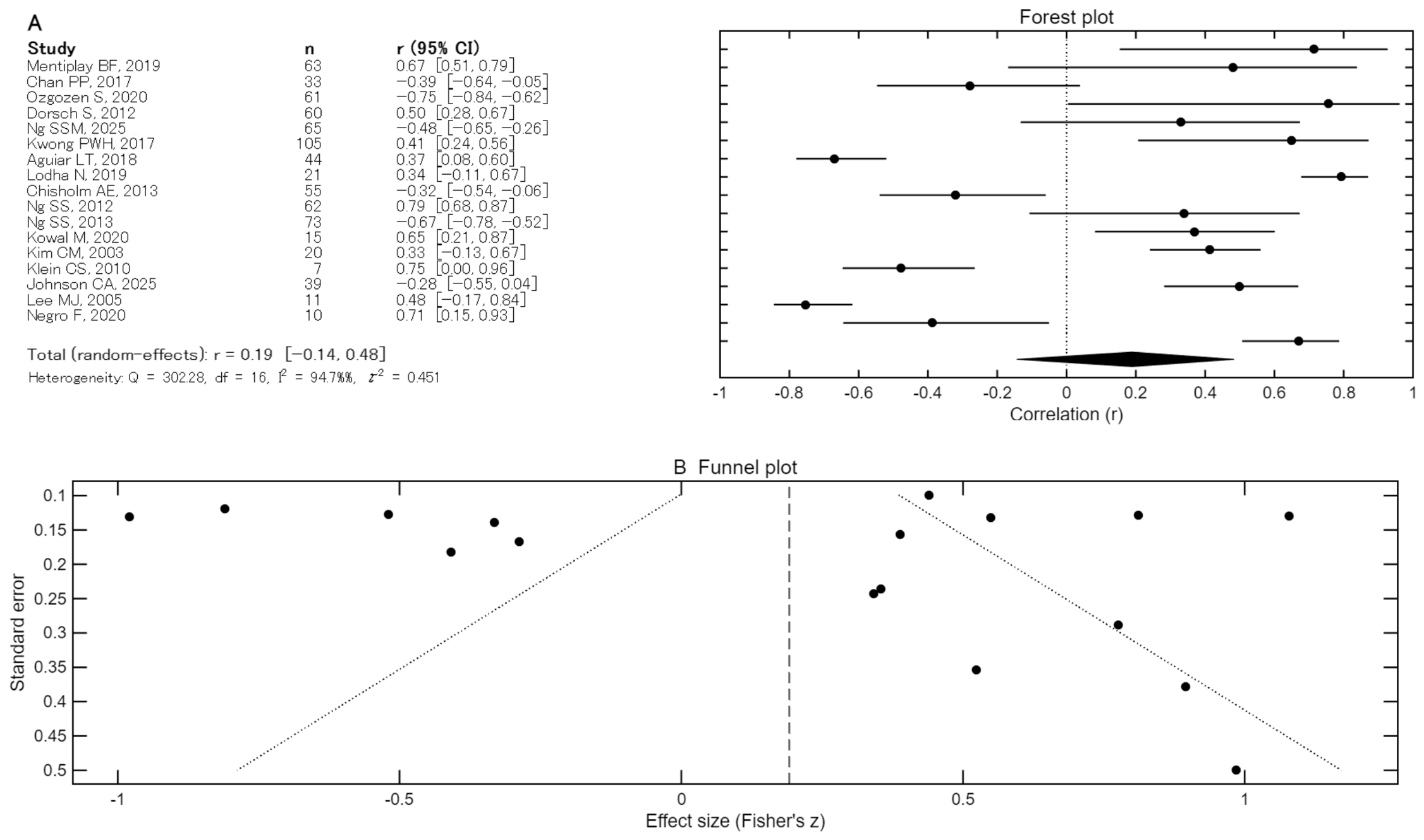
3.4.2. Meta-Analysis Results
4. Discussion
4.1. Summary of Main Findings
4.2. Comparison with Previous Literature
4.3. Assessment Technologies and Their Clinical Implications
4.4. Heterogeneity and Moderating Factors
4.5. Clinical Implications
4.6. Strengths and Limitations
4.7. Recommendations for Future Research
5. Conclusions
Supplementary Materials
Author Contributions
Funding
Institutional Review Board Statement
Informed Consent Statement
Data Availability Statement
Conflicts of Interest
Abbreviations
| IMUs | inertial measurement units |
| sEMG | surface electromyography |
| PRISMA | Preferred Reporting Items for Systematic Reviews and Meta-Analyses |
| MeSH | Medical Subject Headings |
| EEG | electroencephalogram |
| CMC | corticomuscular coherence |
| MVC | maximum voluntary contraction |
| TUG | Timed Up and Go |
| HHD | hand-held dynamometer |
| MMT | manual muscle testing |
| 6 MWT | 6-min walk test |
References
- Moore, S.A.; Boyne, P.; Fulk, G.; Verheyden, G.; Fini, N.A. Walk the talk: Current evidence for walking recovery after stroke, future pathways and a mission for research and clinical practice. Stroke 2022, 53, 3494–3505. [Google Scholar] [CrossRef] [Scilit]
- Li, S.; Francisco, G.E.; Zhou, P. Post-stroke hemiplegic gait: New perspective and insights. Front. Physiol. 2018, 9, 1021. [Google Scholar] [CrossRef] [Scilit]
- Fang, J.; Song, B.; Li, L.; Tong, L.; Jiang, M.; Yan, J. RGX Ensemble Model for Advanced Prediction of Mortality Outcomes in Stroke Patients. BME Front. 2024, 5, 0077. [Google Scholar] [CrossRef] [Scilit] [PubMed]
- Awad, L.N.; Palmer, J.A.; Pohlig, R.T.; Binder-Macleod, S.A.; Reisman, D.S. Walking speed and step length asymmetry modify the energy cost of walking after stroke. Neurorehabilit. Neural Repair 2015, 29, 416–423. [Google Scholar] [CrossRef] [Scilit] [PubMed]
- Skvortsov, D.V.; Kaurkin, S.N.; Grebenkina, N.V.; Ivanova, G.E. Typical changes in gait biomechanics in patients with subacute ischemic stroke. Diagnostics 2025, 15, 511. [Google Scholar] [CrossRef] [Scilit]
- Darak, V.; Karthikbabu, S. Lower limb motor function and hip muscle weakness in stroke survivors and their relationship with pelvic tilt, weight-bearing asymmetry, and gait speed: A cross-sectional study. Curr. J. Neurol. 2020, 19, 1–7. [Google Scholar] [CrossRef] [Scilit]
- Kochman, M.; Kasprzak, M.; Kielar, A. The impact of proprioception impairment on gait function in stroke survivors: A comprehensive review. Front. Neurol. 2025, 16, 1577919. [Google Scholar] [CrossRef] [Scilit]
- Jørgensen, H.S.; Nakayama, H.; Raaschou, H.O.; Olsen, T.S. Recovery of walking function in stroke patients: The Copenhagen Stroke Study. Arch. Phys. Med. Rehabil. 1995, 76, 27–32. [Google Scholar] [CrossRef] [Scilit]
- Peishun, C.; Haiwang, Z.; Taotao, L.; Hongli, G.; Yu, M.; Wanrong, Z. Changes in gait characteristics of stroke patients with foot drop after the combination treatment of foot drop stimulator and moving treadmill training. Neural Plast. 2021, 2021, 9480957. [Google Scholar] [CrossRef] [Scilit]
- Gao, T.; Ma, Z.; Yang, N.; Zhang, S.; Shi, H.; Zhang, H.; Ren, S.; Huang, H. The relationship of peak ankle dorsiflexion angle with lower extremity biomechanics during walking. J. Foot Ankle Res. 2024, 17, e12027. [Google Scholar] [CrossRef] [Scilit] [PubMed]
- Gil-Castillo, J.; Alnajjar, F.; Koutsou, A.; Torricelli, D.; Moreno, J.C. Advances in neuroprosthetic management of foot drop: A review. J. Neuroeng. Rehabil. 2020, 17, 46. [Google Scholar] [CrossRef] [Scilit]
- Alam, M.; Choudhury, I.A.; Bin Mamat, A. Mechanism and design analysis of articulated ankle foot orthoses for drop-foot. Sci. World J. 2014, 2014, 867869. [Google Scholar] [CrossRef] [Scilit]
- Kirdthongkham, T.; Justine, M.; Siriphorn, A. Prognostic accuracy of the Stroke Rehabilitation Assessment of Movement (STREAM) scores on admission for walking independence in stroke patients at discharge and one-month follow-up. PLoS ONE 2025, 20, e0319682. [Google Scholar] [CrossRef] [Scilit]
- Hulleck, A.A.; Menoth Mohan, D.; Abdallah, N.; El Rich, M.; Khalaf, K. Present and future of gait assessment in clinical practice: Towards the application of novel trends and technologies. Front. Med. Technol. 2022, 4, 901331. [Google Scholar] [CrossRef] [Scilit]
- Kumfu, S.; Poncumhak, P. Predictive ability of the three-time stand and walk test to determine frailty and its associations with fear of falling and cognitive function in community-dwelling older adults. Ann. Geriatr. Med. Res. 2022, 26, 316–322. [Google Scholar] [CrossRef] [Scilit] [PubMed]
- Steinbrink, G.M.; Martinez, J.; Swartz, A.M.; Strath, S.J. Sit-to-stand power is a stronger predictor of gait speed than knee extension strength. J. Funct. Morphol. Kinesiol. 2024, 9, 103. [Google Scholar] [CrossRef] [Scilit]
- Dobkin, B.H. Wearable motion sensors to continuously measure real-world physical activities. Curr. Opin. Neurol. 2013, 26, 602–608. [Google Scholar] [CrossRef] [Scilit]
- Xu, D.; Zhou, H.; Jie, T.; Zhou, Z.; Yuan, Y.; Jemni, M.; Quan, W.; Gao, Z.; Xiang, L.; Gusztav, F.; et al. Data-driven deep learning for predicting ligament fatigue failure risk mechanisms. Int. J. Mech. 2025, 301, 110519. [Google Scholar] [CrossRef] [Scilit]
- McCain, E.M.; Dick, T.J.M.; Giest, T.N.; Nuckols, R.W.; Lewek, M.D.; Saul, K.R.; Sawicki, G.S. Mechanics and energetics of post-stroke walking aided by a powered ankle exoskeleton with speed-adaptive myoelectric control. J. Neuroeng. Rehabil. 2019, 16, 57. [Google Scholar] [CrossRef] [Scilit] [PubMed]
- Malouin, F.; Richards, C.L. Mental practice for relearning locomotor skills. Phys. Ther. 2010, 90, 240–251. [Google Scholar] [CrossRef] [Scilit]
- Campanini, I.; Disselhorst-Klug, C.; Rymer, W.Z.; Merletti, R. Surface EMG in clinical assessment and neurorehabilitation: Barriers limiting its use. Front. Neurol. 2020, 11, 934. [Google Scholar] [CrossRef] [Scilit]
- Page, M.J.; McKenzie, J.E.; Bossuyt, P.M.; Boutron, I.; Hoffmann, T.C.; Mulrow, C.D.; Shamseer, L.; Tetzlaff, J.M.; Akl, E.A.; Brennan, S.E.; et al. The PRISMA 2020 statement: An updated guideline for reporting systematic reviews. BMJ 2021, 372, n71. [Google Scholar] [CrossRef] [Scilit]
- Wells, G.A.; Shea, B.; O’Connell, D.; Peterson, J.; Welch, V.; Losos, M.; Tugwell, P. The Newcastle-Ottawa Scale (NOS) for Assessing the Quality of Nonrandomised Studies in Meta-Analyses; Ottawa Hospital Research Institute: Ottawa, ON, Canada, 2014. [Google Scholar]
- Mentiplay, B.F.; Williams, G.; Tan, D.; Adair, B.; Pua, Y.H.; Bok, C.W.; Bower, K.J.; Cole, M.H.; Ng, Y.S.; Lim, L.S.; et al. Gait velocity and joint power generation after stroke: Contribution of strength and balance. Am. J. Phys. Med. Rehabil. 2019, 98, 841–849. [Google Scholar] [CrossRef] [Scilit] [PubMed]
- Chan, P.P.; Si Tou, J.I.; Tse, M.M.; Ng, S.S. Reliability and validity of the timed up and Go test with a motor task in people with chronic stroke. Arch. Phys. Med. Rehabil. 2017, 98, 2213–2220. [Google Scholar] [CrossRef] [Scilit]
- Ozgozen, S.; Guzel, R.; Basaran, S.; Coskun Benlidayi, I. Residual deficits of knee flexors and plantar flexors predict normalized walking performance in patients with poststroke hemiplegia. J. Stroke Cerebrovasc. Dis. 2020, 29, 104658. [Google Scholar] [CrossRef] [Scilit]
- Dorsch, S.; Ada, L.; Canning, C.G.; Al-Zharani, M.; Dean, C. The strength of the ankle dorsiflexors has a significant contribution to walking speed in people who can walk independently after stroke: An observational study. Arch. Phys. Med. Rehabil. 2012, 93, 1072–1076. [Google Scholar] [CrossRef] [Scilit]
- Ng, S.S.M.; Chen, P.; Usman, J.S.; Chan, P.H.; Chau, K.K.; Lee, S.H.; Liang, C.Y.; Man, T.K.; Hsu, C.L.; Li, K.J.J.; et al. Relationship of lower-limb muscle properties with motor control, walking and balance in stroke survivors. Sci. Rep. 2025, 16, 2563. [Google Scholar] [CrossRef] [Scilit]
- Kwong, P.W.H.; Ng, S.S.M.; Chung, R.C.K.; Ng, G.Y.F. A structural equation model of the relationship between muscle strength, balance performance, walking endurance and community integration in stroke survivors. PLoS ONE 2017, 12, e0185807. [Google Scholar] [CrossRef] [Scilit] [PubMed]
- Aguiar, L.T.; Camargo, L.B.A.; Estarlino, L.D.; Teixeira-Salmela, L.F.; Faria, C.D.C.M. Strength of the lower limb and trunk muscles is associated with gait speed in individuals with sub-acute stroke: A cross-sectional study. Braz. J. Phys. Ther. 2018, 22, 459–466. [Google Scholar] [CrossRef] [Scilit] [PubMed]
- Lodha, N.; Patel, P.; Casamento-Moran, A.; Hays, E.; Poisson, S.N.; Christou, E.A. Strength or motor control: What matters in high-functioning stroke? Front. Neurol. 2019, 9, 1160. [Google Scholar] [CrossRef] [Scilit]
- Chisholm, A.E.; Perry, S.D.; McIlroy, W.E. Correlations between ankle-foot impairments and dropped foot gait deviations among stroke survivors. Clin. Biomech. 2013, 28, 1049–1054. [Google Scholar] [CrossRef] [Scilit]
- Ng, S.S.; Hui-Chan, C.W. Contribution of ankle dorsiflexor strength to walking endurance in people with spastic hemiplegia after stroke. Arch. Phys. Med. Rehabil. 2012, 93, 1046–1051. [Google Scholar] [CrossRef] [Scilit]
- Ng, S.S.M.; Hui-Chan, C.W.Y. Ankle dorsiflexor, not plantarflexor strength, predicts the functional mobility of people with spastic hemiplegia. J. Rehabil. Med. 2013, 45, 541–545. [Google Scholar] [CrossRef] [Scilit]
- Kowal, M.; Kołcz, A.; Dymarek, R.; Paprocka-Borowicz, M.; Gnus, J. Muscle torque production and kinematic properties in post-stroke patients: A pilot cross-sectional study. Acta Bioeng. Biomech. 2020, 22, 11–20. [Google Scholar] [CrossRef] [Scilit] [PubMed]
- Kim, C.M.; Eng, J.J. The relationship of lower-extremity muscle torque to locomotor performance in people with stroke. Phys. Ther. 2003, 83, 49–57. [Google Scholar] [CrossRef] [Scilit] [PubMed]
- Klein, C.S.; Brooks, D.; Richardson, D.; McIlroy, W.E.; Bayley, M.T. Voluntary activation failure contributes more to plantar flexor weakness than antagonist coactivation and muscle atrophy in chronic stroke survivors. J. Appl. Physiol. 2010, 109, 1337–1346. [Google Scholar] [CrossRef] [Scilit] [PubMed]
- Johnson, C.A.; Biswas, P.; Tapia, R.; See, J.; Dodakian, L.; Chan, V.; Wang, P.T.; Nenadic, Z.; Do, A.H.; Reinkensmeyer, D.J. The weak relationship between ankle proprioception and gait speed after stroke: A robotic assessment study. Neurorehabilit. Neural Repair 2025, 39, 1031–1045. [Google Scholar] [CrossRef] [Scilit]
- Lee, M.J.; Kilbreath, S.L.; Refshauge, K.M. Movement detection at the ankle following stroke is poor. Aust. J. Physiother. 2005, 51, 19–24. [Google Scholar] [CrossRef] [Scilit]
- Negro, F.; Bathon, K.E.; Nguyen, J.N.; Bannon, C.G.; Orizio, C.; Hunter, S.K.; Hyngstrom, A.S. Impaired firing behavior of individually tracked paretic motor units during fatiguing contractions of the dorsiflexors and functional implications post stroke. Front. Neurol. 2020, 11, 540893. [Google Scholar] [CrossRef] [Scilit]
- Cho, K.H.; Lee, J.Y.; Lee, K.J.; Kang, E.K. Factors related to gait function in post-stroke patients. J. Phys. Ther. Sci. 2014, 26, 1941–1944. [Google Scholar] [CrossRef] [Scilit]
- Kwakkel, G.; Kollen, B.; Twisk, J. Impact of time on improvement of outcome after stroke. Stroke 2006, 37, 2348–2353. [Google Scholar] [CrossRef] [Scilit]
- Lins, C.; Oliveira, A.R.B.; Chagas, M.S.S.; Ribeiro, F.; Cliquet, A.; Pagnano, R.G. Assessment of Isometric and Isokinetic Ankle Strength Measures: A Pilot Study. Acta Ortop. Bras. 2025, 33, e288925. [Google Scholar] [CrossRef] [Scilit]
- Yasacı, Z.; Argut, S.K.; Celik, D. Reliability of a new stabilization device for measurement of muscle strength using a hand-held dynamometer. Muscle Nerve 2024, 70, 1240–1246. [Google Scholar] [CrossRef] [Scilit] [PubMed]
- Sahu, P.K.; Goodstadt, N.; Ramakrishnan, A.; Silfies, S.P. Test-retest reliability and concurrent validity of knee extensor strength measured by a novel device incorporated into a weight stack machine vs. handheld and isokinetic dynamometry. PLoS ONE 2024, 19, e0301872. [Google Scholar] [CrossRef] [Scilit] [PubMed]
- Picot, B.; Maricot, A.; Fourchet, F.; Gokeler, A.; Tassignon, B.; Lopes, R.; Hardy, A. Targeting visual-sensory and cognitive impairments following lateral ankle sprains: A practical framework for functional assessment across the return-to-sport continuum-Part 1. Sensory reweighting and cognitive impairments: What are we really talking about and why clinicians should consider central alterations in return to sport criteria. Front. Sports Act. Living 2025, 7, 1668224. [Google Scholar] [CrossRef] [Scilit]
- Jastifer, J.R. Contemporary Review: Proprioception in Ankle Stability. Foot Ankle Orthop. 2025, 10, 24730114251374662. [Google Scholar] [CrossRef] [Scilit]
- Kwakkel, G.; Kollen, B.J.; van der Grond, J.; Prevo, A.J.H. Probability of regaining dexterity in the flaccid upper limb: Impact of severity of paresis and time since onset in acute stroke. Stroke 2003, 34, 2181–2186. [Google Scholar] [CrossRef] [Scilit]
- Ogborn, D.I.; Bellemare, A.; Bruinooge, B.; Brown, H.; McRae, S.; Leiter, J. Comparison of Common Methodologies for the Determination of Knee Flexor Muscle Strength. Int. J. Sports Phys. Ther. 2021, 16, 350–359. [Google Scholar] [CrossRef] [Scilit]
- Sul, J.H.; Piyathilaka, L.; Moratuwage, D.; Dunu Arachchige, S.; Jayawardena, A.; Kahandawa, G.; Preethichandra, D.M.G. Electromyography Signal Acquisition, Filtering, and Data Analysis for Exoskeleton Development. Sensors 2025, 25, 4004. [Google Scholar] [CrossRef] [Scilit]
- Fang, C.; He, B.; Wang, Y.; Cao, J.; Gao, S. EMG-Centered Multisensory Based Technologies for Pattern Recognition in Rehabilitation: State of the Art and Challenges. Biosensors 2020, 10, 85. [Google Scholar] [CrossRef] [Scilit]
- Hohsoh, N.; Sanghan, T.; Chong, D.Y.R.; Stojanovic, G.; Chatpun, S. Comparative electromyography analysis of subphase gait disorder in chronic stroke survivors. PeerJ 2024, 12, e18473. [Google Scholar] [CrossRef] [Scilit]
- Srivastava, S.; Patten, C.; Kautz, S.A. Altered muscle activation patterns (AMAP): An analytical tool to compare muscle activity patterns of hemiparetic gait with a normative profile. J. Neuroeng. Rehabil. 2019, 16, 21. [Google Scholar] [CrossRef] [Scilit] [PubMed]
- Maffiuletti, N.A.; Aagaard, P.; Blazevich, A.J.; Folland, J.; Tillin, N.; Duchateau, J. Rate of force development: Physiological and methodological considerations. Eur. J. Appl. Physiol. 2016, 116, 1091–1116. [Google Scholar] [CrossRef] [Scilit] [PubMed]
- Winter, D.A.; Yack, H.J. EMG profiles during normal human walking: Stride-to-stride and inter-subject variability. Electroencephalogr. Clin. Neurophysiol. 1987, 67, 402–411. [Google Scholar] [CrossRef] [Scilit]
- Hillier, S.; Immink, M.; Thewlis, D. Assessing proprioception: A systematic review of possibilities. Neurorehabilit. Neural Repair 2015, 29, 933–949. [Google Scholar] [CrossRef] [Scilit]
- Aman, J.E.; Elangovan, N.; Yeh, I.L.; Konczak, J. The effectiveness of proprioceptive training for improving motor function: A systematic review. Front. Hum. Neurosci. 2015, 8, 1075. [Google Scholar] [CrossRef] [Scilit]
- Fulk, G.D.; Echternach, J.L. Test-retest reliability and minimal detectable change of gait speed in individuals undergoing rehabilitation after stroke. J. Neurol. Phys. Ther. 2008, 32, 8–13. [Google Scholar] [CrossRef] [Scilit]
- Flansbjer, U.B.; Holmbäck, A.M.; Downham, D.; Patten, C.; Lexell, J. Reliability of gait performance tests in men and women with hemiparesis after stroke. J. Rehabil. Med. 2005, 37, 75–82. [Google Scholar] [CrossRef] [Scilit]
- Bohannon, R.W. Muscle strength and muscle training after stroke. J. Rehabil. Med. 2007, 39, 14–20. [Google Scholar] [CrossRef] [Scilit]
- Ada, L.; Dorsch, S.; Canning, C.G. Strengthening interventions increase strength and improve activity after stroke: A systematic review. Aust. J. Physiother. 2006, 52, 241–248. [Google Scholar] [CrossRef] [Scilit]



| Study | Year | Sample Size | Sex (Male/Female), N | Age (Years) | Paretic Side (Left/Right), N | Time Since Stroke | Device | Dorsiflexion Index | Value | Walking-Ability Index | Value | Correlation Coefficients |
|---|---|---|---|---|---|---|---|---|---|---|---|---|
| Mentiplay BF [24] | 2019 | 63 | 34/29 | 60 ± 13 | 33/30 | 39 ± 51 (months) | HHD (Lafayette Instrument Company, Lafayette, IN, USA) | Isometric strength (Nm/kg) | 0.13 ± 0.09 (Nm/kg) | Walking speed (m/s) | Habitual pace 0.85 ± 0.37 (m/s) Fast pace 1.07 ± 0.47 (m/s) | 0.67 |
| Chan PP [25] | 2017 | 33 | 22/11 | 60.2 ± 6.4 | 9/24 | 9.35 ± 4.28 (years) | HHD (Lafayette Instrument Company, Lafayette, IN, USA) | Strength (kg) | 9.49 ± 5.47 (kg) | TUG motor time (s) | 18.31 ± 5.77 (s) | −0.39 |
| Ozgozen S [26] | 2020 | 61 | 36/25 | 54.6 ± 11.7 | 31/30 | 19 (months) | HHD (Hoggan Health Industries Inc., West Jordan, UT, USA) | Residual deficits of the muscle groups (%): 100 − [(paretic muscle strength/non-paretic muscle strength) × 100] | 52.6 (%) | Walking speed (m/s) 6-min walk distance (meters) | 0.91 (m/s) 243.7 ± 122.2 (meters) | −0.75 |
| Dorsch S [27] | 2012 | 60 | 42/18 | 69 ± 11 | 28/32 | 1–6 range (years) | HHD | Strength (N) | 66 ± 37 (N) | Walking speed (m/s) | 0.75 ± 0.34 (m/s) | 0.50 |
| Ng SSM [28] | 2025 | 65 | 35/30 | 66.91 ± 6.40 | 31/34 | 9.45 ± 4.99 (years) | HHD (Lafayette Instrument Corp., Lafayette, IN, USA) | Strength (kg) | 9.26 ± 4.62 (kg) | Walking speed (m/s) | Usual 18.20 ± 11.57 (m/s) Maximum 14.00 ± 8.60 (m/s) | −0.48 |
| Kwong PWH [29] | 2017 | 105 | 63/42 | 61.0 ± 6.9 | 48/57 | 6.2 ± 4.9 (years) | HHD (Lafa yette Instrument Company, Lafayette, IN, USA) | Strength (kg) | 8.5 ± 5.0 (kg) | 6 MWT (m) | 219.7 ± 84.4 (m) | 0.41 |
| Aguiar LT [30] | 2018 | 44 | 24/20 | 62 ± 15 | 25/19 | 4 ± 1 (months) | Handheld microFET2® dynamometer (Hoggan Scientific, LLC, Salt Lake City, UT, USA) | Isometric force (Nm/kg) | 5.7 ± 2.4 (Nm/kg) | Walking speed (m/s) | Comfortable 0.8 ± 0.38 (m/s) Maximum 1.1 ± 0.54 (m/s) | 0.37 |
| Lodha N [31] | 2019 | 21 | 13/8 | 65.04 ± 13.72 | 4/17 | 4.79 ± 4.66 (months) | Force transducer (Honeywell, Morristown, NJ, USA) | MVC (N) | 143.64 ± 67.50 (N) | Walking speed (m/s) | NR | 0.34 |
| Chisholm AE [32] | 2013 | 55 | Non-dropped foot: 28/15 Dropped foot: 9/3 | 69.5 ± 12.2 66.8 ± 13.2 | 19/19 (Bilateral 5) 9/2 (Bilateral 1) | 35.0 ± 11.5 (days) 57.9 ± 24.7 (days) | Load cell (Interface Inc., Scottsdale, AZ, USA) | Isometric force (MVC) | NR | Swing peak dorsiflexors | NR | −0.32 |
| Ng SS [33] | 2012 | 62 | 51/11 | 57.4 ± 7.8 | 43/19 | 5.2 ± 3.7(years) | load cell | Peak torque (Nm) | 14.2 ± 6.7 (Nm) | Walking speed(cm/s) 6 MWT (m) | 51.5 ± 26.1 (cm/s) 183.7 ± 84.4 (m) | 0.79 |
| Ng SS [34] | 2013 | 73 | 60/13 | 57.16 ± 7.91 | 45/28 | 5.21 ± 3.63 (years) | Load cell | Peak torque (Nm) | 14.67 ± 7.01 (Nm) | TUG(s) | 27.41 ± 17.63 (s) | −0.67 |
| Kowal M [35] | 2020 | 15 | 7/8 | 57.2 ± 11 | 7/8 | 1.53 ± 0.64 (months) | Biodex System (Biodex Medical Systems Inc., Shirley, NY, USA) | Torque (Nm/kg) | 0.3 (Nm/kg) | Walking speed (m/s) | 0.9 ± 0.1 (m/s) | 0.65 |
| Kim CM [36] | 2003 | 20 | 14/6 | 61.2 ± 8.4 | 9/11 | 4.0 ± 2.6 (years) | Kim-Com isokinetic dynamometer (Chattanooga Group Inc, 4717 Adams Rd, Hixson, TN, USA) | Torque (Nm) | 0.15 ± 0.13 (Nm) | Walking speed (m/s) self-selected pace, maximum | Self-selected pace 0.45 ± 0.25 (m/s) Maximum 0.69 ± 0.35 (m/s) | 0.33 |
| Klein CS [37] | 2010 | 7 | 5/2 | 55.8 ± 3.6 | 4/3 | NR | Load cell (Omega Engineering, Stamford, CT, USA) | MVC torque (Nm) | 56.7 ± 57.4 (Nm) | Walking speed (m/s) | 0.83 ± 0.33 (m/s) | 0.75 |
| Johnson CA [38] | 2025 | 39 | 24/15 | 60 ± 12 | 27/12 | 1155 ± 1096 (days) | Ankle Measuring Proprioceptive Device | Joint Position Reproduction dynamic error (°) | 11.2 ± 5.6 (°) | Walking speed (m/s) 6MWT (m) | 0.37 ± 0.24 (m/s) 114.4 ± 66.4 (m) | −0.28 |
| Lee MJ [39] | 2005 | 11 | 9/2 | 69 ± 11 | 8/3 | 43 ± 32 (months) | linear servo-motor | ROM (°) | 19.5 ± 17.4 (°) | 6MWT (m) | 324.4 ± 173.1 (m) | 0.48 |
| Negro F [40] | 2020 | 10 | 5/5 | 60.4 ± 13 | NR | 15.8 ± 10 (years) | load cell (EMG, Scottsdale, AZ, USA) | MVC (N) | 106. ± 54 (N) | Walking speed (m/s) | NR | 0.71 |
| Cho KH [41] | 2014 | 39 | 23/16 | 67.8 ± 0.9 | 20/19 | 200.1 ± 227.1 (days) | NR (non-instrumented clinical grading; MMT) | MMT | 2.7 ± 1.6 | Walking level (ordinal scale) | 3.6 ± 1.5 | 0.56 |
| 95% CI | ||||||||
|---|---|---|---|---|---|---|---|---|
| Group | Outcome | No. of Studies | Pooled r | Lower | Upper | Model | I2 | τ2 |
| All | Walking speed | 17 | 0.19 | −0.14 | 0.48 | Random-effects | 94.70 | 0.45 |
| Strength | Walking speed | 13 | 0.65 | −0.42 | 0.96 | Random-effects | 94.52 | 0.40 |
Disclaimer/Publisher’s Note: The statements, opinions and data contained in all publications are solely those of the individual author(s) and contributor(s) and not of MDPI and/or the editor(s). MDPI and/or the editor(s) disclaim responsibility for any injury to people or property resulting from any ideas, methods, instructions or products referred to in the content. |
© 2026 by the authors. Licensee MDPI, Basel, Switzerland. This article is an open access article distributed under the terms and conditions of the Creative Commons Attribution (CC BY) license.
Share and Cite
Ito, H.; Yamaguchi, H.; Yamauchi, R.; Kitai, K.; Kodama, T. Measurement Technologies for Ankle-Dorsiflexion Function After Stroke: A Systematic Review and Meta-Analysis of Sensing Approaches and Their Relationships with Gait Performance. Sensors 2026, 26, 3598. https://doi.org/10.3390/s26113598
Ito H, Yamaguchi H, Yamauchi R, Kitai K, Kodama T. Measurement Technologies for Ankle-Dorsiflexion Function After Stroke: A Systematic Review and Meta-Analysis of Sensing Approaches and Their Relationships with Gait Performance. Sensors. 2026; 26(11):3598. https://doi.org/10.3390/s26113598
Chicago/Turabian StyleIto, Hiroki, Hideaki Yamaguchi, Ryosuke Yamauchi, Ken Kitai, and Takayuki Kodama. 2026. "Measurement Technologies for Ankle-Dorsiflexion Function After Stroke: A Systematic Review and Meta-Analysis of Sensing Approaches and Their Relationships with Gait Performance" Sensors 26, no. 11: 3598. https://doi.org/10.3390/s26113598
APA StyleIto, H., Yamaguchi, H., Yamauchi, R., Kitai, K., & Kodama, T. (2026). Measurement Technologies for Ankle-Dorsiflexion Function After Stroke: A Systematic Review and Meta-Analysis of Sensing Approaches and Their Relationships with Gait Performance. Sensors, 26(11), 3598. https://doi.org/10.3390/s26113598