Communication

The Total Ionizing Dose Effects on Perovskite CsPbBr3 Semiconductor Detector

1
School of Nuclear Science and Technology, Xi’an Jiaotong University, No. 28, Xianning West Road, Xi’an 710049, China
2
State Key Laboratory of Intense Pulsed Radiation Simulation and Effect, Northwest Institute of Nuclear Technology, Xi’an 710024, China
3
State Key Laboratory of Radiation Medicine and Protection, Collaborative Innovation Center of Radiological Medicine of Jiangsu Higher Education Institutions, and School for Radiological and Interdisciplinary Sciences (RAD-X), Soochow University, Suzhou 215123, China
4
Sino-French Institute of Nuclear Engineering and Technology, Sun Yat-Sen University, Zhuhai 519082, China
*
Author to whom correspondence should be addressed.
Sensors 2023, 23(4), 2017; https://doi.org/10.3390/s23042017
Submission received: 29 December 2022 / Revised: 18 January 2023 / Accepted: 19 January 2023 / Published: 10 February 2023
(This article belongs to the Section Electronic Sensors)

Abstract

:
Perovskite CsPbBr3 semiconductors exhibit unusually high defect tolerance leading to outstanding and unique optoelectronic properties, demonstrating strong potential for γ-radiation and X-ray detection at room temperature. However, the total dose effects of the perovskite CsPbBr3 must be considered when working in a long-term radiation environment. In this work, the Schottky type of perovskite CsPbBr3 detector was fabricated. Their electrical characteristics and γ-ray response were investigated before and after 60Co γ ray irradiation with 100 and 200 krad (Si) doses. The γ-ray response of the Schottky-type planar CsPbBr3 detector degrades significantly with the increase in total dose. At the total dose of 200 krad(Si), the spectral resolving ability to γ-ray response of the CsPbBr3 detector has disappeared. However, with annealing at room temperature for one week, the device’s performance was partially recovered. Therefore, these results indicate that the total dose effects strongly influence the detector performance of the perovskite CsPbBr3 semiconductor. Notably, it is concluded that the radiation-induced defects are not permanent, which could be mitigated even at room temperature. We believe this work could guide the development of perovskite detectors, especially under harsh radiation conditions.

1. Introduction

The accurate detection of γ-rays from radioactive sources in high resolution at room temperature is of significant interest for industrial, medical imaging, and space applications [1,2,3]. However, the γ-ray photons are in very low flux. Notably, most semiconductors are susceptible to even a small number of defects and impurities that invariably act as signal-killing carrier traps. The traditional γ-ray detector materials are very few and face many challenges. The emergence of inorganic perovskite CsPbBr3 with surprisingly good defect tolerance, which is very promising for high-resolution X-ray and γ-ray detection at room temperature, has attracted extensive attention [4,5,6,7,8,9,10,11,12,13,14,15]. Additionally, the CsPbBr3 detector has shown remarkable energy resolving capability under both X and γ rays, particularly achieving 3.2% (3.9 keV, FWHM) energy resolution for 122 keV 57Co γ-ray [10,16]. Furthermore, the lifetime of the hole in CsPbBr3 detector-grade single crystal was observed to be over 25 μs [16].
For an excellent detector, device stability in a radiation environment is also critical. In addition, electronic devices for space, scientific and medical applications must be tolerant of a wide range of total ionizing doses (TID). However, the spectral degradation of the perovskite CsPbBr3 detector working in high irradiation environments has not been investigated [17,18,19,20].
In this work, we report a first investigation of the spectral response variation of CsPbBr3 detectors under total dose irradiation. The CsPbBr3 detector was fabricated with the asymmetric electrode design, in which the γ-ray response of the Schottky-type planar CsPbBr3 detector was revealed under different total doses. Additionally, the effect of the irradiation damage was evaluated through annealing experiments. Therefore, it is concluded that this device can work stably under the total ionizing dose of 100 krad(Si). We believe that our work will significantly promote and provide guidance for further investigations on the application of halide perovskites.

2. Materials and Methods

2.1. CsPbBr3 Detector Fabrication

High-quality CsPbBr3 single crystals were grown from melt using the Bridgman method, as shown in Figure 1a. After mechanical polishing, the surfaces were deposited with the metal contact (Au and Ga/In), and the typical thickness of gold contact was ~70 nm. After electrode preparation, carbon paste was used to connect the metal electrode with the collection circuit for the front-end electronics. Finally, the test sample is sealed with wax. And the top-view photograph of the CsPbBr3 detectors was illustrated in Figure 1b. More information on the samples is detailed in our previous work [16].

2.2. Irradiation

The CsPbBr3 detectors were irradiated by γ-rays derived from the 60Co source (at the Northwest Institute of Nuclear Technology, Xi’an, China) and had an energy distribution centered typically around 1.25 MeV. The detector was unbiased, with all pins floating during 60Co γ radiation, and tested at a dose rate of 50.0 rad(Si)/s. The sample’s current-voltage (IV) characteristics were measured at the total ionizing doses of 100 and 200 krad(Si), respectively. The spectrum response of the sample was measured at the total ionizing doses of 100 and 200 krad(Si). Finally, the sample’s tested parameters were measured within two hours after each radiation step.

2.3. Measurements

The current-voltage (IV) characteristics of the CsPbBr3 detector were measured using a Keysight B1500A semiconductor device analyzer. The response was tested with a 59.5 keV 241Am γ-ray source. As shown in Figure 2, the CsPbBr3 detectors were positioned inside a vacuum chamber to detect γ-rays. The response signal induced in the CsPbBr3 detector was, in turn, transmitted to the following equipment through coaxial cables: an ORTEC-142A preamplifier, an ORTEC-672 amplifier (shaping time of 3 μs, gain of 1500 times), an ORTEC-ASPEC-927 multi-channel analyzer (MCA), and the MAESTRO software was used to acquire the γ-ray spectrally. An SRS PS350, a high-voltage supply, was used to provide the reverse bias to the CsPbBr3 detector through the preamplifier.

3. Results and Discussion

3.1. I-V and I-T Characteristics

As shown in Figure 3, The pristine I–V curve of the CsPbBr3 detector on the reverse side was compared with the reverse side I–V curves taken after the irradiation with the total dose of 100 krad and 200 krad, respectively. During the test, the sweep voltage was set from −100 V to 0 V, with 4 s intervals between each voltage step. As indicated in Figure 3, with the increase in total dose, there was a decrease in the dark current of the CsPbBr3 detector. Notably, the higher the bias voltage applied, the more the radiation effect will be.
The diffusion model has been utilized for analyzing the I–V characteristics of the CsPbBr3 detector [21,22,23,24,25]. In the diffusion model, the current density J under the reverse bias V in a metal-semiconductor Schottky device can be described as:
J = q 2 μ N c N i ε [ ε q N i d ( V + V i n ) + d 2 ] ( 1 - e q V / k T ) e q φ / k T
where q is the electron charge, μ is the carrier mobility, Nc is the effective density of states in the conduction band, Ni is the concentration of the ionized donor/acceptor centers, ε is the electrical permittivity in the crystal, d is the thickness of the device, Vin is the built-in internal electric field, k is Boltzmann’s constant, T is temperature, and ϕ is Schottky barrier [24]. Accordingly, the J–V curve under the reverse bias of a CsPbBr3 device was fitted according to the above J–V equation, as plotted in Figure 4.
Accordingly, the J-V curve under the reverse bias of an MSM device was fitted according to the above J-V equation, as plotted in Figure 2. The J–V curve in the measured range followed the tendency predicted by the diffusion model. Before irradiation, the Schottky barrier extracted was 0.72 eV, while u is 53.6 cm2/Vs. Following irradiation, the Schottky barrier was not changed, while the μ was dropped with the total dose, and 44.9 cm2/Vs for 100 krad(Si) 38.6 cm2/Vs for 200 krad(Si). Thus, we considered that the radiation induced defects in the CsPbBr3 detector and is the main reason for the decrease in mobility.
The total ionizing dose impacted the photoelectric response of the CsPbBr3 detector. Figure 5 shows the time-dependent currents under the light pulses and 50 V bias. The dark current was about 0.7–1.9 nA. Before radiation, the photocurrent was around 85 nA, and the ON-OFF ratio was about 80. Then, with the increase in total dose, the photocurrent and the ON-OFF ratio dropped. For the total dose of 200 krad(Si), the photocurrent was around 32 nA, and the ON-OFF ratio was reduced to 35. Therefore, this finding indicates that electrical-active defects were likely formed after total irradiation, resulting in the trapping of photo-generated carriers.

3.2. γ-ray Spectral Performance

The γ-ray spectral performance of the CsPbBr3 detector was also influenced by total ionizing dose radiation. The energy resolution is a critical characteristic for evaluating the ability of energy spectral measurement of the detectors. The detector’s energy resolution is usually defined as the ratio between the FWHM and the position of the peak centroid, which is a dimensionless fraction expressed as a percentage. Figure 6 shows the energy resolution of CsPbBr3 detectors to 241Am γ-ray with various bias voltages (−50 V, −100 V, and −150 V) before and after 60Co γ ray total dose irradiation. During the energy spectral test, the radiation-induced charge Q collection was on the Au cathode, while Ga/In anode was grounded, and the γ-ray irradiated from the anode. Figure 6a illustrates the energy spectra under applied voltage (−50 V) with a shaping time of 3 µs. Before radiation, the CsPbBr3 detector showed a well-resolved spectroscopic response with an energy resolution of 21.76% for 59.5 keV 241Am γ-ray. Then the energy resolution degraded to around 34.2% after γ-ray irradiation with a total dose of 100 krad(Si). At the same time, the spectral performance disappeared when the total dose increased to 200 krad(Si). After the radiation device had been annealed at room temperature for one week, the γ-ray spectral response was restored with an energy resolution of 25.35%. During the annealing test, the pins of the device were in floating bias and were placed at room temperature. Figure 6b shows that when the applied bias was −100 V, the TID effects on the spectral performance of the CsPbBr3 detector. Before irradiation, the CsPbBr3 detector showed a more well-resolved spectroscopic response with an energy resolution of 13.89%. After a cumulative total dose of 100 krad(Si), the energy resolution was 20.49%. When the total dose reached 200 krad, the spectral performance disappeared. After annealing, the device’s performance under this voltage bias has not been recovered.
The test result with −150 V bias voltage is demonstrated in Figure 6c. The higher the bias voltage applied the better spectral performance was achieved. However, under higher bias voltage, the influence of irradiation becomes more significant. As shown in Figure 6c, the energy resolution was 12.05% before irradiation and increased to 17.62% after irradiation with a total dose of 100 krad(Si). Similar to the test results under −100 V bias, there was no energy spectrum characteristic when the cumulative total dose was 200 krad(Si) and the test after annealing.
The charge collection efficiency (CCE) is also influenced by the TID effect, as shown in Figure 6d. The CCE of CsPbBr3 detectors can be calculated by using a Si detector as a reference sample, and CCE can be expressed as in Formula (2) [26,27,28].
C C E = P C s P b B r 3 P S i × G S i G C s P b B r 3 × E d C s P b B r 3 E d S i × E α E γ × C C E S i
The PSi and PCsPbBr3 represent the channel numbers of the peak centroid measured by the CsPbBr3 detector and Si (697.52) detector, respectively. The GSi and GCsPbBr3 represent the gain for CsPbBr3 (50) and Si (1500), respectively. The E α and E γ and present the α-particle energy of 239Pu (5156 keV) and γ-ray energy of 241Am (59.59 keV), respectively. The EdSi and EdCsPbBr3 represent the mean ionization energy for CsPbBr3 (5.3 eV) and Si (3.6 eV) [26], respectively. The CCESi is the charge collection efficiency of the reference silicon detector (~100%). The CCE at 150 V bias of the pre-irradiated CsPbBr3 detector was 91.75% and decreased to 84.56% with a total dose of 100 krad (Si). The degradation of energy resolution and CCE can be ascribed to increased trapping centers under gamma radiation, which decrease the mean free path of holes and electrons.

3.3. Photoluminescence

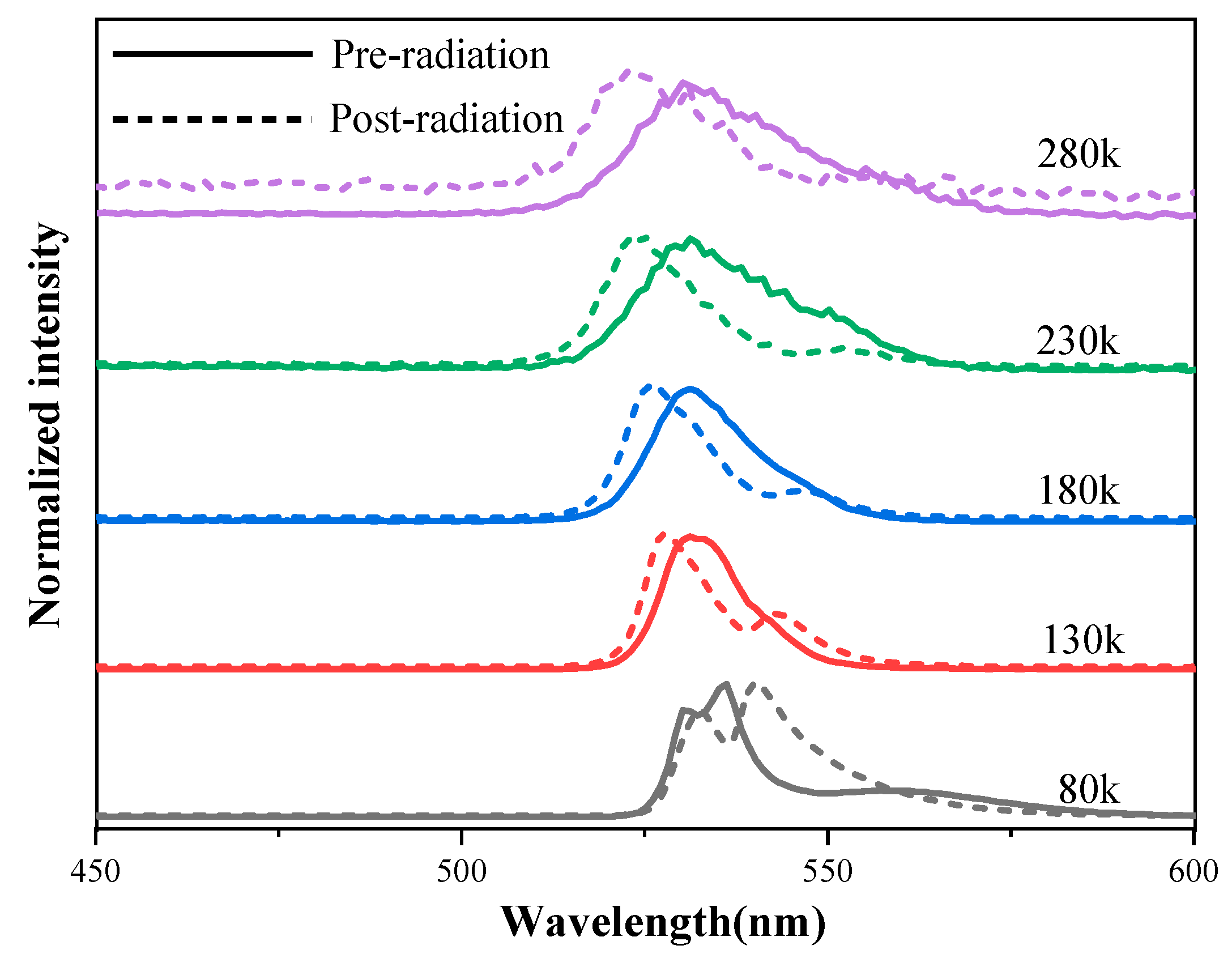
PL spectroscopy was used to characterize the optical properties of the perovskite CsPbBr3 crystal. Figure 7 shows the PL spectrum of as-grown and 60Co γ-ray irradiated samples (the total dose = 165 rad(Si)), and the PL intensities at various temperatures were analyzed in detail. It is evident that the peak value of the PL spectrum has a red shift in the 80 K temperature test, and with the increase of test temperature, the peak value of the PL spectrum after irradiation appeared to have a blue shift. The possible reason for this phenomenon is the total ionizing dose irradiation changes the potential energy at the top of the valence band and at the bottom of the guide band of the halide material [26]. The origin of this difference needs to be further investigated.

4. Discussion

This work investigated the 60Co γ ionization radiation effects on the Schottky type CsPbBr3 detector at the dose rate of 50.0 rad(Si)/s. The ionization radiation has little effect on the I–V curve of the CsPbBr3 detector. With the increase in total dose, the dark current decreased slightly. The I–T character was impacted significantly after ionization radiation, and the pre-irradiated CsPbBr3 detector demonstrated a high ON-OFF ratio of 80. After 200 krad(Si) radiation, the ON-OFF ratio of the CsPbBr3 detector was reduced to 35. At 100 krad(Si), the energy spectral characteristics of the device were still maintained. However, when the total dose accumulated to 200 krad(Si), the spectral performance of the tested CsPbBr3 detector disappeared.
As shown in Table 1, with various bias voltages (−50, −100, and −150 V) during the γ-ray spectral test, there were different responses of energy resolution to irradiation ΔR = R1 − R2, where R1 represents the resolution of post-irradiation and R2 represents the resolution of pre-irradiation. When tested under extensive voltage conditions, the energy resolution caused by radiation after accumulating 100 krad(Si) was deduced at about 6%. Additionally, the performance did not recover within one week of room temperature annealing. Then, under low-voltage test conditions (50 V), the spectral resolution slightly increased by 1.1% after accumulating a total dose of 100 krad(Si). In addition, the main difference was that when tested under low voltage conditions, the energy spectral performance of the CsPbBr3 detector recovered within one week of annealing after irradiation. Additionally, the energy resolution after annealing degraded by about 4.77% compared to that obtained before irradiation. However, this recovery process was not observed when testing at high voltage. Finally, when the total dose was 200 krad(Si), the detector had no spectral characteristics tested under all bias conditions.

5. Conclusions

The 60Co γ ionizing radiation may induce electrically active defects in the bulk of the metal-semiconductor interface of the CsPbBr3 detector. Those defects will likely alter the detector’s performance. The difference in energy spectrum under different test bias voltage may also be due to the defect induced by ionizing radiation in the Schottky interface region. With the increase of the bias applied during the testing process, the Schottky barrier becomes wider, and the space charge region increases, leading to the decrease of carrier lifetime during the energy spectrum testing. Therefore, our future work will continue to study the radiation effects and damage mechanism of the CsPbBr3 detectors to meet the increasing demands for radiation detection in intense radiation fields.

Author Contributions

Conceptualization, Y.H. and L.L.; validation, H.Q. and R.G.; formal analysis, W.M.; writing—original draft preparation, W.M.; writing—review and editing, S.G.; supervision, B.H.; project administration, X.O. All authors have read and agreed to the published version of the manuscript.

Funding

This research was funded by the National Key R&D Program of China (2021YFF0502600), and the National Natural Science Foundation of China (11922507, 12050005, 1205U2267211).

Institutional Review Board Statement

Not applicable.

Informed Consent Statement

Not applicable.

Data Availability Statement

Not applicable.

Conflicts of Interest

The authors declare no conflict of interest. The funders had no role in the design of the study; in the collection, analyses, or interpretation of data; in the writing of the manuscript; or in the decision to publish the results.

References

  1. Schlesinger, T.E.; Toney, J.E.; Yoon, H.; Lee, E.Y.; Brunett, B.A.; Franks, L.; James, R.B. Cadmium zinc telluride and its use as a nuclear radiation detector material. Mater. Sci. Eng. R Rep. 2001, 32, 103–189. [Google Scholar] [CrossRef]
  2. Gehrels, N. The swift gamma-ray burst mission. AIP Conf. Proc. 2004, 727, 637–641. [Google Scholar]
  3. Nikl, M.; Yoshikawa, A. Recent R&D trends in inorganic single-crystal scintillator materials for radiation detection. Adv. Opt. Mater. 2015, 3, 463–481. [Google Scholar]
  4. Zhang, H.; Zhang, Z.; Ma, C.; Liu, Y.Q.; Xie, H.; Luo, S.; Yuan, Y.B.; Gao, Y.L.; Zhang, Y.; Ming, W.Q.; et al. Low Temperature Synthesis of All-Inorganic Perovskite Nanocrystals for UV-Photodetectors. J. Mater. Chem. C. 2019, 7, 5488–5496. [Google Scholar] [CrossRef]
  5. Ciavatti, A.; Sorrentino, R.; Basiricò, L.; Passarella, B.; Caironi, M.; Petrozza, A.; Fraboni, B. High-Sensitivity Flexible X-ray Detectors based on Printed Perovskite Inks. Adv. Funct. Mater. 2021, 31, 2009072. [Google Scholar] [CrossRef]
  6. Fraboni, B.; Ciavatti, A.; Merlo, F.; Pasquini, L.; Cavallini, A.; Quaranta, A. Organic semiconducting single crystals as next generation of low-cost, room-temperature electrical X-ray detectors. Adv. Mater. 2012, 24, 2289–2293. [Google Scholar] [CrossRef]
  7. Pecunia, V. Efficiency and spectral performance of narrowband organic and perovskite photodetectors: A cross-sectional review. J. Phys. Mater. 2019, 2, 042001. [Google Scholar] [CrossRef]
  8. Veeramalai, C.P.; Feng, S.; Zhang, X.; Pammi, S.V.N.; Pecunia, V.; Li, C. Lead–Halide perovskites for next-generation self-powered photodetectors: A comprehensive review. Photonics Res. 2021, 9, 968–991. [Google Scholar] [CrossRef]
  9. Rakita, Y.; Kedem, N.; Gupta, S.; Sadhanala, A.; Kalchenko, V.; Bohm, M.L.; Kulbak, M.; Friend, R.H.; Cahen, D.; Hodes, G. Low Temperature Solution-Grown CsPbBr3 Single Crystals and Their Characterization. Cryst. Growth Des. 2016, 16, 5717–5725. [Google Scholar] [CrossRef]
  10. He, Y.; Liu, Z.; McCall, K.M.; Lin, W.; Chung, D.Y.; Wessels, B.W.; Kanatzidis, M.G. Perovskite CsPbBr3 single crystal detector for alpha-particle spectroscopy. Nucl. Instr. Meth. Phys. Res. A 2019, 922, 217–221. [Google Scholar] [CrossRef]
  11. Liu, Y.; Zai, H.; Xie, H.; Liu, B.; Wang, S.; Zhao, Y.; Niu, D.; Huang, H.; Chen, Q.; Gao, Y. Effects of CsPbBr3 nanocrystals concentration on electronic structure and surface composition of perovskite films. Org. Electron. 2019, 73, 327–331. [Google Scholar] [CrossRef]
  12. Yu, J.; Liu, G.; Chen, C.; Li, Y.; Xu, M.; Wang, T.; Zhao, G.; Zhang, L. Perovskite CsPbBr3 crystals: Growth and applications. J. Mater. Chem. C 2020, 8, 6326–6341. [Google Scholar] [CrossRef]
  13. Zhang, B.B.; Wang, F.; Zhang, H.; Xiao, B.; Sun, Q.; Guo, J.; Hafsia, A.B.; Shao, A.; Xu, Y.; Zhou, J. Defect proliferation in CsPbBr3 crystal induced by ion migration. Appl. Phys. Lett. 2020, 116, 063505. [Google Scholar] [CrossRef]
  14. Peters, J.A.; Liu, Z.; Yu, R.; McCall, K.M.; He, Y.; Kanatzidis, M.G.; Wessels, B.W. Carrier recombination mechanism in CsPbBr3 revealed by time-resolved photoluminescence spectroscopy. Phys. Rev. B 2019, 100, 235305. [Google Scholar] [CrossRef]
  15. Stoumpos, C.C.; Malliakas, C.D.; Peters, J.A.; Liu, Z.; Sebastian, M.; Im, J.; Chasapis, T.C.; Wibowo, A.C.; Chung, D.Y.; Freeman, A.J.; et al. Crystal Growth of the Perovskite Semiconductor CsPbBr3: A New Material for High Energy Radiation Detection. Cryst. Growth Des. 2013, 13, 2722–2727. [Google Scholar] [CrossRef]
  16. He, Y.; Matei, L.; Jung, H.J.; McCall, K.M.; Chen, M.; Stoumpos, C.C.; Liu, Z.; Peters, J.A.; Chung, D.Y.; Wessels, B.W.; et al. High Spectral Resolution of Gamma-rays at Room Temperature by Perovskite CsPbBr3 Single Crystals. Nat. Commun. 2018, 9, 1609. [Google Scholar] [CrossRef]
  17. Fleetwood, D.M. Total Ionizing Dose Effects in MOS and Low-Dose-Rate-Sensitive Linear-Bipolar Devices. IEEE Trans. Nucl. Sci. 2013, 60, 1706–1730. [Google Scholar] [CrossRef]
  18. Fleetwood, D.M. Evolution of total ionizing dose effects in MOS devices with Moore’s law scaling. IEEE Trans. Nucl. Sci. 2018, 65, 1465–1481. [Google Scholar] [CrossRef]
  19. Schwank, J.R.; Shaneyfelt, M.R.; Fleetwood, D.M.; Felix, J.A.; Dodd, P.E.; Paillet, P.; Ferlet-Cavrois, V. Radiation effects in MOS oxides. IEEE Trans. Nucl. Sci. 2008, 55, 1833–1853. [Google Scholar] [CrossRef]
  20. Oldham, T.R.; McLean, F.B. Total ionizing dose effects in MOS oxides and devices. IEEE Trans. Nucl. Sci. 2003, 50, 483–499. [Google Scholar] [CrossRef]
  21. Bolotnikov, A.E.; Boggs, S.E.; Chen, C.H.; Cook, W.R.; Harrison, F.A.; Schindler, S.M. Properties of Pt Schottky type contacts on high-resistivity CdZnTe detectors. Nucl. Instr. Meth. Phys. Res. A 2002, 482, 395–407. [Google Scholar] [CrossRef]
  22. González, R.; Pérez, J.M.; Vela, O.; de Burgos, E.; Oller, J.C.; Gostilo, V. Electrical characterization of large volume CdZnTe coplanar detectors. Nucl. Instr. Meth. Phys. Res. A 2005, 547, 517–534. [Google Scholar] [CrossRef]
  23. Schlaus, A.P.; Spencer, M.S.; Miyata, K.; Liu, F.; Wang, X.; Datta, I.; Lipson, M.; Pan, A.; Zhu, X.Y. How lasing happens in CsPbBr3 perovskite nanowires. Nat. Commun. 2019, 10, 265. [Google Scholar] [CrossRef] [PubMed]
  24. He, Y.; Petryk, M.; Liu, Z.; Chica, D.G.; Hadar, I.; Leak, C.; Ke, W.; Spanopoulos, I.; Lin, W.; Chung, D.Y.; et al. CsPbBr3 perovskite detectors with 1.4% energy resolution for high-energy γ-rays. Nat. Photonics. 2021, 15, 36–42. [Google Scholar] [CrossRef]
  25. Miyata, K.; Meggiolaro, D.; Trinh, M.T.; Joshi, P.P.; Mosconi, E.; Jones, S.C.; Angelis, F.E.; Zhu, X.Y. Large polarons in lead halide perovskites. Sci. Adv. 2017, 3, 1701217. [Google Scholar] [CrossRef]
  26. Wei, K.; Xu, Z.; Chen, R.; Zheng, X.; Cheng, X.; Jiang, T. Temperature-dependent excitonic photoluminescence excited by two-photon absorption in perovskite CsPbBr3 quantum dots. Opt. Lett. 2016, 41, 3821–3824. [Google Scholar] [CrossRef]
  27. Liu, L.; Liu, A.; Bai, S.; Lv, L.; Jin, P.; Ouyang, X.P. Radiation Resistance of Silicon Carbide Schottky Diode Detectors in D-T Fusion Neutron Detection. Sci. Rep. 2017, 7, 13376. [Google Scholar] [CrossRef]
  28. Liu, L.; Li, F.; Bai, S.; Jin, P.; Cao, X.; Ouyang, X.P. Silicon carbide PIN diode detectors used in harsh neutron irradiation. Sens. Actuators A Phys. 2018, 280, 245–251. [Google Scholar] [CrossRef]
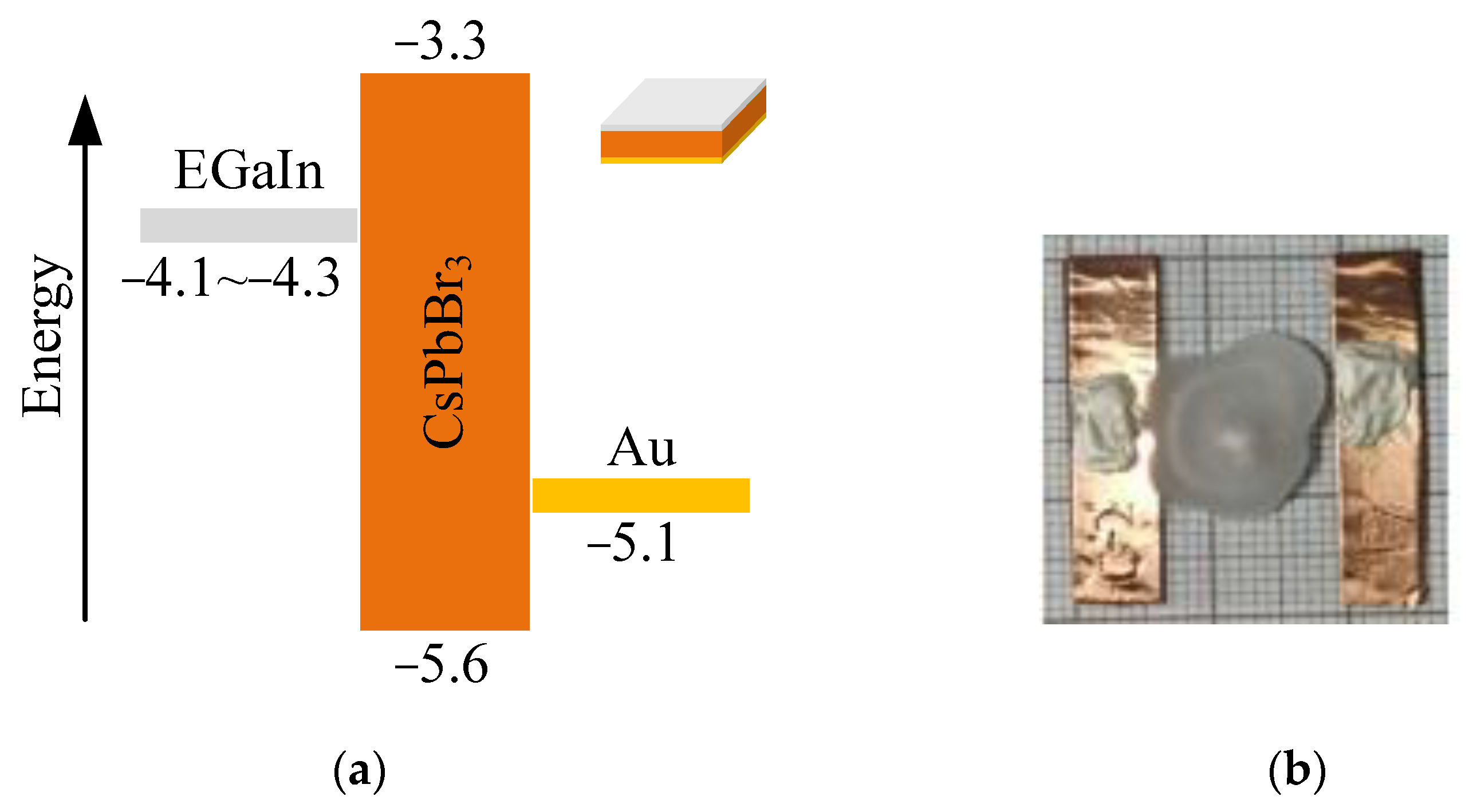
Figure 1. Architectural properties for CsPbBr3 detectors (a) Energy band diagrams for CsPbBr3 detector using asymmetrical electrode materials. (b) Top-view photograph of the CsPbBr3 detectors. As indicated in the insert, the dimension of the crystal is 3 × 3 mm2, where the thickness is 1 mm.
Sensors 23 02017 g001
Figure 2. Schematic diagram of the γ-spectra measurement system.
Sensors 23 02017 g002
Figure 3. Typical dark I–V reverse characteristic curve of the CsPbBr3 detector.
Sensors 23 02017 g003
Figure 4. The J-V characteristics and its tendency fit according to the diffusion model.
Sensors 23 02017 g004
Figure 5. Typical I–T characteristic curve of a Schottky type CsPbBr3 detector after radiation. The light source is a 40 W fluorescent lamp installed in the dark box and switched ON and OFF at 10-s intervals.
Sensors 23 02017 g005
Figure 6. The γ ray spectra by the Schottky type CsPbBr3 detector under a 241Am γ-source with different applied bias voltage before and after radiation. (a) −50 V, (b) −100 V, (c) −150 V. (d) The calculated CCE is based on the spectra indicated in (ac). The curve was smoothed by the Savitzky–Golay method.
Sensors 23 02017 g006
Figure 7. Temperature-dependent PL spectrum excited by a 365 nm laser beam, all normalized to clarify the peak shift.
Sensors 23 02017 g007
Table 1. Total dose response of the CsPbBr3 detectors under different test biases.
Bias Voltage (V)△Energy Resolution
−50−100−150
100 krad(Si)−1.18%6.42%5.57%
After annealing4.77%----
Disclaimer/Publisher’s Note: The statements, opinions and data contained in all publications are solely those of the individual author(s) and contributor(s) and not of MDPI and/or the editor(s). MDPI and/or the editor(s) disclaim responsibility for any injury to people or property resulting from any ideas, methods, instructions or products referred to in the content.