
Multiscale and Hierarchical Feature-Aggregation Network for Segmenting Medical Images


Abstract
:1. Introduction
- ▪
- The proposed model was designed with a multiscale and hierarchical feature-aggregation network to better fuse feature information for the segmentation of medical images.
- ▪
- Guided skip connections from the encoder block to the decoder block are used to improve the segmentation accuracy and the convergence of deep neural networks.
- ▪
- The proposed approach has a good generalization ability according to the results of comparisons with state-of-the-art methods for different challenging tasks involving skin-lesion and tooth segmentation.
2. Related Work
2.1. Multiscale Networks
2.2. Skin-Lesion Segmentation
2.3. Tooth Segmentation
3. Proposed Methodology
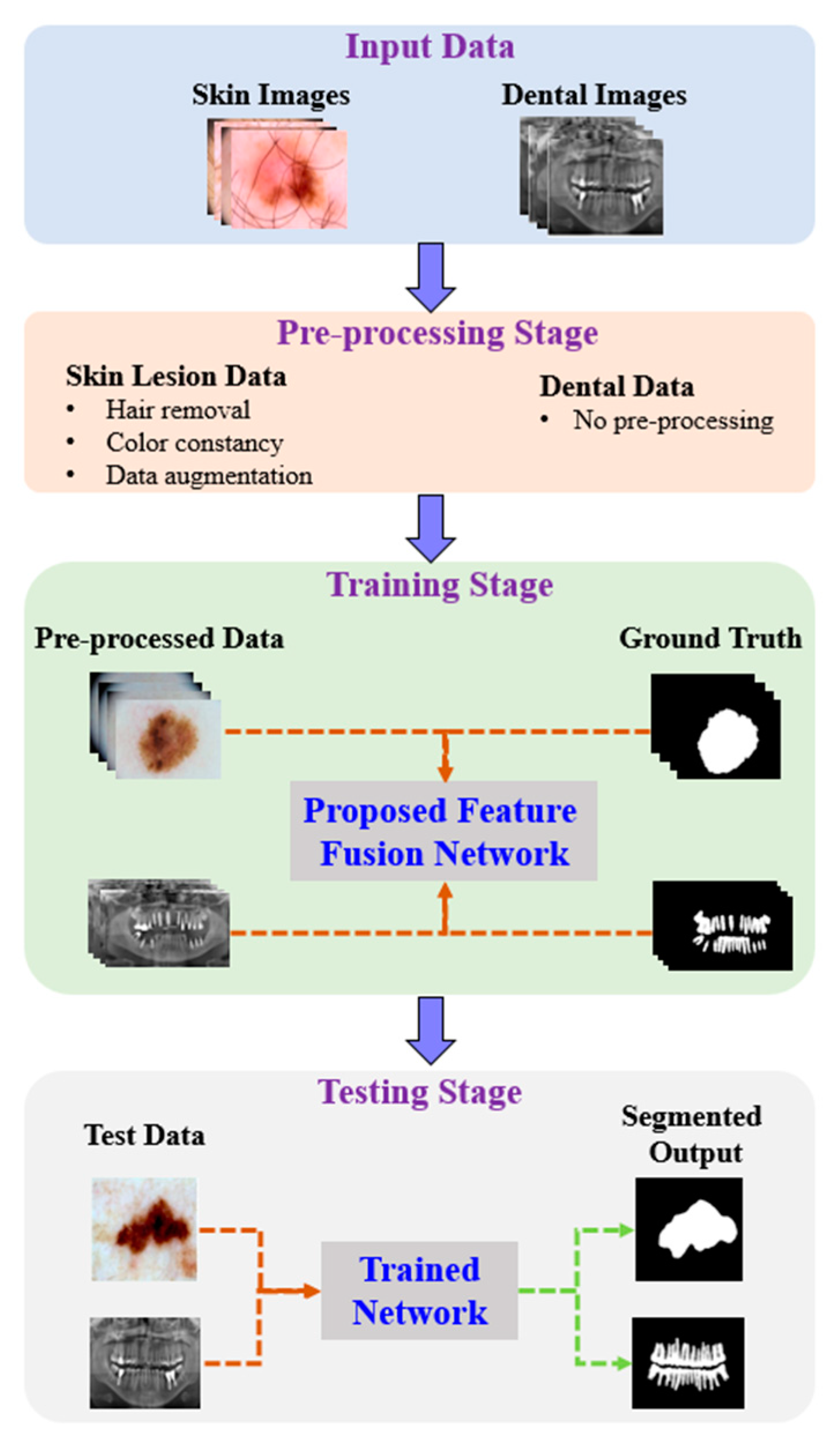
3.1. Overview of Proposed Method
3.2. Proposed Feature-Fusion Architecture
3.2.1. Multiscale Feature Aggregation (MFA)
- Context Encoding Module (CEM)
- Intermediate Module (IM)
- Local Encoding Module (LEM)
3.2.2. Hierarchical Feature Aggregation (HFA)
3.2.3. Encoder and Decoder Blocks
4. Experimental Results
4.1. Datasets
4.1.1. Skin-Lesion Dataset
4.1.2. UFBA-UESC Dental Dataset
4.2. Experimental Settings
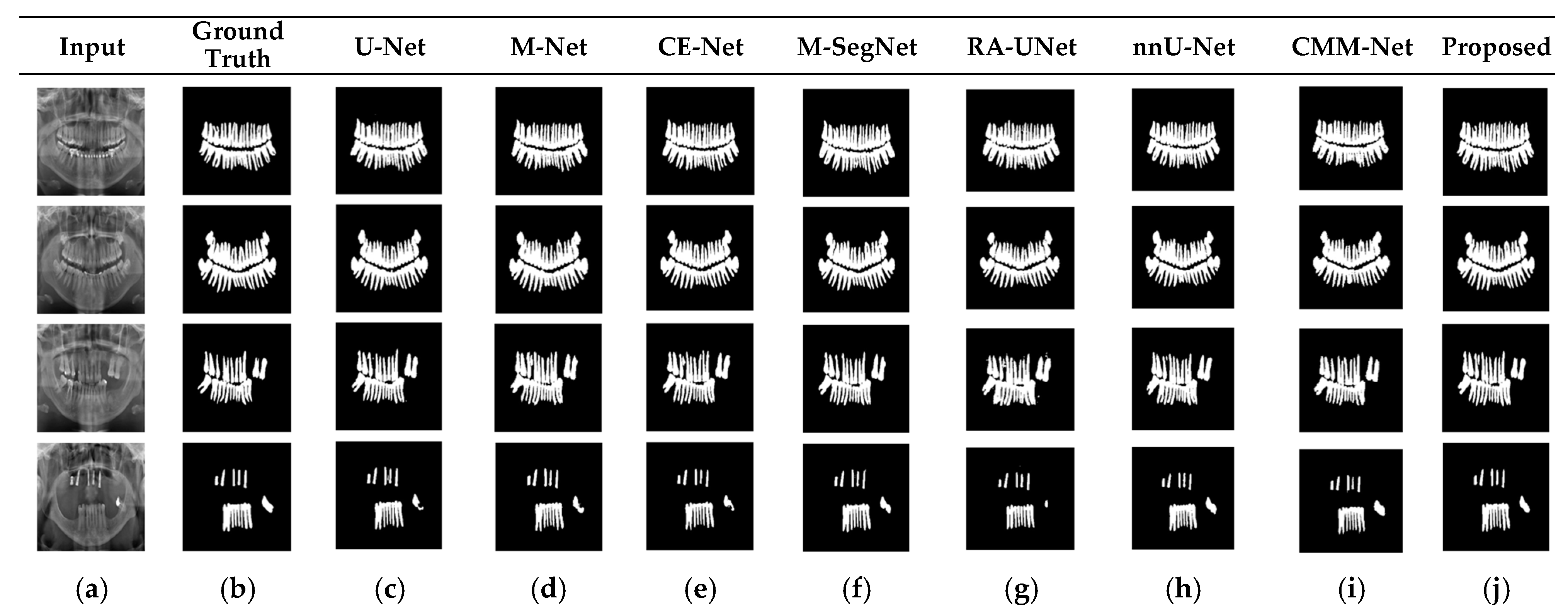
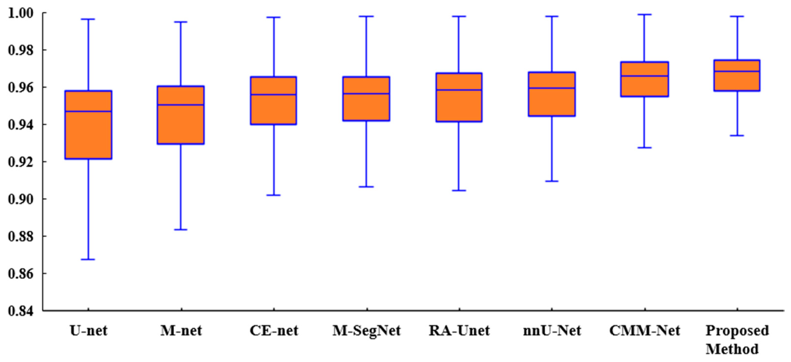
4.3. Results and Discussions
4.4. Ablation Study
5. Conclusions
Author Contributions
Funding
Institutional Review Board Statement
Informed Consent Statement
Data Availability Statement
Conflicts of Interest
References
- Nishitani, Y.; Nakayama, R.; Hayashi, D.; Hizukuri, A.; Murata, K. Segmentation of teeth in panoramic dental X-ray images using U-Net with a loss function weighted on the tooth edge. Radiol. Phys. Technol. 2021, 14, 64–69. [Google Scholar] [CrossRef] [PubMed]
- Lei, B.; Xia, Z.; Jiang, F.; Jiang, X.; Ge, Z.; Xu, Y.; Wang, S. Skin lesion segmentation via generative adversarial networks with dual discriminators. Med. Image Anal. 2020, 64, 101716. [Google Scholar] [CrossRef] [PubMed]
- Gao, J.; Jiang, Q.; Zhou, B.; Chen, D. Convolutional neural networks for computer-aided detection or diagnosis in medical image analysis: An overview. Math. Biosci. Eng. 2019, 16, 6536–6561. [Google Scholar] [CrossRef]
- Wang, G.; Li, W.; Zuluaga, M.A.; Pratt, R.; Patel, P.A.; Aertsen, M.; Doel, T.; David, A.L.; Deprest, J.; Ourselin, S.; et al. Interactive medical image segmentation using deep learning with image-specific fine tuning. IEEE Trans. Med. Imaging 2018, 37, 1562–1573. [Google Scholar] [CrossRef]
- Wu, H.; Pan, J.; Li, Z.; Wen, Z.; Qin, J. Automated Skin Lesion Segmentation Via an Adaptive Dual Attention Module. IEEE Trans. Med. Imaging 2021, 40, 357–370. [Google Scholar] [CrossRef]
- Wirtz, A.; Mirashi, S.G.; Wesarg, S. Automatic Teeth Segmentation in Panoramic X-ray Images Using a Coupled Shape Model in Combination with a Neural Network. In Proceedings of the International Conference on Medical Image Computing and Computer-Assisted Intervention, Granada, Spain, 16–20 September 2018. [Google Scholar]
- Hu, J.; Shen, L.; Sun, G. Squeeze-and-excitation networks. In Proceedings of the 2018 IEEE CVF Conference on Computer Vision and Pattern Recognition, Salt Lake City, UT, USA, 18–23 June 2018; pp. 7132–7141. [Google Scholar]
- Chen, L.-C.; Papandreou, G.; Kokkinos, I.; Murphy, K.; Yuille, A.L. DeepLab: Semantic image segmentation with deep convolutional nets, atrous convolution, and fully connected CRFs. IEEE Trans. Pattern Anal. Mach. Intell. 2018, 40, 834–848. [Google Scholar] [CrossRef] [Green Version]
- Yu, F.; Wang, D.; Shelhamer, E.; Darrell, T. Deep Layer Aggregation. In Proceedings of the 2018 IEEE/CVF Conference on Computer Vision and Pattern Recognition (CVPR), Salt Lake City, UT, USA, 18–23 June 2018; pp. 2403–2412. [Google Scholar]
- Ronneberger, O.; Fischer, P.; Brox, T. U-net: Convolutional networks for biomedical image segmentation. In Proceedings of the Medical Image Computing and Computer Assisted Intervention—MICCAI 2015, Munich, Germany, 5–9 October 2015; Springer: Berlin/Heidelberg, Germany, 2015; pp. 234–241. [Google Scholar]
- Adiga, V.; Sivaswamy, J. FPD-M-net: Fingerprint Image Denoising and Inpainting Using M-net Based Convolutional Neural Networks. In Inpainting and Denoising Challenges; Springer: Berlin/Heidelberg, Germany, 2019. [Google Scholar]
- Zhou, Z.; Rahman Siddiquee, M.M.; Tajbakhsh, N.; Liang, J. UNet++: A Nested U-Net Architecture for Medical Image Segmentation. In Deep Learning in Medical Image Analysis and Multimodal Learning for Clinical Decision Support; Springer: Cham, Switzerland, 2018; pp. 3–11. [Google Scholar]
- Gu, Z.; Cheng, J.; Fu, H.; Zhou, K.; Hao, H.; Zhao, Y.; Liu, J. CE-Net: Context Encoder Network for 2D Medical Image Segmentation. IEEE Trans. Med. Imaging 2019, 38, 2281–2292. [Google Scholar] [CrossRef] [Green Version]
- Yamanakkanavar, N.; Lee, B. A novel M-SegNet with global attention CNN architecture for automatic segmentation of brain MRI. Comput. Biol. Med. 2021, 136, 104761. [Google Scholar] [CrossRef]
- Jin, Q.; Meng, Z.; Sun, C.; Cui, H.; Su, R. RA-UNet: A Hybrid Deep Attention-Aware Network to Extract Liver and Tumor in CT Scans. Front. Bioeng. Biotechnol. 2020, 8, 605132. [Google Scholar] [CrossRef]
- Al-masni, M.A.; Kim, D.H. CMM-Net: Contextual multi-scale multi-level network for efficient biomedical image segmentation. Sci. Rep. 2021, 11, 10191. [Google Scholar] [CrossRef]
- Isensee, F.; Jaeger, F.; Simon, A.; Petersen, J.; Klaus, M.H. nnU-Net: A self-configuring method for deep learning-based biomedical image segmentation. Nat. Methods 2021, 18, 203–211. [Google Scholar] [CrossRef]
- Zhang, J.; Jin, Y.; Xu, J.; Xu, X.; Zhang, Y. MDU-Net: Multi-scale Densely Connected U-Net for biomedical image segmentation. arXiv 2018, arXiv:1812.00352. [Google Scholar]
- Chen, L.C.; Yang, Y.; Wang, J.; Xu, W.; Yuille, A.L. Attention to scale: Scale-aware semantic image segmentation. In Proceedings of the Computer Vision and Pattern Recognition, Las Vegas, NV, USA, 27–30 June 2016; pp. 3640–3649. [Google Scholar]
- Yamanakkanavar, N.; Lee, B. Using a Patch-Wise M-Net Convolutional Neural Network for Tissue Segmentation in Brain MRI Images. IEEE Access 2020, 8, 120946–120958. [Google Scholar] [CrossRef]
- Chen, L.; Papandreou, G.; Schroff, F.; Adam, H. Rethinking Atrous Convolution for Semantic Image Segmentation. arXiv 2017, arXiv:1706.05587. [Google Scholar]
- Yu, F.; Koltun, V. Multi-scale context aggregation by dilated convolutions. arXiv 2015, arXiv:1511.07122. [Google Scholar]
- Dai, J.; Qi, H.; Xiong, Y.; Li, Y.; Zhang, G.; Hu, H.; Wei, Y. Deformable convolutional networks. In Proceedings of the IEEE International Conference on Computer Vision (ICCV), Venice, Italy, 22–29 October 2017; pp. 764–773. [Google Scholar]
- Srivastava, A.; Jha, D.; Chanda, S.; Pal, U.; Johansen, H.D.; Johansen, D.; Riegler, M.; Ali, S.; Halvorsen, P. MSRF-Net: A Multi-Scale Residual Fusion Network for Biomedical Image Segmentation. IEEE J. Biomed. Health Inform. 2021. [Google Scholar] [CrossRef]
- Wang, J.; Sun, K.; Cheng, T.; Jiang, B.; Deng, C.; Zhao, Y.; Liu, D.; Mu, Y.; Tan, M.; Wang, X.; et al. Deep high-resolution representation learning for visual recognition. IEEE Trans. Pattern Anal. Mach. Intell. 2020, 43, 3349–3364. [Google Scholar] [CrossRef] [Green Version]
- Bi, L.; Kim, J.; Ahn, E.; Kumar, A.; Fulham, M.; Feng, D. Dermoscopic image segmentation via multi-stage fully convolutional networks. IEEE Trans. Biomed. Eng. 2017, 64, 2065–2074. [Google Scholar] [CrossRef] [Green Version]
- Yuan, Y.; Chao, M.; Lo, Y.-C. Automatic skin lesion segmentation using deep fully convolutional networks with Jaccard distance. IEEE Trans. Med. Imaging 2017, 36, 1876–1886. [Google Scholar] [CrossRef] [PubMed]
- Goyal, M.; Yap, M.H. Multi-class Semantic Segmentation of Skin Lesions via Fully Convolutional Networks. Bioinformatics. arXiv 2020, arXiv:1711.10449. [Google Scholar]
- Vesal, S.; Patil, S.M.; Ravikumar, N.; Maier, A.K. A multi-task framework for skin lesion detection and segmentation. In OR 2.0 ContextAware Operating Theaters, Computer Assisted Robotic Endoscopy, Clinical Image-Based Procedures, and Skin Image Analysis; Springer: Berlin/Heidelberg, Germany, 2018; pp. 285–293. [Google Scholar]
- Al-Masni, M.A.; Al-Antari, M.A.; Choi, M.-T.; Han, S.-M.; Kim, T.-S. Skin lesion segmentation in dermoscopy images via deep full resolution convolutional networks. Comput. Methods Programs Biomed. 2018, 162, 221–231. [Google Scholar] [CrossRef] [PubMed]
- Amer, Y.Y.; Aqel, M.J. An efficient segmentation algorithm for panoramic dental images. Procedia Comput. Sci. 2015, 65, 718–725. [Google Scholar] [CrossRef] [Green Version]
- Alsmadi, M.K. A hybrid fuzzy C-means and neutrosophic for jaw lesions segmentation. Ain Shams Eng. J. 2018, 9, 697–706. [Google Scholar] [CrossRef] [Green Version]
- Koch, T.L.; Perslev, M.; Igel, C.; Brandt, S.S. Accurate segmentation of dental panoramic radiographs with U-NETS. In Proceedings of the 2019 IEEE 16th International Symposium on Biomedical Imaging (ISBI 2019), Venice, Italy, 8–11 April 2019; pp. 15–19. [Google Scholar]
- Jader, G.; Fontineli, J.; Ruiz, M.; Abdalla, K.; Pithon, M.; Oliveira, L. Deep instance segmentation of teeth in panoramic X-ray images. In Proceedings of the 2018 31st SIBGRAPI Conference on Graphics, Patterns and Images (SIBGRAPI), Paraná, Brazil, 29 October–1 November 2018; IEEE: Piscataway, NJ, USA, 2018; pp. 400–407. [Google Scholar]
- Tuzoff, D.; Tuzova, L.; Bornstein, M.; Krasnov, A.; Kharchenko, M.; Nikolenko, S.; Sveshnikov, M.; Bednenko, G. Tooth detection and numbering in panoramic radiographs using convolutional neural networks. Dentomaxillofacial Radiol. 2019, 48, 20180051. [Google Scholar] [CrossRef]
- Szegedy, C.; Liu, W.; Jia, Y.; Sermanet, P.; Reed, S.; Anguelov, D.; Erhan, D.; Vanhoucke, V.; Rabinovich, A. Going deeper with convolutions. In Proceedings of the 2015 IEEE Conference on Computer Vision and Pattern Recognition (CVPR), Boston, MA, USA, 7–12 June 2015; pp. 1–9. [Google Scholar]
- Codella, N.; Rotemberg, V.; Tschandl, P.; Celebi, M.E.; Dusza, S.; Gutman, D.; Helba, B.; Kalloo, A.; Liopyris, K.; Marchetti, M.; et al. Skin lesion analysis toward melanoma detection: A challenge at 2018. In Proceedings of the IEEE International Symposium on Biomedical Imaging (ISBI), Washington, DC, USA, 4–7 April 2018; pp. 168–172. [Google Scholar]
- Mendonca, T.; Celebi, M.; Mendonca, T.; Marques, J. PH2: A public database for the analysis of dermoscopic images. In Dermoscopy Image Analysis; CRC Press: Boca Raton, FL, USA, 2015. [Google Scholar]
- Silva, G.; Oliveira, L.; Pithon, M. Automatic segmenting teeth in X-ray images: Trends, a novel data set, benchmarking and future perspectives. Expert Syst. Appl. 2018, 107, 15–31. [Google Scholar] [CrossRef] [Green Version]
- Bibiloni, P.; González-Hidalgo, M.; Massanet, S. Skin Hair Removal in Dermoscopic Images Using Soft Color Morphology; Lecture Notes in Computer Science; Springer: Cham, Switzerland, 2017; Volume 10259. [Google Scholar]
- Barata, C.; Celebi, M.E.; Marques, J.S. Improving Dermoscopy Image Classification Using Color Constancy. IEEE J. Biomed. Health Inform. 2015, 19, 1146–1152. [Google Scholar]
- Serra, J. Image Analysis and Mathematical Morphology; Academic Press: Cambridge, MA, USA, 1982; Volume 1. [Google Scholar]


| Models | PH2 [38] | Processing Time | ISIC-2018 [37] | Processing Time | UFBA-UESC [39] | Processing Time | # of Parameters | ||||||
|---|---|---|---|---|---|---|---|---|---|---|---|---|---|
| ACC | DSC | JI | ACC | DSC | JI | ACC | DSC | JI | |||||
| U-Net [10] | 0.92 | 0.89 | 0.80 | 9.1 min | 0.90 | 0.84 | 0.72 | 109.2 min | 0.93 | 0.91 | 0.83 | 64.2 min | 1,946,881 |
| M-Net [11] | 0.93 | 0.90 | 0.82 | 10.4 min | 0.91 | 0.85 | 0.74 | 128.5 min | 0.94 | 0.92 | 0.85 | 79.3 min | 2,337,505 |
| CE-Net [13] | 0.94 | 0.91 | 0.84 | 36.2 min | 0.93 | 0.86 | 0.75 | 188.9 min | 0.95 | 0.92 | 0.85 | 168.9 min | 7,356,929 |
| M-SegNet [14] | 0.96 | 0.93 | 0.87 | 19.2 min | 0.94 | 0.86 | 0.75 | 159.5 min | 0.96 | 0.93 | 0.87 | 123.6 min | 5,468,932 |
| RA-UNet [15] | 0.94 | 0.90 | 0.82 | 12.6 min | 0.93 | 0.86 | 0.75 | 137.8 min | 0.95 | 0.90 | 0.83 | 84.2 min | 2,935,505 |
| nnU-Net [17] | 0.95 | 0.91 | 0.84 | 91.9 min | 0.94 | 0.87 | 0.77 | 767.5 min | 0.95 | 0.91 | 0.84 | 591.3 min | 28,285,984 |
| CMM-Net [16] | 0.96 | 0.94 | 0.87 | 45.3 min | 0.95 | 0.88 | 0.79 | 309.6 min | 0.96 | 0.92 | 0.85 | 268.4 min | 10,252,673 |
| Proposed | 0.97 | 0.95 | 0.90 | 17.8 min | 0.95 | 0.89 | 0.80 | 127.1 min | 0.97 | 0.94 | 0.89 | 108.5 min | 3,165,825 |
| Model | ACC | DSC | JI | Basic | HFA | MFA with Residual | Guided Block | # of Parameters |
|---|---|---|---|---|---|---|---|---|
| Variation 1 | 0.926 | 0.893 | 0.809 | ✓ | × | × | × | 1,946,881 |
| Variation 2 | 0.947 | 0.923 | 0.865 | × | ✓ | × | × | 2,045,089 |
| Variation 3 | 0.932 | 0.903 | 0.828 | ✓ | × | ✓ | × | 1,209,345 |
| Variation 4 | 0.956 | 0.935 | 0.885 | ✓ | × | × | ✓ | 2,119,809 |
| Proposed | 0.972 | 0.951 | 0.903 | ✓ | ✓ | ✓ | ✓ | 3,165,825 |
| Metric | U-Net vs. Proposed | M-Net vs. Proposed | CE-Net vs. Proposed | M-SegNet vs. Proposed | RA-UNet vs. Proposed | nnU-Net vs. Proposed | CMM-Net vs. Proposed |
|---|---|---|---|---|---|---|---|
| DSC | 0.010 | 0.015 | 0.024 | 0.031 | 0.033 | 0.035 | 0.038 |
Publisher’s Note: MDPI stays neutral with regard to jurisdictional claims in published maps and institutional affiliations. |
© 2022 by the authors. Licensee MDPI, Basel, Switzerland. This article is an open access article distributed under the terms and conditions of the Creative Commons Attribution (CC BY) license (https://creativecommons.org/licenses/by/4.0/).
Share and Cite
Yamanakkanavar, N.; Choi, J.Y.; Lee, B. Multiscale and Hierarchical Feature-Aggregation Network for Segmenting Medical Images. Sensors 2022, 22, 3440. https://doi.org/10.3390/s22093440
Yamanakkanavar N, Choi JY, Lee B. Multiscale and Hierarchical Feature-Aggregation Network for Segmenting Medical Images. Sensors. 2022; 22(9):3440. https://doi.org/10.3390/s22093440
Chicago/Turabian StyleYamanakkanavar, Nagaraj, Jae Young Choi, and Bumshik Lee. 2022. "Multiscale and Hierarchical Feature-Aggregation Network for Segmenting Medical Images" Sensors 22, no. 9: 3440. https://doi.org/10.3390/s22093440
APA StyleYamanakkanavar, N., Choi, J. Y., & Lee, B. (2022). Multiscale and Hierarchical Feature-Aggregation Network for Segmenting Medical Images. Sensors, 22(9), 3440. https://doi.org/10.3390/s22093440