
State of the Art in Smart Portable, Wearable, Ingestible and Implantable Devices for Health Status Monitoring and Disease Management


Abstract
:1. Introduction
2. Wearable or Attachable Devices
2.1. Wearable Skin Patches
2.1.1. Monitoring of Body Fluids
2.1.2. Monitoring Body Temperature
2.2. Contact Lens
2.3. Other Wearable Devices
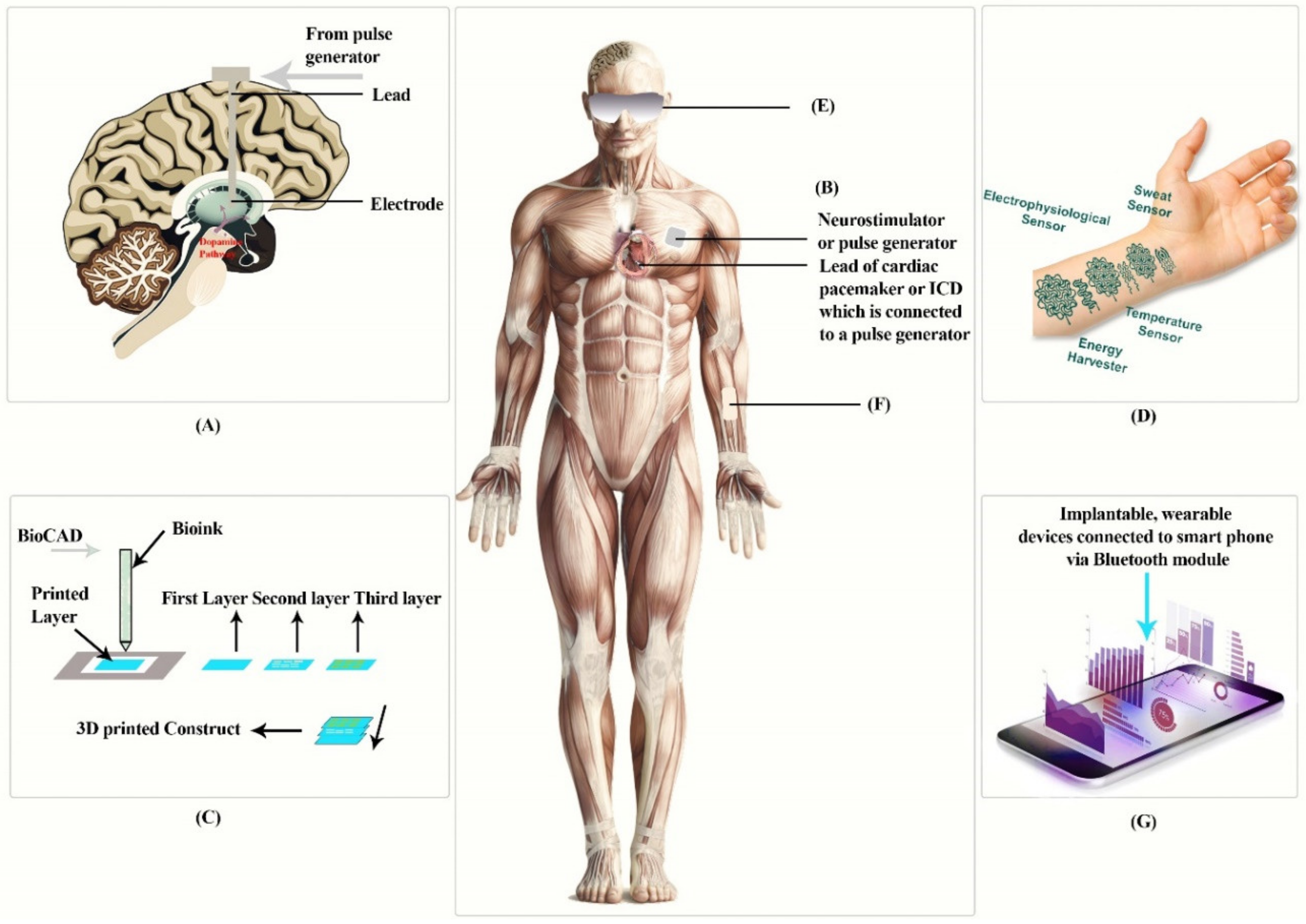
3. Implantable Devices
3.1. Implantable Cardioverter Defibrillators
3.2. Bioinks and 3D Print Implants
3.3. Deep Brain Stimulation
3.4. Other Implantable Devices
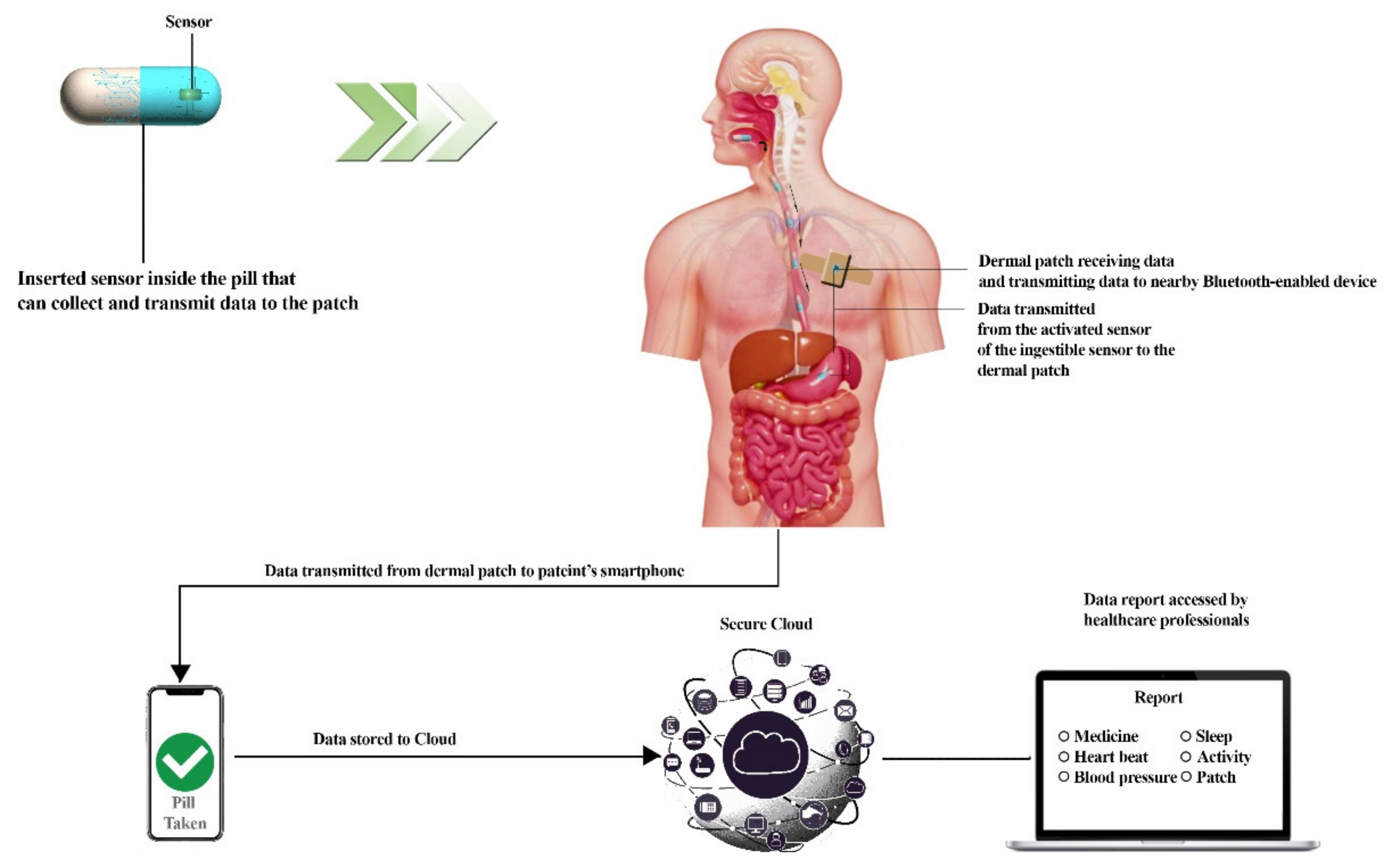
4. Ingestible Pills
4.1. Imaging Capsules
4.2. Temperature-Sensing Capsule
4.3. pH Monitoring and Pressure-Sensing Capsule
4.4. Multifunctional Advanced Capsule
4.5. Gas-Sensing Capsules
4.6. Ultrasound Imaging
4.7. Electro-Chemical Sensing
5. Portable Devices
5.1. Portable Devices for Health Monitoring
5.1.1. Wrist-Mounted Devices
5.1.2. Head-Mounted Devices
5.1.3. E-Textiles or Smart Clothes
5.1.4. Other Portable Devices for Health Monitoring
5.2. Portable Device for Detecting Non-Infectious Diseases
5.2.1. Biosensors and Ovarian Cancer
5.2.2. E-Nose and Colorectal Cancer
5.3. Portable Devices for Detecting Infectious Diseases
5.3.1. COVID-19
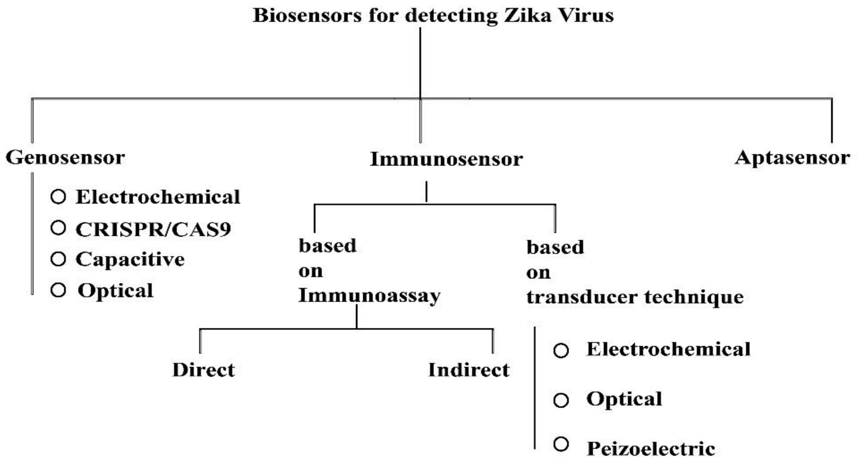
Zika Virus
5.3.2. AIDS
5.3.3. Detection of Human Cytomegalovirus
5.3.4. Tuberculosis and Other Disease-Causing Pathogens
6. Further Developments for the Future in Nanoscale
7. Summary and Conclusions
Funding
Institutional Review Board Statement
Informed Consent Statement
Data Availability Statement
Conflicts of Interest
References
- Guk, K.; Han, G.; Lim, J.; Jeong, K.; Kang, T.; Lim, E.-K.; Jung, J. Evolution of Wearable Devices with Real-Time Disease Monitoring for Personalized Healthcare. Nanomaterials 2019, 9, 813. [Google Scholar] [CrossRef] [PubMed] [Green Version]
- Srivastava, S.K.; van Rijn, C.J.M.; Jongsma, M.A. Biosensor-based detection of tuberculosis. RSC Adv. 2016, 6, 17759–17771. [Google Scholar] [CrossRef] [Green Version]
- Kamišalić, A.; Fister, I.; Turkanović, M.; Karakatiĉ, S. Sensors and Functionalities of Non-Invasive Wrist-Wearable Devices: A Review. Sensors 2018, 18, 1714. [Google Scholar] [CrossRef] [PubMed] [Green Version]
- Wang, B.; Dong, F.; Li, Q.-T.; Yang, D.; Sun, C.; Chen, J.; Song, Z.; Xu, L.; Chu, W.; Xiao, Y.-F.; et al. Visible-Frequency Dielectric Metasurfaces for Multiwavelength Achromatic and Highly Dispersive Holograms. Nano Lett. 2016, 16, 5235–5240. [Google Scholar] [CrossRef]
- Khan, Y.; Ostfeld, A.E.; Lochner, C.M.; Pierre, A.; Arias, A.C. Monitoring of Vital Signs with Flexible and Wearable Medical Devices. Adv. Mater. 2016, 28, 4373–4395. [Google Scholar] [CrossRef]
- Bandodkar, A.J.; Wang, J. Non-invasive wearable electrochemical sensors: A review. Trends Biotechnol. 2014, 32, 363–371. [Google Scholar] [CrossRef]
- Free, C.; Phillips, G.; Galli, L.; Watson, L.; Felix, L.; Edwards, P.; Patel, V.; Haines, A. The Effectiveness of Mobile-Health Technology-Based Health Behaviour Change or Disease Management Interventions for Health Care Consumers: A Systematic Review. PLoS Med. 2013, 10, e1001362. [Google Scholar] [CrossRef] [Green Version]
- Zhu, X.; Liu, W.; Shuang, S.; Nair, M.; Li, C.-Z. Intelligent tattoos, patches, and other wearable biosensors. In Medical Biosensors for Point of Care (POC) Applications; Narayan, R.J., Ed.; Woodhead Publishing: Sawston, UK, 2016; pp. 133–150. [Google Scholar] [CrossRef]
- Walsh, E.P.; Cecchin, F. Recent advances in pacemaker and implantable defibrillator therapy for young patients. Curr. Opin. Cardiol. 2004, 19, 91–96. [Google Scholar] [CrossRef]
- Ryu, S. Book Review: mHealth: New Horizons for Health through Mobile Technologies: Based on the Findings of the Second Global Survey on eHealth (Global Observatory for eHealth Series, Volume 3). Healthc. Informatics Res. 2012, 18, 231–233. [Google Scholar] [CrossRef]
- Haddow, G.; Harmon, S.H.E.; Gilman, L. Implantable Smart Technologies (IST): Defining the ‘Sting’ in Data and Device. Healthc. Care Anal. 2016, 24, 210–227. [Google Scholar] [CrossRef] [Green Version]
- Tian, S.; Yang, W.; le Grange, J.M.; Wang, P.; Huang, W.; Ye, Z. Smart healthcare: Making medical care more intelligent. Glob. Heal. J. 2019, 3, 62–65. [Google Scholar] [CrossRef]
- Lu, L.; Zhang, J.; Xie, Y.; Gao, F.; Xu, S.; Wu, X.; Ye, Z. Wearable Health Devices in Health Care: Narrative Systematic Review. JMIR mHealth uHealth 2020, 8, e18907. [Google Scholar] [CrossRef]
- Kalantar-Zadeh, K.; Ha, N.; Ou, J.Z.; Berean, K.J. Ingestible Sensors. ACS Sens. 2017, 2, 468–483. [Google Scholar] [CrossRef] [PubMed] [Green Version]
- Mandsberg, N.K.; Christfort, J.F.; Kamguyan, K.; Boisen, A.; Srivastava, S.K. Orally ingestible medical devices for gut engineering. Adv. Drug Deliv. Rev. 2020, 165–166, 142–154. [Google Scholar] [CrossRef]
- Lee, W.G.; Kim, Y.-G.; Chung, B.G.; Demirci, U.; Khademhosseini, A. Nano/Microfluidics for diagnosis of infectious diseases in developing countries. Adv. Drug Deliv. Rev. 2010, 62, 449–457. [Google Scholar] [CrossRef] [Green Version]
- Tyagi, H.; Daulton, E.; Bannaga, A.S.; Arasaradnam, R.P.; Covington, J.A. Non-Invasive Detection and Staging of Colorectal Cancer Using a Portable Electronic Nose. Sensors 2021, 21, 5440. [Google Scholar] [CrossRef] [PubMed]
- Michael Okun, B.S.; Zeilman, P.R. A Practical Guide for Patients and Families, 2nd ed.; National Parkinson Foundation: Miami, FL, USA, 2014; Available online: https://books.google.co.in/books?id=ldqAoAEACAAJ (accessed on 25 May 2022).
- Magjarević, R.; Ferek-Petrić, B. Implantable Cardiac Pacemakers—50 Years from the First Implantation. Slov. Med. J. 2010, 79, 55–67. Available online: https://vestnik.szd.si/index.php/ZdravVest/article/view/224 (accessed on 25 May 2022).
- Toledano, M.; Toledano-Osorio, M.; Carrasco-Carmona, Á.; Vallecillo, C.; Toledano, R.; Medina-Castillo, A.L.; Osorio, R. State of the art on biomaterials for soft tissue augmentation in the oral cavity. Part II: Synthetic polymers-based biomaterials. Polymers 2020, 12, 1845. [Google Scholar] [CrossRef]
- Adler, S.N. The history of time for capsule endoscopy. Ann. Transl. Med. 2017, 5, 194. [Google Scholar] [CrossRef] [Green Version]
- Dahad, S.O.; Khadse, A. Pill camera. Int. J. Adv. Res. Comput. Commun. Eng. 2016, 5, 329–338. [Google Scholar] [CrossRef]
- Nakamura, T.; Terano, A. Capsule endoscopy: Past, present, and future. J. Gastroenterol. 2008, 43, 93–99. [Google Scholar] [CrossRef]
- Neumann, H.; Fry, L.C.; Neurath, M.F. Review Article on Current Applications and Future Concepts of Capsule Endoscopy. Digestion 2013, 87, 91–99. [Google Scholar] [CrossRef] [Green Version]
- Shamsudhin, N.; Zverev, V.I.; Keller, H.; Pane, S.; Egolf, P.W.; Nelson, B.J.; Tishin, A.M. Magnetically guided capsule endoscopy. Med. Phys. 2017, 44, e91–e111. [Google Scholar] [CrossRef] [Green Version]
- Basar, M.R.; Malek, F.; Juni, K.M.; Idris, M.S.; Saleh, M.I.M. Ingestible Wireless Capsule Technology: A Review of Development and Future Indication. Int. J. Antennas Propag. 2012, 2012, 1–14. [Google Scholar] [CrossRef]
- Mc Caffrey, C.; Chevalerias, O.; O’Mathuna, C.; Twomey, K. Swallowable-Capsule Technology. IEEE Pervasive Comput. 2008, 7, 23–29. [Google Scholar] [CrossRef]
- Mc Caffrey, C.; Twomey, K.; Ogurtsov, V. Development of a wireless swallowable capsule with potentiostatic electrochemical sensor for gastrointestinal track investigation. Sensors Actuators B Chem. 2015, 218, 8–15. [Google Scholar] [CrossRef]
- Abramson, A.; Dellal, D.; Kong, Y.L.; Zhou, J.; Gao, Y.; Collins, J.; Tamang, S.; Wainer, J.; McManus, R.; Hayward, A.; et al. Ingestible transiently anchoring electronics for microstimulation and conductive signaling. Sci. Adv. 2020, 6, eaaz0127. [Google Scholar] [CrossRef]
- Memon, F.; Touma, G.; Wang, J.; Baltsavias, S.; Moini, A.; Chang, C.; Rasmussen, M.F.; Nikoozadeh, A.; Choe, J.W.; Olcott, E.; et al. Capsule ultrasound device: Further developments. In Proceedings of the 2016 IEEE International Ultrasonics Symposium (IUS), Tours, France, 18–21 September 2016; pp. 1–4. [Google Scholar] [CrossRef]
- Memon, F.; Touma, G.; Wang, J.; Baltsavias, S.; Moini, A.; Chang, C.; Rasmussen, M.F.; Nikoozadeh, A.; Choe, J.W.; Arbabian, A.; et al. Capsule ultrasound device. In Proceedings of the 2015 IEEE International Ultrasonics Symposium (IUS), Taipei, Taiwan, 21–24 October 2015; pp. 1–4. [Google Scholar] [CrossRef]
- Hafezi, H.; Robertson, T.L.; Moon, G.D.; Au-Yeung, K.-Y.; Zdeblick, M.J.; Savage, G.M. An Ingestible Sensor for Measuring Medication Adherence. IEEE Trans. Biomed. Eng. 2014, 62, 99–109. [Google Scholar] [CrossRef]
- Kenry; Yeo, J.C.; Lim, C.T. Emerging flexible and wearable physical sensing platforms for healthcare and biomedical applications. Microsyst. Nanoeng. 2016, 2, 16043. [Google Scholar] [CrossRef]
- Liu, Y.; Pharr, M.; Salvatore, G.A. Lab-on-Skin: A Review of Flexible and Stretchable Electronics for Wearable Health Monitoring. ACS Nano 2017, 11, 9614–9635. [Google Scholar] [CrossRef]
- Noh, S.; Yoon, C.; Hyun, E.; Yoon, H.; Chung, T.; Park, K.; Kim, H. Ferroelectret film-based patch-type sensor for continuous blood pressure monitoring. Electron. Lett. 2014, 50, 143–144. [Google Scholar] [CrossRef]
- Luo, N.; Dai, W.; Li, C.; Zhou, Z.; Lu, L.; Poon, C.C.Y.; Chen, S.-C.; Zhang, Y.; Zhao, N. Flexible Piezoresistive Sensor Patch Enabling Ultralow Power Cuffless Blood Pressure Measurement. Adv. Funct. Mater. 2015, 26, 1178–1187. [Google Scholar] [CrossRef]
- Sonner, Z.; Wilder, E.M.; Heikenfeld, J.; Kasting, G.B.; Beyette, F.R.; Swaile, D.; Sherman, F.F.; Joyce, J.L.; Hagen, J.A.; Kelleyloughnane, N.; et al. The microfluidics of the eccrine sweat gland, including biomarker partitioning, transport, and biosensing implications. Biomicrofluidics 2015, 9, 031301. [Google Scholar] [CrossRef] [PubMed] [Green Version]
- Alizadeh, A.; Burns, A.; Lenigk, R.; Gettings, R.; Ashe, J.; Porter, A.; McCaul, M.; Barrett, R.; Diamond, D.; White, P.; et al. A wearable patch for continuous monitoring of sweat electrolytes during exertion. Lab Chip 2018, 18, 2632–2641. [Google Scholar] [CrossRef]
- Paranjape, M.; Garra, J.; Brida, S.; Schneider, T.; White, R.; Currie, J. A PDMS dermal patch for non-intrusive transdermal glucose sensing. Sens. Actuators A Phys. 2003, 104, 195–204. [Google Scholar] [CrossRef]
- Arora, N.; Martins, D.; Ruggerio, D.; Tousimis, E.; Swistel, A.J.; Osborne, M.P.; Simmons, R.M. Effectiveness of a noninvasive digital infrared thermal imaging system in the detection of breast cancer. Am. J. Surg. 2008, 196, 523–526. [Google Scholar] [CrossRef]
- Kennedy, D.A.; Lee, T.; Seely, D. A Comparative Review of Thermography as a Breast Cancer Screening Technique. Integr. Cancer Ther. 2009, 8, 9–16. [Google Scholar] [CrossRef]
- Yoon, S.; Sim, J.K.; Cho, Y.-H. A Flexible and Wearable Human Stress Monitoring Patch. Sci. Rep. 2016, 6, 23468. [Google Scholar] [CrossRef] [Green Version]
- Trung, T.Q.; Ramasundaram, S.; Hwang, B.-U.; Lee, N.-E. An All-Elastomeric Transparent and Stretchable Temperature Sensor for Body-Attachable Wearable Electronics. Adv. Mater. 2016, 28, 502–509. [Google Scholar] [CrossRef]
- Alexeev, V.L.; Das, S.; Finegold, D.N.; Asher, S.A. Photonic Crystal Glucose-Sensing Material for Noninvasive Monitoring of Glucose in Tear Fluid. Clin. Chem. 2004, 50, 2353–2360. [Google Scholar] [CrossRef] [Green Version]
- March, W.; Lazzaro, D.; Rastogi, S. Fluorescent Measurement in the Non-Invasive Contact Lens Glucose Sensor. Diabetes Technol. Ther. 2006, 8, 312–317. [Google Scholar] [CrossRef]
- Thomas, N.; Lähdesmäki, I.; Parviz, B.A. A contact lens with an integrated lactate sensor. Sens. Actuators B Chem. 2012, 162, 128–134. [Google Scholar] [CrossRef]
- Kim, J.; Imani, S.; de Araujo, W.R.; Warchall, J.; Valdés-Ramírez, G.; Paixão, T.R.L.C.; Mercier, P.P.; Wang, J. Wearable salivary uric acid mouthguard biosensor with integrated wireless electronics. Biosens. Bioelectron. 2015, 74, 1061–1068. [Google Scholar] [CrossRef] [PubMed] [Green Version]
- Mitsubayashi, K.; Arakawa, T. Cavitas Sensors: Contact Lens Type Sensors & Mouthguard Sensors. Electroanalysis 2016, 28, 1170–1187. [Google Scholar] [CrossRef]
- Tseng, P.; Napier, B.; Garbarini, L.; Kaplan, D.L.; Omenetto, F.G. Functional, RF-Trilayer Sensors for Tooth-Mounted, Wireless Monitoring of the Oral Cavity and Food Consumption. Adv. Mater. 2018, 30, 1703257. [Google Scholar] [CrossRef] [PubMed]
- Kim, J.; Campbell, A.S.; de Ávila, B.E.-F.; Wang, J. Wearable biosensors for healthcare monitoring. Nat. Biotechnol. 2019, 37, 389–406. [Google Scholar] [CrossRef]
- Kassanos, P.; Gil Rosa, B.; Keshavarz, M.; Yang, G.-Z. From wearables to implantables—Clinical drive and technical challenges. In Wearable Sensors; Sazonov, E., Ed.; Academic Press: Cambridge, MA, USA, 2020; pp. 29–84. [Google Scholar] [CrossRef]
- Silverman, B.G.; Gross, T.P.; Kaczmarek, R.G.; Hamilton, P.; Hamburger, S. The epidemiology of pacemaker implantation in the United States. Public Health Rep. 1995, 110, 42–46. [Google Scholar]
- Vega, K.; Jiang, N.; Liu, X.; Kan, V.; Barry, N.; Maes, P.; Yetisen, A.; Paradiso, J. The dermal abyss: Interfacing with the skin by tattooing biosensors. In Proceedings of the ISWC ’17: Proceedings of the 2017 ACM International Symposium on Wearable Computers, Maui, Hawaii, 11–15 September 2017; pp. 138–145. [Google Scholar] [CrossRef]
- Liu, X.; Yuk, H.; Lin, S.; Parada, G.A.; Tang, T.-C.; Tham, E.; de la Fuente-Nunez, C.; Lu, T.K.; Zhao, X. 3D Printing of Living Responsive Materials and Devices. Adv. Mater. 2018, 30, 1704821. [Google Scholar] [CrossRef]
- Krauss, J.K.; Lipsman, N.; Aziz, T.; Boutet, A.; Brown, P.; Chang, J.W.; Davidson, B.; Grill, W.M.; Hariz, M.I.; Horn, A.; et al. Technology of deep brain stimulation: Current status and future directions. Nat. Rev. Neurol. 2020, 17, 75–87. [Google Scholar] [CrossRef]
- Jia, W.; Bandodkar, A.J.; Valdés-Ramírez, G.; Windmiller, J.R.; Yang, Z.; Ramírez, J.; Chan, G.; Wang, J. Electrochemical Tattoo Biosensors for Real-Time Noninvasive Lactate Monitoring in Human Perspiration. Anal. Chem. 2013, 85, 6553–6560. [Google Scholar] [CrossRef]
- Guinovart, T.; Bandodkar, A.J.; Windmiller, J.R.; Andrade, F.J.; Wang, J. A potentiometric tattoo sensor for monitoring ammonium in sweat. Analyst 2013, 138, 7031–7038. [Google Scholar] [CrossRef] [PubMed]
- Bandodkar, A.J.; Jia, W.; Yardımcı, C.; Wang, X.; Ramirez, J.; Wang, J. Tattoo-Based Noninvasive Glucose Monitoring: A Proof-of-Concept Study. Anal. Chem. 2015, 87, 394–398. [Google Scholar] [CrossRef] [PubMed]
- Kalantar-Zadeh, K.; Berean, K.J.; Ha, N.; Chrimes, A.F.; Xu, K.; Grando, D.; Ou, J.Z.; Pillai, N.; Campbell, J.L.; Brkljača, R.; et al. A human pilot trial of ingestible electronic capsules capable of sensing different gases in the gut. Nat. Electron. 2017, 1, 79–87. [Google Scholar] [CrossRef]
- Baltsavias, S.; van Treuren, W.; Weber, M.J.; Charthad, J.; Baker, S.; Sonnenburg, J.L.; Arbabian, A. In Vivo Wireless Sensors for Gut Microbiome Redox Monitoring. IEEE Trans. Biomed. Eng. 2020, 67, 1821–1830. [Google Scholar] [CrossRef] [Green Version]
- Belknap, R.; Weis, S.; Brookens, A.; Au-Yeung, K.Y.; Moon, G.; Dicarlo, L.; Reves, R. Feasibility of an Ingestible Sensor-Based System for Monitoring Adherence to Tuberculosis Therapy. PLoS ONE 2013, 8, e53373. [Google Scholar] [CrossRef] [Green Version]
- Frias, J.; Virdi, N.; Raja, P.; Kim, Y.; Savage, G.; Osterberg, L. Effectiveness of Digital Medicines to Improve Clinical Outcomes in Patients with Uncontrolled Hypertension and Type 2 Diabetes: Prospective, Open-Label, Cluster-Randomized Pilot Clinical Trial. J. Med. Internet Res. 2017, 19, e7833. [Google Scholar] [CrossRef]
- Ciuti, G.; Valdastri, P.; Menciassi, A.; Dario, P. Robotic magnetic steering and locomotion of capsule endoscope for diagnostic and surgical endoluminal procedures. Robotica 2010, 28, 199–207. [Google Scholar] [CrossRef] [Green Version]
- Adler, S.N.; Metzger, Y.; Misrachi, C.; Klar, R. Validation of a New Pillcam™ SB2 Video Capsule Versus the Standard Pillcam™ SB for Detection of Small Bowel Disease. Gastrointest. Endosc. 2007, 65, AB165. [Google Scholar] [CrossRef]
- Rajbhandary, P.; Nallathambi, G. Validity and reliability of oral temperature compared to ingestible core temperature pill in free-living conditions. arXiv 2021, arXiv:2108.00537. [Google Scholar]
- McKenzie, J.; Osgood, D. Validation of a new telemetric core temperature monitor. J. Therm. Biol. 2004, 29, 605–611. [Google Scholar] [CrossRef]
- Cutchis, P.N.; Hogrefe, A.F.; Lesho, J.C. The Ingestible Thermal Monitoring System. Johns Hopkins APL Tech. Dig. 1988, 9, 16–22. Available online: https://www.jhuapl.edu/content/techdigest/pdf/V09-N01/09-01 (accessed on 25 May 2022).
- Kwiatek, M.; Pandolfino, J. The Bravo™ pH capsule system. Dig. Liver Dis. 2008, 40, 156–160. [Google Scholar] [CrossRef] [PubMed]
- Kuo, B.; Maneerattanaporn, M.; Lee, A.; Baker, J.R.; Wiener, S.M.; Chey, W.D.; Wilding, G.E.; Hasler, W.L. Generalized Transit Delay on Wireless Motility Capsule Testing in Patients with Clinical Suspicion of Gastroparesis, Small Intestinal Dysmotility, or Slow Transit Constipation. Am. J. Dig. Dis. Sci. 2011, 56, 2928–2938. [Google Scholar] [CrossRef] [PubMed]
- Tran, K.; Brun, R.; Kuo, B. Evaluation of regional and whole gut motility using the wireless motility capsule: Relevance in clinical practice. Ther. Adv. Gastroenterol. 2012, 5, 249–260. [Google Scholar] [CrossRef] [PubMed] [Green Version]
- Ciuti, G.; Menciassi, A.; Dario, P. Capsule Endoscopy: From Current Achievements to Open Challenges. IEEE Rev. Biomed. Eng. 2011, 4, 59–72. [Google Scholar] [CrossRef]
- Karargyris, A.; Koulaouzidis, A.; Koulaouzidis, A. OdoCapsule: Next-Generation Wireless Capsule Endoscopy with Accurate Lesion Localization and Video Stabilization Capabilities. IEEE Trans. Biomed. Eng. 2014, 62, 352–360. [Google Scholar] [CrossRef]
- Ou, J.Z.; Yao, C.; Rotbart, A.; Muir, J.G.; Gibson, P.R.; Kalantar-Zadeh, K. Human intestinal gas measurement systems: In vitro fermentation and gas capsules. Trends Biotechnol. 2015, 33, 208–213. [Google Scholar] [CrossRef]
- Kalantar-Zadeh, K.; Yao, C.K.; Berean, K.J.; Ha, N.; Ou, J.Z.; Ward, S.A.; Pillai, N.; Hill, J.; Cottrell, J.J.; Dunshea, F.R.; et al. Intestinal Gas Capsules: A Proof-of-Concept Demonstration. Gastroenterology 2016, 150, 37–39. [Google Scholar] [CrossRef] [Green Version]
- Lay, H.S.; Qiu, Y.; Al-Rawhani, M.; Beeley, J.; Poltarjonoks, R.; Seetohul, V.; Cumming, D.; Cochran, S.; Cummins, G.; Desmulliez, M.P.Y.; et al. Progress towards a multi-modal capsule endoscopy device featuring microultrasound imaging. In Proceedings of the 2016 IEEE International Ultrasonics Symposium (IUS), Tours, France, 18–21 September 2016; pp. 1–4. [Google Scholar] [CrossRef] [Green Version]
- Wang, J.; Memon, F.; Touma, G.; Baltsavias, S.; Jang, J.H.; Chang, C.; Rasmussen, M.F.; Olcott, E.; Jeffrey, R.B.; Arbabian, A.; et al. Capsule ultrasound device: Characterization and testing results. In Proceedings of the 2017 IEEE International Ultrasonics Symposium (IUS), Washington, DC, USA, 6–9 September 2017; pp. 1–4. [Google Scholar] [CrossRef]
- Lindner, J.R. Microbubbles in medical imaging: Current applications and future directions. Nat. Rev. Drug Discov. 2004, 3, 527–533. [Google Scholar] [CrossRef]
- Capece, S.; Domenici, F.; Brasili, F.; Oddo, L.; Cerroni, B.; Bedini, A.; Bordi, F.; Chiessi, E.; Paradossi, G. Complex interfaces in “phase-change” contrast agents. Phys. Chem. Chem. Phys. 2016, 18, 8378–8388. [Google Scholar] [CrossRef]
- Palmieri, D.; Brasili, F.; Capocefalo, A.; Bizien, T.; Angelini, I.; Oddo, L.; Toumia, Y.; Paradossi, G.; Domenici, F. Improved hybrid-shelled perfluorocarbon microdroplets as ultrasound- and laser-activated phase-change platform. Colloids Surf. A Physicochem. Eng. Asp. 2022, 641, 128522. [Google Scholar] [CrossRef]
- Seneviratne, S.; Hu, Y.; Nguyen, T.; Lan, G.; Khalifa, S.; Thilakarathna, K.; Hassan, M.; Seneviratne, A. A Survey of Wearable Devices and Challenges. IEEE Commun. Surv. Tutor. 2017, 19, 2573–2620. [Google Scholar] [CrossRef]
- Lee, S.-S.; Son, I.-H.; Choi, J.-G.; Nam, N.-H.; Hong, Y.-S.; Lee, W.-B. Estimated Blood Pressure Algorithm for a Wrist-wearable Pulsimeter Using Hall Device. J. Korean Phys. Soc. 2011, 58, 349–352. [Google Scholar] [CrossRef]
- Hsu, Y.-P.; Young, D.J. Skin-surface-coupled personal health monitoring system. In Proceedings of the IEEE 2013 International Conference on Sensors, Baltimore, MD, USA, 3–6 November 2013; pp. 1–4. [Google Scholar] [CrossRef]
- Ishikawa, T.; Hyodo, Y.; Miyashita, K.; Yoshifuji, K.; Komoriya, Y.; Imai, Y. Wearable motion tolerant PPG sensor for instant heart rate in daily activity. In Proceedings of the 10th International Joint Conference on Biomedical Engineering Systems and Technologies (BIOSTEC 2017), Porto, Portugal, 21–23 February 2017; pp. 126–133. [Google Scholar] [CrossRef] [Green Version]
- Vashist, S.K. Non-invasive glucose monitoring technology in diabetes management: A review. Anal. Chim. Acta 2012, 750, 16–27. [Google Scholar] [CrossRef]
- López-Blanco, R.; Velasco, M.A.; Méndez-Guerrero, A.; Romero, J.P.; del Castillo, M.D.; Serrano, J.I.; Rocon, E.; Benito-León, J. Smartwatch for the analysis of rest tremor in patients with Parkinson’s disease. J. Neurol. Sci. 2019, 401, 37–42. [Google Scholar] [CrossRef] [PubMed]
- Tison, G.; Sanchez, J.M.; Ballinger, B.; Singh, A.; Olgin, J.E.; Pletcher, M.J.; Vittinghoff, E.; Lee, E.S.; Fan, S.M.; Gladstone, R.A.; et al. Passive Detection of Atrial Fibrillation Using a Commercially Available Smartwatch. JAMA Cardiol. 2018, 3, 409–416. [Google Scholar] [CrossRef] [PubMed] [Green Version]
- Constant, N.; Douglas-Prawl, O.; Johnson, S.; Mankodiya, K. Pulse-Glasses: An unobtrusive, wearable HR monitor with Internet-of-Things functionality. In Proceedings of the 2015 IEEE 12th International Conference on Wearable and Implantable Body Sensor Networks (BSN), Cambridge, MA, USA, 9–12 June 2015; pp. 1–5. [Google Scholar] [CrossRef]
- Sempionatto, J.R.; Nakagawa, T.; Pavinatto, A.; Mensah, S.T.; Imani, S.; Mercier, P.; Wang, J. Eyeglasses based wireless electrolyte and metabolite sensor platform. Lab Chip 2017, 17, 1834–1842. [Google Scholar] [CrossRef] [PubMed]
- Arakawa, T.; Kuroki, Y.; Nitta, H.; Chouhan, P.; Toma, K.; Sawada, S.-I.; Takeuchi, S.; Sekita, T.; Akiyoshi, K.; Minakuchi, S.; et al. Mouthguard biosensor with telemetry system for monitoring of saliva glucose: A novel cavitas sensor. Biosens. Bioelectron. 2016, 84, 106–111. [Google Scholar] [CrossRef] [Green Version]
- Kim, J.; Valdés-Ramírez, G.; Bandodkar, A.J.; Jia, W.; Martinez, A.G.; Ramírez, J.; Mercier, P.; Wang, J. Non-invasive mouthguard biosensor for continuous salivary monitoring of metabolites. Analyst 2014, 139, 1632–1636. [Google Scholar] [CrossRef]
- Yetisen, A.K.; Martinez-Hurtado, J.L.; Ünal, B.; Khademhosseini, A.; Butt, H. Wearables in Medicine. Adv. Mater. 2018, 30, e1706910. [Google Scholar] [CrossRef]
- Liu, X.; Lillehoj, P.B. Embroidered electrochemical sensors for biomolecular detection. Lab Chip 2016, 16, 2093–2098. [Google Scholar] [CrossRef]
- Pacelli, M.; Loriga, G.; Taccini, N.; Paradiso, R. Sensing fabrics for monitoring physiological and biomechanical variables: E-textile solutions. In Proceedings of the 3rd IEEE-EMBS International Summer School and Symposium on Medical Devices and Biosensors, MIT, Boston, MA, USA, 4–6 September 2006; pp. 1–4. [Google Scholar] [CrossRef]
- Villar, R.; Beltrame, T.B.; Hughson, R.L. Validation of the Hexoskin wearable vest during lying, sitting, standing, and walking activities. Appl. Physiol. Nutr. Metab. 2015, 40, 1019–1024. [Google Scholar] [CrossRef] [PubMed]
- Jung, P.-G.; Lim, G.; Kong, K. A mobile motion capture system based on inertial sensors and smart shoes. In Proceedings of the 2013 IEEE International Conference on Robotics and Automation, Karlsruhe, Germany, 6–10 May 2013; pp. 692–697. [Google Scholar] [CrossRef]
- Lee, S.I.; Park, E.; Huang, A.; Mortazavi, B.; Garst, J.H.; Jahanforouz, N.; Espinal, M.; Siero, T.; Pollack, S.; Afridi, M.; et al. Objectively quantifying walking ability in degenerative spinal disorder patients using sensor equipped smart shoes. Med. Eng. Phys. 2016, 38, 442–449. [Google Scholar] [CrossRef]
- Mishra, R.K.; Hubble, L.; Martín, A.; Kumar, R.; Barfidokht, A.; Kim, J.; Musameh, M.M.; Kyratzis, I.L.; Wang, J. Wearable Flexible and Stretchable Glove Biosensor for On-Site Detection of Organophosphorus Chemical Threats. ACS Sens. 2017, 2, 553–561. [Google Scholar] [CrossRef]
- Berget, C.; Messer, L.H.; Forlenza, G.P. A Clinical Overview of Insulin Pump Therapy for the Management of Diabetes: Past, Present, and Future of Intensive Therapy. Diabetes Spectr. 2019, 32, 194–204. [Google Scholar] [CrossRef]
- Swarup, S.; Makaryus, A.N. Digital stethoscope: Technology update. Med. Devices EÉvid. Res. 2018, 11, 29–36. [Google Scholar] [CrossRef] [PubMed] [Green Version]
- Soares, C.; Clifton, W.; Freeman, W.D. Use of Handheld Video Otoscopy for the Diagnosis of Acute Otitis Media. Cureus 2019, 11, e5547. [Google Scholar] [CrossRef] [PubMed] [Green Version]
- Remais, J.V.; Zeng, G.; Li, G.; Tian, L.; Engelgau, M.M. Convergence of non-communicable and infectious diseases in low- and middle-income countries. Int. J. Epidemiol. 2013, 42, 221–227. [Google Scholar] [CrossRef]
- Anzar, N.; Hasan, M.R.; Akram, M.; Yadav, N.; Narang, J. Systematic and validated techniques for the detection of ovarian cancer emphasizing the electro-analytical approach. Process Biochem. 2020, 94, 126–135. [Google Scholar] [CrossRef]
- Misra, R.; Acharya, S.; Sahoo, S.K. Cancer nanotechnology: Application of nanotechnology in cancer therapy. Drug Discov. Today 2010, 15, 842–850. [Google Scholar] [CrossRef]
- Trabert, B.; Poole, E.M.; White, E.; Visvanathan, K.; Adami, H.-O.; Anderson, G.L.; Brasky, T.M.; Brinton, L.A.; Fortner, R.; Gaudet, M.; et al. Analgesic Use and Ovarian Cancer Risk: An Analysis in the Ovarian Cancer Cohort Consortium. JNCI J. Natl. Cancer Inst. 2019, 111, 137–145. [Google Scholar] [CrossRef] [PubMed]
- Topkaya, S.N.; Azimzadeh, M.; Ozsoz, M. Electrochemical Biosensors for Cancer Biomarkers Detection: Recent Advances and Challenges. Electroanalysis 2016, 28, 1402–1419. [Google Scholar] [CrossRef]
- Phillips, J.A.; Xu, Y.; Xia, Z.; Fan, Z.H.; Tan, W. Enrichment of Cancer Cells Using Aptamers Immobilized on a Microfluidic Channel. Anal. Chem. 2009, 81, 1033–1039. [Google Scholar] [CrossRef] [PubMed] [Green Version]
- Babacan, S.; Pivarnik, P.; Letcher, S.; Rand, A. Evaluation of antibody immobilization methods for piezoelectric biosensor application. Biosens. Bioelectron. 2000, 15, 615–621. [Google Scholar] [CrossRef]
- Tombelli, S.; Minunni, M.; Mascini, M. Piezoelectric biosensors: Strategies for coupling nucleic acids to piezoelectric devices. Methods 2005, 37, 48–56. [Google Scholar] [CrossRef]
- Armaghany, T.; Wilson, J.D.; Chu, Q.; Mills, G. Genetic Alterations in Colorectal Cancer. Gastrointest. Cancer Res. 2012, 5, 19–27. Available online: http://www.ncbi.nlm.nih.gov/pubmed/22574233 (accessed on 30 March 2022).
- Kumar, S.V.; Damodar, G.; Ravikanth, S. An Overview on Infectious Disease. Indian J. Pharm. Sci. Res. 2012, 2, 63–74. Available online: https://www.researchgate.net/publication/260086676_An_Overview_on_Infectious_Disease (accessed on 25 May 2022).
- Suleman, S.; Shukla, S.K.; Malhotra, N.; Bukkitgar, S.D.; Shetti, N.P.; Pilloton, R.; Narang, J.; Tan, Y.N.; Aminabhavi, T.M. Point of care detection of COVID-19: Advancement in biosensing and diagnostic methods. Chem. Eng. J. 2021, 414, 128759. [Google Scholar] [CrossRef]
- Kuiken, T.; Fouchier, R.A.; Schutten, M.; Rimmelzwaan, G.F.; van Amerongen, G.; van Riel, D.; Laman, J.D.; de Jong, T.; van Doornum, G.; Lim, W.; et al. Newly discovered coronavirus as the primary cause of severe acute respiratory syndrome. Lancet 2003, 362, 263–270. [Google Scholar] [CrossRef] [Green Version]
- Zhang, X.; Qi, Q.; Jing, Q.; Ao, S.; Zhang, Z.; Ding, M.; Wu, M.; Liu, K.; Wang, W.; Ling, Y.; et al. Electrical probing of COVID-19 spike protein receptor binding domain via a graphene field-effect transistor. arXiv 2020, arXiv:2003.12529. Available online: http://arxiv.org/abs/2003.12529 (accessed on 25 May 2022).
- Jayamohan, H.; Lambert, C.J.; Sant, H.J.; Jafek, A.; Patel, D.; Feng, H.; Beeman, M.; Mahmood, T.; Nze, U.; Gale, B.K. SARS-CoV-2 pandemic: A review of molecular diagnostic tools including sample collection and commercial response with associated advantages and limitations. Anal. Bioanal. Chem. 2021, 413, 49–71. [Google Scholar] [CrossRef]
- Qiu, G.; Gai, Z.; Tao, Y.; Schmitt, J.; Kullak-Ublick, G.A.; Wang, J. Dual-Functional Plasmonic Photothermal Biosensors for Highly Accurate Severe Acute Respiratory Syndrome Coronavirus 2 Detection. ACS Nano 2020, 14, 5268–5277. [Google Scholar] [CrossRef] [PubMed] [Green Version]
- Shetti, N.P.; Mishra, A.; Bukkitgar, S.D.; Basu, S.; Narang, J.; Reddy, K.R.; Aminabhavi, T.M. Conventional and Nanotechnology-Based Sensing Methods for SARS Coronavirus (2019-nCoV). ACS Appl. Bio Mater. 2021, 4, 1178–1190. [Google Scholar] [CrossRef] [PubMed]
- Thompson, D.; Lei, Y. Mini review: Recent progress in RT-LAMP enabled COVID-19 detection. Sens. Actuators Rep. 2020, 2, 100017. [Google Scholar] [CrossRef] [PubMed]
- Taleghani, N.; Taghipour, F. Diagnosis of COVID-19 for controlling the pandemic: A review of the state-of-the-art. Biosens. Bioelectron. 2021, 174, 112830. [Google Scholar] [CrossRef]
- Alafeef, M.; Dighe, K.; Moitra, P.; Pan, D. Rapid, ultrasensitive, and quantitative detection of SARS-CoV-2 using antisense oligonucleotides directed electrochemical biosensor chip. ACS Nano 2020, 14, 17028–17045. [Google Scholar] [CrossRef] [PubMed]
- Alam, M.A.; Hasan, M.R.; Anzar, N.; Suleman, S.; Narang, J. Diagnostic approaches for the rapid detection of Zika virus—A review. Process Biochem. 2021, 101, 156–168. [Google Scholar] [CrossRef]
- Yang, M.; McGovern, M.E.; Thompson, M. Genosensor technology and the detection of interfacial nucleic acid chemistry. Anal. Chim. Acta 1997, 346, 259–275. [Google Scholar] [CrossRef]
- Adegoke, O.; Morita, M.; Kato, T.; Ito, M.; Suzuki, T.; Park, E.Y. Localized surface plasmon resonance-mediated fluorescence signals in plasmonic nanoparticle-quantum dot hybrids for ultrasensitive Zika virus RNA detection via hairpin hybridization assays. Biosens. Bioelectron. 2017, 94, 513–522. [Google Scholar] [CrossRef]
- Cheng, C.; Wu, J.; Fikrig, E.; Wang, P.; Chen, J.; Eda, S.; Terry, P. Unamplified RNA Sensor for On-Site Screening of Zika Virus Disease in a Limited Resource Setting. ChemElectroChem 2017, 4, 485–489. [Google Scholar] [CrossRef]
- Faria, F.A.M.; Zucolotto, V. Label-free electrochemical DNA biosensor for zika virus identification. Biosens. Bioelectron. 2019, 131, 149–155. [Google Scholar] [CrossRef] [PubMed]
- Hock, B. Antibodies for immunosensors a review. Anal. Chim. Acta 1997, 347, 177–186. [Google Scholar] [CrossRef]
- Jiang, Q.; Chandar, Y.J.; Cao, S.; Kharasch, E.D.; Singamaneni, S.; Morrissey, J.J. Rapid, Point-of-Care, Paper-Based Plasmonic Biosensor for Zika Virus Diagnosis. Adv. Biosyst. 2017, 1, 1700096. [Google Scholar] [CrossRef] [Green Version]
- Kaushik, A.; Yndart, A.; Kumar, S.; Jayant, R.D.; Vashist, A.; Brown, A.N.; Li, C.-Z.; Nair, M. A sensitive electrochemical immunosensor for label-free detection of Zika-virus protein. Sci. Rep. 2018, 8, 1–5. [Google Scholar] [CrossRef] [PubMed] [Green Version]
- Faria, A.M.; Mazon, T. Early diagnosis of Zika infection using a ZnO nanostructures-based rapid electrochemical biosensor. Talanta 2019, 203, 153–160. [Google Scholar] [CrossRef] [PubMed]
- Takemura, K.; Adegoke, O.; Suzuki, T.; Park, E.Y. A localized surface plasmon resonance-amplified immunofluorescence biosensor for ultrasensitive and rapid detection of nonstructural protein 1 of Zika virus. PLoS ONE 2019, 14, e0211517. [Google Scholar] [CrossRef] [PubMed]
- Ilgu, M.; Nilsen-Hamilton, M. Aptamers in analytics. Analyst 2016, 141, 1551–1568. [Google Scholar] [CrossRef] [Green Version]
- Haase, A.T. Perils at mucosal front lines for HIV and SIV and their hosts. Nat. Rev. Immunol. 2005, 5, 783–792. [Google Scholar] [CrossRef]
- Dalgleish, A.G.; Beverley, P.C.L.; Clapham, P.R.; Crawford, D.H.; Greaves, M.F.; Weiss, R.A. The CD4 (T4) antigen is an essential component of the receptor for the AIDS retrovirus. Nature 1984, 312, 763–767. [Google Scholar] [CrossRef]
- World Health Organization. Patient Monitoring Guidelines for HIV Care and Antiretroviral Therapy; WHO: Geneva, Switzerland, 2004; pp. 1–193.
- Lee, S.H.; Kim, S.-W.; Kang, J.Y.; Ahn, C.H. A polymer lab-on-a-chip for reverse transcription (RT)-PCR based point-of-care clinical diagnostics. Lab Chip 2008, 8, 2121–2127. [Google Scholar] [CrossRef]
- Ozcan, A.; Demirci, U. Ultra wide-field lens-free monitoring of cells on-chip. Lab Chip 2007, 8, 98–106. [Google Scholar] [CrossRef] [PubMed]
- Moon, S.J.; Keles, H.O.; Kim, Y.-G.; Kuritzkes, D.; Demirci, U. Lensless imaging for point-of-care testing. In Proceedings of the 2009 Annual International Conference of the IEEE Engineering in Medicine and Biology Society, Minneapolis, MN, USA, 3–6 September 2009; Volume 2009, pp. 6376–6379. [Google Scholar] [CrossRef] [Green Version]
- Hasan, M.R.; Sharma, P.; Anzar, N.; Pundir, C.; Pilloton, R.; Narang, J.; Shetti, N.P. Analytical methods for detection of human cytomegalovirus clinched biosensor a cutting-edge diagnostic tool. Biomed. Eng. Adv. 2021, 1, 100006. [Google Scholar] [CrossRef]
- Sun, H.; Qi, C.; Niu, Y.; Kang, T.; Wei, Y.; Jin, G.; Dong, X.; Wang, C.; Zhu, W. Detection of Cytomegalovirus Antibodies Using a Biosensor Based on Imaging Ellipsometry. PLoS ONE 2015, 10, e0136253. [Google Scholar] [CrossRef]
- Geitmann, M.; Danielson, U.H. Studies of substrate-induced conformational changes in human cytomegalovirus protease using optical biosensor technology. Anal. Biochem. 2004, 332, 203–214. [Google Scholar] [CrossRef] [PubMed]
- Narang, J.; Singhal, C.; Mathur, A.; Sharma, S.; Singla, V.; Pundir, C.S. Portable bioactive paper based genosensor incorporated with Zn-Ag nanoblooms for herpes detection at the point-of-care. Int. J. Biol. Macromol. 2018, 107, 2559–2565. [Google Scholar] [CrossRef] [PubMed]
- Pai, M.; Behr, M.A.; Dowdy, D.; Dheda, K.; Divangahi, M.; Boehme, C.C.; Ginsberg, A.; Swaminathan, S.; Spigelman, M.; Getahun, H.; et al. Tuberculosis. Nat. Rev. Dis. Prim. 2016, 2, 16076. [Google Scholar] [CrossRef] [PubMed]
- Aly, A.H.; Mohamed, D.; Zaky, Z.A.; Matar, Z.S.; El-Gawaad, N.S.A.; Shalaby, A.S.; Tayeboun, F.; Mohaseb, M. Novel Biosensor Detection of Tuberculosis Based on Photonic Band Gap Materials. Mater. Res. 2021, 24, e20200483. [Google Scholar] [CrossRef]
- Yesudasu, V.; Pradhan, H.S.; Pandya, R.J. Recent progress in surface plasmon resonance based sensors: A comprehensive review. Heliyon 2021, 7, e06321. [Google Scholar] [CrossRef]
- Zeng, G.; Zhang, L.; Dong, H.; Chen, Y.; Zhang, J.; Zhu, Y.; Yuan, Y.; Xie, Y.; Fang, W. Pathway and mechanism of nitrogen transformation during composting: Functional enzymes and genes under different concentrations of PVP-AgNPs. Bioresour. Technol. 2018, 253, 112–120. [Google Scholar] [CrossRef]
- Brainina, K.; Kozitsina, A.; Beikin, J. Electrochemical immunosensor for Forest-Spring encephalitis based on protein A labeled with colloidal gold. Anal. Bioanal. Chem. 2003, 376, 481–485. [Google Scholar] [CrossRef]
- Yuan, R.; Zhang, L.; Li, Q.; Chai, Y.; Cao, S. A label-free amperometric immunosenor based on multi-layer assembly of polymerized o-phenylenediamine and gold nanoparticles for determination of Japanese B encephalitis vaccine. Anal. Chim. Acta 2005, 531, 1–5. [Google Scholar] [CrossRef]
- Purvis, D.; Leonardova, O.; Farmakovsky, D.; Cherkasov, V. An ultrasensitive and stable potentiometric immunosensor. Biosens. Bioelectron. 2003, 18, 1385–1390. [Google Scholar] [CrossRef]
- Lee, W.E.; Thompson, H.G.; Hall, J.G.; Fulton, R.E.; Wong, J.P. Rapid immunofiltration assay of Newcastle disease virus using a silicon sensor. J. Immunol. Methods 1993, 166, 123–131. [Google Scholar] [CrossRef]
- Pejcic, B.; de Marco, R.; Parkinson, G. The role of biosensors in the detection of emerging infectious diseases. Analyst 2006, 131, 1079–1090. [Google Scholar] [CrossRef] [PubMed]
- Tang, D.; Yuan, R.; Chai, Y.; Dai, J.; Zhong, X.; Liu, Y. A novel immunosensor based on immobilization of hepatitis B surface antibody on platinum electrode modified colloidal gold and polyvinyl butyral as matrices via electrochemical impedance spectroscopy. Bioelectrochemistry 2004, 65, 15–22. [Google Scholar] [CrossRef]
- Muhammad-Tahir, Z.; Alocilja, E.C.; Grooms, D.L. Rapid detection of bovine viral diarrhea virus as surrogate of bioterrorism agents. IEEE Sens. J. 2005, 5, 757–762. [Google Scholar] [CrossRef]
- Lee, W.E.; Thompson, H.G. Detection of Newcastle disease virus using an evanescent wave immuno-based biosensor. Can. J. Chem. 1996, 74, 707–712. [Google Scholar] [CrossRef]
- Johari-Ahar, M.; Rashidi, M.R.; Barar, J.; Aghaie, M.; Mohammadnejad, D.; Ramazani, A.; Karami, P.; Coukos, G.; Omidi, Y. An ultra-sensitive impedimetric immunosensor for detection of the serum oncomarker CA-125 in ovarian cancer patients. Nanoscale 2015, 7, 3768–3779. [Google Scholar] [CrossRef]
- Hong, W.; Lee, S.; Cho, Y. Dual-responsive immunosensor that combines colorimetric recognition and electrochemical response for ultrasensitive detection of cancer biomarkers. Biosens. Bioelectron. 2016, 86, 920–926. [Google Scholar] [CrossRef]
- Zhang, Y.; Guo, X.; Fan, L.; Zhang, Q.; Sang, S. A Novel Magnetoelastic Immunosensor for Ultrasensitively Detecting Carcinoembryonic Antigen. Nanoscale Res. Lett. 2018, 13, 258. [Google Scholar] [CrossRef]
- Gedi, V.; Kil Song, C.; Kim, G.B.; Lee, J.O.; Oh, E.; Shin, B.S.; Jung, M.; Shim, J.; Lee, H.; Kim, Y.-P. Sensitive on-chip detection of cancer antigen 125 using a DNA aptamer/carbon nanotube network platform. Sensors Actuators B Chem. 2018, 256, 89–97. [Google Scholar] [CrossRef]
- Seo, G.; Lee, G.; Kim, M.J.; Baek, S.-H.; Choi, M.; Ku, K.B.; Lee, C.-S.; Jun, S.; Park, D.; Kim, H.G.; et al. Rapid Detection of COVID-19 Causative Virus (SARS-CoV-2) in Human Nasopharyngeal Swab Specimens Using Field-Effect Transistor-Based Biosensor. ACS Nano 2020, 14, 5135–5142. [Google Scholar] [CrossRef] [Green Version]
- Chen, Q.; Bian, Z.; Chen, M.; Hua, X.; Yao, C.; Xia, H.; Kuang, H.; Zhang, X.; Huang, J.; Cai, G.; et al. Real-time monitoring of the strand displacement amplification (SDA) of human cytomegalovirus by a new SDA-piezoelectric DNA sensor system. Biosens. Bioelectron. 2009, 24, 3412–3418. [Google Scholar] [CrossRef] [PubMed]
- Fasolato, C.; Giantulli, S.; Capocefalo, A.; Toumia, Y.; Notariello, D.; Mazzarda, F.; Silvestri, I.; Postorino, P.; Domenici, F. Antifolate SERS-active nanovectors: Quantitative drug nanostructuring and selective cell targeting for effective theranostics. Nanoscale 2019, 11, 15224–15233. [Google Scholar] [CrossRef] [PubMed]
- Capocefalo, A.; Mammucari, D.; Brasili, F.; Fasolato, C.; Bordi, F.; Postorino, P.; Domenici, F. Exploring the Potentiality of a SERS-Active pH Nano-Biosensor. Front. Chem. 2019, 7, 413. [Google Scholar] [CrossRef] [Green Version]
- Capocefalo, A.; Bizien, T.; Sennato, S.; Ghofraniha, N.; Bordi, F.; Brasili, F. Responsivity of Fractal Nanoparticle Assemblies to Multiple Stimuli: Structural Insights on the Modulation of the Optical Properties. Nanomaterials 2022, 12, 1529. [Google Scholar] [CrossRef]
- Mehlenbacher, R.D.; Kolbl, R.; Lay, A.; Dionne, J.A. Nanomaterials for in vivo imaging of mechanical forces and electrical fields. Nat. Rev. Mater. 2017, 3, 17080. [Google Scholar] [CrossRef]


| Portable Device/Biosensor | Disease | Causative Agent | Principle | Disease Type/Marker | Limit of Detection | Advantages | Disadvantages | Reference(s) |
|---|---|---|---|---|---|---|---|---|
| Amperometric | New castle disease | Paramyxovirus | Enzyme label immunoassay | Infectious; Antigen of New Castle Disease | 11.1 ng mL−1 | Short time to detect | Solution contamination | [144] |
| Amperometric | Forest spring encephalitis | Tick-borne encephalitis virus | Sandwich gold-labelled immunoassay | Infectious; Antigen and Protein A | 0.0000001 mg mL−1 | Can be detected in a wide concentration range | Unstable substrates may limit use of such sensors | [145] |
| Amperometric | Japanese b encephalitis | Japanese encephalitis virus | Probe and label-free immunoassay | Infectious; Fe2+/3+ probe | 0.000000006 lg pfu mL−1 | Quick plaque formation | - | [146] |
| Geno- and immunosensors | Zika fever | Zika virus | (Genosensor) isothermal amplification of viral RNA via nucleic acid sequence-based amplification. (Immunosensor) IDE (interdigitated electrodes) Gold array | Infectious; Zika RNA Zika Protein | 3 fM 10 pM | Short time to detect Highly specific Low cost | Elapsed time | [120] |
| Potentiometric | Hepatitis B | Hepatitis B virus | Enzyme label immunoassay | Infectious; Enzyme labeled with horse radish peroxidase | 50 fM (approx) | Rapid detection High stability Highly sensitive | Extremely dependent on polymerization Controlled conditions are required | [147] |
| Light Adressable Potentiometric (LAPS) | New castle disease | Paramyxovirus | Sandwich enzyme-label immunoassay; Field effect transistor technology | Infectious; N type silicon doped with phosphorus | 2 ng mL−1 | Rapid detection Highly sensitiveResponds on a wide range | Variation of sensitivity | [148] |
| Light Adressable Potentiometric (LAPS) | Venezuelan equine encephalitis | Venezuelan equine encephalitis virus | Sandwich enzyme-label immunoassay; Field effect transistor technology | Infectious; N type silicon doped with phosphorus, immunofiltration enzyme assay in conjugation with LAPS | 30 ng mL−1 | Rapid detection Highly sensitive Responds on a wide range | - | [149] |
| Impedance Spectroscopy | Hepatitis B | Hepatitis B virus | Immunoassay | Infectious; Before and after antigen–antibody contact, the electron transfer resistance of a redox probe varies. | 8 ng mL−1 | Rapid detection Highly sensitive Responds on a wide range | On absence of Au nanoparticle and PVB, it retained sensitivity of 27.6%. Antigen and serum may interfere to cause inhibition impedance | [150] |
| Conductometric | Bovine viral diarrhea | Pestivirus | Sandwich immunoassay | Infectious; Antigen–antibody interaction using conducting polyalanine label | 100–10,000 CCID mL−1 | Responds on a wide range Reaction occurs in less than a minute | Monoclonal antibodies are significantly more susceptible to epitope loss as a result of chemical treatment | [151] |
| Fiber optic evanescent wave biosensor | New castle disease | Paramyxovirus | Sandwich immunoassay with fluorescein labeling | Infectious; Polyclonal antibody covalently attached to an aminosilane-coated quartz fiber, fluorescein-labeled anti-ND used to identify it | 10 ng mL−1 | Sample analysis can be done along long distances Rapid detection Responds on wide range | - | [152] |
| Electrochemical Immunosensor | Ovarian cancer | -Type- I-genome alterations in KRAS, BRAF, PTEN, PIK3CA, ARID1A. -Type- II- TP53 mutations | Sandwich-based method conjugation of nanoparticles and antigen | Non-infectious; CA-125 tumor marker | 0.0016 U/mL | Highly specific Proteins other than CA-125 showed no interference on high sensitivity or high specificity. | - | [153] |
| Colorimetric Biosensor | Ovarian cancer | -Type- I-genome alterations in KRAS, BRAF, PTEN, PIK3CA, ARID1A. -Type- II- TP53 mutations | Electric field approach; biotin doped polypyrrole immunosensor based on colorimetric methods | Non-infectious; CA-125, PSA, CEA | PSA-0.7 pg/mL CA-125-0.0005 U/mL CEA-0.8 pg/mL | Rapid detection Direct detection | - | [154] |
| Mass-Based Biosensor | Ovarian cancer | -Type- I-genome alterations in KRAS, BRAF, PTEN, PIK3CA, ARID1A. -Type- II- TP53 mutations | Based on gold nano material | Non-infectious; CEA | 2.5 pg/mL | Highly specific Precise analyte binding | - | [155] |
| Optical Biosensor | Ovarian cancer | -Type- I-genome alterations in KRAS, BRAF, PTEN, PIK3CA, ARID1A. -Type- II- TP53 mutations | Biochip-based assay | Non-infectious;CA-125 | - | 10–100 times sensitive Early detection Uses Aptamers which is more stable and versatile than antibodies. | [156] | |
| (Portable Electronic Nose) PEN 3 (E-Nose) | Colorectal cancer | TP53, KRAS, BRAF, and MMR gene Alleles mutation, 18qLOH, CpG methylation | Metal oxide-based detection and analysis through neural network and random forest | Non-infectious; Aromatic, aliphatic, hydrogen, methane, sulfur as substrates for sensing | - | Highly sensitive | Less specificity Cross sensitivity to possible inorganic gases | [17] |
| GC-TOF-MS (Gas Chromatography-Time of Flight-Mass Spectrometry) | Colorectal cancer | TP53, KRAS, BRAF and MMR gene alleles mutation, 18qLOH, CpG methylation | Volatile organic compound-based detection and analysis through neural network and random forest | Non-infectious; Different volatile organic compounds used as markers for quantification | - | Highly sensitive Highly specific | Detects molecules, having more than 3 carbon atoms only | [17] |
| COVID-19 FET Sensor | COVID-19 | SARS-Cov2 virus | Field effect transistor | Infectious; Antibody conjugated graphene | 1 fg/mL | Highly sensitive Rapid and real-time Highly specific | - | [157] |
| RT-LAMP NBS | COVID-19 | SARS-Cov2 virus | Reverse transcription loop mediate isothermal amplification; F1ab and nucleoproteins-based nano biosensor | Infectious; F1 ab and nucleoproteins | 12 copies | 100% sensitivity 100% specificity Takes less time | - | [116] |
| Electrochemical DNA Sensor | HCMV Associated disease | Human cytomegalo virus | Based on an EPAD that includes Zn–Ag nanoblooms | Infectious; HHV-5 DNA | 97 copies per mL | Quick fabrication technique can be used to develop it | Expensive wax printers are required After wax deposition, a further heating step is required | [140] |
| Optical Biosensor | HCMV Associated disease | Human cytomegalo virus | SPR-dependent method | Infectious; Protease and peptidase reaction of HCMV | - | Reusable Label free High sensitivity | Low selectivity Nonspecific binding | [139] |
| Piezoelectric Biosensor | HCMV Associated disease | Human cytomegalo virus | The technique of strand displacement amplification was used | Infectious; HCMV Nucleic acid | - | Takes less time to detect Highly sensitive Real time detection | High temperature sensitivity Inability to amplify long sequences | [158] |
| On Chip Flow cytometry | AIDS | HIV | Flow cytometry | Infectious; CD4+ detection | 10 µL (whole blood required to detect) | Label-free detection Lens-less imaging | Difficult for clinical use unless modified with sheath-less focusing techniques | [16] |
| Chip NMR Biosensor | Tuberculosis | Mycobacterium Tuberculosis | Miniaturized diagnostic magnetic resonance | Infectious; Mycobacterium detection from sputum | 1 ng (approx.) | High Sensitivity and specificity | Micro-coil resistance Signal detecting circuitry is monolithically integrated on a single integrated circuit chip | [134] |
Publisher’s Note: MDPI stays neutral with regard to jurisdictional claims in published maps and institutional affiliations. |
© 2022 by the authors. Licensee MDPI, Basel, Switzerland. This article is an open access article distributed under the terms and conditions of the Creative Commons Attribution (CC BY) license (https://creativecommons.org/licenses/by/4.0/).
Share and Cite
Mukherjee, S.; Suleman, S.; Pilloton, R.; Narang, J.; Rani, K. State of the Art in Smart Portable, Wearable, Ingestible and Implantable Devices for Health Status Monitoring and Disease Management. Sensors 2022, 22, 4228. https://doi.org/10.3390/s22114228
Mukherjee S, Suleman S, Pilloton R, Narang J, Rani K. State of the Art in Smart Portable, Wearable, Ingestible and Implantable Devices for Health Status Monitoring and Disease Management. Sensors. 2022; 22(11):4228. https://doi.org/10.3390/s22114228
Chicago/Turabian StyleMukherjee, Shouvik, Shariq Suleman, Roberto Pilloton, Jagriti Narang, and Kirti Rani. 2022. "State of the Art in Smart Portable, Wearable, Ingestible and Implantable Devices for Health Status Monitoring and Disease Management" Sensors 22, no. 11: 4228. https://doi.org/10.3390/s22114228
APA StyleMukherjee, S., Suleman, S., Pilloton, R., Narang, J., & Rani, K. (2022). State of the Art in Smart Portable, Wearable, Ingestible and Implantable Devices for Health Status Monitoring and Disease Management. Sensors, 22(11), 4228. https://doi.org/10.3390/s22114228