
Automated Detection and Removal of Cardiac and Pulse Interferences from Neonatal EEG Signals


Abstract
1. Introduction
- This is the first method that is able to automatically detect and remove both electrical cardiac and pulsatile interference while preserving the neonatal brain activity.
- It can save time and reduce mistakes related to the expertise of the operator, who detects artefactual segments by visual inspection.
- It employs ICA, which is a familiar tool for clinicians pre-processing EEG recordings.
- It uses information derived from the real ECG—typically recorded simultaneously with the EEG in the NICU—to improve the separation performance of the ICA algorithm.
- Its performance has been statistically assessed in terms of accuracy, sensitivity and false omission rate and in the quality of the reconstructed artefact-free EEG signals.
- It can be used in conjunction with other methods dedicated to the correction of other types of artefacts and prior to analytical methods developed for diagnostic purposes (such as for the detection and classification of seizures).
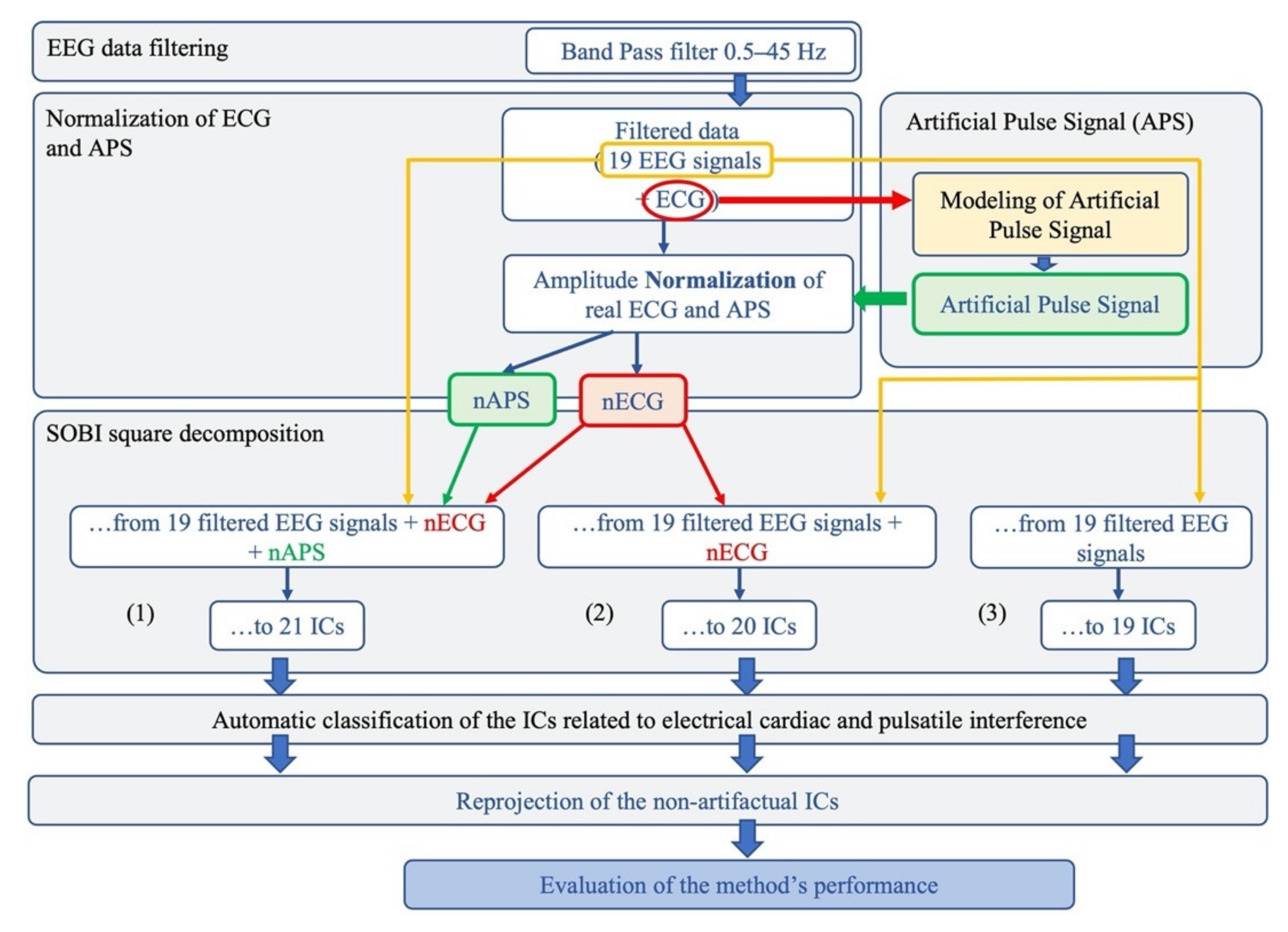
2. Materials and Methods
2.1. EEG Recordings
2.2. EEG Data Analysis
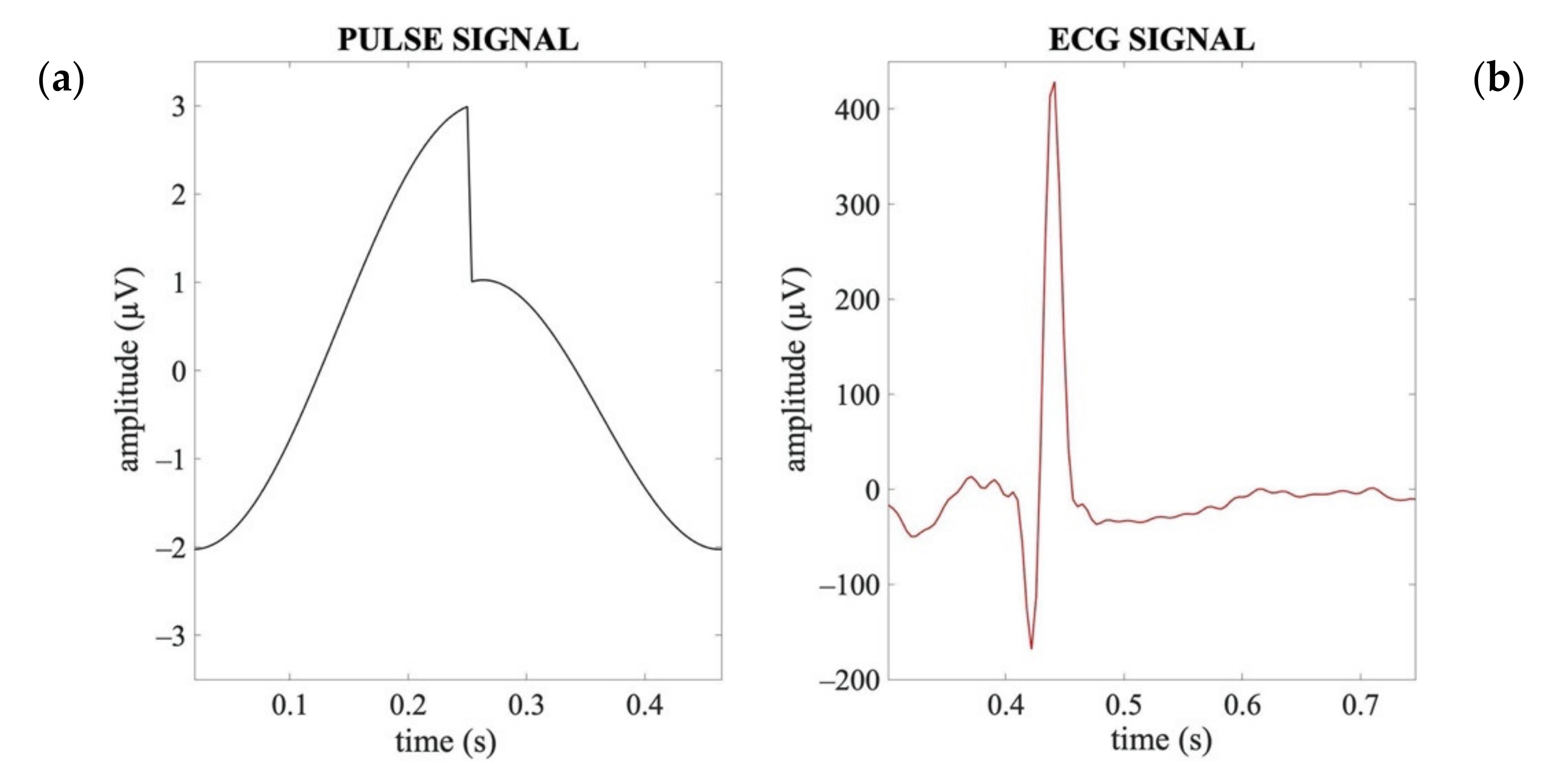
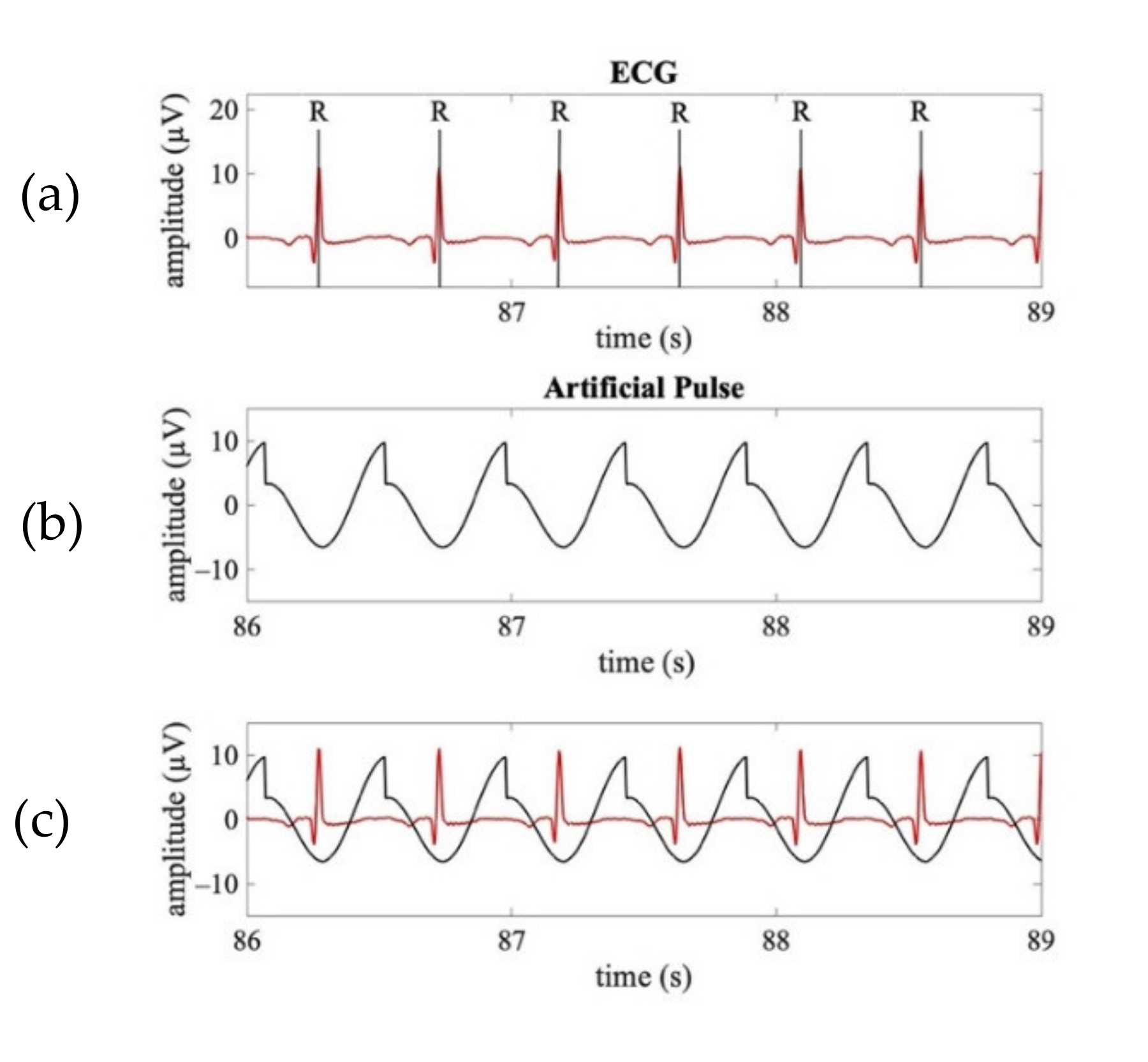
2.2.1. Artificial Pulse Signal
2.2.2. Normalization of the ECG and APS signals
2.2.3. BBS Decomposition
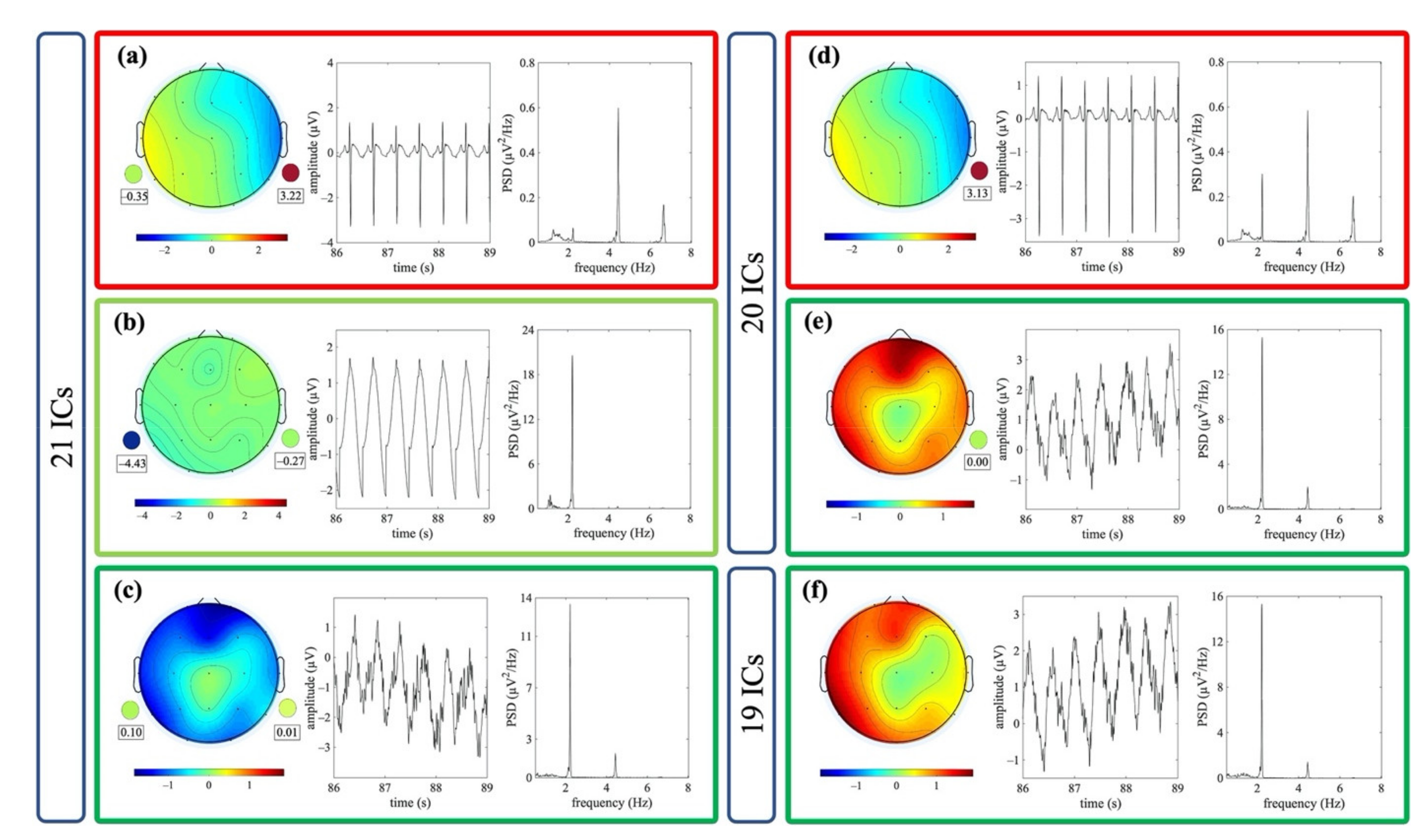
- Group 1 (G1): 21 signals, including the 19 filtered neonatal EEG signals and the normalized APS and ECG signals;
- Group 2 (G2): 20 signals, including the 19 filtered neonatal EEG signals and the normalized ECG signal;
- Group 3 (G3): 19 signals, including only the 19 filtered neonatal EEG signals.
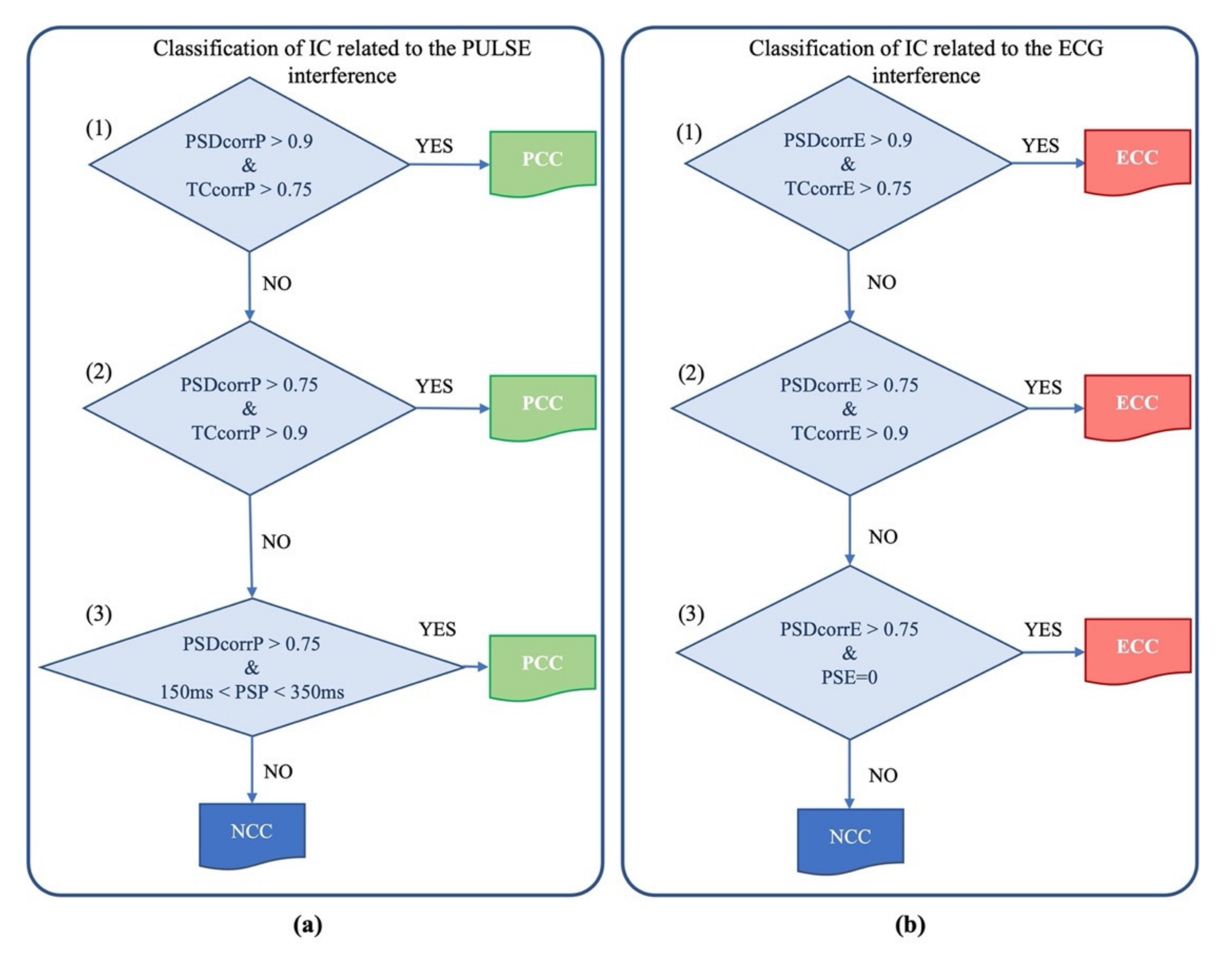
2.2.4. Automated Classification of the ICs Containing Cardiac Electrical and Pulsatile Interferences
- For the cardiac electrical interference: the time delay between the peaks of the IC and the peaks of the nECG signal must be equal to 0;
2.3. Evaluation of the Method’s Effectiveness
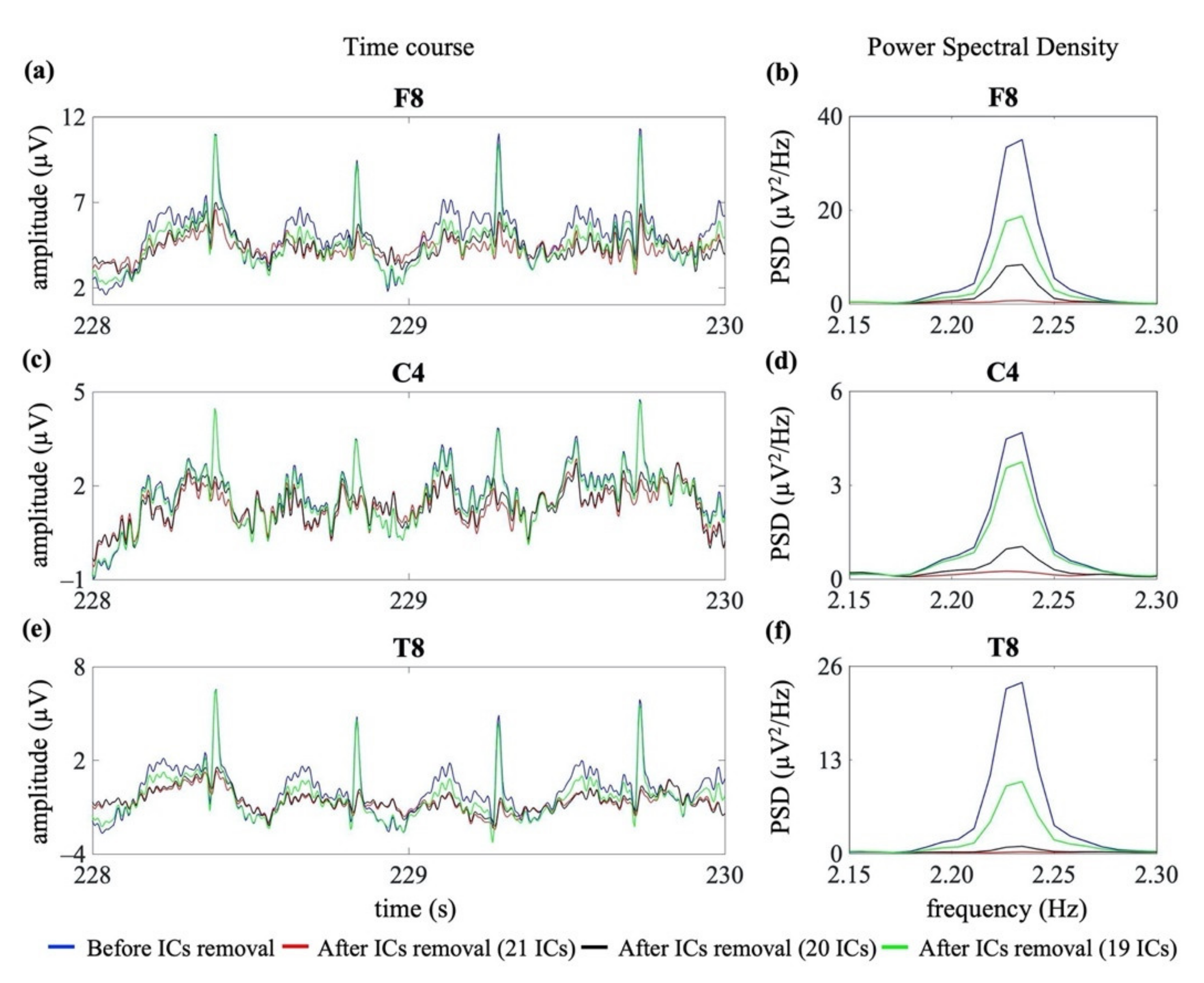
2.3.1. Evaluation of the Influence of Using the nECG and nAPS Signals in the SOBI Decomposition
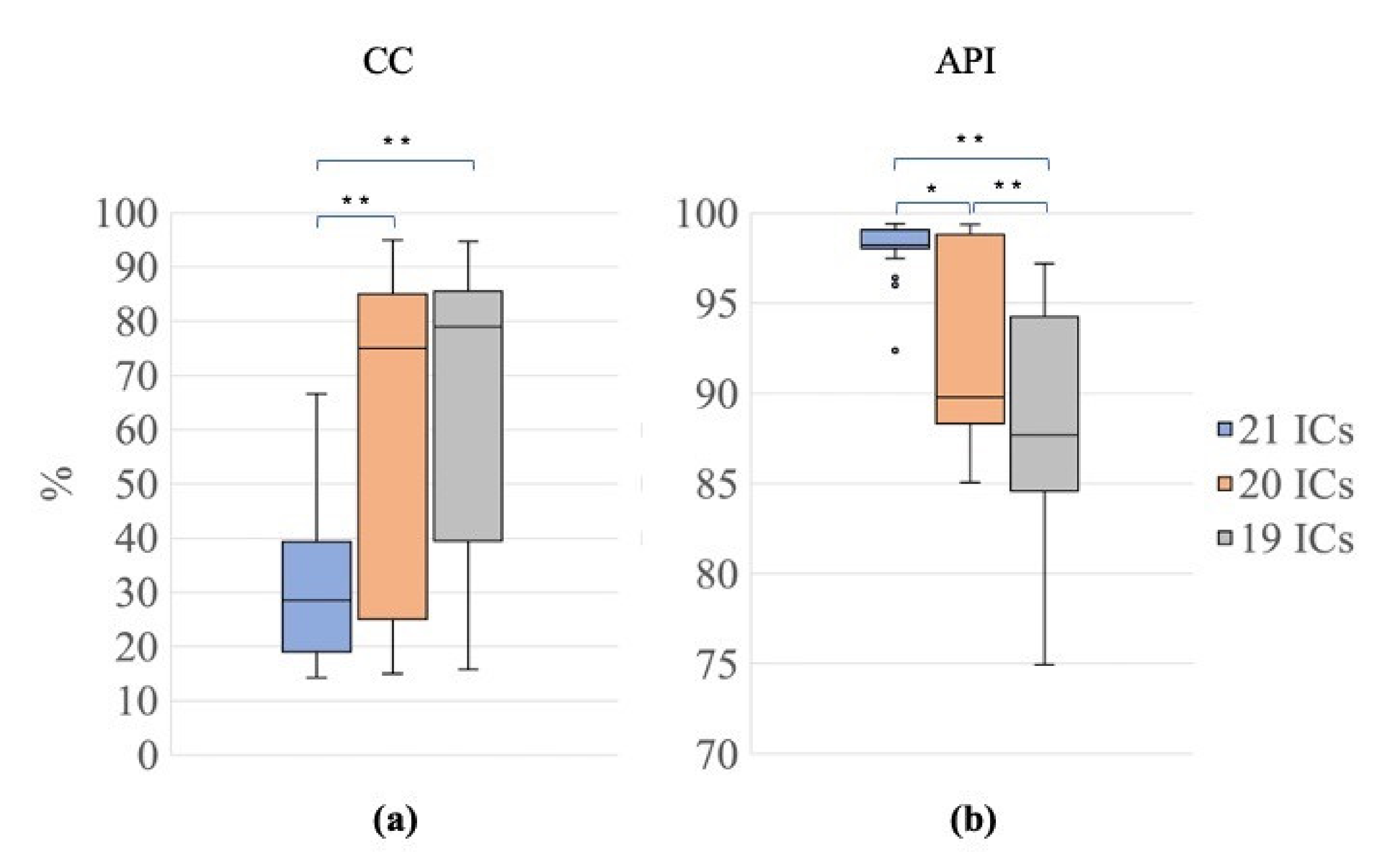
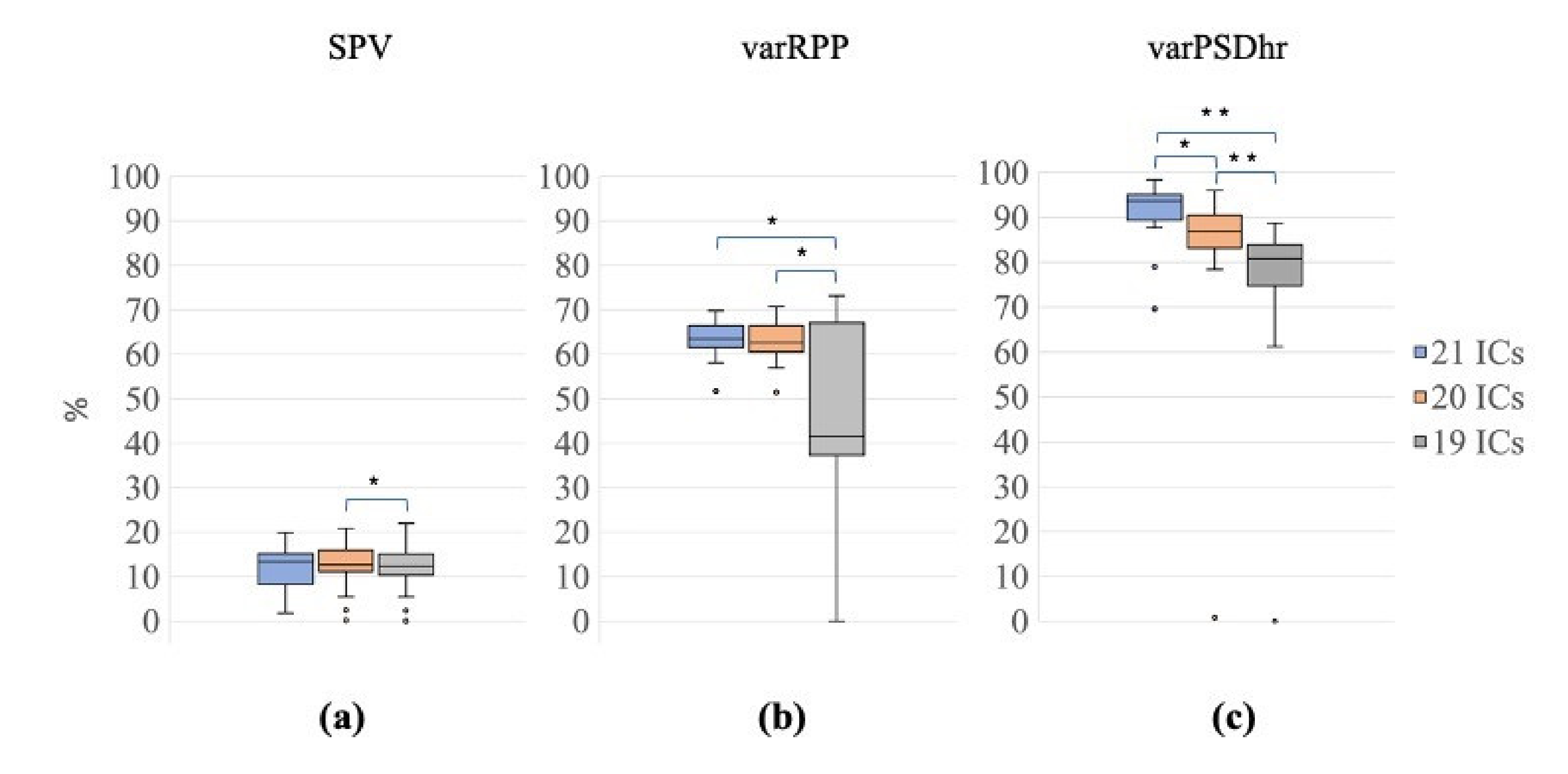
2.3.2. Evaluation of the Quality of the Reconstructed EEG Signals
2.4. Statistical Analysis
2.4.1. Assessment of the Effectiveness of the Proposed Decomposition Method for Separating Cardiac-Related ICs with the Three Signal Groups Composed with Seizure-Free EEG Segments (G1, G2 and G3) and with the Signal Group of EEG Segments with Seizures (G1s)
2.4.2. Validation of the Automated Classification of the ICs: Comparison with the Expert Classification of the ICs
- (1)
- True Positives (TP): the number of artefactual ICs correctly classified as CRCs by the algorithm;
- (2)
- True Negative (TN): the number of non-artefactual ICs correctly classified as NCCs by the algorithm;
- (3)
- False Negative (FN): the number of artefactual ICs wrongly classified as non-artefactual (i.e., as NCCs) by the algorithm;
- (4)
- False Positive (FP): the number of non-artefactual ICs wrongly classified as artefactual (i.e., as CRCs) by the algorithm.
3. Results
3.1. Testing of the Proposed Method on the Seizure-Free EEG Recordings
3.1.1. Evaluation of the Effectiveness of Using the nECG and nAPS Signals in the SOBI Decomposition
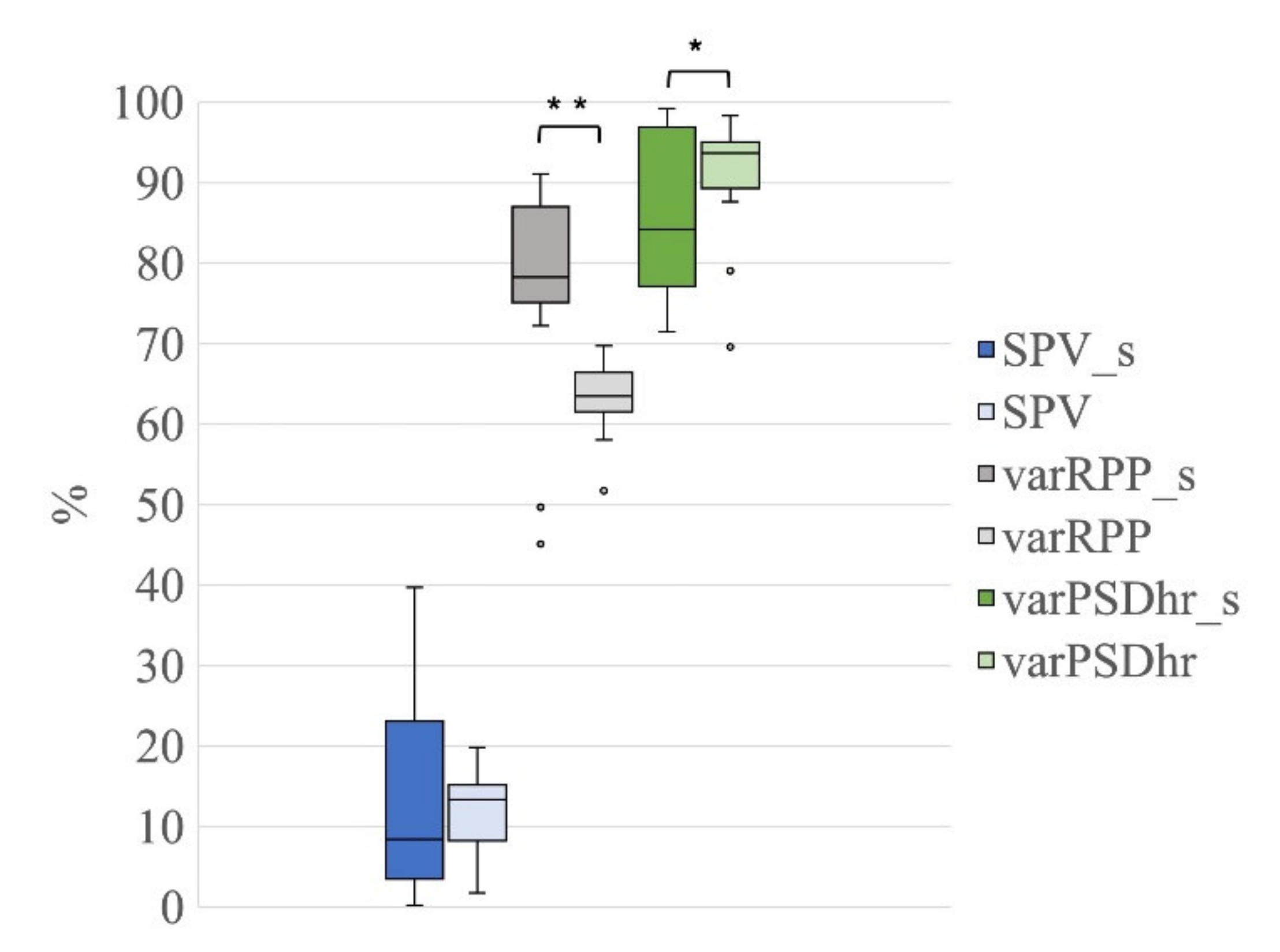
3.1.2. Evaluation of the Quality of the Reconstructed EEG Signals
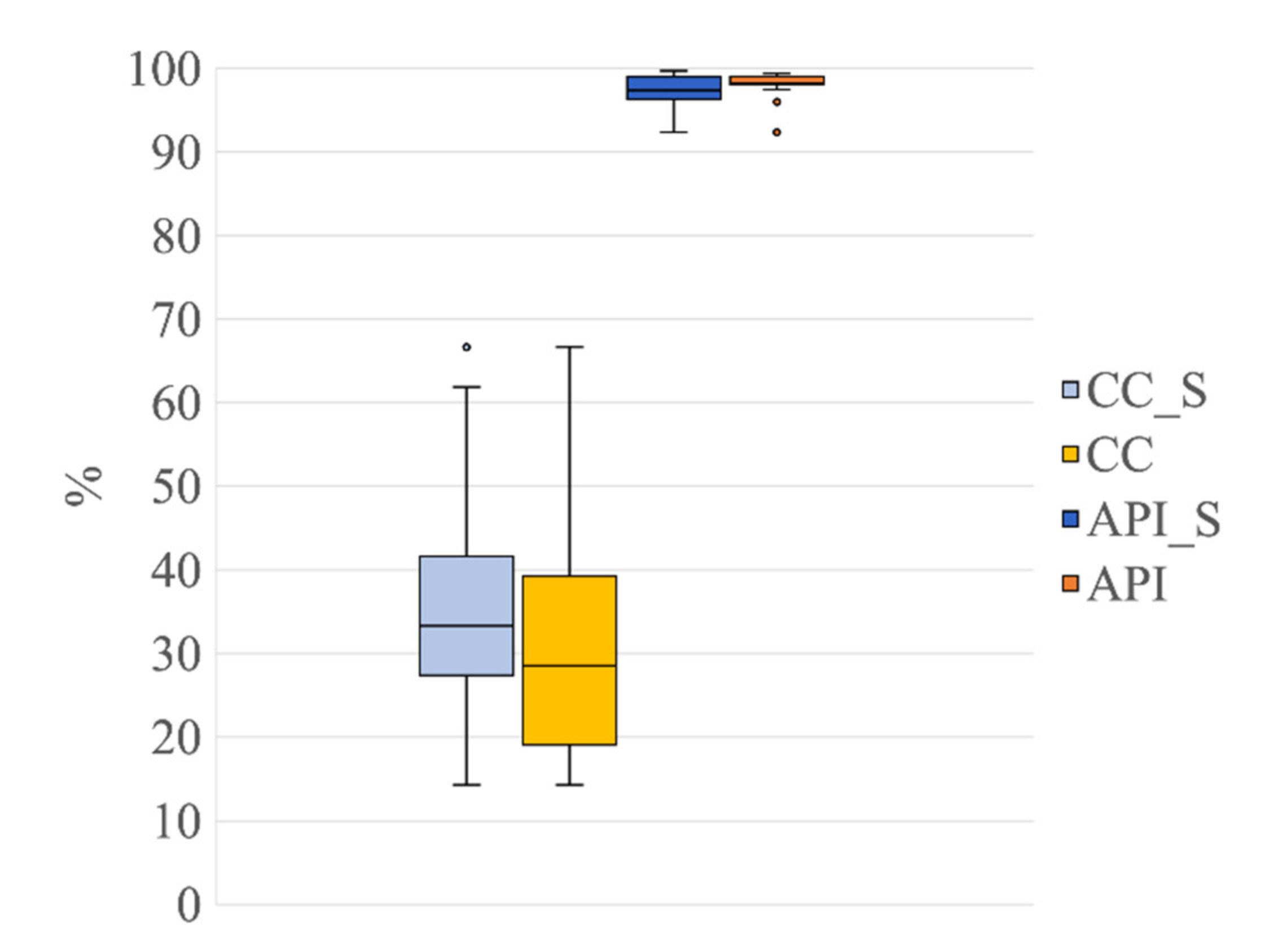
3.1.3. Validation of The Automated Classification of the ICs in Comparison with the Expert ICs Classification
3.2. Validation of the Proposed Method on the EEG Recordings Containing Seizures
4. Discussion
5. Conclusions
Author Contributions
Funding
Data Availability Statement
Conflicts of Interest
References
- Levene, M. The Clinical Conundrum of Neonatal Seizures. Arch. Dis. Child. Fetal Neonatal Ed. 2002, 86, F75–F77. [Google Scholar] [CrossRef]
- Rennie, J.M.; Chorley, G.; Boylan, G.B.; Pressler, R.; Nguyen, Y.; Hooper, R. Non-Expert Use of the Cerebral Function Monitor for Neonatal Seizure Detection. Arch. Dis. Child. Fetal Neonatal Ed. 2004, 89, F37–F40. [Google Scholar] [CrossRef] [PubMed]
- Ansari, A.H.; Cherian, P.J.; Caicedo, A.; Dereymaeker, A.; Jansen, K.; De Wispelaere, L.; Dielman, C.; Vervisch, J.; Govaert, P.; De Vos, M.; et al. NeoGuard: A Public, Online Learning Platform for Neonatal Seizures. arXiv 2019, arXiv:1905.12382. [Google Scholar]
- Ansari, A.H.; De Wel, O.; Pillay, K.; Dereymaeker, A.; Jansen, K.; Van Huffel, S.; Naulaers, G.; De Vos, M. A Convolutional Neural Network Outperforming State-of-the-Art Sleep Staging Algorithms for Both Preterm and Term Infants. J. Neural Eng. 2020, 17, 016028. [Google Scholar] [CrossRef]
- Abdelhameed, A.M.; Bayoumi, M. Semi-Supervised EEG Signals Classification System for Epileptic Seizure Detection. IEEE Signal Process. Lett. 2019, 26, 1922–1926. [Google Scholar] [CrossRef]
- Bhattacharyya, S.; Biswas, A.; Mukherjee, J.; Majumdar, A.K.; Majumdar, B.; Mukherjee, S.; Singh, A.K. Detection of Artifacts from High Energy Bursts in Neonatal EEG. Comput. Biol. Med. 2013, 43, 1804–1814. [Google Scholar] [CrossRef] [PubMed]
- Becker, T.; Vandecasteele, K.; Chatzichristos, C.; Van Paesschen, W.; Valkenborg, D.; Van Huffel, S.; De Vos, M. Classification with a Deferral Option and Low-Trust Filtering for Automated Seizure Detection. Sensors 2021, 21, 1046. [Google Scholar] [CrossRef] [PubMed]
- Bhattacharyya, A.; Ranta, R.; Le Cam, S.; Louis-Dorr, V.; Tyvaert, L.; Colnat-Coulbois, S.; Maillard, L.; Pachori, R.B. A Multi-Channel Approach for Cortical Stimulation Artefact Suppression in Depth EEG Signals Using Time-Frequency and Spatial Filtering. IEEE Trans. Biomed. Eng. 2019, 66, 1915–1926. [Google Scholar] [CrossRef]
- Dereymaeker, A.; Pillay, K.; Vervisch, J.; Van Huffel, S.; Naulaers, G.; Jansen, K.; De Vos, M. An Automated Quiet Sleep Detection Approach in Preterm Infants as a Gateway to Assess Brain Maturation. Int. J. Neural Syst. 2017, 27, 1750023. [Google Scholar] [CrossRef]
- Duffy, F.H.; Als, H. A Stable Pattern of EEG Spectral Coherence Distinguishes Children with Autism from Neuro-Typical Controls—A Large Case Control Study. BMC Med. 2012, 10, 64. [Google Scholar] [CrossRef]
- Sánchez Castillo, S.; Smith, L.; Díaz Suárez, A.; López Sánchez, G.F. Levels of Physical Activity in Spanish Asthmatics: A Cross-Sectional Study. Medicina 2020, 56, E643. [Google Scholar] [CrossRef] [PubMed]
- Costa, F. Development of an Algorithm for the Automatic Detection of Artifacts in Neonatal Electroencephalography. Ph.D. Thesis, University of Lisbon, Lisbon, Portugal, 2017. [Google Scholar]
- De Vos, M.; Deburchgraeve, W.; Cherian, P.J.; Matic, V.; Swarte, R.M.; Govaert, P.; Visser, G.H.; Van Huffel, S. Automated Artifact Removal as Preprocessing Refines Neonatal Seizure Detection. Clin. Neurophysiol. 2011, 122, 2345–2354. [Google Scholar] [CrossRef] [PubMed]
- Saby, J.N.; Marshall, P.J. The Utility of EEG Band Power Analysis in the Study of Infancy and Early Childhood. Dev. Neuropsychol. 2012, 37, 253–273. [Google Scholar] [CrossRef] [PubMed]
- Park, H.-J.; Jeong, D.-U.; Park, K.-S. Automated Detection and Elimination of Periodic ECG Artifacts in EEG Using the Energy Interval Histogram Method. IEEE Trans. Biomed. Eng. 2002, 49, 1526–1533. [Google Scholar] [CrossRef] [PubMed]
- Zhou, W.; Gotman, J. Removal of EMG and ECG Artifacts from EEG Based on Wavelet Transform and ICA. In Proceedings of the 26th Annual International Conference of the IEEE Engineering in Medicine and Biology Society, San Francisco, CA, USA, 1–5 September 2004; pp. 392–395. [Google Scholar] [CrossRef]
- Correa, A.G.; Laciar, E.; Patiño, H.D.; Valentinuzzi, M.E. Artifact Removal from EEG Signals Using Adaptive Filters in Cascade. J. Phys. Conf. Ser. 2007, 90, 012081. [Google Scholar] [CrossRef]
- Devuyst, S.; Dutoit, T.; Stenuit, P.; Kerkhofs, M.; Stanus, E. Cancelling ECG Artifacts in EEG Using a Modified Independent Component Analysis Approach. EURASIP J. Adv. Sig. Proc. 2008. [Google Scholar] [CrossRef]
- Hamaneh, M.B.; Chitravas, N.; Kaiboriboon, K.; Lhatoo, S.D.; Loparo, K.A. Automated Removal of EKG Artifact from EEG Data Using Independent Component Analysis and Continuous Wavelet Transformation. IEEE Trans. Biomed. Eng. 2014, 61, 1634–1641. [Google Scholar] [CrossRef]
- Urigüen, J.A.; Garcia-Zapirain, B. EEG Artifact Removal-State-of-the-Art and Guidelines. J. Neural Eng. 2015, 12, 031001. [Google Scholar] [CrossRef]
- Dora, C.; Biswal, P. Robust ECG Artifact Removal from EEG Using Continuous Wavelet Transformation and Linear Regression. In Proceedings of the 2016 International Conference on Signal Processing and Communications (SPCOM), Bangalore, India, 12–15 June 2016; pp. 1–5. [Google Scholar]
- Dora, C.; Biswal, P. Efficient Detection and Correction of Variable Strength ECG Artifact from Single Channel EEG. Biomed. Signal Process. Control 2019, 50, 168–177. [Google Scholar] [CrossRef]
- Radüntz, T.; Scouten, J.; Hochmuth, O.; Meffert, B. Automated EEG Artifact Elimination by Applying Machine Learning Algorithms to ICA-Based Features. J. Neural Eng. 2017, 14, 046004. [Google Scholar] [CrossRef]
- Waser, M.; Garn, H.; Jennum, P.J.; Sørensen, H.B.D. A Blind Source-Based Method for Automated Artifact-Correction in Standard Sleep EEG. In Proceedings of the 40th Annual International Conference of the IEEE Engineering in Medicine and Biology Society, Honolulu, HI, USA, 18–21 July 2018; pp. 6010–6013. [Google Scholar]
- Kumar, N. Removal of ECG Artifact from EEG Data Using Independent Component Analysis and S-Transform. Available online: https://www.semanticscholar.org/paper/Removal-of-ECG-Artifact-from-EEG-data-using-and-Kumar/c1eefc3b36674da0c1880de9de24eb64be9d5e72 (accessed on 28 July 2021).
- Tamburro, G.; Stone, D.B.; Comani, S. Automatic Removal of Cardiac Interference (ARCI): A New Approach for EEG Data. Front. Neurosci. 2019, 13, 441. [Google Scholar] [CrossRef]
- Issa, M.; Tuboly, G.; Kozmann, G.; Juhasz, Z. Automatic ECG Artefact Removal from EEG Signals. Meas. Sci. Rev. 2019, 19, 101–108. [Google Scholar] [CrossRef]
- Taha, L.Y.; Abdel-Raheem, E. EEG Signal Extraction Utilizing Null Space Approach. In Proceedings of the 2019 IEEE International Symposium on Signal Processing and Information Technology (ISSPIT), Ajman, UAE, 10–12 December 2019; pp. 1–5. [Google Scholar]
- Jiang, X.; Bian, G.-B.; Tian, Z. Removal of Artifacts from EEG Signals: A Review. Sensors 2019, 19, 987. [Google Scholar] [CrossRef]
- Moeyersons, J.; Smets, E.; Morales, J.; Villa, A.; De Raedt, W.; Testelmans, D.; Buyse, B.; Van Hoof, C.; Willems, R.; Van Huffel, S.; et al. Artefact Detection and Quality Assessment of Ambulatory ECG Signals. Comput. Methods Programs Biomed. 2019, 182, 105050. [Google Scholar] [CrossRef]
- Islam, K.; Rastegarnia, A.; Sanei, S. Signal Artifacts and Techniques for Artifacts and Noise Removal. In Signal Processing Techniques for Computational Health Informatics; Springer: Cham, Switzerland, 2020; ISBN 978-3-030-54932-9. [Google Scholar]
- Abbasi, S.F.; Awais, M.; Zhao, X.; Chen, W. Automatic Denoising and Artifact Removal from Neonatal EEG. In Proceedings of the BIBE 2019: The Third International Conference on Biological Information and Biomedical Engineering, Hangzhou, China, 20–22 July 2019; pp. 1–5. [Google Scholar]
- Khlif, M.S.; Mesbah, M.; Boashash, B.; Colditz, P. Influence of EEG Artifacts on Detecting Neonatal Seizure. In Proceedings of the 10th International Conference on Information Science, Signal Processing and their Applications (ISSPA 2010), Kuala Lumpur, Malaysia, 10–13 May 2010; pp. 500–503. [Google Scholar]
- Stevenson, N.; O’Toole, J.; Korotchikova, I.; Boylan, G. Artefact Detection in Neonatal EEG. In Proceedings of the 36th Annual International Conference of the IEEE Engineering in Medicine and Biology Society (EMBC 2014), Chicago, IL, USA, 27–31 August 2014; pp. 926–929. [Google Scholar] [CrossRef]
- Kauppila, M.; Vanhatalo, S.; Stevenson, N.J. Artifact Detection in Neonatal EEG Using Gaussian Mixture Models. In Proceedings of the EMBEC & NBC 2017, Tampere, Finland, 11–15 June 2017; pp. 221–224. [Google Scholar]
- Webb, L.; Kauppila, M.; Roberts, J.A.; Vanhatalo, S.; Stevenson, N.J. Automated Detection of Artefacts in Neonatal EEG with Residual Neural Networks. Comput. Methods Programs Biomed. 2021, 208, 106194. [Google Scholar] [CrossRef] [PubMed]
- Navarro-Sune, X.; Poree, F.; Carrault, G. ECG Removal in Preterm EEG Combining Empirical Mode Decomposition and Adaptive Filtering. In Proceedings of the 2012 IEEE International Conference on Acoustics, Speech and Signal Processing (ICASSP), Kyoto, Japan, 25–30 March 2012; pp. 661–664. [Google Scholar]
- Navarro, X.; Porée, F.; Beuchée, A.; Carrault, G. Denoising Preterm EEG by Signal Decomposition and Adaptive Filtering: A Comparative Study. Med. Eng. Phys. 2015, 37, 315–320. [Google Scholar] [CrossRef][Green Version]
- Janardhan, A.; Rao, K.K. Application of Signal Separation Algorithms for Artifact Removal from EEG Signals. Int. J. Mod. Commun. Technol. Res. 2015, 3, 6. [Google Scholar]
- Govindan, R.B.; Kota, S.; Al-Shargabi, T.; Massaro, A.N.; Chang, T.; du Plessis, A. Effect of Electrocardiogram Interference on Cortico-Cortical Connectivity Analysis and a Possible Solution. J. Neurosci. Methods 2016, 270, 76–84. [Google Scholar] [CrossRef]
- Matić, V.; Huffel, S. Comparison of ICA Algorithms for ECG Artifact Removal from EEG Signals. In Proceedings of the 4th Annual Symposium of the Benelux Chapter (pp. 137–140) of the IEEE Engineering in Medicine and Biology Society (EMBS), Enschede, The Netherlands, 9–10 November 2009. [Google Scholar]
- Tamburro, G.; Fiedler, P.; Stone, D.; Haueisen, J.; Comani, S. A New ICA-Based Fingerprint Method for the Automatic Removal of Physiological Artifacts from EEG Recordings. PeerJ 2018, 6, e4380. [Google Scholar] [CrossRef]
- Jung, T.P.; Makeig, S.; Humphries, C.; Lee, T.W.; McKeown, M.J.; Iragui, V.; Sejnowski, T.J. Removing Electroencephalographic Artifacts by Blind Source Separation. Psychophysiology 2000, 37, 163–178. [Google Scholar] [CrossRef]
- Castellanos, N.P.; Makarov, V.A. Recovering EEG Brain Signals: Artifact Suppression with Wavelet Enhanced Independent Component Analysis. J. Neurosci. Methods 2006, 158, 300–312. [Google Scholar] [CrossRef] [PubMed]
- Delorme, A.; Sejnowski, T.; Makeig, S. Enhanced Detection of Artifacts in EEG Data Using Higher-Order Statistics and Independent Component Analysis. Neuroimage 2007, 34, 1443–1449. [Google Scholar] [CrossRef] [PubMed]
- Inuso, G.; Foresta, F.L.; Mammone, N.; Morabito, F.C. Wavelet-ICA Methodology for Efficient Artifact Removal from Electroencephalographic Recordings. In Proceedings of the IEEE International Conference on Neural Networks, Orlando, FL, USA, 12–17 August 2007; pp. 1524–1529. [Google Scholar]
- Viola, F.C.; Thorne, J.; Edmonds, B.; Schneider, T.; Eichele, T.; Debener, S. Semi-Automatic Identification of Independent Components Representing EEG Artifact. Clin. Neurophysiol. 2009, 120, 868–877. [Google Scholar] [CrossRef]
- Nolan, H.; Whelan, R.; Reilly, R.B. FASTER: Fully Automated Statistical Thresholding for EEG Artifact Rejection. J. Neurosci. Methods 2010, 192, 152–162. [Google Scholar] [CrossRef]
- Mur, A.; Dormido, R.; Duro, N. An Unsupervised Method for Artefact Removal in EEG Signals. Sensors 2019, 19, 2302. [Google Scholar] [CrossRef]
- Mognon, A.; Jovicich, J.; Bruzzone, L.; Buiatti, M. ADJUST: An Automatic EEG Artifact Detector Based on the Joint Use of Spatial and Temporal Features. Psychophysiology 2011, 48, 229–240. [Google Scholar] [CrossRef]
- Miljković, N.; Matić, V.; Van Huffel, S.; Popović, M.B. Independent Component Analysis (ICA) Methods for Neonatal EEG Artifact Extraction: Sensitivity to Variation of Artifact Properties. In Proceedings of the 10th Symposium on Neural Network Applications in Electrical Engineering, Belgrade, Serbia, 23–25 September 2010; pp. 19–21. [Google Scholar]
- Tamburro, G.; Croce, P.; Zappasodi, F.; Comani, S. Is Brain Dynamics Preserved in the EEG After Automated Artifact Removal? A Validation of the Fingerprint Method and the Automatic Removal of Cardiac Interference Approach Based on Microstate Analysis. Front. Neurosci. 2020, 14, 577160. [Google Scholar] [CrossRef] [PubMed]
- Widmann, A.; Schröger, E.; Maess, B. Digital Filter Design for Electrophysiological Data—A Practical Approach. J. Neurosci. Methods 2015, 250, 34–46. [Google Scholar] [CrossRef] [PubMed]
- Delorme, A.; Makeig, S. EEGLAB: An Open Source Toolbox for Analysis of Single-Trial EEG Dynamics Including Independent Component Analysis. J. Neurosci. Methods 2004, 134, 9–21. [Google Scholar] [CrossRef]
- Kwon, Y.; Jacobs, D.R.; Lutsey, P.L.; Brumback, L.; Chirinos, J.A.; Mariani, S.; Redline, S.; Duprez, D.A. Sleep Disordered Breathing and ECG R-Wave to Radial Artery Pulse Delay, The Multi-Ethnic Study of Atherosclerosis. Sleep Med. 2018, 48, 172–179. [Google Scholar] [CrossRef]
- Pépin, J.-L.; Delavie, N.; Pin, I.; Deschaux, C.; Argod, J.; Bost, M.; Levy, P. Pulse Transit Time Improves Detection of Sleep Respiratory Events and Microarousals in Children. Chest 2005, 127, 722–730. [Google Scholar] [CrossRef] [PubMed]
- Rajala, S.; Lindholm, H.; Taipalus, T. Comparison of Photoplethysmogram Measured from Wrist and Finger and the Effect of Measurement Location on Pulse Arrival Time. Physiol. Meas. 2018, 39, 075010. [Google Scholar] [CrossRef] [PubMed]
- Belouchrani, A.; Abed-Meraim, K.; Cardoso, J.-F.; Moulines, E. A Blind Source Separation Technique Using Second-Order Statistics. IEEE Trans. Signal Process. 1997, 45, 434–444. [Google Scholar] [CrossRef]
- Agnetti, A.; Greco, C.; Tchana, B. L’ECG in Eta Pediatrica. Assoc. Cult. Pediatri 2016, 23, 271–275. [Google Scholar]
- Fleming, S.; Thompson, M.; Stevens, R.; Heneghan, C.; Plüddemann, A.; Maconochie, I.; Tarassenko, L.; Mant, D. Normal Ranges of Heart Rate and Respiratory Rate in Children from Birth to 18 Years of Age: A Systematic Review of Observational Studies. Lancet 2011, 377, 1011–1018. [Google Scholar] [CrossRef]
- Cohen, J. Statistical Power Analysis for the Behavioral Sciences, 2nd ed.; Routledge: New York, NY, USA, 1988; ISBN 978-0-203-77158-7. [Google Scholar]
- Fritz, C.O.; Morris, P.E.; Richler, J.J. Effect Size Estimates: Current Use, Calculations, and Interpretation. J. Exp. Psychol. Gen. 2012, 141, 2–18. [Google Scholar] [CrossRef] [PubMed]
- Tomczak, M.; Tomczak, E. The Need to Report Effect Size Estimates Revisited. An Overview of Some Recommended Measures of Effect Size. Trends Sport Sci. 2014, 21, 19–25. [Google Scholar]
- National Research Council (US) Committee on Vision. Emergent Techniques for Assessment of Visual Performance; National Academies Press: Washington, DC, USA, 1985. [Google Scholar]


| Artefactual IC | Decomposition Level | Median | 95th CI | 𝜒2 | p | Comparison | Z | pw | Effect Size |
|---|---|---|---|---|---|---|---|---|---|
| ECC | 21 ICs | 1.0 | 0.00 | 21 ICs vs. 20 ICs | 0.00 | 1.00 | 0.00 | ||
| 20 ICs | 1.0 | 0.00 | 14.70 | <0.001 | 21 ICs vs. 19 ICs | 3.74 | <0.001 | 0.59 | |
| 19 ICs | 0.0 | 0.21 | 20 ICs vs. 19 ICs | 3.74 | <0.001 | 0.59 | |||
| PCC | 21 ICs | 2.00 | 0.25 | 21 ICs vs. 20 ICs | 3.61 | <0.05 | 0.57 | ||
| 20 ICs | 1.00 | 0.17 | 19.43 | <0.001 | 21 ICs vs. 19 ICs | 4.24 | <0.001 | 0.67 | |
| 19 ICs | 1.00 | 0.27 | 20 ICs vs. 19 ICs | 2.24 | <0.05 | 0.35 |
| Signal Group | Dec. level | No. of ICs | ECC | PCC | TP | TN | FP | FN | Acc | FOR | Sens |
|---|---|---|---|---|---|---|---|---|---|---|---|
| G1 | 21 ICs | 420 | 21(20) | 36(34) | 53 | 362 | 1 | 4 | 0.99 | 0.01 | 0.93 |
| G2 | 20 ICs | 400 | 20(20) | 28(21) | 41 | 352 | 0 | 7 | 0.98 | 0.02 | 0.85 |
| G3 | 19 ICs | 380 | 20(6) | 27(16) | 22 | 333 | 0 | 25 | 0.93 | 0.07 | 0.47 |
| Signal Group | Dec. level | No. ofICs | ECC | PCC | TP | TN | FP | FN | Acc | FOR | Sens |
|---|---|---|---|---|---|---|---|---|---|---|---|
| G1s | 21 ICs | 420 | 20(17) | 29(29) | 46 | 371 | 0 | 3 | 0.993 | 0.008 | 0.939 |
| G1 | 21 ICs | 420 | 21(20) | 36(34) | 53 | 362 | 1 | 4 | 0.988 | 0.011 | 0.930 |
Publisher’s Note: MDPI stays neutral with regard to jurisdictional claims in published maps and institutional affiliations. |
© 2021 by the authors. Licensee MDPI, Basel, Switzerland. This article is an open access article distributed under the terms and conditions of the Creative Commons Attribution (CC BY) license (https://creativecommons.org/licenses/by/4.0/).
Share and Cite
Tamburro, G.; Croce, P.; Zappasodi, F.; Comani, S. Automated Detection and Removal of Cardiac and Pulse Interferences from Neonatal EEG Signals. Sensors 2021, 21, 6364. https://doi.org/10.3390/s21196364
Tamburro G, Croce P, Zappasodi F, Comani S. Automated Detection and Removal of Cardiac and Pulse Interferences from Neonatal EEG Signals. Sensors. 2021; 21(19):6364. https://doi.org/10.3390/s21196364
Chicago/Turabian StyleTamburro, Gabriella, Pierpaolo Croce, Filippo Zappasodi, and Silvia Comani. 2021. "Automated Detection and Removal of Cardiac and Pulse Interferences from Neonatal EEG Signals" Sensors 21, no. 19: 6364. https://doi.org/10.3390/s21196364
APA StyleTamburro, G., Croce, P., Zappasodi, F., & Comani, S. (2021). Automated Detection and Removal of Cardiac and Pulse Interferences from Neonatal EEG Signals. Sensors, 21(19), 6364. https://doi.org/10.3390/s21196364