Deep Learning in Physiological Signal Data: A Survey


Abstract
1. Introduction
2. Related Works
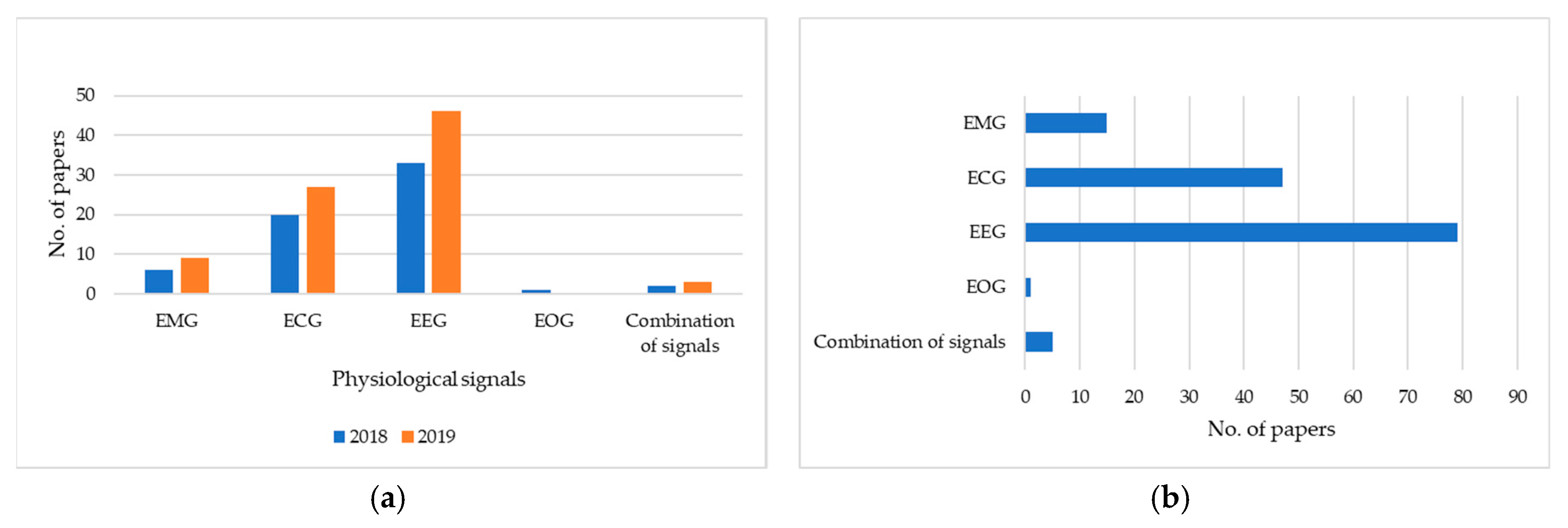
3. Physiological Signal Analysis and Modality
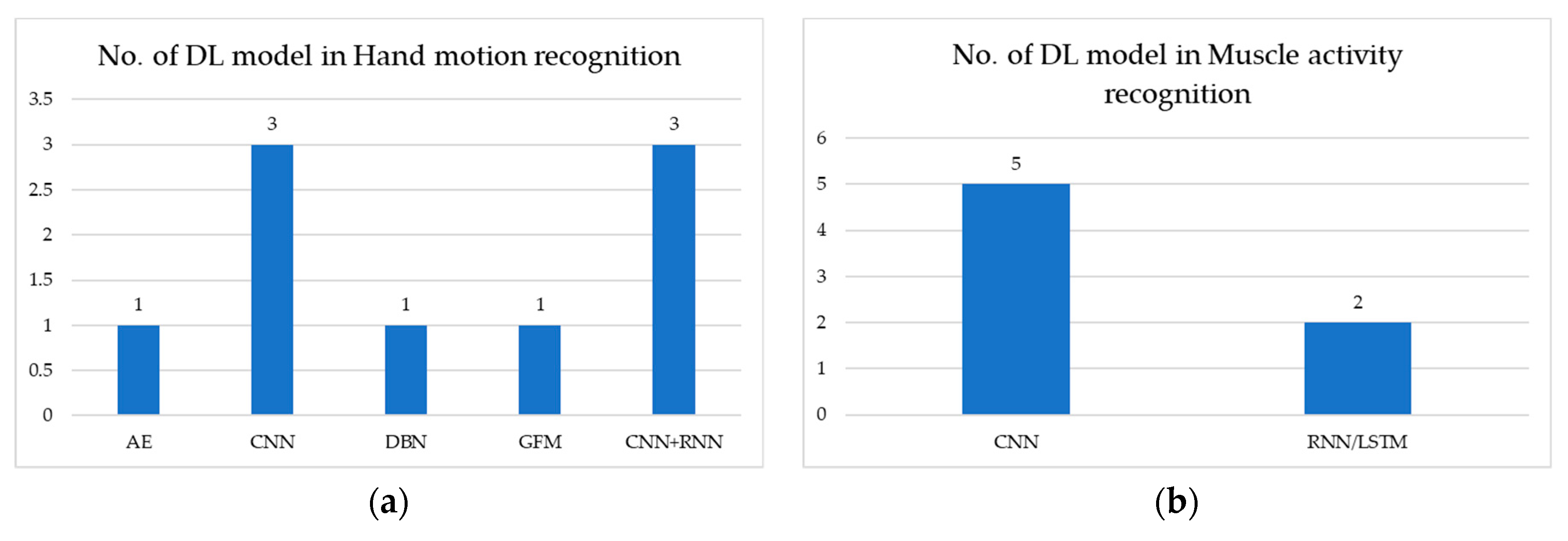
3.1. Deep Learning with Electromyogram (EMG)
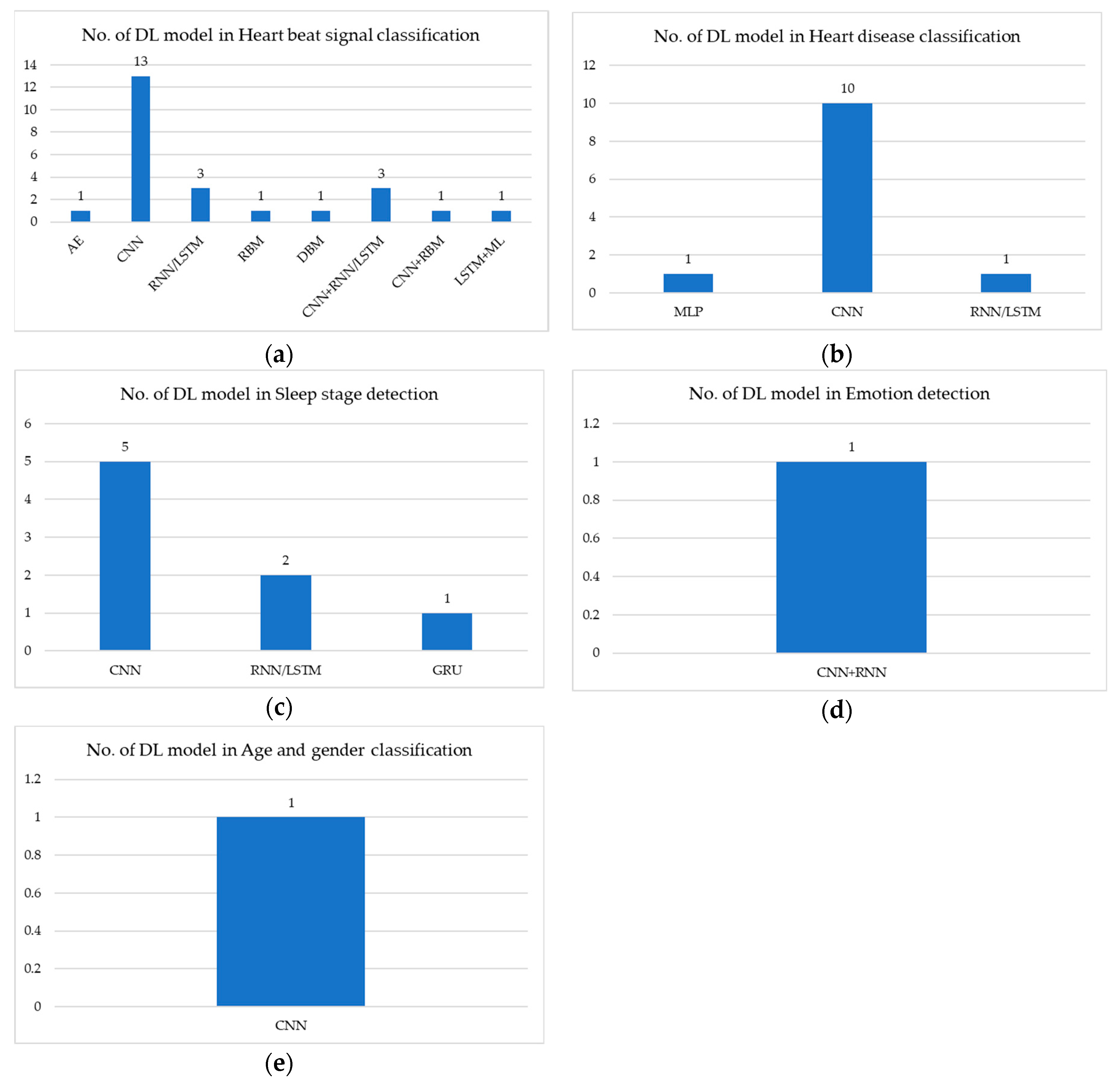
3.2. Deep Learning with Electrocardiogram (ECG)
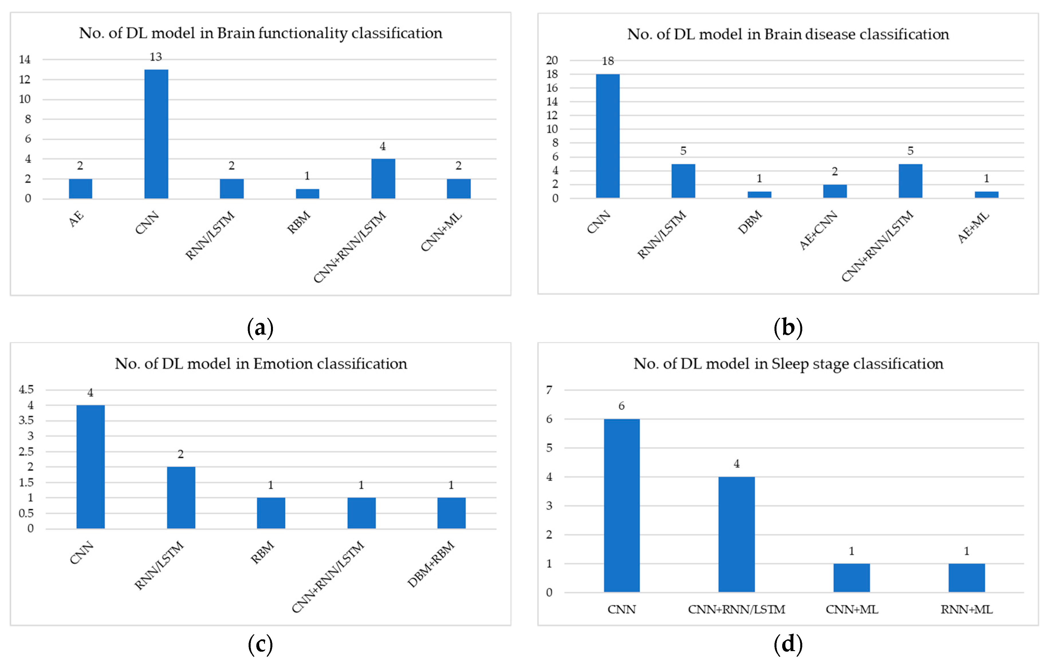
3.3. Deep Learning with Electroencephalogram (EEG)
3.4. Deep Learning with Electrooculogram (EOG)
3.5. Deep Learning with a Combination of Signals
4. Training Architecture
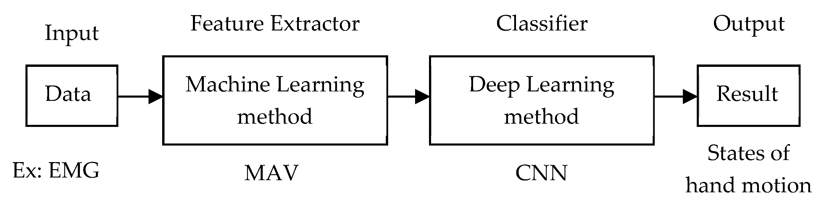
4.1. Traditional Machine Learning as Feature Extractor and Deep Learning as Classifier
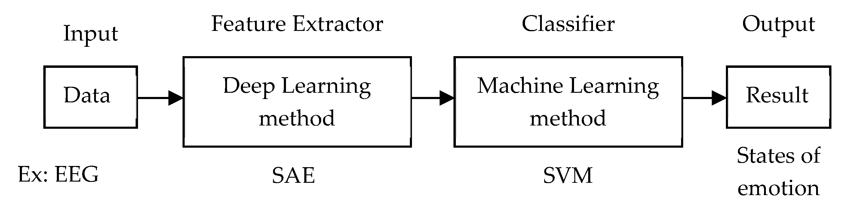
4.2. Deep Learning as Feature Extractor and Traditional Machine Learning as Classifier
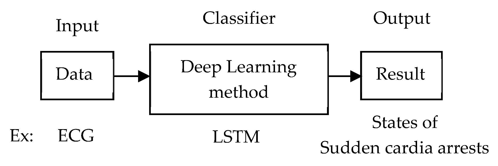
4.3. End-to-End Learning
5. Dataset Sources
6. Discussion
6.1. Discussion of the Deep-Learning Task
6.2. Discussion of the Deep-Learning Model
6.3. Discussion of the Training Architecture
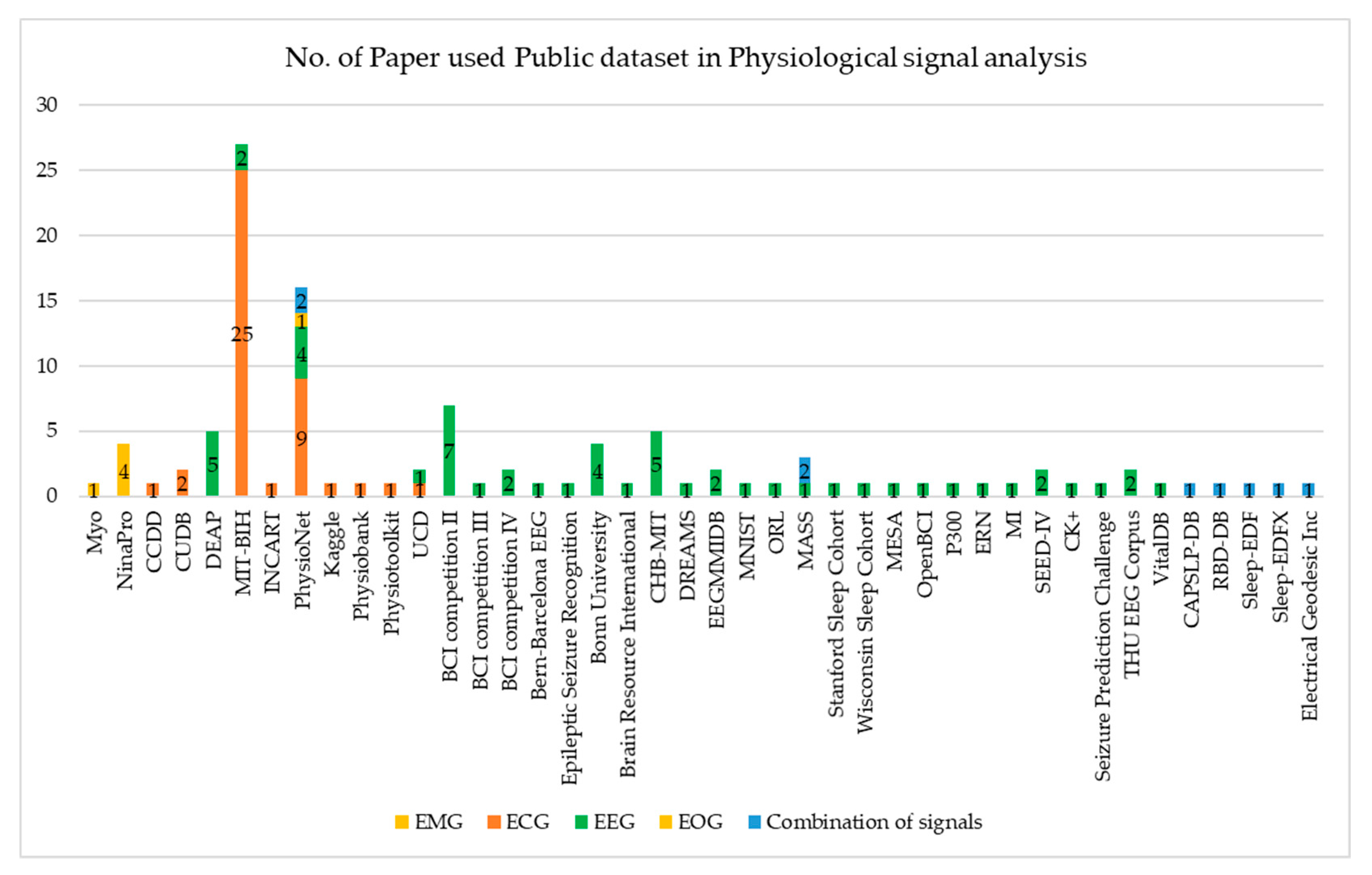
6.4. Discussion of the Dataset Source
7. Conclusions
Author Contributions
Acknowledgments
Conflicts of Interest
Abbreviations
| ADHD | Attention Deficit Hyperactivity Disorder |
| AE | Auto-encoder |
| ANN | Artificial neural network |
| AUC | Area under the curve |
| AUPRC | Area under the precision–recall curve |
| AUROC | Area under the receiver operating characteristic curve |
| BB | Bern-Barcelona EEG database |
| BCI | Brain-computer interface |
| BRNN | Bi-directional recurrent neural network |
| CAM-ICU | Confusion assessment method for the ICU |
| CapsNet | Capsule network |
| CNNE | Convolutional neural network as a feature extractor |
| CP-MixedNet | Channel-projection mixed-scale convolutional neural network |
| CssC DBM | Contractive Slab and Spike Convolutional Deep Boltzmann Machine |
| DBLSTM-WS | Bi-directional LSTM network-based wavelet sequences |
| DBM | Deep Boltzmann Machine |
| DBN | Deep belief network |
| DBN-GC | Deep belief networks with glia chains |
| DCNN | Deep convolution neural network |
| DCssC DBM | Discriminative version of CssCDBM |
| DL-CCANet | Dual-lead ECGs - canonical correlation analysis and cascaded convolutional network |
| DN-AE-NTM | Deep network - auto-encoder - neural Turing machine |
| DNN | Deep neural network |
| EBR | Error Backpropagation |
| ED | Emergency department |
| EEG-fNIRs | EEG-Functional near-infrared spectroscopy |
| EL-SDAE | Ensemble SDAE classifier with local information preservation |
| ERP | Event-related potential |
| ESR | Epileptic Seizure Recognition dataset |
| ETLE | Extra-temporal lobe epilepsy |
| FPR | False prediction rates |
| GAN | Generative adversarial network |
| GFM | Generative flow model |
| GRU | Gated-recurrent unit |
| HGD | High gamma dataset |
| HMM | Hidden Markov models |
| IC | Independent component |
| KFs | Polynomial Kalman filters |
| LSTM | Long short-term memory |
| MEG | Magnetoencephalographic |
| MLP | Multilayer perceptron |
| MLR | Multilayer logistic regression |
| MMDPN | Multi-view multi-level deep polynomial network |
| MPCNN | Multi-perspective convolutional neural network |
| MTLE | Mesial temporal lobe epilepsy |
| NIP | Neural interface processor |
| NMSE | Normalised mean square error |
| OCNN | Orthogonal convolutional neural network |
| PCANet | Integrating the principal component analysis (PCA) and a deep-learning model |
| R3DCNN | 3D convolutional neural networks |
| RA | Region aggregation |
| RASS | Richmond agitation-sedation scale |
| RBM | Restricted Boltzmann machine |
| RCNN | Recurrent convolutional neural network |
| RNN | Recurrent neural network |
| RR | Respiratory rate |
| SAE | Stacked auto-encoder |
| SDAE | Stacked denoising auto-encoder |
| SEED | SJTU emotion EEG dataset |
| SNN | Spiking neural network |
| STFT | Short-term Fourier transform |
| SVEB | Supraventricular ectopic beat |
| SVM | Support vector machine |
| SWT | Stationary wavelet transforms |
| TCN | Temporal convolutional network |
| TL-CCANet | Three-lead ECGs - canonical correlation analysis and cascaded convolutional network |
| TLE | Temporal lobe epilepsy |
| VAE | Variational auto-encoder |
| VEB | Ventricular ectopic beat |
References
- Mu, R.; Zeng, X. A Review of Deep Learning Research. TIISs 2019, 13, 1738–1764. [Google Scholar] [CrossRef]
- Zhang, L.; Jia, J.; Li, Y.; Gao, W.; Wang, M. Deep Learning based Rapid Diagnosis System for Identifying Tomato Nutrition Disorders. KSII Trans. Internet Inf. Syst. 2019, 13, 2012–2027. [Google Scholar] [CrossRef]
- Ganapathy, N.; Swaminathan, R.; Deserno, T.M. Deep learning on 1-D biosignals: A taxonomy-based survey. Yearbook Med. Inf. 2018, 27, 98–109. [Google Scholar] [CrossRef] [PubMed]
- Zhang, D.; Yao, L.; Chen, K.; Wang, S.; Chang, X.; Liu, Y. Making Sense of Spatio-Temporal Preserving Representations for EEG-Based Human Intention Recognition. IEEE Trans. Cybern. 2019, 1–12. [Google Scholar] [CrossRef] [PubMed]
- PubMed. Available online: https://www.ncbi.nlm.nih.gov/pubmed/ (accessed on 31 October 2019).
- Faust, O.; Hagiwara, Y.; Hong, T.J.; Lih, O.S.; Acharya, U.R. Deep learning for healthcare applications based on physiological signals: A review. Comput. Methods Programs Biomed. 2018, 161, 1–13. [Google Scholar] [CrossRef] [PubMed]
- Tobore, I.; Li, J.; Yuhang, L.; Al-Handarish, Y.; Abhishek, K.; Zedong, N.; Lei, W. Deep Learning Intervention for Health Care Challenges: Some Biomedical Domain Considerations. JMIR mHealth uHealth 2019, 7, e11966. [Google Scholar] [CrossRef]
- Baig, M.Z.; Kavkli, M. A Survey on Psycho-Physiological Analysis & Measurement Methods in Multimodal Systems. Multimodal Technol. Interact 2019, 3, 37. [Google Scholar] [CrossRef]
- Yu, Y.; Chen, X.; Cao, S.; Zhang, X.; Chen, X. Exploration of Chinese Sign Language Recognition Using Wearable Sensors Based on Deep Belief Net. IEEE J. Biomed. Health Inf. 2019. [Google Scholar] [CrossRef]
- Hu, Y.; Wong, Y.; Wei, W.; Du, Y.; Kankanhalli, M.; Geng, W. A novel attention-based hybrid CNN-RNN architecture for sEMG-based gesture recognition. PLoS ONE 2018, 13, e0206049. [Google Scholar] [CrossRef]
- Wei, W.; Dai, Q.; Wong, Y.; Hu, Y.; Kankanhalli, M.; Geng, W. Surface Electromyography-based Gesture Recognition by Multi-view Deep Learning. IEEE Trans. Biomed. Eng. 2019, 66, 2964–2973. [Google Scholar] [CrossRef]
- Cote-Allard, U.; Fall, C.L.; Drouin, A.; Campeau-Lecours, A.; Gosselin, C.; Glette, K.; Laviolette, F.; Gosselin, B. Deep learning for electromyographic hand gesture signal classification using transfer learning. IEEE Trans. Neural Syst. Rehabil. Eng. 2019, 27, 760–771. [Google Scholar] [CrossRef] [PubMed]
- Sun, W.; Liu, H.; Tang, R.; Lang, Y.; He, J.; Huang, Q. sEMG-Based Hand-Gesture Classification Using a Generative Flow Model. Sensors 2019, 19, 1952. [Google Scholar] [CrossRef] [PubMed]
- Li, C.; Ren, J.; Huang, H.; Wang, B.; Zhu, Y.; Hu, H. PCA and deep learning based myoelectric grasping control of a prosthetic hand. Biomed. Eng. Online 2018, 17, 107. [Google Scholar] [CrossRef] [PubMed]
- Rehman, M.Z.; Waris, A.; Gilani, S.O.; Jochumsen, M.; Niazi, I.K.; Jamil, M.; Farina, D.; Kamavuako, E.N. Multiday EMG-based classification of hand motions with deep learning techniques. Sensors 2018, 18, 2497. [Google Scholar] [CrossRef]
- Wang, W.; Chen, B.; Xia, P.; Hu, J.; Peng, Y. Sensor Fusion for Myoelectric Control Based on Deep Learning with Recurrent Convolutional Neural Networks. Artif. Organs 2018, 42, E272–E282. [Google Scholar] [CrossRef]
- Xia, P.; Hu, J.; Peng, Y. EMG-based estimation of limb movement using deep learning with recurrent convolutional neural networks. Artif. Organs 2018, 42, E67–E77. [Google Scholar] [CrossRef]
- Olsson, A.E.; Sager, P.; Andersson, E.; Bjorkman, A.; Malesevic, N.; Antfolk, C. Extraction of Multi-Labelled Movement Information from the Raw HD-sEMG Image with Time-Domain Depth. Sci. Rep. 2019, 9, 7244. [Google Scholar] [CrossRef]
- Khowailed, I.A.; Abotabl, A. Neural muscle activation detection: A deep learning approach using surface electromyography. J. Biomech. 2019, 95, 109322. [Google Scholar] [CrossRef]
- Rance, L.; Ding, Z.; McGregor, A.H.; Bull, A.M.J. Deep learning for musculoskeletal force prediction. Ann. Biomed. Eng. 2019, 47, 778–789. [Google Scholar] [CrossRef]
- Dantas, H.; Warren, D.J.; Wendelken, S.M.; Davis, T.S.; Clark, G.A.; Mathews, V.J. Deep Learning Movement Intent Decoders Trained with Dataset Aggregation for Prosthetic Limb Control. IEEE Trans. Biomed. Eng. 2019, 66, 3192–3203. [Google Scholar] [CrossRef]
- Ameri, A.; Akhaee, M.A.; Scheme, E.; Englehart, K. Real-time, simultaneous myoelectric control using a convolutional neural network. PLoS ONE 2018, 13, e0203835. [Google Scholar] [CrossRef]
- Nodera, H.; Osaki, Y.; Yamazaki, H.; Mori, A.; Izumi, Y.; Kaji, R. Deep learning for waveform identification of resting needle electromyography signals. Clin. Neurophysiol. 2019, 130, 617–623. [Google Scholar] [CrossRef]
- Ribeiro, A.L.P.; Paixao, G.M.M.; Gomes, P.R.; Ribeiro, M.H.; Ribeiro, A.H.; Canazart, J.A.; Oliveira, D.M.; Ferreira, M.P.; Lima, E.M.; Moraes, J.L.; et al. Tele-electrocardiography and bigdata: The CODE (Clinical Outcomes in Digital Electrocardiography) study. J. Electrocardiol. 2019, 57, S75–S78. [Google Scholar] [CrossRef]
- Cano-Espinosa, C.; Gonzalez, G.; Washko, G.R.; Cazorla, M.; Estepar, R.S.J. Automated Agatston score computation in non-ECG gated CT scans using deep learning. In Proceedings of the Medical Imaging 2018: Image Processing, Houston, TX, USA, 2 March 2018. [Google Scholar] [CrossRef]
- Chauhan, S.; Vig, L.; Ahmad, S. ECG anomaly class identification using LSTM and error profile modeling. Comput. Biol. Med. 2019, 109, 14–21. [Google Scholar] [CrossRef]
- Xia, Y.; Wulan, N.; Wang, K.; Zhang, H. Detecting atrial fibrillation by deep convolutional neural networks. Comput. Biol. Med. 2018, 93, 84–92. [Google Scholar] [CrossRef]
- Tan, J.H.; Hagiwara, Y.; Pang, W.; Lim, I.; Oh, S.L.; Adam, M.; Tan, R.S.; Chen, M.; Acharya, U.R. Application of stacked convolutional and long short-term memory network for accurate identification of CAD ECG signals. Comput. Biol. Med. 2018, 94, 19–26. [Google Scholar] [CrossRef]
- Smith, S.W.; Walsh, B.; Grauer, K.; Wang, K.; Rapin, J.; Li, J.; Fennell, W.; Taboulet, P. A deep neural network learning algorithm outperforms a conventional algorithm for emergency department electrocardiogram interpretation. J. Electrocardiol. 2019, 52, 88–95. [Google Scholar] [CrossRef]
- Wang, L.; Zhou, X. Detection of congestive heart failure based on LSTM-based deep network via short-term RR intervals. Sensors 2019, 19, 1502. [Google Scholar] [CrossRef]
- Attia, Z.I.; Sugrue, A.; Asirvatham, S.J.; Acherman, M.J.; Kapa, S.; Freidman, P.A.; Noseworthy, P.A. Noninvasive assessment of dofetilide plasma concentration using a deep learning (neural network) analysis of the surface electrocardiogram: A proof of concept study. PLoS ONE 2018, 13, e0201059. [Google Scholar] [CrossRef]
- Chen, M.; Wang, G.; Xie, P.; Sang, Z.; Lv, T.; Zhang, P.; Yang, H. Region Aggregation Network: Improving Convolutional Neural Network for ECG Characteristic Detection. In Proceedings of the 2018 40th Annual International Conference of the IEEE Engineering in Medicine and Biology Society (EMBC), Honolulu, HI, USA, 18–21 July 2018. [Google Scholar]
- Wang, F.; Ma, Q.; Liu, W.; Chang, S.; Wang, H.; He, J.; Huang, Q. A novel ECG signal compression method using spindle convolutional auto-encoder. Comput. Methods Programs Biomed. 2019, 175, 139–150. [Google Scholar] [CrossRef]
- Wang, E.K.; Xi, I.; Sun, R.; Wang, F.; Pan, L.; Cheng, C.; Dimitrakopoulou-Srauss, A.; Zhe, N.; Li, Y. A new deep learning model for assisted diagnosis on electrocardiogram. Math. Biosci. Eng. MBE 2019, 16, 2481–2491. [Google Scholar] [CrossRef] [PubMed]
- Saadatnejad, S.; Oveisi, M.; Hashemi, M. LSTM-Based ECG Classification for Continuous Monitoring on Personal Wearable Devices. IEEE J. Biomed. Health Inf. 2019, 24, 515–523. [Google Scholar] [CrossRef] [PubMed]
- Brito, C.; Machado, A.; Sousa, A. Electrocardiogram Beat-Classification Based on a ResNet Network. Stud. Health technol. Inf. 2019, 264, 55–59. [Google Scholar] [CrossRef]
- Xu, S.S.; Mak, M.W.; Cheung, C.C. Towards end-to-end ECG classification with raw signal extraction and deep neural networks. IEEE J. Biomed. Health Inf. 2018, 23, 1574–1584. [Google Scholar] [CrossRef]
- Yildirim, Ö. A novel wavelet sequence based on deep bidirectional LSTM network model for ECG signal classification. Comput. Biol. Med. 2018, 96, 189–202. [Google Scholar] [CrossRef]
- Zhao, W.; Hu, J.; Jia, D.; Wang, H.; Li, Z.; Yan, C.; You, T. Deep Learning Based Patient-Specific Classification of Arrhythmia on ECG signal. In Proceedings of the 2019 41st Annual International Conference of the IEEE Engineering in Medicine and Biology Society (EMBC); IEEE: Berlin, Germany, 2019. [Google Scholar] [CrossRef]
- Niu, J.; Tang, Y.; Sun, Z.; Zhang, W. Inter-Patient ECG Classification with Symbolic Representations and Multi-Perspective Convolutional Neural Networks. IEEE J. Biomed. Health Inf. 2019. [Google Scholar] [CrossRef]
- Attia, Z.I.; Kapa, S.; Yao, X.; Lopez-Jimenez, F.; Mohan, T.L.; Pellikka, P.A.; Carter, R.E.; Shah, N.D.; Friedman, P.A.; Noseworthy, P.A. Prospective validation of a deep learning electrocardiogram algorithm for the detection of left ventricular systolic dysfunction. J. Cardiovascu Electrophysiol. 2019, 30, 668–674. [Google Scholar] [CrossRef]
- Yang, W.; Si, Y.; Wang, D.; Zhang, G. A Novel Approach for Multi-Lead ECG Classification Using DL-CCANet and TL-CCANet. Sensors 2019, 19, 3214. [Google Scholar] [CrossRef]
- Yoon, D.; Lim, H.; Jung, K.; Kim, T.; Lee, S. Deep Learning-Based Electrocardiogram Signal Noise Detection and Screening Model. Healthcare Inf. Res. 2019, 25, 201–211. [Google Scholar] [CrossRef]
- Ansari, S.; Gryak, J.; Najarian, K. Noise Detection in Electrocardiography Signal for Robust Heart Rate Variability Analysis: A Deep Learning Approach. In Proceedings of the 2018 40th Annual International Conference of the IEEE Engineering in Medicine and Biology Society (EMBC), Honolulu, HI, USA, 18–21 July 2018. [Google Scholar]
- Jeon, E.; Jung, B.; Nam, Y.; Lee, H. Classification of Premature Ventricular Contraction using Error Back-Propagation. KSII Trans. Internet Inf. Syst. 2018, 12, 988–1001. [Google Scholar] [CrossRef]
- Byeon, Y.; Pan, S.; Kwak, K. Intelligent deep models based on scalograms of electrocardiogram signals for biometrics. Sensors 2019, 19, 935. [Google Scholar] [CrossRef] [PubMed]
- Mathews, S.M.; Kambhamettu, C.; Barner, K.E. A novel application of deep learning for single-lead ECG classification. Comput. Biol. Med. 2018, 99, 53–62. [Google Scholar] [CrossRef] [PubMed]
- Picon, A.; Irusta, U.; Alvarez-Gila, A.; Aramendi, E.; Alonso-Atienza, F.; Figuera, C.; Ayala, U.; Garrote, E.; Wik, L.; Kramer-Johansen, J.; et al. Mixed convolutional and long short-term memory network for the detection of lethal ventricular arrhythmia. PLoS ONE 2019, 14, e0216756. [Google Scholar] [CrossRef] [PubMed]
- Yildirim, O.; Baloglu, U.B.; Tan, R.; Ciaccio, E.J.; Acharya, U.R. A new approach for arrhythmia classification using deep coded features and LSTM networks. Comput. Methods Programs Biomed. 2019, 176, 121–133. [Google Scholar] [CrossRef] [PubMed]
- Oh, S.L.; Ng, E.Y.K.; Tan, R.S.; Acharya, U.R. Automated diagnosis of arrhythmia using combination of CNN and LSTM techniques with variable length heart beats. Comput. Biol. Med. 2018, 102, 278–287. [Google Scholar] [CrossRef] [PubMed]
- He, Z.; Zhang, X.; Cao, Y.; Liu, Z.; Zhang, B.; Wang, X. LiteNet: Lightweight neural network for detecting arrhythmias at resource-constrained mobile devices. Sensors 2018, 18, 1229. [Google Scholar] [CrossRef]
- Erdenebayar, U.; Kim, H.; Park, J.; Kang, D.; Lee, K. Automatic Prediction of Atrial Fibrillation Based on Convolutional Neural Network Using a Short-term Normal Electrocardiogram Signal. J. Korean Med. Sci. 2019, 34. [Google Scholar] [CrossRef]
- Oh, S.L.; Ng, E.Y.K.; Tan, R.S.; Acharya, U.R. Automated beat-wise arrhythmia diagnosis using modified U-net on extended electrocardiographic recordings with heterogeneous arrhythmia types. Comput. Biol. Med. 2019, 105, 92–101. [Google Scholar] [CrossRef]
- Savalia, S.; Emamian, V. Cardiac arrhythmia classification by multi-layer perceptron and convolution neural networks. Bioengineering 2018, 5, 35. [Google Scholar] [CrossRef]
- Yıldırım, Ö.; Plawiak, P.; Tan, R.S.; Acharya, U.R. Arrhythmia detection using deep convolutional neural network with long duration ECG signals. Comput. Biol. Med. 2018, 102, 411–420. [Google Scholar] [CrossRef]
- Hannun, A.Y.; Rajpurkar, P.; Haghpanahi, M.; Tison, G.H.; Bourn, C.; Turakhia, M.P.; Ng, A.Y. Cardiologist-level arrhythmia detection and classification in ambulatory electrocardiograms using a deep neural network. Nat. Med. 2019, 25, 65–69. [Google Scholar] [CrossRef] [PubMed]
- Yildirim, O.; Talo, M.; Ay, B.; Baloglu, U.B.; Aydin, G.; Acharya, U.R. Automated detection of diabetic subject using pre-trained 2D-CNN models with frequency spectrum images extracted from heart rate signals. Comput. Biol. Med. 2019, 113, 103387. [Google Scholar] [CrossRef] [PubMed]
- Xiao, R.; Xu, Y.; Pelter, M.M.; Mortara, D.W.; Hu, X. A deep learning approach to examine ischemic ST changes in ambulatory ECG recordings. AMIA Summits Transl. Sci. Proc. 2018, 2018, 256. [Google Scholar]
- Ji, Y.; Zhang, S.; Xiao, W. Electrocardiogram Classification Based on Faster Regions with Convolutional Neural Network. Sensors 2019, 19, 2558. [Google Scholar] [CrossRef] [PubMed]
- Liu, M.; Kim, Y. Classification of Heart Diseases Based On ECG Signals Using Long Short-Term Memory. In Proceedings of the 2018 40th Annual International Conference of the IEEE Engineering in Medicine and Biology Society (EMBC), Honolulu, HI, USA, 18–21 July 2018. [Google Scholar]
- Sbrollini, A.; Jongh, M.C.; Haar, C.C.T.; Treskes, R.W.; Man, S.; Burattini, L.; Swenne, C.A. Serial electrocardiography to detect newly emerging or aggravating cardiac pathology: A deep-learning approach. Biomed. Eng. Online 2019, 18, 15. [Google Scholar] [CrossRef]
- Seo, W.; Kim, N.; Lee, C.; Park, S. Deep ECG-Respiration Network (DeepER Net) for Recognizing Mental Stress. Sensors 2019, 19, 3021. [Google Scholar] [CrossRef]
- Nguyen, M.T.; Nguyen, B.V.; Kim, K. Deep Feature Learning for Sudden Cardiac Arrest Detection in Automated External Defibrillators. Sci. Rep. 2018, 8, 17196. [Google Scholar] [CrossRef]
- Wang, L.; Lin, Y.; Wang, J. A RR interval based automated apnea detection approach using residual network. Comput. Methods Programs Biomed. 2019, 176, 93–104. [Google Scholar] [CrossRef]
- Dey, D.; Chaudhuri, S.; Munshi, S. Obstructive sleep apnoea detection using convolutional neural network based deep learning framework. Biomed. Eng. lett. 2018, 8, 95–100. [Google Scholar] [CrossRef]
- Kido, K.; Tamura, T.; Ono, N.; Altaf-Ul-Amin, M.; Sekine, M.; Kanaya, S.; Huang, M. A Novel CNN-Based Framework for Classification of Signal Quality and Sleep Position from a Capacitive ECG Measurement. Sensors 2019, 19, 1731. [Google Scholar] [CrossRef]
- Erdenebayar, U.; Kim, Y.J.; Park, J.; Joo, E.; Lee, K. Deep learning approaches for automatic detection of sleep apnea events from an electrocardiogram. Comput. Methods Programs Biomed. 2019, 180, 105001. [Google Scholar] [CrossRef] [PubMed]
- Wang, T.; Lu, C.; Shen, G.; Hong, F. Sleep apnea detection from a single-lead ECG signal with automatic feature-extraction through a modified LeNet-5 convolutional neural network. PeerJ 2019, 7. [Google Scholar] [CrossRef] [PubMed]
- Hwang, B.; You, J.; Vaessen, T.; Myin-Germeys, I.; Park, C.; Zhang, B.T. Deep ECGNet: An optimal deep learning framework for monitoring mental stress using ultra short-term ECG signals. TELEMEDICINE e-HEALTH 2018, 24, 753–772. [Google Scholar] [CrossRef] [PubMed]
- Attia, Z.I.; Friedman, P.A.; Noseworthy, P.A.; Ladewig, D.J.; Satam, G.; Pellikka, P.A.; Munger, T.M.; Asirvatham, S.J.; Scott, C.G.; Carter, R.E.; et al. Age and sex estimation using artificial intelligence from standard 12-lead ECGs. Circ. Arrhythmia Electrophysiol. 2019, 12. [Google Scholar] [CrossRef]
- Toraman, S.; Tuncer, S.A.; Balgetir, F. Is it possible to detect cerebral dominance via EEG signals by using deep learning? Med. Hypotheses 2019, 131. [Google Scholar] [CrossRef]
- Doborjeh, Z.G.; Kasabov, N.; Doborjeh, M.G.; Sumich, A. Modelling peri-perceptual brain processes in a deep learning spiking neural network architecture. Sci. Rep. 2018, 8. [Google Scholar] [CrossRef]
- Kshirsagar, G.B.; Londhe, N.D. Improving Performance of Devanagari Script Input-Based P300 Speller Using Deep Learning. IEEE Trans. Biomed. Eng. 2018, 66, 2992–3005. [Google Scholar] [CrossRef]
- Roy, S.; Kiral-Kornek, I.; Harrer, S. Deep learning enabled automatic abnormal EEG identification. In Proceedings of the 2018 40th Annual International Conference of the IEEE Engineering in Medicine and Biology Society (EMBC), Honolulu, HI, USA, 18–21 July 2018. [Google Scholar]
- Lei, B.; Liu, X.; Liang, S.; Hang, W.; Wang, Q.; Choi, K.; Qin, J. Walking imagery evaluation in brain computer interfaces via a multi-view multi-level deep polynomial network. IEEE Trans. Neural Syst. Rehabil. Eng. 2019, 27, 497–506. [Google Scholar] [CrossRef]
- Li, J.; Yu, Z.L.; Gu, Z.; Wu, W.; Li, Y.; Jin, L. A hybrid network for ERP detection and analysis based on restricted Boltzmann machine. IEEE Trans. Neural Syst. Rehabil. Eng. 2018, 26, 563–572. [Google Scholar] [CrossRef]
- Vahid, A.; Bluschke, A.; Roessner, V.; Stober, S.; Beste, C. Deep Learning Based on Event-Related EEG Differentiates Children with ADHD from Healthy Controls. J. Clin. Med. 2019, 8, 1055. [Google Scholar] [CrossRef]
- Tjepkema-Cloostermans, M.C.; Carvalho, R.C.V.; Putten, M.J.A.M. Deep learning for detection of focal epileptiform discharges from scalp EEG recordings. Clin. Neurophysiol. 2018, 129, 2191–2196. [Google Scholar] [CrossRef] [PubMed]
- Yang, S.; Yin, Z.; Wang, Y.; Zhang, W.; Wang, Y.; Zhang, J. Assessing cognitive mental workload via EEG signals and an ensemble deep learning classifier based on denoising autoencoders. Comput. Biol. Med. 2019, 109, 159–170. [Google Scholar] [CrossRef] [PubMed]
- Ravindran, A.S.; Mobiny, A.; Cruz-Garza, J.G.; Paek, A.; Kopteva, A.; Vidal, J.L.C. Assaying neural activity of children during video game play in public spaces: A deep learning approach. J. Neural Eng. 2019, 16, 12. [Google Scholar] [CrossRef]
- Zhao, D.; Tang, F.; Si, B.; Feng, X. Learning joint space–time–frequency features for EEG decoding on small labeled data. Neural Networks 2019, 114, 67–77. [Google Scholar] [CrossRef]
- Zhang, P.; Wang, X.; Zhang, W.; Chen, J. Learning Spatial–Spectral–Temporal EEG Features With Recurrent 3D Convolutional Neural Networks for Cross-Task Mental Workload Assessment. IEEE Trans. Neural Syst. Rehabil. Eng. 2018, 27, 31–42. [Google Scholar] [CrossRef]
- Sakhavi, S.; Guan, C.; Yan, S. Learning temporal information for brain-computer interface using convolutional neural networks. IEEE Trans. Neural Networks Learn. Syst. 2018, 29, 5619–5629. [Google Scholar] [CrossRef]
- Ha, K.W.; Jeong, J.W. Motor Imagery EEG Classification Using Capsule Networks. Sensors 2019, 19, 2854. [Google Scholar] [CrossRef]
- Majidov, I.; Whangbo, T. Efficient Classification of Motor Imagery Electroencephalography Signals Using Deep Learning Methods. Sensors 2019, 19, 1736. [Google Scholar] [CrossRef]
- Li, Y.; Zhang, X.R.; Zhang, B.; Lei, M.Y.; Cui, W.G.; Guo, Y.Z. A Channel-Projection Mixed-Scale Convolutional Neural Network for Motor Imagery EEG Decoding. IEEE Trans. Neural Syst. Rehabil. Eng. 2019, 27, 1170–1180. [Google Scholar] [CrossRef]
- Abbas, W.; Khan, N.A. DeepMI: Deep Learning for Multiclass Motor Imagery Classification. In Proceedings of the 2018 40th Annual International Conference of the IEEE Engineering in Medicine and Biology Society (EMBC), Honolulu, HI, USA, 18–21 July 2018. [Google Scholar]
- Tayeb, Z.; Fedjaev, J.; Ghaboosi, N.; Richter, C.; Everding, L.; Qu, X.; Wu, Y.; Cheng, G.; Conradt, J. Validating deep neural networks for online decoding of motor imagery movements from EEG signals. Sensors 2019, 19, 210. [Google Scholar] [CrossRef]
- Lee, H.C.; Ryu, H.G.; Chung, E.J.; Jung, C.W. Prediction of bispectral index during target-controlled infusion of propofol and remifentanil. Anesthesiology 2018, 128, 492–501. [Google Scholar] [CrossRef] [PubMed]
- Zhang, P.; Wang, X.; Chen, J.; You, W.; Zhang, W. Spectral and Temporal Feature Learning With Two-Stream Neural Networks for Mental Workload Assessment. IEEE Trans. Neural Syst. Rehabil. Eng. 2019, 27, 1149–1159. [Google Scholar] [CrossRef] [PubMed]
- Zhang, X.; Wu, D. On the Vulnerability of CNN Classifiers in EEG-Based BCIs. IEEE Trans. Neural Syst. Rehabil. Eng. 2019, 17, 814–825. [Google Scholar] [CrossRef] [PubMed]
- Ahmedt-Aristizabal, D.; Fooke, C.; Denman, S.; Nguyen, K.; Sridharan, S.; Dionisio, S. Aberrant epileptic seizure identification: A computer vision perspective. Seizure 2019, 65, 65–71. [Google Scholar] [CrossRef]
- Mumtaz, W.; Qayyum, A. A deep learning framework for automatic diagnosis of unipolar depression. Int. J. Med. Inform. 2019, 132. [Google Scholar] [CrossRef]
- Kim, S.; Kim, J.; Chun, H.W. Wave2Vec: Vectorizing Electroencephalography Bio-Signal for Prediction of Brain Disease. Int. J. Environ. Res. Publ. Health 2018, 15, 1750. [Google Scholar] [CrossRef]
- Golmohammadi, M.; Torbati, A.H.H.N.; Diego, S.L.; Obeid, I.; Picone, J. Automatic analysis of EEGs using big data and hybrid deep learning architectures. Front. Hum. Neurosci. 2019, 13, 76. [Google Scholar] [CrossRef]
- Zhou, Y.; Xu, T.; Li, S.; Li, S. Confusion State Induction and EEG-based Detection in Learning. In Proceedings of the 2018 40th Annual International Conference of the IEEE Engineering in Medicine and Biology Society (EMBC), Honolulu, HI, USA, 18–21 July 2018. [Google Scholar]
- Acharya, U.R.; Oh, S.L.; Hagiwara, Y.; Tan, J.H.; Adeli, H.; Subha, D.P. Automated EEG-based screening of depression using deep convolutional neural network. Comput. Methods Programs Biomed. 2018, 161, 103–113. [Google Scholar] [CrossRef]
- Bi, X.; Wang, H. Early Alzheimer’s disease diagnosis based on EEG spectral images using deep learning. Neural Networks 2019, 114, 119–135. [Google Scholar] [CrossRef]
- Wei, X.; Zhou, L.; Zhang, Z.; Chen, Z.; Zhou, Y. Early prediction of epileptic seizures using a long-term recurrent convolutional network. J. Neurosci. Methods 2019, 327, 108395. [Google Scholar] [CrossRef]
- Kim, D.; Kim, K. Detection of Early Stage Alzheimer’s Disease using EEG Relative Power with Deep Neural Network. In Proceedings of the 2018 40th Annual International Conference of the IEEE Engineering in Medicine and Biology Society (EMBC), Honolulu, HI, USA, 18–21 July 2018. [Google Scholar]
- Dai, M.; Zheng, D.; Na, R.; Wang, S.; Zhang, S. EEG Classification of Motor Imagery Using a Novel Deep Learning Framework. Sensors 2019, 19, 551. [Google Scholar] [CrossRef] [PubMed]
- Tian, X.; Deng, Z.; Ying, W.; Choi, K.S.; Wu, D.; Qin, B.; Wan, J.; Shen, H.; Wang, S. Deep multi-view feature learning for EEG-based epileptic seizure detection. IEEE Trans. Neural Syst. Rehabil. Eng. 2019, 27, 1962–1972. [Google Scholar] [CrossRef] [PubMed]
- Jonas, S.; Rossetti, A.O.; Oddo, M.; Jenni, S.; Favaro, P.; Zubler, F. EEG-based outcome prediction after cardiac arrest with convolutional neural networks: Performance and visualization of discriminative features. Hum. Brain Mapp. 2019, 40, 4606–4617. [Google Scholar] [CrossRef] [PubMed]
- Türk, Ö.; Özerdem, M.S. Epilepsy Detection by Using Scalogram Based Convolutional Neural Network from EEG Signals. Brain Sci. 2019, 9, 115. [Google Scholar] [CrossRef]
- Hao, Y.; Khoo, H.M.; Ellenrieder, N.; Zazubovits, N.; Gotman, J. DeepIED: An epileptic discharge detector for EEG-fMRI based on deep learning. NeuroImage Clin. 2018, 17, 962–975. [Google Scholar] [CrossRef]
- San-Segundo, R.; Gil-Martin, M.; D’Haro-Enriquez, L.F.; Pardo, J.M. Classification of epileptic EEG recordings using signal transforms and convolutional neural networks. Comput. Biol. Med. 2019, 109, 148–158. [Google Scholar] [CrossRef]
- Korshunova, I.; Kindermans, P.J.; Degrave, J.; Verhoeven, T.; Brinkmann, B.H.; Dambre, J. Towards improved design and evaluation of epileptic seizure predictors. IEEE Trans. Biomed. Eng. 2018, 65, 502–510. [Google Scholar] [CrossRef]
- Daoud, H.; Bayoumi, M. Efficient Epileptic Seizure Prediction based on Deep Learning. IEEE Trans. Biomed. Circ. Syst. 2019, 13, 804–813. [Google Scholar] [CrossRef]
- Kiral-Kornek, I.; Roy, S.; Nurse, E.; Mashford, B.; Karoly, P.; Carroll, T.; Payne, D.; Saha, S.; Baldassano, S.; O’Brien, T.; et al. Epileptic seizure prediction using big data and deep learning: Toward a mobile system. EBioMedicine 2018, 27, 103–111. [Google Scholar] [CrossRef]
- Hussein, R.; Palangi, H.; Ward, R.K.; Wang, Z.J. Optimized deep neural network architecture for robust detection of epileptic seizures using EEG signals. Clin. Neurophysiol. 2019, 130, 25–37. [Google Scholar] [CrossRef]
- Tsiouris, Κ.Μ.; Pezoulas, V.C.; Zervaski, M.; Konitsiotis, S.; Doutsouris, D.D.; Fotiadis, D.I. A Long Short-Term Memory deep learning network for the prediction of epileptic seizures using EEG signals. Comput. Biol. Med. 2018, 99, 24–37. [Google Scholar] [CrossRef]
- Phang, C.R.; Norman, F.; Hussain, H.; Ting, C.M.; Ombao, H. A Multi-Domain Connectome Convolutional Neural Network for Identifying Schizophrenia from EEG Connectivity Patterns. IEEE J. Biomed. Health Inf. 2019. [Google Scholar] [CrossRef]
- Gleichgerrcht, E.; Munsell, B.; Bhatia, S.; Vandergrift III, W.A.; Rorden, C.; McDonald, C.; Edwards, J.; Kuzniecky, R.; Bonilha, L. Deep learning applied to whole-brain connectome to determine seizure control after epilepsy surgery. Epilepsia 2018, 59, 1643–1654. [Google Scholar] [CrossRef]
- Sirpal, P.; Kassab, A.; Pouliot, P.; Nguyen, D.K.; Lesage, F. fNIRS improves seizure detection in multimodal EEG-fNIRS recordings. J. Biomed. Opt. 2019, 24, 051408. [Google Scholar] [CrossRef]
- Emami, A.; Kunii, N.; Matsuo, T.; Shinozaki, T.; Kawai, K.; Takahashi, H. Seizure detection by convolutional neural network-based analysis of scalp electroencephalography plot images. NeuroImage Clin. 2019, 22, 101684. [Google Scholar] [CrossRef]
- Acharya, U.R.; Oh, S.L.; Hagiwara, Y.; Tan, J.H.; Adeli, H. Deep convolutional neural network for the automated detection and diagnosis of seizure using EEG signals. Comput. Biol. Med. 2018, 100, 270–278. [Google Scholar] [CrossRef]
- Wei, X.; Zhou, L.; Chen, Z.; Zhang, L.; Zhou, Y. Automatic seizure detection using three-dimensional CNN based on multi-channel EEG. BMC Med. Inf. Decis. Making 2018, 18, 111. [Google Scholar] [CrossRef]
- Yuan, Y.; Xun, G.; Jia, K.; Zhang, A. A Multi-View Deep Learning Framework for EEG Seizure Detection. IEEE J. Biomed. Health Inf. 2018, 23, 83–94. [Google Scholar] [CrossRef]
- Ahmedt-Aristizabal, D.; Fookes, C.; Nguyen, K.; Sridharan, S. Deep classification of epileptic signals. In Proceedings of the 2018 40th Annual International Conference of the IEEE Engineering in Medicine and Biology Society (EMBC), Honolulu, HI, USA, 18–21 July 2018. [Google Scholar]
- Jang, H.J.; Cho, K.O. Dual deep neural network-based classifiers to detect experimental seizures. The Korean Can. J. Physiol. Pharmacol. 2019, 23, 131–139. [Google Scholar] [CrossRef]
- Sun, H.; Kimchi, E.; Akeju, O.; Nagaraj, S.B.; McClain, L.M.; Zhou, D.W.; Boyle, E.; Zheng, W.L.; Ge, W.; Westover, M.B. Automated tracking of level of consciousness and delirium in critical illness using deep learning. NPJ Digital Med. 2019, 2, 1–8. [Google Scholar] [CrossRef]
- Kwon, Y.H.; Shin, S.B.; Kim, S.D. Electroencephalography based fusion two-dimensional (2D)-convolution neural networks (CNN) model for emotion recognition system. Sensors 2018, 18, 1383. [Google Scholar] [CrossRef] [PubMed]
- Chao, H.; Dong, L.; Liu, Y.; Lu, B. Emotion Recognition from Multiband EEG Signals Using CapsNet. Sensors 2019, 19, 2212. [Google Scholar] [CrossRef] [PubMed]
- Zhang, T.; Zheng, W.; Cui, Z.; Zong, Y.; Li, Y. Spatial–temporal recurrent neural network for emotion recognition. IEEE Trans. Cybern. 2018, 49, 839–847. [Google Scholar] [CrossRef] [PubMed]
- Bălan, O.; Moise, G.; Moldoveanu, A.; Leordeanu, M.; Moldoveanu, F. Fear Level Classification Based on Emotional Dimensions and Machine Learning Techniques. Sensors 2019, 19, 1738. [Google Scholar] [CrossRef]
- Zheng, W.L.; Liu, W.; Lu, Y.; Lu, B.L.; Cichocki, A. Emotionmeter: A multimodal framework for recognizing human emotions. IEEE Trans. Cybern. 2018, 49, 1110–1122. [Google Scholar] [CrossRef]
- Chao, H.; Zhi, H.; Dong, L.; Liu, Y. Recognition of emotions using multichannel EEG data and DBN-GC-based ensemble deep learning framework. Comput. Intell. Neurosci. 2018, 2018, 9750904. [Google Scholar] [CrossRef]
- Yohanandan, S.A.C.; Kiral-Kornek, I.; Tang, J.; Mashford, B.S.; Asif, U.; Harrer, S. A Robust Low-Cost EEG Motor Imagery-Based Brain-Computer Interface. In Proceedings of the 2018 40th Annual International Conference of the IEEE Engineering in Medicine and Biology Society (EMBC), Honolulu, HI, USA, 18–21 July 2018. [Google Scholar]
- Choi, E.J.; Kim, D.K. Arousal and Valence Classification Model Based on Long Short-Term Memory and DEAP Data for Mental Healthcare Management. Healthcare Inf. res. 2018, 24, 309–316. [Google Scholar] [CrossRef]
- Chambon, S.; Thorey, V.; Arnal, P.J.; Mignot, E.; Gramfort, A. DOSED: A deep learning approach to detect multiple sleep micro-events in EEG signal. J. Neurosci. Methods 2019, 321, 64–78. [Google Scholar] [CrossRef]
- Ma, Y.; Chen, B.; Li, R.; Wang, C.; Wang, J.; She, Q.; Luo, Z.; Zhang, Y. Driving fatigue detection from EEG using a modified PCANet method. Comput. Intell. Neurosci. 2019, 2019, 9. [Google Scholar] [CrossRef]
- Van Leeuwen, K.G.; Sun, H.; Tabaeizadeh, M.; Struck, A.F.; Putten, M.J.A.M.; Westover, M.B. Detecting abnormal electroencephalograms using deep convolutional networks. Clin. Neurophysiology 2019, 130, 77–84. [Google Scholar] [CrossRef]
- Kulkarni, P.M.; Xiao, Z.; Robinson, E.J.; Jami, A.S.; Zhang, J.; Zhou, H.; Henin, S.E.; Liu, A.A.; Osorio, R.S.; Wang, J.; et al. A deep learning approach for real-time detection of sleep spindles. J. Neural Eng. 2019, 16, 036004. [Google Scholar] [CrossRef] [PubMed]
- Bresch, E.; Grossekathofer, U.; Garcia-Molina, G. Recurrent deep neural networks for real-time sleep stage classification from single channel EEG. Front. Comput. Neurosci. 2018, 12, 85. [Google Scholar] [CrossRef] [PubMed]
- Phan, H.; Andreotti, F.; Cooray, N.; Chen, O.Y.; Vos, M.D. DNN filter bank improves 1-max pooling CNN for single-channel EEG automatic sleep stage classification. In Proceedings of the 2018 40th Annual International Conference of the IEEE Engineering in Medicine and Biology Society (EMBC), Honolulu, HI, USA, 18–21 July 2018. [Google Scholar]
- Phan, H.; Andreotti, F.; Cooray, N.; Chen, O.Y.; Vos, M.D. Automatic sleep stage classification using single-channel eeg: Learning sequential features with attention-based recurrent neural networks. In Proceedings of the 2018 40th Annual International Conference of the IEEE Engineering in Medicine and Biology Society (EMBC), Honolulu, HI, USA, 18–21 July 2018. [Google Scholar]
- Zhang, J.; Wu, Y. Complex-valued unsupervised convolutional neural networks for sleep stage classification. Comput. Methods Programs Biomed. 2018, 164, 181–191. [Google Scholar] [CrossRef] [PubMed]
- Mousavi, S.; Afghah, F.; Acharya, U.R. SleepEEGNet: Automated sleep stage scoring with sequence to sequence deep learning approach. PLoS ONE 2019, 14, e0216456. [Google Scholar] [CrossRef] [PubMed]
- Mousavi, Z.; Rezaii, T.Y.; Sheykhivand, S.; Farzamnia, A.; Razavi, S.N. Deep convolutional neural network for classification of sleep stages from single-channel EEG signals. J. Neurosci. Methods 2019, 324, 108312. [Google Scholar] [CrossRef]
- Zhang, J.; Yao, R.; Ge, W.; Gao, J. Orthogonal convolutional neural networks for automatic sleep stage classification based on single-channel EEG. Comput. Methods Programs Biomed 2019, 183, 105089. [Google Scholar] [CrossRef]
- Malafeev, A.; Laptev, D.; Bauer, S.; Omlin, X.; Wierzbicka, A.; Wichniak, A.; Jernajczyk, W.; Riener, R.; Buhmann, J.; Achermann, P. Automatic human sleep stage scoring using deep neural networks. Front. Neurosci. 2018, 12, 781. [Google Scholar] [CrossRef]
- Goh, S.K.; Abbass, H.A.; Tan, K.C.; Al-Mamun, A.; Thakor, N.; Bezerianos, A.; Li, J. Spatio–Spectral Representation Learning for Electroencephalographic Gait-Pattern Classification. IEEE Trans. Neural Syst. Rehabil. Eng. 2018, 26, 1858–1867. [Google Scholar] [CrossRef]
- Ma, X.; Qiu, S.; Du, C.; Xing, J.; He, H. Improving EEG-Based Motor Imagery Classification via Spatial and Temporal Recurrent Neural Networks. In Proceedings of the 2018 40th Annual International Conference of the IEEE Engineering in Medicine and Biology Society (EMBC), Honolulu, HI, USA, 18–21 July 2018. [Google Scholar]
- Villalba-Diez, J.; Zheng, X.; Schmidt, D.; Molina, M. Characterization of Industry 4.0 Lean Management Problem-Solving Behavioral Patterns Using EEG Sensors and Deep Learning. Sensors 2019, 19, 2841. [Google Scholar] [CrossRef]
- Ruffini, G.; Ibanez, D.; Castellano, M.; Dubreuil-Vall, L.; Soria-Frisch, A.; Postuma, R.; Gagnon, J.F.; Montplaisir, J. Deep learning with EEG spectrograms in rapid eye movement behavior disorder. Front. Neurol. 2019, 10, 1–9. [Google Scholar] [CrossRef]
- Van Putten, M.J.A.M.; Olbrich, S.; Arns, M. Predicting sex from brain rhythms with deep learning. Sci. Rep. 2018, 8, 3069. [Google Scholar] [CrossRef] [PubMed]
- Boloukian, B.; Safi-Esfahani, F. Recognition of words from brain-generated signals of speech-impaired people: Application of autoencoders as a neural Turing machine controller in deep neural networks. Neural Networks 2019, 121, 186–207. [Google Scholar] [CrossRef] [PubMed]
- Kostas, D.; Pang, E.W.; Rudzicz, F. Machine learning for MEG during speech tasks. Sci. Rep. 2019, 9, 1609. [Google Scholar] [CrossRef] [PubMed]
- Lee, W.; Kim, Y. Interactive sleep stage labelling tool for diagnosing sleep disorder using deep learning. In Proceedings of the 2018 40th Annual International Conference of the IEEE Engineering in Medicine and Biology Society (EMBC), Honolulu, HI, USA, 18–21 July 2018. [Google Scholar]
- Sokolovsky, M.; Guerrero, F.; Paisarnsrisomsuk, S.; Ruiz, C.; Alvarez, S.A. Deep learning for automated feature discovery and classification of sleep stages. IEEE/ACM Trans. Comput. Biol. Bioinf. 2019. [Google Scholar] [CrossRef]
- Chambon, S.; Galtier, M.N.; Arnal, P.J.; Wainrib, G.; Gramfort, A. A deep learning architecture for temporal sleep stage classification using multivariate and multimodal time series. IEEE Trans. Neural Syst. Rehabil. Eng. 2018, 26, 758–769. [Google Scholar] [CrossRef]
- Andreotti, F.; Phan, H.; Cooray, N.; Lo, C.; Hu, M.T.M.; Vos, M.D. Multichannel sleep stage classification and transfer learning using convolutional neural networks. In Proceedings of the 2018 40th Annual International Conference of the IEEE Engineering in Medicine and Biology Society (EMBC), Honolulu, HI, USA, 18–21 July 2018. [Google Scholar]
- Yildirim, O.; Baloglu, U.B.; Acharya, U.R. A deep learning model for automated sleep stages classification using psg signals. Int. J. Environ. Res. Publ. Health 2019, 16, 599. [Google Scholar] [CrossRef]
- Croce, P.; Zappasodi, F.; Marzetti, L.; Merla, A.; Pizzella, V.; Chiarelli, A.M. Deep Convolutional Neural Networks for feature-less automatic classification of Independent Components in multi-channel electrophysiological brain recordings. IEEE Trans. Biomed. Eng. 2018, 66, 2372–2380. [Google Scholar] [CrossRef]
- Goodfellow, I.; Bengio, Y.; Courville, A. Practical Methodology. In Deep Learning; MIT Press: Cambridge, MA, USA, 2016; pp. 416–437. [Google Scholar]


| Signal Modality | Medical Application |
|---|---|
| EMG | Hand motion recognition [9,10,11,12,13,14,15,16,17], Muscle activity recognition [18,19,20,21,22,23] |
| ECG | Heartbeat signal classification [24,25,26,27,28,29,30,31,32,33,34,35,36,37,38,39,40,41,42,43,44,45,46,47,48], Heart disease classification [49,50,51,52,53,54,55,56,57,58,59,60,61,62,63], Sleep-stage classification [64,65,66,67,68], Emotion classification [69], age and gender prediction [70] |
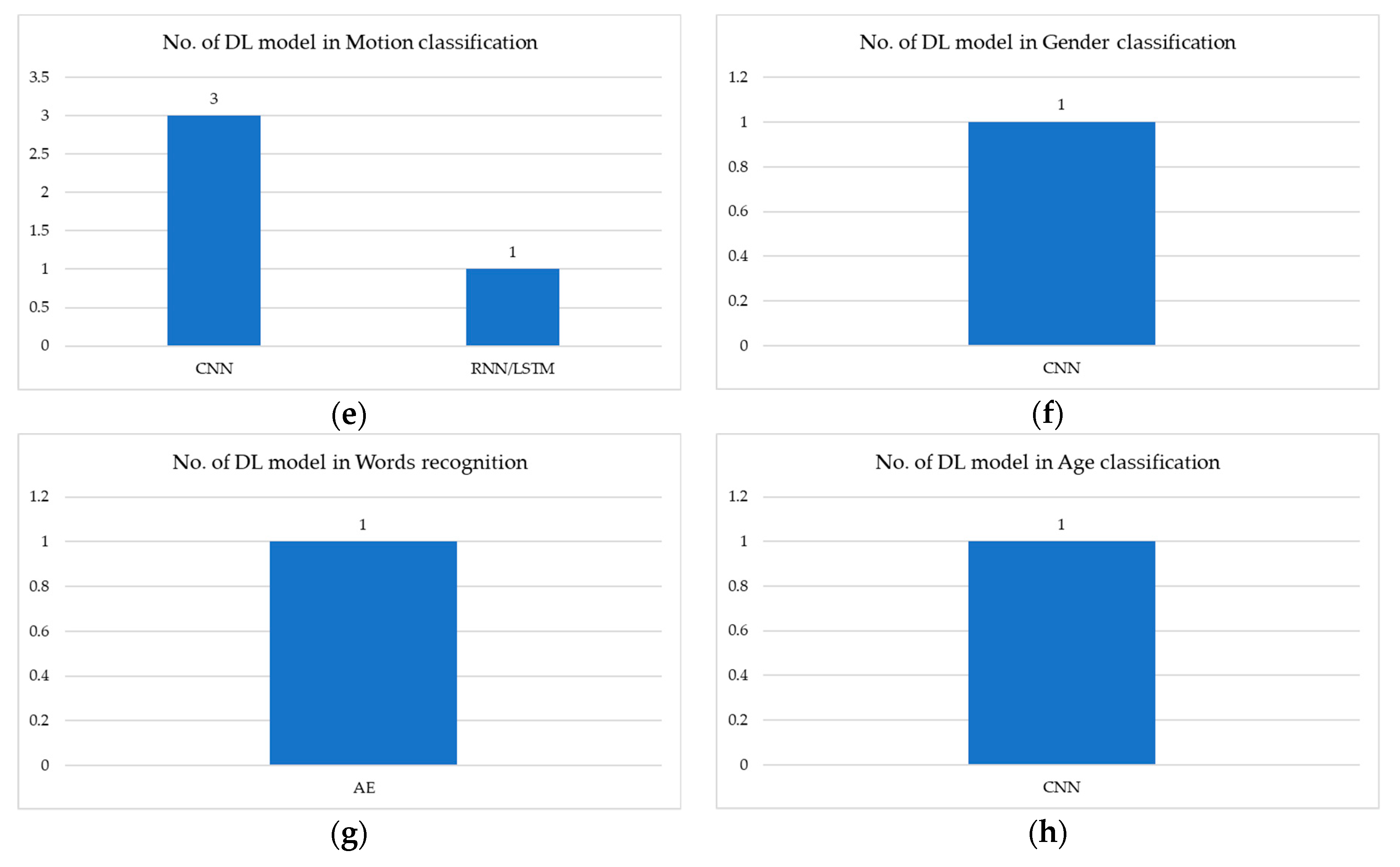
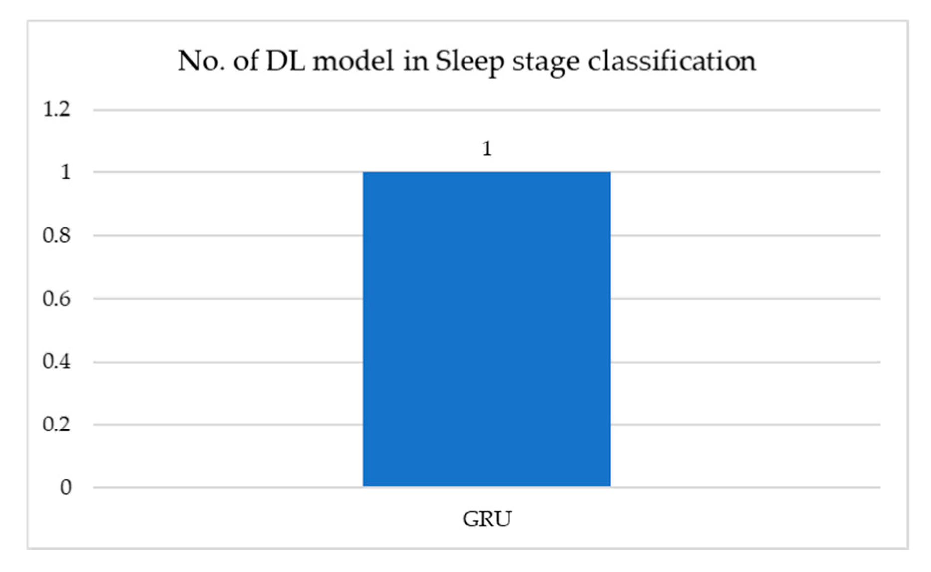
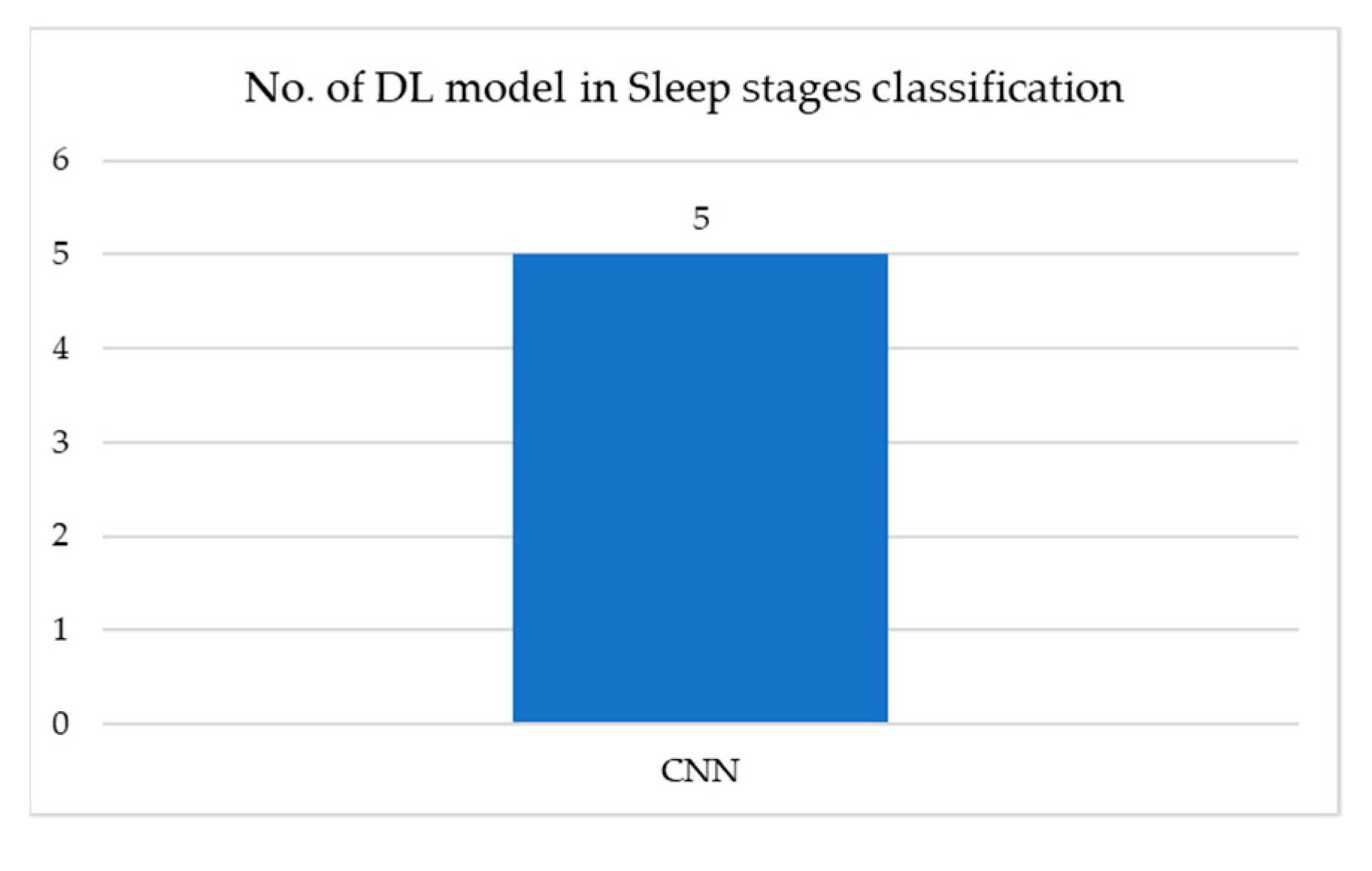
| EEG | Brain functionality classification [71,72,73,74,75,76,77,78,79,80,81,82,83,84,85,86,87,88,89,90,91], Brain disease classification [92,93,94,95,96,97,98,99,100,101,102,103,104,105,106,107,108,109,110,111,112,113,114,115,116,117,118,119,120,121], Emotion classification [122,123,124,125,126,127,128,129], Sleep-stage classification [130,131,132,133,134,135,136,137,138,139,140,141], Motion classification [142,143,144,145], Gender classification [146], Words classification [147], Age classification [148] |
| EOG | Sleep-stage classification [149] |
| Combination of signals | Sleep-stage classification [150,151,152,153,154] |
| Signal Modality | Public Dataset | Private Dataset | Hybrid Dataset |
|---|---|---|---|
| EMG | Table 3 | Table 4 | |
| ECG | Table 5 | Table 6 | Table 7 |
| EEG | Table 8 | Table 9 | Table 10 |
| EOG | Table 11 | ||
| Combination of signals | Table 12 |
| Medical Application | Medical Task | DL Model | Dataset Source | No. of Subjects | Performance |
|---|---|---|---|---|---|
| Hand motion recognition | Gesture recognition [10] | CNN+RNN | NinaProDB1 | 27 | Accuracy = 87.0% |
| NinaProDB2 | 40 | Accuracy = 82.2% | |||
| BioPatRec sub-database | 17 | Accuracy = 94.1% | |||
| CapgMyo sub-database | 18 | Accuracy = 99.7% | |||
| csl-hdemg databases | 5 | Accuracy = 94.5% | |||
| Gesture recognition [11] | CNN | NinaPro | 128 | Accuracy = 85.78% | |
| BioPatRec | 53 | Accuracy = 94.0% | |||
| Gesture signal classification [12] | CNN | MYO | 17 | Accuracy = 98.31% | |
| NinaPro | 10 | Accuracy = 68.98% | |||
| Hand gesture classification [13] | GFM | NinaPro database | 10 | Accuracy = 63.86 ± 5.12% | |
| Hand movement classification [16] | CNN+RNN | Ninapro project dataset | 78 | Accuracy = 87.3 ± 4.9% |
| Medical Application | Medical Task | DL Model | Dataset Source | No. of Subjects | Performance |
|---|---|---|---|---|---|
| Hand motion recognition | Chinese sign language recognition [9] | DBN | 6-D inertial sensor (3D-ACC and 3D-GYRO) | 8 | Accuracy = 95.1% (user-dependent test), Acc = 88.2% (user-independent test) |
| Hand-grasping classification [14] | SAE | MYO | 15 | Accuracy = 95%, SD = 3.58~1.25% | |
| Hand motion classification [15] | CNN | MYO | 7 | mean CE ± SD = 9.79 ± 4.57 | |
| Limb movement estimation [17] | CNN+RNN | EMG system (NCC Medical Co., LTD, Shanghai, China) | 8 | mean R2 = 90.3 ± 4.5% | |
| Muscle activity recognition | Multi-labeled movement information extraction [18] | CNN | ELSCH064NM3 from OT Bioelettronica, Turin, Italy | 14 | mean exact match rate = 78.7% and a mean Hamming loss = 2.9% |
| Muscle activity detection [19] | RNN | Vastus Lateralis and the Lateral Hamstring of a runner | N/A | Signal-to-noise ration < 5 | |
| Musculoskeletal force prediction [20] | CNN | Trigno Wireless EMG system, Delsys, USA | 156 | RMSE = 0.25, Std. = 0.13 | |
| Prosthetic limb control, Movement Intent decoder [21] | CNN | Grapevine NIP system (Ripple, Salt Lake City, UT, USA) | 2 | NMSE = 0.033 ± 0.017 | |
| LSTM | NMSE = 0.096 ± 0.013 | ||||
| Real-time, simultaneous myoelectric control system [22] | CNN | Eight pairs of bipolar surface electrodes (g.HiAmp, g-tec Inc.) | 17 | Accuracy = 91.61%, Standard error = 0.39 | |
| Wave form identification [23] | CNN | Tokushima University Hospital | 83 | Accuracy = 86% (test set), Accuracy = 100% (train set) |
| Medical Application | Medical Task | DL Model | Dataset Source | No. of Subject/Data | Performance |
|---|---|---|---|---|---|
| Heartbeat signal classification | Anomaly class identification [26] | LSTM+SVM, LSTM+MLR, LSTM+MLP | MIT-BIH Arrhythmia | 43 input features | LSTM+SVM = 42.86% LSTM+MLR = 51.43% LSTM+MLP = 50.0% |
| Atrial fibrillation detection [27] | STFT+CNN, SWT+CNN | MIT-BIH Atrial fibrillation | 23 annotated ECG recordings | STFT+CNN: Sensitivity = 98.34%, Specificity = 98.24%, Accuracy = 98.29%. SWT+CNN: Sensitivity = 98.79%, Specificity = 97.87%, Accuracy = 98.63% | |
| CAD ECG signals detection [28] | LSTM+CNN | PhysioNet | 47 | Accuracy = 99.85% | |
| Congestive heart failure detection [30] | LSTM | BIDMC-CHF | 15 | Accuracy = 99.22% | |
| MIT-BIH NSR | 18 | Accuracy = 98.85% | |||
| Fantasia | 40 | Accuracy = 98.92% | |||
| Dofetilide plasma concentrations prediction [31] | CNN | PhysioNet | 42 | Correlation (r = 0.85) | |
| ECG Characteristic detection [32] | CNN+RA | QT database (MIT-BIH Arrhythmia+ ST-T Database+ several other ECG databases) | 23 records (test set) | P-on = 0.4 ± 14.4 P-peak = −0.4 ± 10.1 P-off = −2.0 ± 12.7 QRS-on = −0.7 ± 10.9 QRS-off = −4.8 ± 13.1 T-peak = −0.3 ± 10.5 T-off = −0.3 ± 18.5 | |
| ECG signal compression [33] | AE | MIT-BIH arrhythmia | 48 records | Compression ratio = 106.45, Root mean square difference = 8.00% | |
| Electrocardiogram diagnosis [34] | CNN+BRNN | Chinese Cardiovascular Disease Database | 19K | Accuracy = 87.69% | |
| Heartbeat classification for continuous monitoring [35] | LSTM | MIT-BIH arrhythmia | N/A | VEB: Accuracy = 99.2%, Sensitivity = 93.0%, Specificity = 99.8% F1 = 95.5% SVEB: Accuracy = 98.3%, Sensitivity = 66.9%, Specificity = 99.8% F1 = 78.8% | |
| Heartbeat classification [36] | CNN | MIT-BIH Arrhythmia | 48 records | Accuracy = 96%, F1-score = 90% | |
| Heartbeat types classification [37] | CNN+RBM | MIT-BIH arrhythmia | 47 | AUC = 0.999 | |
| Heartbeats classification [38] | DBLSTM-WS | MIT-BIH arrhythmia | 48 records | Accuracy = 99.39% | |
| Heartbeats classification [39] | CNN | MIT-BIH arrhythmia | 48 records | Accuracy = 98.6% | |
| Multi-lead ECG classification [42] | DL-CCANet, TL-CCANet | MIT-BIH database | 48 records | DL-CCANet: Accuracy = 95.2% | |
| INCART database | 78 records | TL-CCANet: Accuracy = 95.52% | |||
| Premature ventricular contraction classification [45] | EBR | MIT-BIH arrhythmia | 119 records | Precision = 100%, Recall = 100%, Accuracy = 100% | |
| Ventricular and supraventricular heart beats detection [47] | RBM+DBM | MIT-BIH database | 44 records | Ventricular ectopic beats (Acc = 93.63%), Supraventricular ectopic beats (Acc = 95.57%) | |
| Heart disease classification | Arrhythmia classification [49] | AE+LSTM | MIT-BIH arrhythmia | 47 | Accuracy = 99.0%, Root mean square difference = 0.70% |
| Arrhythmia diagnosis [50] | CNN+LSTM | MIT-BIT arrhythmia | 47 | Accuracy = 98.10%, Sensitivity = 97.50%, Specificity = 98.70% | |
| Arrhythmias detection [51] | CNN | MIT-BIH arrhythmia | 48 | DB1: Accuracy = 97.87% DB2: Accuracy = 99.30% | |
| Atrial fibrillation (AF) automatically prediction [52] | CNN | MIT-BIH | 139 records | Accuracy = 98.7%, Sensitivity = 98.6%, Specificity = 98.7%. | |
| Beat-wise arrhythmia diagnosis [53] | AE+U-net | MIT-BIH AFDB + PAFDB + MIT-BIH NSRDB | 74 (evaluate), 65 (test) | Accuracy = 98.7% Sensitivity = 98.7% Specificity = 98.6% | |
| Cardiac Arrhythmia classification [54] | MLP, CNN | PhysioBank | 208 ECG recordings | Accuracy = 88.7% | |
| Kaggle | Accuracy = 83.5% | ||||
| Cardiac arrhythmias classification [55] | 1D-CNN | MIT-BIH Arrhythmia | 45 | Accuracy = 91.33% | |
| Cardiologist-Level Arrhythmia detection and classification [56] | CNN | Ziomonitor (iRhythm Technologies Inc, San Francisco, CA) | 53,877 patients | AUC = 0.97, Fi-score = 0.837, Sensitivity = 0.780 | |
| Early detection of myocardial ischemia [58] | CNN | PhysioNet | N/A | AUC = 89.6% Sensitivity = 84.4% Specificity = 84.9%, F1-score = 89.2% | |
| Heart Disease classification [59] | Faster RCNN | MIT-BIH | 47 | Accuracy = 99.21% | |
| Heart Diseases classification [60] | LSTM | PhysioNet | Accuracy = 98.4% | ||
| Sudden cardiac arrests (SCA) detection [63] | CNN | Creighton University Ventricular Tachyarrhythmia + MIT-BIH Malignant Ventricular Arrhythmia | 35 records + 22 records | Accuracy = 99.26% Sensitivity = 97.07% Specificity = 99.44% | |
| Sleep-stage classification | Apnea detection [64] | CNN | PhysioNet | 35 | Accuracy = 94.4% Sensitivity = 93.0% Specificity = 94.9% |
| Signal quality and sleep position classification [66] | CNN | MIT-BIH arrhythmia | 12 | C1 class: Precision = 0.99, Recall = 0.99 Sleep position: Precision = 0.99, Recall = 0.99 | |
| Sleep Apnea detection [68] | CNN | PhysioNet Apnea + University College Dublin | 70 records + 25 records | Accuracy = 87.6% Sensitivity = 83.1% Specificity = 90.3% AUC = 0.950 |
| Medical Application | Medical Task | DL Model | Dataset Source | No. of Subject/Data | Performance |
|---|---|---|---|---|---|
| Heartbeat signal classification | 6 types of ECG abnormalities classification [24] | CNN | Telehealth Network of Minas Gerais, Brazil | 1,558,415 patients | F1-score > 80% Specificity > 99% |
| Cardiologs and veritas detection [29] | CNN | ECGs recorded in the ED of HCMC | 1500 records | Cardiologs: Accuracy = 92.2% Sensitivity = 88.7% Specificity = 94.0% Veritas: Accuracy = 87.2% Sensitivity = 92.0% Specificity = 84.7% | |
| Left ventricular systolic dysfunction detection [41] | CNN | Mayo Clinic ECG | 16 056 adult patients | Accuracy = 86.5% Sensitivity = 82.5% Specificity = 86.8% | |
| Noise detection and screening model [43] | CNN | trauma intensive-care unit | 165,142,920 ECG II (10-second lead II electrocardiogram) | Positive prediction = 0.74, Negative prediction = 0.96, Sensitivity = 0.88, Specificity = 0.89, F1-score = 0.80, AUC = 0.93 | |
| Scalogram of ECG classification [46] | ResNet | Physikalisch-Technische Bundesanstalt (PTB)-ECG | 290 | Accuracy = 0.73 | |
| Chosun University (CU)-ECG | 100 | Accuracy = 0.94 | |||
| Heart disease classification | Diabetic subject detection [57] | 1D-CNN | Kasturba Medical Hospital (KMH), Manipal, India | 30 | Accuracy = 97.62%, Sensitivity = 100% |
| Heart failure detection on patients in ischemia and post-infarction [61] | CNN | Heart failure database (HFDB) | 128 ECG pairs | AUC = 84% | |
| Ischemia database (IDB) | 482 ECG pairs | AUC = 83% | |||
| Mental stress recognition [62] | CNN+LSTM | Zephyr BioHarness 3.0 | 18 | Accuracy = 83.9%, F1-score = 0.81, AUC = 0.92 | |
| Sleep-stage classification | Sleep apnea detection [67] | DNN, 1D-CNN, 2D-CNN, RNN, LSTM, GRU | SA dataset | 86 | Accuracy = 99.0%, Recall = 99.0% (1D-CNN and GRU) |
| Emotion classification | Stressful state classification [69] | RNN+CNN | Kwangwoon University in Korea | 13 | Accuracy = 87.39% |
| KU Leuven University in Belgium | 9 | Accuracy = 73.96% | |||
| Age and gender prediction | Age and gender prediction [70] | CNN | Mayo Clinic digital data vault | 275,056 | Accuracy = 90.4%, ACU = 0.97 (independent test data) |
| Medical Application | Medical Task | DL Model | Dataset Source | No. of Subject/Data | Performance |
|---|---|---|---|---|---|
| Heartbeat signal classification | Ventricular fibrillation detection [48] | 1D-CNN+ LSTM | PhysioNet MIT-BIH Malignant Ventricular Arrhythmia + Creighton University Ventricular Tachyarrhythmia + American Heart Association ECG Database | N/A | BAC = 99.3%, Sensitivity = 99.7%, Specificity = 98.9% |
| OHCA patients | N/A | BAC = 98.0%, Sensitivity = 99.2%, Specificity = 96.7% |
| Medical Application | Medical Task | DL Model | Dataset Source | No. of Subject/Data | Performance |
|---|---|---|---|---|---|
| Brain functionality classification | EEG session normal or abnormal detection [74] | 1D-CNN+RNN | TUH Abnormal EEG Corpus | 1488 abnormal + 1529 normal EEG sessions | Accuracy = 76.9% |
| Event-related potential (ERP) detection and analysis [76] | CNN | BCI competition II and III | 2 | AUC = 0.825 ± 0.064 | |
| Brain activity detection [81] | CNN | BCIC IV 2a. BCI competition IV data set 2a | 9 | Accuracy = 69% | |
| BCIC IV 2b. BCI competition IV 2b | 9 | Accuracy = 83% | |||
| Upper limb movement | 15 | Accuracy = 31% | |||
| Motor Imagery classification [83] | RNN+3D-CNN | BCI competition IV-2a 4-class Motor Imagery (MI) dataset | 9 | Accuracy = 74.46% | |
| Motor Imagery EEG classification [85] | CNN | BCI Competition IV | 9 | Accuracy = 87.94% | |
| Motor Imagery EEG Decoding [86] | CP-MixedNet | BCI competition IV 2a | 9 | Accuracy = 74.6% Precision = 73.9% Recall = 74.7% F1-score = 0.743 | |
| HGD dataset | 14 | Accuracy = 93.7% Precision = 73.7% Recall = 93.7% F1-score = 0.937 | |||
| Multiclass Motor Imagery classification [87] | CNN | BCI Competition Dataset 2a | 9 | Mean kappa = 0.61 St. Dev = 0.101 | |
| Online decoding of Motor Imagery movement [88] | LSTM, CNN, RCNN | BCI Competition IV | 20 | LSTM: Accuracy = 66.97 ± 6.45% CNN: Accuracy = 66.2 ± 7.21% RCNN: Accuracy = 77.72 ± 6.5% | |
| Prediction of bispectral index during target-controlled infusion of propofol and remifentanil [89] | LSTM | vitaldb | 180 data points | concordance correlation coefficient (95% CI) = 0.561 (0.560 to 0.562) | |
| EEG-based BCIs classification [91] | CNN | P300 Evoked Potentials (P300) | 8 | EEGNet: SNRs = 20.43 DeepCNN: SNRs = 20.50 ShallowCNN: SNRs = 20.53 | |
| Feedback Error-Related Negativity (ERN) | 26 | EEGNet: SNRs = 20.26 DeepCNN: SNRs = 20.39 ShallowCNN: SNRs = 20.31 | |||
| MI | 9 | EEGNet: SNRs = 25.50 DeepCNN: SNRs = 25.57 ShallowCNN: SNRs = 25.60 | |||
| Brain disease classification | Aberrant epileptic seizure identification [92] | CNN+LSTM | University of Bonn | 28 | AUC = 0.9703 Accuracy = 90% |
| Brain disorders diagnosis [95] | HMM+SDAE | TUH EEG Corpus | 13,500 patients | Sensitivity > 90% Specificity < 5% | |
| Depression screening [97] | CNN | Bonn University | 15 normal + 15 depressed patients | Left hemisphere: Accuracy = 93.5% Right hemisphere: Accuracy = 96.0% | |
| EEG-based epileptic seizure detection [102] | CNN | CHB-MIT dataset | 23 | Accuracy = 98.3% Sensitivity = 96.7% Specificity = 99.1% | |
| Epilepsy detection by using scalogram [104] | CNN | Bonn University | A: healthy 100 segment B: healthy 100 segment C: patient 100 segment D: patient 100 segment E: patient 100 segment | A-E: Accuracy = 99.5% A-D: Accuracy = 100% D-E: Accuracy = 98.5% A-D-E: Accuracy = 99.0% A-B-C-D-E: Accuracy = 93.6% | |
| Epileptic EEG recording classification [106] | CNN | Bern-Barcelona EEG | 5 | Accuracy = 98.9 ± 0.08% | |
| Epileptic Seizure Recognition datasets | 500 | Accuracy = 99.8 ± 0.13% | |||
| Epileptic Seizure prediction [107] | CNN | Seizure Prediction Challenge | 5 | AUC = 0.79 | |
| Epileptic Seizure prediction [108] | CNN+LSTM | CHB-MIT EEG dataset | 22 | Accuracy = 99.6% | |
| Epileptic seizures detection using EEG [110] | LSTM | Bonn University | A: healthy 100 segment B: healthy 100 segment C: patient 100 segment D: patient 100 segment E: patient 100 segment | Accuracy = 100% Sensitivity = 100% Specificity = 100% | |
| Epileptic seizures prediction [111] | LSTM | Open CHB-MIT Scalp | 23 | Sensitivity = 100% Specificity = 99.28% | |
| Seizure detection in multimodal EEG-fNIRs [114] | LSTM | BCI competition IV 2b dataset | 40 | Sensitivity = 89.7% Specificity = 95.5% | |
| Seizure Detection [118] | CNN+AE | CHB-MIT dataset | 23 | Accuracy = 94.37% F1-score = 85.34% | |
| Seizure detection [119] | LSTM | University of Bonn | A: healthy 100 segment B: healthy 100 segment C: patient 100 segment D: patient 100 segment E: patient 100 segment | Accuracy = 95.54% AUC = 0.9582 | |
| Emotion classification | Emotion recognition [122] | 2D-CNN | DEAP dataset | 32 | Accuracy = 73.4% |
| Emotion Recognition [124] | RNN | SJTU emotion EEG dataset | 15 | Accuracy = 89.5% | |
| CK+ facial expression | 327 images | Accuracy = 95.4% | |||
| Fear level classification based on emotional dimensions [125] | DNN | DEAP database | 32 | Accuracy = 59.84% F1-score = 58.78% | |
| Human emotion recognition [126] | RBM | SEED-IV dataset | 15 | Accuracy = 85.11% | |
| Recognition of emotion [127] | DBN-GC+RBM | DEAP dataset | 32 | Arousal: Accuracy = 75.92% Valence: Accuracy = 76.83% | |
| Relaxation classification [128] | CNN | OpenBCI | 7 | 1s temporal window: Accuracy = 55.46% 2s temporal window: Accuracy = 98.96% | |
| Valence and arousal classification [129] | LSTM | DEAP dataset | 32 | Arousal: Accuracy = 74.65% Valence: Accuracy = 78% | |
| Sleep-stage classification | Detect multiple sleep micro-events in EEG [130] | CNN | Montreal Archives of Sleep Studies dataset | 19 | Precision = 0.3 Recall = 0.95 |
| Stanford Sleep Cohort dataset | 26 | Precision = 0.58 Recall = 0.43 | |||
| Wisconsin Sleep Cohort dataset | 30 | Precision = 0.79 Recall = 0.1 | |||
| MESA dataset | 1000 | N/A | |||
| Real-time detection of sleep spindles [133] | CNN+RNN | Montreal archive of sleep studies | 19 | Sensitivity = 90.07 ± 2.16% Specificity = 96.19 ± 0.71% FDR = 30.36 ± 5.88% F1-score = 0.75 ± 0.05 AUROC = 98.97 ± 0.13% | |
| DREAMS database | 8 | Sensitivity = 77.85 ± 4.28% Specificity = 94.2 ± 1.26% FDR = 61.96 ± 7.39% F1-score = 0.48 ± 0.07 AUROC = 95.97 ± 0.96% | |||
| Sleep-stage classification [135] | CNN | PhysioNet (Sleep- EDF dataset) | 20 | Setting 1: Accuracy = 79.8% Setting 2: Accuracy = 82.6% | |
| Sleep-stage classification [136] | RNN+SVM | PhysioNet (Sleep-EDF dataset) | 20 | Setting 1: Accuracy = 79.1% Setting 2: Accuracy = 82.5% | |
| Sleep-stage classification [137] | CU-CNN | UCD dataset | 25 | Accuracy = 87% Kappa = 0.8 | |
| MIT-BIH datasets | 16 records | Accuracy = 99.9% Kappa = 0.904 | |||
| Sleep-stage scoring/detection [138] | CNN+RNN | PhysioNet (Sleep-EDF datasets) | 258 | Accuracy = 84.26% F1-score = 79.66% Kappa = 0.79 | |
| Sleep stages classification from single-channel EEG [139] | CNN | PhysioNet | 8 | Accuracy = 98.10%, 96.86%, 93.11%, 92.95%, 93.55%, Kappa = 0.98%, 0.94%, 0.90%, 0.86%,0.89%, | |
| Motion classification | Movement intention recognition of disable person [143] | LSTM | MI-based eegmmidb dataset | 12 | Accuracy = 68.20% |
| Gender classification | Gender prediction from brain rhythms [146] | CNN | Brain Resource International Database | 1308 | Accuracy > 80% (p < 10−5) |
| Words classification | Words recognition of speech-impaired people from brain-generated signals [147] | DN-AE-NTM | P300 EEG dataset | 9 | Accuracy = 97.5% |
| EEG recording of individuals with alcoholism and control individuals | 64 | Accuracy = 95% | |||
| EEGMMIDB | 109 | Accuracy = 98% | |||
| MNIST | 60K samples | Accuracy = 99.4% | |||
| ORL | 10 images | Accuracy = 99.1% |
| Medical Application | Medical Task | DL Model | Dataset Source | No. of Subject/Data | Performance |
|---|---|---|---|---|---|
| Brain functionality classification | Cerebral Dominance detection [71] | CNN+SVM | Firat University Hospital (Nicolet EEG v32 device) | 67 | AUC = 0.83 ± 0.05 |
| Complexity of peri-perceptual processes of familiarity detection [72] | SNN | “Hamrah Clinic” of Tabriz, Iran | 20 | Accuracy = 83% Sensitivity = 84% Specificity = 86% F1-score = 84% | |
| Devanagari script input-based P300 speller detection [73] | SAE, DCNN | National Institute of Technology Raipur (ctiCAP Xpress V-amp EEG recorder) | 10 | Accuracy = 88.22% | |
| Walking Imagery Evaluation [75] | MMDPN | Biosemi ActiveTwo system | 9 | Text-MMDPN: AUC = 0.7984 VE-MMDPN: AUC = 0.9424 | |
| EEG event-related classification on children with ADHD from healthy controls [77] | CNN+RNN | Technical University of Dresden | 144 | Accuracy = 83% | |
| Focal epileptiform discharges detection [78] | CNN+RNN | Department of Clin. Neurophysiology and Neurology, Medisch Spectrum Twente, Enschede, The Netherlands | 50 | AUC = 0.94 Sensitivity = 47.4% Specificity = 98.0% | |
| Human Mental workload Recognition [79] | EL-SDAE | Simulated Human Machine systems | 8 | Accuracy = 92.02% | |
| Identify patterns of brain activity of children at idle time and playing videogame time [80] | CNN | University of Houston | 233 | Accuracy = 67% | |
| Cross-task mental workload assessment [82] | RNN+3D-CNN | Tsinghua University | 20 | Accuracy = 88.9%, | |
| Spectral and temporal feature learning for mental workload assessment [90] | CNN+TCN | Tsinghua University | 17 | Accuracy = 91.9%, | |
| Brain disease classification | Automatic diagnosis of unipolar depression [93] | 1D-CNN, 1D-CNN+LSTM | hospital Universiti Sains Malaysia (HUSM) | 63 | 1D-CNN: Accuracy = 98.32% Precision = 99.78% Recall = 98.34% F-score = 97.65% 1D-CNN+LSTM: Accuracy = 95.97% Precision = 99.23% Recall = 93.67% F-score = 95.14% |
| Brain disease detection [94] | CNN, RNN, DNN | EEG data of the University of California Irvine | 122 | CNN: F1-score = 0.94 RNN: F1-score = 0.73 DNN: F1-score = 0.70 | |
| Confusion state induction and detection [96] | CNN | Emotiv Epoc+ | 16 | Accuracy = 71.36% | |
| Early Alzheimer’s disease diagnosis [98] | DCssCDBM | Beijing Easy monitor Technology | 14 | Accuracy = 95.04% | |
| Early prediction of epileptic seizure [99] | CNN+LSTM | Department of Neurology at the First Affiliated Hospital of Xinjiang Medical University | 15 | Accuracy = 93.40% Sensitivity = 91.88% Specificity = 86.13% | |
| Early stage Alzheimer disease detection [100] | CNN | Chosun University Hospital (CUH, Gwangju, S. Korea) and Gwangju Optimal Dementia Center located in Gwangju Senior Technology Center (Gwangju, S. Korea) | 10 | Accuracy = 59.4% Std. = 22.7 | |
| Epileptic discharge detection [105] | CNN | EEG/fMRI study | 30 | Sensitivity = 84.2% | |
| Epileptic seizure prediction [109] | CNN | Intracranial electrodes (magenta circles) | 10 | Sensitivity = 69% | |
| Identifying Schizophrenia from EEG connectivity Patterns [112] | CNN | Lomonosov Moscow State University | 84 | Accuracy = 91.69% | |
| Seizure classification [113] | CNN | Diagnosis of medication refractory TLE based on International League Against Epilepsy (ILAE) criteria | 50 | Positive Predictio n = 88 ± 7%, Negative Prediction = 79 ± 8%, Accuracy < 50% | |
| Seizure detection [117] | 3D-CNN | Hospital of Xinjiang Medical University | 13 | Accuracy = 90.00% Sensitivity = 88.90% Specificity = 93.78% | |
| Seizure detection [120] | CNN | Department of Physiology, College of Medicine, The Catholic University of Korea | 249 | Sensitivity = 100% Positive Prediction = 98% | |
| Tracking both the level of consciousness and delirium [121] | CNN+LSTM | Partners Institutional Review Board (IRB) | 174 | Accuracy = 70% Sensitivity = 69% Specificity = 3% AUC = 0.80 | |
| Emotion classification | Human Intention Recognition [4] | CNN+LSTM | BCI2000 instrumentation | 108 subjects, 3,145,160 EEG records | Accuracy = 98.3% |
| Sleep-stage classification | Driving Fatigue detection from EEG [131] | PCANet+SVM | Guangdong Provincial Work Injury Rehabilitation Center | 6 | Accuracy = 95% |
| Identifying abnormal EEGs, age and sleep-stage classification [132] | CNN | Department of Neurology in Massachusetts General Hospital | 8522 EEGs | EEGs: AUC = 0.917 EEGs+Age: AUC = 0.924 EEGs+Age+Sleep: AUC = 0.925 | |
| Sleep stages classification [141] | CNN+LSTM | Chronobiology and Sleep Research, Institute of Pharmacology and Toxicology, University of Zurich, Zurich, Switzerland | 75 records | Kappa = 0.8 | |
| Motion classification | Problem-solving behavioral pattern characterization [144] | CNN | Fakultät Management und Vertrieb, Hochschule Heilbronn Campus Schwäbisch Hall, 74523 Schwäbisch Hall, Germany | 26 | Accuracy = 99% |
| Rapid eye movement behavior disorder [145] | CNN | Center for Advanced Research in Sleep Medicine of the Hôpital du Sacrè- Coeur de Montréal | 212 | Accuracy = 80 ± 1% AUC = 87 ± 1% |
| Medical Application | Medical Task | DL Model | Dataset Source | No. of Subject/Data | Performance |
|---|---|---|---|---|---|
| Brain disease classification | EEG classification of Motor Imagery [101] | CNN + VAE | BCI Competition IV dataset 2b | 9 | Kappa = 0.564 |
| Ag-AgCl electrodes | 5 | 3-electrode EEG: Kappa = 0.568 5-electrode EEG: Kappa = 0.603 | |||
| Sleep-stage classification | Real-time sleep-stage classification [134] | CNN+LSTM | SIESTA database | 19 | Kappa = 0.760 ± 0.022 |
| Data Science, Philips Research, Eindhoven, Netherlands | 29 | Kappa = 0.727 ± 0.005 | |||
| Age classification | Age of children classification on performing a verb-generation task, a monosyllable speech-elicitation task [148] | CNN | BCI Competition IV | 9 | Accuracy = 95% |
| University of Toronto, Toronto, Canada | 92 |
| Medical Application | Medical Task | DL Model | Dataset Source | No. of Subject/Data | Performance |
|---|---|---|---|---|---|
| Sleep stages classification | Sleep-stage labeling [149] | GRU | PhysioNet | 6 sleep stages and 6 sleep disorders | Accuracy = 69.25% |
| Medical Application | Medical Task | DL Model | Dataset Source | No. of Subject/Data | Performance |
|---|---|---|---|---|---|
| Sleep stages classification | Sleep stages classification [150] | CNN | PhysioNet | 20 | Accuracy = 81% F1-score = 72% |
| Sleep-stage classification [151] | CNN | MASS dataset - session 3 | 62 records | Sensitivity = 85% Specificity = 100% | |
| Sleep-stage classification [152] | CNN | PhysioNet Sleep-EDF Database (SLPEDF-DB) | 19 | Kappa = 0.67 ± 0.05 | |
| Montreal Archive of Sleep Studies (MASS-DB) | 200 | Kappa = 0.74 ± 0.01 | |||
| CAP Sleep Database (CAPSLP-DB) | 112 | Kappa = 0.61 ± 0.01 | |||
| RBD Database (RBD-DB) | 21 | Kappa = 0.48 ± 0.07 | |||
| Sleep-stage classification [153] | 1D-CNN | Sleep-EDF | 9 | 6 sleep classes: Accuracy = 98.06%, 94.64%, 92.36%, 91.22%, 91.00% | |
| Sleep-EDFX | 61 | 6 sleep classes: Accuracy = 97.62%, 94.34%, 92.33%, 90.98%, 89.54% | |||
| Classification of brain and artifactual independent component (IC) [154] | CNN | Electrical Geodesic Inc, EEG System Net 300 | 2048 samples | EEG: Accuracy = 92.4% MEG: Accuracy = 95.4% EEG+MEG: Accuracy = 95.6% |
© 2020 by the authors. Licensee MDPI, Basel, Switzerland. This article is an open access article distributed under the terms and conditions of the Creative Commons Attribution (CC BY) license (http://creativecommons.org/licenses/by/4.0/).
Share and Cite
Rim, B.; Sung, N.-J.; Min, S.; Hong, M. Deep Learning in Physiological Signal Data: A Survey. Sensors 2020, 20, 969. https://doi.org/10.3390/s20040969
Rim B, Sung N-J, Min S, Hong M. Deep Learning in Physiological Signal Data: A Survey. Sensors. 2020; 20(4):969. https://doi.org/10.3390/s20040969
Chicago/Turabian StyleRim, Beanbonyka, Nak-Jun Sung, Sedong Min, and Min Hong. 2020. "Deep Learning in Physiological Signal Data: A Survey" Sensors 20, no. 4: 969. https://doi.org/10.3390/s20040969
APA StyleRim, B., Sung, N.-J., Min, S., & Hong, M. (2020). Deep Learning in Physiological Signal Data: A Survey. Sensors, 20(4), 969. https://doi.org/10.3390/s20040969