Wearable Sensors Detect Differences between the Sexes in Lower Limb Electromyographic Activity and Pelvis 3D Kinematics during Running


 , ,
, ,
Abstract
1. Introduction
2. Materials and Methods
2.1. Participants
2.2. Procedure
2.3. Data Analysis
2.4. Statistical Analysis
3. Results
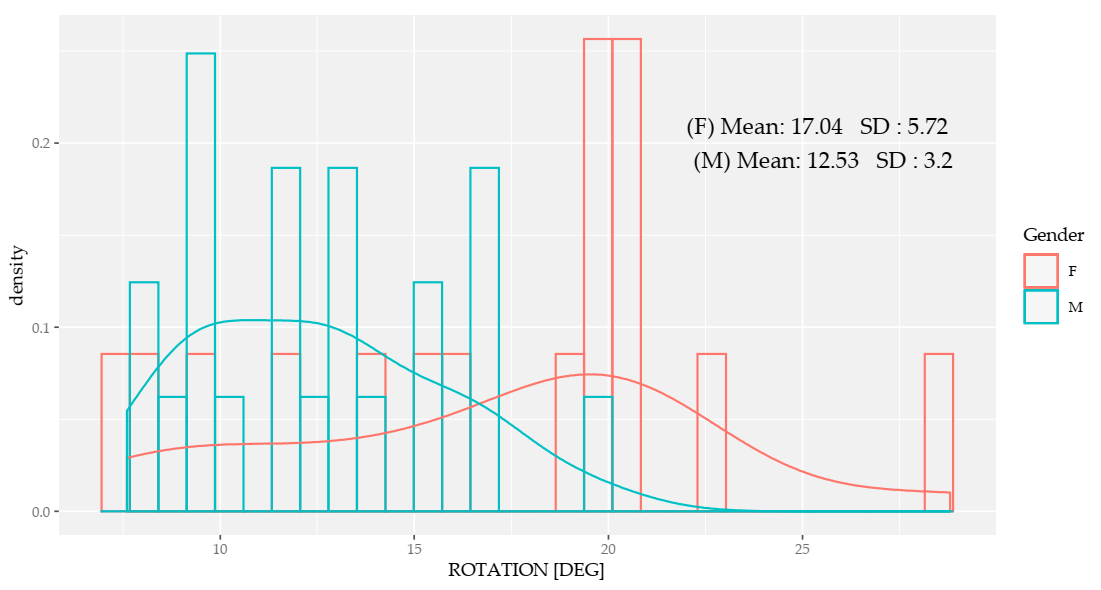
3.1. Kinematics of the Pelvis
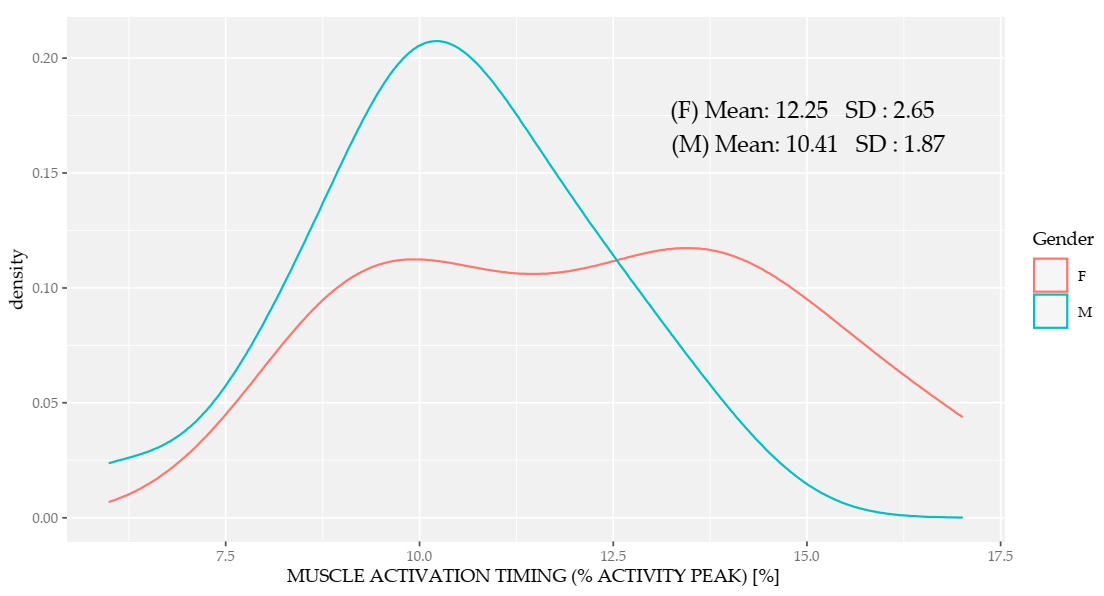
3.2. Mean Running Cycle Muscle Activation
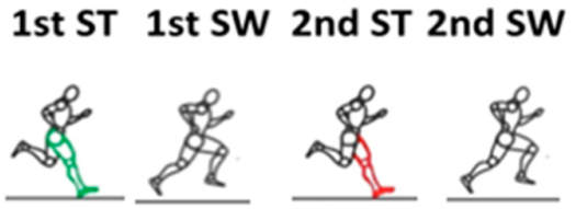
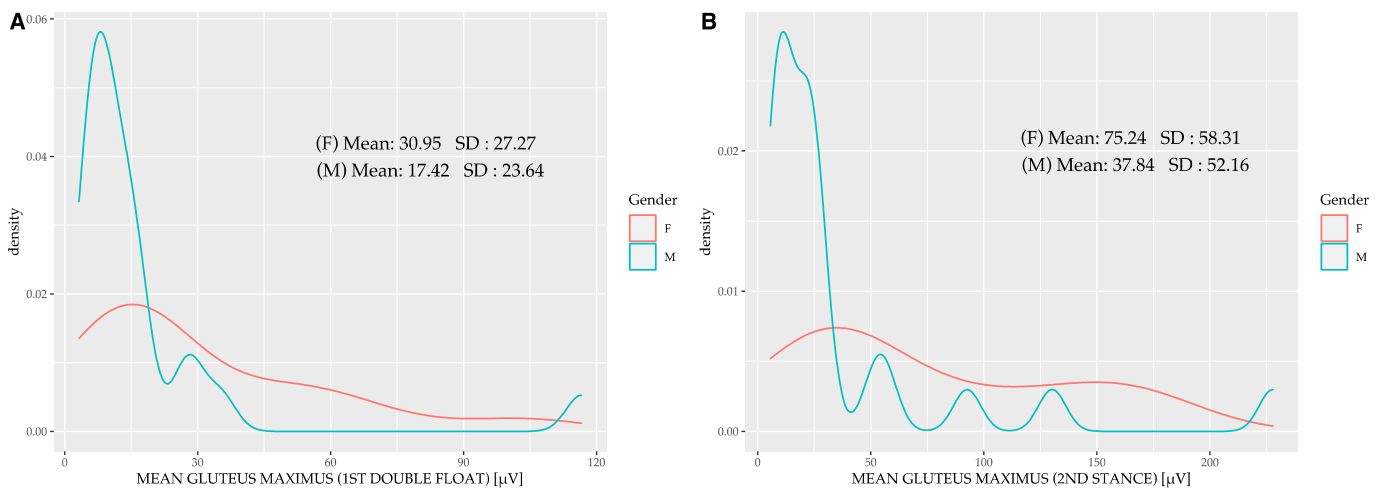
3.3. Muscle Activation for Each of the Phases
4. Discussion
5. Conclusions
Author Contributions
Funding
Acknowledgments
Conflicts of Interest
References
- Vidal, A.B.; Monezi, L.A.; Sarro, K.; De Barros, R.M.L. Analysis of required coefficient of friction in running and walking. Sports Biomech. 2019, 1–13. [Google Scholar] [CrossRef]
- Bramah, C.; Preece, S.J.; Gill, N.; Herrington, L. Is There a Pathological Gait Associated With Common Soft Tissue Running Injuries? Am. J. Sports Med. 2018, 46, 3023–3031. [Google Scholar] [CrossRef]
- Abadía, S.; Medina, F.X.; Sánchez, R.; Bantulà, J.; Fornons, D.; Bastida, N.; Augé, A.; Corderas, F.; Vega, S.; Pujadas, X. Entre el boom atlético y la cooperación social. Las carreras solidarias y el ejemplo de la Trailwalker España 2013. Península Inval. 2014, 9, 105–123. [Google Scholar] [CrossRef][Green Version]
- Fields, K.B. Running Injuries—Changing Trends and Demographics. Curr. Sports Med. Rep. 2011, 10, 299–303. [Google Scholar] [CrossRef] [PubMed]
- Saragiotto, B.T.; Di Pierro, C.; Lopes, A.D. Risk factors and injury prevention in elite athletes: A descriptive study of the opinions of physical therapists, doctors and trainers. Braz. J. Phys. Ther. 2014, 18, 137–143. [Google Scholar] [CrossRef] [PubMed]
- Salas Sanchez, J.; Latorre Roman, P.A.; Soto Hermoso, V.M. Body Composition and Strength of the Veteran Athlete: Effect of Aging. Apunt. Med. Sport 2013, 48, 137–142. [Google Scholar]
- Boling, M.; Padua, D.; Marshall, S.; Guskiewicz, K.; Pyne, S.; Beutler, A. Gender differences in the incidence and prevalence of patellofemoral pain syndrome. Scand. J. Med. Sci. Sports 2010, 20, 725–730. [Google Scholar] [CrossRef]
- Preece, S.J.; Bramah, C.; Mason, D. The biomechanical characteristics of high-performance endurance running. Eur. J. Sport Sci. 2019, 19, 784–792. [Google Scholar] [CrossRef]
- Williams, D.S.B.; Welch, L.M. Male and female runners demonstrate different sagittal plane mechanics as a function of static hamstring flexibility. Rev. Bras. Fisioter. 2015, 19, 421–428. [Google Scholar] [CrossRef]
- Barnes, K.R.; Hopkins, W.G.; McGuigan, M.R.; Northuis, M.E.; Kilding, A.E. Effects of Resistance Training on Running Economy and Cross-country Performance. Med. Sci. Sports Exerc. 2013, 45, 2322–2331. [Google Scholar] [CrossRef]
- Taunton, J.E.; Ryan, M.B.; Clement, D.B.; McKenzie, D.C.; Lloyd-Smith, D.R.; Zumbo, B.D. A prospective study of running injuries: The Vancouver Sun Run “In Training” clinics. Br. J. Sports Med. 2003, 37, 239–244. [Google Scholar] [CrossRef] [PubMed]
- Taunton, J.E.; Ryan, M.B.; Clement, D.B.; McKenzie, D.C.; Lloyd-Smith, D.R.; Zumbo, B.D. A retrospective case-control analysis of 2002 running injuries. Br. J. Sports Med. 2002, 36, 95–101. [Google Scholar] [CrossRef] [PubMed]
- Dorn, T.W.; Schache, A.G.; Pandy, M.G. Muscular strategy shift in human running: Dependence of running speed on hip and ankle muscle performance. J. Exp. Biol. 2012, 215, 1944–1956. [Google Scholar] [CrossRef] [PubMed]
- Zamparo, P.; Perini, R.; Peano, C.; Di Prampero, P.E. The Self Selected Speed of Running in Recreational Long Distance Runners. Int. J. Sports Med. 2001, 22, 598–604. [Google Scholar] [CrossRef]
- Ekkekakis, P.; Hall, E.; Petruzzello, S. Affective Responses to a Graded Treadmill Test: Is the Ventilatory Threshold the Turning Point Toward Negativity? J. Sport Exerc. Psychol. 2002, 24, 52–53. [Google Scholar]
- Liu, S.-H.; Lin, C.-B.; Chen, Y.; Chen, W.; Huang, T.-S.; Hsu, C.-Y. An EMG Patch for the Real-Time Monitoring of Muscle-Fatigue Conditions during Exercise. Sensors 2019, 19, 3108. [Google Scholar] [CrossRef]
- Yoong, N.K.M.; Perring, J.; Mobbs, R.J. Commercial Postural Devices: A Review. Sensors 2019, 19, 5128. [Google Scholar] [CrossRef]
- Stirling, L.M.; Von Tscharner, V.; Kugler, P.; Nigg, B.M. Classification of muscle activity based on effort level during constant pace running. J. Electromyogr. Kinesiol. 2011, 21, 566–571. [Google Scholar] [CrossRef]
- Von Tscharner, V.; Eskofier, B.; Federolf, P. Removal of the electrocardiogram signal from surface EMG recordings using non-linearly scaled wavelets. J. Electromyogr. Kinesiol. 2011, 21, 683–688. [Google Scholar] [CrossRef]
- Farina, D.; Merletti, R.; Enoka, R.M.; Cope, K.A.; Watson, M.T.; Foster, W.M.; Sehnert, S.S.; Risby, T.H. The extraction of neural strategies from the surface EMG. J. Appl. Physiol. 2004, 96, 1486–1495. [Google Scholar] [CrossRef]
- Schubert, A.G.; Kempf, J.; Heiderscheit, B.C. Influence of Stride Frequency and Length on Running Mechanics: A Systematic Review. Sports Health 2014, 6, 210–217. [Google Scholar] [CrossRef] [PubMed]
- Howard, R.M.; Conway, R.; Harrison, A.J. Muscle activity in sprinting: A review. Sports Biomech. 2018, 17, 1–17. [Google Scholar] [CrossRef] [PubMed]
- León Sánchez, J.M. Determinación De Los Datos Normativos De La Actividad Muscular Del Miembro Inferior Y De Los Parámetros Espacio-Temporales Durante La Carrera. Ph.D. Thesis, Universidad CEU Cardenal Herrera, Moncada, Spain, 2017. [Google Scholar]
- Perpiña, S.M. Fiabilidad Del Sistema Inercial Durante El Análisis Biomecánico De La carrera a Pie En Triatletas: Establecimiento Del Patrón Cinemático Normativo. Ph.D. Thesis, Universidad CEU Cardenal Herrera, Moncada, Spain, 2017. [Google Scholar]
- Heise, G.D.; Shinohara, M.; Binks, L. Biarticular Leg Muscles and Links to Running Economy. Int. J. Sports Med. 2008, 29, 688–691. [Google Scholar] [CrossRef] [PubMed]
- Bergamini, E.; Picerno, P.; Pillet, H.; Natta, F.; Thoreux, P.; Camomilla, V. Estimation of temporal parameters during sprint running using a trunk-mounted inertial measurement unit. J. Biomech. 2012, 45, 1123–1126. [Google Scholar] [CrossRef]
- Gazendam, M.G.J.; Hof, A.L. Averaged EMG profiles in jogging and running at different speeds. Gait Posture 2007, 25, 604–614. [Google Scholar] [CrossRef]
- Cappellini, G.; Ivanenko, Y.P.; Poppele, R.E.; Lacquaniti, F. Motor Patterns in Human Walking and Running. J. Neurophysiol. 2006, 95, 3426–3437. [Google Scholar] [CrossRef]
- Wall-Scheffler, C.M.; Myers, M.J. The Biomechanical and Energetic Advantages of a Mediolaterally Wide Pelvis in Women. Anat. Rec. 2017, 300, 764–775. [Google Scholar] [CrossRef]
- Schache, A.G.; Blanch, P.; Rath, D.; Wrigley, T.; Bennell, K. Differences between the sexes in the three-dimensional angular rotations of the lumbo-pelvic-hip complex during treadmill running. J. Sports Sci. 2003, 21, 105–118. [Google Scholar] [CrossRef]
- Ferber, R.; Davis, I.M.; Iii, D.S.W. Gender differences in lower extremity mechanics during running. Clin. Biomech. 2003, 18, 350–357. [Google Scholar] [CrossRef]
- Taborri, J.; Keogh, J.; Kos, A.; Santuz, A.; Umek, A.; Urbanczyk, C.; Van Der Kruk, E.; Rossi, S. Sport Biomechanics Applications Using Inertial, Force, and EMG Sensors: A Literature Overview. Appl. Bionics Biomech. 2020, 2020, 1–18. [Google Scholar] [CrossRef]
- Queen, R.M.; Gross, M.T.; Liu, H.-Y. Repeatability of lower extremity kinetics and kinematics for standardized and self-selected running speeds. Gait Posture 2006, 23, 282–287. [Google Scholar] [CrossRef] [PubMed]
- Lavcanska, V.; Taylor, N.F.; Schache, A.G. Familiarization to treadmill running in young unimpaired adults. Hum. Mov. Sci. 2005, 24, 544–557. [Google Scholar] [CrossRef] [PubMed]
- McNair, P.J.; Marshall, R.N. Kinematic and kinetic parameters associated with running in different shoes. Br. J. Sports Med. 1994, 28, 256–260. [Google Scholar] [CrossRef] [PubMed]
- Hermens, H.J.; Freriks, B.; Disselhorst-Klug, C.; Rau, G. Development of recommendations for SEMG sensors and sensor placement procedures. J Electromyogr. Kinesiol. 2000, 10, 361–374. [Google Scholar] [CrossRef]
- BTS. 2020. Available online: Https://www.Btsbioengineering.Com/ (accessed on 12 November 2020).
- Stegeman, D.F.; Blok, J.H.; Hermens, H.J.; Roeleveld, K. Surface EMG models: Properties and applications. J. Electromyogr. Kinesiol. 2000, 10, 313–326. [Google Scholar] [CrossRef]
- Stegeman, D.F.; Linsenn, W.F. Muscle fiber action potential changues and surface EMG: A simulation study. J. Electromyogr. Kinesiol. 1992, 2, 130–140. [Google Scholar] [CrossRef]
- Õunpuu, S. The Biomechanics of Walking and Running. Clin. Sports Med. 1994, 13, 843–863. [Google Scholar] [CrossRef]
- Lussiana, T.; Gindre, C. Feel your stride and find your preferred running speed. Biol. Open 2016, 5, 45–48. [Google Scholar] [CrossRef]
- Kong, P.W.; Candelaria, N.G.; Tomaka, J. Perception of self-selected running speed is influenced by the treadmill but not footwear. Sports Biomech. 2009, 8, 52–59. [Google Scholar] [CrossRef]
- Novacheck, T.F. Running injuries: A biomechanical approach. J. Bone Jt. Surg. Am. 1998, 80A, 1220–1233. [Google Scholar] [CrossRef]
- Lewis, C.L.; Laudicina, N.M.; Khuu, A.; Loverro, K.L. The Human Pelvis: Variation in Structure and Function During Gait. Anat. Rec. 2017, 300, 633–642. [Google Scholar] [CrossRef] [PubMed]
- Aura, O.; Komi, P.V. Mechanical Efficiency of Pure Positive and Pure Negative Work with Special Reference to the Work Intensity. Int. J. Sports Med. 1986, 7, 44–49. [Google Scholar] [CrossRef]
- Anderson, P.T. Biomechanics and Running Economy. Sports Med. 1996, 22, 76–89. [Google Scholar] [CrossRef]
- Novacheck, T.F. The biomechanics of running. Gait Posture 1998, 7, 77–95. [Google Scholar] [CrossRef]
- Vaughan, C.L. Biomechanics of running gait. Crit. Rev. Biomed. Eng. 1984, 12, 1–48. [Google Scholar]
- Bramah, C.; Preece, S.J.; Gill, N.; Herrington, L. A 10% Increase in Step Rate Improves Running Kinematics and Clinical Outcomes in Runners With Patellofemoral Pain at 4 Weeks and 3 Months. Am. J. Sports Med. 2019, 47, 3406–3413. [Google Scholar] [CrossRef]
- Neal, M.; Fleming, N.; Eberman, L.; Games, K.; Vaughan, J. Effect of Body-Weight-Support Running on Lower-Limb Biomechanics. J. Orthop. Sports Phys. Ther. 2016, 46, 784–793. [Google Scholar] [CrossRef]
- Bazuelo-Ruiz, B.; Durá, J.-V.; Palomares, N.; Medina, E.; Llana-Belloch, S. Effect of fatigue and gender on kinematics and ground reaction forces variables in recreational runners. PeerJ 2018, 6, e4489. [Google Scholar] [CrossRef]
- Lessi, G.C.; Dos Santos, A.F.; Batista, L.F.; De Oliveira, G.C.; Serrão, F.V. Effects of fatigue on lower limb, pelvis and trunk kinematics and muscle activation: Gender differences. J. Electromyogr. Kinesiol. 2017, 32, 9–14. [Google Scholar] [CrossRef]
- Willson, J.D.; Loss, J.R.; Willy, R.W.; Meardon, S.A. Sex differences in running mechanics and patellofemoral joint kinetics following an exhaustive run. J. Biomech. 2015, 48, 4155–4159. [Google Scholar] [CrossRef]
- Clemente, C.J.; Dick, T.J.M.; Wheatley, R.; Gaschk, J.; Nasir, A.F.A.A.; Cameron, S.F.; Wilson, R.S. Moving in complex environments: A biomechanical analysis of locomotion on inclined and narrow substrates. J. Exp. Biol. 2019, 222, jeb189654. [Google Scholar] [CrossRef] [PubMed]
- Orchard, J.W. Hamstrings are most Susceptible to Injury during the Early Stance Phase of Sprinting. Br. J. Sports Med. 2012, 46, 88–89. [Google Scholar] [CrossRef] [PubMed][Green Version]
- Lieberman, D.E.; Venkadesan, M.; Werbel, W.A.; Daoud, A.I.; D’Andrea, S.; Davis, I.S.; Mang’Eni, R.O.; Pitsiladis, Y. Foot strike patterns and collision forces in habitually barefoot versus shod runners. Nature 2010, 463, 531–535. [Google Scholar] [CrossRef]
- Higashihara, A.; Nagano, Y.; Ono, T.; Fukubayashi, T. Differences in activation properties of the hamstring muscles during overground sprinting. Gait Posture 2015, 42, 360–364. [Google Scholar] [CrossRef]
- Gantchev, G.N.; Draganova, N. Muscular synergies during different conditions of postural activity. Acta Physiol. Pharmacol. Bulg. 1986, 12, 58–65. [Google Scholar]
- Okada, M. An electromyographic estimation of the relative muscular load in different human postures. J. Hum. Ergol. 1973, 1, 75–93. [Google Scholar]
- Evangelidis, P.E.; Pain, M.T.G.; Folland, J. Angle-specific hamstring-to-quadriceps ratio: A comparison of football players and recreationally active males. J. Sports Sci. 2015, 33, 309–319. [Google Scholar] [CrossRef]
- Chumanov, E.S.; Wille, C.M.; Michalski, M.P.; Heiderscheit, B. Changes in muscle activation patterns when running step rate is increased. Gait Posture 2012, 36, 231–235. [Google Scholar] [CrossRef]
- Yu, B.; Li, L. Research in prevention and rehabilitation of hamstring muscle strain injury. J. Sport Health Sci. 2017, 6, 253–254. [Google Scholar] [CrossRef]
- Gleim, G.W.; Stachenfeld, N.S.; Nicholas, J.A. The influence of flexibility on the economy of walking and jogging. J. Orthop. Res. 1990, 8, 814–823. [Google Scholar] [CrossRef]
- Asmussen, E.; Bonde-Petersen, F. Apparent Efficiency and Storage of Elastic Energy in Human Muscles during Exercise. Acta Physiol. Scand. 1974, 92, 537–545. [Google Scholar] [CrossRef] [PubMed]
- Panagiotopoulos, E.; Strzelczyk, P.; Herrmann, M.; Scuderi, G. Cadaveric study on static medial patellar stabilizers: The dynamizing role of the vastus medialis obliquus on medial patellofemoral ligament. Knee Surg. Sports Traumatol. Arthrosc. 2006, 14, 7–12. [Google Scholar] [CrossRef] [PubMed]
- Mellor, R.; Hodges, P.W. Effect of knee joint angle on motor unit synchronization. J. Orthop. Res. 2006, 24, 1420–1426. [Google Scholar] [CrossRef] [PubMed]
- Lin, C.-F.; Liu, H.; Garrett, W.E.; Yu, B. Effects of a Knee Extension Constraint Brace on Selected Lower Extremity Motion Patterns during a Stop-Jump Task. J. Appl. Biomech. 2008, 24, 158–165. [Google Scholar] [CrossRef] [PubMed]
- Bennett, H.J.; Shen, G.; Cates, H.E.; Zhang, S. Effects of toe-in and toe-in with wider step width on level walking knee biomechanics in varus, valgus, and neutral knee alignments. Knee 2017, 24, 1326–1334. [Google Scholar] [CrossRef] [PubMed]
- Sharma, S.K.; Yadav, S.L.; Singh, U.; Wadhwa, S. Muscle Activation Profiles and Co-Activation of Quadriceps and Hamstring Muscles around Knee Joint in Indian Primary Osteoarthritis Knee Patients. J. Clin. Diagn. Res. 2017, 11, RC09–RC14. [Google Scholar] [CrossRef]
- McLean, S.G.; Neal, R.J.; Myers, P.T.; Walters, M.R. Knee joint kinematics during the sidestep cutting maneuver: Potential for injury in women. Med. Sci. Sports Exerc. 1999, 31, 959–968. [Google Scholar] [CrossRef]
- Oya, Y.; Nakamura, M.; Tabata, E.; Morizono, R.; Mori, S.; Kimuro, Y.; Horikawa, E. Fall risk assessment and knee extensor muscle activity in elderly people. Nippon. Ronen Igakkai Zasshi. Jpn. J. Geriatr. 2008, 45, 308–314. [Google Scholar] [CrossRef][Green Version]
- Tenan, M.S.; Hackney, A.C.; Griffin, L. Entrainment of vastus medialis complex activity differs between genders. Muscle Nerve 2016, 53, 633–640. [Google Scholar] [CrossRef]
- Tucker, R.; Noakes, T.D. The physiological regulation of pacing strategy during exercise: A critical review. Br. J. Sports Med. 2009, 43, e1. [Google Scholar] [CrossRef]
- Peng, Y.-L.; Tenan, M.S.; Griffin, L. Hip position and sex differences in motor unit firing patterns of the vastus medialis and vastus medialis oblique in healthy individuals. J. Appl. Physiol. 2018, 124, 1438–1446. [Google Scholar] [CrossRef] [PubMed]
- Montgomery, W.H.; Pink, M.; Perry, J. Electromyographic Analysis of Hip and Knee Musculature during Running. Am. J. Sports Med. 1994, 22, 272–278. [Google Scholar] [CrossRef] [PubMed]
- Foss, K.D.B.; Myer, G.D.; Hewett, T.E. Epidemiology of Basketball, Soccer, and Volleyball Injuries in Middle-School Female Athletes. Phys. Sportsmed. 2014, 42, 146–153. [Google Scholar] [CrossRef] [PubMed]


| Value | ||
|---|---|---|
| Female | Male | |
| Participants, n | 16 | 22 |
| Age, years | 27.07 ± 9.16 | 26.39 ± 6.61 |
| Weight, Kg | 58.31 ± 7.06 | 70.14 ± 8.3 |
| Height, cm | 166.3 ± 0.06 | 177.5 ± 0.07 |
| Weekly number of training sessions | 3.93 ± 1.03 | 4.87 ± 1.14 |
| Female (avg) | Female SD | Male (avg) | Male SD | Wilcoxon p-Value | |
|---|---|---|---|---|---|
| Speed (km/h) | 9.22 | 1.59 | 10.61 | 1.56 | 0.009 * |
| Distance (Km) | 0.79 | 0.13 | 0.9 | 0.14 | 0.02 * |
| Variable | Mean Men | Mean Women | p-Value |
|---|---|---|---|
| Rotation | 12.53 (SD: 3.2) | 17.04 (SD: 5.72) | 0.011 * |
| Obliquity | 7.57 (SD: 1.99) | 7.82 (SD: 1.61) | 0.391 |
| Tilt | 7.41 (SD: 1.68) | 8.51 (SD: 2.11) | 0.086 |
| Muscle | % Activation Women | % Activation Men |
|---|---|---|
| Gluteus maximus | 12 (11.25–15.50) | 12 (11–13) |
| Gluteus medius | 12 (11–13) | 11.50 (10.75–13) |
| Femoral rectus | 12 (11–14) | 13.50 (12–15.25) |
| Vastus medial | 12.50 (9.25–14) * | 10 (9–12) * |
| Semitendinosus | 14 (13–15.75) | 13 (11.75–16) |
| Femoral biceps | 14.50 (13.25–17.30) | 15.00 (13–15) |
| Medial gastrocnemius | 10.50 (9–12) | 11.00 (10–12) |
| Soleus | 10.00 (10–11.75) | 12 (11–13.70) |
| Muscle | 1st Stance | 1st Double Float | 2nd Stance | 2nd Double Float |
|---|---|---|---|---|
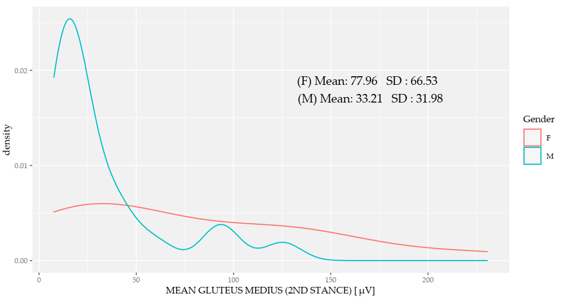
| Gluteus maximus | p = 0.114 | p = 0.013 * | p = 0.003 * | p = 0.647 |
| Gluteus medius | p = 0.198 | p = 0.057 | p = 0.028 * | p = 0.584 |
Publisher’s Note: MDPI stays neutral with regard to jurisdictional claims in published maps and institutional affiliations. |
© 2020 by the authors. Licensee MDPI, Basel, Switzerland. This article is an open access article distributed under the terms and conditions of the Creative Commons Attribution (CC BY) license (http://creativecommons.org/licenses/by/4.0/).
Share and Cite
Moltó, I.N.; Albiach, J.P.; Amer-Cuenca, J.J.; Segura-Ortí, E.; Gabriel, W.; Martínez-Gramage, J. Wearable Sensors Detect Differences between the Sexes in Lower Limb Electromyographic Activity and Pelvis 3D Kinematics during Running. Sensors 2020, 20, 6478. https://doi.org/10.3390/s20226478
Moltó IN, Albiach JP, Amer-Cuenca JJ, Segura-Ortí E, Gabriel W, Martínez-Gramage J. Wearable Sensors Detect Differences between the Sexes in Lower Limb Electromyographic Activity and Pelvis 3D Kinematics during Running. Sensors. 2020; 20(22):6478. https://doi.org/10.3390/s20226478
Chicago/Turabian StyleMoltó, Iván Nacher, Juan Pardo Albiach, Juan José Amer-Cuenca, Eva Segura-Ortí, Willig Gabriel, and Javier Martínez-Gramage. 2020. "Wearable Sensors Detect Differences between the Sexes in Lower Limb Electromyographic Activity and Pelvis 3D Kinematics during Running" Sensors 20, no. 22: 6478. https://doi.org/10.3390/s20226478
APA StyleMoltó, I. N., Albiach, J. P., Amer-Cuenca, J. J., Segura-Ortí, E., Gabriel, W., & Martínez-Gramage, J. (2020). Wearable Sensors Detect Differences between the Sexes in Lower Limb Electromyographic Activity and Pelvis 3D Kinematics during Running. Sensors, 20(22), 6478. https://doi.org/10.3390/s20226478