Use of Cyanobacterial Luminescent Bioreporters to Report on the Environmental Impact of Metallic Nanoparticles


Abstract
1. Introduction
2. Materials and Methods
2.1. Biological Materials and Culture Conditions
2.2. Chemicals
2.3. Characterization of NPs
2.4. Bioluminescence Assays
2.5. Spiking Experiments: Performance of the ROS-Detecting Bioreporters in Environmental Matrices Artificially Contaminated with Metallic NPs
2.6. Statistical Analysis
3. Results
3.1. Metallic NPs Physicochemical Characterization
3.2. Response of Nostoc sp. CPB4337 to Metallic NPs: Toxicity
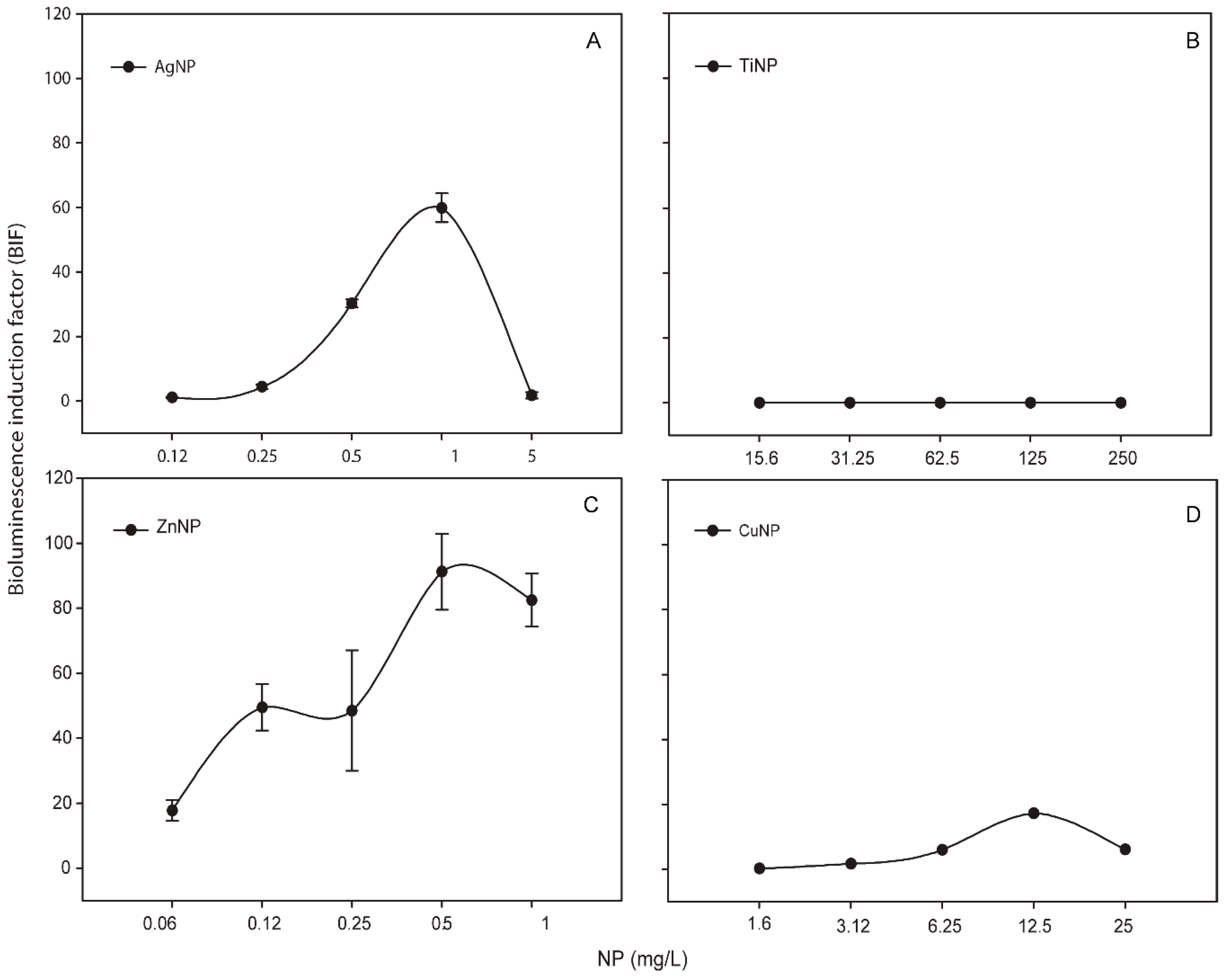
3.3. Response of Synechococcus Elongatus sp. PCC7942 pBG2120 to Released Free-Ions from Metallic NPs
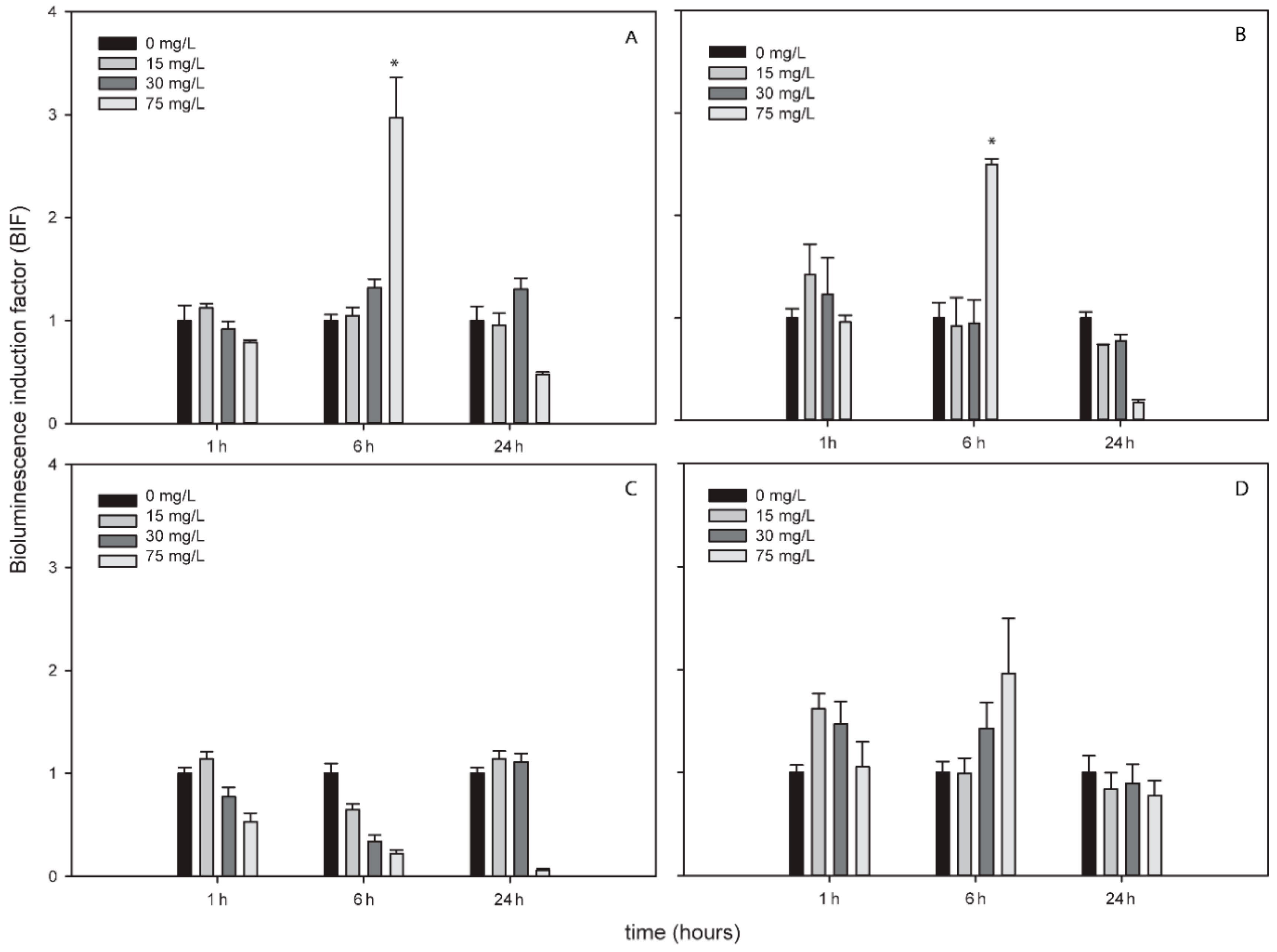
3.4. Response of ROS-Detecting Nostoc sp. PCC7120 Bioreporters to Metallic NPs
Response of ROS-Detecting Nostoc sp. PCC7120 Bioreporters to Metallic NPs Added to Environmental Water Samples (Spiking Experiments)
4. Discussion
5. Conclusions
Supplementary Materials
Author Contributions
Funding
Conflicts of Interest
References
- Nel, A.; Xia, T.; Mädler, L.; Li, N. Toxic potential of materials at the nanolevel. Science 2006, 311, 622–627. [Google Scholar] [CrossRef] [PubMed]
- Rengel, Z. Heavy metals as essential nutrients. In Heavy Metal Stress in Plants; Springer: Berlin/Heidelberg, Germany, 2004; pp. 271–294. [Google Scholar]
- Tchounwou, P.B.; Yedjou, C.G.; Patlolla, A.K.; Sutton, D.J. Heavy metal toxicity and the environment. In Molecular, Clinical and Environmental Toxicology; Springer: Berlin/Heidelberg, Germany, 2012; pp. 133–164. [Google Scholar]
- Rodea-Palomares, I.; Fernández-Piñas, F.; González-García, C.; Leganés, F. Use of lux-marked cyanobacterial bioreporters for assessment of individual and combined toxicities of metals in aqueous samples. Handb. Cyanobact. Biochem. Biotechnol. Appl. 2009, 283–304. [Google Scholar]
- Durán, N.; Durán, M.; de Jesus, M.B.; Seabra, A.B.; Fávaro, W.J.; Nakazato, G. Silver nanoparticles: A new view on mechanistic aspects on antimicrobial activity. Nanomed. Nanotechnol. Biol. Med. 2016, 12, 789–799. [Google Scholar] [CrossRef] [PubMed]
- Abdal Dayem, A.; Hossain, M.; Lee, S.; Kim, K.; Saha, S.; Yang, G.-M.; Choi, H.; Cho, S.-G. The role of reactive oxygen species (ROS) in the biological activities of metallic nanoparticles. Int. J. Mol. Sci. 2017, 18, 120. [Google Scholar] [CrossRef] [PubMed]
- Anreddy, R.N.R.; Yellu, N.R.; Devarakonda, K.R. Oxidative biomarkers to assess the nanoparticle-induced oxidative stress. In Oxidative Stress and Nanotechnology; Springer: Berlin/Heidelberg, Germany, 2013; pp. 205–219. [Google Scholar]
- Anjum, N.A.; Gill, S.S.; Duarte, A.C.; Pereira, E. Oxidative Stress Biomarkers and Antioxidant Defense in Plants Exposed to Metallic Nanoparticles. In Nanomaterials and Plant Potential; Springer: Berlin/Heidelberg, Germany, 2019; pp. 427–439. [Google Scholar]
- Touyz, R.M. Molecular and cellular mechanisms in vascular injury in hypertension: Role of angiotensin II–editorial review. Curr. Opin. Nephrol. Hypertens. 2005, 14, 125–131. [Google Scholar] [CrossRef] [PubMed]
- Latifi, A.; Ruiz, M.; Zhang, C.-C. Oxidative stress in cyanobacteria. FEMS Microbiol. Rev. 2009, 33, 258–278. [Google Scholar] [CrossRef]
- Augusto, O.; Miyamoto, S. Oxygen radicals and related species. Princ. Free Radic. Biomed. 2011, 1, 19–42. [Google Scholar]
- Von Moos, N.; Slaveykova, V.I. Oxidative stress induced by inorganic nanoparticles in bacteria and aquatic microalgae–state of the art and knowledge gaps. Nanotoxicology 2014, 8, 605–630. [Google Scholar] [CrossRef]
- Stone, V.; Donaldson, K. Signs of stress. Nat. Nanotechnol. 2006, 1, 23–24. [Google Scholar] [CrossRef]
- Niazi, J.H.; Gu, M.B. Toxicity of metallic nanoparticles in microorganisms—A review. In Atmospheric and Biological Environmental Monitoring; Springer: Berlin/Heidelberg, Germany, 2009; pp. 193–206. [Google Scholar]
- Zuberek, M.; Grzelak, A. Nanoparticles-caused oxidative imbalance. In Cellular and Molecular Toxicology of Nanoparticles; Springer: Berlin/Heidelberg, Germany, 2018; pp. 85–98. [Google Scholar]
- Handy, R.D.; van den Brink, N.; Chappell, M.; Mühling, M.; Behra, R.; Dušinská, M.; Simpson, P.; Ahtiainen, J.; Jha, A.N.; Seiter, J. Practical considerations for conducting ecotoxicity test methods with manufactured nanomaterials: What have we learnt so far? Ecotoxicology 2012, 21, 933–972. [Google Scholar] [CrossRef]
- Eduok, S.; Martin, B.; Villa, R.; Nocker, A.; Jefferson, B.; Coulon, F. Evaluation of engineered nanoparticle toxic effect on wastewater microorganisms: Current status and challenges. Ecotoxicol. Environ. Saf. 2013, 95, 1–9. [Google Scholar] [CrossRef]
- Duester, L.; Burkhardt, M.; Gutleb, A.C.; Kaegi, R.; Macken, A.; Meermann, B.; von der Kammer, F. Toward a comprehensive and realistic risk evaluation of engineered nanomaterials in the urban water system. Front. Chem. 2014, 2, 39. [Google Scholar] [CrossRef]
- Girotti, S.; Ferri, E.N.; Fumo, M.G.; Maiolini, E. Monitoring of environmental pollutants by bioluminescent bacteria. Anal. Chim. Acta 2008, 608, 2–29. [Google Scholar] [CrossRef]
- Mallevre, F.; Alba, C.; Milne, C.; Gillespie, S.; Fernandes, T.; Aspray, T. Toxicity testing of pristine and aged silver nanoparticles in real wastewaters using bioluminescent Pseudomonas putida. Nanomaterials 2016, 6, 49. [Google Scholar] [CrossRef]
- Holden, P.A.; Nisbet, R.M.; Lenihan, H.S.; Miller, R.J.; Cherr, G.N.; Schimel, J.P.; Gardea-Torresdey, J.L. Ecological nanotoxicology: Integrating nanomaterial hazard considerations across the subcellular, population, community, and ecosystems levels. Acc. Chem. Res. 2012, 46, 813–822. [Google Scholar] [CrossRef]
- Sadik, O.; Zhou, A.; Kikandi, S.; Du, N.; Wang, Q.; Varner, K. Sensors as tools for quantitation, nanotoxicity and nanomonitoring assessment of engineered nanomaterials. J. Environ. Monit. 2009, 11, 1782–1800. [Google Scholar] [CrossRef]
- Whitton, B.A.; Potts, M. The Ecology of Cyanobacteria: Their Diversity in Time and Space; Springer Science & Business Media: Berlin, Germany, 2007. [Google Scholar]
- Mur, R.; Skulberg, O.M.; Utkilen, H. Cyanobacteria in the Environment; WHO: Geneva, Switzerland, 1999. [Google Scholar]
- Flores, F.G. The Cyanobacteria: Molecular Biology, Genomics, and Evolution; Horizon Scientific Press: Sevilla, Spain, 2008. [Google Scholar]
- Harms, H.; Wells, M.C.; van der Meer, J.R. Whole-cell living biosensors—Are they ready for environmental application? Appl. Microbiol. Biotechnol. 2006, 70, 273–280. [Google Scholar] [CrossRef]
- Van Der Meer, J.R.; Belkin, S. Where microbiology meets microengineering: Design and applications of reporter bacteria. Nat. Rev. Microbiol. 2010, 8, 511. [Google Scholar] [CrossRef]
- Rodea-Palomares, I.; Gonzalez-Garcia, C.; Leganes, F.; Fernandez-Pinas, F. Effect of pH, EDTA, and anions on heavy metal toxicity toward a bioluminescent cyanobacterial bioreporter. Arch. Environ. Contam. Toxicol. 2009, 57, 477. [Google Scholar] [CrossRef]
- Hurtado-Gallego, J.; Martín-Betancor, K.; Rodea-Palomares, I.; Leganés, F.; Rosal, R.; Fernández-Piñas, F. Two novel cyanobacterial bioluminescent whole-cell bioreporters based on superoxide dismutases MnSod and FeSod to detect superoxide anion. Chemosphere 2018, 201, 772–779. [Google Scholar] [CrossRef]
- Hurtado-Gallego, J.; Redondo-López, A.; Leganés, F.; Rosal, R.; Fernández-Piñas, F. Peroxiredoxin (2-cys-prx) and catalase (katA) cyanobacterial-based bioluminescent bioreporters to detect oxidative stress in the aquatic environment. Chemosphere 2019, 236, 124395. [Google Scholar] [CrossRef]
- Martin-Betancor, K.; Rodea-Palomares, I.; Munoz-Martin, M.; Leganes, F.; Fernandez-Pinas, F. Construction of a self-luminescent cyanobacterial bioreporter that detects a broad range of bioavailable heavy metals in aquatic environments. Front. Microbiol. 2015, 6, 186. [Google Scholar] [CrossRef]
- Fernández-Pinas, F.; Wolk, C.P. Expression of luxCD-E in Anabaena sp. can replace the use of exogenous aldehyde for in vivo localization of transcription by luxAB. Gene 1994, 150, 169–174. [Google Scholar] [CrossRef]
- Gonzalo, S.; Llaneza, V.; Pulido-Reyes, G.; Fernández-Piñas, F.; Bonzongo, J.C.; Leganes, F.; Rosal, R.; García-Calvo, E.; Rodea-Palomares, I. A colloidal singularity reveals the crucial role of colloidal stability for nanomaterials in-vitro toxicity testing: nZVI-microalgae colloidal system as a case study. PLoS ONE 2014, 9, e109645. [Google Scholar] [CrossRef]
- Rosal, R.; Rodea-Palomares, I.; Boltes, K.; Fernández-Piñas, F.; Leganés, F.; Gonzalo, S.; Petre, A. Ecotoxicity assessment of lipid regulators in water and biologically treated wastewater using three aquatic organisms. Environ. Sci. Pollut. Res. 2010, 17, 135–144. [Google Scholar] [CrossRef]
- Rodea-Palomares, I.; Leganés, F.; Rosal, R.; Fernández-Pinas, F. Toxicological interactions of perfluorooctane sulfonic acid (PFOS) and perfluorooctanoic acid (PFOA) with selected pollutants. J. Hazard. Mater. 2012, 201, 209–218. [Google Scholar] [CrossRef]
- González-Pleiter, M.; Gonzalo, S.; Rodea-Palomares, I.; Leganés, F.; Rosal, R.; Boltes, K.; Marco, E.; Fernández-Piñas, F. Toxicity of five antibiotics and their mixtures towards photosynthetic aquatic organisms: Implications for environmental risk assessment. Water Res. 2013, 47, 2050–2064. [Google Scholar] [CrossRef]
- Rodea-Palomares, I.; Boltes, K.; Fernández-Pinas, F.; Leganés, F.; García-Calvo, E.; Santiago, J.; Rosal, R. Physicochemical characterization and ecotoxicological assessment of CeO2 nanoparticles using two aquatic microorganisms. Toxicol. Sci. 2010, 119, 135–145. [Google Scholar] [CrossRef]
- Rodea-Palomares, I.; Gonzalo, S.; Santiago-Morales, J.; Leganés, F.; García-Calvo, E.; Rosal, R.; Fernández-Pinas, F. An insight into the mechanisms of nanoceria toxicity in aquatic photosynthetic organisms. Aquat. Toxicol. 2012, 122, 133–143. [Google Scholar] [CrossRef]
- Martín-de-Lucía, I.; Campos-Mañas, M.C.; Agüera, A.; Rodea-Palomares, I.; Pulido-Reyes, G.; Leganés, F.; Fernández-Piñas, F.; Rosal, R. Reverse Trojan-horse effect decreased wastewater toxicity in the presence of inorganic nanoparticles. Environ. Sci. Nano 2017, 4, 1273–1282. [Google Scholar] [CrossRef]
- Kahru, A.; Dubourguier, H.-C. From ecotoxicology to nanoecotoxicology. Toxicology 2010, 269, 105–119. [Google Scholar] [CrossRef]
- Kahru, A.; Ivask, A.; Blinova, I.; Kasemets, K.; Dubourguier, H.C. Bioavailability and ecotoxicology of nanoparticles. Toxicol. Lett. 2008, S20. [Google Scholar] [CrossRef]
- Douterelo, I.; Perona, E.; Mateo, P. Use of cyanobacteria to assess water quality in running waters. Environ. Pollut. 2004, 127, 377–384. [Google Scholar] [CrossRef]
- Barrán-Berdón, A.L.; Rodea-Palomares, I.; Leganés, F.; Fernández-Piñas, F. Free Ca2+ as an early intracellular biomarker of exposure of cyanobacteria to environmental pollution. Anal. Bioanal. Chem. 2011, 400, 1015–1029. [Google Scholar] [CrossRef]
- Rosal, R.; Rodríguez, A.; Perdigón-Melón, J.A.; Petre, A.; García-Calvo, E.; Gómez, M.J.; Agüera, A.; Fernández-Alba, A.R. Occurrence of emerging pollutants in urban wastewater and their removal through biological treatment followed by ozonation. Water Res. 2010, 44, 578–588. [Google Scholar] [CrossRef]
- Ritz, C.; Streibig, J.C. Bioassay analysis using R. J. Stat. Softw 2005, 12, 1–22. [Google Scholar] [CrossRef]
- Pulido-Reyes, G.; Leganes, F.; Fernández-Piñas, F.; Rosal, R. Bio-nano interface and environment: A critical review. Environ. Toxicol. Chem. 2017, 36, 3181–3193. [Google Scholar] [CrossRef]
- Mazumder, B.; Ray, S.; Pal, P.; Pathak, Y. Nanotechnology: Therapeutic, Nutraceutical, and Cosmetic Advances; CRC Press: Boca Raton, FL, USA, 2019. [Google Scholar]
- Hurtado-Gallego, J.; Pulido-Reyes, G.; González-Pleiter, M.; Fernández-Piñas, F. Luminescent Microbial Bioassays and Microalgal Biosensors as Tools for Environmental Toxicity Evaluation. In Handbook of Cell Biosensors; Thouand, G., Ed.; Springer International Publishing: Cham, Switzerland, 2019; pp. 1–58. [Google Scholar]
- Pulido-Reyes, G.; Martín, E.; Coronado, J.G.; Leganes, F.; Rosal, R.; Fernández-Piñas, F. Physicochemical and biological interactions between cerium oxide nanoparticles and a 1,8-naphthalimide derivative. J. Photochem. Photobiol. B Biol. 2017, 172, 61–69. [Google Scholar] [CrossRef]
- Bundschuh, M.; Seitz, F.; Rosenfeldt, R.R.; Schulz, R. Effects of nanoparticles in fresh waters: Risks, mechanisms and interactions. Freshw. Biol. 2016, 61, 2185–2196. [Google Scholar] [CrossRef]
- McShan, D.; Ray, P.C.; Yu, H. Molecular toxicity mechanism of nanosilver. J. Food Drug Anal. 2014, 22, 116–127. [Google Scholar] [CrossRef]
- Ivask, A.; Bondarenko, O.; Jepihhina, N.; Kahru, A. Profiling of the reactive oxygen species-related ecotoxicity of CuO, ZnO, TiO2, silver and fullerene nanoparticles using a set of recombinant luminescent Escherichia coli strains: Differentiating the impact of particles and solubilised metals. Anal. Bioanal. Chem. 2010, 398, 701–716. [Google Scholar] [CrossRef]
- Hwang, E.T.; Lee, J.H.; Chae, Y.J.; Kim, Y.S.; Kim, B.C.; Sang, B.I.; Gu, M.B. Analysis of the toxic mode of action of silver nanoparticles using stress-specific bioluminescent bacteria. Small 2008, 4, 746–750. [Google Scholar] [CrossRef]
- Völker, C.; Oetken, M.; Oehlmann, J. The biological effects and possible modes of action of nanosilver. In Reviews of Environmental Contamination and Toxicology; Springer: Berlin/Heidelberg, Germany, 2013; Volume 223, pp. 81–106. [Google Scholar]
- Nair, P.M.G.; Park, S.Y.; Choi, J. Evaluation of the effect of silver nanoparticles and silver ions using stress responsive gene expression in Chironomus riparius. Chemosphere 2013, 92, 592–599. [Google Scholar] [CrossRef]
- Tlili, A.; Cornut, J.; Behra, R.; Gil-Allué, C.; Gessner, M.O. Harmful effects of silver nanoparticles on a complex detrital model system. Nanotoxicology 2016, 10, 728–735. [Google Scholar] [CrossRef]
- Shah, S.N.A.; Shah, Z.; Hussain, M.; Khan, M. Hazardous effects of titanium dioxide nanoparticles in ecosystem. Bioinorg. Chem. Appl. 2017, 2017, 4101735. [Google Scholar] [CrossRef]
- Ramsden, C.; Henry, T.; Handy, R. Sub-lethal effects of titanium dioxide nanoparticles on the physiology and reproduction of zebrafish. Aquat. Toxicol. 2013, 126, 404–413. [Google Scholar] [CrossRef]
- Ates, M.; Demir, V.; Adiguzel, R.; Arslan, Z. Bioaccumulation, subacute toxicity, and tissue distribution of engineered titanium dioxide nanoparticles in goldfish (Carassius auratus). J. Nanomater. 2013, 2013, 9. [Google Scholar] [CrossRef]
- Iswarya, V.; Bhuvaneshwari, M.; Alex, S.A.; Iyer, S.; Chaudhuri, G.; Chandrasekaran, P.T.; Bhalerao, G.M.; Chakravarty, S.; Raichur, A.M.; Chandrasekaran, N. Combined toxicity of two crystalline phases (anatase and rutile) of Titania nanoparticles towards freshwater microalgae: Chlorella sp. Aquat. Toxicol. 2015, 161, 154–169. [Google Scholar] [CrossRef]
- Sharma, V.K. Aggregation and toxicity of titanium dioxide nanoparticles in aquatic environment—A review. J. Environ. Sci. Health Part A 2009, 44, 1485–1495. [Google Scholar] [CrossRef]
- Maness, P.-C.; Smolinski, S.; Blake, D.M.; Huang, Z.; Wolfrum, E.J.; Jacoby, W.A. Bactericidal activity of photocatalytic TiO2 reaction: Toward an understanding of its killing mechanism. Appl. Environ. Microbiol. 1999, 65, 4094–4098. [Google Scholar]
- Zhao, X.; Wang, S.; Wu, Y.; You, H.; Lv, L. Acute ZnO nanoparticles exposure induces developmental toxicity, oxidative stress and DNA damage in embryo-larval zebrafish. Aquat. Toxicol. 2013, 136, 49–59. [Google Scholar] [CrossRef]
- Huang, C.-C.; Aronstam, R.S.; Chen, D.-R.; Huang, Y.-W. Oxidative stress, calcium homeostasis, and altered gene expression in human lung epithelial cells exposed to ZnO nanoparticles. Toxicol. Vitr. 2010, 24, 45–55. [Google Scholar] [CrossRef]
- Applerot, G.; Lellouche, J.; Lipovsky, A.; Nitzan, Y.; Lubart, R.; Gedanken, A.; Banin, E. Understanding the antibacterial mechanism of CuO nanoparticles: Revealing the route of induced oxidative stress. Small 2012, 8, 3326–3337. [Google Scholar] [CrossRef]
- Liu, Y.; Wang, S.; Wang, Z.; Ye, N.; Fang, H.; Wang, D. TiO2, SiO2 and ZrO2 nanoparticles synergistically provoke cellular oxidative damage in freshwater microalgae. Nanomaterials 2018, 8, 95. [Google Scholar] [CrossRef]
- Xia, B.; Chen, B.; Sun, X.; Qu, K.; Ma, F.; Du, M. Interaction of TiO2 nanoparticles with the marine microalga Nitzschia closterium: Growth inhibition, oxidative stress and internalization. Sci. Total Environ. 2015, 508, 525–533. [Google Scholar] [CrossRef]
- Wang, F.; Guan, W.; Xu, L.; Ding, Z.; Ma, H.; Ma, A.; Terry, N. Effects of Nanoparticles on Algae: Adsorption, Distribution, Ecotoxicity and Fate. Appl. Sci. 2019, 9, 1534. [Google Scholar] [CrossRef]
- Levard, C.; Hotze, E.M.; Lowry, G.V.; Brown Jr, G.E. Environmental transformations of silver nanoparticles: Impact on stability and toxicity. Environ. Sci. Technol. 2012, 46, 6900–6914. [Google Scholar] [CrossRef]
- Lee, S.; Kim, K.; Shon, H.; Kim, S.D.; Cho, J. Biotoxicity of nanoparticles: Effect of natural organic matter. J. Nanopart. Res. 2011, 13, 3051–3061. [Google Scholar] [CrossRef]
- Blinova, I.; Ivask, A.; Heinlaan, M.; Mortimer, M.; Kahru, A. Ecotoxicity of nanoparticles of CuO and ZnO in natural water. Environ. Pollut. 2010, 158, 41–47. [Google Scholar] [CrossRef]


| Gene System | Characteristics and Culture Conditions | References | |
|---|---|---|---|
| Nostoc sp. CPB4337 | luxCDABE genes in the chromosome | Toxicity bioreporter based on Nostoc sp. PCC7120. NmR in AA/8+N growth medium | [32] |
| Synechococcus elongatus sp. PCC7942 pBG2120 | Plasmid pBG2120: Psmt::luxCDABE smt encodes the transcriptional SmtB and the metallothionein SmtA | Bioavailable heavy metal bioreporter based on Synechococcus elongatus sp. PCC7942 expressing the plasmid pBG2120. CmR in BG11 growth medium | [31] |
| Nostoc sp. PCC7120 pBG2154 | Plasmid pBG2154; PsodA::luxCDABE sodA encodes a Mn-superoxide dismutase | Specific superoxide anion bioreporter based on Nostoc sp. PCC7120 expressing the plasmid pBG2154. SpR in AA/8+N growth medium | [29] |
| Nostoc sp. PCC7120 pBG2165 | Plasmid pBG2165; PsodB::luxCDABE sodB encodes a Fe-superoxide dismutase enzyme | Specific superoxide anion bioreporter based on Nostoc sp. PCC7120 expressing the plasmid pBG2165. SpR in AA/8+N growth medium | [29] |
| Nostoc sp. PCC7120 pBG2172 | Plasmid pBG2172; P2-cys-prx:: luxCDABE 2-cys-prx encodes a peroxiredoxin enzyme | Superoxide anion and H2O2 bioreporter based on Nostoc sp. PCC 7120 expressing the plasmid pBG2172. SpR in AA/8+N growth medium | [30] |
| Nostoc sp. PCC7120 pBG2173 | Plasmid pBG2173; PkatA:: luxCDABE katA encodes a Mn-catalase enzyme | Specific superoxide anion bioreporter based on Nostoc sp. PCC 7120 expressing the plasmid pBG2173. SpR in AA/8+N | [30] |
| Size (nm) | |||||
|---|---|---|---|---|---|
| Ultrapure Water (pH 6.5) | AA/8+N (pH 7) | BG11 (pH 7.6) | Glx1 (pH 6.9) | WWTP (pH 7.5) | |
| Without NPs | - | 711.0 ± 181.18 171.8 ± 36.4 | 772.1 ± 122.6 | 227.9 ± 49.6 | 148.4 ± 10.2 |
| AgNPs | 55.7 ± 20.5 9.2 ± 3.3 | 635.9 ± 154.9 46.6 ± 10.4 8.5 ± 1.9 | 306.8 ± 39.1 28.96 ± 3.0 | 76.7 ± 38.1 10.3 ± 4.4 | 169.4 ± 110.0 16.5 ± 4.8 |
| TiNPs | 2333.0 ± 377.5 | 1197.0 ± 87.4 | 5433.0 ± 283.0 | 4213.0 ± 909.9 | 1099.0 ± 114.0 |
| ZnNPs | 264.8 ± 55 | 637.0 ± 120.3 | 1193.0 ± 87.5 | - | 509.3 ± 118.3 |
| CuNPs | 242.0 ± 120.2 | 204.6 ± 73.9 | 921.2±112.8 | 256.2 ± 91.1 | 249.1 ± 99.7 |
| ζ-potential (mV) | |||||
| Without NPs | - | −27.4 ± 1.0 | −11.51 ± 2.8 | −9.8 ± 1.4 | −9.9 ± 2.2 |
| AgNPs | −10.9 ± 2.4 | −24.2 ± 1.7 | −21.0 ± 11.8 | −4.7 ± 0.3 | −11.7 ± 11.5 |
| TiNPs | −17.1 ± 2.1 | −23.6 ± 1.0 | −18.2 ± 2.0 | −17.2 ± 0.8 | −12.5 ± 0.7 |
| ZnNPs | −17.4 ± 0.6 | −27.5 ± 1.3 | −19.0 ± 2.1 | −13.7 ± 3.3 | −13.3 ± 1.4 |
| CuNPs | −24.1 ± 0.2 | −28.7 ± 0.9 | −26.1 ± 1.0 | −22.4 ± 0.4 | −14.5 ± 0.1 |
| Time (h) | Metallic NPs | “R” Model Fitted | IC10 (mg/L) | IC50 (mg/L) | IC90 (mg/L) |
|---|---|---|---|---|---|
| 1 | Ag | LL.4 | 0.13 ± 0.04 | 0.23 ± 0.04 | 0.43 ± 0.17 |
| Ti | - | - | - | - | |
| Zn | LL2.3 | - | 2.04 ± 0.8 | 17.9 ± 5.5 | |
| Cu | W1.3 | 17.01 ± 3 | 32.8 ± 2.1 | 50.01 ± 4.23 | |
| 6 | Ag | W1.3 | 0.02 ± 0.02 | 0.14 ± 0.03 | 1.92 ± 0.6 |
| Ti | W1.4 | 0.55 ± 0.4 | 8.94 ± 2.0 | 52.77 ± 25.0 | |
| Zn | LL2.3 | - | 1.23 ± 0.1 | 2.96 ± 0.2 | |
| Cu | W2.3 | 14.06 ± 3.2 | 18.58 ± 2.26 | 28.77 ± 2.04 | |
| 24 | Ag | W1.3 | 0.4 ± 0.04 | 0.71 ± 0.03 | 1.03 ± 0.05 |
| Ti | LL.3 | 3.92 ± 0.77 | 15.23 ± 1.18 | 59.2 ± 8.05 | |
| Zn | W1.4 | 0.07 ± 0.04 | 0.38 ± 0.07 | 1.06 ± 0.33 | |
| Cu | W2.3 | 9.17 ± 0.41 | 12.45 ± 0.78 | 20.10 ± 3.69 |
© 2019 by the authors. Licensee MDPI, Basel, Switzerland. This article is an open access article distributed under the terms and conditions of the Creative Commons Attribution (CC BY) license (http://creativecommons.org/licenses/by/4.0/).
Share and Cite
Hurtado-Gallego, J.; Leganés, F.; Rosal, R.; Fernández-Piñas, F. Use of Cyanobacterial Luminescent Bioreporters to Report on the Environmental Impact of Metallic Nanoparticles. Sensors 2019, 19, 3597. https://doi.org/10.3390/s19163597
Hurtado-Gallego J, Leganés F, Rosal R, Fernández-Piñas F. Use of Cyanobacterial Luminescent Bioreporters to Report on the Environmental Impact of Metallic Nanoparticles. Sensors. 2019; 19(16):3597. https://doi.org/10.3390/s19163597
Chicago/Turabian StyleHurtado-Gallego, Jara, Francisco Leganés, Roberto Rosal, and Francisca Fernández-Piñas. 2019. "Use of Cyanobacterial Luminescent Bioreporters to Report on the Environmental Impact of Metallic Nanoparticles" Sensors 19, no. 16: 3597. https://doi.org/10.3390/s19163597
APA StyleHurtado-Gallego, J., Leganés, F., Rosal, R., & Fernández-Piñas, F. (2019). Use of Cyanobacterial Luminescent Bioreporters to Report on the Environmental Impact of Metallic Nanoparticles. Sensors, 19(16), 3597. https://doi.org/10.3390/s19163597