Dual-Color Fluorescence Imaging of EpCAM and EGFR in Breast Cancer Cells with a Bull’s Eye-Type Plasmonic Chip


Abstract
:1. Introduction
2. Materials and Methods
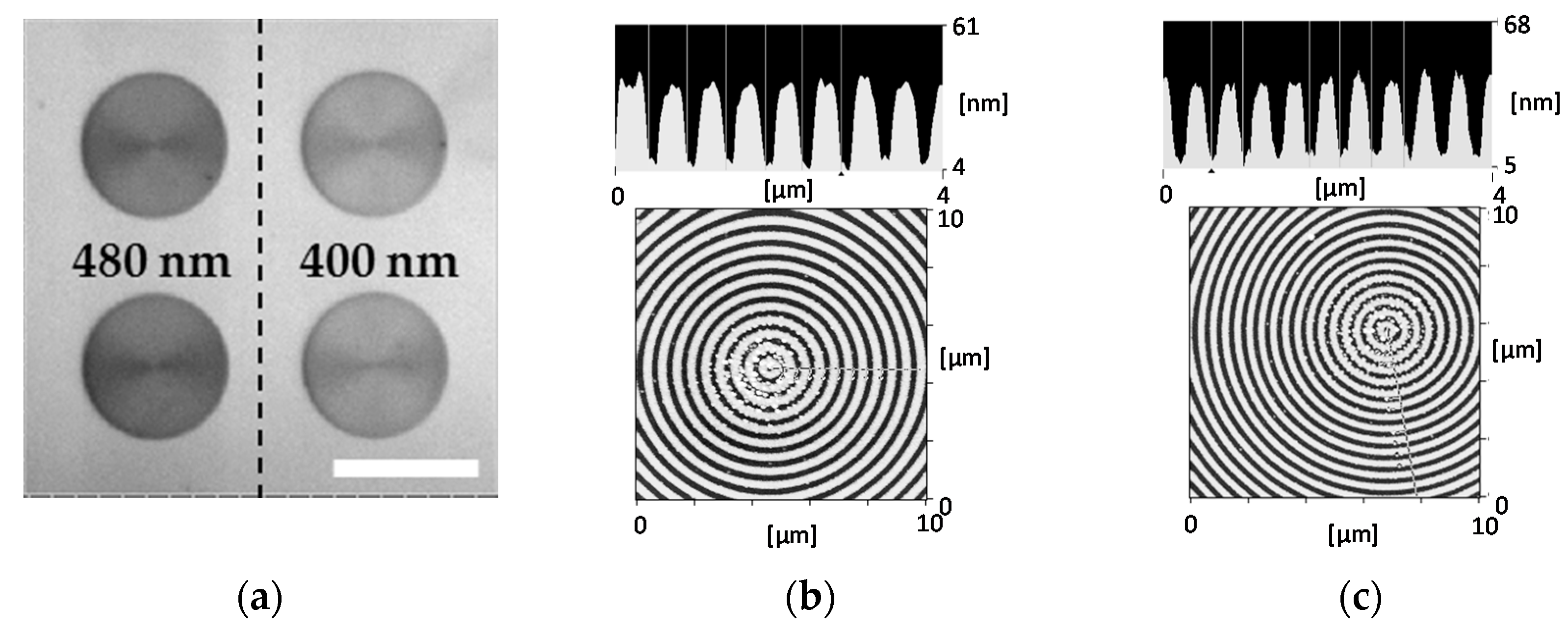
2.1. Fabrication of a Bull’s Eye-Plasmonic Chip
2.2. Cell Culture and Preparation for Microscopic Observation on a Glass Slide and a Plasmonic Chip
2.3. Microscopy
3. Results and Discussion
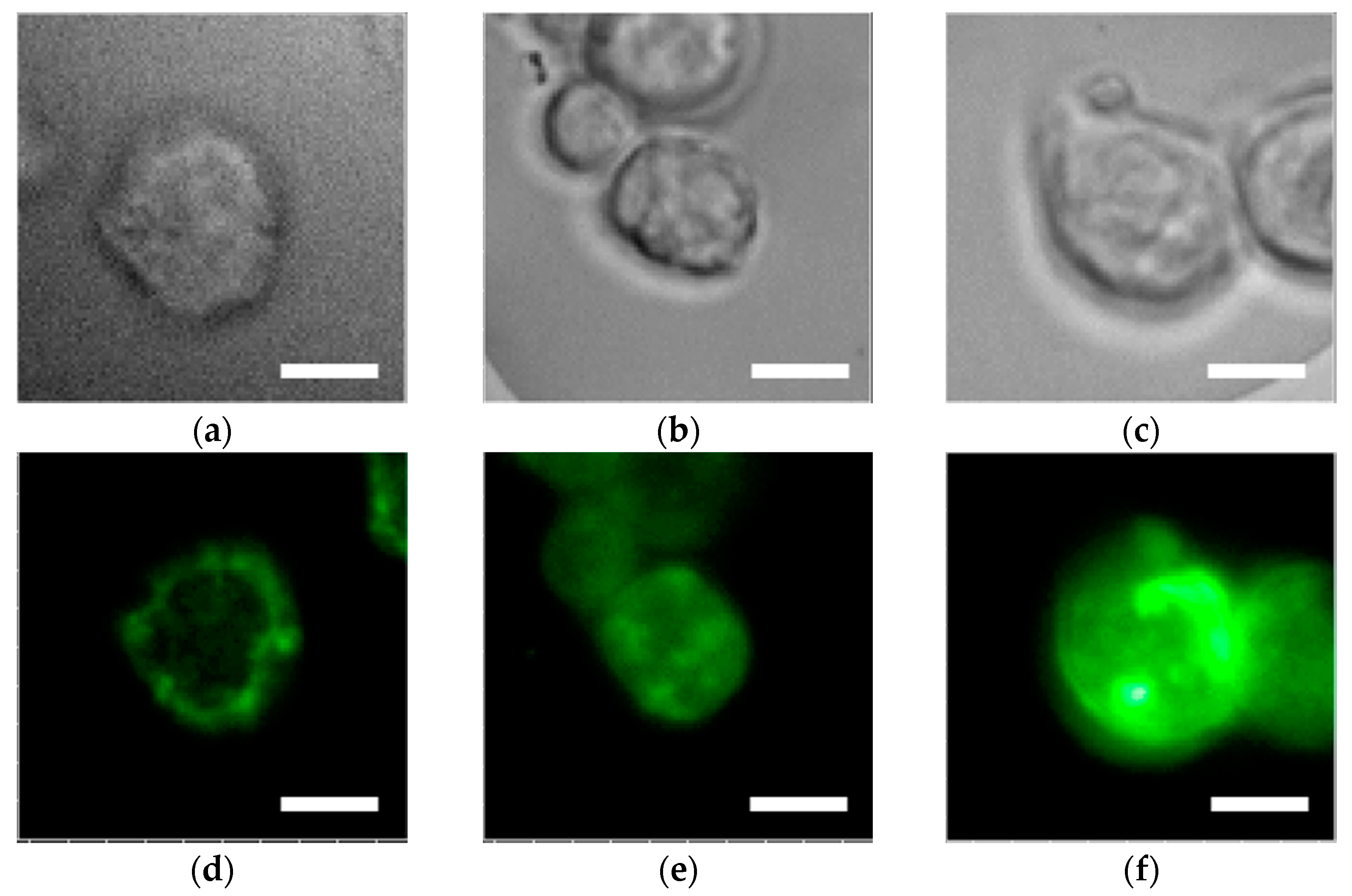
3.1. Enhanced Fluorescence Microscopic Observation of EGFR in Shorter Wavelength (Green Color)
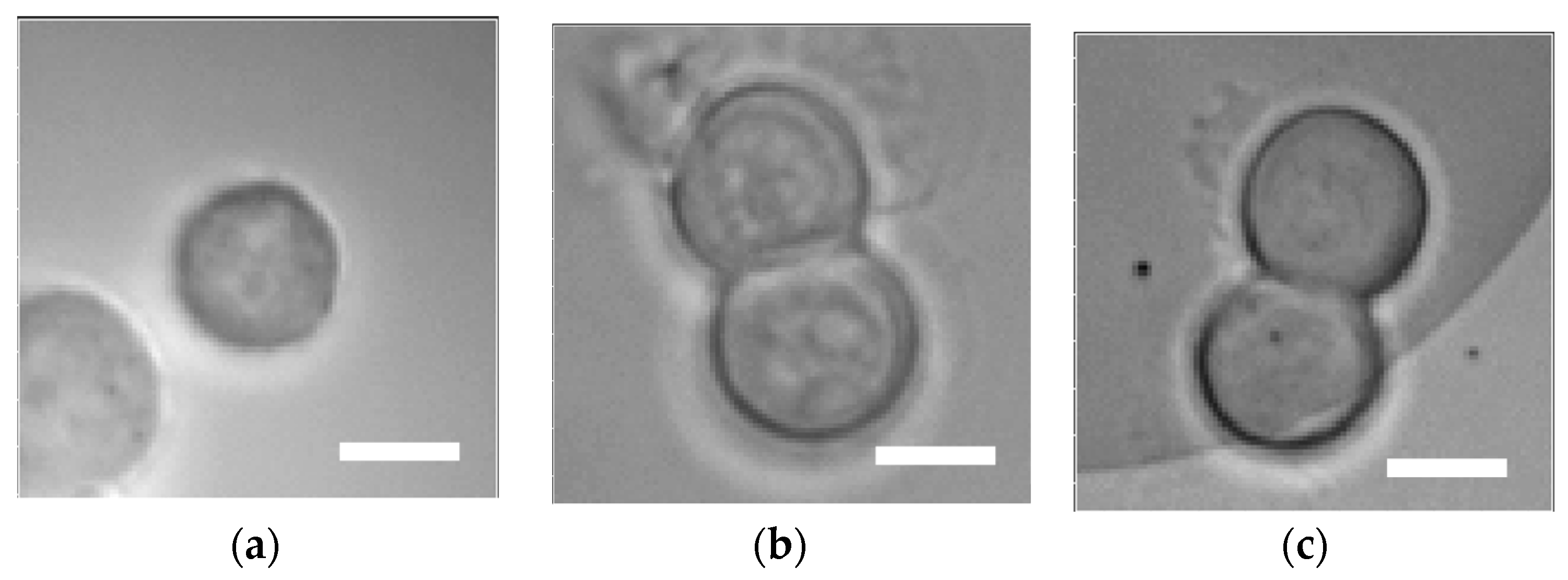
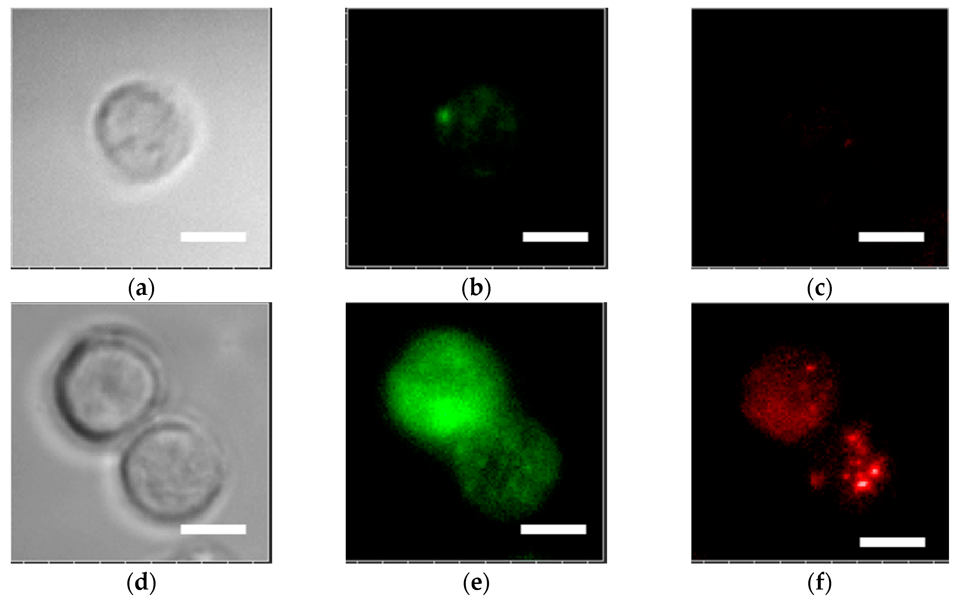
3.2. Dual-Color Fluorescence Microscopic Observation in MDA-MB-231
4. Conclusions
Acknowledgments
Author Contributions
Conflicts of Interest
References
- Homola, J. Surface Plasmon Resonance Sensors for Detection of Chemical and Biological Species. Chem. Rev. 2008, 108, 462–493. [Google Scholar] [CrossRef] [PubMed]
- Anker, J.N.; Hall, W.P.; Lyandres, O.; Shah, N.C.; Zhao, J.; Van Duyne, R.P. Biosensing with Plasmonic Nanosensors. Nat. Mater. 2008, 7, 442–453. [Google Scholar] [CrossRef] [PubMed]
- Vareiro, M.L.M.; Liu, J.; Knoll, W.; Zak, K.; Williams, D.; Jenkins, A.T.A. Surface plasmon fluorescence measurements of human chorionic gonadotrophin: Role of antibody orientation in obtaining enhanced sensitivity and limit of detection. Anal. Chem. 2005, 77, 2426–2431. [Google Scholar] [CrossRef] [PubMed]
- Chang, Y.F.; Chen, R.C.; Lee, Y.J.; Chao, S.C.; Su, L.C.; Li, Y.C.; Chou, C. Localized surface plasmon coupled fluorescence fiber-optic biosensor for alpha-fetoprotein detection in human serum. Biosens. Bioelectron. 2009, 24, 1610–1614. [Google Scholar] [CrossRef] [PubMed]
- Tawa, K.; Kondo, F.; Sasakawa, C.; Nagae, K.; Nakamura, Y.; Nozaki, A.; Kaya, T. Sensitive detection of a tumor marker, α-fetoprotein, with a sandwich assay on a plasmonic chip. Anal. Chem. 2015, 87, 3871–3876. [Google Scholar] [CrossRef] [PubMed]
- Zhao, J.; Zhang, X.Y.; Yonzon, C.R.; Haes, A.J.; Van Duyne, R.P. Localized surface plasmon resonance biosensors. Nanomedicine 2006, 1, 219–228. [Google Scholar] [CrossRef] [PubMed]
- Lal, S.; Link, S.; Halas, N.J. Nano-optics from sensing to waveguiding. Nat. Photonics 2007, 1, 641–648. [Google Scholar] [CrossRef]
- Chiu, N.F.; Yu, C.; Nien, S.-Y.; Lee, J.-H.; Kuan, C.-H.; Wu, K.-C.; Lee, C.-K.; Lin, C.-W. Enhancement and tunability of active plasmonic by multilayer grating coupled emission. Opt. Express 2007, 15, 11608–11615. [Google Scholar] [CrossRef] [PubMed]
- Neubrech, F.; Pucci, A.; Cornelius, T.W.; Karim, S.; Garcia-Etxarri, A.; Javier, A. Resonant plasmonic and vibrational coupling in a tailored nanoantenna for infrared detection. Phys. Rev. Lett. 2008, 101, 157403. [Google Scholar] [CrossRef] [PubMed]
- Eustis, S.; El-Sayed, M.A. Why gold nanoparticles are more precious than pretty gold: Noble metal surface plasmon resonance and its enhancement of the radiative and nonradiative properties of nanocrystals of different shapes. Chem. Soc. Rev. 2006, 35, 209–217. [Google Scholar] [CrossRef] [PubMed]
- Link, S.; El-Sayed, M.A. Spectral properties and relaxation dynamics of surface plasmon electronic oscillations in gold and silver nanodots and nanorods. J. Phys. Chem. 1999, 103, 8410–8426. [Google Scholar] [CrossRef]
- Knoll, W. Interfaces and thin films as seen by bound electromagnetic waves. Ann. Rev. Phys. Chem. 1998, 49, 569–638. [Google Scholar] [CrossRef] [PubMed]
- Raether, R. Surface Plasmons on Smooth and Rough Surfaces and on Gratings, 1st ed.; Springer: Heidelberg, Germany, 1988; pp. 1–133. [Google Scholar]
- Tawa, K.; Hori, H.; Kintaka, K.; Kiyosue, K.; Tatsu, Y.; Nishii, J. Optical microscopic observation of fluorescence enhanced by grating-coupled surface plasmon resonance. Opt. Express 2008, 16, 9781–9790. [Google Scholar] [CrossRef] [PubMed]
- Tawa, K.; Nakayama, T.; Kintaka, K. Optimal structure of a plasmonic chip for the sensitive bio-detection with the grating-coupled surface plasmon-field enhanced fluorescence (GC-SPF). Materials 2017, 10, 1063. [Google Scholar] [CrossRef] [PubMed]
- Tawa, K.; Yasui, C.; Hosokawa, C.; Aota, H.; Nishii, J. In situ sensitive fluorescence imaging of neurons cultured on a plasmonic dish using fluorescence microscopy. ACS Appl. Mater. Interfaces 2014, 6, 20010–20015. [Google Scholar] [CrossRef] [PubMed]
- Malicka, J.; Gryczynski, I.; Gryczynski, Z.; Lakowicz, J.R. DNA Hybridization Using Surface Plasmon-Coupled Emission. Anal. Chem. 2003, 75, 6629–6633. [Google Scholar] [CrossRef] [PubMed]
- Tawa, K.; Izumi, S.; Hosokawa, C.; Toma, M. Enhanced fluorescence microscopy with the Bull’s eye-plasmonic chip. Opt. Express 2017, 25, 10622–10631. [Google Scholar] [CrossRef] [PubMed]
- Peters, I.T.; Hilders, C.G.; Sier, C.F.; Vahrmeijer, A.L.; Smit, V.T.H.; Baptist Trimbos, J.; Kuppen, P.J. Identification of cell-surface markers for detecting breast cancer cells in ovarian tissue. Gynecol. Oncol. 2016, 294, 385–393. [Google Scholar] [CrossRef] [PubMed]
- Ki, J.; Arumugam, P.; Song, J.M. TIRF high-content assay development for the evaluation of drug efficacy of chemotherapeutic agents against EGFR-/HER2-positive breast cancer cell lines. Anal. Bioanal. Chem. 2016, 408, 3233–3238. [Google Scholar] [CrossRef] [PubMed]
- Zhang, Y.; Zhang, B.; Liu, F.; Luo, J.; Bai, J. In vivo tomographic imaging with fluorescence and MRI using tumor-targeted dual-labeled nanoparticles. Int. J. Nanomed. 2014, 9, 33–41. [Google Scholar] [CrossRef] [PubMed]
- Ke, S.; Wen, X.; Gurfinkel, M.; Charnsangavej, C.; Wallace, S.; Sevick-Muraca, E.M.; Li, C. Near-Infrared Optical Imaging of Epidermal Growth Factor Receptor in Breast Cancer Xenografts. Cancer Res. 2003, 63, 7870–7875. [Google Scholar] [PubMed]
- Tawa, K.; Yamamura, S.; Sasakawa, C.; Shibata, I.; Kataoka, M. Sensitive detection of cell surface membrane proteins in living breast cancer cells using multicolor fluorescence microscopy with a plasmonic chip. ACS Appl. Mater. Interface 2016, 8, 29893–29898. [Google Scholar] [CrossRef] [PubMed]
- Martowicz, A.; Spizzo, G.; Gastl, G.; Untergasser, G. Phenotype-dependent effects of EpCAM expression on growth and invasion of human breast cancer cell lines. BMC Cancer 2012, 12, 501. [Google Scholar] [CrossRef] [PubMed]
- Osta, W.A.; Chen, Y.; Mikhitarian, K.; Mitas, M.; Salem, M.; Hannun, Y.A.; Cole, D.J.; Gillanders, W.E. EpCAM is overexpressed in breast cancer and is a potential target for breast cancer gene therapy. Cancer Res. 2004, 64, 5818–5824. [Google Scholar] [CrossRef] [PubMed]
- Yamamura, S.; Yatsushiro, S.; Yamaguchi, Y.; Abe, K.; Shinohara, Y.; Tamiya, E.; Baba, Y.; Kataoka, M. Accurate detection of carcinoma cells by use of a cell microarray chip. PLoS ONE 2012, 7, e32370. [Google Scholar] [CrossRef] [PubMed]
- Yi, J.-M.; Cuche, A.; Devaux, E.; Genet, C.; Ebbesen, T.W. Beaming visible light with a plasmonic aperture antenna. ACS Photonics 2014, 1, 365–370. [Google Scholar] [CrossRef] [PubMed]
- Aouani, H.; Mahboub, O.; Devaux, E.; Rigneault, H.; Ebbesen, T.W.; Wenger, J. Plasmonic antennas for directional sorting of fluorescence emission. Nano Lett. 2011, 11, 2400–2406. [Google Scholar] [CrossRef] [PubMed]
- Mohtashami, A.; Osorio, C.I.; Koenderink, A.F. Angle-resolved polarimetry measurements of antenna-mediated fluorescence. Phys. Rev. Appl. 2015, 4, 054014-1–054014-7. [Google Scholar] [CrossRef]
- Arabi, H.E.; Joe, H.-E.; Nazari, T.; Min, B.-K.; Oh, K. A high throughput supra-wavelength plasmonic bull’s eye photon sorter spatially and spectrally multiplexed on silica optical fiber facet. Opt. Express 2013, 21, 28083–28094. [Google Scholar] [CrossRef] [PubMed]
- Aouani, H.; Mahboub, O.; Bonod, N.; Devaux, E.; Popov, E.; Rigneault, H.; Ebbesen, T.W.; Wenger, J. Bright unidirectional fluorescence emission of molecules in a nanoaperture with plasmonic corrugations. Nano Lett. 2011, 11, 637–644. [Google Scholar] [CrossRef] [PubMed]
- Wenger, J.; Aouani, H.; Gérard, D.; Blair, S.; Ebbesen, T.W.; Rigneault, H. Enhanced fluorescence from metal nanoapertures: Physical characterizations and biophotonic applications. Proc. SPIE 2010, 75770J. [Google Scholar] [CrossRef]
- Tawa, K.; Morigaki, K. Substrate-supported phospholipid membranes studied by surface plasmon resonance and surface plasmon fluorescence spectroscopy. Biophys. J. 2005, 89, 2750–2758. [Google Scholar] [CrossRef] [PubMed]
- Chance, R.R.; Prock, A.; Silbey, R. Molecular fluorescence and energy transfer near interfaces. Adv. Chem. Phys. 1978, 37, 1–65. [Google Scholar] [CrossRef]
- Takabatake, D.; Fujita, T.; Shien, T.; Kawasaki, K.; Taira, N.; Yoshitomi, S.; Takahashi, H.; Ishibe, Y.; Ogasawara, Y.; Doihara, H. Tumor inhibitory effect of gefitinib (ZD1839, Iressa) and taxane combination therapy in EGFR-overexpressing breast cancer cell lines (MCF7/ADR, MDA-MB-231). Int. J. Cancer 2006, 120, 181–188. [Google Scholar] [CrossRef] [PubMed]
- Sterzynska, K.; Kempisty, B.; Zawierucha, P.; Zabel, M. Analysis of the specificity and selectivity of anti-EpCAM antibodies in breast cancer cell line. Folia Histochemica et Cytobiologica 2012, 50, 534–541. [Google Scholar] [CrossRef] [PubMed]
- Wang, Z.-Q.; Zhao, J.; Zeng, J.; Wu, K.-J.; Chen, Y.-L.; Wang, X.-Y.; Chang, L.S.; He, D.-L. Specific survivin dual fluorescence resonance energy transfer molecular beacons for detection of human bladder cancer cells. Acta Pharmacol. Sin. 2011, 32, 1522–1528. [Google Scholar] [CrossRef] [PubMed]
- Liu, S.; Zhang, H.; Liu, W.; Zhou, B.; Ma, Q.; Ge, J.; Wu, J.; Wang, P. Investigation of biological cell–small molecular interactions based gold surface plasmon resonance sensor using a laser scanning confocal imaging-surface plasmon resonance system. RSC Adv. 2016, 6, 65930–65935. [Google Scholar] [CrossRef]


© 2017 by the authors. Licensee MDPI, Basel, Switzerland. This article is an open access article distributed under the terms and conditions of the Creative Commons Attribution (CC BY) license (http://creativecommons.org/licenses/by/4.0/).
Share and Cite
Izumi, S.; Yamamura, S.; Hayashi, N.; Toma, M.; Tawa, K. Dual-Color Fluorescence Imaging of EpCAM and EGFR in Breast Cancer Cells with a Bull’s Eye-Type Plasmonic Chip. Sensors 2017, 17, 2942. https://doi.org/10.3390/s17122942
Izumi S, Yamamura S, Hayashi N, Toma M, Tawa K. Dual-Color Fluorescence Imaging of EpCAM and EGFR in Breast Cancer Cells with a Bull’s Eye-Type Plasmonic Chip. Sensors. 2017; 17(12):2942. https://doi.org/10.3390/s17122942
Chicago/Turabian StyleIzumi, Shota, Shohei Yamamura, Naoko Hayashi, Mana Toma, and Keiko Tawa. 2017. "Dual-Color Fluorescence Imaging of EpCAM and EGFR in Breast Cancer Cells with a Bull’s Eye-Type Plasmonic Chip" Sensors 17, no. 12: 2942. https://doi.org/10.3390/s17122942
APA StyleIzumi, S., Yamamura, S., Hayashi, N., Toma, M., & Tawa, K. (2017). Dual-Color Fluorescence Imaging of EpCAM and EGFR in Breast Cancer Cells with a Bull’s Eye-Type Plasmonic Chip. Sensors, 17(12), 2942. https://doi.org/10.3390/s17122942