Article

Morphological and Molecular Identification of New Puccinia Species from Qinghai, China

1
College of Agriculture and Animal Husbandry, Qinghai University, Xining 810016, China
2
Datong County Forestry Station, Xining 810199, China
3
Academy of Agriculture and Forestry Sciences, Qinghai University, Xining 810016, China
4
Huzhu County Agriculture, Rural Affairs, and Science & Technology Bureau, Haidong 810599, China
5
Hualong County Forestry and Grassland Bureau, Haidong 810900, China
*
Author to whom correspondence should be addressed.
These authors contributed equally to this work.
Diversity 2026, 18(8), 487; https://doi.org/10.3390/d18080487
Submission received: 7 July 2026 / Revised: 8 August 2026 / Accepted: 12 August 2026 / Published: 14 August 2026
(This article belongs to the Special Issue Fungal Diversity—2nd Edition)

Abstract

Rust fungi of the genus Puccinia are important plant pathogens responsible for substantial economic losses in agriculture and forestry. During field surveys conducted in Qinghai Province, China, a novel rust species, Puccinia ligulari-przewalskii, was discovered parasitizing Ligularia przewalskii. Morphological characteristics of its teliospores were examined using light microscopy (LM) and scanning electron microscopy (SEM). Molecular phylogenetic analysis based on the concatenated internal transcribed spacer (ITS) and large subunit (LSU) ribosomal DNA sequences supported its taxonomic distinction as a new species within the genus Puccinia. Phylogenetic reconstructions inferred from maximum likelihood and Bayesian inference showed that the new species forms a distinct, well-supported lineage, clustering as sister to a clade comprising Puccinia argentata and Puccinia komarovii. P. ligulari-przewalskii is characterized by two-celled, spindle-shaped teliospores exhibiting sparse verrucose ornamentation on the spore wall. Only the telial stage was observed across multi-year surveys, and the complete life cycle remains to be determined. This study contributes to the taxonomic knowledge of rust fungi in China and underscores the ecological and phytopathological significance of rust fungi in the Qinghai–Tibet Plateau region.

1. Introduction

Approximately 8400 species of rust fungi have been identified worldwide to date, classified into 24 families and 160 genera. Many of these species cause significant economic losses to agricultural or forestry crops [1,2,3,4]. More than half of these species belong to the genus Puccinia, which is characterized by two-celled teliospores [2].
Rust fungi exhibit highly diverse life cycle patterns. The macrocyclic life cycle produces all five spore stages—spermogonia (0), aecia (I), uredinia (II), telia (III), and basidia (IV)—and can be completed on a single host (autoecious) or on two phylogenetically unrelated hosts (heteroecious) [2]. Derived, reduced life cycles include the demicyclic type (uredinial stage omitted), the hemicyclic type (spermogonial and aecial stages absent), and the microcyclic type, in which the life cycle is reduced to the telial and basidial stages, with or without spermogonia [2,5,6]. Microcyclic rusts do not produce uredinia or aecia and have no known alternate hosts [7,8]. A significantly higher proportion of microcyclic species occurs in arcto-alpine and high-elevation environments compared with temperate regions—a pattern attributed to abbreviated growing seasons that favor the elimination of spore stages requiring two hosts or multiple infection cycles [6,9].
The identification of Puccinia species is primarily based on morphological characteristics, such as teliospore shape, size, surface ornamentation, germ pore position, presence or absence of pore caps, cell wall thickness, pedicel length, and the germ pore position of urediniospores, combined with information on host occurrence and host relationships [2,10,11,12,13,14,15,16]. In recent years, DNA-based phylogenetic approaches, particularly those employing the internal transcribed spacer (ITS) and large subunit (LSU) ribosomal DNA regions, have become essential for precise species delimitation in Puccinia [4,17,18,19].
Globally, more than 5000 species of the genus Puccinia have been documented, with approximately 520 species and varieties reported in China [4,12,13,20,21,22,23,24,25,26,27]. Puccinia species parasitize plants extensively from the families Asteraceae, Cyperaceae, Liliaceae, and Poaceae in China [12,13], causing diseases that significantly affect plant growth and development. Such diseases reduce the yields of economically important crops and have profound impacts on forest ecosystem functionality, warranting increased attention and research. Approximately eleven rust taxa have previously been recorded on Ligularia (Asteraceae) worldwide, including Aecidium nikkense, Aecidium ligulariae, Coleosporium ligulariae, Coleosporium saussureae, Coleosporium senecionis, Puccinia cnici-oleracei, Puccinia eriophori, Puccinia expansa, Puccinia glomerata, Puccinia ligulariae, and Uromyces ligulariae [3,13,15,28,29,30,31]. Among these, only P. cnici-oleracei has been characterized by molecular phylogenetic data. P. ligulariae is listed only by name and host association in the Chinese taxonomic literature, without accompanying morphological descriptions [15,30]. P. eriophori is the teleomorph of A. ligulariae [32,33]; however, its uredinial and telial stages have never been recorded in China, despite the wide distribution of its reported telial host, Eriophorum (Cyperaceae), in the northern part of the country [32]. None of the previously recorded rust taxa on Ligularia have been supported by both morphological and molecular phylogenetic evidence in combination.
During a survey of rust diseases in Qinghai Province, a novel species of Puccinia parasitizing Ligularia przewalskii (Maxim.) Diels was identified and initially recorded in a floristic checklist of the Sanjiangyuan region [34]. Prior to this study, no Puccinia species on Ligularia had been formally described with supporting molecular phylogenetic data. In this study, its morphological characteristics and molecular data were analyzed, its phylogenetic relationships with related Puccinia species were evaluated, and its taxonomic novelty was confirmed. This paper provides a detailed description and comprehensive analysis of this newly discovered rust fungus.

2. Materials and Methods

2.1. Specimens

The specimens analyzed in this study were collected in 2023 from forested areas along the Mako River, Guoluo Tibetan Autonomous Prefecture, Qinghai Province, China (Table 1). These specimens are deposited in the Plant Pathology Herbarium of Qinghai University under voucher numbers QHU2023090 and QHU2023092, respectively, and were used for subsequent morphological observation and phylogenetic analysis. Two additional collections from the same locality (QHU2021116, QHU2022139) were previously examined under a light microscope; only telia were observed in these collections, and teliospore morphology was consistent with that of the holotype.

2.2. Morphological Observations

The morphological classification system used in this study follows the 14-family classification framework outlined in the 10th edition of the Dictionary of Fungi [35]. Morphological characteristics were primarily described with reference to the Fungi Identification Manual [36] and Chinese Fungi Flora [12,13,14,15,16], along with other relevant books and literature [20,35,37]. The morphological features of teliospores, including type, shape, attachment position, and color of spore clusters, were examined using a Nikon SMZ800N stereomicroscope (DM; Nikon Corporation, Tokyo, Japan), an optical microscope (LM; Olympus, Tokyo, Japan), and a scanning electron microscope (SEM; Hitachi, Tokyo, Japan). Fifty teliospores were randomly selected for measurements of their length, width, and wall thickness. The surface ornamentation and ultrastructure of teliospores were observed using a Hitachi S-4800 scanning electron microscope (Hitachi, Tokyo, Japan) operating at 15 kV. The measurements obtained were compared with the descriptions of related species in published literature.

2.3. DNA Extraction, PCR Amplification, and Sequencing

DNA extraction was performed following the methods of Tian et al. [38] and Yang [39]. Approximately 100–200 rust fungus spores were placed between two slides, and a few drops of DNA extraction buffer were added. The spores were then thoroughly ground to disrupt the cell walls. The presence of a slurry, observed under an optical microscope, indicated successful cell lysis. The slides were then carefully separated, and DNA was extracted. The primer pairs ITS5-u (5′-CAAGGTTTCTGTAGGTG-3′) [40] and ITS4rust (5′-CAGATTACAAATTTGGGCT-3′) [41] and NL1(5′-GCATATCAATAAGCGGAGGAAAAG-3′) and NL4(5′-GGTCCGTGTTTCAAGACGG-3′) [42], were used to amplify the ITS and LSU regions, respectively. Polymerase chain reaction (PCR) amplifications were performed in a total volume of 25 µL. The PCR protocol for ITS was as follows: initial denaturation at 94 °C for 5 min, followed by 30 cycles of denaturation at 94 °C for 30 s, annealing at 48 °C for 40 s, extension at 72 °C for 60 s, and a final extension at 72 °C for 10 min. The PCR protocol for LSU was as follows: initial denaturation at 94 °C for 5 min, followed by 30 cycles of denaturation at 94 °C for 30 s, annealing at 55 °C for 40 s, extension at 72 °C for 60 s, and a final extension at 72 °C for 10 min. Amplification products were separated by electrophoresis on a 1.2% agarose gel in 1× TAE buffer at 120 V for 40–50 min, and visualized under UV light after staining with ethidium bromide or GelRed. The PCR products with bright and distinct single bands were sequenced by Sangon Biotech (Shanghai, China) Co., Ltd.

2.4. Phylogenetic Analyses

The newly generated sequences were subjected to BLASTn searches (https://blast.ncbi.nlm.nih.gov (accessed on 11 March 2024 and 25 June 2026)) against the NCBI database. Sequences of closely related taxa were downloaded from GenBank (https://www.ncbi.nlm.nih.gov/genbank) (Table 2). The newly generated sequences were assigned corresponding GenBank accession numbers. The final combined dataset contained a total of 102 taxa, including 100 ingroup strains of Puccinia and two strains of Austropuccinia psidii designated as outgroup taxa. Alignments were constructed separately for each gene fragment using Multiple Alignment using Fast Fourier Transform (MAFFT) version 7 [43], and manually refined and trimmed in BioEdit version 7.2.5 [44]. The two aligned locus matrices were concatenated into a combined dataset using PhyloSuite v2 [45]. The best-fit nucleotide substitution models for each partition were selected by the built-in ModelFinder under the Bayesian Information Criterion (BIC). Phylogenetic trees were reconstructed using both maximum likelihood (ML) and Bayesian inference (BI). ML analysis was performed in IQ-TREE v3.0.1 [46,47], with branch support assessed via 1000 ultrafast bootstrap replicates. BI analysis was conducted with MrBayes v3.2.7 [48] using two Markov chain Monte Carlo (MCMC) runs of 2 million generations each, sampling every 200 generations, with the first 25% discarded as burn-in. Taxon names, GenBank accession numbers, and collection localities are indicated on the tree. Branch support values are presented as ML ultrafast bootstrap support (BS) followed by Bayesian posterior probability (PP) on the final phylogenetic tree.

2.5. Results

Molecular Phylogeny

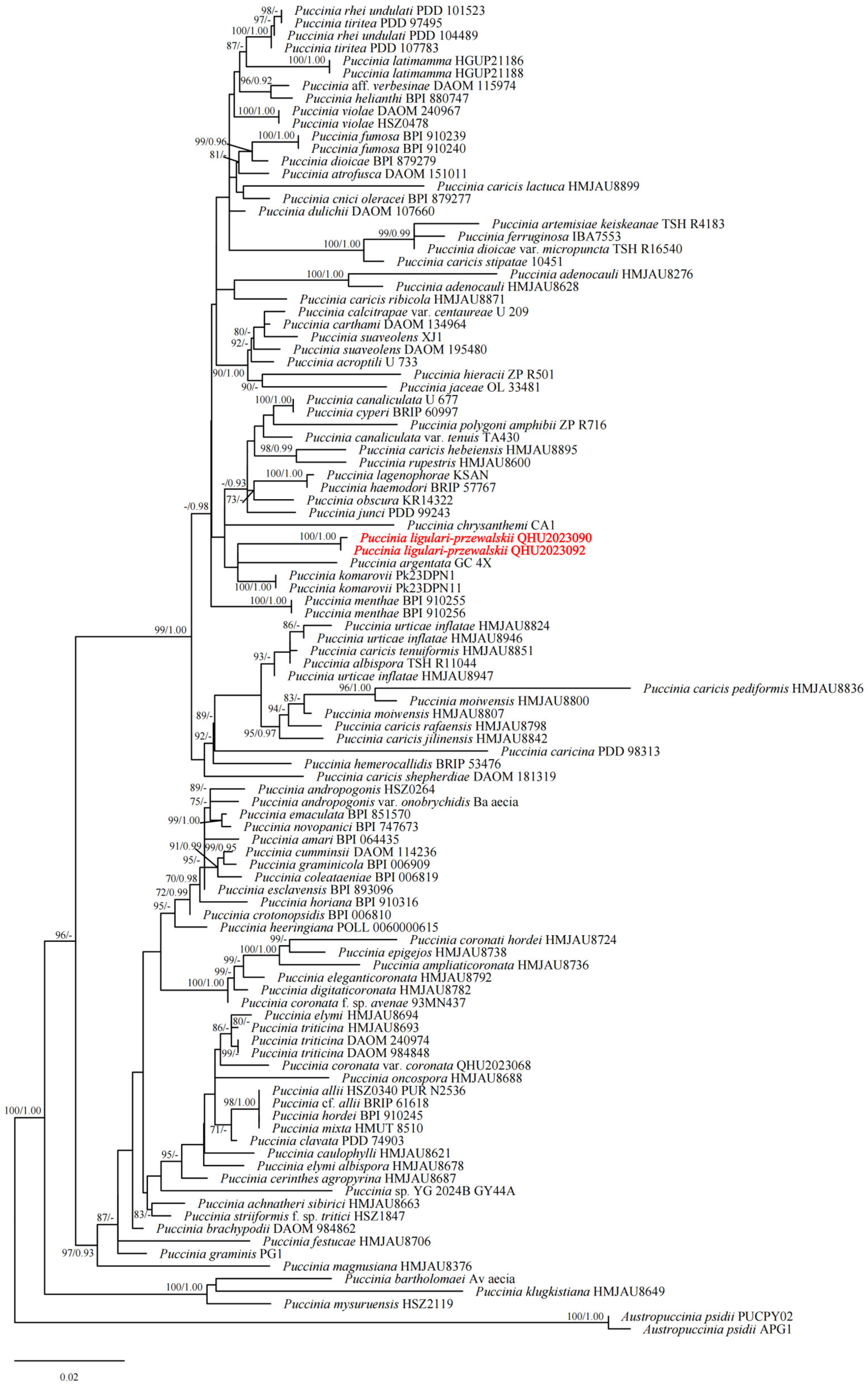
The combined sequence data of LSU and ITS comprised 104 strains of Puccinia and two strains of A. psidii as outgroup taxa (Figure 1). A total of 1540 characters, including gaps, were obtained for the phylogenetic analysis. The IQ-TREE analysis of the combined dataset yielded a best-scoring tree with a final ML optimization likelihood value of −8803.859. The matrix had 720 distinct alignment patterns, containing 1041 constant sites, 285 parsimony-informative sites and 214 singleton variable sites; 3 sites contained only gaps or ambiguous characters. The estimated base frequencies were as follows: A = 0.328, C = 0.151, G = 0.232, T = 0.289; substitution rates AC = 1.52670, AG = 3.32150, AT = 1.20335, CG = 0.77246, CT = 5.02495, GT = 1.00000; the proportion of invariable sites was 0.352, and the gamma distribution shape parameter α was 0.528. The topologies recovered by ML and Bayesian inference were highly congruent. Our new isolates of Puccinia ligulari-przewalskii (QHU2023090T and QHU2023092) formed a distinct monophyletic clade with 100% ML and 1.00 BYPP values, and showed a close phylogenetic affinity with Puccinia argentata (strain GC 4X) and Puccinia komarovii (strains Pk23DPN1 and Pk23DPN11) (Figure 1).

2.6. Taxonomy

Puccinia ligulari-przewalskii L.C. Bai & Q. Xu, sp. nov.
Holotype: CHINA. QINGHAI PROVINCE: Makehe District, Guoluo Tibetan Autonomous Prefecture (100°57′33″ E, 32°40′54″ N; 3368.5 ma.s.l.), on Ligularia przewalskii, 25 Aug 2023, Q. Xu, QHU2023090. GenBank: ITS = PZ575731; LSU = PP469520. Other spore types, except for the teliospore type, have not been found.
Etymology: The epithet ligulari-przewalskii refers to the host plant Ligularia przewalskii, from which the rust fungus was isolated.
Description: Spermogonia, aecia and uredinia: Unknown.
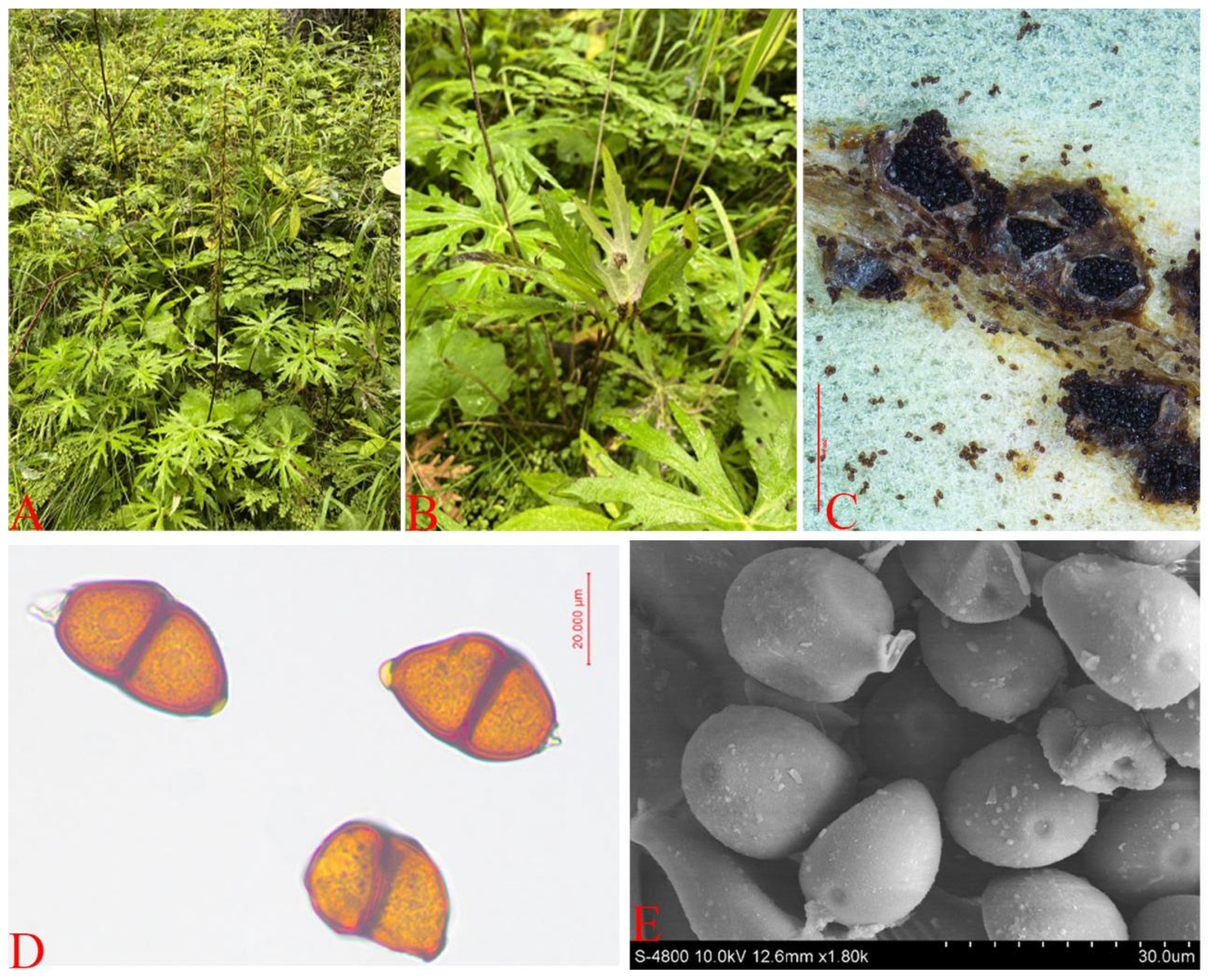
Telia: Present on the underside of leaves, aggregated, round to elliptical, 0.1–0.5 mm in diameter, chestnut-brown, initially covered by the host plant’s epidermis but later rupturing and exposed.
Teliospores: Elliptical, spindle-shaped, or polygonal, two-celled, slightly constricted at the septum, measuring 30.7–44.8 × 17.4–25.3 µm, chestnut-brown, with lateral walls 1.4–2.3 µm thick; papillae 1.6–4.4 µm long, yellow, with apical germ pores; spore surface relatively smooth with sparse warts. (Figure 2).
Ecology and distribution: III (teliospores) on L. przewalskii, known only from Qinghai in China.
Notes: L. przewalskii is a perennial herbaceous plant belonging to the genus Ligularia in the Asteraceae family. It is mainly distributed in the eastern part and marginal areas of the Qinghai–Tibet Plateau. In traditional Tibetan medicine, the entire herb of L. przewalskii can be used as medicine, which has the effects of clearing away heat and toxic substances, dispersing blood stasis and reducing swelling. It is commonly used to treat sore throat, carbuncles and sores, bruises and other diseases. Mashing its fresh product for external application has a significant relieving effect on the redness, swelling, heat and pain in the early stage of sores.

3. Discussion

A new rust species, P. ligulari-przewalskii sp. nov., is described in this study based on both morphological and molecular phylogenetic evidence. It represents the first record of a Puccinia species occurring on Ligularia (Asteraceae) in China to be characterized by both morphological and molecular phylogenetic data. Prior to this discovery, eleven rust taxa had been documented on Ligularia spp.: A. nikkense (on Ligularia stenocephala in China and Japan) [3,28], A. ligulariae (reported from Finland and China without specified host species) [29,31], C. ligulariae (infecting Ligularia jaluensis, Ligularia przewalskii and other species in China, Finland and Romania) [3,13,30], C. saussureae (occurring on Ligularia dentata, L. stenocephala and other hosts, with distribution reflecting host range) [3,13], and C. senecionis (found on Ligularia gigantea and Ligularia macrophylla across multiple regions) [3], P. cnici-oleracei (recorded on Ligularia tsangchanensis and other Asteraceae) [13,15], P. eriophori (reported on Ligularia speciosa and other Ligularia spp. in China) [15,30], P. expansa (recorded on Ligularia sibirica and Ligularia spp. in China) [15,30], P. glomerata (occurring on L. achyrotricha and other Ligularia spp.) [3,15], P. ligulariae (recorded on Ligularia spp. in China) [30], and U. ligulariae (on Ligularia tussilaginea var. formosana in Taiwan) [3].
A. nikkense was originally described from Japan on Ligularia stenocephala var. comosa [28] and has since been recorded on L. stenocephala in Hubei (Shennongjia), China, and on Farfugium japonicum in Japan [3,32]. The Flora Fungorum Sinicorum Vol. 69 treats it as an independent anamorphic species known only from its aecial stage; no telial connection has been established, and no ITS or LSU sequence data are available [32]. It may be noted that the Catalogue of Life China (2023) lists A. nikkense as a synonym of Uromyces veratri (DC.) J. Schröt. [49]; however, the Flora Fungorum Sinicorum Vol. 25 records Aecidium cacaliae Thüm., not A. nikkense, as the aecial stage of U. veratri [14]. The accuracy of this synonymy therefore remains to be verified.
A. ligulariae was originally described from Finland [29]. Tranzschel [50] identified this fungus as the aecial stage of P. eriophori Thüm., and this treatment was subsequently adopted by Tai [30] and Zhuang [33]. Zhuang (1988) listed A. ligulariae as the aecial synonym of P. eriophori and recorded the uredinial and telial stages on Eriophorum (Cyperaceae) [33]. The aeciospores of A. ligulariae recorded in the Chinese rust flora (18–25 × 15–20 µm, with densely and finely verrucose walls) [32] are morphologically consistent with those of Puccinia eriophori var. eriophori described from North America by Savile (1972) (16–24.5 × 12.5–21 µm, with similarly densely and finely verrucose walls) [51]. The species has been recorded on multiple Ligularia species across northern China, including Qinghai Province [32]. Although Eriophorum spp. (Cyperaceae)—the host of P. eriophori recorded—are widely distributed in northern China, the uredinial and telial stages have never been observed on these plants in the country [32]. Savile (1972) described the telial stage of P. eriophori var. eriophori in detail based on North American and European material on Eriophorum [51]: the teliospores are smooth, light yellow-brown to light chestnut, (34–)37–71(–83) × (14–)16.5–28(–29.5) µm, with a rounded or conic apical cap concolorous or paler, and pedicels moderately firm, 48–75 µm long. By contrast, the teliospores of P. ligulari-przewalskii are sparsely verrucose, distinctly smaller (30.7–44.8 × 17.4–25.3 µm), bear a prominent yellow apical papilla 1.6–4.4 µm, and have pedicels that are readily detached (Table 3).
In summary, A. nikkense remains an anamorphic name whose telial connection is uncertain and disputed across authoritative sources. A. ligulariae is recognized as the aecial stage of P. eriophori, a rust for which Eriophorum (Cyperaceae) is recorded as the host in the Chinese rust flora; however, the uredinial and telial stages of P. eriophori on Eriophorum (Cyperaceae) have never been recorded in China. The teliospores of P. ligulari-przewalskii are morphologically distinct from those of P. eriophori described by Savile (1972) [51] (Table 3). The available evidence does not support a connection between the uredinial or telial stages of A. nikkense or A. ligulariae and the telial material on L. przewalskii described here. P. ligulari-przewalskii is therefore proposed as a new species on the basis of the morphological and molecular data presented. The ITS and LSU sequences, voucher specimens, and morphological documentation from this study are available for comparison should the teleomorph of A. nikkense or A. ligulariae be identified.
The three Coleosporium species recorded on LigulariaC. ligulariae, C. saussureae, and C. senecionis—differ fundamentally from Puccinia in their teliospore morphology: Coleosporium teliospores are one-celled, cylindrical, thin-walled, colorless, and formed in a single-layered crust, germinating without dormancy by producing an external basidium [3,13]. In contrast, Puccinia teliospores are two-celled, thick-walled, pigmented, pedicellate, and germinate via apical germ pores [2]. The three Coleosporium species and U. ligulariae are readily distinguished from Puccinia by their one-celled teliospores [3,13], in contrast to the two-celled teliospores of P. ligulari-przewalskii. Unlike the aforementioned Uromyces and Coleosporium species having one-celled teliospores, P. ligulari-przewalskii is assigned to Puccinia, characterized by its two-celled teliospores.
Phylogenetic analysis based on the concatenated ITS and LSU sequences places P. ligulari-przewalskii within the genus Puccinia, clustering as a sister lineage to P. argentata and P. komarovii, further corroborating its generic placement. The teliospores of P. argentata are smooth or very finely obscurely verruculose, ellipsoid or oblong, measuring 25–40 × 14–21 µm, with a distinct hyaline pore cap 3–5 µm and an apex that is not thickened [13]. P. ligulari-przewalskii produces sparsely verrucose teliospores that are ellipsoid, fusiform or polygonal, 30.7–44.8 × 17.4–25.3 µm, with a yellow apical papilla 1.6–4.4 µm (Figure 2; Table 3). The teliospores of P. komarovii are smooth or nearly smooth, subrectangular or obovoid, 25–40 × 16–23 µm, with a uniform wall thickness of approximately 2.0 µm and a distinct hyaline pore cap [13]. P. ligulari-przewalskii differs in its sparsely verrucose ornamentation, ellipsoid to fusiform shape, and the presence of a yellow apical papilla. Furthermore, P. argentata and P. komarovii both occur on Impatiens (Balsaminaceae) [13], whereas the new species parasitizes Ligularia (Asteraceae).
Among the morphologically compared species, Puccinia gentianae merits particular attention. It is widely distributed in the North Temperate Zone, including the Qinghai–Tibet Plateau region, where it parasitizes various Gentiana species (Gentianaceae) [13]. In the Sanjiangyuan region, P. gentianae has been recorded on Gentiana straminea [34], indicating that it co-occurs with P. ligulari-przewalskii in the same geographic area. P. gentianae has a macrocyclic life cycle, producing spermogonia, aecia, uredinia, and telia [13]. The teliospores of P. gentianae are smooth and broadly ellipsoid to oblong, measuring 28–43 × 20–30 µm, with uniformly thickened lateral walls (2.0–3.0 µm) and a colorless small pore cap at the apical germ pore [13]. In contrast, P. ligulari-przewalskii has sparsely verrucose, ellipsoid to fusiform teliospores (30.7–44.8 × 17.4–25.3 µm), with thinner lateral walls (1.4–2.3 µm) and a distinct yellow apical papilla (1.6–4.4 µm) (Figure 2; Table 3). The host genera Gentiana (Gentianaceae) and Ligularia (Asteraceae) are phylogenetically distant, further corroborating the distinction between these two species.
Among the Puccinia species previously recorded on Ligularia, Puccinia glomerata is the most comparable taxon [3,15]. Its teliospores are smooth, ellipsoid or irregularly obovoid, 33–53 × 15–28 µm, with a hyaline pore cap 3–5 µm [13]. The new species produces smaller, sparsely verrucose teliospores with a yellow papilla. Moreover, P. glomerata produces two teliospore morphs, small and large, according to Cummins [52], whereas P. ligulari-przewalskii produces a single teliospore type. P. cnici-oleracei, recorded on L. tsangchanensis [15], has smooth, clavate or oblong-ellipsoid teliospores measuring 33–58 × 13–20 µm, with a considerably thickened apical wall of 3–13 µm [13], clearly distinct from the sparsely verrucose, papilla-bearing spores of the new species. P. expansa, also recorded on Ligularia [15,30], is morphologically similar to P. glomerata and has been treated as distinct by several authors [53]; however, both P. expansa and P. glomerata lack molecular characterization and differ from the new species in teliospore ornamentation and pore structure.
P. ligulari-przewalskii is characterized by a unique combination of sparsely verrucose teliospores, a yellow apical papilla 1.6–4.4 µm, and the absence of a hyaline pore cap. This combination distinguishes it from all other Puccinia species compared in this study (Table 3). Among these, Puccinia cremanthodii has reticulate-rugose spores; Puccinia hieracii, although finely verrucose and similarly lacking a pore cap, has more broadly ellipsoid spores with the lower cell germ pore positioned away from the septum; Puccinia minussensis has verrucose spores with a distinct hyaline pore cap; Puccinia violae is densely verrucose and occurs on Violaceae; P. senecionis and Puccinia artemisiae-keiskeanae both have smooth teliospores; and Puccinia ferruginosa, shown to be polyphyletic by Engkhaninun et al. [54], has smooth, elongate or clavate spores with a thickened apex [55].
Only the telial stage was observed in the specimens of P. ligulari-przewalskii examined in this study. Specimens were collected from L. przewalskii at the type locality on multiple occasions between 2021 and 2023, with collection efforts concentrated in July through September, corresponding to the flowering and fruiting period of the host [56]. No spermogonia, aecia, or uredinia were detected in any collection. However, because the surveys did not span the entire growing season—particularly the early season (May–June) when leaf emergence and the development of spermogonial and aecial stages of rust fungi typically occur—the possibility that additional spore stages are present during unsampled periods cannot be excluded.
Several lines of evidence suggest that P. ligulari-przewalskii may not be microcyclic. First, its closest phylogenetic relatives, P. argentata and P. komarovii, are both non-microcyclic: P. argentata is macrocyclic and heteroecious, producing spermogonia and aecia on Adoxa moschatellina and uredinia and telia on Impatiens species [13], while P. komarovii produces uredinia and telia on Impatiens [13]. The derivation of a microcyclic species from within this non-microcyclic lineage would represent an evolutionary exception, though not an impossibility. Second, microcyclic Puccinia species are rare on Asteraceae. Among the more than 1000 rust species reported on this family, the vast majority are macrocyclic or demicyclic [6], and only a handful of microcyclic taxa have been documented, e.g., P. cremanthodii on Cremanthodium at elevations above 4000 m on the Qinghai–Tibet Plateau [13]. P. glomerata, previously considered microcyclic on Ligularia [3,15], has recently been shown to occasionally produce urediniospores [13]. Third, collections were made during the flowering and fruiting phase of the host [56], which is suboptimal for detecting spermogonial and aecial stages that typically develop on newly emerged leaves in early summer.
Nevertheless, the type locality lies in a high-elevation alpine environment (3370 m) on the Qinghai–Tibet Plateau, where abbreviated growing seasons are known to favor life cycle reduction. Microcyclic rusts are disproportionately represented in arcto-alpine floras, a pattern attributed to the selective advantage of completing the life cycle within a short thermal window without reliance on an alternate host [6,9]. Whether P. ligulari-przewalskii is microcyclic, demicyclic, or macrocyclic remains an open question. Full-season phenological monitoring from leaf emergence (June) through senescence (September), combined with basidiospore inoculation experiments on healthy L. przewalskii seedlings, would be required to definitively establish the life cycle type.
Collectively, both morphological and molecular evidence support the recognition of P. ligulari-przewalskii as a species new to science, and it represents the first Puccinia species occurring on Ligularia (Asteraceae) in China to be characterized by both morphological and molecular phylogenetic data.

Author Contributions

Conceptualization, L.B. and Y.Z.; Methodology, Q.X. and L.Q.; Sample collection, L.B., Q.X. and W.Z.; Writing—Original Draft Preparation, Q.X. and L.Q.; Writing—Review & Editing, L.B. and Y.C. All authors have read and agreed to the published version of the manuscript.

Funding

This paper was supported by Qinghai Province Key Research and Development and Transformation Plan (2023-SF-119).

Institutional Review Board Statement

Not applicable.

Informed Consent Statement

Not applicable.

Data Availability Statement

The original contributions presented in this study are included in the article. Further inquiries can be directed to the corresponding author.

Acknowledgments

We thank the Qinghai Provincial Department of Science & Technology for technical guidance and Sanjiangyuan National Administration for ecological stewardship.

Conflicts of Interest

The authors declare no competing interests.

References

  1. Aime, M.C.; McTaggart, A.R. A higher-rank classification for rust fungi, with notes on genera. Fungal Syst. Evol. 2021, 7, 21–47. [Google Scholar] [CrossRef] [Scilit] [PubMed]
  2. Cummins, G.B.; Hiratsuka, Y. Illustrated Genera of Rust Fungi, 3rd ed.; APS Press: St. Paul, MN, USA, 2003. [Google Scholar]
  3. Hiratsuka, N.; Sato, S.; Katsuya, K.; Kakishima, M.; Hiratsuka, Y.; Kaneko, S.; Ono, Y.; Sato, T.; Harada, Y.; Hiratsuka, T.; et al. The Rust Flora of Japan; Tsukuba Shuppankai: Ibaraki, Japan, 1992. [Google Scholar]
  4. Zhao, P.; Zhang, Z.F.; Hu, D.M.; Tsui, K.M.; Qi, X.H.; Phurbu, D.; Gafforov, Y.; Cai, L. Contribution to rust flora in China I, tremendous diversity from natural reserves and parks. Fungal Divers. 2021, 110, 1–58. [Google Scholar] [CrossRef] [Scilit]
  5. Hennen, J.F.; Buriticá, P. A brief summary of modern rust taxonomic and evolutionary theory. Rep. Tottori Mycol. Inst. 1980, 18, 243–256. [Google Scholar]
  6. Ono, Y. The diversity of nuclear cycle in microcyclic rust fungi (Uredinales) and its ecological and evolutionary implications. Mycoscience 2002, 43, 421–439. [Google Scholar] [CrossRef] [Scilit]
  7. Demers, J.E.; Romberg, M.K.; Castlebury, L.A. Microcyclic rusts of hollyhock (Alcea rosea). IMA Fungus 2015, 6, 477–482. [Google Scholar] [CrossRef] [Scilit] [PubMed]
  8. Avasthi, S.; Gautam, A.K.; Niranjan, M.; Verma, R.K.; Karunarathna, S.C.; Kumar, A.; Suwannarach, N. Insights into diversity, distribution, and systematics of rust genus Puccinia. J. Fungi 2023, 9, 639. [Google Scholar] [CrossRef] [Scilit] [PubMed]
  9. Savile, D.B.O. Short-season adaptations in the rust fungi. Mycologia 1953, 45, 75–87. [Google Scholar] [CrossRef] [Scilit]
  10. Abbasi, M.; Darvishnia, M. Puccinia fritschii sp. nov.—A new rust species from Iran. Schlechtendalia 2015, 28, 77–79. [Google Scholar] [CrossRef] [PubMed]
  11. Abbasi, M.; Klimek, J.F.; Aime, M.C. First report of rust disease caused by Puccinia liliacearum on Ornithogalum umbellatum from Indiana and Maryland with notes on the spread of the rust fungus in the United States. Plant Dis. 2016, 100, 2169. [Google Scholar] [CrossRef] [Scilit]
  12. Zhuang, J.Y.; Wei, S.X.; Wang, Y.C. Flora Fungorum Sinicorum. Vol. 10. Uredinales (I); Science Press: Beijing, China, 1998. [Google Scholar]
  13. Zhuang, J.Y.; Wei, S.X.; Wang, Y.C. Flora Fungorum Sinicorum. Vol. 19. Uredinales (II); Science Press: Beijing, China, 2003. [Google Scholar]
  14. Zhuang, J.Y.; Wei, S.X.; Wang, Y.C. Flora Fungorum Sinicorum. Vol. 25. Uredinales (III); Science Press: Beijing, China, 2005. [Google Scholar]
  15. Zhuang, J.Y.; Wei, S.X.; Wang, Y.C. Flora Fungorum Sinicorum. Vol. 41. Uredinales (IV); Science Press: Beijing, China, 2012. [Google Scholar]
  16. Zhuang, J.Y. Rust Order of China (V); Science Press: Beijing, China, 2021. [Google Scholar]
  17. Marin-Felix, Y.; Groenewald, J.Z.; Cai, L.; Chen, Q.; Marincowitz, S.; Barnes, I.; Bensch, K.; Braun, U.; Camporesi, E.; Damm, U.; et al. Genera of phytopathogenic fungi: GOPHY 1. Stud. Mycol. 2017, 86, 99–216. [Google Scholar] [CrossRef] [Scilit] [PubMed]
  18. Sun, C.; Liu, Y.F.; Liang, Y.M.; Wang, L. Four new species of Puccinia from herbaceous plants in China. Mycologia 2024, 116, 309–321. [Google Scholar] [CrossRef] [Scilit] [PubMed]
  19. Gao, Y.; Tibpromma, S.; McKenzie, E.H.C.; Eungwanichayapant, P.D.; Gautam, A.K.; Lu, L.; Jayawardena, R.S.; Hyde, K.D.; Xu, J.C.; Gui, H. Two new species of Puccinia (Pucciniaceae, Pucciniales) from herbaceous plants in Yunnan Province, China. Phytotaxa 2025, 694, 25–45. [Google Scholar] [CrossRef] [Scilit]
  20. Cao, Z.M.; Li, Z.Q.; Zhuang, J.Y. Uredinales from the Qinling Mountains (Continued I). Mycosystema 2000, 19, 181–192. [Google Scholar]
  21. Dai, Y.C.; Zhuang, J.Y. Numbers of fungal species hitherto known in China. Mycosystema 2015, 29, 652–658. [Google Scholar]
  22. Liu, M.; Hambleton, S. Puccinia chunjii, a close relative of the cereal stem rusts revealed by molecular phylogeny and morphological study. Mycologia 2012, 104, 1056–1067. [Google Scholar] [CrossRef] [Scilit] [PubMed]
  23. Liu, T.Z.; Yang, X.P.; Zhuang, J.Y. A new species and a new record of Puccinia on Poaceae from China. Mycosystema 2014, 33, 773–776. [Google Scholar] [CrossRef]
  24. Liu, T.Z.; Zhuang, J.Y. A new species and a new Chinese record of Puccinia on Asteraceae from China. Mycosystema 2015, 34, 341–344. [Google Scholar] [CrossRef] [Scilit]
  25. Liu, T.Z.; Zhuang, J.Y. A new species of Puccinia and a new variety of P. acetosae from Inner Mongolia, China. Mycosystema 2016, 35, 549–552. [Google Scholar]
  26. Liu, T.Z.; Zhuang, J.Y.; Yang, X.P. A new variety and a new record of Puccinia from China. Mycosystema 2016, 35, 1489–1492. [Google Scholar]
  27. Zhuang, J.Y.; Wei, S.X. Materials for study on rust fungi of eastern rimland of Qinghai–Xizang (Tibet) Plateau VI. Two new species of Puccinia. Mycosystema 1999, 18, 117–120. [Google Scholar]
  28. Ito, S. Mycological Flora of Japan. Vol. II. Basidiomycetes. No. 3. Uredinales-Pucciniaceae, Uredinales Imperfecti; Yokendo Ltd.: Tokyo, Japan, 1950. [Google Scholar]
  29. Liro, J.I. Uredineae Fennicae: Finlands Rostsvampar; Finska Litteratursällskapets Tryckeri: Helsinki, Finland, 1908. [Google Scholar]
  30. Tai, F.L. Sylloge Fungorum Sinicorum; Science Press; Academica Sinica: Beijing, China, 1979. [Google Scholar]
  31. Zhuang, W.Y. Fungi of Northwestern China; Mycotaxon, Ltd.: Ithaca, NY, USA, 2005. [Google Scholar]
  32. Zhuang, J.Y.; Wei, S.X.; Wang, Y.C. Flora Fungorum Sinicorum. Vol. 69, Uredinales (VII); Science Press: Beijing, China, 2025. [Google Scholar]
  33. Zhuang, J.Y. Species of Puccinia on the Cyperaceae in China. Mycosystema 1988, 1, 115–148. [Google Scholar]
  34. Xu, Q.; Bai, L. The diversity and floristic analysis of rust diseases in the Sanjiangyuan forest plants. J. Fungi 2024, 10, 425. [Google Scholar] [CrossRef] [Scilit] [PubMed]
  35. Kirk, P.M.; Cannon, P.F.; Minter, D.W.; Stalpers, J.A. Ainsworth & Bisby’s Dictionary of the Fungi, 10th ed.; CAB International: Wallingford, UK, 2008. [Google Scholar]
  36. Wei, J.C. Handbook of Fungal Identification; Shanghai Science and Technology Press: Shanghai, China, 1979. [Google Scholar]
  37. Cao, Z.M.; Li, Z.Q. Qinling Rust Fungus; China Forestry Publishing Press: Beijing, China, 1999. [Google Scholar]
  38. Tian, C.M.; Shang, Y.Z.; Zhuang, J.Y.; Wang, Y.C.; Kakishima, M. Morphological and molecular phylogenetic analysis of Melampsora species on poplars in China. Mycoscience 2004, 45, 56–66. [Google Scholar] [CrossRef] [Scilit]
  39. Yang, T. Phylogenetic and Taxonomic Studies of Pucciniastrum s.l. Ph.D. Thesis, Beijing Forestry University, Beijing, China, 2015. [Google Scholar]
  40. Pfunder, M.; Schürch, S.; Roy, B.A. Sequence variation and geographic distribution of pseudoflower-forming rust fungi (Uromyces pisi s. lat.) on Euphorbia cyparissias. Mycol. Res. 2001, 105, 57–66. [Google Scholar] [CrossRef] [Scilit]
  41. Beenken, L.; Zoller, S.; Berndt, R. Rust fungi on Annonaceae II: The genus Dasyspora Berk. & M.A. Curtis. Mycologia 2012, 104, 659–681. [Google Scholar] [CrossRef] [Scilit] [PubMed]
  42. O’Donnell, K. Fusarium and its near relatives. In The Fungal Holomorph: Mitotic, Meiotic and Pleomorphic Speciation in Fungal Systematics; Reynolds, D.R., Taylor, J.W., Eds.; CAB International: Wallingford, UK, 1993; pp. 225–233. [Google Scholar]
  43. Katoh, K.; Standley, D.M. MAFFT multiple sequence alignment software version 7: Improvements in performance and usability. Mol. Biol. Evol. 2013, 30, 772–780. [Google Scholar] [CrossRef] [Scilit] [PubMed]
  44. Hall, T.A. BioEdit: A user-friendly biological sequence alignment editor and analysis program for Windows 95/98/NT. Nucleic Acids Symp. Ser. 1999, 41, 95–98. [Google Scholar]
  45. Zhao, D.; Ye, T.; Gao, F.; Jakovlić, I.; La, Q.; Tong, Y.; Liu, X.; Song, R.; Liu, F.; Lian, Z.M.; et al. PhyloSuite v2: The development of an all-in-one, efficient and visualization-oriented suite for molecular dating analysis and other advanced features. iMeta 2025, 4, e70095. [Google Scholar] [CrossRef] [Scilit] [PubMed]
  46. Nguyen, L.T.; Schmidt, H.A.; von Haeseler, A.; Minh, B.Q. IQ-TREE: A fast and effective stochastic algorithm for estimating maximum-likelihood phylogenies. Mol. Biol. Evol. 2015, 32, 268–274. [Google Scholar] [CrossRef] [Scilit] [PubMed]
  47. Minh, B.Q.; Schmidt, H.A.; Chernomor, O.; Schrempf, D.; Woodhams, M.D.; von Haeseler, A.; Lanfear, R. IQ-TREE 2: New models and efficient methods for phylogenetic inference in the genomic era. Mol. Biol. Evol. 2020, 37, 1530–1534. [Google Scholar] [CrossRef] [Scilit] [PubMed]
  48. Ronquist, F.; Teslenko, M.; van der Mark, P.; Ayres, D.L.; Darling, A.; Höhna, S.; Larget, B.; Liu, L.; Suchard, M.A.; Huelsenbeck, J.P. MrBayes 3.2: Efficient Bayesian phylogenetic inference and model choice across a large model space. Syst. Biol. 2012, 61, 539–542. [Google Scholar] [CrossRef] [Scilit] [PubMed]
  49. The Biodiversity Committee of Chinese Academy of Sciences. Catalogue of Life China: 2022 Annual Checklist, Version 1.1; Chinese Academy of Sciences (CAS): Beijing, China, 2023. [CrossRef]
  50. Tranzschel, V.G. Kulturversuche mit Uredineen im Jahre 1907. Ann. Mycol. 1907, 5, 418. [Google Scholar]
  51. Savile, D.B.O. Some rusts of Scirpus and allied genera. Can. J. Bot. 1972, 50, 2579–2596. [Google Scholar] [CrossRef] [Scilit]
  52. Cummins, G.B. Rust Fungi on Legumes and Composites in North America; University of Arizona Press: Tucson, AZ, USA, 1978. [Google Scholar]
  53. Gäumann, E. Die Rostpilze Mitteleuropas; Buchdruckerei Büchler: Bern, Switzerland, 1959. [Google Scholar]
  54. Engkhaninun, J.; Ono, Y.; Kakishima, M. Phylogenetic relationships of four Puccinia species parasitic on Artemisia in Japan. Mycoscience 2005, 46, 61–65. [Google Scholar] [CrossRef] [Scilit]
  55. Preston, C.D.; Harries, D.J.; Kruse, J.; Stringer, R.N. Puccinia ferruginosa, a second rust on Artemisia vulgaris in Britain. Field Mycol. 2023, 24, 128–136. [Google Scholar] [CrossRef] [Scilit]
  56. Editorial Committee of Flora of Qinghai. Flora of Qinghai. Vol. 3; Qinghai People’s Publishing House: Xining, China, 1996; pp. 401–402. [Google Scholar]
Figure 1. Phylogram generated from ML analysis based on the combined LSU and ITS of Puccinia species. Bootstrap support values for ML equal to or higher than 65% and BYPP equal to or greater than 0.90 are shown above the nodes. The new species are highlighted in red font. The tree is rooted with A. psidii (APG1 and PUCPY02). The scale bar represents 0.02 nucleotide substitutions per site.
Diversity 18 00487 g001
Figure 2. Morphological characteristics of P. ligulari-przewalskii on L. przewalskii. (A,B) Host plant habitat. (C) Telia. (D) Teliospores (LM). (E) Teliospores (SEM).
Diversity 18 00487 g002
Table 1. Herbarium specimens used for morphological examination and molecular phylogenetic analyses.
Host PlantsLocalityTimeElevation/mLongitude &
Latitude
Specimen No.Mycobank No.
Ligularia
przewalskii
Makehe25 August 20233368.5100°57′33″ E
32°40′54″ N
QHU2023090 (Holotype)
QHU2023092 (Isotype)
860240
Table 2. Sequence data of Puccinia species from GenBank and used for phylogenetic analyses.
Taxon NameStrain NumberITSLSUHostCountry
Austropuccinia psidiiPUCPY02MH513948MH513948Eucalyptus benthamiiParaguay
Austropuccinia psidiiAPG1MN238767MN238767GuavaBrazil
Puccinia achnatheri-sibiriciHMJAU8663MW404809MW404969Deyeuxia purpureaChina
Puccinia acroptiliU-733JN204196JN204196Rhaponticum repensKazakhstan
Puccinia adenocauliHMJAU8276MW447796MW414403Adenocaulon himalaicumChina
Puccinia adenocauliHMJAU8628MK785270MK785295Adenocaulon himalaicumChina
Puccinia aff. verbesinaeDAOM 115974MW009569MW009569Verbesina sp.Mexico
Puccinia albisporaTSH R11044MT560796MT560813 South Africa
Puccinia alliiHSZ0340AF511087AF511087AlliumUSA
Puccinia amariBPI 064435TKX190836KX190836Panicum amarumUSA
Puccinia ampliaticoronataHMJAU8736TMW404765MW404925Melica grandifloraChina
Puccinia andropogonisHSZ0264DQ344519DQ344519Andropogon spp.USA
Puccinia andropogonis var. onobrychidisBa aeciaEF583818EF583818Baptisia australisUSA
Puccinia argentataGC-4XPP273009PP266821 China
Puccinia artemisiae-keiskeanaeTSH-R4183AB188133AB190893Artemisia keiskeanaJapan
Puccinia atrofuscaDAOM:151011MW009474MW009474Carex rossiiCanada
Puccinia bartholomaeiAv aeciaEF583820EF583820Asclepias viridisUSA
Puccinia brachypodiiDAOM:984862MT965646MT965646Brachypodium sylvaticumUK
Puccinia calcitrapae var. centaureaeU-209JN204183JN204183Centaurea calcitrapaTurkey
Puccinia canaliculataU-677HQ412647HQ412647Cyperus rotundusOman
Puccinia canaliculata var. tenuisTA430OL437018OL437033Cyperus esculentusBenin
Puccinia caricinaPDD:98313KX985750KX985750Carex geminataNew Zealand
Puccinia caricis-hebeiensisHMJAU8895TMW447818MW414427Carex lanceolataChina
Puccinia caricis-jilinensisHMJAU8842TMW447898MW414367Carex siderostictaChina
Puccinia caricis-lactucaeHMJAU8899TMW447778NG_228873Lactuca sp. China
Puccinia caricis-pediformisHMJAU8836TMW447855MW414323Carex pediformisChina
Puccinia caricis-rafaensisHMJAU8798TMW447892MW414360Carex sp.China
Puccinia caricis-ribicolaHMJAU8871MW447805MW414413Carex dispalataChina
Puccinia caricis-shepherdiaeDAOM:181319MW009426MW009426Shepherdia canadensisCanada
Puccinia caricis-stipatae10451AB188137AB190887Carex stipataJapan
Puccinia caricis-tenuiformisHMJAU8851TMW447858MW414326Carex sp. China
Puccinia carthamiDAOM:134964MW009544MW009544Carthamus tinctoriusCanada
Puccinia caulophylliHMJAU8621MK785279MK785304Milium effusumChina
Puccinia cerinthes-agropyrinaHMJAU8687MW404828MW404988Clematis ternifloraChina
Puccinia cf. alliiBRIP 61618KU296870KU296870Allium sativumAustralia
Puccinia chrysanthemiCA1EU816926EU816926ChrysanthemumUSA
Puccinia clavataPDD:74903KX985761KX985761Clematis foetidaNew Zealand
Puccinia cnici-oleraceiBPI 879277GU058017GU058017Aster sp.Mexico
Puccinia coleataeniaeBPI 006819TKX190843KX190843Coleataenia ancepsUSA
Puccinia coronata f. sp. avenae93MN437AY114290AY114290AvenaeUSA
Puccinia coronata var. coronataQHU2023068PP471872.1PP469628.1Clematis sp.China
Puccinia coronati-hordeiHMJAU8724MW404779MW404939Poa sp.China
Puccinia crotonopsidisBPI 006810TKX190844KX190844Croton michauxiiUSA
Puccinia cumminsiiDAOM 114236TKX190845KX190845Panicum sp.USA
Puccinia cyperiBRIP 60997KU296885KU296885Cyperus iriaAustralia
Puccinia digitaticoronataHMJAU8782TMW404711MW404871Poa sp.China
Puccinia dioicaeBPI 879279GU058019GU058019Oenothera fruticosaUSA
Puccinia dioicae var. micropunctaTSH:R16540AB188129LC626796Carex breviculmisJapan
Puccinia dulichiiDAOM:107660MW009481MW009481Dulichium arundinaceumUSA
Puccinia eleganticoronataHMJAU8792TMW404723MW404883Poa sphondylodesChina
Puccinia elymiHMJAU8694MW404827MW404987PoaceaeChina
Puccinia elymi-albisporaHMJAU8678TMW404819MW404979Elymus ciliarisChina
Puccinia emaculataBPI 851570TKX190848KX190848Panicum capillareUSA
Puccinia epigejosHMJAU8738MW404769MW404929Calamagrostis epigeiosChina
Puccinia esclavensisBPI 893096KX190866KX190866Mirabilis melanotrichaMexico
Puccinia ferruginosaIBA7553AB193001AB190901Artemisia freynianaJapan
Puccinia festucaeHMJAU8706MW404786MW404946Festuca extremiorientalisChina
Puccinia fumosaBPI 910240KY764123KY764123Loeselia mexicanaUSA
Puccinia fumosaBPI 910239KY764122KY764122Loeselia mexicanaUSA
Puccinia graminicolaBPI 006909TKX190868KX190868Panicum virgatumUSA
Puccinia graminisPG1AY874153MK952782Triticum aestivumUSA
Puccinia haemodoriBRIP:57767KF690675KF690693Anigozanthos sp.Australia
Puccinia heeringianaPOLL_0060000615OR568547OR568547Tanacetum partheniumGermany
Puccinia helianthinBPI 880747KY764127KY764127Helianthus annuusUSA
Puccinia hemerocallidisBRIP:53476KM249855KM249855Hemerocallis sp.Australia
Puccinia hieraciiZP-R501MK518930MK518610 China
Puccinia hordeiBPI 910245KY764129KY764129Ornithogalum sp.USA
Puccinia horianaBPI 910316KY798398KY798398Chrysanthemum x morifoliumUSA
Puccinia jaceaeOL 33481KX468974KX468974Centaurea macrocephalaCzech Republic
Puccinia junciPDD:99243KX985745KX985745Juncus tenuisNew Zealand
Puccinia klugkistianaHMJAU8649MW406957MW397084Cleistogenes hackeliiChina
Puccinia komaroviiPk23DPN11OR475557OR475550Impatiens parvifloraPoland
Puccinia komaroviiPk23DPN1OR475551OR473652Impatiens parvifloraPoland
Puccinia lagenophoraeKSANFJ655869FJ669236Senecio arenariusSouth Africa
Puccinia latimammaHGUP21188OR470000OR548163Pleuropterus multiflorusChina
Puccinia latimammaHGUP21186OR469998OR548161Pleuropterus multiflorusChina
Puccinia ligulari-przewalskiiQHU2023090TPZ575731PP469520Ligularia przewalskiiChina
Puccinia ligulari-przewalskiiQHU2023092PZ575735PZ575738Ligularia przewalskiiChina
Puccinia magnusianaHMJAU8376MW406953MW397080Clematis ternifloraChina
Puccinia menthaeBPI 910256KY764142KY764142Mentha sp.USA
Puccinia menthaeBPI 910255KY764141KY764141Mentha sp.USA
Puccinia mixtaHMUT 8510MW429286MW429286Allium platyspathumChina
Puccinia moiwensisHMJAU8807MW447888MW414356Carex sp.China
Puccinia moiwensisHMJAU8800MW447882MW414350Urtica angustifoliaChina
Puccinia mysuruensisHSZ2119KC847089KC847089Psychotria nervosaUSA
Puccinia novopaniciBPI 747673TKX190874KX190874Panicum virgatumUSA
Puccinia obscuraKR14322FJ655874FJ669234Luzula sylvaticaGermany
Puccinia oncosporaHMJAU8688MW404825MW404985Trillium kamtschaticumChina
Puccinia polygoni-amphibiiZP-R716MK519001MK518706 China
Puccinia rhei-undulatiPDD:101523KX985739KX985739Rheum rhabarbarumNew Zealand
Puccinia rhei-undulatiPDD:104489KX985743KX985743Rheum rhabarbarumNew Zealand
Puccinia rupestrisHMJAU8600PP937689PP930982Flueggea suffruticosaChina
Puccinia sp. YG-2024BGY44ATPQ435764PQ435766PoaceaeChina
Puccinia suaveolensDAOM:195480MW009554MW009554Cirsium arvenseCanada
Puccinia suaveolensXJ1OR600240OR598614 China
Puccinia striiformis f. sp. triticiHSZ1847GU382671GU382671Triticum aestivumUSA
Puccinia tiriteaPDD:107783KX985736KX985736Muehlenbeckia australisNew Zealand
Puccinia tiriteaPDD:97495KX985742KX985742Muehlenbeckia australisNew Zealand
Puccinia triticinaHMJAU8693MW404826MW404986 China
Puccinia triticinaDAOM:240974MT965572MT965572Triticum sp.Canada
Puccinia triticinaDAOM:984848MT965605MT965605Triticum sp.Canada
Puccinia urticae-inflataeHMJAU8824MW447878MW414346Carex sp.China
Puccinia urticae-inflataeHMJAU8946MW447879MW414347Carex sp.China
Puccinia urticae-inflataeHMJAU8947MW447880MW414348Carex sp.China
Puccinia violaeHSZ0478KM096424KM096424Viola sp.India
Puccinia violaeDAOM 240967HQ317513HQ317513Viola sp.Canada
Note: Sequences from type collections are indicated with (T). The new taxa are in bold.
Table 3. Morphological comparison of teliospores among P. ligulari-przewalskii and related species.
SpeciesSize (µm)ShapeWallThickness (µm)Germ Pore & Pore CapHost
P. ligulari-przewalskii30.7–44.8 × 17.4–25.3Ellipsoid, spindle-shaped or polygonalSparsely verruculoseLateral wall 1.4–2.3Apical germ pore; yellow papilla 1.6–4.4 µm longL. przewalskii
P. eriophori var. eriophori(34–)37–71(–83) × (14–)16.5–28(–29.5)Ellipsoid to oblong, slightly to moderately constrictedSmoothLateral (0.7–)1.0–1.8(–2.3); apical 4.5–14(–19)Upper cell apical, slightly to moderately eccentric; lower cell near septum; rounded or conic cap, concolorous or palerEriophorum spp. (Cyperaceae)
P. argentata25–40 × 14–21Ellipsoid or oblongSmooth or very finely obscurely verruculose1.5–2.0; apex not thickenedUpper cell apical, lower cell near septum; distinct hyaline pore cap 3–5 µmImpatiens spp.
P. komarovii25–40 × 16–23Subrectangular or obovoidSmooth or nearly smooth~2.0, uniformUpper cell apical, lower cell near septum; distinct hyaline pore capImpatiens spp.
P. gentianae28–43 × 20–30Broadly ellipsoid or oblongSmooth2–3Upper cell apical, lower cell near septum or slightly below; colorless small pore capGentiana spp.
P. cnici-oleracei33–58(–73) × 13–20(–25)Clavate or oblong-ellipsoidSmoothLateral wall 1–2.5; apical wall 3–13(–18)Upper cell apical; lower cell near septumAsteraceae (L. tsangchanensis)
P. ferruginosa(38–)45–77 × 14–30Elongate or clavateSmoothApex thickened up to 8Apical; apex thickenedArtemisia spp.
P. senecionis28–38 × 14–23Broadly ellipsoid or obovoidSmooth1.5–2.0 (apex + cap 2–3)Upper cell apical, lower cell near septum or slightly below; small hyaline pore capSenecio spp.
P. hieracii(25–)29–40(–45) × (18–)21–25(–29)Oblong, ellipsoid or broadly ellipsoidFinely verrucose1.5–2.5; apex not thickenedUpper cell apical or slightly depressed; lower cell away from septum, median; no pore capTaraxacum spp., Crepis spp., Hieracium spp.
P. artemisiae-keiskeanae35–55 × 20–25Ellipsoid or oblongSmoothLateral wall 1.5–3.0; apical wall 4–13(–18)Upper cell apical, lower cell near septumArtemisia spp.
P. violae(20–)30–45 × 17–23(–28)Ellipsoid or obovoidDensely verrucose1.5–2.5Upper cell apical, lower cell near septum; pale hyaline pore capViola spp.
P. glomerata33–53 × 15–28Ellipsoid or irregularly obovoidSmooth1.5–2Upper cell apical, lower cell near septum; hyaline pore cap 3–5 µm thickLigularia spp., Cacalia spp.
P. cremanthodii(32–)35–45(–48) × (19–)22–28(–32)Ellipsoid or subobovoidReticulate-rugose2.0–3.5; apex + pore cap up to 5Upper cell apical, lower cell near septumCremanthodium spp.
P. minussensis(27–)30–38(–45) × (16–)19–25(–28)Ellipsoid or obovoidVerrucose1.5–2.5; apex not thickenedUpper cell apical, lower cell median or below; distinct hyaline pore capIxeris spp., Lactuca spp.
Disclaimer/Publisher’s Note: The statements, opinions and data contained in all publications are solely those of the individual author(s) and contributor(s) and not of MDPI and/or the editor(s). MDPI and/or the editor(s) disclaim responsibility for any injury to people or property resulting from any ideas, methods, instructions or products referred to in the content.