Article

High-Mg Calcite Biomineralization in Pelagic Sargassum spp.: Structural and Compositional Evidence from the Mexican Caribbean


by
Daniel Lardizábal-Gutierrez
1,
Joan Sebastian Salas-Leiva
1,
Caleb Carreño-Gallardo
1,
Armando Reyes-Rojas
1,
Elisabeth Restrepo-Parra
2,* and
Harby Alexander Martinez-Rodriguez
1,3,*
1
Centro de Investigación en Materiales Avanzados (CIMAV), Miguel de Cervantes 120, Chihuahua 31109, Chihuahua, Mexico
2
Laboratorio de Física del Plasma, Universidad Nacional de Colombia Sede Manizales, Manizales 170003, Colombia
3
Grupo de Propiedades Térmicas, Dieléctricas de Compositos, Universidad Nacional de Colombia Sede Manizales, Manizales 170004, Colombia
*
Authors to whom correspondence should be addressed.
Diversity 2026, 18(7), 412; https://doi.org/10.3390/d18070412
Submission received: 25 May 2026 / Revised: 30 June 2026 / Accepted: 30 June 2026 / Published: 6 July 2026
(This article belongs to the Section Marine Diversity)

Abstract

Sargassum biomass has attracted increasing attention due to its massive accumulation along the Mexican Caribbean coast (Riviera Maya) and its potential role in carbon cycling. Although previous studies have reported calcium carbonate formation associated with Sargassum, the crystallographic nature of these biomineralized phases and the possible incorporation of Mg into the carbonate lattice remain poorly understood. In this study, carbonate phases associated with Sargassum collected from the Mexican Caribbean were investigated using X-ray diffraction (XRD), Rietveld refinement, Fourier-transform infrared spectroscopy (FTIR), scanning electron microscopy coupled with energy-dispersive spectroscopy (SEM-EDS), and transmission electron microscopy (TEM). Structural, morphological, and compositional analyses consistently revealed calcite as the dominant carbonate phase, exhibiting lattice modifications associated with Mg incorporation. Rietveld refinement identified crystallographic changes consistent with Mg substitution within the calcite lattice, while complementary characterization confirmed Mg-bearing carbonate domains and local structural distortions characteristic of high-Mg calcite (HMC). The combined results provide strong evidence for the formation of HMC associated with Sargassum, demonstrating that Mg incorporation occurs within the carbonate structures of a non-calcifying brown macroalga, a process previously reported predominantly in calcifying organisms and calcareous algae. These findings expand the current understanding of biomineralization pathways in marine ecosystems and suggest that Sargassum can promote the transformation of dissolved inorganic carbon into carbonate minerals. The occurrence of HMC highlights the potential role of Sargassum as a natural bioremediator and a contributor to transient carbon fixation through carbonate formation, providing new insights into the role of brown macroalgae in carbonate production and carbon cycling.

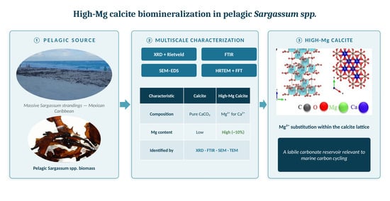
Graphical Abstract

1. Introduction

The massive proliferation of pelagic Sargassum spp. in the tropical Atlantic and Caribbean Sea since 2011 has generated significant ecological, economic, and environmental impacts, particularly along the Mexican Caribbean coast [1,2]. Although pelagic Sargassum spp. historically constituted an important floating habitat supporting marine biodiversity in oligotrophic waters [3], recent bloom magnitudes have exceeded historical records, producing extensive coastal accumulations that affect tourism, fisheries, and nearshore ecosystems [4,5]. These blooms, primarily consisting of S. fluitans III and S. natans VIII, have transformed from important ecological components into major coastal nuisances, with significant environmental and economic consequences [2]. Despite these negative impacts, the unprecedented biomass expansion has stimulated interest in the potential role of pelagic Sargassum in marine carbon cycling and atmospheric CO2 capture, particularly given its capacity to assimilate carbon through photosynthesis and promote carbonate formation [6].
Marine biomineralization processes play a critical role in long-term carbon storage through the formation of calcium carbonate (CaCO3) mineral phases in biological systems [7]. In marine organisms, CaCO3 commonly occurs as calcite, aragonite, and Mg-bearing carbonate phases, whose formation depends on environmental conditions such as temperature, Mg/Ca ratio, carbonate availability, and biological regulation mechanisms [8,9]. Among these phases, high-Mg calcite (HMC) is of particular interest because of its structural complexity, metastable nature, and relevance in carbonate cycling and ocean acidification studies [10,11]. The partial substitution of Ca2+ by Mg2+ within the calcite lattice generates local crystallographic distortions and modifies physicochemical properties such as solubility, stability, and dissolution kinetics [12].
Although brown algae are generally not classified as calcifying organisms, previous studies demonstrated that Sargassum biomass can produce calcium carbonate and contribute to CO2 fixation through biomineralization pathways [13].
That study reported the presence of calcite associated with different anatomical structures of Sargassum, suggesting that these macroalgae may play a role in marine carbonate cycling processes. However, the crystallographic nature of the carbonate phases formed in Sargassum, particularly regarding Mg incorporation, lattice distortion, and the possible formation of high-Mg calcite, remains poorly understood. In addition, the spatial distribution and compositional heterogeneity of these biomineral phases within the algal matrix have not been comprehensively characterized.
Recent studies have shown that biomineralization in marine systems may involve complex interactions among organic matrices, microbial activity, carbonate chemistry, and ion regulation processes [13,14]. In brown algae, polysaccharides such as alginate can interact with Ca2+ ions and potentially influence carbonate nucleation and mineral stabilization [15,16]. Furthermore, Mg incorporation into carbonate structures has been associated with kinetic effects, environmental conditions, and biologically mediated crystallization pathways in several marine organisms [7,17]. Nevertheless, the mechanisms governing Mg-bearing carbonate formation in Sargassum biomass remain largely unexplored.
In this study, we investigate the crystallographic and compositional characteristics of carbonate biomineralization in Sargassum biomass collected from the Mexican Caribbean coast. X-ray diffraction (XRD), Rietveld refinement, Fourier-transform infrared spectroscopy (FTIR), scanning electron microscopy coupled with energy-dispersive spectroscopy (SEM-EDS), and transmission electron microscopy (TEM) were employed to identify and characterize the carbonate phases associated with the algal matrix. The results provide experimental evidence for the formation of high-Mg calcite in Sargassum, revealing Mg incorporation within the calcite lattice and structural features associated with biomineralized carbonate phases.
These findings contribute to the understanding of carbonate biomineralization in non-calcifying marine macroalgae and suggest that Sargassum may promote the conversion of dissolved inorganic carbon into carbonate minerals through biomineralization, potentially contributing to transient carbon fixation, although the long-term stability of these phases requires further investigation.

2. Materials and Methods

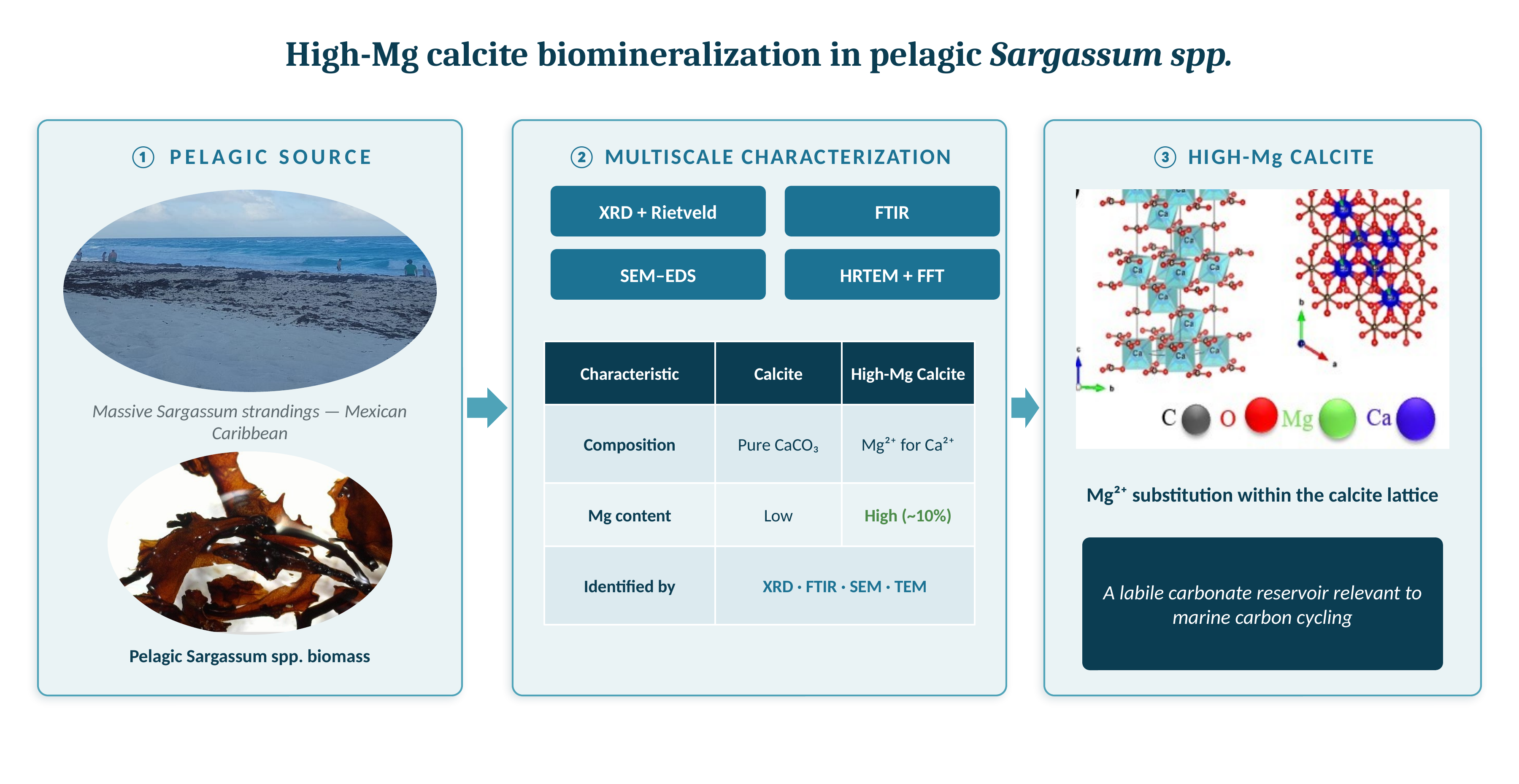
The experimental workflow employed for the structural and compositional characterization of biomineralized carbonate phases in Sargassum biomass is summarized in Figure 1. The analytical sequence combined structural, spectroscopic, morphological, and nanoscale characterization techniques to evaluate Mg incorporation and high-Mg calcite formation within the algal matrix.
The experimental procedures associated with each characterization technique are described in detail in the following sections. Particular emphasis was placed on correlating crystallographic, spectroscopic, and compositional information to evaluate the presence of Mg-bearing carbonate phases and their structural relationship with calcite biomineralization in Sargassum.
Sargassum biomass samples were collected from the Caribbean coast near Cancún, Quintana Roo, Mexico, during August 2024. The sampled material corresponded to naturally occurring mixed pelagic assemblages predominantly composed of S. fluitans III and S. natans VIII. Accordingly, the term Sargassum spp. is used throughout the manuscript to refer to this naturally occurring mixed pelagic assemblage rather than to a single species. Sampling was conducted at two coastal accumulation sites influenced by open-sea Sargassum influxes: Marlin Beach (21.1037281° N, −86.7649134° W) and Dolphins Beach (21.169379° N, −86.844882° W). Biomass was collected directly from freshly deposited beach accumulations at both locations on 12 August and 13 August 2024, ensuring minimal post-depositional alteration. Approximately 5 kg of material was obtained through manual collection along representative accumulation zones at each site. Sample collection and preliminary cleaning procedures were adapted from previous studies on Sargassum biomineralization [6], with specific modifications described below.
The collected biomass was rinsed three times with distilled water to remove adhered epibionts, fine sediments (silt), organic matter, and salts. The samples were then air-dried at room temperature (approximately 25 °C) for 72 h until complete drying was achieved. Subsequently, the dried material was manually fragmented and ground using an agate mortar. To remove the remaining organic fraction and preserve the inorganic mineral phases associated with biomineralization, the material was thermally treated in a furnace at 550 °C for 2 h under air atmosphere. This thermal treatment was selected based on previous studies [6] and supported by thermogravimetric analyses indicating that the organic fraction is effectively removed while the inorganic carbonate residue is preserved. The resulting material was further ground to obtain a homogeneous fine powder suitable for structural and spectroscopic analyses.
X-ray diffraction (XRD) analyses were performed using a Panalytical X’Pert PRO MPD diffractometer (Malvern PANalytical, Almelo, The Netherlands) equipped with a PW3011/20 X’Celerator detector operating in Bragg–Brentano geometry with Cu Kα radiation (λ = 1.5406 Å). Diffraction patterns were acquired at room temperature over an angular range appropriate for carbonate phase identification. Phase identification was performed using reference crystallographic databases for calcite, magnesian calcite, and anhydrite phases. Quantitative phase analysis and lattice parameter refinement were carried out through the Rietveld method using GSAS-II software (Version 2.0, Advanced Photon Source, Argonne National Laboratory, Argonne, IL, USA) [18]. The refinement included background correction, scale factors, lattice parameters, peak profile adjustment, and phase fraction quantification. Structural visualization and crystallographic modeling were performed using VESTA software (Version 3, National Institute for Materials Science, Tsukuba, Japan) [19].
Fourier-transform infrared spectroscopy (FTIR) analyses were performed using an IR Affinity-1S spectrometer (Shimadzu Corporation, Kyoto, Japan) equipped with an attenuated total reflectance (ATR) accessory with a diamond crystal (Specac Ltd., Orpington, UK). Spectra were collected at room temperature in the range of 4000–400 cm−1 with a resolution of 4 cm−1 and 32 scans per spectrum to identify the characteristic vibrational modes of carbonate compounds associated with calcium carbonate and Mg-bearing carbonate phases.
Morphological characterization was performed using a Hitachi SU3500 scanning electron microscope (SEM) (Hitachi High-Tech Corporation, Tokyo, Japan). Secondary electron images were acquired at an accelerating voltage of 15 kV. Elemental analysis and chemical mapping were conducted using an Oxford Aztec energy-dispersive X-ray spectroscopy (EDS) (Oxford Instruments plc, Abingdon, UK) detector operated at 20 kV.
Elemental mapping analyses were carried out to evaluate the spatial distribution of carbon, oxygen, calcium, and magnesium within the biomineralized structures associated with Sargassum spp. Semi-quantitative compositional analyses were obtained from selected regions of interest identified during SEM observation.
Transmission electron microscopy (TEM) analyses were performed to evaluate the crystallinity and nanoscale structural features of the carbonate phases formed in Sargassum spp. High-resolution TEM (HRTEM) images were acquired using a JEM-2200FS transmission electron microscope (JEOL Ltd., Tokyo, Japan) equipped with a field-emission gun (FEG) and an omega energy filter, operating at an accelerating voltage of 200 kV, from selected mineral particles obtained after thermal treatment.
Fast Fourier transform (FFT) analysis was conducted using ImageJ software (Version 1.54g, National Institutes of Health, Bethesda, MD, USA) to estimate interplanar distances and evaluate crystallographic ordering within the Mg-bearing carbonate phases. The experimentally observed lattice spacings were compared with the crystallographic parameters obtained from XRD and Rietveld refinement analyses.

3. Results

3.1. Structural Evidence of Carbonate Biomineralization in Sargassum

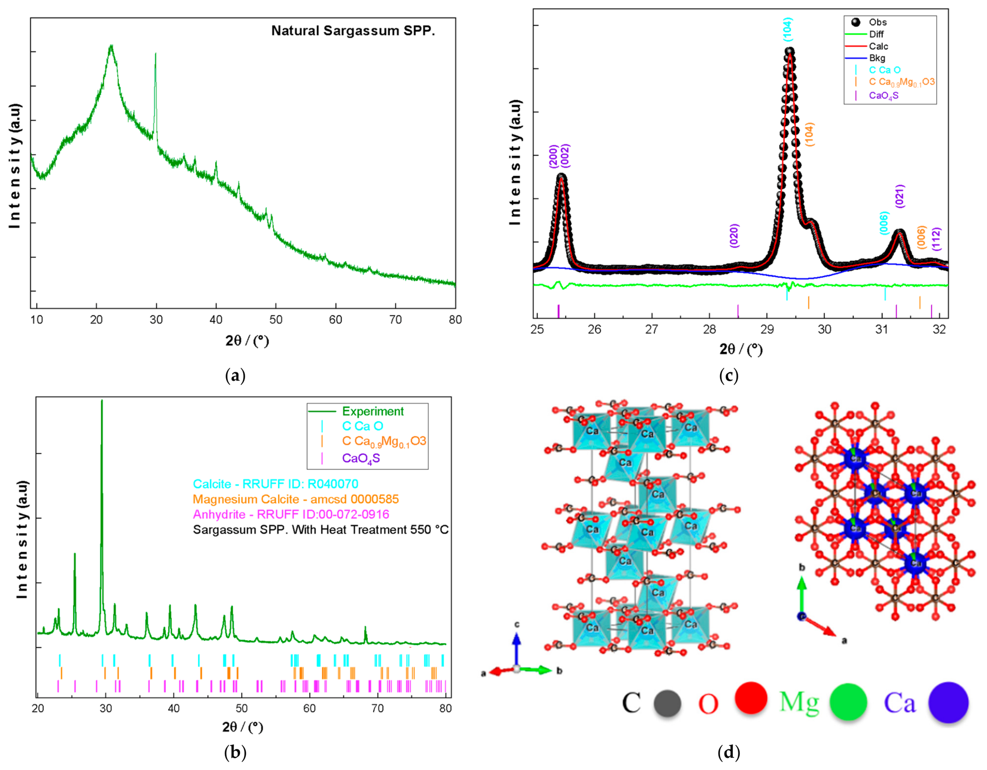
The X-ray diffraction (XRD) patterns obtained from untreated and thermally treated Sargassum biomass samples are presented in Figure 2. The diffraction profile of the untreated biomass exhibited predominantly amorphous characteristics with low-intensity reflections attributable to calcite phases, indicating the presence of biomineralized carbonate structures within the natural algal matrix. After thermal treatment at 550 °C, the diffraction peaks became significantly more defined due to the removal of the organic fraction, allowing accurate phase identification and crystallographic refinement.
Figure 2a compares the diffraction patterns obtained from untreated and calcined Sargassum spp. samples together with the reference pattern corresponding to magnesian calcite. The thermally treated sample exhibited characteristic reflections associated with calcite-type carbonate phases, including the dominant peak near 29.4° (2θ), corresponding to the (104) crystallographic plane.
The indexed diffraction peaks obtained after thermal treatment are shown in Figure 2b. Phase identification revealed the presence of calcite (CaCO3), magnesian calcite (Ca0.9Mg0.1CO3), and anhydrite (CaSO4). Quantitative phase analysis and crystallographic refinement were performed through the Rietveld method using GSAS-II software [18]. The refinement profile obtained between 25° and 32° (2θ) is presented in Figure 2c, showing good agreement between the experimental and calculated diffraction patterns.
The Rietveld refinement yielded agreement factors of R w p = 2.52%, R p = 1.89%, R e x p = 1.98%, and χ 2 = 2.37, indicating satisfactory fitting quality. The refined lattice parameters obtained for calcite and magnesian calcite phases are summarized in Table 1. The magnesian calcite phase exhibited slightly contracted lattice parameters relative to stoichiometric calcite, with values of a = b = 4.899 Å and c = 16.876 Å, whereas pure calcite exhibited lattice parameters of a = b = 4.933 Å and c = 17.195 Å.
The refined phase fractions corresponded to 57.9% calcite, 26.3% magnesian calcite, and 15.8% anhydrite. Structural visualization of the refined calcite lattice generated using VESTA software [19] is presented in Figure 2d. The modeled trigonal structure (space group R-3c, No. 167) exhibited local bond distortions associated with Mg incorporation while preserving the overall rhombohedral symmetry of the calcite phase.

3.2. Spectroscopic Evidence of Mg-Bearing Carbonate Phases

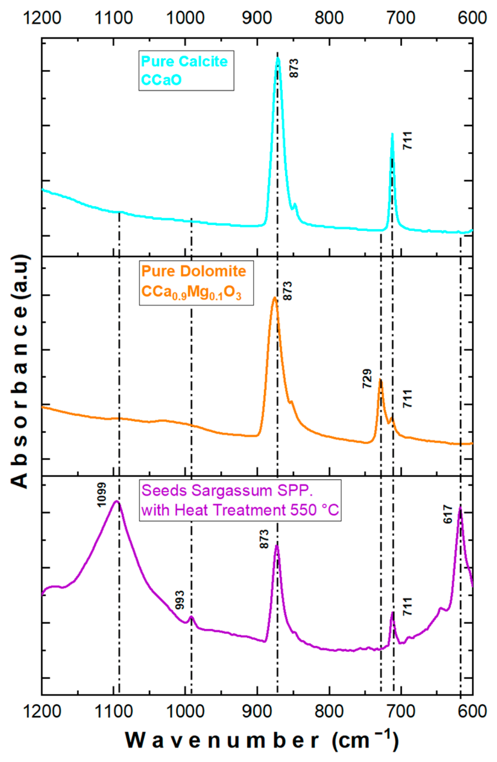
Fourier-transform infrared spectroscopy (FTIR) was employed to evaluate the vibrational characteristics of the carbonate phases associated with Sargassum biomass after thermal treatment. The FTIR spectra obtained from the calcined samples are presented in Figure 3.
The spectra exhibited characteristic absorption bands associated with carbonate-containing mineral phases. The principal vibrational bands identified in the spectra and their corresponding assignments are summarized in Table 2. The most intense absorption band was observed near 1097 cm−1 and corresponds to the asymmetric stretching vibration mode of carbonate groups (CO32−). Additional bands located near 991 cm−1, 873 cm−1, and 711 cm−1 were assigned to symmetric stretching and bending vibrational modes characteristic of carbonate structures [7,9,10].
Figure 3 shows the FTIR spectrum obtained from the biomineralized carbonate phases associated with Sargassum biomass. The spectra exhibited well-defined carbonate vibrational modes within the characteristic spectral region of calcite-type phases. A low-intensity band near 617 cm−1 was additionally identified and attributed to Mg–O-related vibrational contributions associated with Mg-bearing carbonate phases [7,8,10].
The observed spectral features are consistent with the carbonate phases identified through XRD and Rietveld refinement analyses. Slight band broadening was observed in several carbonate vibrational modes relative to stoichiometric calcite references. The FTIR results therefore confirm the presence of carbonate structures containing calcite-type coordination environments associated with the biomineralized inorganic fraction of Sargassum biomass.
The principal vibrational bands identified in the FTIR spectra and their corresponding spectral assignments are summarized in Table 2.

3.3. Morphological and Compositional Heterogeneity of Biomineralized Carbonate Phases

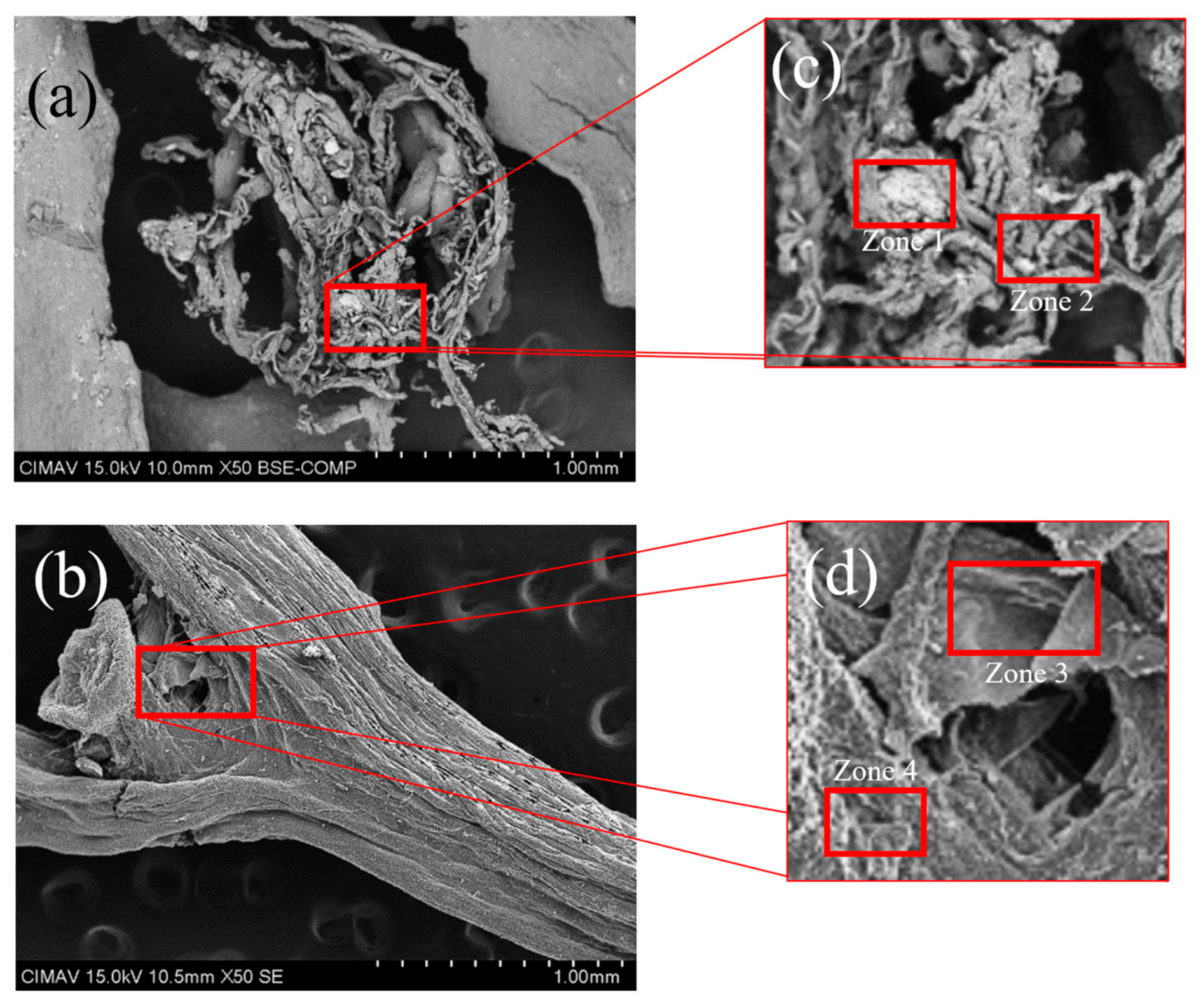
Scanning electron microscopy coupled with energy-dispersive X-ray spectroscopy (SEM-EDS) was employed to investigate the morphology and elemental distribution of the biomineralized carbonate phases associated with Sargassum biomass. Representative SEM micrographs and elemental mapping analyses are presented in Figure 4.
The SEM micrographs revealed irregularly distributed mineralized domains embedded within the inorganic residue obtained after thermal treatment. The observed structures exhibited heterogeneous morphologies, including compact agglomerates and localized crystalline regions with variable particle size and surface texture.
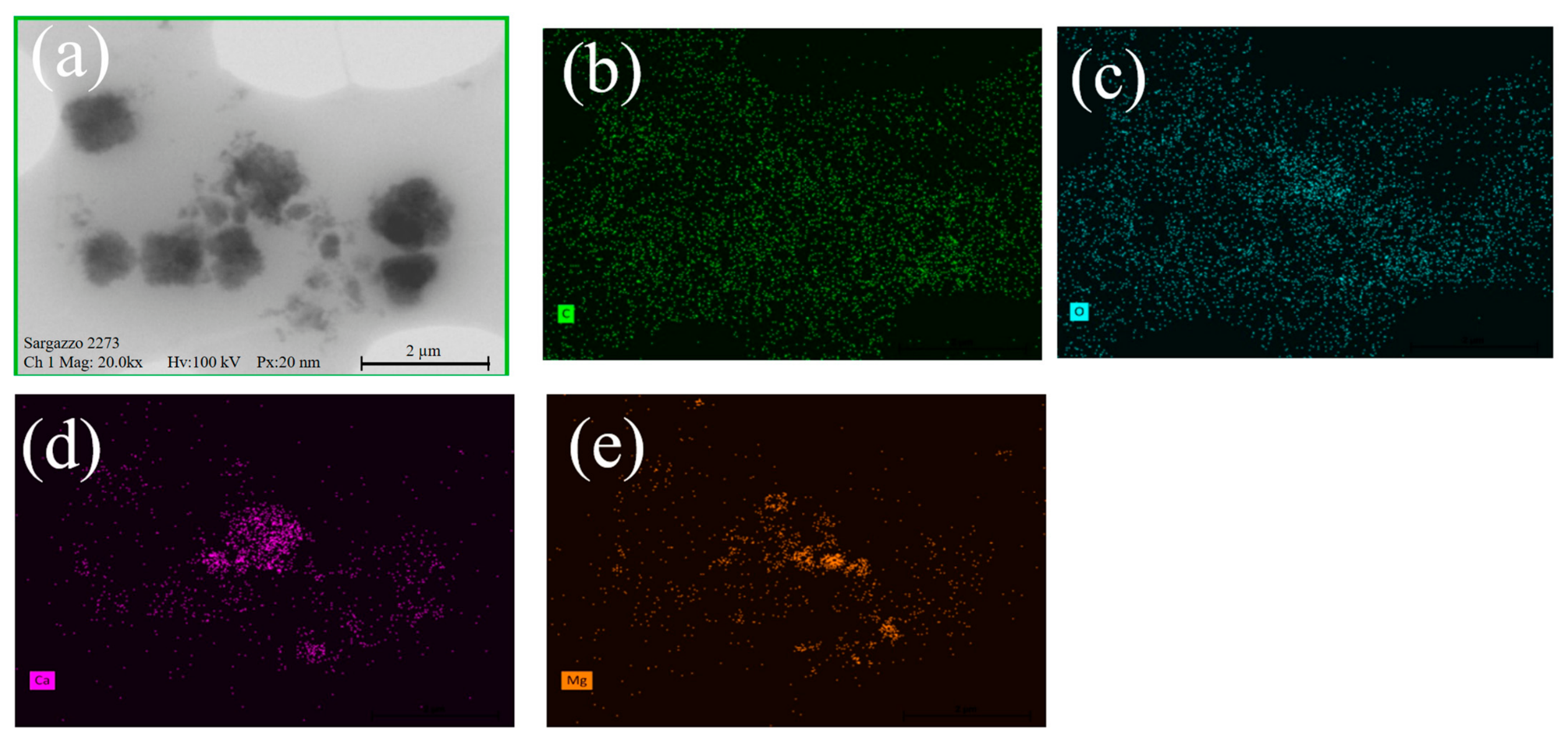
Figure 5 shows the distribution of the principal elements identified within the analyzed regions. Elemental mapping analyses confirmed the presence of Ca, Mg, O, and C associated with the biomineralized structures. The elemental distributions obtained from SEM-EDS revealed spatial heterogeneity among the analyzed domains, particularly for Mg and Ca-rich regions. Figure 5a presents the SEM micrograph of the analyzed area, while Figure 5b–e show the corresponding elemental distributions of carbon, oxygen, calcium, and magnesium, respectively.
Semi-quantitative EDS analyses obtained from selected regions of interest are summarized in Table 3. Zone 1 exhibited the highest Mg content among the analyzed regions, with 2.3 wt% Mg, whereas Zone 2 showed negligible Mg concentration and higher Ca content. Zones 3 and 4 exhibited intermediate Mg concentrations between 1.6 and 1.7 wt%.
The elemental distributions obtained through EDS mapping are consistent with the carbonate phases identified by XRD and FTIR analyses. The coexistence of Ca- and Mg-containing regions within the biomineralized structures indicates compositional variability throughout the analyzed carbonate domains. These compositional variations are consistent with the heterogeneous microstructural domains identified in Figure 4.

3.4. Nanoscale Crystallographic Evidence of High-Mg Calcite Formation

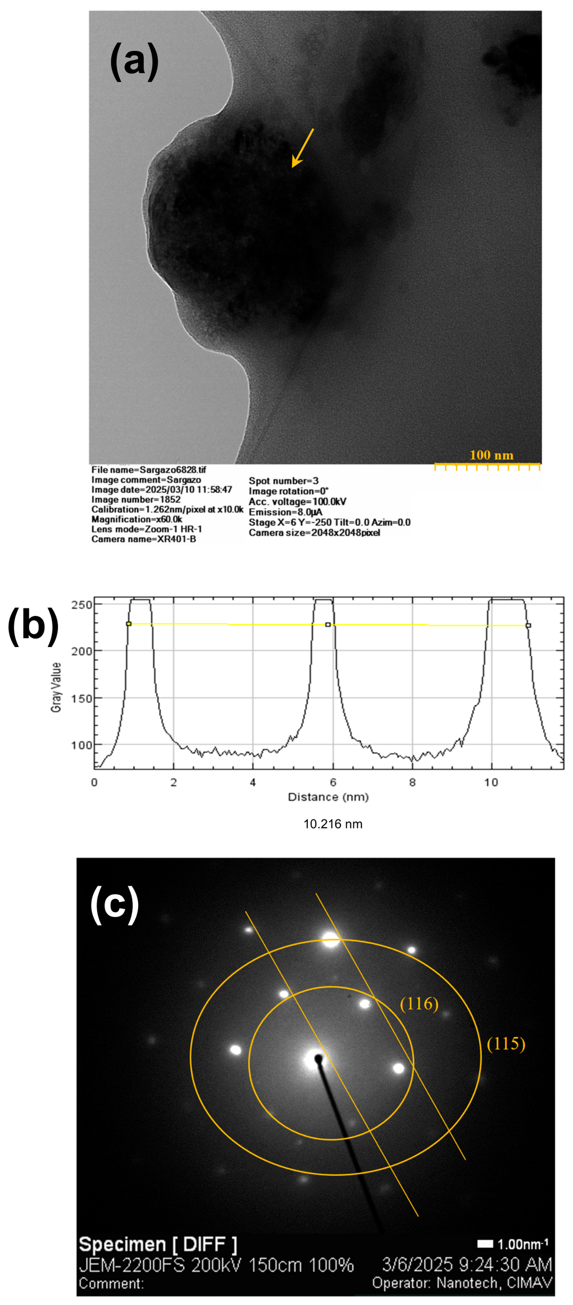
Transmission electron microscopy (TEM) and high-resolution transmission electron microscopy (HRTEM) analyses were performed to evaluate the nanoscale crystallographic organization of the biomineralized carbonate phases associated with Sargassum biomass. Representative HRTEM images, intensity profiles, and fast Fourier transform (FFT) analyses are presented in Figure 6.
The HRTEM micrographs revealed localized crystalline domains distributed within the biomineralized carbonate structures. Figure 6a shows a representative crystalline region exhibiting well-defined lattice fringes associated with ordered carbonate phases. The selected crystalline area indicated nanoscale structural organization within the mineralized matrix.
The intensity profile obtained from the selected lattice fringes is presented in Figure 6b. Periodic variations in the intensity distribution confirmed the presence of ordered interplanar arrangements within the analyzed crystalline domains. The measured lattice fringes exhibited spacings consistent with carbonate mineral structures identified through XRD analyses.
The corresponding FFT diffraction pattern is shown in Figure 6c. The (116) and (115) reflections correspond to specific crystallographic planes of the calcite structure and confirm the presence of well-ordered crystalline domains at the nanoscale. The diffraction reflections were indexed to the (116) and (115) crystallographic planes associated with calcite-type carbonate structures. The experimentally observed interplanar spacings exhibited slight deviations relative to stoichiometric calcite reference values [21].
These observations, together with the lattice contraction determined from Rietveld refinement, the FTIR band shifts, and the heterogeneous Mg distribution revealed by SEM-EDS, collectively support the presence of local structural distortions consistent with partial Mg incorporation into the calcite lattice. The HRTEM and FFT results are consistent with the crystallographic information obtained from XRD and Rietveld refinement analyses, confirming the presence of crystalline carbonate domains containing structural modifications associated with Mg-bearing phases at the nanoscale. The combined structural and compositional evidence is consistent with partial isomorphic substitution of Mg2+ for Ca2+ within the rhombohedral calcite structure. The smaller ionic radius of Mg2+ relative to Ca2+ provides a crystallographic basis for the observed local lattice distortions, while the preservation of long-range crystallographic order indicates that Mg incorporation occurs without disruption of the overall crystal symmetry.
These results are consistent with biologically influenced biomineralization processes, in which local physicochemical conditions within the algal matrix or associated microenvironments may facilitate Mg incorporation during carbonate formation, as commonly reported for high-Mg calcite in marine systems [9].

4. Discussion

4.1. Evidence of High-Mg Calcite Formation in Sargassum

The combined multiscale characterization results obtained in this study provide consistent evidence supporting the formation of Mg-bearing carbonate phases associated with Sargassum biomass collected from the Mexican Caribbean. The integration of XRD, FTIR, SEM-EDS, and HRTEM analyses revealed structural, compositional, and nanoscale features compatible with high-Mg calcite (HMC) biomineralization within the algal matrix.
The XRD and Rietveld refinement analyses provided the first indication of Mg incorporation into the carbonate structure through the detection of lattice contraction relative to stoichiometric calcite. The refined lattice parameters obtained for the magnesian calcite phase were systematically smaller than those corresponding to pure calcite, consistent with partial substitution of Ca2+ by Mg2+ within the rhombohedral calcite lattice. Similar crystallographic behavior has been extensively reported in marine HMC systems, where the smaller ionic radius of Mg2+ generates local lattice distortion and contraction of interplanar spacings [8,11].
Importantly, the coexistence of calcite and magnesian calcite phases identified through Rietveld refinement indicates that biomineralization in Sargassum biomass does not occur as a chemically homogeneous process. Instead, the results suggest the formation of carbonate domains exhibiting variable Mg incorporation levels, which is characteristic of biologically mediated carbonate systems formed under non-equilibrium marine conditions [7,18]. The presence of approximately 26.3% magnesian calcite within the refined phase composition therefore represents strong crystallographic evidence supporting HMC formation associated with the algal matrix.
The FTIR spectra further reinforced the structural evidence obtained through XRD analyses. In particular, the carbonate vibrational bands exhibited slight broadening and spectral shifts relative to stoichiometric calcite references, suggesting local structural disorder associated with Mg incorporation into the carbonate framework. These findings, together with the vibrational assignments summarized in Table 3, are consistent with the presence of a Mg-bearing carbonate phase exhibiting non-stoichiometric characteristics. Similar features have been reported in biologically influenced carbonate systems [20,22]. The Mg-related vibrational contribution identified near 617 cm−1 additionally supports the presence of Mg-bearing carbonate structures, in agreement with previous studies on magnesian calcite systems [8,10,23].
SEM-EDS elemental mapping demonstrated heterogeneous spatial distributions of Ca and Mg throughout the biomineralized structures, indicating that Mg incorporation is not uniformly distributed within the carbonate domains. Such heterogeneity is commonly observed in biologically mediated carbonate systems, where local variations in ion concentration, nucleation kinetics, and organic matrix interactions influence mineral composition at the microscale [14,15]. The elevated Mg concentration observed in specific EDS regions, particularly in Zone 1, correlates well with the lattice distortions identified through XRD refinement and the spectral broadening observed in FTIR analyses.
The nanoscale evidence obtained through HRTEM and FFT analyses further strengthens the interpretation of HMC formation in Sargassum biomass. The observed lattice fringes and indexed crystallographic reflections corresponding to the (116) and (115) planes confirmed the presence of highly ordered carbonate domains exhibiting local structural distortions. Slight deviations in interplanar spacing relative to pure calcite references are consistent with nanoscale lattice contraction induced by Mg substitution, as previously reported for marine Mg-bearing carbonate systems [10,11].
Taken together, the multiscale observations obtained in this study strongly support the interpretation that Sargassum biomass can promote the formation of structurally modified carbonate phases compatible with high-Mg calcite biomineralization. These findings expand previous reports describing calcite formation in Sargassum biomass [13] by providing direct crystallographic, spectroscopic, compositional, and nanoscale evidence of Mg incorporation within the carbonate lattice.
Importantly, the presence of high-Mg calcite suggests that these biomineralized phases may be particularly sensitive to environmental changes, as Mg-rich carbonates are known to exhibit higher solubility under decreasing pH conditions. This behavior is consistent with previous studies on ocean acidification, which report generally negative and variable effects on marine carbonate-forming organisms [24].

4.2. Possible Biomineralization Mechanisms in Sargassum Biomass

Although the precise mechanisms controlling Mg-bearing carbonate formation in Sargassum biomass remain unresolved, the combined structural and compositional evidence obtained in this study suggests that biomineralization may involve complex physicochemical and biologically mediated processes occurring within the algal matrix.
Such processes are consistent with recent studies on marine macroalgae, where biological activity, environmental conditions, and microbially mediated interactions have been shown to influence carbonate formation, including photosynthetic microbially induced carbonate precipitation (MICP) pathways that may contribute to localized carbonate nucleation in algal systems [22,25].
The coexistence of calcite and high-Mg calcite domains, together with the heterogeneous spatial distribution of Mg identified through SEM-EDS analyses, indicates that carbonate precipitation likely occurs under localized microenvironmental conditions rather than through homogeneous inorganic crystallization.
One possible mechanism involves the interaction between seawater cations and the polysaccharide-rich extracellular matrix associated with brown algae. In particular, alginate is known to exhibit strong affinity for divalent cations such as Ca2+ and Mg2+ through cooperative binding processes commonly described by the “egg-box” model [17]. These interactions may promote localized ion accumulation and generate favorable nucleation environments for carbonate precipitation within or near the algal surface. Similar ion-mediated stabilization mechanisms have been previously associated with carbonate mineralization processes in biologically influenced marine systems [14,15].
An important aspect to consider is whether the formation of Mg-bearing carbonate phases requires direct metabolic energy investment by the algal host. Based on current knowledge, such biomineralization processes do not necessarily rely on active metabolic control by Sargassum. Instead, they may arise from the combined effects of physicochemical and biologically mediated mechanisms. For instance, the polysaccharide-rich extracellular matrix of brown algae, particularly alginate, can facilitate ion binding and locally modify the Ca2+/Mg2+ activity ratios, promoting carbonate nucleation without requiring active ion transport mechanisms [16,17]. In addition, microbially induced carbonate precipitation (MICP) associated with the Sargassum microbiome may contribute to localized mineral formation through metabolic activity occurring at the algal surface [13,25]. In both scenarios, the incorporation of Mg into carbonate structures is promoted by microenvironmental conditions that facilitate kinetic pathways rather than equilibrium processes. However, distinguishing between passive physicochemical processes and active biological or microbiome-driven contributions remains an open question that warrants further investigation.
The incorporation of Mg into calcite structures is thermodynamically unfavorable under equilibrium conditions due to the strong hydration energy and smaller ionic radius of Mg2+ relative to Ca2+, which significantly affects nucleation and crystal growth kinetics [8,11,25]. Consequently, the formation of high-Mg calcite commonly requires kinetically controlled or biologically mediated crystallization pathways capable of facilitating partial Mg substitution within the carbonate lattice [18]. The lattice contraction observed through Rietveld refinement, together with the nanoscale distortions identified by HRTEM and FFT analyses, are consistent with this type of non-equilibrium incorporation process.
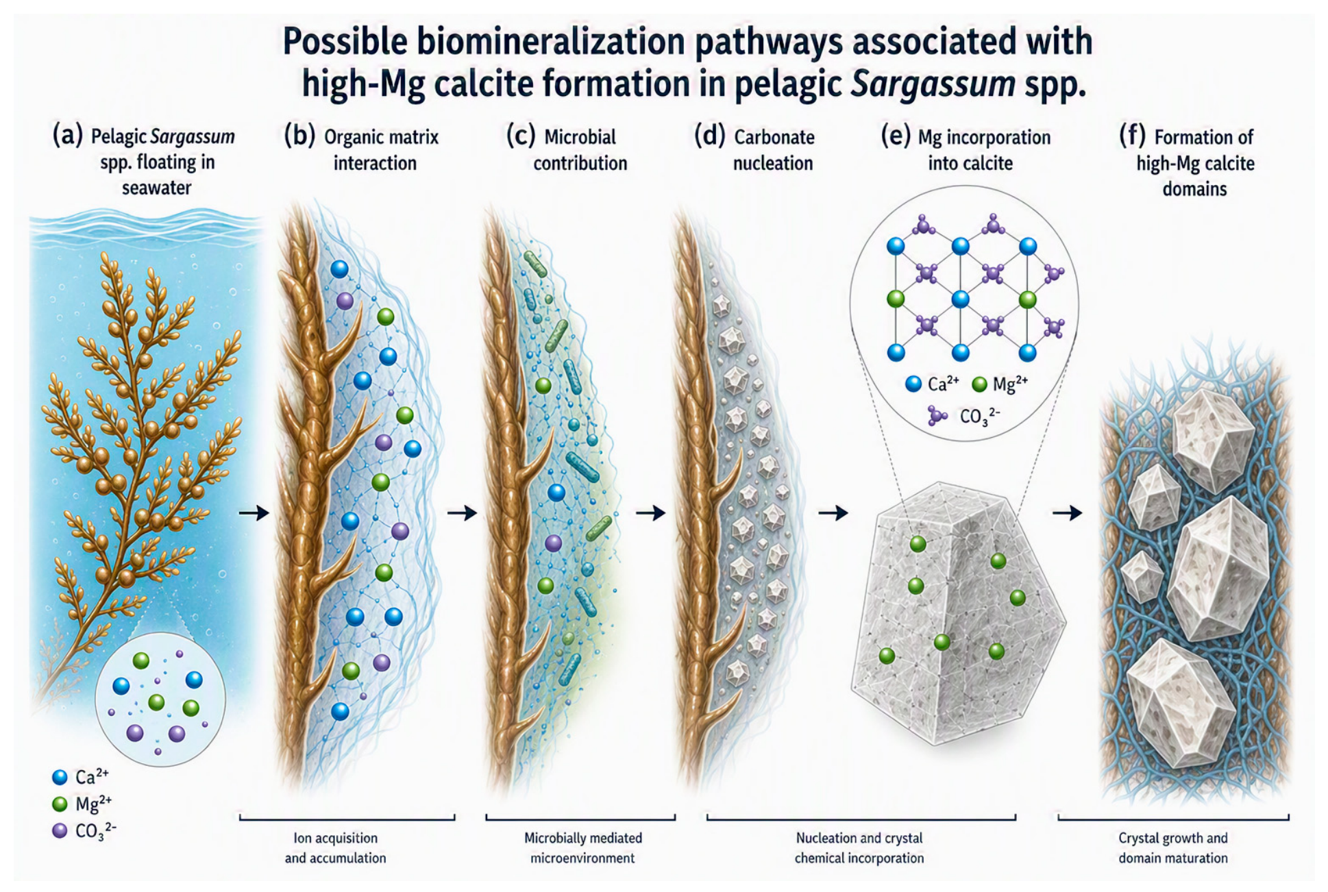
Microbial activity associated with the Sargassum holobiont may additionally contribute to localized carbonate precipitation. Previous studies have demonstrated that microbial biofilms and extracellular polymeric substances can influence carbonate nucleation by modifying local pH, carbonate saturation, and ion availability within biologically active microenvironments [13,14]. Although microbiological analyses were beyond the scope of the present study, the conceptual model proposed in Figure 7 considers the potential contribution of microbial processes as part of the biomineralization pathway associated with Mg-bearing carbonate formation.
The heterogeneous Mg distribution observed throughout the biomineralized structures additionally suggests that carbonate formation may occur through multiple nucleation and growth events influenced by local chemical variability. Such heterogeneity is characteristic of several marine biomineralization systems where crystal growth is regulated by fluctuating environmental conditions, ion transport processes, and organic matrix interactions [7,18]. The combined evidence therefore supports the interpretation that Sargassum biomass may act as a biologically active substrate capable of promoting localized high-Mg calcite formation under marine environmental conditions.
These observations expand the current understanding of biomineralization processes in non-calcifying marine macroalgae and highlight the potential complexity of carbonate formation pathways associated with large-scale Sargassum accumulations. Because high-Mg calcite is more soluble and less thermodynamically stable than stoichiometric calcite [8,11], the carbon fixed within these phases would represent a comparatively labile carbonate reservoir, sensitive to seawater carbonate saturation and to the dissolution dynamics that accompany ocean acidification. This carries direct implications for the role of recurrent pelagic blooms in carbon cycling. Further studies integrating microbiological characterization, in situ mineralization analyses, and geochemical monitoring will be necessary to clarify the mechanisms governing Mg incorporation and carbonate stabilization within these marine systems.

5. Conclusions

This study provides multiscale experimental evidence supporting the formation of high-Mg calcite (HMC) associated with Sargassum biomass collected from the Mexican Caribbean. Lattice contraction relative to stoichiometric calcite, carbonate vibrational features indicative of local structural disorder, heterogeneous Ca–Mg domains, and nanoscale lattice distortions converged on a single interpretation: the partial substitution of Ca2+ by Mg2+ within the rhombohedral calcite lattice through a kinetically controlled, biologically mediated pathway operating far from thermodynamic equilibrium.
Because high-Mg calcite is more soluble and less stable than stoichiometric calcite, its formation within massive, recurrent pelagic blooms has direct implications for carbon cycling. The capacity of these macroalgae to fix inorganic carbon as a metastable carbonate phase reframes large-scale strandings not merely as an ecological nuisance but as a transient, environmentally sensitive carbon reservoir whose persistence depends on local carbonate saturation and seawater chemistry.
Whereas earlier study established only that calcite forms in Sargassum biomass [12], the present study resolves the crystallographic identity of that carbonate, demonstrating that it is not pure calcite but a structurally modified, Mg-bearing phase. This distinction matters: identifying high-Mg calcite, rather than calcite, in Sargassum places this non-calcifying macroalga within the same biomineralization framework as recognized marine calcifiers and provides the first direct evidence that Mg substitution operates within its carbonate lattice.
Although the biochemical and microbiological mechanisms controlling Mg incorporation remain unresolved, the present study establishes a crystallographic and compositional framework for future investigations on biomineralization processes in pelagic macroalgae. Further studies integrating mineralogical, microbiological, and in situ geochemical analyses will be essential to evaluate the environmental significance and long-term stability of Mg-bearing carbonate phases associated with large-scale Sargassum accumulations.

Author Contributions

D.L.-G.: Conceptualization, Supervision, Funding acquisition, Writing—review & editing. J.S.S.-L. and C.C.-G.: Formal analysis, Methodology, Writing—review & editing. A.R.-R.: Conceptualization, Supervision, Writing—review & editing. H.A.M.-R.: Conceptualization, Investigation, Methodology, Writing—original draft, Writing—review & editing. E.R.-P.: Writing—review & editing, Corresponding author. All authors have read and agreed to the published version of the manuscript.

Funding

This research received no external funding.

Data Availability Statement

The data supporting the findings of this study are available from the corresponding authors upon reasonable request.

Acknowledgments

The authors appreciate the support from CIMAV, Nanotech, and the Universidad Nacional de Colombia for providing the necessary infrastructure to carry out this research. Special thanks to Andrés González for support with X-ray diffraction (XRD) analysis, Jose Eduardo Garcia Bejar for FT-IR analysis, and to C. Leyva-Porras for assistance with scanning electron microscopy (SEM) characterization. During the preparation of this study, the authors used Illustrae (https://illustrae.co, accessed on 25 June 2026), an AI-based scientific illustration tool, to assist in the creation of the conceptual schematics shown in Figure 1 (experimental workflow) and Figure 7 (conceptual biomineralization model). The authors designed the conceptual content of these figures, verified the scientific accuracy of all elements, and take full responsibility for their content. These figures do not contain experimental data. No generative AI tools were used to produce or interpret the experimental results, data, or scientific conclusions reported in this study.

Conflicts of Interest

The authors declare that they have no known competing financial interests or personal relationships that could have appeared to influence the study reported in this paper.

References

  1. Wang, M.; Hu, C.; Barnes, B.B.; Mitchum, G.; Lapointe, B.; Montoya, J.P. The Great Atlantic Sargassum Belt. Science 2019, 365, 83–87. [Google Scholar] [CrossRef] [PubMed]
  2. Johns, E.M.; Lumpkin, R.; Putman, N.F.; Smith, R.H.; Muller-Karger, F.E.; Rueda-Roa, D.T.; Hu, C. The establishment of a pelagic Sargassum population in the tropical Atlantic: Biological consequences of a basin-scale long-distance dispersal event. Prog. Oceanogr. 2020, 182, 102269. [Google Scholar] [CrossRef]
  3. Witherington, B.; Hirama, S.; Hardy, R. Young sea turtles of the pelagic Sargassum-dominated drift community. Mar. Ecol. Prog. Ser. 2012, 463, 1–22. [Google Scholar] [CrossRef]
  4. Franks, J.S.; Johnson, D.R.; Ko, D.S. Pelagic Sargassum in the tropical North Atlantic. Gulf Caribb. Res. 2016, 27, SC6–SC11. [Google Scholar] [CrossRef]
  5. Magaña-Gallegos, E.; García-Sánchez, M.; Graham, C.; Olivos-Ortiz, A.; Siuda, A.N.S.; van Tussenbroek, B.I. Growth rates of pelagic Sargassum species in the Mexican Caribbean. Aquat. Bot. 2023, 185, 103614. [Google Scholar] [CrossRef]
  6. Paraguay-Delgado, F.; Carreño-Gallardo, C.; Estrada-Guel, I.; Zabala-Arceo, A.; Martinez-Rodriguez, H.A.; Lardizábal-Gutierrez, D. Pelagic Sargassum spp. capture CO2 and produce calcite. Environ. Sci. Pollut. Res. 2020, 27, 25794–25800. [Google Scholar] [CrossRef] [PubMed]
  7. Gilbert, P.U.P.A.; Bergmann, K.D.; Boekelheide, N.; Tambutté, S.; Mass, T.; Marin, F.; Adkins, J.F.; Erez, J.; Gilbert, B.; Knutson, V.; et al. Biomineralization: Integrating mechanism and evolutionary history. Sci. Adv. 2022, 8, eabl9653. [Google Scholar] [CrossRef] [PubMed]
  8. Morse, J.W.; Andersson, A.J.; Mackenzie, F.T. Initial responses of carbonate-rich shelf sediments to rising atmospheric pCO2 and ocean acidification: Role of high Mg-calcites. Geochim. Cosmochim. Acta 2006, 70, 5814–5830. [Google Scholar] [CrossRef]
  9. Purgstaller, B.; Mavromatis, V.; Goetschl, K.E.; Steindl, F.R.; Dietzel, M. Effect of temperature on the transformation of amorphous calcium magnesium carbonate with near-dolomite stoichiometry into high Mg-calcite. CrystEngComm 2021, 23, 1969–1981. [Google Scholar] [CrossRef]
  10. Lammers, L.N.; Mitnick, E.H. Magnesian calcite solid solution thermodynamics inferred from authigenic deep-sea carbonate. Geochim. Cosmochim. Acta 2019, 246, 409–432. [Google Scholar] [CrossRef]
  11. Alkhatib, M.; Qutob, M.; Alkhatib, S.; Eisenhauer, A. Influence of precipitation rate and temperature on the partitioning of magnesium and strontium in calcite overgrowths. Chem. Geol. 2022, 599, 120841. [Google Scholar] [CrossRef]
  12. Sulpis, O.; Jeansson, E.; Dinauer, A.; Lauvset, S.K.; Middelburg, J.J. Aragonite dissolution protects calcite at the seafloor. Nat. Commun. 2022, 13, 1104. [Google Scholar] [CrossRef] [PubMed]
  13. Dupraz, C.; Reid, R.P.; Braissant, O.; Decho, A.W.; Norman, R.S.; Visscher, P.T. Processes of carbonate precipitation in modern microbial mats. Earth-Sci. Rev. 2009, 96, 141–162. [Google Scholar] [CrossRef]
  14. Braissant, O.; Cailleau, G.; Dupraz, C.; Verrecchia, E.P. Bacterially induced mineralization of calcium carbonate in terrestrial environments: The role of exopolysaccharides and amino acids. J. Sediment. Res. 2003, 73, 485–490. [Google Scholar] [CrossRef]
  15. Rodriguez-Blanco, J.D.; Shaw, S.; Benning, L.G. The kinetics and mechanisms of amorphous calcium carbonate crystallization to calcite, via vaterite. Nanoscale 2011, 3, 265–271. [Google Scholar] [CrossRef] [PubMed]
  16. Cao, L.; Lu, W.; Mata, A.; Nishinari, K.; Fang, Y. Egg-box model-based gelation of alginate and pectin: A review. Carbohydr. Polym. 2020, 242, 116389. [Google Scholar] [CrossRef] [PubMed]
  17. Stephenson, A.E.; De Yoreo, J.J.; Wu, L.; Wu, K.J.; Hoyer, J.; Dove, P.M. Peptides enhance magnesium signature in calcite: Insights into origins of vital effects. Science 2008, 322, 724–727. [Google Scholar] [CrossRef] [PubMed]
  18. Toby, B.H.; Von Dreele, R.B. GSAS-II: The genesis of a modern open-source all purpose crystallography software package. J. Appl. Crystallogr. 2013, 46, 544–549. [Google Scholar] [CrossRef]
  19. Momma, K.; Izumi, F. VESTA 3 for three-dimensional visualization of crystal, volumetric and morphology data. J. Appl. Crystallogr. 2011, 44, 1272–1276. [Google Scholar] [CrossRef]
  20. Leplastrier, A.; Rintoul, M.; Opdyke, B.; Nand, V.; Branson, O.; Eggins, S.M.; Ellwood, M.J. Sulphate reduction and carbonate precipitation in a high-energy algal rim framework. Coral Reefs 2024, 43, 951–968. [Google Scholar] [CrossRef]
  21. Zhang, Y.; Dawe, R.A. Influence of Mg2+ on the kinetics of calcite precipitation and calcite crystal morphology. Chem. Geol. 2000, 163, 129–138. [Google Scholar] [CrossRef]
  22. Krzemińska, M.; Piwoni-Piórewicz, A.; Boonzaaier-Davids, M.K.; Florence, W.; Waeschenbach, A.; Najorka, J.; Jenkins, H.L.; Kukliński, P. Carbonate mineralogy and geochemistry of bryozoans along the South African coast. Mar. Biol. 2025, 172, 8. [Google Scholar] [CrossRef]
  23. Bianco-Stein, N.; Polishchuk, I.; Lang, A.; Portal, L.; Dejoie, C.; Katsman, A.; Pokroy, B. High-Mg calcite nanoparticles within a low-Mg calcite matrix: A widespread phenomenon in biomineralization. Proc. Natl. Acad. Sci. USA 2022, 119, e2120177119. [Google Scholar] [CrossRef] [PubMed]
  24. Kroeker, K.J.; Kordas, R.L.; Crim, R.N.; Singh, G.G. Meta-analysis reveals negative yet variable effects of ocean acidification on marine organisms. Ecol. Lett. 2010, 13, 1419–1434. [Google Scholar] [CrossRef] [PubMed]
  25. Zúñiga-Barra, H.; Ostojić, C.; Torres-Aravena, Á.; Rivas, M.; Vílchez, C.; Jeison, D. Use of photosynthetic MICP to induce calcium carbonate precipitation: Prospecting the role of the microorganism in the formation of CaCO3 crystals. Algal Res. 2024, 80, 103499. [Google Scholar] [CrossRef]
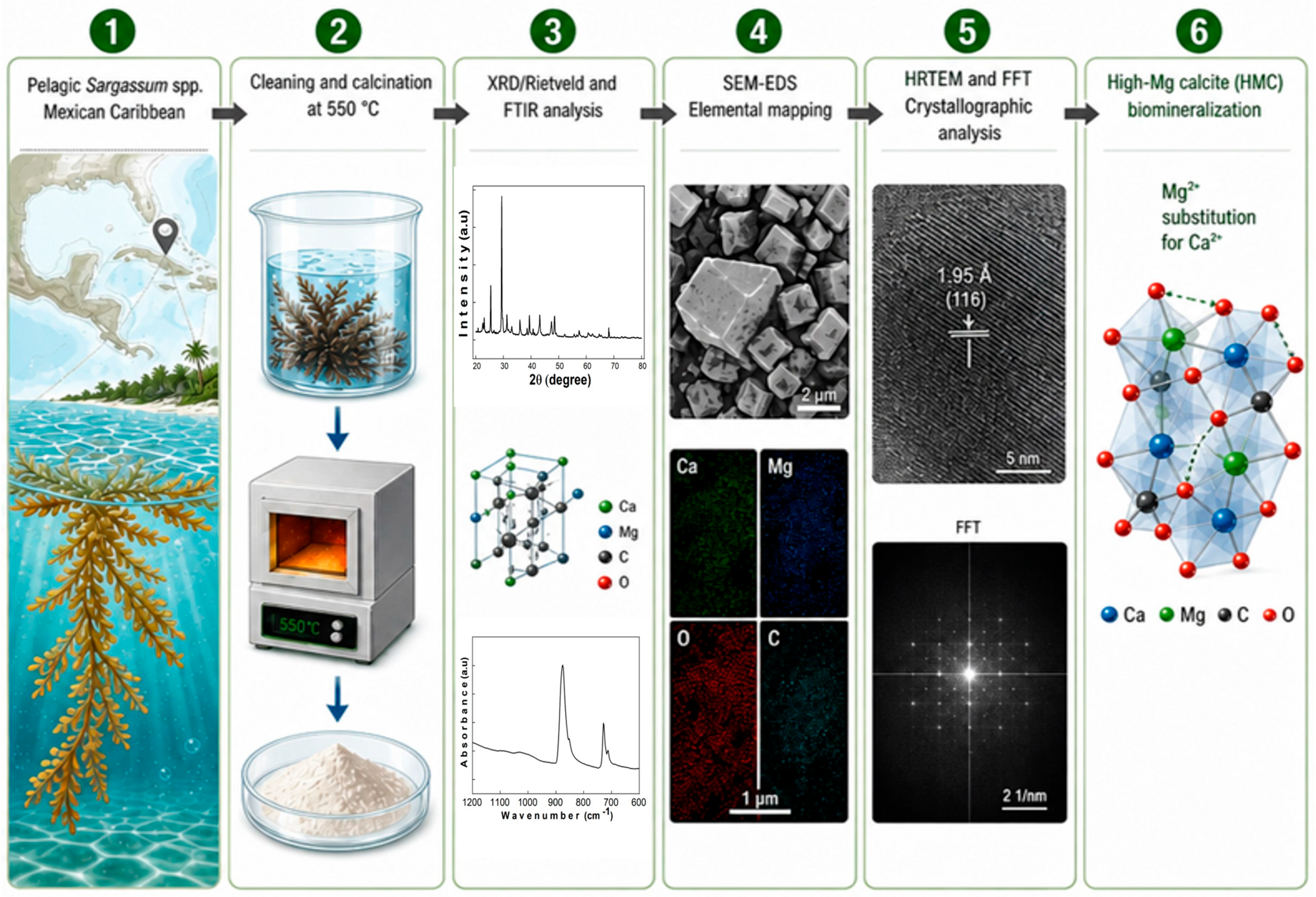
Figure 1. Experimental workflow used for the identification and characterization of biomineralized high-Mg calcite (HMC) in Sargassum biomass collected from the Mexican Caribbean. The methodology included sample preparation, X-ray diffraction (XRD) and Rietveld refinement, FTIR spectroscopy, SEM-EDS elemental mapping, and high-resolution TEM analysis to evaluate the structural and compositional features associated with Mg-bearing carbonate phases.
Diversity 18 00412 g001
Figure 2. (a) Comparative X-ray diffraction (XRD) patterns obtained from untreated and thermally treated Sargassum biomass samples together with the reference diffraction pattern of magnesian calcite. (b) Indexed diffraction peaks corresponding to calcite, magnesian calcite, and anhydrite phases identified after thermal treatment. (c) Rietveld refinement profile of the calcined sample between 25° and 35° (2θ). (d) Crystallographic model of the refined calcite structure generated using VESTA software based on the Rietveld refinement results.
Diversity 18 00412 g002
Figure 3. FTIR spectra obtained from biomineralized carbonate phases associated with Sargassum biomass after thermal treatment. The spectra exhibit characteristic carbonate vibrational bands corresponding to calcite-type mineral phases together with Mg-related vibrational contributions.
Diversity 18 00412 g003
Figure 4. SEM micrographs revealing a heterogeneous microstructural organization in thermally treated Sargassum biomass. Image (a) shows a collapsed and filamentous morphology of the residual matrix, whereas image (b) highlights more compact and fibrous structures. The magnified regions in (c) indicate Zone 1 as a dense aggregate and Zone 2 as a more porous and irregular domain. Image (d) shows Zone 3 with smoother surfaces and Zone 4 with a rough, granular texture. These variations reflect significant spatial heterogeneity in the distribution of mineralized carbonate phases within the sample.
Diversity 18 00412 g004
Figure 5. (a) SEM micrograph of biomineralized carbonate structures associated with Sargassum biomass and corresponding EDS elemental mapping of (b) carbon (C), (c) oxygen (O), (d) calcium (Ca), and (e) magnesium (Mg). The elemental maps reveal heterogeneous distributions of Ca- and Mg-containing carbonate domains within the biomineralized matrix.
Diversity 18 00412 g005
Figure 6. (a) High-resolution TEM micrograph of the crystal derived from Sargassum spp. treated at 550 °C. The yellow arrow indicates the crystalline region selected for structural analysis. (b) Intensity profile obtained from the selected lattice fringes. The yellow horizontal line indicates the reference level used for measuring the periodic lattice spacing between adjacent fringes. (c) Fast Fourier transform (FFT) diffraction pattern corresponding to the analyzed crystalline domain, showing reflections indexed to the (116) and (115) crystallographic planes. The measured interplanar spacing was 1.95 Å and was indexed to the (116) plane.
Diversity 18 00412 g006
Figure 7. Conceptual model illustrating possible pathways associated with high-Mg calcite biomineralization in Sargassum biomass. The schematic integrates the multiscale experimental evidence obtained in this study and summarizes the sequence of processes involved: (a) pelagic Sargassum spp. floating in seawater and initial ion acquisition; (b) interaction between seawater ions and the algal organic matrix; (c) development of microbially influenced microenvironments associated with the algal surface; (d) carbonate nucleation within localized regions; (e) incorporation of Mg2+ into the calcite structure through partial isomorphic substitution; and (f) growth and maturaEtion of localized high-Mg calcite domains.
Diversity 18 00412 g007
Table 1. Rietveld refinement parameters and phase composition obtained for biomineralized carbonate phases associated with Sargassum biomass.
PhaseCaCO3 (Calcite)Ca0.9Mg0.1CO3
(Magnesian Calcite)
CaSO4
(Anhydrite)
Space GroupR-3c (No. 167)R-3c (No. 167)Cmcm (No. 63)
Lattice parametersa = b = 4.933 Å
c = 17.195 Å
a = b = 4.899 Å
c = 16.876 Å
a = 6.996 Å
b = 6.238 Å
c = 6.991 Å
Volume (Å3)362.37350.76305.09
Phase %57.926.315.8
Rietveld Refinement StatisticsRwp = 2.523; χ2: = 2.37; Max Shift/σ = 0.002
Table 2. Principal FTIR vibrational bands identified in biomineralized carbonate phases associated with Sargassum biomass and their corresponding spectral assignments.
Wavenumber (cm−1)Vibration TypeSpectral AssignmentReference Sources
1097Asymmetric C-O stretchingCarbonate group (CO32−)[11]
991Symmetric C-O stretchingCarbonate group (CO32−)[11]
873Out-of-plane CO32− bendingCarbonate group (CO32−)[8,9]
711In-plane CO32− bendingCarbonate group (CO32−)[8,10]
617bending modeMg-Oxygen (Mg-O) vibration[8,20]
Table 3. Semi-quantitative EDS elemental composition obtained from selected biomineralized regions associated with Sargassum biomass. Values are expressed as wt% ± standard deviation (SD).
ZoneOxygen (wt%)±SDCarbon (wt%)±SDCalcium (wt%)±SDMagnesium (wt%)±SD
151.50.926.50.9190.52.30.2
230.12.119.72.1491.8----
343.42.331.72.221.61.31.60.4
439.91.540.21.816.30.71.70.2
Disclaimer/Publisher’s Note: The statements, opinions and data contained in all publications are solely those of the individual author(s) and contributor(s) and not of MDPI and/or the editor(s). MDPI and/or the editor(s) disclaim responsibility for any injury to people or property resulting from any ideas, methods, instructions or products referred to in the content.