Article

The Novel Halotolerant, Filamentous Cyanobacterium Krienitziella sambharensis gen. et sp. nov. (Nodosilineales, Cyanobacteriophyta) Isolated from an Indian Wetland (Sambhar Salt Lake, India)


1
Department of Microbiology, School of Life Sciences, Central University of Rajasthan, Bandarsindri 305817, Ajmer, India
2
Department of Botany, Institute of Science, Banaras Hindu University, Varanasi 221005, India
3
Department of Biology, University of North Florida, Jacksonville, FL 32250, USA
*
Authors to whom correspondence should be addressed.
Diversity 2026, 18(3), 181; https://doi.org/10.3390/d18030181
Submission received: 13 February 2026 / Revised: 4 March 2026 / Accepted: 15 March 2026 / Published: 17 March 2026

Abstract

Soda lakes are ecologically significant habitats characterized by high salinity, alkaline pH, and intense evaporation. These milieus are hostile to most life, though these lakes could be a rich source for discovering novel halotolerant and halophilic cyanobacterial taxa. The Indian subcontinent is endowed with shallow saline–alkaline lakes whose cyanobacterial diversity has been little explored. The present study was undertaken to explore the cyanobacterial diversity in an inland saline–alkaline lake (Sambhar Lake) in India using a polyphasic approach. Two thin, filamentous strains encapsulated within thick sheaths and capable of nodule formation under normal light conditions were recovered. Both isolates exhibited growth at up to 4% salinity, indicating their halotolerant nature. The studied strains exhibited <95% 16S rRNA gene similarity with closely related taxa from the genera Thainema and Insularia and formed a distinct evolutionary lineage in phylogenetic tree supported by a high bootstrap value. Additionally, the secondary structures of the 16S-23S Internal Transcribed Spacer (ITS) regions (D1-D1′ and BoxB) of the studied strains showed remarkable differences from phylogenetically closely related taxa, indicating these strains represent a new genus in the Nodosilineales: Krienitziella sambharensis gen. et sp. nov., in accordance with the International Code of Nomenclature for Algae, Fungi, and Plants (ICN).

1. Introduction

Cyanobacteria are prokaryotic, photoautotrophic microorganisms that thrive in a myriad of habitats, including extreme environments (e.g., deserts, polar regions, geothermal springs, caves, volcanic soils, and hypersaline soda lakes) [1]. In the last two decades, research on extremophilic cyanobacteria has substantially increased [2,3]. Consequently, numerous novel taxa have been described, such as Desertifilum [4], Euryhalinema [5], Copelandiella [6], Trichothermofontia [7], Eurychoronema [8], and Reofilinostoc [9].
The current classification system of cyanobacteria posits >4500 validly described species [10,11,12], with the thin, filamentous cyanobacteria placed into the orders Nodosilineales, Oculatellales, and Leptolyngbyales [12]. Furthermore, the family Nodosilineaceae (Nodosilineales) mainly encompasses halotolerant cyanobacteria [2,4,5,8,13,14,15,16], although genera from freshwater [17] and microplastic particles [18] have also been described.
The Indian subcontinent encompasses diverse ecological zones that are rich in cyanobacteria [4,5,19,20,21]. However, cyanobacterial diversity from the north-western region of India remains underexplored. Sambhar Lake is the largest halo-alkaline lake of India, situated in the gaps of the Aravalli Mountain range and covering an area of 190–230 km2 de-pending on seasonal variation [22,23,24]. This lake has solar-salt pans (kyars) and shallow brine pools with a salinity of up to 35% [25]. While the kyars are dominated by microorganisms from all three domains of life [26,27,28,29,30,31], the diversity of cyanobacteria remains poorly documented and needs to be studied using modern methods.
While the exploration of cyanobacterial biodiversity in India has lagged behind that in Europe [4], a recent surge of taxonomic investigations has led to numerous descriptions of novel taxa [5,32,33,34,35,36]. The present study investigates the cyanobacterial diversity in Sambhar Lake, a little-explored, inland halo-alkaline lake, using a polyphasic approach. Two strains were collected from saline soil crusts near the littoral zone and free-floating near the shore. Based on 16S rDNA sequences, the isolated strains formed a distinct lineage in the Nodosilineales, and the secondary structure of 16S-23S ITS region (D1-D1′ helix and BoxB helix) exhibited substantial differences from the other closest related strains. Hereby, we propose a new genus and species, Krienitziella sambharensis gen. et sp. nov., in accordance with the rules of the International Code of Nomenclature for Algae, Fungi, and Plants (ICN) [37].

2. Materials and Methods

2.1. Sampling, Isolation, and Cultivation of Strains

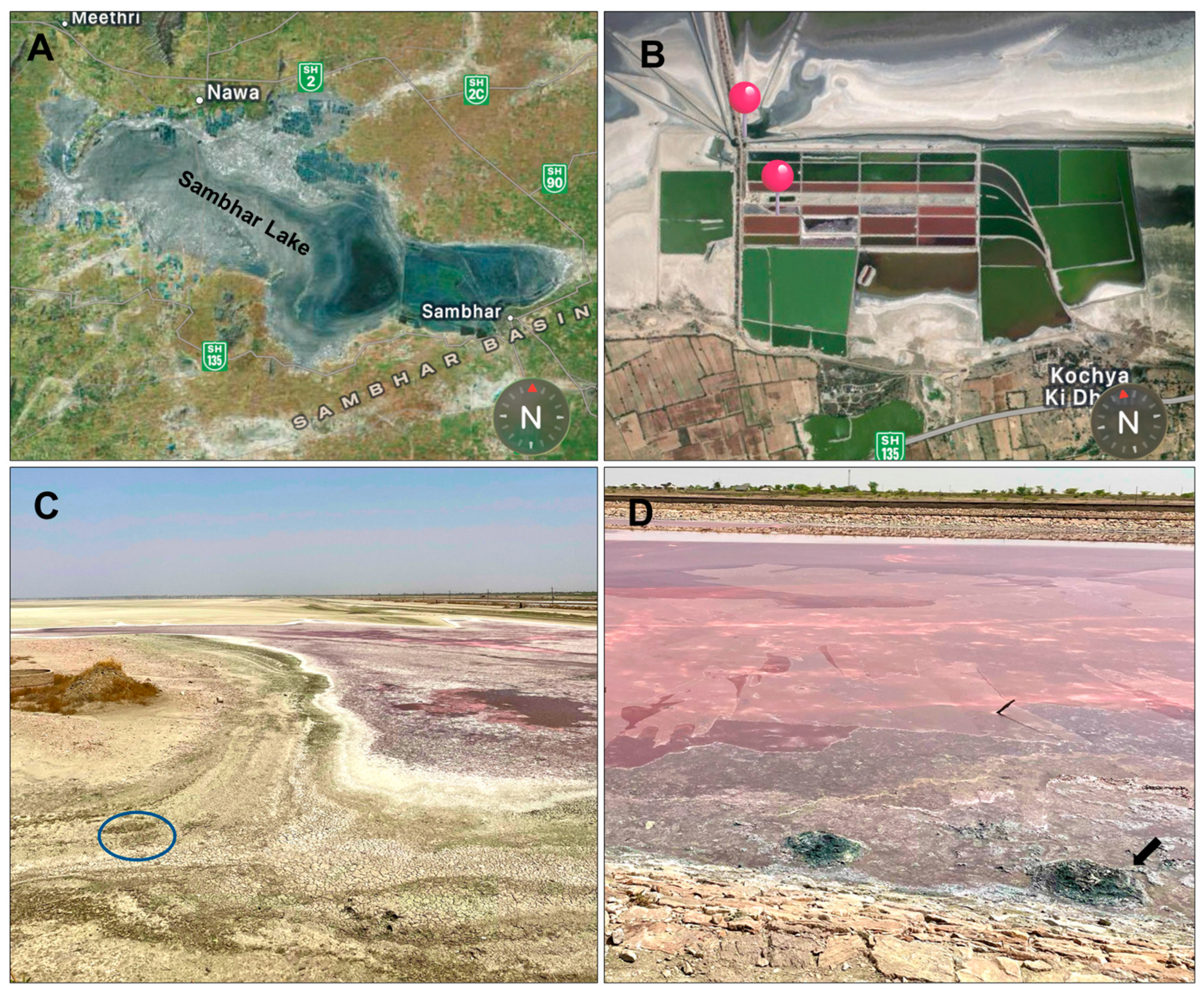
Biological crusts from sandy surfaces (26.91380° N, 75.12085° E) and the water surface (26.91019° N, 75.12175° E) were collected in April 2022 and November 2023 (Figure 1). The physicochemical properties of the sampling site (i.e., pH, total dissolved solids, salinity, and electrical conductivity) were recorded onsite using multi-parameter PCSTestrTM35 (Eutech instrument, Oakton, Singapore) (Table S1). The collected samples were brought to the laboratory and inoculated into 50 mL culture tubes containing BG-11 medium (HiMedia Laboratories, Thane, India) [38], and Spirulina platensis (SP) medium (HiMedia Laboratories, Thane, India) with varying salt concentrations (2–10%). Once the macroscopic green mats formed, the colonies were aseptically picked and observed under an optical microscope (Axio Lab A1, Carl Zeiss, Goettingen, Germany). Uni-algal cultures of SN2022/16 and SN2023/15 strains were established by picking single filaments and inoculating them into SPM medium with 2.5% salinity [38]. The culture tubes were kept under white, fluorescent lights (35 μE·m−2·s−1), at 26 ± 2 °C, and a 12 h light:dark cycle.

2.2. Morphological Analysis

The cyanobacterial strains were observed under an optical microscope (Axio Lab Al, Carl Zeiss, Goettingen, Germany) to record the morphological characters. Trichome size was measured from ≥50 individuals at 400× and 1000× magnifications. Differential Interference Contrast (DIC) microphotographs were taken with a Leica DMi8 Confocal Inverted Microscope (Wetzlar, Germany), available in the Central Instrumentation Facility (CIF) of Central University of Rajasthan. The images were analyzed and annotated in Adobe Photoshop ver.11 to improve clarity, while maintaining the original appearance of the filaments. A preliminary identification of the strains was performed according to standard taxonomic keys, with updated taxonomy [39,40].

2.3. Effect of Salinity on Growth of Cyanobacterial Strains

The samples were collected from Sambhar Salt Lake, an ecosystem known for its high salinity and alkaline pH. Therefore, the isolated strains were cultivated in varying NaCl (HiMedia Laboratories, Thane, India) concentrations to assess their salinity tolerance because this is the most abundant dissolved salt present in saline environments. Hereafter, five NaCl treatments were employed: SPM supplemented with NaCl at concentrations of 2%, 4%, 6%, 8%, and 10% (w/v). Standard SPM (contains 0.2% NaCl as its constituent) served as the control, while SPM completely devoid of NaCl represented the 0% NaCl concentration. Each culture flask was inoculated with a standardized 5% (v/v) inoculum and subsequently incubated under fluorescent illumination. Biomass was harvested at 7, 14, 21, and 28 days post inoculation via centrifugation (Allegra 64-R, Beckman–Coulter, Brea, CA, USA) at 10,000 RPM for 5 min, followed by three consecutive washing cycles with distilled water to eliminate residual media. The harvested biomass was subjected to chlorophyll (Chl) a quantification [41]. One mL of chilled methanol (HiMedia Laboratories, Thane, India) was added and was incubated in a 60 °C water bath for 30 min to ensure complete pigment extraction. After cooling to an ambient temperature, the samples were scanned spectrophotometrically across a wavelength range of 200–800 nm using a UV-2600 spectrophotometer (Shimadzu, (Asia Pacific), Singapore). Chl-a content (μg/mL) was subsequently calculated using the following formula [41]:
Chl-a content (µg/mL) = 16.29 × (A665.2 − A750) − 8.54 × (A652 − A670)
The influence of varying NaCl concentrations on cyanobacterial growth was evaluated using a two-way ANOVA, with NaCl concentration and harvest time as independent variables. All analyses were performed in triplicate for each treatment group.

2.4. Molecular Characterization

Genomic DNA was extracted from freshly grown cultures using NucleoSpin Plant II DNA isolation kits (Macherey–Nagel GmbH & Co. KG, Dueren, Germany) following the manufacture’s protocol and stored at −20 °C for further experiments. The amplification of the 16S rRNA gene and 16S-23S ITS region was performed using primers 8F [42] and 340R [43]. The PCR reaction was performed in C1000 Touch™ Thermal Cycler (Bio-Rad, Jaipur, India) and the 25 μL PCR cocktail contained 2.5 μL of 10X PCR buffer (Qiagen, Hilden, Germany), 2 μL of template DNA, 0.5 μL of 10 mM primers, 0.625 μL of 20 mM deoxynucleoside triphosphates (Qiagen, Hilden, Germany) in equimolar proportions, and 0.15 μL of Taq DNA polymerase (5 U/mL, Qiagen, Hilden, Germany). The amplification of the 16S rRNA gene and the 16S-23S ITS region was performed using the conditions described by Pal et al. [35]. Amplified fragments were visualized on a 1% agarose gel stained with SYBR-Safe dye (HiMedia Laboratories, Thane, India) and purified using the HiPurA® PCR Product Purification Kit (Himedia, Thane, India) according to the manufacturer’s protocol. The 16S rRNA gene was sequenced commercially (Eurofins Genomics, Bangalore, India) with 8F and 1495R primers [42], along with internal primers [44,45], and the 16S-23S ITS region was sequenced with 322F and 340R primers [43].

2.5. Phylogenetic Analysis

Sequences were assembled in SeqAssem v07/2008 software [46] and a consensus sequence of 1449 bp was obtained. Similar sequences were queried in the BLASTn database (https://www.ncbi.nlm.nih.gov/, accessed on 7 January 2026) for use in phylogenetic analyses. Sequences were aligned in MAFFT v7.5 software along with the reference strains available in NCBI GenBank database and CyanoSeq v1.3 [47]. Final alignment consisted of 107 sequences of 1325 bp. The Maximum Likelihood (ML) tree was constructed in IQ-TREE v2.1.3 [48] using Gloeobacter violaceus PCC7421 (AF132790.1) as an outgroup. The best-fit model was determined by ModelFinder [49], inbuilt into IQ-TREE, by giving the command iqtree2 -s alignment.nex -m MFP, and the model TPM3 + I + G4 was selected based on Bayesian Information Criterion (BIC) scores. The standard nonparametric bootstrap value was set to 1000 replicates and the ultrafast bootstrap to 10,000 replicates in order to evaluate the robustness of the tree topology [50]. The Bayesian inference (BI) tree was built in MrBayes v3.2.7a [51] using GTR + I + G model. The BI tree was constructed in two independent runs with four Markov chains that were run for up to 15 million generations, with 25% burn-in frequency. The print frequency, sampling frequency, and diagnostic frequency were set to 1000, and the run was continued until the standard deviation of split frequency reached <0.01. The final tree depicted in the study was visualized and edited in FigTree v1.4.4 [52].
The P-distance matrix of 16S rRNA gene of studied strains with related taxa was built in MEGA 11 [53]. The secondary structure of D1-D1′ helix and BoxB helix was folded in RNA structure v6.4 [54] and redrawn in Inkscape v1.3.2 software [55]. The sequences were submitted to the NCBI database under the GenBank accession numbers PX684461 (SN2022/16) and PX684462 (SN2023/15).

3. Results

3.1. Taxonomic Description

Krienitziella S. Sonam, N. Pareek, P. Singh, D.A. Casamatta et P.K. Dadheech gen. nov.:
Diagnosis: The studied strains were morphologically distinct from related genera by having thin trichomes encapsulated within a thick sheath and presence of compact nodules formed in older cultures. Moreover, the 16S rRNA gene sequence of studied strains indicated low similarity with phylogenetically related genera (Thainema and Insularia) and they formed an independent clade in Nodosilineaceae family. In addition, significant differences in ITS secondary structures (D1-D1′ and BoxB helix) were noted.
Description: Filament straight to slightly wavy, long, solitary or in clusters, forming nodules under ambient light conditions in older cultures. Sheath thick, colorless, non-lamellated. Trichomes up to 6.6 µm wide, constricted at cross wall, immotile, not attenuated at terminal ends. Cells blue green to pale greenish, wider than long, with homogenous cytoplasm and cyanophycean granules present. Apical cells rounded, without calyptra. Reproduction with the help of necridic cells.
Type Species: Krienitziella sambharensis S. Sonam, N. Pareek, P. Singh, D.A. Casamatta and P.K. Dadheech sp. nov.
Etymology: Krienitziella: named in honor of Dr. Lothar Krienitz, a German phycologist who conducted considerable research on cyanobacteria inhabit various continents including Sambhar Lake, which is one of the homes of the Lesser Flamingo.
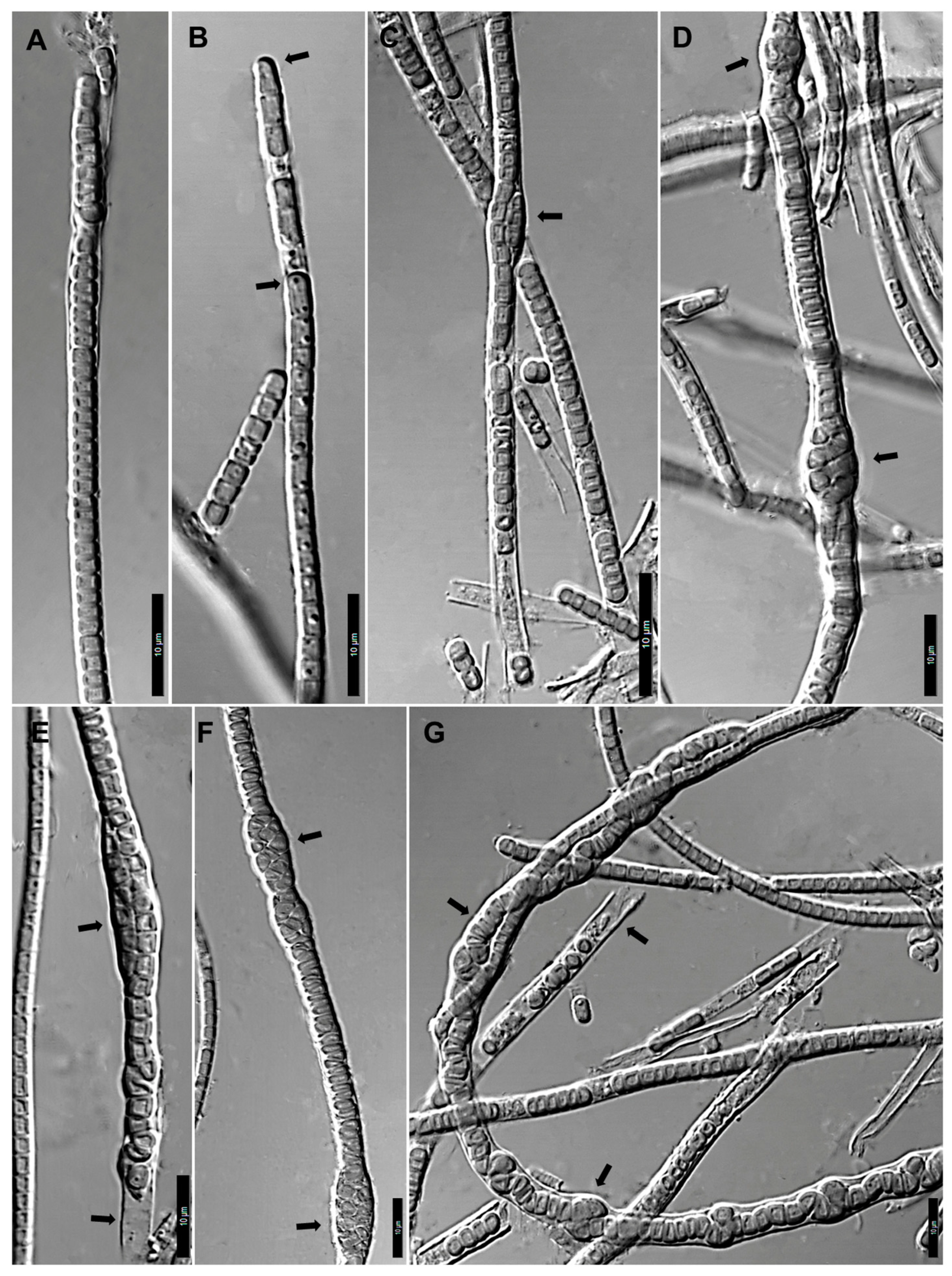
Krienitziella sambharensis S. Sonam, N. Pareek, P. Singh, D.A. Casamatta and P.K. Dadheech sp. nov. (Figure 2):
Description: In culture forming dense, dark, blue-green macroscopic mats. Filaments straight to slightly wavy, 3.5–6.6 µm wide, and unbranched. Sheaths thick, diffluent, wider, open at ends, non-lamellated. Trichomes uniseriate, sometimes becoming multiseriate due to division in multiple planes, forming compact spiral nodules (4.5–7.7 µm wide) under normal light condition. Filaments unattenuated, immotile, constricted at the cross-walls. Cells dark blue-green to pale greenish, 2.0–3.3 µm wide and 1.2–2.5 µm long, barrel to discoid in shape, one prominent granule per cell present. Apical cells rounded, dome-shaped, non-capitate. Reproduction with hormogonia formed via necridic cells.
Reference Strain: SN2022/16
Type Locality: Sambhar Lake (26.91380° N, 75.12085° E), Sambhar, Rajasthan, India.
Holotype: Dry specimen of the reference strain was deposited in University of Rajasthan Herbarium (RUBL), Jaipur, Rajasthan, INDIA with accession number RUBL22018.
Isotype: The culture of Krienitziella sambharensis (accession number GCC 202513) is preserved in metabolically inactive form in the Global Collection of Cyanobacteria (GCC) (https://ccinfo.wdcm.org/details?regnum=1165, accessed on 20 April 2025), Varanasi, India.
Etymology: The specific epithet “sambharensis” (samb.har.en’sis. N.L. fem. adj) refers to the fact that the sample was isolated from the Sambhar Lake of Rajasthan, India.
Habitat: Saline soil crust from episodically wetted soils near littoral zone of Sambhar Lake.

3.2. Evaluation of Growth in Response to Salinity

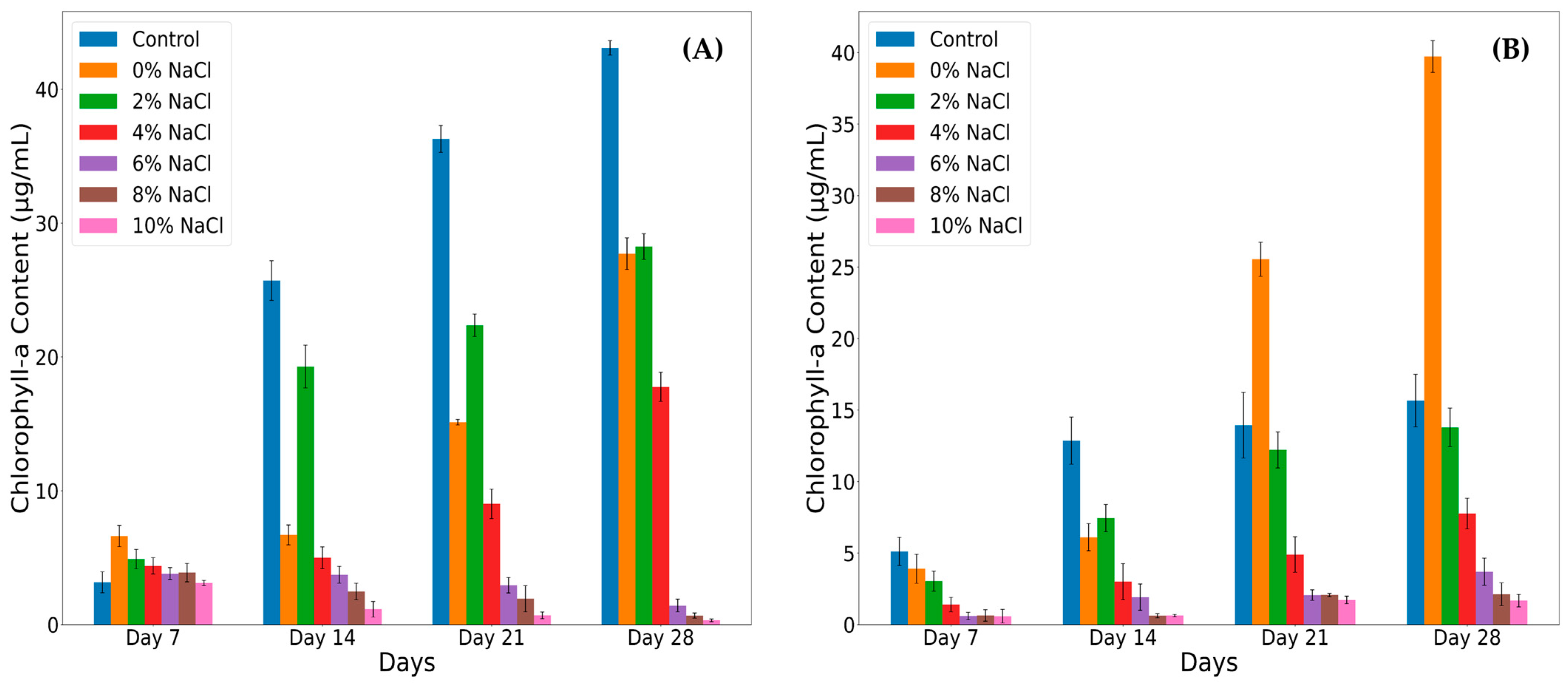
The effects of NaCl concentrations were evaluated by quantifying chl-a content across 7, 14, 21, and 28 days (Figure 3). A two-way ANOVA revealed a significant effect of NaCl treatment on chl-a content (F = 6.075, p = 0.000121), indicating that varying NaCl concentration substantially modulates photosynthetic pigment synthesis and growth in both strains. Both Krienitziella sambharensis SN2022/16 and SN2023/15 strains exhibited robust and steady growth, evidenced by a progressive increase in chl-a content under low-to-moderate salinity conditions (control, 2% NaCl, and 0% NaCl). Krienitziella sambharensis SN2022/16 achieved its highest mean chl-a content of 43.48 μg/mL by day 28 in the control treatment, demonstrating optimal performance under standard conditions. Similarly, Krienitziella sambharensis SN2023/15 performed optimally in low-salinity environments, with the 0% NaCl treatment yielding the highest overall chl-a content for the strain, reaching 39.73 μg/mL after 28 days. Although both strains grew in the 4% NaCl treatment, their chl-a content consistently remained lower than the control, 2% NaCl, and 0% NaCl conditions. Conversely, exposure to elevated salt concentrations (6%, 8%, and 10% NaCl) markedly suppressed the growth of both isolates. The 10% NaCl treatment resulted in a pronounced inhibition, exhibiting the lowest recorded Chl-a content of 0.32 μg/mL and 1.67 μg/mL, respectively, after 28 days. These findings collectively demonstrate that salinity directly influences the growth dynamics of both cyanobacterial isolates in a comparable manner. Krienitziella sambharensis tolerates and maintains robust growth up to 4% NaCl. However, concentrations exceeding 4% NaCl significantly inhibit growth (Figure 3).

3.3. Molecular and Phylogenetic Analysis

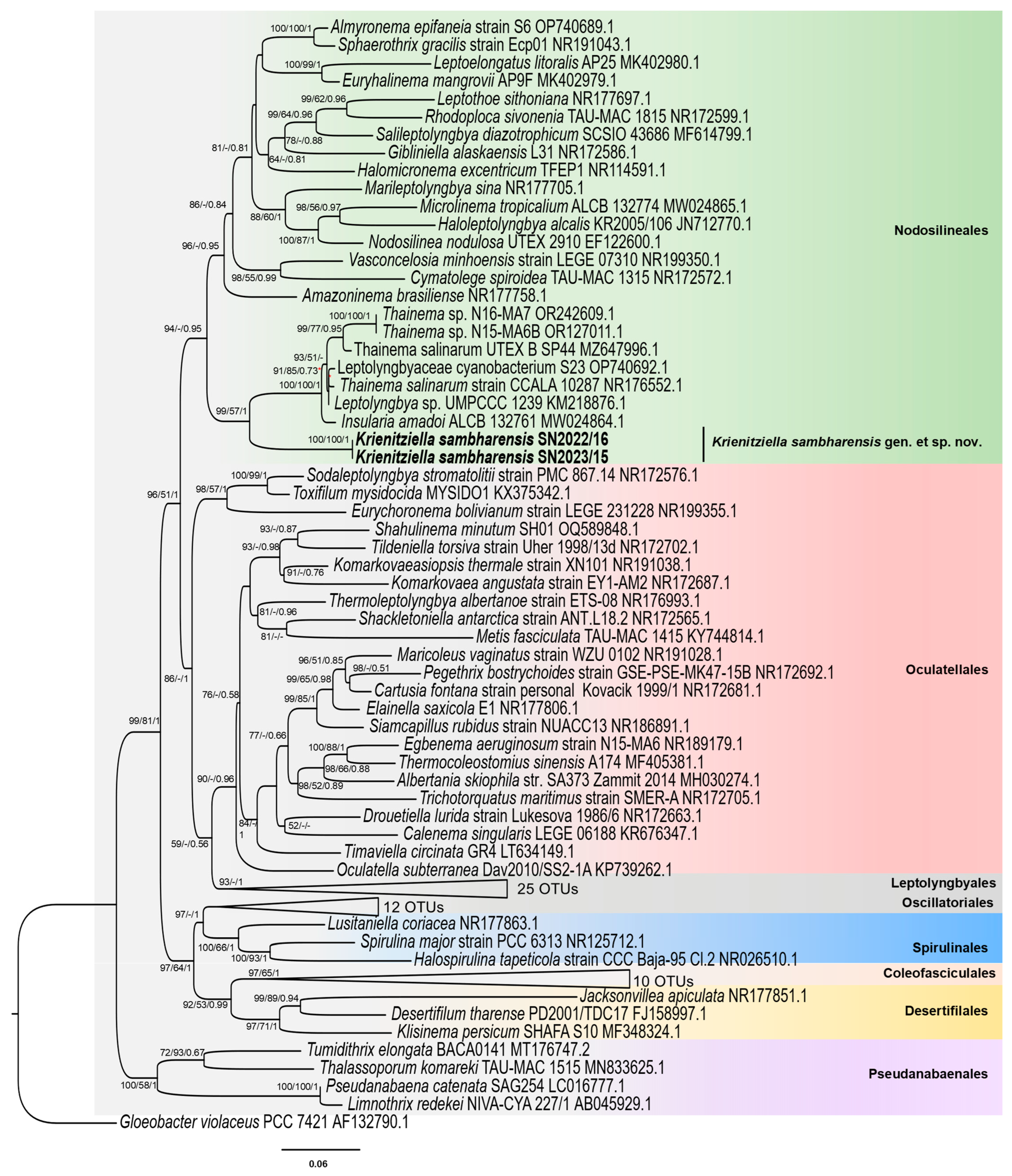
The BLASTn search of 16S rRNA gene sequence revealed <95% similarities with the most hits in the NCBI database. The uncorrected 16S rRNA gene p-distance matrix indicated that Krienitziella sambharensis shared the highest similarity (94.16%) with an unclassified strain of Thainema (“Leptolyngbya” sp. UMPCCC 1239) and a low percentage similarity of 91.38% with T. salinarum CCALA 10287 (Table S2). Moreover, the phylogenetic tree (Figure 4) demonstrated that Krienitziella sambharensis formed a distinct, highly supported clade (ML = 100, BI = 1) within the order Nodosilineales. The sister clade to Krienitziella sambharensis includes filamentous cyanobacteria with thin trichomes such as T. salinarum CCALA 10287, Insularia amadoi ALCB 132761, T. salinarum UTEX B SP44, Thainema sp. N15-MA6B, Thainema sp. N16-MA7, Leptolyngbya sp. UMPCCC 1239 and Leptolyngbyaceae cyanobacterium S23.

3.4. Secondary Structure of the 16S-23S ITS

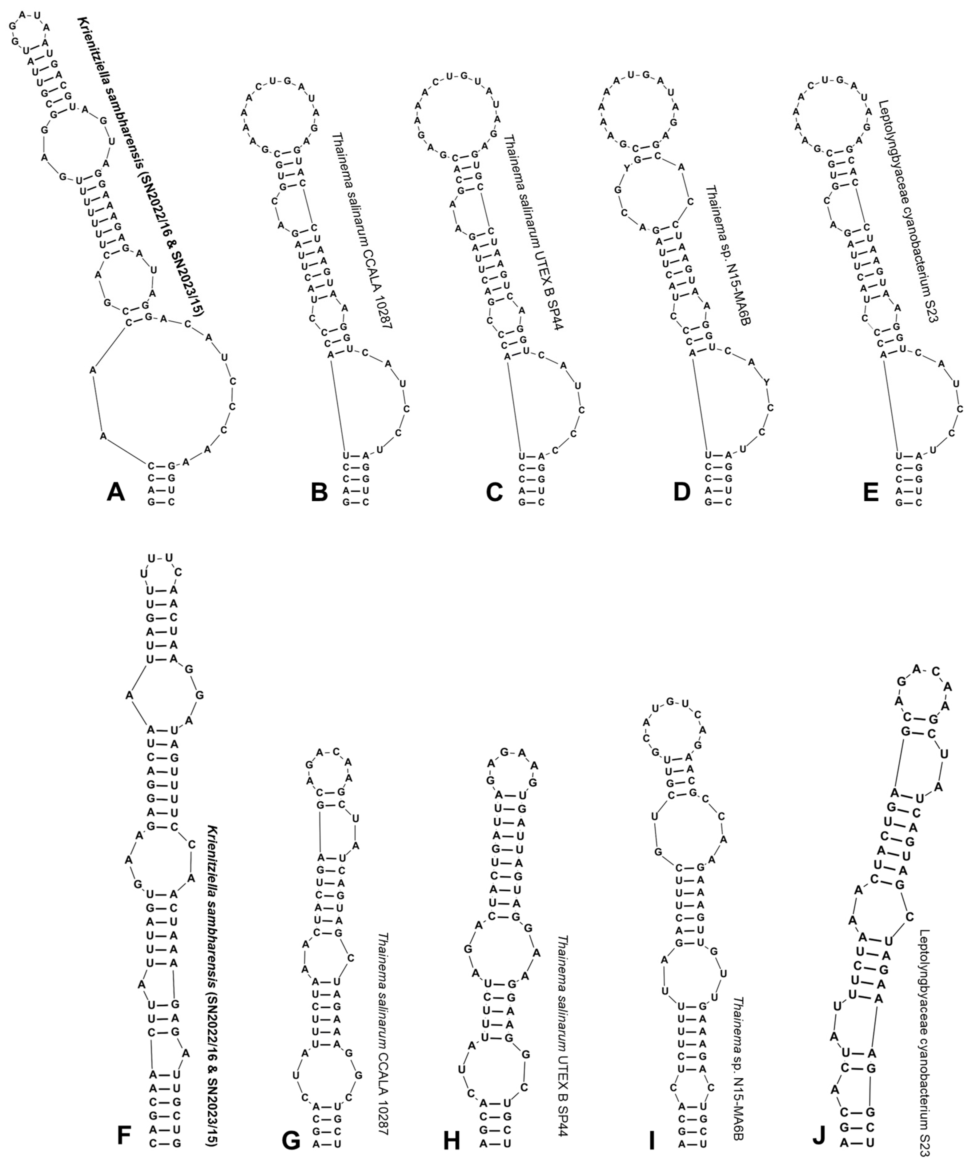
The 16S-23 ITS sequences of 500 bp were recovered from both studied strains (SN2022/16 and SN2023/15), and they possessed identical ITS sequences and the presence of both tRNAile and tRNAala genes. The analysis of ITS secondary structures revealed some unique features compared to close relatives, although no ITS sequences for Insularia were available. The D1-D1′ helix of Krienitziella sambharensis was 70 nucleotides (nt) long, and the basal stem was formed with four base pairs (5′-GACC: GGUC-3′), which varied from related strains that had a 5 bp long stem sequence (Figure 5). The stem sequence was proceeded by an asymmetric bilateral loop having two bases (A) on the 5ʹ side and nine bases on the 3ʹ side, which was further extended by a small asymmetric bilateral loop (5′-GA: AUA-3′). The terminal loop was 5 nt long, mainly formed by purine-rich bases (except U base), which was subtended by an asymmetric bilateral loop (5′-GAG: AGUA-3′) with an unequal distribution of bases on the 5′ and 3′ sides (Figure 5).
The BoxB helix basal stem of Krienitziella sambharensis comprised 6 bp (5′-CAGCAA: UUGCUG-3′), which was unique in comparison to related strains (T. salinarum CCALA 10287, T. salinarum UTEX B SP44, Thainema sp. N15-MA6B and Leptolyngbyaceae cyanobacterium S23), which had a smaller stem sequence (4 bp only). Two unilateral bulges in opposite symmetry around helix were observed, with only one nucleotide (A), which was further proceeded by two asymmetric bilateral loops that were mainly purine-rich. Moreover, the terminal loop was extremely small, being formed by four unpaired bases (UUUA) (Figure 5).

4. Discussion

Cyanobacterial systematics has a long and dynamic history, beginning with their initial description as “blue-green algae”, followed by several disagreements among scientists regarding their taxonomic placements [56,57,58,59,60,61,62] to their current recognition as phylum Cyanobacteriophyta [63]. Traditionally, they were identified solely based on their morphological characters [39,40,64], though this approach has proven inadequate, especially for thin filamentous cyanobacteria, which have fewer characters for separation. In fact, only a few thin filamentous genera possess distinct morphological apomorphies. After the advent of molecular techniques, particularly 16S rRNA gene and 16S-23S ITS sequencing, many morphologically defined taxa were found to be polyphyletic [12,65,66,67]. Thus, a polyphasic approach that integrates morphological, molecular, phylogenetic, and ecological data is needed for robust phylogenetic delineation [12,66], which we employed in the current study.
Morphologically depauperate cyanobacterial lineages (e.g., Nodosilineales) can be challenging to identify and assess taxonomically. Outside of a few distinct morphological apomorphies (e.g., nodules in Nodosilinea [14,65] and pigment particles in apical cells of Oculatella [68], there are few visible characters to separate these lineages. The studied strains were morphologically more or less similar to Nodosilinea nodulosa, e.g., thin filaments forming compact nodules. However, a difference between N. nodulosa and Krienitziella sambharensis is in the development of nodules; for instance, in N. nodulosa, the nodules were formed under low-light conditions, while in Krienitziella sambharensis, they develop in ambient light. Additionally, the cells of Krienitziella sambharensis were 3x wider (2.0–3.3 µm) than the N. nodulosa cells (1.1–1.5 µm), although the cell length remained the same (up to 2.5 µm). Morphologically, the studied strains also deviated from phylogenetically related taxa Thainema and Insularia in regard to cell size and shape; cells of Krienitziella sambharensis were wider than they were long, while Thainema and Insularia have cells that are longer than they are wide (Table 1). Moreover, trichomes of Krienitziella sambharensis developed nodules in older cultures that were encapsulated in thick non-lamellated sheaths, whereas Thainema and Insularia were characterized by thin sheaths and the absence of nodules.
According to the updated classification system [12], Krienitziella sambharensis was clustered into Nodosilineales and formed a distinct lineage with high bootstrap/posterior probability values (Figure 4). The sister clade encompasses the genera Thainema, Insularia and some unclassified strains of Thainema. Furthermore, in a recent study conducted by [8], the authors considered transferring genera Toxifilum, Sodaleptolyngbya and Metis from Oculatellales to Nodosilineales based on their phylogenetic analyses. However, these inferences are questionable, as the alignment employed contained only 1152 nucleotides, which is very short and could compromise the phylogenetic resolution. Additionally, in our phylogenetic tree, these genera (Toxifilum, Sodaleptolyngbya and Metis) fall into Oculatellales, which is consistent with [12]. Therefore, we recommend using an alignment of at least more than 1200 nucleotides because longer sequences will enhance phylogenetic resolution and allow for reproducibility in taxonomic studies.
The 16S rRNA gene p-distance values showed that Krienitziella sambharensis exhibited the highest similarity of 94.16% with unclassified Thainema (Leptolyngbya sp. UMPCCC 1239) (Table S2). These threshold values were below the currently accepted value (<94.5%) for the establishment of a new genus, as suggested by [69,70,71]. However, as noted in [72], these values are not necessarily fixed. However, the low p-distance values coupled with one apomorphic character (the presence of nodules) distinguishes Krienitziella sambharensis from phylogenetically related taxa and reinforces the delineation of the studied strains as a novel genus in this study.
The 16S-23S ITS secondary structures are an important facet of modern taxonomy while describing new cyanobacteria [43,73,74]. The secondary structure of D1-D1′ and BoxB helices of Krienitziella sambharensis (Figure 5) was compared with closest relatives to confirm its distinctness at the taxonomic level. Both D1-D1′ and BoxB helices of Krienitziella sambharensis varied significantly from related strains T. salinarum CCALA 10287, T. salinarum UTEX B SP44, Thainema sp. N15-MA6B and Leptolyngbyaceae cyanobacterium S23 in sequence length, asymmetric bulges, terminal loop and stem sequence (Figure 5, Table S3). Moreover, the terminal loop of Krienitziella sambharensis D1-D1′ helix was extremely small, with 5 bases, while in related strains the loop was formed of 13–15 bases. Another remarkable feature that makes the D1-D1′ helix of Krienitziella sambharensis distinct from others was the presence of a bilateral asymmetric bulge (5′-AA: ACAUCCCAA-3′) formed above the basal stem sequence; in related strains, a unilateral basal bulge is observed.
In addition to morphology and molecular analyses, ecology may play a pivotal role in cyanobacterial taxonomic studies [40,66,75,76,77]. The studied strains were isolated from a saline lake characterized by salinity levels of ~350 ppt [25]; nonetheless, their tolerance to salinity ranges from 45.2 to 49.9 ppt and the pH for their optimum growth ranges from 9 to 10. The ability to thrive under no- to moderate-salinity conditions, with a predictable growth reduction only at higher levels, classifies both isolates as halotolerant to slightly halophilic (Figure 3). However, the related taxa Thainema [78] was reported from a saltern pond in Thailand with a salinity of 90–250 ppt and Insularia [79] was found to thrive in a marine environment in Brazil; notably, the physicochemical characteristics of Insularia sampling site were not available for comparison. Despite being reported from a similar niche, Krienitziella sambharensis exhibits distinct morphological characteristics such as the development of nodules and the presence of thick sheaths, which might be an adaptive strategy to endure salinity stress and irradiance [80,81]. Furthermore, saline lakes are rich in nutrients that support the growth of Limnospira (formerly Spirulina), a filamentous, non-heterocytous cyanobacterium which is a primary food source for migratory flamingo populations [82]. Thus, the isolation of Krienitziella sambharensis from saline lake provides an alternative dietary source for flamingo birds that could be investigated in further studies focusing on nutraceutical aspects.
Table 1. Morphological comparison of Krienitziella sambharensis with phylogenetically related genera.
Krienitziella sambharensisThainema salinarum [78]Insularia amadoi [79]
FilamentsStraight or slightly wavyStraight or flexuousStraight to slightly wavy
Cell width (µm)2.0–3.3<31.0–2.2
Cell length (µm)1.2–2.52–3.51.4–2.9
Cell shapeWider than longSlightly longer than wide or mostly isodiametric Longer than wide, elongated
SheathThick, colorless, wideThin, colorlessThin, hyaline, firm
ConstrictionsDistinct SlightDistinct
Necridic cellsPresentAbsentAbsent
Apical cellsRounded, dome shapedRoundedRounded
NodulesPresentAbsentAbsent
Salinity toleranceHalotolerant or slightly halophilic Halophilic to halotolerant-
OccurrenceSaline soil crust from littoral zone, Sambhar Lake, Rajasthan, IndiaWet soil surface, Ban Laem district Petchaburi Province, ThailandMarine, Abrolhos Archipelago, Brazil
Based on the polyphasic approach that combined morphological characters, molecular and phylogenetic studies, and ecology, we propose a new cyanobacterial taxon, Krienitziella sambharensis gen. et sp. nov. in Nodosilineales (Cyanobacteriophyta).

Supplementary Materials

The following supporting information can be downloaded at: https://www.mdpi.com/article/10.3390/d18030181/s1, Figure S1: Uncompressed maximum likelihood phylogenetic tree of Krienitziella sambharensis along with 148 cyanobacterial taxa.; Table S1: Physiochemical properties of the sampling site located in Sambhar, Jaipur, Rajasthan; Table S2: Similarity matrix (p-distance) of 16S rRNA gene of Krienitziella sambharensis (SN2022/16 and SN2023/15) with closest BLAST search results; Table S3: Comparison of ITS region lengths (nt) of D1-D1′ and BoxB helix folded structures of K. sambharensis and related taxa.

Author Contributions

Conceptualization, S.S. and P.K.D.; investigation, S.S. and S.A.; writing—original draft preparation, S.S.; writing—review and editing, S.S., N.P., P.S., D.A.C. and P.K.D.; supervision, P.K.D.; funding acquisition, P.K.D. All authors have read and agreed to the published version of the manuscript.

Funding

This study was supported by the Department of Science & Technology, Government of Rajasthan, India, Project No. P.7 (3) DST/BT/R&D/EAC/2018/3168. The APC was funded by University of North Florida, Jacksonville, FL 32250, USA.

Institutional Review Board Statement

Not applicable.

Data Availability Statement

The data presented in this study are openly available in GenBank [https://www.ncbi.nlm.nih.gov/genbank/] under accession no PX684461 (SN2022/16) and PX684462 (SN2023/15); Global Collection of Cyanobacteria (GCC) [https://ccinfo.wdcm.org/details?regnum=1165 accessed on 14 March 2026] under accession number GCC 202513 and University of Rajasthan Herbarium (RUBL), with accession number RUBL22018.

Acknowledgments

We thank Department of Microbiology, Central University of Rajasthan for providing the necessary facilities for research work. Special thanks to Hansa for technical assistance while taking photos with Confocal Microscope available in Central Instrumentation Facility (CIF) of Central University of Rajasthan.

Conflicts of Interest

The authors declare no conflicts of interest.

References

  1. Seckbach, J. Cellular Origin, Life in Extreme Habitats and Astrobiology; Springer: Dordrecht, The Netherlands, 1999; Available online: www.springer.com/series/5775 (accessed on 10 January 2026).
  2. Genuário, D.B.; Vaz, M.G.; Santos, S.N.; Kavamura, V.N.; Melo, I.S. Cyanobacteria from Brazilian extreme environments: Toward functional exploitation. In Microbial Diversity in the Genomic Era; Academic Press: Cambridge, MA, USA, 2019; pp. 265–284. [Google Scholar]
  3. Saini, N.; Pal, K.; Sujata, D.B.; Mona, S. Thermophilic algae: A new prospect towards environmental sustainability. J. Clean. Prod. 2021, 324, 129277. [Google Scholar] [CrossRef] [Scilit]
  4. Dadheech, P.K.; Abed, R.M.; Mahmoud, H.; Mohan, M.K.; Krienitz, L. Polyphasic characterization of cyanobacteria isolated from desert crusts, and the description of Desertifilum tharense gen. et sp. nov. (Oscillatoriales). Phycologia 2012, 51, 260–270. [Google Scholar] [CrossRef] [Scilit]
  5. Chakrabort, S.; Maruthanay, V.; Achari, A.; Pramanik, A.; Jaisankar, P.; Mukherjee, J. Euryhalinema mangrovii gen. nov., sp. nov. and Leptoelongatus litoralis gen. nov., sp. nov. (Leptolyngbyaceae) isolated from an Indian mangrove forest. Phytotaxa 2019, 422, 58–74. [Google Scholar] [CrossRef] [Scilit]
  6. Kaštovský, J.; Johansen, J.R.; Hauerová, R.; Akagha, M.U. Hot is rich—An enormous diversity of simple trichal cyanobacteria from Yellowstone hot springs. Diversity 2023, 15, 975. [Google Scholar] [CrossRef] [Scilit]
  7. Tang, J.; Zhou, H.; Jiang, Y.; Yao, D.; Waleron, K.F.; Du, L.M.; Daroch, M. Characterization of a novel thermophilic cyanobacterium within Trichocoleusaceae, Trichothermofontia sichuanensis gen. et sp. nov., and its CO2-concentrating mechanism. Front. Microbiol. 2023, 14, 1111809. [Google Scholar] [CrossRef] [Scilit]
  8. Hentschke, G.S.; Ciancas Jiménez, J.C.; Hoepfner, C.; Guzmán, D.; Mesquita, M.J.; Vasconcelos, V. The extremophile Eurychoronema bolivianum gen. et sp. nov. (Nodosilineales, Cyanobacteria) and Leptolyngbya aquatica comb. nov. Phycologia 2024, 63, 510–519. [Google Scholar] [CrossRef] [Scilit]
  9. Garduno-Solorzano, G.; Martinez-Garcia, M.; Hentschke, G.S.; Solorza, L.F.M.; Vasconcelos, V.M. A phylogenetically distant clade of Nostoc–like (Cyanobacteria) taxa with the description of Reofilinostoc matlalcueyense gen. et sp. nov. from an extreme environment. Phytotaxa 2024, 655, 125–143. [Google Scholar] [CrossRef] [Scilit]
  10. de Oliveira, F.L.; Hentschke, G.S.; Morais, J.; Silva, R.; Cruz, P.; Vasconcelos, V.M. Exploring the cyanobacterial diversity in Portugal: Description of four new genera from LEGE-CC using the polyphasic approach. J. Phycol. 2024, 60, 1285–1304. [Google Scholar] [CrossRef] [Scilit]
  11. Kaštovský, J. Welcome to the jungle! An overview of modern taxonomy of cyanobacteria. Hydrobiologia 2024, 851, 1063–1077. [Google Scholar] [CrossRef] [Scilit]
  12. Strunecký, O.; Ivanova, A.P.; Mareš, J. An updated classification of cyanobacterial orders and families based on phylogenomic and polyphasic analysis. J. Phycol. 2023, 59, 12–51, Erratum in J. Phycol. 2023, 59, 635. [Google Scholar] [CrossRef] [Scilit] [PubMed]
  13. Abed, R.M.; Garcia-Pichel, F.; Hernández-Mariné, M. Polyphasic characterization of benthic, moderately halophilic, moderately thermophilic cyanobacteria with very thin trichomes and the proposal of Halomicronema excentricum gen. nov., sp. nov. Arch. Microbiol. 2002, 177, 361–370. [Google Scholar] [CrossRef] [Scilit]
  14. Perkerson, R.B., III; Johansen, J.R.; Kovácik, L.; Brand, J.; Kaštovský, J.; Casamatta, D.A. A unique Pseudanabaenalean (Cyanobacteria) genus Nodosilinea gen. nov. based on morphological and molecular data. J. Phycol. 2011, 47, 1397–1412. [Google Scholar] [CrossRef] [Scilit]
  15. Roy, A.R.; Chakraborty, S.; Karmakar, A.; de los Santos-Villalobos, S.; Mukherjee, J. Almyronema epifaneia gen. & sp. nov. (Cyanobacteria, Nodosilineaceae) isolated from an Indian mangrove forest. Phycologia 2024, 63, 89–106. [Google Scholar] [CrossRef] [Scilit]
  16. Zhou, W.G.; Ding, D.W.; Yang, Q.S.; Ahmad, M.; Zhang, Y.Z.; Lin, X.C.; Dong, J.D. Marileptolyngbya sina gen. nov., sp. nov. and Salileptolyngbya diazotrophicum gen. nov., sp. nov. (Synechococcales, Cyanobacteria), species of cyanobacteria isolated from a marine ecosystem. Phytotaxa 2018, 383, 75–92. [Google Scholar] [CrossRef] [Scilit]
  17. Strunecky, O.; Raabova, L.; Bernardova, A.; Ivanova, A.P.; Semanova, A.; Crossley, J.; Kaftan, D. Diversity of cyanobacteria at the Alaska North Slope with description of two new genera: Gibliniella and Shackletoniella. FEMS Microbiol. Ecol. 2020, 96, fiz189. [Google Scholar] [CrossRef] [Scilit]
  18. Curren, E.; Kuwahara, V.S.; Yoshida, T.; Leong, S.C.Y. Sphaerothrix gracilis gen. et sp. nov. (Nodosilineales, Cyanobacteria): A novel filamentous cyanobacterium isolated from tropical coastal microplastics. Phycol. Res. 2024, 72, 205–214. [Google Scholar] [CrossRef] [Scilit]
  19. Chavadar, M.; Saraf, A.; Suradkar, A.; Mishra, D.; Kumar, N.; Singh, P. Constrictifilum karadense gen. et sp. nov., a new Nostocalean genus from Maharashtra, India. FEMS Microbiol. Lett. 2021, 368, fnab057. [Google Scholar] [CrossRef] [Scilit] [PubMed]
  20. Kumar, N.; Saraf, A.; Pal, S.; Mishra, D.; Singh, P. Insights into the phylogenetic inconsistencies of the genus Amazonocrinis and description of epilithic Amazonocrinis malviyae sp. nov. (Cyanobacteria, Nostocales) from Jammu and Kashmir, India. Int. J. Syst. Evol. Microbiol. 2022, 72, 005658. [Google Scholar] [CrossRef] [Scilit]
  21. Pant, H.; Kumar, N.; Pal, S.; Singh, P. Exploring cyanobacteria from diverse habitats of the Konkan region of India, unveiling novel species of the genera Desikacharya, Pseudoaliinostoc, and Chlorogloeopsis using a polyphasic approach. J. Phycol. 2025, 61, 194–217. [Google Scholar] [CrossRef] [Scilit]
  22. Jakher, G.R.; Bhargava, S.C.; Sinha, R.K. Comparative limnology of Sambhar and Didwana lakes (Rajasthan, NW India). Hydrobiologia 1990, 197, 245–256. [Google Scholar] [CrossRef] [Scilit]
  23. Kulshreshtha, S.; Sharma, B.K.; Sharma, S. The Ramsar sites of Rajasthan: Ecology and conservation of Sambhar Salt Lake, Jaipur and Keoladeo National Park, Bharatpur. In Faunal Heritage of Rajasthan, India: Conservation and Management of Vertebrates; Springer International Publishing: Cham, Switzerland, 2013; pp. 173–219. [Google Scholar] [CrossRef] [Scilit]
  24. Bhatt, H.H.; Pasricha, R.; Upasani, V.N. Isolation and characterization of a halophilic cyanobacterium Euhalothece SLVH01 from Sambhar Salt Lake, India. Int. J. Curr. Microbiol. Appl. Sci. 2016, 5, 215–224. [Google Scholar] [CrossRef] [Scilit]
  25. Singh, S.; Goyal, A.; Dutt, K. Bioprospecting for moderately halophilic eubacteria for potential biotechnological applications from Sambhar Lake, Rajasthan, India. Appl. Biol. Chem. J. 2024, 5, 12–21. [Google Scholar] [CrossRef] [Scilit]
  26. Yadav, A.K.; Vardhan, S.; Kashyap, S.; Yandigeri, M.; Arora, D.K. Actinomycetes diversity among rRNA gene clones and cellular isolates from Sambhar Salt Lake, India. Sci. World J. 2013, 2013, 781301. [Google Scholar] [CrossRef] [Scilit]
  27. Gaur, A. Halophiles of Sambhar Salt Lake. In Microbes: In Action; AGROBIOS (INDIA): Jodhpur, India, 2015; p. 355. [Google Scholar]
  28. Gaur, A.; Sharma, G.; Mohan, D. Isolation and characterization of moderate halophilic Salinicoccus sp. producing extracellular hydrolytic enzymes. J. Biosci. 2015, 4, 3379–3383. [Google Scholar]
  29. Deshmukh, S.K.; Verekar, S.A.; Chavan, Y.G. Keratinophilic fungi from the vicinity of salt pan soils of Sambhar Lake Rajasthan (India). J. Med. Mycol. 2018, 28, 457–461. [Google Scholar] [CrossRef] [Scilit]
  30. Kaushik, G.; Raza, K. Potential of novel Dunaliella salina from sambhar Salt Lake, India, for bioremediation of hexavalent chromium from aqueous effluents: An optimized green approach. Ecotoxicol. Environ. Saf. 2019, 180, 430–438. [Google Scholar] [CrossRef] [Scilit]
  31. Kajale, S.; Deshpande, N.; Pali, S.; Shouche, Y.; Sharma, A. Natrialba swarupiae sp. nov., a halophilic archaeon isolated from a hypersaline lake in India. Int. J. Syst. Evol. Microbiol. 2020, 70, 1876–1881. [Google Scholar] [CrossRef] [Scilit]
  32. Singh, P.; Dubey, N.; Bagchi, S.N. Westiellopsis ramosa sp. nov., intensely branched species of Westiellopsis (cyanobacteria) from a freshwater habitat of Jabalpur, Madhya Pradesh, India. Plant Syst. Evol. 2017, 303, 1239–1249. [Google Scholar] [CrossRef] [Scilit]
  33. Saraf, A.G.; Dawda, H.G.; Singh, P. Desikacharya gen. nov., a phylogenetically distinct genus of Cyanobacteria along with the description of two new species, Desikacharya nostocoides sp. nov. and Desikacharya soli sp. nov., and reclassification of Nostoc thermotolerans to Desikacharya thermotolerans comb. nov. Int. J. Syst. Evol. Microbiol. 2019, 69, 307–315. [Google Scholar] [CrossRef] [Scilit]
  34. Maltsev, Y.; Kezlya, E.; Maltseva, S.; Karthick, B.; Dvořák, P.; Kociolek, J.P.; Kulikovskiy, M. A new species of the previously monotypic genus Iningainema (Cyanobacteria, Scytonemataceae) from the Western Ghats, India. Eur. J. Phycol. 2021, 56, 348–358. [Google Scholar] [CrossRef] [Scilit]
  35. Pal, S.; Saraf, A.; Kumar, N.; Singh, P. Phycological exploration of the global biodiversity hotspots of Northeast India: Discovery of a new species of soil-dwelling cyanobacteria, Desikacharya kailashaharensis sp. nov. FEMS Microbiol. Lett. 2022, 369, fnac099. [Google Scholar] [CrossRef] [Scilit]
  36. Bhandari, M.; Sonam, S.; Negi, R.; Singh, P.; Singh, R.K.; Dadheech, P.K. Description of a novel cyanobacterial species Elainella ringii sp. nov. (Oculatellales) from Himalayan hot spring, Uttarakhand, India using polyphasic approach. Phytotaxa 2025, 718, 163–177. [Google Scholar] [CrossRef] [Scilit]
  37. Turland, N.J.; Wiersema, J.H.; Barrie, F.R.; Gandhi, K.N.; Gravendyck, J.; Greuter, W.; Hawksworth, D.L.; Herendeen, P.S.; Klopper, R.R.; Knapp, S.; et al. International Code of Nomenclature for Algae, Fungi, and Plants (Madrid Code); Regnum Vegetabile 162; University of Chicago Press: Chicago, IL, USA, 2025. [Google Scholar] [CrossRef] [Scilit]
  38. Rippka, R.; Deruelles, J.; Waterbury, J.B.; Herdman, M.; Stanier, R.Y. Generic assignments, strain histories and properties of pure cultures of cyanobacteria. Microbiology 1979, 111, 1–61. [Google Scholar] [CrossRef] [Scilit]
  39. Desikachary, T.V. Cyanophyta ICAR Monographs of Algae; ICAR: New Delhi, India, 1959; 686p. [Google Scholar]
  40. Komárek, J.; Anagnostidis, K. Cyanoprokaryota 2. Teil/2nd part: Oscillatoriales. In Susswasserflora von Mitteleuropa; Springer: Berlin/Heidelberg, Germany, 2005; Volume 19, pp. 1–759. [Google Scholar]
  41. Bhandari, M.; Prajapati, S.K. Use of reverse osmosis reject from drinking water plant for microalgal biomass production. Water Res. 2022, 210, 117989. [Google Scholar] [CrossRef] [Scilit] [PubMed]
  42. Lane, D.J. 16S/23S rRNA Sequencing. In Nucleic Acid Techniques in Bacterial Systematics; Stackebrandt, E., Goodfellow, M., Eds.; John Wiley and Sons: New York, NY, USA, 1991; pp. 115–175. [Google Scholar]
  43. Iteman, I.; Rippka, R.; Tandeau de Marsac, N.; Herdman, M. Comparison of conserved structural and regulatory domains within divergent 16S rRNA–23S rRNA spacer sequences of cyanobacteria. Microbiology 2000, 146, 1275–1286. [Google Scholar] [CrossRef] [Scilit] [PubMed]
  44. Edwards, U.; Rogall, T.; Blöcker, H.; Emde, M.; Böttger, E.C. Isolation and direct complete nucleotide determination of entire genes. Characterization of a gene coding for 16S ribosomal RNA. Nucleic Acids Res. 1989, 17, 7843–7853. [Google Scholar] [CrossRef] [Scilit] [PubMed]
  45. Nübel, U.; Garcia-Pichel, F.; Muyzer, G. PCR primers to amplify 16S rRNA genes from cyanobacteria. Appl. Environ. Microbiol. 1997, 63, 3327–3332. [Google Scholar] [CrossRef] [Scilit]
  46. Hepperle, D. SeqAssem Ver. 07/2008. Licensed. Available online: https://www.sequentix.de/ (accessed on 14 March 2026).
  47. Lefler, F.W.; Berthold, D.E.; Laughinghouse, H.D., IV. Cyanoseq: A database of cyanobacterial 16S rRNA gene sequences with curated taxonomy. J. Phycol. 2023, 59, 470–480. [Google Scholar] [CrossRef] [Scilit]
  48. Minh, B.Q.; Schmidt, H.A.; Chernomor, O.; Schrempf, D.; Woodhams, M.D.; von Haeseler, A.; Lanfear, R. IQ-TREE 2: New models and efficient methods for phylogenetic inference in the genomic era. Mol. Biol. Evol. 2020, 37, 1530–1534, Erratum in Mol. Biol. Evol. 2020, 37, 2461. [Google Scholar] [CrossRef] [Scilit]
  49. Kalyaanamoorthy, S.; Minh, B.Q.; Wong, T.K.; Von Haeseler, A.; Jermiin, L.S. ModelFinder: Fast model selection for accurate phylogenetic estimates. Nat. Methods 2017, 14, 587–589. [Google Scholar] [CrossRef] [Scilit]
  50. Hoang, D.T.; Chernomor, O.; Von Haeseler, A.; Minh, B.Q.; Vinh, L.S. UFBoot2: Improving the ultrafast bootstrap approximation. Mol. Biol. Evol. 2018, 35, 518–522. [Google Scholar] [CrossRef] [Scilit]
  51. Ronquist, F.; Teslenko, M.; Van Der Mark, P.; Ayres, D.L.; Darling, A.; Höhna, S.; Huelsenbeck, J.P. MrBayes 3.2: Efficient Bayesian phylogenetic inference and model choice across a large model space. Syst. Biol. 2012, 61, 539–542. [Google Scholar] [CrossRef] [Scilit]
  52. Rambaut, A. Figtree, Version 1.4.4; Institute of Evolutionary Biology, University of Edinburgh: Edinburgh, UK, 2018. Available online: http://tree.bio.ed.ac.uk/software/figtree (accessed on 17 January 2026).
  53. Tamura, K.; Stecher, G.; Kumar, S. MEGA11: Molecular evolutionary genetics analysis version 11. Mol. Biol. Evol. 2021, 38, 3022–3027. [Google Scholar] [CrossRef] [Scilit]
  54. Reuter, J.S.; Mathews, D.H. RNAstructure: Software for RNA secondary structure prediction and analysis. BMC Bioinform. 2010, 11, 129. [Google Scholar] [CrossRef] [Scilit]
  55. Inkscape Project. Inkscape, Version 1.2.1. 2022. Available online: https://inkscape.org (accessed on 17 January 2026).
  56. Wallroth, K.F.W. Flora Cryptogamica Germaniae: Algas et Fungos. 2; Schragius: Nuremberg, Germany, 1833; Volume 4. [Google Scholar]
  57. Rabenhorst, L. Kryptogamen Flora von Sachsen, der ober Laüsitz, Thuringen, und Nordböhmen. J. Cell Sci. 1865, 2, 46–48. [Google Scholar] [CrossRef] [Scilit]
  58. Stanier, R.Y.; Van Niel, C.B. The concept of a bacterium. Arch. Mikrobiol. 1962, 42, 17–35. [Google Scholar] [CrossRef] [Scilit] [PubMed]
  59. Stanier, R.Y. Division I. The Cyanobacteria. In Bergey’s Manual of Systematic Bacteriology, 8th ed.; Buchanan, R.E., Gibbons, N.E., Eds.; Williams & Wilkins: Baltimore, MD, USA, 1974; p. 22. [Google Scholar]
  60. Gibbons, N.E.; Murray, R.G.E. Validation of Cyanobacteriales Stanier in Gibbons and Murray 1978 as a new order of the kingdom Procaryotae Murray 1968, and of the use of neuter plural endings for photobacteria and scotobacteria classes nov. Gibbons and Murray 1978: Request for an opinion. Int. J. Syst. Evol. Microbiol. 1978, 28, 332–333. [Google Scholar] [CrossRef] [Scilit]
  61. Komárek, J.; Anagnostidis, K. Cyanoprokaryota. In Süsswasserflora von Mitteleuropa; Ettl, H., Gärtner, G., Heynig, H., Mollenhauer, D., Eds.; Gustav Fischer: Jena, Germany, 1998. [Google Scholar]
  62. Oren, A.; Mareš, J.; Rippka, R. Validation of the names Cyanobacterium and Cyanobacterium stanieri, and proposal of Cyanobacteriota phyl. nov. Int. J. Syst. Evol. Microbiol. 2022, 72, 005528. [Google Scholar] [CrossRef] [Scilit] [PubMed]
  63. Aleksovski, B.; Krstić, S.; Guiry, M.D. The names of phyla and classes of blue–green algae (cyanobacteria, cyanophytes) and a typified name: Cyanobacteriophyta Oren, Mareš et Rippka. Fottea 2025, 25, 212–217. [Google Scholar] [CrossRef] [Scilit]
  64. Komárek, J.; Kaštovský, J.; Mareš, J.; Johansen, J.R. Taxonomic classification of cyanoprokaryotes (cyanobacterial genera) 2014, using a polyphasic approach. Preslia 2014, 86, 295–335. [Google Scholar]
  65. Li, Z.; Brand, J. Leptolyngbya nodulosa sp. nov. (Oscillatoriaceae), a subtropical marine cyanobacterium that produces a unique multicellular structure. Phycologia 2007, 46, 396–401. [Google Scholar] [CrossRef] [Scilit]
  66. Komárek, J. A polyphasic approach for the taxonomy of cyanobacteria: Principles and applications. Eur. J. Phycol. 2016, 51, 346–353. [Google Scholar] [CrossRef] [Scilit]
  67. Mai, T.; Johansen, J.R.; Pietrasiak, N.; Bohunická, M.; Martin, M.P. Revision of the Synechococcales (Cyanobacteria) through recognition of four families including Oculatellaceae fam. nov. and Trichocoleaceae fam. nov. and six new genera containing 14 species. Phytotaxa 2018, 365, 1–59. [Google Scholar] [CrossRef] [Scilit]
  68. Zammit, G.; Billi, D.; Albertano, P. The subaerophytic cyanobacterium Oculatella subterranea (Oscillatoriales, Cyanophyceae) gen. et sp. nov.: A cytomorphological and molecular description. Eur. J. Phycol. 2012, 47, 341–354. [Google Scholar] [CrossRef] [Scilit]
  69. Stackebrandt, E.; Ebers, J. Taxonomic parameters revisited: Tarnished gold standards. Microb. Today 2006, 33, 152. [Google Scholar]
  70. Yarza, P.; Yilmaz, P.; Pruesse, E.; Glöckner, F.O.; Ludwig, W.; Schleifer, K.H.; Rosselló-Móra, R. Uniting the classification of cultured and uncultured bacteria and archaea using 16S rRNA gene sequences. Nat. Rev. Microbiol. 2014, 12, 635–645. [Google Scholar] [CrossRef] [Scilit]
  71. Komárek, J. Recent changes (2008) in cyanobacteria taxonomy based on a combination of molecular background with phenotype and ecological consequences (genus and species concept). Hydrobiologia 2010, 639, 245–259. [Google Scholar] [CrossRef] [Scilit]
  72. Dvořák, P.; Skoupý, S.; Stanojković, A.; Johansen, J.R.; Villanueva, C.; Jung, P.; Casamatta, D.A. A hitchhiker’s guide to modern, practical cyanobacterial taxonomy. J. Phycol. 2025, 61, 1536–1552. [Google Scholar] [CrossRef] [Scilit]
  73. Boyer, S.L.; Johansen, J.R.; Flechtner, V.R.; Howard, G.L. Phylogeny and genetic variance in terrestrial Microcoleus (cyanophyceae) species based on sequence analysis of the 16s rRNA gene and associated 16s–23s its region. J. Phycol. 2002, 38, 1222–1235. [Google Scholar] [CrossRef] [Scilit]
  74. Johansen, J.R.; Kovacik, L.; Casamatta, D.A.; Iková, K.F.; Kastovský, J. Utility of 16S-23S ITS sequence and secondary structure for recognition of intrageneric and intergeneric limits within cyanobacterial taxa: Leptolyngbya corticola sp. nov. (Pseudanabaenaceae, Cyanobacteria). Nova Hedwig. 2011, 92, 283. [Google Scholar] [CrossRef] [Scilit]
  75. Nübel, U.; Garcia-Pichel, F.; Muyzer, G. The halotolerance and phylogeny of cyanobacteria with tightly coiled trichomes (Spirulina Turpin) and the description of Halospirulina tapeticola gen. nov., sp. nov. Int. J. Syst. Evol. Microbiol. 2000, 50, 1265–1277. [Google Scholar] [CrossRef] [Scilit]
  76. Řeháková, K.; Johansen, J.R.; Casamatta, D.A.; Xuesong, L.; Vincent, J. Morphological and molecular characterization of selected desert soil cyanobacteria: Three species new to science including Mojavia pulchra gen. et sp. nov. Phycologia 2007, 46, 481–502. [Google Scholar] [CrossRef] [Scilit]
  77. Strunecký, O.; Komárek, J.; Elster, J. Biogeography of Phormidium autumnale (Oscillatoriales, Cyanobacteria) in western and central Spitsbergen. Pol. Polar Res. 2012, 369–382. [Google Scholar] [CrossRef] [Scilit]
  78. Rasouli-Dogaheh, S.; Komárek, J.; Chatchawan, T.; Hauer, T. Thainema gen. nov. (Leptolyngbyaceae, Synechococcales): A new genus of simple trichal cyanobacteria isolated from a solar saltern environment in Thailand. PLoS ONE 2022, 17, e0261682. [Google Scholar] [CrossRef] [Scilit]
  79. de Araújo, V.L.; Ferreira dos Santos, M.; Selbach Schnadelbach, A.; de Castro Nunes, J.M.; Aguiar Caires, T. New genera of thin homocyted cyanobacteria from Brazilian tropical and subtropical marine islands. Eur. J. Phycol. 2023, 58, 457–468. [Google Scholar] [CrossRef] [Scilit]
  80. Jung, P.; Sommer, V.; Karsten, U.; Lakatos, M. Salty twins: Salt-tolerance of terrestrial Cyanocohniella strains (Cyanobacteria) and description of C. rudolphia sp. nov. point towards a marine origin of the genus and terrestrial long distance dispersal patterns. Microorganisms 2022, 10, 968. [Google Scholar] [CrossRef] [Scilit] [PubMed]
  81. Singh, R.P.; Yadav, P.; Kujur, R.; Pandey, K.D.; Gupta, R.K. Cyanobacteria and salinity stress tolerance. In Cyanobacterial Lifestyle and Its Applications in Biotechnology; Academic Press: Cambridge, MA, USA, 2022; pp. 253–280. [Google Scholar] [CrossRef] [Scilit]
  82. Krienitz, L. Lesser Flamingos: Descendants of Phoenix; Springer: Berlin/Heidelberg, Germany, 2018. [Google Scholar]
Figure 1. Sampling location situated in the semi-arid terrain of Rajasthan, India. (A,B) Satellite overview of Sambhar Lake (pins denoting sampling locations); (C,D) sampling habitat of studied strains (circle and arrow showing the samples collection area).
Diversity 18 00181 g001
Figure 2. Differential Interference Contrast (DIC) microphotographs of Krienitziella sambharensis (SN2022/16 and SN2023/15). (A,B) Straight to slightly wavy filaments of Krienitziella sambharensis with arrow indicating refractive granules and shape of apical cells; (C,E) Straight filament, arrow indicating multiseriate arrangement of trichomes observed in older cultures and sheaths; (D,F) Filament showing mature nodules (arrow pointing towards nodules); (G) Spirally coiled trichomes encapsulated in thick mucilage (arrow showing thick sheath and initiation of nodule formation).
Diversity 18 00181 g002
Figure 3. Effect of varying NaCl concentration on chl-a content (μg/mL) of cyanobacterial isolates Krienitziella sambharensis SN2022/16 (A), and SN2023/15 (B) to assess their tolerance under saline conditions. Standard SPM (0.2% NaCl as a constituent) was used as control and SPM prepared without adding any NaCl was taken as 0% NaCl concentration.
Diversity 18 00181 g003
Figure 4. Maximum likelihood phylogenetic tree constructed using 16S rRNA gene sequence of 107 cyanobacteria and Gloeobacter violaceus as the outgroup. Numbers at the node represent statistical values in the following order: ultrafast bootstrap/standard bootstrap/posterior probability. Values ≥ 50% (ML) are shown and red dot represent statistical value for Leptolyngbyaceae cyanobacterium S23 and Thainema salinarum CCALA 10287 clade. The full un-collapsed tree with more OTUs is available in Figure S1. Scale bar = 0.06 nucleotide substitutions per site.
Diversity 18 00181 g004
Figure 5. Secondary structures of Krienitziella sambharensis and phylogenetically close taxa. (AE) D1-D1′ helices; (FJ) BoxB helices.
Diversity 18 00181 g005
Disclaimer/Publisher’s Note: The statements, opinions and data contained in all publications are solely those of the individual author(s) and contributor(s) and not of MDPI and/or the editor(s). MDPI and/or the editor(s) disclaim responsibility for any injury to people or property resulting from any ideas, methods, instructions or products referred to in the content.