Article

Lots of Lancelets or Not? Diversity of Cephalochordates in the Tropical Eastern Pacific

by
Maycol Ezequiel Madrid Concepcion
1,2,*,
Kenneth S. Macdonald III
3,
Amy C. Driskell
3,
Regina Wetzer
4,
Maikon Di Domenico
5 and
Rachel Collin
1,*
1
NAOS Marine and Molecular Laboratories, Smithsonian Tropical Research Institute, Ancón 0843-03092, Panama
2
Departamento de Botánica, Escuela de Biología, Universidad de Panamá, Bella Vista 0824-03366, Panama
3
Laboratories of Analytical Biology, National Museum of Natural History, Smithsonian Institution, Washington, DC 20560, USA
4
Marine Biodiversity Center, Natural History Museums of Los Angeles County, Los Angeles, CA 90007, USA
5
Center for Marine Studies (CEM), Universidade Federal do Paraná, (UFPR), Pontal do Paraná 83255-976, PR, Brazil
*
Authors to whom correspondence should be addressed.
Diversity 2025, 17(6), 411; https://doi.org/10.3390/d17060411
Submission received: 8 April 2025 / Revised: 2 June 2025 / Accepted: 3 June 2025 / Published: 10 June 2025
(This article belongs to the Special Issue Patterns Of Marine Benthic Biodiversity)

Abstract

As close relatives of the vertebrates, cephalochordates have been the focus of significant evo–devo and genomic research; however, their biodiversity and systematics remain poorly known. In particular, few species have been documented in the eastern Pacific and there are few published observations for this region. Using sequences from COI and 16S DNA barcode markers and morphological observations from 16 animals collected incidentally during other studies, we document the presence of three species of amphioxus on the Pacific coast and one from the Caribbean coast of Panama. The high genetic diversity recovered from so few samples suggests that the application of molecular taxonomy to neotropical amphioxus would likely uncover additional species and could help to more easily delineate morphological differences among taxa.

1. Introduction

An irony of modern biology is that species that serve as laboratory model systems, and are thus the subject of intensive research effort, often belong to groups whose natural history, ecology and diversity are poorly documented in the wild. This is particularly true of cephalochordates (commonly known as amphioxus or lancelets), a small group of fish-like or worm-like animals which are usually thought of as living semi-infaunally in clean, coarse sand in shallow waters [1]. More recently, they have also been documented to occur in areas with high fecal steroid concentrations, seagrass beds, near thermal vents, and associated with whale falls deeper than 200 m [2,3,4,5,6].
As close relatives of vertebrates, and as the group of invertebrates that shares the most morphological similarities with vertebrates, cephalochordates have been the focus of significant evo–devo and genomic research [7,8,9,10]. This situation has also led to interest in their phylogenetic relationships, with large transcriptomic and genomic datasets being applied to determine the relationships among cephalochordate taxa and to estimate the dates of divergence between the genera [11,12,13]. However, these analyses have focused on the model taxa and have generally included few of the species described. As is often the case, when molecular taxonomic methods are first applied to the taxonomy of a group, integrative systematics approaches have helped to clarify the taxonomy and detect cryptic diversity [14,15,16]. Here, as part of a larger DNA barcoding survey of the marine invertebrates of Panama, we apply this approach to make a first assessment of amphioxus diversity in Panama, using DNA barcoding to delimit putative species.
Cephalochordates all fall into the family Branchiostomatidae Bonaparte, 1846, which currently includes 3 genera [17]. The genus Asymmetron Andrews, 1893 has two species Asymmetron inferum Nishikawa, 2004 and A. lucayanum Andrews, 1893. Both species have COI sequences available in BOLD, although molecular phylogenetic analyses support the existence of four or five species [12,16]. This genetic diversity is reflected in the COI sequences attributed to A. lucayanum in BOLD, which fall into 4 BINS. The genus Epigonichthys Peters, 1876 has 5 species, E. cultellus Peters, 1877, and E. maldivensis (Forster Cooper, 1903), which have COI sequences available in BOLD, and E. australis (Raffe, 1912), E. bassanus (Günther, 1884), and E. hectori (Benham, 1901), which do not. The most diverse genus is Branchiostoma Costa, 1834, which includes 23 species, of which six (B. belcheri (Gray, 1847), B. floridae Hubbs, 1922, B. japonicum (Willey, 1897), B. lanceolatum (Pallas, 1774), B. malayanum Webb, 1956, B. virginiae Hubbs, 1922) have COI sequences publicly available in BOLD. Branchiostoma platae Hubbs, 1922 appears in the database, but does not have publicly available sequences, and B. californiense Andrews, 1893 has three previously unpublished COI sequences, available from the BioGenome project [18]. 16S sequences are also available for many of these species.
The genera are distinguished by the morphology of the gonad, with Branchiostoma characterized by bilaterally paired gonads and Epigonichthys and Asymmetron both characterized by gonads only on the right side. Asymmetron species have a “urostyloid” or caudal process, which is lacking in Epigonichthys and Branchiostoma [5]. At the species level, diagnoses use meristic characteristics as well as the shape of the tail and rostrum [19]. However, numerous studies have demonstrated that there is sufficient intraspecific variation that characteristic states of multiple species can overlap [19]. This is especially true for species with wide latitudinal ranges, such as Branchiostoma californiense, which occurs from California to Panama and has a total myotome count ranging from 59 to 79 [3,19,20,21,22,23]. This morphological variation, availability of DNA sequences for fewer than half the currently accepted species, and the fact that most publications use either morphological or molecular, but not both kinds of data, limit our understanding of the diversity and distribution of these animals. In particular, little information is available on amphioxus in the Eastern Pacific, a region which only has reports of two species that are latitudinally separated (Figure 1). Here, based primarily on the analysis of DNA barcode data, we report the previously unrecognized diversity of amphioxus living in the intertidal region of the Pacific coast of Panama.

2. Materials and Methods

2.1. Sample Collection

Larval amphioxus were collected in a survey of invertebrate larvae from the two coasts of Panama [29,30,31,32,33,34,35] (Figure 2). Samples were collected in the mornings between 8:00 AM and 12:00 PM by towing a 125 µm or 500 µm mesh plankton net behind a small boat. Horizontal tows were conducted at variable depths, but the depth did not exceed 40 m. Samples were sorted alive using a Nikon SMZ 1500 stereomicroscope (Nikon Corporation, Tokyo, Japan) and larvae were moved to dishes of filtered sea water. Adults were collected as by-catch from sieving for intertidal micro-gastropods in the intertidal regions of Panama Oeste and Agua Dulce Provinces on the Pacific coast of Panama (Figure 2). A subset of animals were photographed alive, often moving, in a depression slide or small petri-dish under a Nikon SMZ 1500 dissecting stereomicroscope (Nikon Corporation, Tokyo, Japan) or with a Canon EOS Rebel T3i camera (Canon Inc., Tokyo, Japan) equipped with two Godox TT600 flashes (GODOX Photo Equipment Co., Ltd., Shenzhen, China) and a Canon EF 100 mm f/2.8 USM Macro Lens (Canon Inc., Tokyo, Japan). Notes were taken on the overall appearance, morphological details, and approximate size of each animal before they were preserved for sequencing. Entire larvae were transferred directly to extraction buffer, while a small sample of the tail was cut from each adult. Adult morphological descriptions followed Poss and Boschung [19], while larval descriptions followed Urata et al. [36].

2.2. DNA Extraction and Sequencing

Animals were preserved in 150 µL of M2 extraction buffer (AutoGen, Holliston, MA, USA), frozen and shipped to the Smithsonian’s Laboratories of Analytical Biology (LAB) for DNA extraction and sequencing. DNA was extracted from plates of samples using an AutoGenprep 965 extraction robot (AutoGen) after overnight digestion in the AutoGen buffer with proteinase-K. The resuspension volume of extracted DNA was 50 µL. The ~600 bp DNA barcode fragment of the cytochrome c oxidase subunit I (COI) was amplified, primarily using the primer pair jgLCO1490/jgHCO2198 [37], although dgLCO1490/dgHCO2198 [38] were also used. The 10 µL PCR cocktail for COI included 5 µL GoTaq Hot Start Mix (Promega Corporation, Madison, WI, USA), 0.1 µL BSA, and 0.3 µL of each 10 mM primer. For amplification and sequencing of a ~490 bp fragment of 16S, the primer pairs 16Sar/16Sbr [39] or 16SL2/16SH2 [40,41] were used. The cocktail for 16Sar/16Sbr used Biolase Taq (Meridian Bioscience, Inc., Cincinnati, OH, USA) with the addition of 0.5 µL 50 mM MgCl2. The cocktail for 16SL2/16SH2 followed that for COI. The annealing temperature was 48 °C for COI and 50 °C for 16S.

2.3. Analysis of DNA Sequences

Sequences were screened for quality and used to generate contigs of forward and reverse amplicons with Sequencher version 5.4.6 (Gene Codes Corporation, Ann Arbor, MI, USA). Only sequences with more than 90% of the expected length and with a Phred quality score of at least 30 for more than 85% of the bases were combined into contigs and used for analyses. We attempted to identify these animals from comparison with published sequences from amphioxus in BOLD and GenBank using the BLASTn website https://blast.ncbi.nlm.nih.gov/Blast.cgi (accessed on 13 March 2025). We downloaded all COI sequences from BOLD and augmented this with five sequences from Panama extracted from entire mitochondrial genomes [42] and three COI sequences of B. californiense from the BioGenome project [18]. This COI dataset was supplemented with all available COI sequences from GenBank, resulting in a final dataset of 460 sequences (Table S1). The dataset was aligned using MAFFT version 7.490 [43,44] in Geneious version 2025.0.3 (Dotmatics, Boston, MA, USA). Due to low coverage in the 5′ region of the COI gene, we had to remove 34 sequences from GenBank that were primarily located in the 3′ region. The final dataset included 426 COI sequences of Branchiostomatidae. We then used MEGA version 12 [45] to make a neighbor-joining (NJ) cluster using p-distances model. Pairwise distances and barcoding gap analyses were made on the SPdel python pipeline using p-distances [46]. Potential OTUs that included sequences from Panama were identified based on visual inspection of this cluster, combined with BIN numbers generated from the COI sequences in BOLD. We aligned our new 16S sequences and 174 16S sequences available from GenBank and BOLD. We analyzed these following the same procedure as for the COI dataset. Gaps in the 16S dataset were treated using pairwise deletion in the NJ cluster construction and pairwise distance calculations in SPdel.
The visual species delimitation inspections were compared with the results of four species delimitation methods. We applied two distance-based approaches: Automatic Barcode Gap Discovery (ABGD) [47] and Assemble Species by Automatic Partitioning (ASAP) [48], and two phylogeny-based methods: Poisson Tree Processes (PTP) [49] and multi-rate Poisson Tree Processes (mPTP) [50]. ABGD and ASAP analyses were performed on the COI and 16S datasets using the SPART Explorer taxonomic web platform https://spartexplorer.mnhn.fr/ (accessed on 9 May 2025). Each dataset was analyzed separately using simple pairwise distances (p-distances) and default parameters (relative gap width X = 1.5; minimum prior intraspecific divergence Pmin = 0.001; maximum prior intraspecific divergence Pmax = 0.1; 10 steps between Pmin and Pmax; 20 bins). PTP and mPTP analyses were conducted using the web server at https://mcmc-mptp.h-its.org/ (accessed on 9 May 2025). For each gene dataset, we generated a RAxML tree as input [51], using the RAxML version 8 plugin in Geneious Prime version 2025.1.2 with the GTR + G + I substitution model, as selected by PartitionFinder 2 version 2.1.1 [52]. Default parameters were used for the mPTP and PTP analysis, with the null model as the starting delimitation and MCMC settings of 1000 burn-in steps, sampling every 1000 steps for a total of 100,000 steps.
A phylogenetic analysis was conducted using a concatenated dataset of COI and 16S sequences from each uniquely delimited species identified by the previously described method. The most appropriate substitution model for each gene and analysis was selected using PartitionFinder 2 [52]. Randomized Axelerated Maximum Likelihood (RAxML) analysis was conducted using a concatenated dataset of COI and 16S. Maximum likelihood analysis was performed in Geneious Prime using RAxML [51] with the GTR + I + G substitution model and 1000 rapid bootstrap replicates, followed by search for the best-scoring tree. Bayesian inferences were made with the same concatenated dataset on Geneious using the MrBayes version 3.2.6 plugin [53]. The substitution model was set to GTR with an invgamma rate variation. MCMC were run for 10 million generations, sampling every 1000 generations. A 50% majority rule consensus tree was constructed from two independent MCMC runs after discarding the first 20% of generations as burn-in, and posterior probabilities were calculated. Petromyzon marinus (Cyclostomata, GenBank accession U11880), Scyliorhinus canicula (Chondrichthyes, GenBank accession Y16067), and Balanoglossus carnosus (Hemichordata, GenBank accession AF051097) were chosen as outgroups for all analyses, following Kon et al. [4] and Subirana et al. [16].
DNA sequences generated by this project have been deposited in GenBank (accession numbers: PV366828—PV366830, PV711376—PV711384 for COI; PV697390—PV697399 for 16S; Table 1), and the COI and 16S dataset has been assigned the figshare https://figshare.com/s/897b6beeaace21c57e50 (created on 14 May 2025).

3. Results

3.1. DNA Barcoding and Phylogeny of Branchiostoma Species from the Americas

Barcode gap analysis of all published COI sequences for American amphioxus identified a gap at 3.6–6.1% (Figure 3). Based on this gap and on the BINS in BOLD (both of which were in agreement), we determined that the 16 samples from Panama fell into four molecular operational taxonomic units (mOTUs) and the three from California fell into a different OTU, all of which are distinct from any previously sequenced amphioxus (Table 1). Sixteen 16S sequences were obtained for the Pacific samples and recovered the same 4 mOTUs as the COI sequences for these samples (Table 1). Therefore, the 2 individuals lacking a COI sequence (RCMicro 586, 256) could be attributed to one of the two Pacific mOTUs using this marker. The barcoding gap observed in the 16S dataset, ranging from 2.0% to 11%, was larger than the gap found in the COI dataset (Figure 3).
Seven animals from the Pacific, five larvae, two adults and one GenBank sequence, fell into BIN ADE2554, which could not be identified for species using the available reference databases (Table 1, Figure 4). For these, the highest similarity discovered by BLASTn was an 86% similarity to Branchiostoma belcheri collected from Asia and a similarity of 85% to B. floridae collected from Florida (Figure 4). The nearest neighbor of this BIN ADE2554 found within publicly available COI sequences was from another Panamanian BIN (AEX3153) at 87.69%. The 16S BLAST recovered a similarity of 88–89% to B. floridae collected from Florida (Table 2, Figure 5). The BOLD workbench indicated that the nearest neighbor of BIN ADE2554 was a single unpublished sequence from Peru, which was only identified to genus level, with a p-distance from our BIN of 8.46%.
The second BIN AEX3153 from the Pacific coast of Panama consisted of 3 individuals, 1 larva and 2 adults. Only one individual was sequenced successfully for COI, but all 3 generated 16S sequences. As with BIN ADE2554 this mOTU could not be identified through comparison with reference databases. The COI sequence had 86% similarity to B. belcheri from China in a BLASTn search and 84% similarity to E. cultellus from Japan. A comparative BLAST within our dataset showed that the COI sequence for this OTU was 87.69% similar to our other Pacific OTU (ADE2554) and 89% to another OTU from the Pacific coast of Panama (OTU3) (Table 2, Figure 4). A BLASTn search for the 16S sequences recovered an 88% similarity to B. floridae (Table 2, Figure 5).
The 4 remaining GenBank sequences from Sevigny et al. [42] fell into one additional OTU (OTU3). This mOTU also showed ~84–88% similarity with other amphioxus in GenBank and 89% to one of the OTUs from the Pacific coast of Panama (AEX3153) (Table 2, Figure 4). Unfortunately, the photographs that can be attributed to one of these individuals are of an immature specimen. They do not capture the most important taxonomic features and cannot therefore shed additional light on the identity of these two OTUs.
The single larval sample from the Caribbean, which was only successfully sequenced for COI, fell into its own OTU (AEY2366), with a highest BLAST match with 84% similarity to both B. floridae from Florida and B. belcheri from Asia and 82% similarity to E. cultellus from Asia. The nearest neighbor of this OTU was another from the Pacific coast of Panama at 87.94% (Table 2, Figure 4). We were not able to obtain a 16S sequence for this OTU.
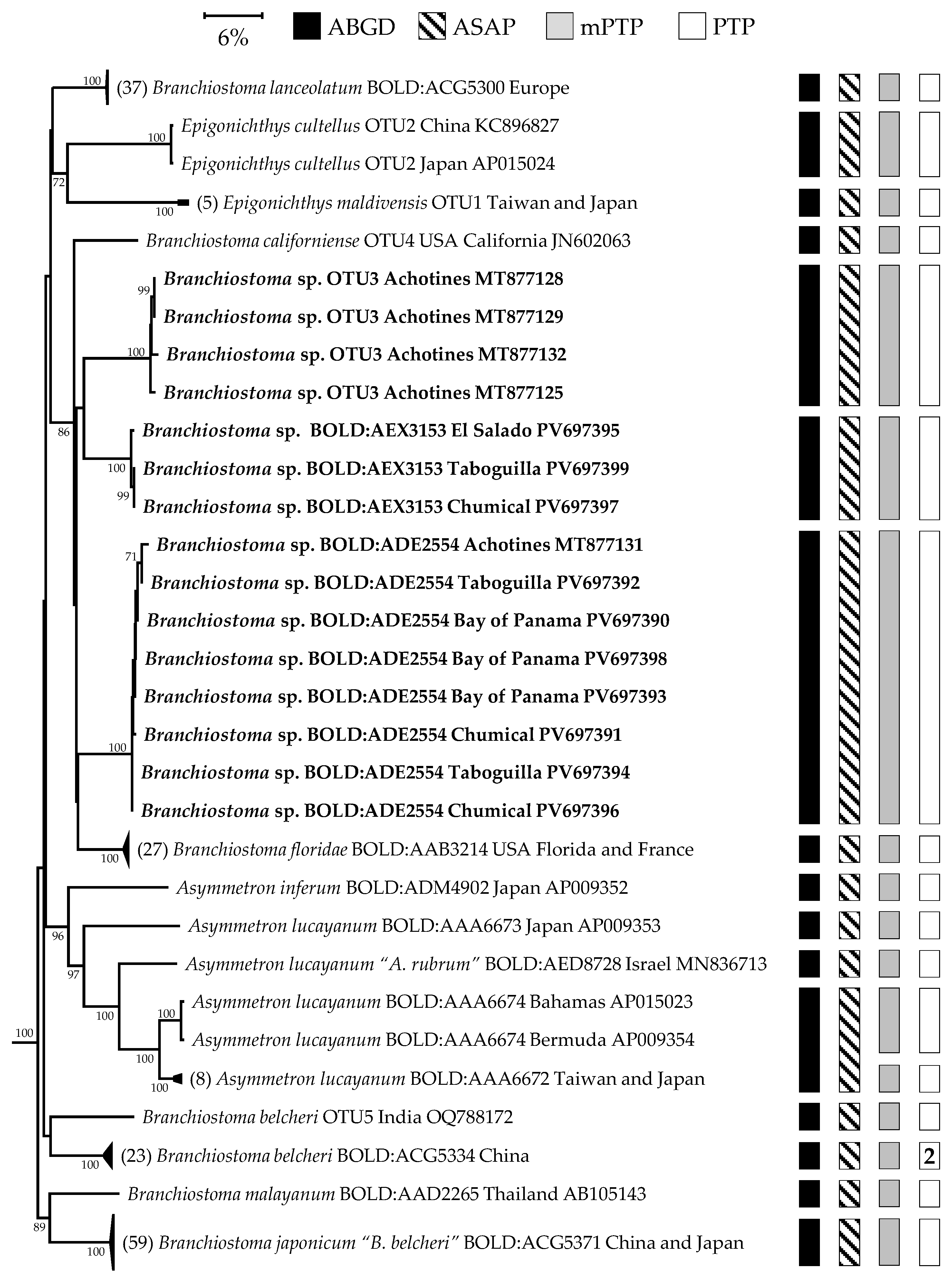
The three COI amphioxus sequences from California fell into their own mOTU (OTU4). BLAST results of these sequences showed a percentage identity of 82.6% to B. floridae from Florida. The nearest neighbor of this Californian BIN was another OTU from the Pacific coast of Panama (AEX3153) at 85.5% (Table 2, Figure 4). Unfortunately, these three samples did not have 16S sequences. A single 16S sequence (GenBank accession JN602063) of a Californian sample identified as B. californiense was obtained from the DNA project on marine fish fauna of California. This sequence showed the closest similarity to B. floridae from Florida at 88% (Table 2, Figure 5). The RAxML phylogenetic analysis of the concatenated COI + 16S dataset resolved the monophyletic clade Branchiostomatidae into two clades, with the genus Asymmetron sister to the Branchiostoma sp. and Epigonichthys sp. clades (100% Bootstrap BS, 1.00 posterior probability PP). The genus Branchiostoma was resolved as a fully supported clade that included all the sequences from the American specimens, confirming the identity of all Panamanian OTUs within this genus (100% BS, 1.00 PP) (Figure 6). The phylogeny based on COI and 16S combined for this genus was poorly resolved (Figure 6). Notable exceptions include the larvae from Bocas del Toro (BOLD:AEY2366, Caribbean), which were recovered as sister to one of the species from Achotines (OTU3), with strong support (92% BS, 0.98 PP; Figure 6), and the close relationship between B. floridae and B. virginiae, which form a fully supported clade (100% BS, 1.00 PP) (Figure 6). However, more data on B. virginiae from other genes are needed to assess the possibility of synonymy, as two distance-based species delimitation methods suggest that they may represent the same species, whereas two phylogeny-based methods split them into distinct species (Figure 4). All three species from the Pacific coast of Panama were resolved into distinct clades with full or very high support, none of which included B. californiense (Figure 6).

3.2. Morphological Descriptions of Panamanian Branchiostoma Species

Examination of the photographs from adult specimens clearly indicates that they have bilateral gonads (supporting the placement in the genus Branchiostoma). The buccal cirri are pinnate, with 9 to 20 cirri visible on each side (Figure 7B,E and Figure 8B).
Branchiostoma sp. BOLD:ADE2554 (Figure 7A–C) was examined based on measurements taken from a single adult individual (RCMicro 580). The myotome formula consists of 44 pre-atriopore, 16 atriopore-to-anus, and 10 post-anal myotomes, totaling 70. The specimen has 331 dorsal fin chambers and 54 preanal ventral fin chambers. There are 31 gonads on one side. The tallest dorsal fin chamber has a height-to-width ratio of 11, and the post-atriopore length is approximately 39.8% of the pre-atriopore length. The total body length is 3.30 cm, with a width of 0.35 cm.
Branchiostoma sp. BOLD:AEX3153 (Figure 7D–F) was examined based on measurements taken from a single adult individual (RCMicro 256). The myotome formula consists of 43 pre-atriopore, 13 atriopore-to-anus, and 10 postanal myotomes, totaling 66. The specimen has 351 dorsal fin chambers and 63 preanal ventral fin chambers. There are 27 gonads on one side. The tallest dorsal fin chamber has a height-to-body-width ratio of 8, and the post-atriopore length is approximately 23.1% of the pre-atriopore length. The total body length is 3.8 cm, with a width of 0.29 cm.
Branchiostoma sp. OTU3 (Figure 8A,B) was examined based on measurements taken from a single immature individual (GenBank accession MT877128). The myotome formula consists of 48 pre-atriopore, 16 atriopore-to-anus, and 9 postanal myotomes, totaling 73. The specimen has 453 dorsal fin chambers and 44 preanal ventral fin chambers. Gonads were not observed due to the immaturity status of the individual. The tallest dorsal fin chamber has a height-to-width ratio of 24, and the post-atriopore length is approximately 44.4% of the pre-atriopore length. Body length and width could not be measured due to the lack of a scale bar.
Examination showed that Ln stage larva [54] was typical of amphioxus larvae found in this study (Figure 7C,F and Figure 8C,D). We were able to make morphological descriptions for all the Branchiostoma mOTU from the Pacific coast. We were unable to obtain an adult specimen of the Caribbean mOTU (AEY2366) and, because of that, we only present the photographs of the Ln larvae stage with eight gill slits (Figure 8D).

4. Discussion

Our results suggest a surprising molecular diversity of Branchiostoma species in Panama, with the presence of three species of Branchiostoma on the Pacific coast of Panama and one in the Caribbean. The large distances between the clades (exceeding the barcode gap) and the high support of the clades in the combined phylogenetic analysis support the status of these as distinct species. The taxonomic identity of these species is more difficult to determine due to the limited number of samples in our morphological dataset. Traditional cephalochordate taxonomy relies on numerous morphometric measurements from hundreds of adult specimens to define species boundaries [19]. However, due to our limited sample size, we are not able to provide formal species descriptions for the newly discovered molecular operational taxonomic units.
Published reports of amphioxus from the Pacific coast usually indicate that B. californiense is the only amphioxus occurring in the region and that it ranges from California, through Mexico, El Salvador, Costa Rica and south to Panama [3,19,20,25,27,55]. None of our three mOTUs from the Pacific coast of Panama appear to belong to this species, as they are all only ~84–88% similar to the single partial (255 bp) 16S sequence available from B. californiense (GenBank: JN602063), and 83–85% similar to the three COI sequences of B. californiense from the BioGenome project (GenBank: PV366828, PV366829, PV366830) [18]. All four sequences of B. californiense were collected near the type locality of B. californiense from the shore of San Diego, California (Figure 2). In Figure 2 of their paper, Del Moral-Flores et al. [20], indicate the occurrence of Branchiostoma aff. californiense along the Pacific coast of Mexico, but the paper provides no further description of this putative taxon. The only other species reported from the region is Asymmetron lucayanum, which was observed in Cocos Island, Costa Rica [56]. This morphospecies has been shown to be comprised of at least 4 mOTUs based on mitogenomic data [16]. The sequences from Panama do not fall within any of these 4 mOTUs (Figure 4, Figure 5 and Figure 6), although no material from the Tropical Eastern Pacific has been sequenced so far (Figure 2). The collecting locality of the animals in OTU3 and ADE2554, near Achotines on the Azuero Peninsula, has a strong oceanic influence, and therefore may show some faunal similarities with Cocos Island. However, the photograph of the juvenile specimen MT877128 (Maikon pers. com.) does not show the extended tail typical of Asymmetron species, confirming that at least OTU3 does not belong to that genus. Finally, the only species reported from the South East Pacific are E. maldivensis from Rapa Nui [57] and B. elongatum from along the mainland coast of Chile and Peru [28] (Figure 1). There are no available sequences for B. elongatum to allow a comparison with our material and none of the Panamanian sequences were similar to those from E. maldivensis (Figure 4 and Figure 5).
To summarize, four names have been applied to animals from the eastern Pacific, based on morphological assessments, but none of these appear to correspond to the three mOTUs we found on the Pacific coast of Panama. Large sequence divergences suggest that these OTUs represent new species. This result is not surprising as vast numbers of tropical species from other taxonomic groups remain undiscovered and undescribed [58], and virtually all of the publications on amphioxus diversity and distributions from the Americas are based on morphological characteristics, which may often underestimate species diversity. The fact that three new mOTUs could be discovered from samples of only 15 animals and with little dedicated sampling effort, suggests that targeted sampling may result in detection of even higher diversity as less abundant species are detected. For example, Chen [59] showed that five species occur in Hong Kong; the three Branchiostoma species (B. belcheri, B. japonicum, and B. malayanum) are common, while only a very small number of two others (Asymmetron lucayanum and Epigonichthys culltellus) were detected after intensive sampling [59,60].
To further refine our understanding of the Tropical Eastern Pacific amphioxus species, it is essential to broaden our current sampling effort in the region. Results from this study suggest that efforts should be made to identify the southern limits of B. californiense, as researchers working in Central America should not automatically assume that animals collected on the Pacific coast belong to this species. In addition, to document the distributions of the new OTUs and to resolve their taxonomic status will require extensive sampling and sequencing efforts, and careful morphological descriptions and larger sample sizes. The inclusion of nuclear genetic markers would be likely to help better resolve the phylogeny of amphioxus species and evaluate the possibility of hybridization [61], a task beyond the scope of this study. Finally, the significant sequence divergences observed among our OTUs do not support the hypothesis that amphioxus species exhibit a notably slow rate of evolution [12,62]. Instead, the divergence levels observed here are consistent with those found in other marine taxa, with barcode gaps of approximately 5% [63,64].

Supplementary Materials

The following supporting information can be downloaded at: https://www.mdpi.com/article/10.3390/d17060411/s1, Table S1: DNA sequences from BOLD and GenBank used in all the analyses.

Author Contributions

Conceptualization, R.C.; methodology, R.C. and A.C.D.; software, M.E.M.C.; validation, all authors; formal analysis, M.E.M.C.; investigation, R.C., R.W., A.C.D., K.S.M.III and M.D.D.; resources, R.C., A.C.D. and K.S.M.III; data curation, M.E.M.C., R.C., A.C.D. and K.S.M.III; writing—original draft preparation, M.E.M.C. and R.C.; writing—review and editing, all authors; visualization, M.E.M.C.; supervision, R.C.; project administration, R.C.; funding acquisition, R.C. and A.C.D. All authors have read and agreed to the published version of the manuscript.

Funding

This research was supported by the Smithsonian Institution. Funding for collections in Achotines was supported by a Global Genome Initiative (GGI) award (Grant No. GGI-Rolling-2015-020) to Francesca Leasi.

Institutional Review Board Statement

Not applicable.

Data Availability Statement

COI and 16S dataset can be downloaded from the figshare dataset https://figshare.com/s/897b6beeaace21c57e50 (created on 14 May 2025).

Acknowledgments

We thank Tania Romero, Victor Lara and the participants of the 2013 Larval Invertebrate diversity, form, and function course for help in collecting and imaging animals and Linda Holland for providing helpful information about amphioxus life histories. This work was performed with permission from the Panamanian Ministry of the Environment (MiAmbiente), permit numbers SC/AP-5-15 and SEX/P-5879-16 (2016), and SEX/P-33-17, and from the ARAP Collecting permit No. 47 in 2013 and No. 06 in 2014 and export permit Nos. 37 and 80 (2013–2014).

Conflicts of Interest

The authors declare no conflicts of interest. The funders had no role in the design of the study; in the collection, analyses, or interpretation of data; in the writing of the manuscript; or in the decision to publish the results.

References

  1. Weng, Z.; Xie, Y.; Fang, Q.; Li, J.; Liu, J.; Wang, J.; Huang, L.; Qu, T.; Xie, W. Current Population, Habitat Status and Species of Amphioxus in Dongshan Bay, Fujian Province. J. Ocean. Univ. China 2025, 24, 417–426. [Google Scholar] [CrossRef]
  2. Barboza, C.A.D.M.; Hadlich, H.L.; Sandrini-Neto, L.; Martins, C.D.C.; Lana, P.D.C. Is the Distribution of the Lancelet Branchiostoma caribaeum Affected by Sewage Discharges? An Analysis at Multiple Scales of Variability. Mar. Pollut. Bull. 2013, 69, 178–188. [Google Scholar] [CrossRef] [PubMed]
  3. Campos-Dávila, L.; Pérez-Estrada, C.J.; Rodríguez-Estrella, R.; Morales-Bojórquez, E.; Brun-Murillo, F.G.; Balart, E.F. Seagrass Halodule wrightii as a New Habitat for the Amphioxus Branchiostoma californiense (Cephalochordata, Branchiostomidae) in the Southern Gulf of California, Mexico. ZooKeys 2019, 873, 113–131. [Google Scholar] [CrossRef]
  4. Kon, T.; Nohara, M.; Yamanoue, Y.; Fujiwara, Y.; Nishida, M.; Nishikawa, T. Phylogenetic Position of a Whale-Fall Lancelet (Cephalochordata) Inferred from Whole Mitochondrial Genome Sequences. BMC Evol. Biol. 2007, 7, 127. [Google Scholar] [CrossRef]
  5. Nishikawa, T. A New Deep-Water Lancelet (Cephalochordata) from off Cape Nomamisaki, SW Japan, with a Proposal of the Revised System Recovering the Genus Asymmetron. Zool. Sci. 2004, 21, 1131–1136. [Google Scholar] [CrossRef]
  6. Rodríguez-Uribe, M.C.; Chávez-Dagostino, R.M.; Del Moral-Flores, L.F.; Bravo-Olivas, M.L. First Record of Amphioxus Branchiostoma californiense (Amphioxiformes: Branchiostomatidae) Adjacent to a Shallow Submarine Hydrothermal System at Banderas Bay (Mexico). Diversity 2019, 11, 227. [Google Scholar] [CrossRef]
  7. Bertrand, S.; Escriva, H. Evolutionary Crossroads in Developmental Biology: Amphioxus. Development 2011, 138, 4819–4830. [Google Scholar] [CrossRef]
  8. Holland, L.Z.; Albalat, R.; Azumi, K.; Benito-Gutiérrez, È.; Blow, M.J.; Bronner-Fraser, M.; Brunet, F.; Butts, T.; Candiani, S.; Dishaw, L.J.; et al. The Amphioxus Genome Illuminates Vertebrate Origins and Cephalochordate Biology. Genome Res. 2008, 18, 1100–1111. [Google Scholar] [CrossRef]
  9. Holland, L.Z.; Holland, N.D. Cephalochordates: A Window into Vertebrate Origins. In Current Topics in Developmental Biology; Elsevier: Amsterdam, The Netherlands, 2021; Volume 141, pp. 119–147. ISBN 978-0-12-814968-3. [Google Scholar]
  10. Marlétaz, F.; Firbas, P.N.; Maeso, I.; Tena, J.J.; Bogdanovic, O.; Perry, M.; Wyatt, C.D.R.; De La Calle-Mustienes, E.; Bertrand, S.; Burguera, D.; et al. Amphioxus Functional Genomics and the Origins of Vertebrate Gene Regulation. Nature 2018, 564, 64–70. [Google Scholar] [CrossRef]
  11. Bi, C.; Lu, N.; Huang, Z.; Chen, J.; He, C.; Lu, Z. Whole-genome Resequencing Reveals the Pleistocene Temporal Dynamics of Branchiostoma belcheri and Branchiostoma floridae Populations. Ecol. Evol. 2020, 10, 8210–8224. [Google Scholar] [CrossRef]
  12. Igawa, T.; Nozawa, M.; Suzuki, D.G.; Reimer, J.D.; Morov, A.R.; Wang, Y.; Henmi, Y.; Yasui, K. Evolutionary History of the Extant Amphioxus Lineage with Shallow-Branching Diversification. Sci. Rep. 2017, 7, 1157. [Google Scholar] [CrossRef] [PubMed]
  13. Zhang, Q.-L.; Zhang, G.-L.; Yuan, M.-L.; Dong, Z.-X.; Li, H.-W.; Guo, J.; Wang, F.; Deng, X.-Y.; Chen, J.-Y.; Lin, L.-B. A Phylogenomic Framework and Divergence History of Cephalochordata Amphioxus. Front. Physiol. 2018, 9, 1833. [Google Scholar] [CrossRef] [PubMed]
  14. Caccavale, F.; Osca, D.; D’Aniello, S.; Crocetta, F. Molecular Taxonomy Confirms That the Northeastern Atlantic and Mediterranean Sea Harbor a Single Lancelet, Branchiostoma lanceolatum (Pallas, 1774) (Cephalochordata: Leptocardii: Branchiostomatidae). PLoS ONE 2021, 16, e0251358. [Google Scholar] [CrossRef]
  15. Kon, T.; Nohara, M.; Nishida, M.; Sterrer, W.; Nishikawa, T. Hidden Ancient Diversification in the Circumtropical Lancelet Asymmetron lucayanum Complex. Mar. Biol. 2006, 149, 875–883. [Google Scholar] [CrossRef]
  16. Subirana, L.; Farstey, V.; Bertrand, S.; Escriva, H. Asymmetron Lucayanum: How Many Species Are Valid? PLoS ONE 2020, 15, e0229119. [Google Scholar] [CrossRef]
  17. WoRMS Editorial Board World Register of Marine Species. VLIZ. Available online: https://www.marinespecies.org (accessed on 13 March 2025).
  18. Lewin, H.A.; Richards, S.; Lieberman Aiden, E.; Allende, M.L.; Archibald, J.M.; Bálint, M.; Barker, K.B.; Baumgartner, B.; Belov, K.; Bertorelle, G.; et al. The Earth BioGenome Project 2020: Starting the Clock. Proc. Natl. Acad. Sci. USA 2022, 119, e2115635118. [Google Scholar] [CrossRef]
  19. Poss, S.G.; Boschung, H.T. Lancelets (Cephalochordata: Branchiostomatidae): How Many Species Are Valid? Isr. J. Zool. 1996, 42, S13–S66. [Google Scholar]
  20. Del Moral-Flores, L.F.; Guadarrama-Martínez, M.Á.; Flores-Coto, C. Taxonomic Composition and Distribution of Cephalochordates (Cephalochordata: Amphioxiformes) from Mexico. Lat. Am. J. Aquat. Res. 2017, 44, 497–503. [Google Scholar] [CrossRef]
  21. Galván-Villa, C.M.; Ríos-Jara, E.; Ayón-Parente, M. New Records of the Californian Lancelet Branchiostoma Californiense (Cephalochordata: Branchiostomidae) from the Pacific Coast of Mexico. Rev. Mex. Biodivers. 2017, 88, 995–998. [Google Scholar] [CrossRef]
  22. Hubbs, C.L. A List of the Lancelets of the World with Diagnosis of Five New Species of Branchiostoma. In Occasional Papers of the Museum of Zoology; University of Michigan: Ann Arbor, MI, USA, 1922; Volume 105, pp. 1–16. [Google Scholar]
  23. Kirkaldy, T.W. A Revision of the Genera and Species of the Branchiostomidae. J. Cell Sci. 1895, 2, 303–324. [Google Scholar] [CrossRef]
  24. Global Biodiversity Information Facility GBIF. Occurrence Download 2025. The Dataset Includes 7838 Records from 269 Constituent Datasets. Available online: https://www.gbif.org/occurrence/download/0007176-250310093411724 (accessed on 13 March 2025).
  25. Barraza Sandoval, J.E.; Melara Pérez, V.E. New Records of Branchiostoma californiense (Amphioxiformes: Branchiostomatidae) in El Salvador. Cuad. Investig. UNED 2022, 14, e4302. [Google Scholar] [CrossRef]
  26. Mujica, A.; Nava, M.L.; Leiva-Dietz, F. On the Presence of Branchiostoma elongatum Juveniles (Cephalochordata: Branchiostomatidae) on the North-Central Coast of Chile. Lat. Am. J. Aquat. Res. 2022, 50, 622–627. [Google Scholar] [CrossRef]
  27. Velázquez-Velázquez, E. First Record of Branchiostoma californiense (Cephalochordata: Branchiostomatidae) in a Lagoon-Estuarine System from Chiapas, Mexico. Hidrobiológica 2021, 30, 107–110. [Google Scholar] [CrossRef]
  28. Vergara, M.; Oliva, M.E.; Riascos, J.M. Population Dynamics of the Amphioxus Branchiostoma elongatum from Northern Chile. J. Mar. Biol. Ass. 2012, 92, 591–599. [Google Scholar] [CrossRef]
  29. Collin, R.; Venera-Pontón, D.E.; Macdonald, K.; Driskell, A.C.; Boyle, M.J. Knots, Spoons, and Cloches: DNA Barcoding Unusual Larval Forms Helps Document the Diversity of Neotropical Marine Annelids. Invertebr. Biol. 2021, 140, e12311. [Google Scholar] [CrossRef]
  30. Collin, R.; Madrid, M.; Venera-Pontón, D.E.; Macdonald, K.S.; De León, A.; Vrdoljak, D.; Boyle, M.J.; Bryant, P.; Arehart, T.; Driskell, A.C. Diversity and Genetic Connectivity of Heteropod (Pterotracheoidea) Gastropods in the Tropical Eastern Pacific. Invertebr. Biol. 2023, 142, e12395. [Google Scholar] [CrossRef]
  31. Collin, R.; Venera-Pontón, D.E.; Paulay, G.; Boyle, M.J. World Travelers: DNA Barcoding Unmasks the Origin of Cloning Asteroid Larvae from the Caribbean. Biol. Bull. 2020, 239, 73–79. [Google Scholar] [CrossRef]
  32. Collin, R.; Venera-Pontón, D.E.; Driskell, A.C.; Macdonald, K.S.; Boyle, M.J. Unexpected Molecular and Morphological Diversity of Hemichordate Larvae from the Neotropics. Invertebr. Biol. 2019, 138, e12273. [Google Scholar] [CrossRef]
  33. Collin, R.; Venera-Pontón, D.E.; Driskell, A.C.; Macdonald, K.S.; Boyle, M.J. How I Wonder What You Are: Can DNA Barcoding Identify the Larval Asteroids of Panama? Invertebr. Biol. 2020, 139, e12303. [Google Scholar] [CrossRef]
  34. Collin, R.; Venera-Pontón, D.E.; Driskell, A.C.; Macdonald, K.S.; Chan, K.K.; Boyle, M.J. Documenting Neotropical Diversity of Phoronids with DNA Barcoding of Planktonic Larvae. Invertebr. Biol. 2019, 138, e12242. [Google Scholar] [CrossRef]
  35. Collin, R.; Venera-Pontón, D.E.; Driskell, A.C.; Macdonald, K.S.; Geyer, L.B.; Lessios, H.A.; Boyle, M.J. DNA Barcoding of Echinopluteus Larvae Uncovers Cryptic Diversity in Neotropical Echinoids. Invertebr. Biol. 2020, 139, e12292. [Google Scholar] [CrossRef]
  36. Urata, M.; Yamaguchi, N.; Henmi, Y.; Yasui, K. Larval Development of the Oriental Lancelet, Branchiostoma belcheri, in Laboratory Mass Culture. Zool. Sci. 2007, 24, 787–797. [Google Scholar] [CrossRef] [PubMed]
  37. Geller, J.; Meyer, C.; Parker, M.; Hawk, H. Redesign of PCR Primers for Mitochondrial Cytochrome c Oxidase Subunit I for Marine Invertebrates and Application in All-taxa Biotic Surveys. Mol. Ecol. Resour. 2013, 13, 851–861. [Google Scholar] [CrossRef] [PubMed]
  38. Meyer, C.P. Molecular Systematics of Cowries (Gastropoda: Cypraeidae) and Diversification Patterns in the Tropics. Biol. J. Linn. Soc. 2003, 79, 401–459. [Google Scholar] [CrossRef]
  39. Palumbi, S.R.; Benzie, J. Large Mitochondrial DNA Differences between Morphologically Similar Penaeid Shrimp. Mol. Mar. Biol. Biotechnol. 1991, 1, 27–34. [Google Scholar]
  40. Arnedo, M.A.; Oromí, P.; Ribera, C. Radiation of the Spider Genus Dysdera (Araneae, Dysderidae) in the Canary Islands: Cladistic Assessment Based on Multiple Data Sets. Cladistics 2001, 17, 313–353. [Google Scholar] [CrossRef]
  41. Schubart, C.D.; Cuesta, J.A.; Felder, D.L. Glyptograpsidae, a New Brachyuran Family from Central America: Larval and Adult Morphology, and a Molecular Phylogeny of the Grapsoidea. J. Crustac. Biol. 2002, 22, 28–44. [Google Scholar] [CrossRef]
  42. Sevigny, J.; Leasi, F.; Simpson, S.; Di Domenico, M.; Jörger, K.M.; Norenburg, J.L.; Thomas, W.K. Target Enrichment of Metazoan Mitochondrial DNA with Hybridization Capture Probes. Ecol. Indic. 2021, 121, 106973. [Google Scholar] [CrossRef]
  43. Katoh, K. MAFFT: A Novel Method for Rapid Multiple Sequence Alignment Based on Fast Fourier Transform. Nucleic Acids Res. 2002, 30, 3059–3066. [Google Scholar] [CrossRef]
  44. Katoh, K.; Standley, D.M. MAFFT Multiple Sequence Alignment Software Version 7: Improvements in Performance and Usability. Mol. Biol. Evol. 2013, 30, 772–780. [Google Scholar] [CrossRef]
  45. Kumar, S.; Stecher, G.; Suleski, M.; Sanderford, M.; Sharma, S.; Tamura, K. MEGA12: Molecular Evolutionary Genetic Analysis Version 12 for Adaptive and Green Computing. Mol. Biol. Evol. 2024, 41, msae263. [Google Scholar] [CrossRef] [PubMed]
  46. Ramirez, J.L.; Valdivia, P.; Rosas-Puchuri, U.; Valdivia, N.L. SPdel: A Pipeline to Compare and Visualize Species Delimitation Methods for Single-locus Datasets. Mol. Ecol. Resour. 2023, 23, 1959–1965. [Google Scholar] [CrossRef] [PubMed]
  47. Puillandre, N.; Lambert, A.; Brouillet, S.; Achaz, G. ABGD, Automatic Barcode Gap Discovery for Primary Species Delimitation. Mol. Ecol. 2012, 21, 1864–1877. [Google Scholar] [CrossRef]
  48. Puillandre, N.; Brouillet, S.; Achaz, G. ASAP: Assemble Species by Automatic Partitioning. Mol. Ecol. Resour. 2021, 21, 609–620. [Google Scholar] [CrossRef]
  49. Zhang, J.; Kapli, P.; Pavlidis, P.; Stamatakis, A. A General Species Delimitation Method with Applications to Phylogenetic Placements. Bioinformatics 2013, 29, 2869–2876. [Google Scholar] [CrossRef]
  50. Kapli, P.; Lutteropp, S.; Zhang, J.; Kobert, K.; Pavlidis, P.; Stamatakis, A.; Flouri, T. Multi-Rate Poisson Tree Processes for Single-Locus Species Delimitation under Maximum Likelihood and Markov Chain Monte Carlo. Bioinformatics 2017, 33, 1630–1638. [Google Scholar] [CrossRef]
  51. Stamatakis, A. RAxML Version 8: A Tool for Phylogenetic Analysis and Post-Analysis of Large Phylogenies. Bioinformatics 2014, 30, 1312–1313. [Google Scholar] [CrossRef]
  52. Lanfear, R.; Frandsen, P.B.; Wright, A.M.; Senfeld, T.; Calcott, B. PartitionFinder 2: New Methods for Selecting Partitioned Models of Evolution for Molecular and Morphological Phylogenetic Analyses. Mol. Biol. Evol. 2016, 34, 772–773. [Google Scholar] [CrossRef]
  53. Ronquist, F.; Teslenko, M.; Van Der Mark, P.; Ayres, D.L.; Darling, A.; Höhna, S.; Larget, B.; Liu, L.; Suchard, M.A.; Huelsenbeck, J.P. MrBayes 3.2: Efficient Bayesian Phylogenetic Inference and Model Choice across a Large Model Space. Syst. Biol. 2012, 61, 539–542. [Google Scholar] [CrossRef]
  54. Carvalho, J.E.; Lahaye, F.; Yong, L.W.; Croce, J.C.; Escrivá, H.; Yu, J.-K.; Schubert, M. An Updated Staging System for Cephalochordate Development: One Table Suits Them All. Front. Cell Dev. Biol. 2021, 9, 668006. [Google Scholar] [CrossRef]
  55. Vargas, J.A.; Dean, H.K. On Branchiostoma californiense (Cephalochordata) from the Gulf of Nicoya Estuary, Costa Rica. Rev. Biol. Tropical 2010, 58, 1143–1148. [Google Scholar] [CrossRef] [PubMed]
  56. Sibaja-Cordero, J.A.; Troncoso, J.S.; Cortés, J. The Lancelet Asymmetron lucayanum Complex in Cocos Island National Park, Pacific Costa Rica. Pac. Sci. 2012, 66, 523–528. [Google Scholar] [CrossRef]
  57. Meerhoff, E.; Veliz, D.; Vega-Retter, C.; Yannicelli, B. The Amphioxus Epigonichthys maldivensis (Forster Cooper, 1903) (Cephalochordata Branchiostomatidae) Larvae in the Plankton from Rapa Nui (Chile) and Ecological Implications. Biodivers. J. 2016, 7, 7–11. [Google Scholar]
  58. Ellison, C.I.; Frey, M.R.; Sanford, E.; Maslakova, S. Ribbon Worms (Phylum Nemertea) from Bodega Bay, California: A Largely Undescribed Diversity. ZooKeys 2024, 1204, 15–64. [Google Scholar] [CrossRef]
  59. Chen, Y. The Ecology and Biology of Amphioxus in Hong Kong. Ph.D. Thesis, Department of Biology and Chemistry, City University of Hong Kong, Hong Kong, China, 2007. [Google Scholar]
  60. Au, M.F.F.; Au, H.M.; Chu, W.K.V.; Kwok, C.K.; Cheung, S.G.; Leung, K.M.Y.; Qiu, J.-W. Spatial Distribution, Abundance, Seasonality and Environmental Relationship of Amphioxus in Subtropical Hong Kong Waters. Reg. Stud. Mar. Sci. 2023, 57, 102726. [Google Scholar] [CrossRef]
  61. Holland, N.D.; Holland, L.Z.; Heimberg, A. Hybrids Between the Florida Amphioxus (Branchiostoma floridae) and the Bahamas Lancelet (Asymmetron lucayanum): Developmental Morphology and Chromosome Counts. Biol. Bull. 2015, 228, 13–24. [Google Scholar] [CrossRef]
  62. Holland, N.D.; Holland, L.Z. The Ups and Downs of Amphioxus Biology: A History. Int. J. Dev. Biol. 2017, 61, 575–583. [Google Scholar] [CrossRef]
  63. Krug, P.J.; Vendetti, J.E.; Rodriguez, A.K.; Retana, J.N.; Hirano, Y.M.; Trowbridge, C.D. Integrative Species Delimitation in Photosynthetic Sea Slugs Reveals Twenty Candidate Species in Three Nominal Taxa Studied for Drug Discovery, Plastid Symbiosis or Biological Control. Mol. Phylogenet. Evol. 2013, 69, 1101–1119. [Google Scholar] [CrossRef]
  64. Krämer, D.; Schmidt, C.; Podsiadlowski, L.; Beckers, P.; Horn, L.; Von Döhren, J. Unravelling the Lineus ruber/Viridis Species Complex (Nemertea, Heteronemertea). Zool. Scr. 2017, 46, 111–126. [Google Scholar] [CrossRef]
Figure 1. Distribution map of the American Branchiostoma species from the Pacific coast. Distribution records obtained from www.GBIF.org/occurrence/download/0007176-250310093411724 (accessed on 13 March 2025) [24] and several regional articles [6,21,25,26,27,28]. Branchiostoma californiense type locality San Diego, California (32.646011, −117.237562). Branchiostoma elongatum type locality Chincha Island, Peru (−13.63328, −76.407). A list of all the coordinates used to create this map can be found in the figshare repository https://figshare.com/s/897b6beeaace21c57e50 (created on 14 May 2025).
Diversity 17 00411 g001
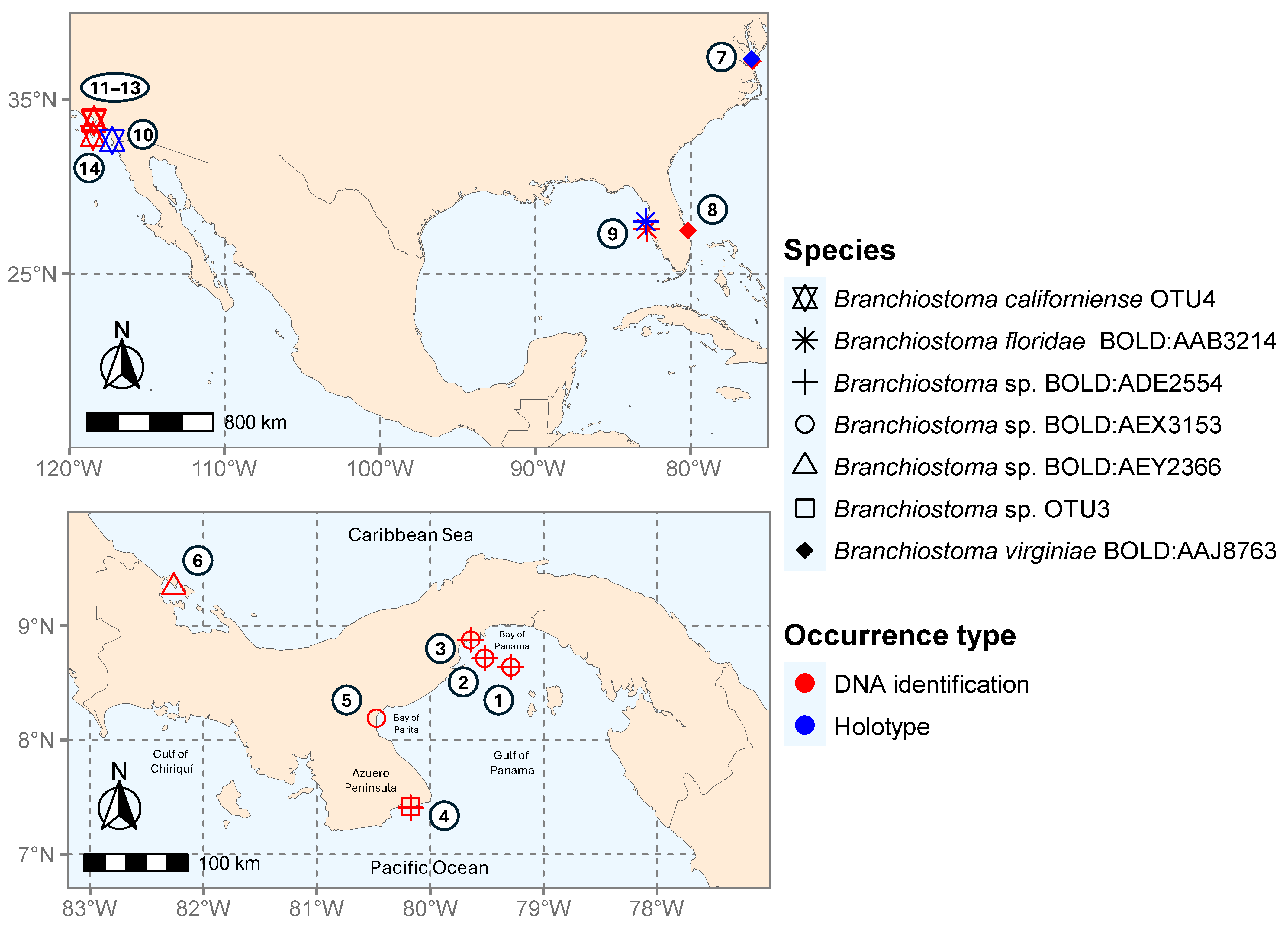
Figure 2. DNA verified distribution map of the American Branchiostoma species. The color of the points on the map represents the type of occurrence, while their shape indicates the different species. White circles with numbers indicate the collection sites: (1) Bay of Panama, (8.64, −79.29). (2) Southeast side of Taboguilla Island (8.80, −79.50). (3) Chumical (8.876, −79.644). (4) Achotines (7.41793, −80.17573). (5) El Salado (8.192, −80.475). (6) Bahía Almirante, Bocas del Toro, (9.34, −82.26). (7) Branchiostoma virginiae type locality Sewell’s Point, on Chesapeake Bay, Virginia (37.32732, −76.088638). (8) Fort Pierce, reef offshore, Florida (27.486, −80.185). (9) Branchiostoma floridae type locality Tampa Bay, by the Steamer Fish Hawk Sta. 7121, Florida (28.000915, −82.892243). (10) Branchiostoma californiense type locality San Diego, California (32.646011, −117.237562). (11) Redondo Beach, Los Angeles, California (33.837, −118.393). (12) Christmas tree cove, Los Angeles, California (33.76 −118.424). (13) Point Vicente, Los Angeles, California (33.741, −118.421) (14) San Clement Island, California (32.9, −118.483).
Diversity 17 00411 g002
Figure 3. (A) COI and (B) 16S DNA barcoding gap of Branchiostoma species from the Americas and their nearest neighbors. Species included in the analysis are detailed in Table 2.
Diversity 17 00411 g003
Figure 4. Phylogram illustrating the neighbor-joining cluster generated from 429 COI (1567 bp) sequences of Branchiostomatidae analyzed in this study. Sequences from Panama and California are highlighted in bold. Tip triangles represent collapsed clades from the same BOLD BIN, and the number in parentheses indicates the number of sequences in each collapsed clade. Bootstrap values on the branches represent support values greater than 70% from 10,000 replicates. The cluster scale represents pairwise distances calculated using the p-distance model. Branch tip labels indicate the species, BOLD BIN, locality, and GenBank accession numbers. The root (GenBank accessions: AF051097, U11880, Y16067) has been removed to aid visualization.
Diversity 17 00411 g004
Figure 5. Phylogram illustrating the neighbor-joining cluster generated from 187 16S rDNA (1447 bp) sequences of Branchiostomatidae analyzed in this study. Sequences from Panama and California are highlighted in bold. Diagram explanation is similar to that of Figure 4.
Diversity 17 00411 g005
Figure 6. RAxML phylogeny of Branchiostomatidae generated from a concatenated dataset of 16S + COI (3014 bp) sequences. Sequences from Panama and California are highlighted in bold. Values above or below the tree branches represent bootstrap support (BS) from the RAxML analysis and posterior probability (PP) from the Bayesian analysis, shown only when BS > 70 and PP > 0.90. An asterisk represents full support (100% BS, 1.00 PP). The tree scale indicates the percentage of base pair substitutions. Branch tip labels include species, BOLD BIN, locality, and 16S/COI GenBank accession numbers in between parenthesis. The tree root (GenBank accessions: AF051097, U11880, Y16067) has been removed to aid visualization.
Diversity 17 00411 g006
Figure 7. Branchiostoma adults and larvae from the Pacific coast of Panama. (A) Whole adult body and (B) magnified view of the anterior part from sample ABBAL1292-21, BIN BOLD:ADE2554. (C) Magnified view of the anterior part of post-metamorphic amphioxus ABBAK059-17, BIN BOLD:ADE2554. (D) Whole adult body and (E) magnified view of the anterior part from sample ABBAL968-21, BIN BOLD:AEX3153. (F) Magnified view of the anterior part of a pre-metamorphic larva from sample ABBAK058-17, BIN BOLD:AEX3153. Abbreviations: (a) anus, (ap) atriopore, (bc) buccal cirri, (cf) caudal fin, (df) dorsal fin, (n) notochord, (o) ovaries, (ps) pharyngeal gill slits, (r) rostrum, (vf) ventral fin, (vt) velar tentacle.
Diversity 17 00411 g007
Figure 8. Branchiostoma juvenile and larvae from the Pacific and Caribbean coasts of Panama belonging to BIN AEY2366. Juvenile (A) anterior and (B) posterior magnified view from sample MDD02-FG02-Achotines-D1 (GenBank accession MT877128), photos taken by Maikon Di Domenico. (C) Whole body, (D) anterior and (E) posterior part of a pre-metamorphic larvae from sample ABBAI111-15, BOLD:AEY2366. Abbreviations: (a) anus, (ap) atriopore, (bc) buccal cirri, (df) dorsal fin, (n) notochord, (r) rostrum, (vf) ventral fin.
Diversity 17 00411 g008
Table 1. Summary of material collected and sequenced from Panama (PA) and California (CA).
BOLD BIN#BOLD Process IDSample IDCOI16SLife StageLocality Number in Figure 2Collection DateDepth
PACIFIC
ADE2554ABBAK057-17RCMB0657PV711381PV697394Larva (w/photo)(2) SE side of Taboguilla Island PA18 November 2014Plankton tow
ABBAK059-17RCMB0659PV711378PV697392Larva (w/photo)(2) SE side of Taboguilla Island PA18 November 2014Plankton tow
ABBAI1544-16RCMB084PV711379PV697393Larva (w/photo)(1) Bay of Panama PA13 March 2014Plankton tow
ABBAI1545-16RCMB085PV711376PV697390Larva (1) Bay of Panama PA13 March 2014Plankton tow
ABBAI1546-16RCMB086PV711383PV697398Larva (1) Bay of Panama PA 13 March 2014Plankton tow
ABBAL1292-21RCMicro 580PV711377PV697391Adult (w/photo)(3) Chumical PA 10 March 2020Intertidal
ABBAL1297-21RCMicro 585PV711382PV697396Adult (3) Chumical PA10 March 2020Intertidal
LANGB001-22Achotines-F1MT877131MT877131Adult(4) Achotines PA [42]8 March 20161 m
AEX3153ABBAK058-17RCMB0658PV711384PV697399Larva (w/photo)(2) SE side of Taboguilla Island PA 18 November 2014Plankton tow
ABBAL1298-21RCMicro 586-PV697397Adult(3) Chumical PA10 March 2024Intertidal
ABBAL968-21RCMicro 256-PV697395Adult (w/photo)(5) El Salado PA14 January 2020Intertidal
OTU3LANGB005-22Achotines-E1MT877132MT877132Adult(4) Achotines PA [42]8 March 20161 m
LANGB004-22Achotines-H1MT877125MT877125Adult(4) Achotines PA [42]8 March 20161 m
LANGB003-22Achotines-D1MT877128MT877128Adult (w/photo)(4) Achotines PA [42]8 March 20161 m
LANGB002-22Achotines-G1MT877129MT877129Adult(4) Achotines PA [42]8 March 20161 m
OTU4-15904PV366828-Adult(11) Redondo Beach CA21 August 20199.75 m
-16987PV366829-Adult(12) Christmas Tree Cove CA 26 August 201920 m
-17911PV366830-Adult(13) Point Vicente CA26 August 201930 m
-SIO 10-94-JN602063Adult(14) San Clement Island CA, unpublished1 July 201022.86 m
CARIBBEAN
AEY2366ABBAI111-15CCLV062PV711380-Larva (w/photo)(4) Bahía Almirante PA1 July 2013Plankton tow
Bold GenBank accession numbers represent new sequences generated from this study.
Table 2. Pairwise distance summaries of the Branchiostoma BINs found in the Americas and its nearest neighbors. NA values indicate species for which only a single sequence is available, and no intraspecific distance could be calculated.
LociSpecies and BINMean
Intra. (%)
Maximum
Intra. (%)
Nearest NeighborMin. Dist.
to NN (%)
COIB. belcheri BOLD:ACG53340.853.65Branchiostoma sp. BOLD:AEX315313.08
B. californiense OTU40.510.76Branchiostoma sp. BOLD:AEX315314.5
B. floridae BOLD:AAB32141.513.17B. virginiae BOLD:AAJ87636.11
Branchiostoma sp. BOLD:AEX3153NANABranchiostoma sp. OTU310.99
Branchiostoma sp. BOLD:ADE25540.30.77Branchiostoma sp. BOLD:AEX315312.31
Branchiostoma sp. BOLD:AEY2366NANABranchiostoma sp. OTU312.06
Branchiostoma sp. OTU31.131.61Branchiostoma sp. BOLD:AEX315310.99
B. virginiae BOLD:AAJ87630.40.76B. floridae BOLD:AAB32146.11
16SB. floridae BOLD:AAB32140.942.03Branchiostoma sp. BOLD:ADE255410.99
Branchiostoma sp. BOLD:ADE25540.230.4B. floridae BOLD:AAB321410.99
Branchiostoma sp. BOLD:AEX31530.370.55B. floridae BOLD:AAB321411.44
Branchiostoma sp. OTU30.751.02Branchiostoma sp. BOLD:AEX315312.15
B. californiense OTU4NANAB. floridae BOLD:AAB321411.86
Disclaimer/Publisher’s Note: The statements, opinions and data contained in all publications are solely those of the individual author(s) and contributor(s) and not of MDPI and/or the editor(s). MDPI and/or the editor(s) disclaim responsibility for any injury to people or property resulting from any ideas, methods, instructions or products referred to in the content.