Article

Different Ribotypes of Akashiwo sanguinea Harbor Distinct Bacterial Communities in Their Phycospheres

1
CAS Key Laboratory of Marine Ecology and Environmental Sciences, Institute of Oceanology, Chinese Academy of Sciences, Qingdao 266071, China
2
Laboratory of Marine Ecology and Environmental Science, Qingdao National Laboratory for Marine Science and Technology, Qingdao 266237, China
3
Center for Ocean Mega-Science, Chinese Academy of Sciences, Qingdao 266071, China
4
University of Chinese Academy of Sciences, Beijing 100049, China
5
Qingdao Innovation and Development Center, Harbin Engineering University, Qingdao 266000, China
6
College of Fisheries, Guangdong Ocean University, Zhanjiang 524088, China
*
Author to whom correspondence should be addressed.
These authors contributed equally to this work.
Diversity 2025, 17(6), 400; https://doi.org/10.3390/d17060400
Submission received: 24 April 2025 / Revised: 26 May 2025 / Accepted: 4 June 2025 / Published: 5 June 2025

Abstract

:
The unarmored dinoflagellate Akashiwo sanguinea is a cosmopolitan harmful algal species known for forming intense blooms leading to mass mortality of fish, shellfish, and seabirds. Globally distributed populations of A. sanguinea have been classified into four ribotypes based on their characteristic sequences in LSU rRNA gene and primary geographic distributions. In this study, we compared the bacterial communities co-existing with the six strains of A. sanguinea from China and the USA (belonging to two ribotypes) using high-throughput sequencing of 16S rRNA gene amplicons. Generally, a bacterial microbiome with high diversity was found to be associated with laboratory-cultured A. sanguinea strains from different geographic origins. Based on ribotype classification, the six samples were divided into two groups (ribotype A: AsCHINA; ribotype C: AsUSA) for subsequent comparative analyses of their bacterial communities. Beta diversity analysis revealed a clear separation between the two groups, reflecting significant differences in bacterial community composition between the two ribotypes. Significantly higher abundance of nitrogen-fixing bacteria was found in the AsUSA group, suggesting that ribotype C may benefit from external nitrogen sources provided by their bacterial associates. If this also holds true in natural environments, this nitrogen-fixing partnership likely confers a competitive advantage to ribotype C in oligotrophic offshore waters, and potentially extends bloom duration when environmental nitrogen is depleted. Our study raised the possibility that different ribotypes of A. sanguinea may harbor distinct prokaryotic microbiomes in their phycospheres under stable cultivation conditions. Further comprehensive comparison among more isolates across all four ribotypes is highly necessary to validate this hypothesis.

1. Introduction

The unarmored dinoflagellate Akashiwo sanguinea (Hirasaka) G. Hansen et Moestrup, previously known as Gymnodinium sanguinea Hirasaka [1], is a cosmopolitan species commonly found in estuarine and coastal waters [2]. As a harmful algal bloom (HAB)-forming species, A. sanguinea blooms have been frequently reported around the world, with cell densities often exceeding 105 cells L−1 [3]. Since 1998, persistent blooms of this species have been observed along the coast of China, causing significant economic losses to local aquaculture industries [4]. Particularly, a devastating HAB event covering 1440 km2 occurred in Rongcheng offshore in Shandong Province, China, from late November to early December 2021, with A. sanguinea identified as one of the dominant causative species [5,6]. The bloom affected a wide area of coastal waters extending from west to east along the northern Shandong peninsula and had a devastating blow to the local kelp cultivation industry, which caused an estimated direct economic loss of CNY 2 billion [5]. Although no definitive evidence of toxin production by A. sanguinea has been documented, its blooms have been associated with mass mortality events of fish, shellfish, and seabirds [7,8,9]. Acute toxicity of A. sanguinea to finfish, shellfish, and zooplankton was also demonstrated in laboratory experiments [10]. Due to its frequent bloom events as well as the serious ecological damage and economic losses, A. sanguinea has garnered increasing attention globally.
Dinoflagellates grow in tight association with bacteria (heterotrophic bacteria, photosynthetic bacteria, e.g., cyanobacteria, etc.), which has led to a co-evolutionary relationship characterized by intricate interactions that not only modify their respective physiological behaviors but also significantly impact broader biogeochemical cycles [11,12]. These bacterial communities interact with dinoflagellates in diverse ways, such as nutrient exchange, secretion of pathogenic compounds, and participation in the synthesis of bioactive metabolites [12]. With the rising global incidence of dinoflagellate HABs in recent decades, the role of dinoflagellate–bacteria interactions has gained increasing attention due to accumulated evidence of linkages between bacterioplankton and the population dynamics of dinoflagellates in the blooming field [13,14,15]. The most common methods for studying dinoflagellate-associated bacteria include culture-dependent assessments and field-based environmental sampling [12]. Bacteria within certain lineages (e.g., Roseobacter within the phylum α-proteobacteria, Alteromonas within the phylum γ-proteobacteria, and Bacteroides within the phylum Bacteroidota) have been commonly found both in the phycospheres of laboratory-cultured A. sanguinea [11,16] and field samples during A. sanguinea blooms [15,17,18,19,20,21], suggesting that specific interactions could be established between A. sanguinea and particular groups of bacteria. However, in case studies on bacterial community dynamics during blooms, these interactions were often difficult to accurately assess, partly due to the difficulty in separating dinoflagellate-associated bacteria from free-living bacterioplankton [11]. Meanwhile, the relationships between phytoplankton and surrounding bacteria are intimate, complex, and dynamic [22]. Therefore, more intensive and detailed investigations using culture-dependent methods are also highly desirable, since this approach enables us to obtain critical insights into the interaction between host and specific microorganisms [12].
Cryptic species are genetically distinct but lack discernible morphological differences [23]. Cryptic species have been characterized within several dinoflagellate taxa, such as Scrippsiella acuminata (formerly Scrippsiella trochoidea) [24], Peridinium limbatum [25], Alexandrium minutum [26], Oxyrrhis marina [27], A. tamarense [28], Prorocentrum lima [29], Bysmatrum subsalsum [30], and Protoceratium reticulatum [31]. Many studies have also demonstrated that cryptic marine species exhibit distinct eco-physiological traits (e.g., optimal environmental requirements and/or nutrient utilization) [32,33]. For the species A. sanguinea, Tang and Gobler reported that its distinct ribotypes originated from different geographic regions [3]. Luo et al. further provided evidence for cryptic speciation among ribotypes in this species by integrating morphological, genetic, and eco-physiological analyses [34]. Based on characteristic sequences in LSU rRNA gene and primary geographic distributions, A. sanguinea has been proposed to be a species complex comprising at least four genetically distinct ribotypes which lack distinguishing morphological features [34]. Ribotype A has a global distribution, whereas ribotype B, with a more restricted range, has been recorded in the Pacific region, including Malaysia, Singapore, South Korea, Mexico, and China [34]. Ribotype C has been exclusively recorded on the east coast of the United States [3], while ribotype D appears restricted to the Mediterranean Sea [35]. Both field temporal distribution and laboratory eco-physiological experiments consistently showed that ribotype A is a winter ecotype preferring lower temperatures, while ribotype B is a summer ecotype adapting to higher temperatures, suggesting differentiated ecological niches between the two ribotypes [34]. Until now, bacterial microbiomes associated with different ribotypes of A. sanguinea remained underexplored.
In this study, we characterized the bacterial communities co-existing with the six strains of A. sanguinea from China and the USA using high-throughput sequencing of 16S rRNA gene amplicons. Based on ribotype classification, the six strains were divided into two groups for comparative analyses of their bacterial communities. The findings collectively suggested that the two ribotypes harbored distinct bacterial communities in their phycospheres characterized by different metabolic capabilities in nitrogen acquisition. Our study provided new insights into the potential interactions between bacterial communities and their dinoflagellate hosts of different ribotypes, as well as the possible contributions of these associations to the physiological functions of A. sanguinea.

2. Materials and Methods

2.1. Algal Cultures of Akashiwo sanguinea

A total of six strains of A. sanguinea were used in this study (Table 1). The strains ASND1 and ASND2 were isolated from Ningde, Fujian, China in 2016. The strain CCMA256 was isolated from the coastal water collected from Xiamen, Fujian, China in 2011. The other three strains were isolated from Northport Bay, New York, USA in 2011 [3]. The cultures were routinely maintained in f/2 medium without silicate [36] prepared with pre-filtered (0.22 μm membrane filter; Millipore, Billerica, MA, USA) and autoclaved (121 °C for 30 min) natural seawater with a salinity of approximately 32. Cultures were grown and maintained in an incubator (20 ± 1 °C; 12 h: 12 h light: dark cycle), with cool white fluorescent light providing a photon flux of ~100 μmol photons m−2 s−1. The stock cultures were maintained in the exponential growth stage by transferring into fresh f/2-Si medium bi-weekly.

2.2. Sample Collection and DNA Extraction

Cultures at the exponential growth phase were inoculated into six-well culture plates (Corning, Corning, NY, USA), each containing 10 mL of f/2-Si medium prepared with natural seawater. They were then incubated for 15 days under the routine culture conditions as mentioned above and collected at their stationary growth stage, as previously determined [37]. Each sample (approximately 104~105 cells) were pelleted in a 1.5 mL centrifuge tube and immediately used for genomic DNA isolation. Genomic DNA was extracted using the Plant DNA Extraction Kit (Tiangen, Beijing, China) and eluted with 50 μL TE buffer. Nuclear-free water processed through DNA extraction was used as sample blanks. The DNA quality and purity were estimated by NanoDropTM 1000 spectrophotometer (Thermo Fisher Scientific, Waltham, MA, USA), then stored at −80 °C for further PCR amplification.

2.3. Amplicon Sequencing of 28 and 16S rRNA Genes

The highly variable D2 domain and parts of the more conservative D1 and D3 domains of the eukaryotic 28S rRNA gene were amplified using a karyotic universal primer set of LSU335 (5′-ACCGATAGCA(G)AACAAGTA-3′) and LSU714 (5′-TCCTTGGTCCGTGTTTCA-3′), following the PCR protocol described by Chai et al. [38]. The V3-V4 variable region of the bacterial 16S rRNA gene was amplified using the primer set of 341F (5′-CCTACGGGNGGCWGCAG-3′) and 805R (5′-GACTACHVGGGTATCTAATCC-3′) [39], following the PCR protocol described by Deng et al. [11]. The 5′ ends of the primers were tagged with specific barcodes per sample and sequenced using universal primers. All PCR reactions were conducted in a 25 μL volume, including 12.5 μL of 2× Phusion® Hot Start Flex Master Mix, 2.5 μL of each primer (1 μM), and 50 ng of template DNA. Nuclease-free water served as negative control. The resulting amplicons were checked on an agarose gel electrophoresis and purified with the Gel Extraction Kit (Axygen Biosciences, USA). The size and quantity of the purified amplicon libraries were assessed on Agilent 2100 Bioanalyzer (Agilent, Santa Clara, CA, USA) and then pyrosequenced on the NovaSeq PE250 platform (LC-Bio Technology Company, Hangzhou, China).

2.4. Sequencing Data Processing and Bioinformatics Analysis

The sequencing primers were removed from de-multiplexed raw sequences using Cutadapt (version 1.9.1). Then, paired-end reads were merged using FLASH (version 1.2.8). The low-quality reads (quality scores < 20), short reads (<100 bp), and reads containing more than 5% “N” records were trimmed by using the sliding-window algorithm method in in Fqtrim (version 0.9.7). Chimeric sequences were further filtered using Vsearch program [40]. The DADA2 package [41] was applied for denoising and generating amplicon sequence variants (ASVs). The eukaryotic and bacterial ASVs were taxonomically classified against the NCBI GenBank and SILVA databases using the consensus BLAST (2.15.0) method implemented in the “feature-classifier” plugin of QIIME 2 [42]. Relative abundance of each ASV was estimated based on its read counts normalized to the total number of good quality reads. Alpha diversity indices (Shannon diversity, Simpson evenness, Chao1 richness, observed species richness, and Goods coverage) and beta diversity of principal component analysis (PCA) based on the Bray–Curtis distance were calculated via QIIME 2 plugin [42]. The significance of variance between or among samples was tested with one-way ANOVA or t-test using the software SPSS 22.0 (SAS Institute Inc., Cary, NC, USA). The significance level in all statistical analyses was set at 0.05 unless otherwise stated.

2.5. Functional Predictions of the Presented Bacterial Communities

Functional prediction of bacterial assemblages was explored from 16S rRNA gene-based microbial species compositions using the PICRUSt2 (Phylogenetic Investigation of Communities by Reconstruction of Unobserved States) algorithm [43] to make inferences from KEGG database [44]. The differentially abundant gene families and pathways were assessed using the software STAMP (v2.1.3) [45] subjected to t-test and Tukey–Kramer post hoc analysis with Benjamini–Hochberg FDR multiple comparison correction. A significant difference was inferred when p < 0.05.

2.6. Phylogenetic Analysis for the LSU rRNA Gene Sequences

The collected LSU rRNA gene sequences were aligned using MAFFT v7.475 with the default settings [46], and alignments were manually checked with BioEdit v7.2.5 [47]. Bayesian inference (BI) tree was constructed using the MrBayes v3.2.1 [48]. Posterior probability was estimated using four chains running 1,000,000 generations sampling every 100 generations. The first 25% sampled trees were considered burn-in trees and were discarded prior to constructing a 50% majority-rule consensus tree. FigTree (v1.4.4) was used to view and edit trees for publication.

3. Results

3.1. General Descriptions of Pyrosequencing the Eukaryotic 28S rRNA and the Prokaryotic 16S rRNA Gene Amplicons

The eukaryotic 28S rRNA gene pyrosequencing yielded 512,516 raw reads, with an average of 85,419 sequences per sample (Supplementary Table S1). The raw sequencing data were deposited in the NCBI Short Read Archive (SRA) database with the accession number PRJNA1131083. Good coverage value was 1.00 in all six samples (Supplementary Table S2), indicating the sequencing depth of these samples is sufficient to detect the characteristic diversity of eukaryotic taxa. A total of 498,192 clean reads were obtained, with effective sequences per sample varied from 81,785 to 84,235 (Supplementary Table S1). Dereplication using DADA2 plugin within the QIIME2 tool yielded 347 amplicon sequencing variants (ASVs) (Supplementary Table S3). Based on the annotations in the NCBI database, the ASVs assigned to “Akashiwo sanguinea” accounted for 98.33–99.93% of the total ASVs in all six samples (Supplementary Table S3), indicating the identity of all cultures used in our study as Akashiwo sanguinea.
The prokaryotic 16S rRNA gene sequencing generated 507,429 raw reads (BioProject: PRJNA771505), with sequences per sample ranging from 80,326 to 86,583 (Supplementary Table S1). Goods coverage of all samples were 1.00 (Supplementary Table S2), indicating sufficient sequences were harvested to reveal the majority of taxa in the prokaryotic assemblages. A total of 423,419 clean reads were obtained from the six samples (Supplementary Table S1). The dereplication using DADA2 plugin yielded 536 ASVs (Supplementary Table S3). All these ASVs were further annotated against the SILVA database.

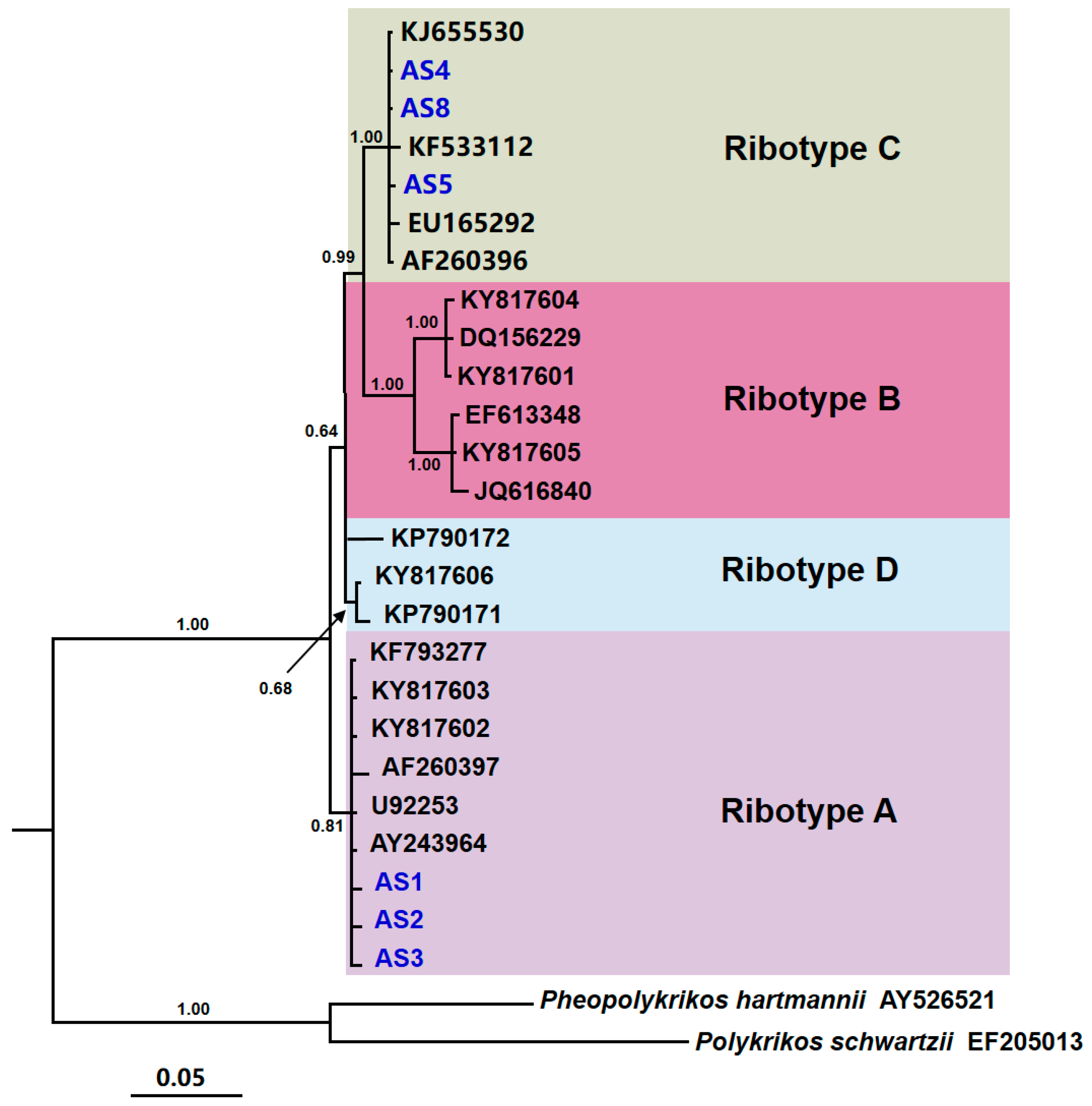
3.2. Phylogenetic Analysis

Four well-resolved ribotypes were recovered in phylogenetic reconstruction of Akashiwo sanguinea based on LSU rDNA sequences (Figure 1). The three newly sequenced Chinese strains (AS1, AS2, and AS3) clustered within the ribotype A clade, together with reference strains from China (KF793277, KY817603, KY817602), South Africa (AY243964), New Zealand (U92253), and Canada (AF260397) (Figure 1). In contrast, the three strains from USA (AS4, AS5, and AS8) clustered within the ribotype C with strong nodal support (BI = 1.00), which exclusively contained strains from the east coast of the United States (KJ655530, KF533112, EU165292, and AF260396) (Figure 1). Therefore, samples AS1, AS2, and AS3 were designated as group “AsCHINA”, while AS4, AS5, and AS8 were categorized as group “AsUSA” for subsequent comparative analyses of structural composition and functional predictions.

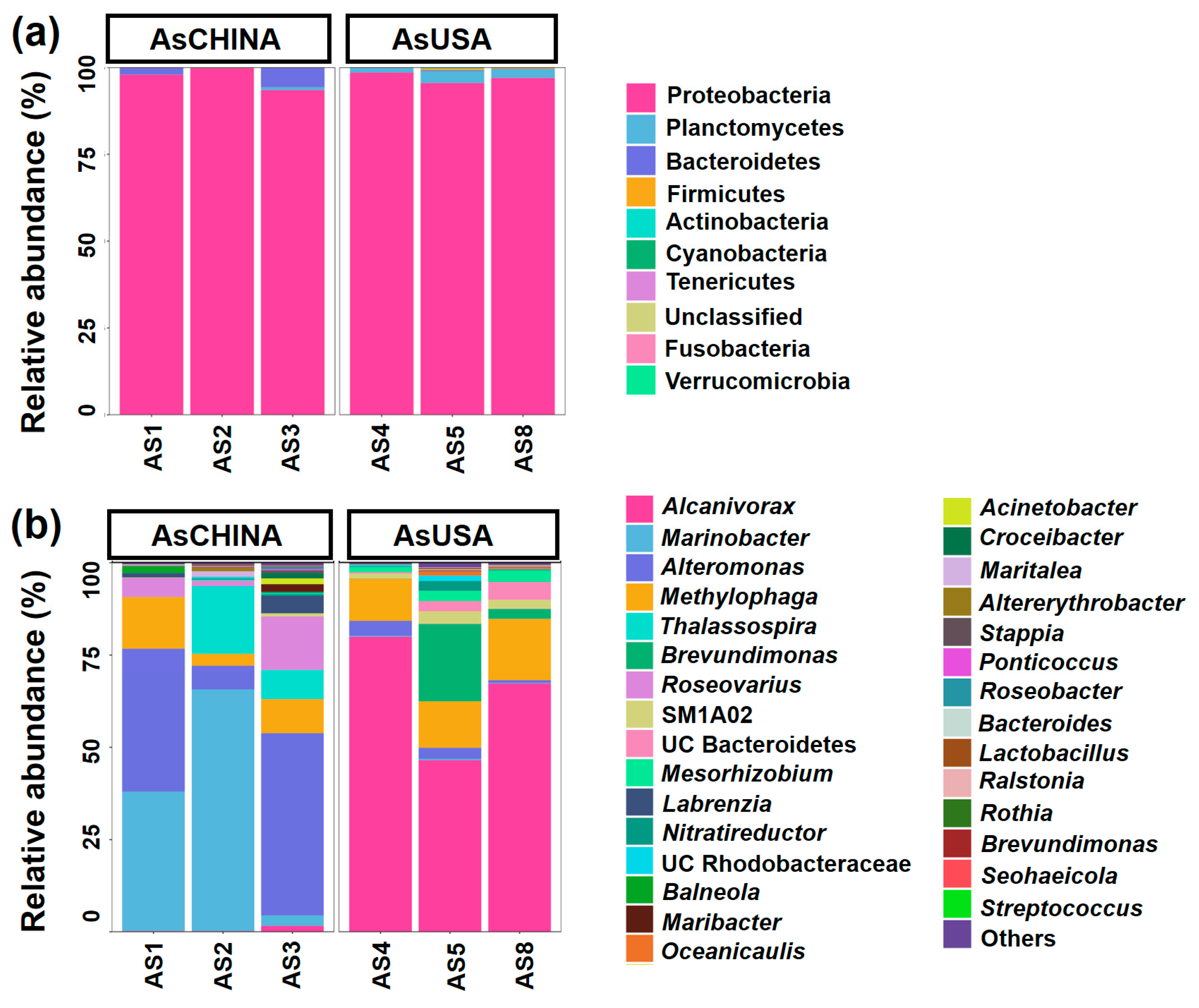
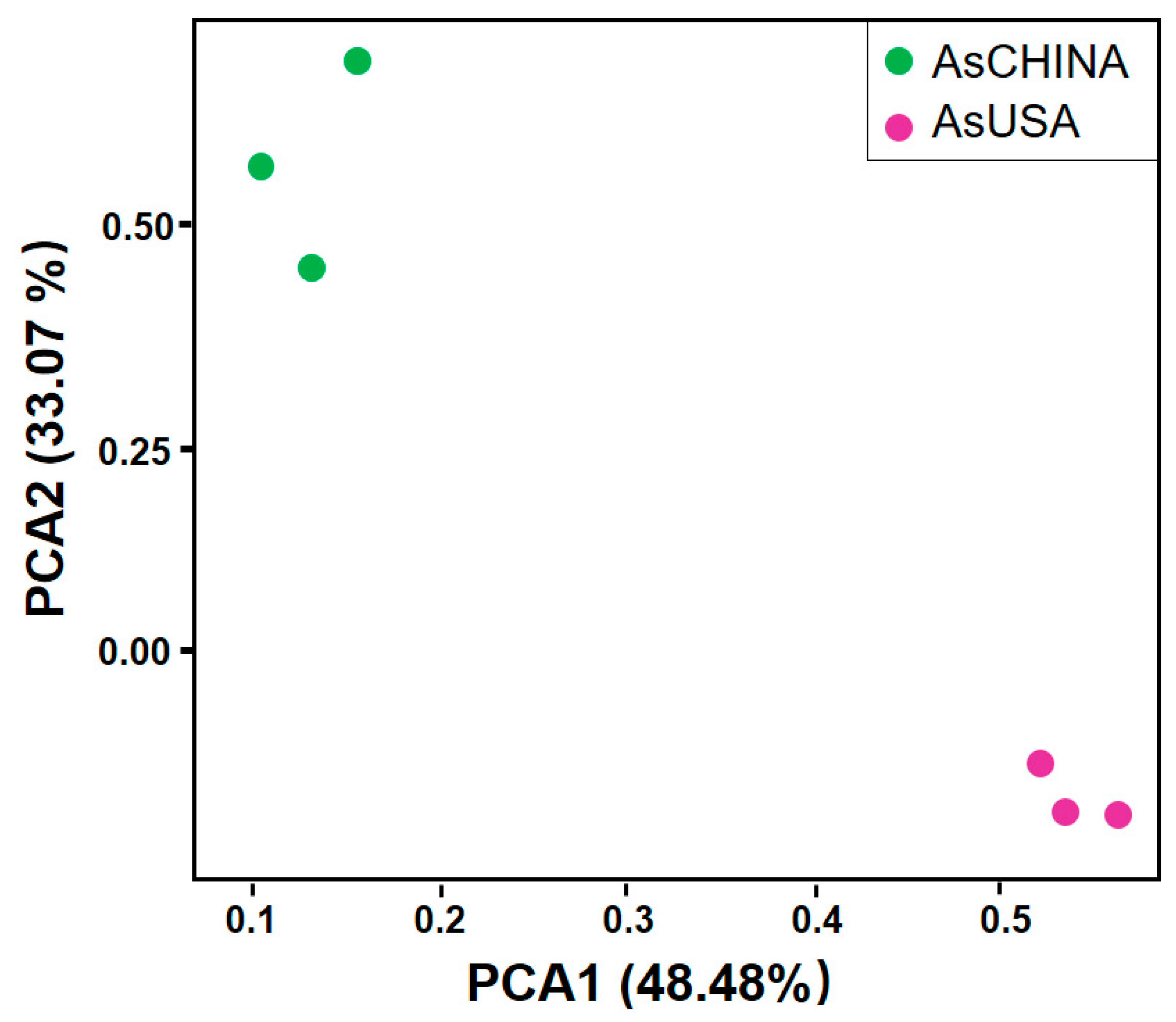
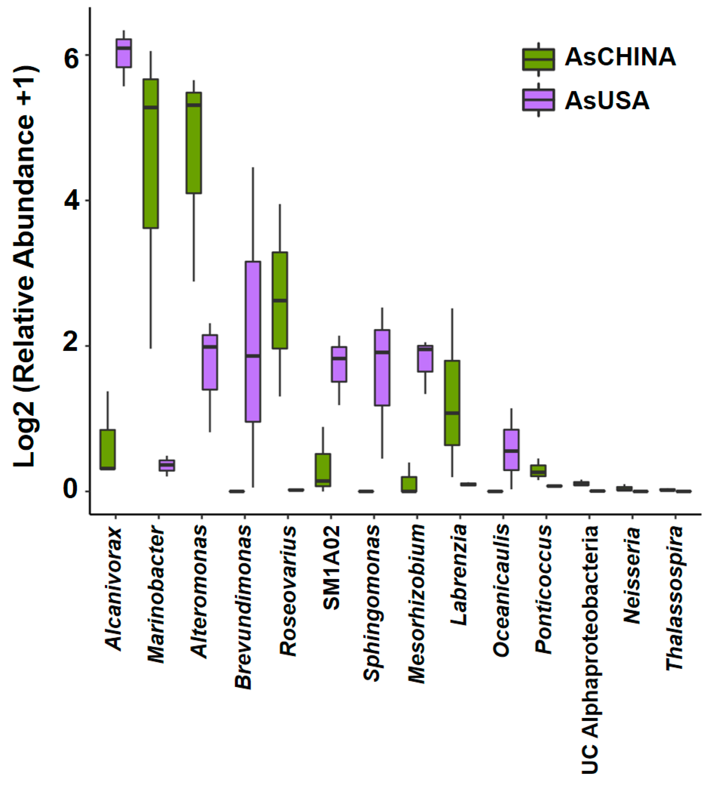
3.3. Comparative Analysis of ASV Diversity and Structural Composition of Bacterial Communities Between AsCHINA and AsUSA Groups

Based on the assignments in the SILVA database, the entire prokaryotic assemblage covered 12 phyla, 26 classes, 67 orders, 115 families, 164 genera, and 360 species. The number of recovered bacteria at the ASV level per sample varied from 65 to 133 (mean = 97) (Supplementary Table S3). Generally, Proteobacteria was the absolutely predominant phylum, accounting for 97.02% of all the ASVs, followed by Planctomycetes (1.37%), Bacteroidetes (1.30%), and Firmicutes (0.20%) (Figure 2a). At the genus level, Alcanivorax (32.65%), Marinobacter (17.84%), Alteromonas (17.03%), Methylophaga (11.17%), Thalassospira (4.40%), Roseovarius (3.52%), SM1A02 (3.94%), UC (unclassified) Bacteroidetes (1.37%), Mesorhizobium (1.32%), and Labrenzia (1.31%) were the most dominant genera (relative abundance > 1%), which together contributed up to 94.55% bacterial taxa for all samples analyzed (Figure 2b).
Comparing AsCHINA and AsUSA groups, no significant differences were detected in alpha diversity indices (including Shannon diversity, Simpson evenness, and Observed species; ANOVA, p > 0.05; Supplementary Figure S1a). Further beta diversity analysis was conducted to illustrate similarity and dissimilarity in ASVs complexity. PCA plot showed a distinct separation between the two groups (Figure 3). At the phylum level, while the two groups shared eight phyla in common, AsCHINA and AsUSA groups had one and three unique phyla, respectively (Supplementary Figure S1b). Significantly higher relative abundance of Planctomycetes was found in the AsUSA group than that in the AsCHINA group. At the class level, the two groups shared 13 common classes, while AsCHINA and AsUSA covered four and nine unique classes, respectively (Supplementary Figure S1b). At the genus level, 46 genera were shared by both groups, whereas 76 and 42 unique genera were present in AsUSA and AsCHINA groups, respectively (Supplementary Figure S1b). Compared with AsCHINA group, AsUSA was notably enriched with genera including Alcanivorax, Brevundimonas, SM1A02, Sphingomonas, Mesorhizobium, Oceanicaulis, but it had significantly lowered relative abundances of Marinobacter, Alteromonas, Roseovarius, Labrenzia, Ponticoccus, UC Alphaproteobacteria, Neisseria, Thalassospira (Figure 4).

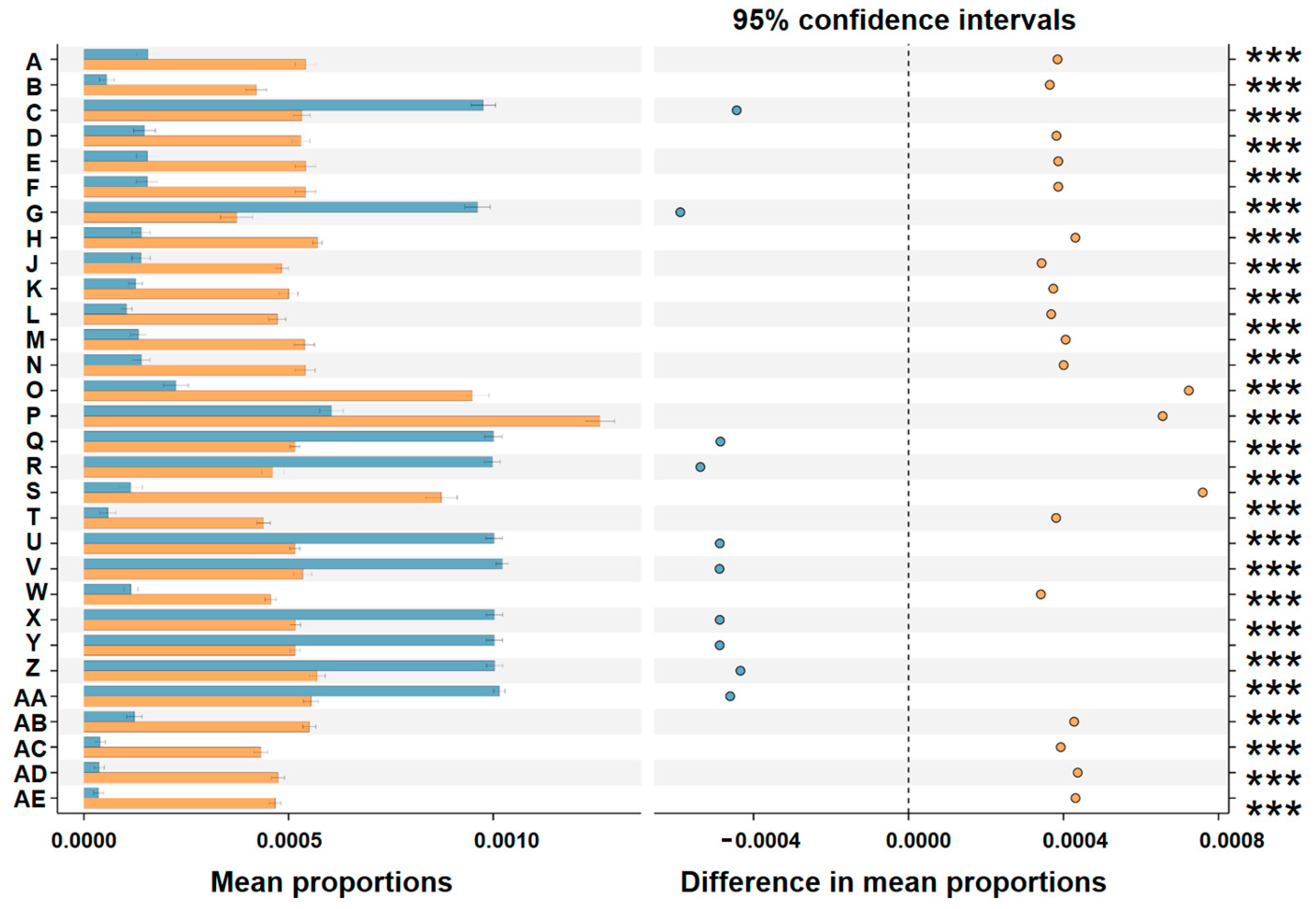
3.4. Comparative Analysis of Predicted Function of Bacterial Communities Between AsCHINA and AsUSA Groups

To assess the metabolic inferences of bacterial communities of AsCHINA and AsUSA groups, the functional repertoire was predicted using the PICRUSt algorithm. Generally, several differences in the metabolic potential of bacterial communities were predicted between the two groups. Three categories, namely genetic information processing, metabolism, and unclassified function, showed significantly higher abundances in the AsUSA group, while organismal systems, environmental information processing, and cellular processes were significantly predominant in the AsCHINA group (KEGG level 1; Supplementary Figure S2a). For the secondary functional modules (KEGG level 2), eight categories of functions (signal transduction, metabolism of other amino acids, transport and catabolism, amino acid metabolism, membrane transport, cell motility, glycan biosynthesis and metabolism, energy metabolism) were greatly enriched in the AsCHINA group. While the AsUSA group had significantly higher abundances in xenobiotics biodegradation and metabolism, metabolism, replication and repair, poorly characterized, carbohydrate metabolism, signal transduction, transcription, lipid metabolism, metabolism of cofactors and vitamins, transcription, and enzyme families (Supplementary Figure S2b). Differential function prediction at KO (KEGG Orthology) assignments also showed that ten entries annotated membrane protein K07058, phosphoglucomutase, DNA recombination protein RmuC, nitrogenase molybdenum-iron protein beta chain, UDP-glucose 6-dehydrogenase, isocitrate dehydrogenase, tRNA-uridine 2-sulfurtransferase, DNA processing protein, aspartate-semialdehyde dehydrogenase, and nitrogenase iron protein NifH were predominantly higher in the AsUSA group than those in the AsCHINA group (Figure 5).

4. Discussion

4.1. Highly Diverse Bacterial Communities Associated with Laboratory-Cultured A. sanguinea Strains from Different Geographic Origins

Compared with the microbiota which co-existed with sessile plants, the bacterial consortia associated with phytoplankton in aquatic habitats are particularly difficult to accurately define. This is largely because most microalgae are unicellular organisms living in highly dynamic and ever-changing environments [11,49]. Although previous studies have documented several common and/or important bacterial taxa interacting with some dinoflagellate hosts, the association of bacterial taxa with multiple strains of a specific dinoflagellate species, which could indicate “intimate” associations [49], has not yet been widely investigated [50,51,52]. In this study, six strains of A. sanguinea from distinct geographic origins were employed, and all these clonal cultures have been maintained under routine laboratory conditions for five to ten years. Through high-throughput metabarcoding of partial 16S rRNA gene amplicons, we detected 536 prokaryotic ASVs in the bacterial microbiome, spanning 12 phyla, 26 classes, 67 orders, 115 families, 164 genera, and 360 species. Compared with the extremely high microbial diversity usually found in terrestrial plant rhizospheres (e.g., rhizospheres typically harbor >103 bacterial taxa; [53] and references therein), lower microbial diversity is commonly observed in phycospheres surrounding microalgae [54], particularly in laboratory-cultured microalgae [55,56]. Generally, fewer than 30 bacterial associates on a species level were affiliated with these microalgae–bacteria biofilms ([54] and the references therein). Therefore, our results suggest that highly diverse bacterial communities were associated with laboratory-cultured A. sanguinea strains from different geographic origins. Given that the six clonal cultures were maintained for 4–9 years in our laboratory, the persistent bacterial associations observed in laboratory-cultured A. sanguinea strongly suggests mutualistic and/or commensal relationships between the algae and bacteria, which supports long-term stable coexistence rather than competitive exclusion.

4.2. Ribotype A and Ribotype C Harbor Distinct Bacterial Communities Characterized by Different Metabolic Capabilities in Nitrogen Acquisition

Previous phylogenetic analyses have clearly differentiated A. sanguinea populations of different geographic origins into four ribotypes [34]. Our phylogenetic analyses confirmed that the three Chinese isolates belong to ribotype A, whereas the three strains from USA belong to ribotype C. According to ribotype classification, the six samples were divided into two groups (ribotype A: AsCHINA; ribotype C: AsUSA) for subsequent comparative analyses of their bacterial communities. Although no significant differences were found in alpha diversity, beta diversity analysis revealed a clear separation between the two groups, reflecting significant differences in bacterial community composition between the two ribotypes. Only 46 genera were shared by both groups, whereas 76 and 42 unique genera were present in AsUSA and AsCHINA groups, respectively. A total of six bacterial genera exhibited significantly higher abundances in the AsUSA group, in which three genera (Brevundimonas, Sphingomonas, Mesorhizobium) have been previously reported to have nitrogen-fixing ability. Brevundimonas sp. with nitrogen-fixing potential has been used as plant growth-promoting bacteria to enhance plant growth by supplying nitrogen [57]. The genus Sphingomonas comprises metabolically versatile members, with some species capable of nitrogen fixation [58,59]. Mesorhizobium, a genus best known for its nitrogen-fixing symbiosis with terrestrial legumes [60], has also been found in association with the marine polychaete Meganerilla bactericola [61]. Consistently, the differential function prediction based on KO (KEGG Orthology) also showed that two entries annotated as components of the nitrogenase enzyme system (nitrogenase molybdenum-iron protein beta chain [EC:1.18.6.1], nitrogenase iron protein NifH [EC:1.18.6.1]) responsible for biological nitrogen fixation [62,63] were significantly more abundant in the AsUSA group. These results collectively suggest that ribotype A and ribotype C harbored distinct bacterial communities, which exhibited different metabolic capabilities in nitrogen acquisition.
Biological nitrogen fixation, the metabolic conversion of atmospheric nitrogen (N2) into ammonia (NH3), is performed exclusively by certain bacteria and archaea [64]. It has a long evolutionary history and makes a significant contribution to the amount of “new” nitrogen available in a wide variety of marine habitats [64,65,66]. Prokaryotes involved in nitrogen fixation and other nitrogen transformations have been documented in association with a range of eukaryotic hosts in the marine environment, such as corals, polychaetes, sponges, microalgae, and shipworms [61,65,67]. These associations range from temporary, non-specific external associations to permanent intracellular endosymbioses [65]. Synergistic nitrogen utilization between dinoflagellates and associated bacteria has been documented in multiple species. A total of 18 dinoflagellate species were observed to feed on the nitrogen-fixing Synechococcus spp., implying that mixotrophic dinoflagellates may meet their nitrogen requirements through cyanobacterial ingestion [68]. Further evidence also confirmed that nitrogen-fixing bacteria can form either endosymbiotic or ectosymbiotic relationships with their dinoflagellate hosts [69,70,71]. The association with nitrogen-fixing bacteria provides dinoflagellates with a means to acquire nitrogen, especially under nitrogen-limited conditions [12,68,70,71]. Here, higher abundance of bacteria with nitrogen-fixing potential was found in the AsUSA group, suggesting that ribotype C may benefit from external nitrogen sources provided by their bacterial associates. If this also holds true in natural environments, this nitrogen-fixing partnership likely confers a competitive advantage to ribotype C in oligotrophic offshore waters, and potentially extends bloom duration when environmental nitrogen is depleted. In this study, the differential taxonomic and functional composition of bacterial communities associated with six strains of laboratory-raised A. sanguinea raised the possibility that different ribotypes of A. sanguinea may harbor distinct prokaryotic microbiomes in their phycospheres under stable cultivation conditions. Due to the limited number of strains examined in this study, comprehensive comparison among more isolates across all four ribotypes is required to further validate this hypothesis in the future.

Supplementary Materials

The following supporting information can be downloaded at: https://www.mdpi.com/article/10.3390/d17060400/s1, Table S1: The statistics of generated sequences data of eukaryotic 28S rRNA gene and prokaryotic 16S rRNA gene amplicons sequencing; Table S2: Alpha diversity indices of the 6 samples; Table S3: Annotation and statistics of different classification levels; Figure S1: Alpha diversity analysis and Venn diagrams of AsCHINA and AsUSA groups; Figure S2: Prediction of the differential function of bacterial communities between AsCHINA and AsUSA groups in KEGG categories.

Author Contributions

Conceptualization, Y.Z.T. and Y.D.; methodology, F.L. and Z.H.; software, J.L.; validation, L.S.; formal analysis, H.Z.; investigation, F.L.; resources, Y.D.; data curation, H.Z.; writing—original draft preparation, H.Z. and F.L.; writing—review and editing, Y.D. and Y.Z.T.; visualization, H.Z. and F.L.; supervision, Y.D.; project administration, Y.D. and Y.Z.T.; funding acquisition, Y.D. and L.S. All authors have read and agreed to the published version of the manuscript.

Funding

This research was financially supported by the National Science Foundation of China (Grant No. 42376138), the Open Research Fund Program of the Key Laboratory of Marine Ecosystem Dynamics, Ministry of Natural Resources (MED202304), and the Shandong Provincial Natural Science Foundation (ZR2021QD025).

Institutional Review Board Statement

Not applicable.

Data Availability Statement

The data is contained within the article and Supplementary Material.

Acknowledgments

We would like to express our gratitude to the anonymous reviewers for their constructive suggestions and comments. We acknowledge financial support from the National Science Foundation of China (Grant No. 42376138), the Open Research Fund Program of the Key Laboratory of Marine Ecosystem Dynamics, Ministry of Natural Resources (MED202304), and the Shandong Provincial Natural Science Foundation (ZR2021QD025).

Conflicts of Interest

The authors declare no conflicts of interest.

Abbreviations

The following abbreviations are used in this manuscript:
HABHarmful algal bloom
ASVsAmplicon sequence variants
PCAPrincipal component analysis
BIBayesian inference

References

  1. Daugbjerg, N.; Hansen, G.; Larsen, J.; Moestrup, O. Phylogeny of some of the major genera of dinoflagellates based on ultrastructure and partial LSU rDNA sequence data, including the erection of three new genera of unarmoured dinoflagellates. Phycologia 2000, 39, 302–317. [Google Scholar] [CrossRef]
  2. Steidinger, K.A.; Tangen, K. Dinoflagellates. In Identifying Marine Diatoms and Dinoflagellates; Tomas, C.R., Ed.; Academic Press: New York, NY, USA, 1996; pp. 387–598. [Google Scholar]
  3. Tang, Y.Z.; Gobler, C.J. Sexual resting cyst production by the dinoflagellate Akashiwo sanguinea: A potential mechanism contributing to the ubiquitous distribution of a harmful alga. J. Phycol. 2015, 51, 298–309. [Google Scholar] [CrossRef] [PubMed]
  4. Chen, B.; Kang, W.; Hui, L. Akashiwo sanguinea blooms in Chinese waters in 1998–2017. Mar. Pollut. Bull. 2019, 149, 110652. [Google Scholar] [CrossRef]
  5. Li, X.D.; Su, L.; Li, X.J.; Li, J.; Xu, Y.J.; Chang, L.R.; Yu, R.C.; Yang, D.Z.; Pang, S.J. Comprehensive analysis of large-scale Saccharina japonica damage in the principal farming area of Rongcheng in Shandong Province from 2021 to 2022. J. Agric. Sci. Technol. 2023, 25, 206–222. [Google Scholar]
  6. Geng, H.X.; Kong, F.Z.; Wang, J.X.; Zhang, Q.C.; Li, F.; Hong, X.; Song, M.J.; Lian, Z.R.; Cai, Y.L.; Yu, R.C. An unusual winter bloom of dinoflagellates with notable damage to kelp cultivation around Shandong peninsula, China. Mar. Environ. Res. 2024, 201, 106687. [Google Scholar] [CrossRef]
  7. Horner, R.A.; Garrison, D.L.; Plumley, F.G. Harmful algal blooms and red tide problems on the US west coast. Limnol. Oceanogr. 1997, 42, 1076–1088. [Google Scholar] [CrossRef]
  8. Matsubara, T.; Nagasoe, S.; Yamasaki, Y.; Shikata, T.; Shimasaki, Y.; Oshima, Y.; Honjo, T. Effects of temperature, salinity, and irradiance on the growth of the dinoflagellate Akashiwo sanguinea. J. Exp. Mar. Biol. Ecol. 2007, 342, 226–230. [Google Scholar] [CrossRef]
  9. Jessup, D.A.; Miller, M.A.; Ryan, J.P.; Nevins, H.M.; Kerkering, H.A.; Mekebri, A.; Crane, D.B.; Johnson, T.A.; Kudela, R.M. Mass stranding of marine birds caused by a surfactant-producing red tide. PLoS ONE 2009, 4, e4550. [Google Scholar] [CrossRef]
  10. Xu, N.; Wang, M.; Tang, Y.; Zhang, Q.; Duan, S.; Gobler, C.J. Acute toxicity of the cosmopolitan bloom-forming dinoflagellate Akashiwo sanguinea to finfish, shellfish, and zooplankton. Aquat. Microb. Ecol. 2017, 80, 209–222. [Google Scholar] [CrossRef]
  11. Deng, Y.Y.; Wang, K.; Hu, Z.X.; Tang, Y.Z. Abundant species diversity and essential functions of bacterial communities associated with dinoflagellates as revealed from metabarcoding sequencing for laboratory-raised clonal cultures. Int. J. Environ. Res. Public Health 2022, 19, 4446. [Google Scholar] [CrossRef]
  12. Yang, X.; Liu, Z.; Zhang, Y.; Shi, X.; Wu, Z. Dinoflagellate–Bacteria Interactions: Physiology, Ecology, and Evolution. Biology 2024, 13, 579. [Google Scholar] [CrossRef] [PubMed]
  13. Zhou, J.; Chen, G.F.; Ying, K.Z.; Jin, H.; Song, J.T.; Cai, Z.H. Phycosphere microbial succession patterns and assembly mechanisms in a marine dinoflagellate bloom. Appl. Environ. Microbiol. 2019, 85, e00349-19. [Google Scholar] [CrossRef] [PubMed]
  14. Patin, N.V.; Brown, E.; Chebli, G.; Garfield, C.; Kubanek, J.; Stewart, F.J. Microbial and chemical dynamics of a toxic dinoflagellate bloom. Peer J. 2020, 8, e9493. [Google Scholar] [CrossRef] [PubMed]
  15. Hu, J.M.; Camerón, H.; Rilling, J.I.; Campos, M.; Ruiz-Gil, T.; Gonzalez, M.A.; Gajardo, G.; Vergara, K.; Guzmán, L.; Espinoza-González, O.; et al. Differentiation of microbial communities in coastal seawater before and during an Akashiwo sanguinea (Dinophyceae) bloom in the urban area of Antofagasta city (northern Chile). Harmful Algae 2025, 142, 102782. [Google Scholar] [CrossRef]
  16. Li, S.F.; Chen, M.C.; Chen, Y.F.; Tong, J.; Wang, L.Y.; Xu, Y.; Hu, Z.L.; Chen, H. Epibiotic bacterial community composition in red-tide dinoflagellate Akashiwo sanguinea culture under various growth conditions. FEMS Microbiol. Ecol. 2019, 95, fiz057. [Google Scholar] [CrossRef]
  17. Yang, C.Y.; Li, Y.; Zhou, Y.Y.; Zheng, W.; Tian, Y.; Zheng, T.L. Bacterial community dynamics during a bloom caused by Akashiwo sanguinea in the Xiamen sea area, China. Harmful Algae 2012, 20, 132–141. [Google Scholar] [CrossRef]
  18. Yang, C.Y.; Li, Y.; Zhou, B.; Zhou, Y.Y.; Zheng, W.; Tian, Y.; Van Nostrand, J.D.; Wu, L.Y.; He, Z.L.; Zhou, J.Z.; et al. Illumina sequencing-based analysis of free-living bacterial community dynamics during an Akashiwo sanguinea bloom in Xiamen sea, China. Sci. Rep. 2015, 5, 8476. [Google Scholar]
  19. Yang, C.Y.; Li, Y.; Zhou, Y.Y.; Lei, X.Q.; Zheng, W.; Tian, Y.; Van Nostrand, J.D.; He, Z.L.; Wu, L.Y.; Zhou, J.Z.; et al. A comprehensive insight into functional profiles of free-living microbial community responses to a toxic Akashiwo sanguinea bloom. Sci. Rep. 2016, 6, 34645. [Google Scholar] [CrossRef]
  20. Chen, H.R.; Jiang, J.J.; Jiang, F.J.; Li, S.F.; Hu, Z.L. Temporal variability of free-living microbial culturability and community composition after an Akashiwo sanguinea bloom in Shenzhen, China. Ecotoxicology 2021, 30, 975–985. [Google Scholar] [CrossRef]
  21. Kang, J.; Park, J.S.; Jung, S.W.; Kim, H.J.; Joo, H.M.; Kang, D.; Seo, H.; Kim, S.; Jang, M.C.; Lee, K.W.; et al. Zooming on dynamics of marine microbial communities in the phycosphere of Akashiwo sanguinea (Dinophyta) blooms. Mol. Ecol. 2021, 30, 207–221. [Google Scholar] [CrossRef]
  22. Cirri, E.; Pohnert, G. Algae-bacteria interactions that balance the planktonic microbiome. New Phytol. 2019, 223, 100–106. [Google Scholar] [CrossRef] [PubMed]
  23. Bickford, D.; Lohman, D.J.; Sodhi, N.S.; Ng, P.K.L.; Meier, R.; Winker, K.; Ingram, K.K.; Das, I. Cryptic species as a window on diversity and conservation. Trends Ecol. Evol. 2007, 22, 148–155. [Google Scholar] [CrossRef] [PubMed]
  24. Montresor, M.; Sgrosso, S.; Procaccini, G.; Kooistra, W.H. Intraspecific diversity in Scrippsiella trochoidea (Dinopbyceae): Evidence for cryptic species. Phycologia 2003, 42, 56–70. [Google Scholar] [CrossRef]
  25. Kim, E.; Wilcox, L.; Graham, L.; Graham, J. Genetically distinct populations of the dinoflagellate Peridinium limbatum in neighboring Northern Wisconsin lakes. Microb. Ecol. 2004, 48, 521–527. [Google Scholar] [CrossRef]
  26. Lilly, E.L.; Halanych, K.M.; Anderson, D.M. Phylogeny, biogeography, and species boundaries within the Alexandrium minutum group. Harmful Algae 2005, 4, 1004–1020. [Google Scholar] [CrossRef]
  27. Lowe, C.D.; Montagnes, D.J.; Martin, L.E.; Watts, P.C. High genetic diversity and fine-scale spatial structure in the marine flagellate Oxyrrhis marina (Dinophyceae) uncovered by microsatellite loci. PLoS ONE 2010, 5, e15557. [Google Scholar] [CrossRef]
  28. Genovesi, B.; Shin-Grzebyk, M.S.; Grzebyk, D.; Laabir, M.; Gagnaire, P.A.; Vaquer, A.; Pastoureaud, A.; Lasserre, B.; Collos, Y.; Berrebi, P. Assessment of cryptic species diversity within blooms and cyst bank of the Alexandrium tamarense complex (Dinophyceae) in a Mediterranean lagoon facilitated by semimultiplex PCR. J. Plankton Res. 2011, 33, 405–414. [Google Scholar] [CrossRef]
  29. Nagahama, Y.; Murray, S.; Tomaru, A.; Fukuyo, Y. Species boundaries in the toxic dinoflagellate Prorocentrum lima (Dinophyceae, Prorocentrales), based on morphological and phylogenetic characters. J. Phycol. 2011, 47, 178–189. [Google Scholar] [CrossRef]
  30. Anglès, S.; Reñé, A.; Garcés, E.; Lugliè, A.; Sechi, N.; Camp, J.; Satta, C.T. Morphological and molecular characterization of Bysmatrum subsalsum (Dinophyceae) from the western Mediterranean Sea reveals the existence of cryptic species. J. Phycol. 2017, 53, 833–847. [Google Scholar] [CrossRef]
  31. Wang, N.; Mertens, K.N.; Krock, B.; Luo, Z.H.; Derrien, A.; Pospelova, V.; Liang, Y.B.; Bilien, G.; Smith, K.F.; De Schepper, S.; et al. Cryptic speciation in Protoceratium reticulatum (Dinophyceae): Evidence from morphological, molecular and ecophysiological data. Harmful Algae 2019, 88, 101610. [Google Scholar] [CrossRef]
  32. Rissler, L.J.; Apodaca, J.J. Adding more ecology into species delimitation: Ecological niche models and phylogeography help define cryptic species in the black salamander (Aneides flavipunctatus). Syst. Biol. 2007, 56, 924–942. [Google Scholar] [CrossRef] [PubMed]
  33. De Meester, N.; Derycke, S.; Bonte, D.; Moens, T. Salinity effects on the coexistence of cryptic species: A case study on marine nematodes. Mar. Biol. 2011, 158, 2717–2726. [Google Scholar] [CrossRef]
  34. Luo, Z.H.; Yang, W.D.; Leaw, C.P.; Pospelova, V.; Bilien, G.; Liow, G.R.; Lim, P.T.; Gu, H.F. Cryptic diversity within the harmful dinoflagellate Akashiwo sanguinea in coastal Chinese waters is related to differentiated ecological niches. Harmful Algae 2017, 66, 88–96. [Google Scholar] [CrossRef] [PubMed]
  35. Reñé, A.; Camp, J.; Garcés, E. Diversity and phylogeny of Gymnodiniales (Dinophyceae) from the NW Mediterranean Sea revealed by a morphological and molecular approach. Protist 2015, 166, 234–263. [Google Scholar] [CrossRef]
  36. Guillard, R.R.L. Culture of Phytoplankton for Feeding Marine Invertebrates. In Culture of Marine Invertebrate Animals; Smith, W.L., Chanley, M.H., Eds.; Plenum Press: New York, NY, USA, 1975; pp. 29–60. [Google Scholar]
  37. Deng, Y.Y.; Hu, Z.X.; Zhan, Z.F.; Ma, Z.P.; Tang, Y.Z. Differential expressions of an Hsp70 gene in the dinoflagellate Akashiwo sanguinea in response to temperature stress and transition of life cycle and its implications. Harmful Algae 2015, 50, 57–64. [Google Scholar] [CrossRef]
  38. Chai, Z.Y.; He, Z.L.; Deng, Y.Y.; Yang, Y.F.; Tang, Y.Z. Cultivation of seaweed Gracilaria lemaneiformis enhanced biodiversity in a eukaryotic plankton community as revealed via metagenomic analyses. Mol. Ecol. 2018, 27, 1081–1093. [Google Scholar] [CrossRef]
  39. Logue, J.B.; Stedmon, C.A.; Kellerman, A.M.; Nielsen, N.J.; Andersson, A.F.; Laudon, H.; Lindström, E.S.; Kritzberg, E.S. Experimental insights into the importance of aquatic bacterial community composition to the degradation of dissolved organic matter. ISME J. 2016, 10, 533–545. [Google Scholar] [CrossRef]
  40. Rognes, T.; Flouri, T.; Nichols, B.; Quince, C.; Mahé, F. VSEARCH: A versatile open source tool for metagenomics. Peer J. 2016, 4, e2584. [Google Scholar] [CrossRef]
  41. Callahan, B.J.; McMurdie, P.J.; Rosen, M.J.; Han, A.W.; Johnson, A.J.A.; Holmes, S.P. DADA2: High-resolution sample inference from Illumina amplicon data. Nat. Methods 2016, 13, 581–583. [Google Scholar] [CrossRef]
  42. Bolyen, E.; Rideout, J.R.; Dillon, M.R.; Bokulich, N.; Abnet, C.C.; Al-Ghalith, G.A.; Alexander, H.; Alm, E.J.; Arumugam, M.; Asnicar, F.; et al. Reproducible, interactive, scalable and extensible microbiome data science using QIIME 2. Nat. Biotechnol. 2019, 37, 852–857. [Google Scholar] [CrossRef]
  43. Douglas, G.M.; Maffei, V.J.; Zaneveld, J.R.; Yurgel, S.N.; Brown, J.R.; Taylor, C.M.; Huttenhower, C.; Langille, M.G.I. PICRUSt2 for prediction of metagenome functions. Nat. Biotechnol. 2020, 38, 685–688. [Google Scholar] [CrossRef] [PubMed]
  44. Langille, M.G.; Zaneveld, J.; Caporaso, J.G.; McDonald, D.; Knights, D.; Reyes, J.A.; Clemente, J.C.; Burkepile, D.E.; Thurber, R.L.V.; Knight, R.; et al. Predictive functional profiling of microbial communities using 16S rRNA marker gene sequences. Nat. Biotechnol. 2013, 31, 814–821. [Google Scholar] [CrossRef] [PubMed]
  45. Parks, D.H.; Tyson, G.W.; Hugenholtz, P.; Beiko, R.G. STAMP: Statistical analysis of taxonomic and functional profiles. Bioinformatics 2014, 30, 3123–3124. [Google Scholar] [CrossRef] [PubMed]
  46. Katoh, K.; Misawa, K.; Kuma, K.I.; Miyata, T. MAFFT: A novel method for rapid multiple sequence alignment based on fast Fourier transform. Nucleic Acids Res. 2002, 30, 3059–3066. [Google Scholar] [CrossRef]
  47. Hall, T.A. BioEdit: A user-friendly biological sequence alignment editor and analysis program for Windows 95/98/NT. Nucleic Acids Symp. Ser. 1999, 41, 95–98. [Google Scholar]
  48. Ronquist, F.; Huelsenbeck, J.P. MrBayes 3: Bayesian phylogenetic inference under mixed models. Bioinformatics 2003, 19, 1572–1574. [Google Scholar] [CrossRef]
  49. Behringer, G.; Ochsenkühn, M.A.; Fei, C.; Fanning, J.; Koester, J.A.; Amin, S.A. Bacterial communities of diatoms display strong conservation across strains and time. Front. Microbiol. 2018, 9, 659. [Google Scholar] [CrossRef]
  50. Lawson, C.A.; Raina, J.B.; Kahlke, T.; Seymour, J.R.; Suggett, D.J. Defining the core microbiome of the symbiotic dinoflagellate, Symbiodinium. Environ. Microbiol. Rep. 2018, 10, 7–11. [Google Scholar] [CrossRef]
  51. Maire, J.; Girvan, S.K.; Barkla, S.E.; Perez-Gonzalez, A.; Suggett, D.J.; Blackall, L.L.; van Oppen, M.J.H. Intracellular bacteria are common and taxonomically diverse in cultured and in hospite algal endosymbionts of coral reefs. ISME J. 2021, 15, 2028–2042. [Google Scholar] [CrossRef]
  52. Deng, Y.Y.; Wang, K.; Hu, Z.X.; Hu, Q.; Tang, Y.Z. Identification and implications of a core bacterial microbiome in 19 clonal cultures laboratory-reared for months to years of the cosmopolitan dinoflagellate Karlodinium veneficum. Front. Microbiol. 2022, 13, 967610. [Google Scholar] [CrossRef]
  53. Mendes, R.; Garbeva, P.; Raaijmakers, J.M. The rhizosphere microbiome: Significance of plant beneficial, plant pathogenic, and human pathogenic microorganisms. Fems Microbiol. Rev. 2013, 37, 634–663. [Google Scholar] [CrossRef] [PubMed]
  54. Krohn-Molt, I.; Alawi, M.; Förstner, K.U.; Wiegandt, A.; Burkhardt, L.; Indenbirken, D.; Thiess, M.; Grundhoff, A.; Kehr, J.; Tholey, A.; et al. Insights into microalga and bacteria interactions of selected phycosphere biofilms using metagenomic, transcriptomic, and proteomic approaches. Front. Microbiol. 2017, 8, 1941. [Google Scholar] [CrossRef] [PubMed]
  55. Buchan, A.; LeCleir, G.R.; Gulvik, C.A.; González, J.M. Master recyclers: Features and functions of bacteria associated with phytoplankton blooms. Nat. Rev. Microbiol. 2014, 12, 686–698. [Google Scholar] [CrossRef] [PubMed]
  56. Seymour, J.R.; Amin, S.A.; Raina, J.B.; Stocker, R. Zooming in on the phycosphere: The ecological interface for phytoplankton-bacteria relationships. Nat. Microbiol. 2017, 2, 17065. [Google Scholar] [CrossRef]
  57. Kumar, V.; Gera, R. Isolation of a multi-trait plant growth promoting Brevundimonas sp. and its effect on the growth of Bt-cotton. 3 Biotech 2014, 4, 97–101. [Google Scholar] [CrossRef]
  58. Cavicchioli, R.; Fegatella, F.; Ostrowski, M.; Eguchi, M.; Gottschal, J. Sphingomonads from marine environments. J. Ind. Microbiol. Biotechnol. 1999, 23, 268–272. [Google Scholar] [CrossRef]
  59. Xie, C.H.; Yokota, A. Sphingomonas azotifigens sp. nov., a nitrogen-fixing bacterium isolated from the roots of Oryza sativa. Int. J. Syst. Evol. Microbiol. 2006, 56, 889–893. [Google Scholar] [CrossRef]
  60. Laranjo, M.; Alexandre, A.; Oliveira, S. Legume growth-promoting rhizobia: An overview on the Mesorhizobium genus. Microbiol. Res. 2014, 169, 2–17. [Google Scholar] [CrossRef]
  61. Summers, M.M.; Katz, S.; Allen, E.E.; Rouse, G.W. Association of rhizobia with a marine polychaete. Environ. Microbiol. Rep. 2013, 5, 492–498. [Google Scholar] [CrossRef]
  62. Eady, R.R.; Postgate, J.R. Nitrogenase. Nature 1974, 249, 805–810. [Google Scholar] [CrossRef]
  63. Kim, J.; Rees, D.C. Nitrogenase and biological nitrogen fixation. Biochemistry 1994, 33, 389–397. [Google Scholar] [CrossRef] [PubMed]
  64. Zehr, J.P.; Capone, D.G. Changing perspectives in marine nitrogen fixation. Science 2020, 368, eaay9514. [Google Scholar] [CrossRef] [PubMed]
  65. Fiore, C.L.; Jarett, J.K.; Olson, N.D.; Lesser, M.P. Nitrogen fixation and nitrogen transformations in marine symbioses. Trends Microbiol. 2010, 18, 455–463. [Google Scholar] [CrossRef] [PubMed]
  66. Sohm, J.A.; Webb, E.A.; Capone, D.G. Emerging patterns of marine nitrogen fixation. Nat. Rev. Microbiol. 2011, 9, 499–508. [Google Scholar] [CrossRef]
  67. Lamas, A.; Leon-Miranda, E.; Tejada-Jimenez, M. Microalgal and nitrogen-fixing bacterial consortia: From interaction to biotechnological potential. Plants-Basel. 2023, 12, 2476. [Google Scholar] [CrossRef]
  68. Jeong, H.J.; Park, J.Y.; Nho, J.H.; Park, M.O.; Ha, J.H.; Seong, K.A.; Jeng, C.; Seong, C.N.; Lee, K.Y.; Yih, W.H. Feeding by red-tide dinoflagellates on the cyanobacterium Synechococcus. Aquat. Microb. Ecol. 2005, 41, 131–143. [Google Scholar] [CrossRef]
  69. Hansen, P.J. The role of photosynthesis and food uptake for the growth of marine mixotrophic dinoflagellates. J. Eukaryot. Microbiol. 2011, 58, 203–214. [Google Scholar] [CrossRef]
  70. Nakayama, T.; Nomura, M.; Takano, Y.; Tanifuji, G.; Shiba, K.; Inaba, K.; Inagaki, Y.; Kawata, M. Single-cell genomics unveiled a cryptic cyanobacterial lineage with a worldwide distribution hidden by a dinoflagellate host. Proc. Natl. Acad. Sci. USA 2019, 116, 15973–15978. [Google Scholar] [CrossRef]
  71. Wu, Z.; Yang, X.; Lin, S.; Lee, W.H.; Lam, P.K. A Rhizobium bacterium and its population dynamics under different culture conditions of its associated toxic dinoflagellate Gambierdiscus balechii. Mar. Life Sci. Tech. 2021, 3, 542–551. [Google Scholar] [CrossRef]
Figure 1. Phylogenetic tree inferred from partial large subunit rDNA sequences based on Bayesian inference (BI) showing four ribotypes of Akashiwo sanguinea. Numbers at the nodes represent BI posterior probabilities. The 6 new generated sequences are highlighted in blue color. The sequences of Polykrikos schwartzii (EF205013) and Pheopolykrikos hartmannii (AY526521) were used as outgroup.
Diversity 17 00400 g001
Figure 2. Relative abundance (%) of bacteria communities present in the six samples. (a) Top 10 phylum; (b) top 30 genera. The X-axis shows sample IDs. The Y-axis shows the relative abundance (%) in total effective ASVs. UC: unclassified.
Diversity 17 00400 g002
Figure 3. Principal component analysis (PCA) of AsCHINA and AsUSA groups. The analysis was conducted at ASV level for 16S rRNA gene amplicons via QIIME (version 2) plugin.
Diversity 17 00400 g003
Figure 4. Bar plot of the 14 bacterial genera displaying significantly differential abundance between AsCHINA and AsUSA groups. The Y-axis shows the relative abundance of these genera. The analysis was conducted at the genus level for 16S rRNA gene amplicons via QIIME (version 2) plugin.
Diversity 17 00400 g004
Figure 5. Prediction of the differentially functional inferences of bacterial communities between AsCHINA (orange) and AsUSA (blue haze) groups in KO (KEGG Orthology) assignments. Gene functions were predicted from 16S rRNA gene−based microbial compositions at the ASV level using the PICRUSt algorithm to make inferences from KEGG annotated databases via QIIME (version 2) plugin. Relative signal intensity was normalized by the number of genes for each indicated metabolic pathway. The 95% confidence interval is where 95% of the samples fall within this range. The right longitudinal axis is the p values calculated by the t−test method. Symbol *** indicates the difference at significant level with p < 0.001. Mean proportions indicate the mean proportional of the functional term in the two groups. Difference in mean proportions is shown as the distance from the dots to the dashed line, representing different values between the relative abundance of a certain functional term in a group and the mean proportions of the certain functional term. The dots of the two groups are on each side of the dashed line, and in the group in which the relative abundance is higher, the dots will be shown as the color of the group. A: flagellar L-ring protein precursor, flgH; B: uncharacterized protein, K07157; C: membrane protein, K07058; D: multiple antibiotic resistance protein, marC; E: flagellar hook-associated protein, flgL; F: flagellar basal-body rod protein, flgF; G: phosphoglucomutase [EC: 5.4.2.2]; H: membrane protease subunit HflK [EC: 3.4.-.-]; J: flagella basal body P-ring formation protein, flgA; K: 2-dehydro-3-deoxyphosphogluconate aldolase, eda; L: 6-phosphogluconolactonase [EC: 3.1.1.31]; M: flagellar assembly protein FliH; N: flagellar motor switch protein, FliG; O: formate dehydrogenase major subunit [EC: 1.17.1.9]; P: protein SCO1/2; Q: DNA recombination protein RmuC; R: nitrogenase molybdenum–iron protein beta chain [EC: 1.18.6.1]; S: choline dehydrogenase [EC: 1.1.99.1]; T: adenine phosphoribosyltransferase [EC: 2.4.2.7]; U: UDP-glucose 6-dehydrogenase [EC: 1.1.1.22]; V: isocitrate dehydrogenase [EC: 1.1.1.42]; W: glycolate oxidase iron-sulfur subunit; X: tRNA-uridine 2-sulfurtransferase [EC: 2.8.1.13]; Y: DNA processing protein; Z: aspartate–semialdehyde dehydrogenase [EC: 1.2.1.11]; AA: nitrogenase iron protein NifH [EC: 1.18.6.1]; AB: uroporphyrin-III C-methyltransferase [EC: 2.1.1.107]; AC: uracil phosphoribosyltransferase [EC: 2.4.2.9]; AD: error-prone DNA polymerase [EC: 2.7.7.7]; AE: membrane glycosyltransferase [EC: 2.4.1.-].
Diversity 17 00400 g005
Table 1. Details on the six strains of Akashiwo sanguinea used in the current study.
GroupingSample IDStrain NumberOrigin/Isolation Date
AsCHINAAS1ASND1Ningde, Fujian province, China, 2016
AsCHINAAS2ASND2Ningde, Fujian Province, China, 2016
AsCHINAAS3CCMA256 1Xiamen, Fujian province, China, 2011
AsUSAAS4ASNP6Northport Bay, New York, USA, 2011
AsUSAAS5ASNP2Northport Bay, New York, USA, 2011
AsUSAAS8ASNP2GNorthport Bay, New York, USA, 2011
1 The culture was obtained from Center for Collections of Marine Algae (CCMA, Xiamen University, China).
Disclaimer/Publisher’s Note: The statements, opinions and data contained in all publications are solely those of the individual author(s) and contributor(s) and not of MDPI and/or the editor(s). MDPI and/or the editor(s) disclaim responsibility for any injury to people or property resulting from any ideas, methods, instructions or products referred to in the content.

