Article

A New Species of Myxobolus (Cnidaria: Myxosporea: Myxobolidae) from the Mesenteries of Blackspotted Topminnow, Fundulus olivaceus (Cyprinodontiformes: Fundulidae), from the Upper Ouachita River Drainage, Arkansas, USA †

1
Division of Natural Sciences, Northeast Texas Community College, 2886 FM 1735, Chapel Hill Road, Mt. Pleasant, TX 75455, USA
2
La Crosse Fish Health Center–Midwest Fisheries Center, U. S. Fish and Wildlife Service, Onalaska, WI 54650, USA
3
P.O. Box 197, Burdett, KS 67523, USA
4
Department of Pathology, College of Veterinary Medicine, University of Georgia, Athens, GA 30677, USA
5
16 Lake Avenue, Newbury, NH 03255, USA
6
Fork State Fish Hatchery, Indiana Department of Natural Resources, 5807 E. 825 S., Montgomery, IN 47558, USA
7
Department of Biology, Texas A&M University, College Station, TX 77843, USA
8
Department of Biological and Environmental Science, Texas A&M University-Commerce, Commerce, TX 75428, USA
9
602 Big Creek Drive, Sherwood, AR 72120, USA
*
Author to whom correspondence should be addressed.
ZooBank: urn:lsid:zoobank.org:pub:DA207773-EBFC-4C76-B20C-7EE5706A50CF.
Diversity 2025, 17(3), 192; https://doi.org/10.3390/d17030192
Submission received: 7 January 2025 / Revised: 4 February 2025 / Accepted: 18 February 2025 / Published: 7 March 2025

Abstract

:
Between March and August 2024, three species of Fundulus topminnows, including two northern studfish, Fundulus catenatus, six blackspotted topminnows, Fundulus olivaceus, and eleven blackstriped topminnows, Fundulus notatus, were collected from various Ouachita River drainage watersheds in Arkansas, USA. They were examined for myxozoans by having their gill, gallbladder, fins, integument, mesenteries, musculature, and other major organs examined. The mesenteries of two (33%) F. olivaceus from Town Creek (Fulton County, Arkansas, USA) were infected with a new myxozoan, Myxobolus storeri sp. n. Molecular data consisted of a 1726 base pair sequence of the partial small subunit ribosomal RNA gene for M. storeri sp. n. Histologically, plasmodia were limited to mesenteric adipose and hepatic parenchyma, causing slight compression of neighboring tissue but producing no associated inflammatory response in the examined histosections. This is the first time a myxozoan has been described from F. olivaceus and is the seventh species recorded from any member of the fish family Fundulidae.

1. Introduction

The subphylum Myxozoa Grassé, 2021, includes at least three classes, three orders, 17 families, 62 genera, and over 2000 species [1,2]. They are parasites that possess a rather complex two-host life cycle that, in the majority of cases, involves a vertebrate intermediate host fish, but in uncommon events include amphibians [1], reptiles [1], birds [3], and mammals [4,5]. The definitive host is usually some sort of annelid (polychaete) or a bryozoan [6].
One relatively common myxozoan genus is Myxobolus Bütschli, 1882, within the family Myxobolidae Thélohan, 1892. Myxobolus is an extremely species-rich genus and comprises nearly 50% of all hitherto known myxozoans that, by 2015, included more than 800 nominal species associated with 1100 hosts [7,8]. In addition, during the period 2014–2020 more 122 nominal species were described [9]. Interestingly, the genus includes more than 133 described species reported from the freshwater fishes of North America [7,8,9].
One such fish family that has been reported to harbor myxozoans is the Fundulidae Günther, 1866. These fish occur in fresh, brackish, and marine waters of North America south to the Yucatán Peninsula, and also in Cuba and Bermuda [10]. To date, at least six species of Myxobolus and two unknown species (Myxobolus sp.) have been previously reported from these fish (see Table 1).
One common North American fundulid species, the blackspotted topminnow, Fundulus olivaceus (Storer, 1845), has an extensive range, including the Gulf Slope, from the Galveston Bay drainage of Texas, east to the Choctawhatchee River system, Florida, and middle Chattahoochee River drainage, Georgia, and west from the Mississippi River basin and the Gulf Coast to southeastern Kansas, central Missouri, southern Illinois, western Kentucky, and eastern Tennessee [10,11], USA. In Arkansas, USA, F. olivaceus occurs in all river drainages across the state [12]. This fish prefers surface waters and the water column in quiet or flowing, relatively clear water, with sand or gravel bottom headwaters, creeks, and small rivers, where it often occurs along margins near thick stands of emergent vegetation. The blackspotted topminnow is an omnivore who feeds on terrestrial and aquatic insects and various other arthropods (small crustaceans), algae, diatoms, and plant matter [13,14].
Surprisingly, this fish has been the subject of relatively few studies on its parasites. A monogenean, Gyrodactylus megacanthus Wellborn and Rogers, 1967, from Alabama, Arkansas, and Mississippi, USA, and an acanthocephalan, Neoechinorhychus cylindratus (Van Cleave, 1913) Van Cleave, 1919, have been reported from F. olivaceus [15,16]. In Arkansas, McAllister et al. [17] examined 44 specimens of F. olivaceus and reported the monogenean, Salsuginus umbraensis (Mizelle, 1938) Murith and Beverley-Burton, 1985, a digenean, Creptotrema sp., and two nematodes, larval Eustrongylides sp. and Rhabdochona cascadilla Wigdor, 1918. However, they did not observe myxozoans on their gills or any other anatomical locale. Here, we provide a description of a new species of Myxobolus, the first known myxozoan from F. olivaceus, using a combination of morphological, histopathological, and molecular data, as well as documenting the seventh report of a myxozoan from a member of the Fundulidae.

2. Methods

2.1. Collection and Necropsy

Between March and August 2024, using a backpack electrofisher, 3 species of Fundulus Lacépède, 1803 (n = 19) were collected from the Ouachita and White river drainage watersheds in Arkansas, USA, including the following: 2 northern studfish, Fundulus catenatus (Storer, 1846) (mean ± SD total length [TL] = 84.0 ± 8.0, range 76–92 mm TL) from Butcherknife Creek at Big Fork, Polk County (34°29′10.81″ N, −93°58′52.06762″ W), 6 F. olivaceus (100.1 ± 12.7, 85–122 mm TL) from Town Creek off West Fairview Road in Salem, Fulton County (White River drainage) (36°21′57.89″ N, −91°49′22.40″ W), and 8 blackstripe topminnows, Fundulus notatus (Rafinesque, 1820) (66.8 ± 7.0, 56–81 mm TL) from the Caddo River at Caddo Gap, Montgomery County (n = 3; 34°23′58.19″ N, −93°37′20.13″ W) and Wilson (n = 5), Brushy (n = 2), and Moore (n = 1) creeks, Nevada County (~33°53′30.77″ N, −93°19′40.26″ W). Specimens were placed in aerated habitat water, taken to the laboratory, and necropsied within 24 h, following guidelines for the use of fish in research by the American Fisheries Society [18]. Individuals were euthanized by immersion in a solution of concentrated (250 mg/L) tricaine methanesulfonate solution (TMS-222), and the fins, the first and second gill arches, integument, contents of the gallbladder, major organs, mesenteries, and musculature were placed in Petri dishes containing 0.9% (v/v) saline and macroscopically examined for visible plasmodia with an Am Scope PM240 stereomicroscope at 20–30× magnification (AmScope PM240, Irvine, CA, USA). Samples of infected tissues were also saved in 80% (v/v) molecular grade ethanol for DNA analysis and 10% (v/v) neutral-buffered formalin (NBF) for histology.

2.2. Morphological Characterization

With the use of a stereomicroscope, samples of fresh mesentery tissues, as well as 10% NBF-fixed abdominal tissues, were placed in Petri dishes containing 0.9% (v/v) saline. Biopsies of these tissues revealed numerous myxozoan plasmodia. The plasmodia were isolated, placed on microscopic slides, and ruptured with minimal coverslip pressure to release the myxospores. Those samples were examined and photographed in unstained temporary wet smears at magnification of 40–1000× and archived as digital images taken with a Swift model M10 (Microscope World, Carlsbad, California, CA, USA) or an Accuscope 300-LED Series phase-contrast microscope (Accu-Scope Inc., Commack, New York, NY, USA) and camera. Measurements of myxozoans (to the nearest 0.1 µm) were made from digital images according to Lom and Arthur [19] and Hoffman [16], and terminology for shapes of myxospores and polar capsules follow Clopton [20]. Dimensions of the myxospores are given in micrometers (µm) as mean length ± 1SD × mean width ± 1SD with the ranges in parentheses.
Syntype and voucher specimens of parasites were deposited in the Harold W. Manter Laboratory of Parasitology (HWML) database, the University of Nebraska, Lincoln, Nebraska, USA. Host voucher specimens were deposited in the Northeast Texas Community College Vertebrate Collection (NTCCVC), Mt. Pleasant, TX, USA. Prevalence is provided in accordance with terminology given in Bush et al. [21]. Fish common and scientific names follow Page et al. [22].

2.3. Histology

For histologic evaluation, NBF-fixed samples of the mesentery tissues from 2 fish were placed in tissue cassettes and processed routinely by dehydrating in a graded series of ethanol solutions of increasing strength, followed by clearing in a series of xylenes and infiltration with paraffin using a Tissue-Tek VIP5 automated processor (Sakura Finetek USA Inc., Torrance, California, CA, USA). The prepared tissues were embedded in paraffin blocks and sectioned at 5 µm with a RM2255 rotary microtome (Leica Biosystems, Nussloch, Germany) before staining with hematoxylin and eosin (H&E) using a Tissue-Tek Prisma Plus automatic stainer with a Tissue-Tek Glas coverslipper (Sakura Finetek). Additional sections were manually stained with a modified Brown and Hopps tissue Gram stain and a Ziehl–Neelsen acid fast stain. The stained sections were evaluated using an Olympus BX 51 microscope equipped with a DP74 camera and cellSens software (Dimension 4.2, Olympus America, Center Valley, Pennsylvania, PA, USA).

2.4. Molecular Characterization

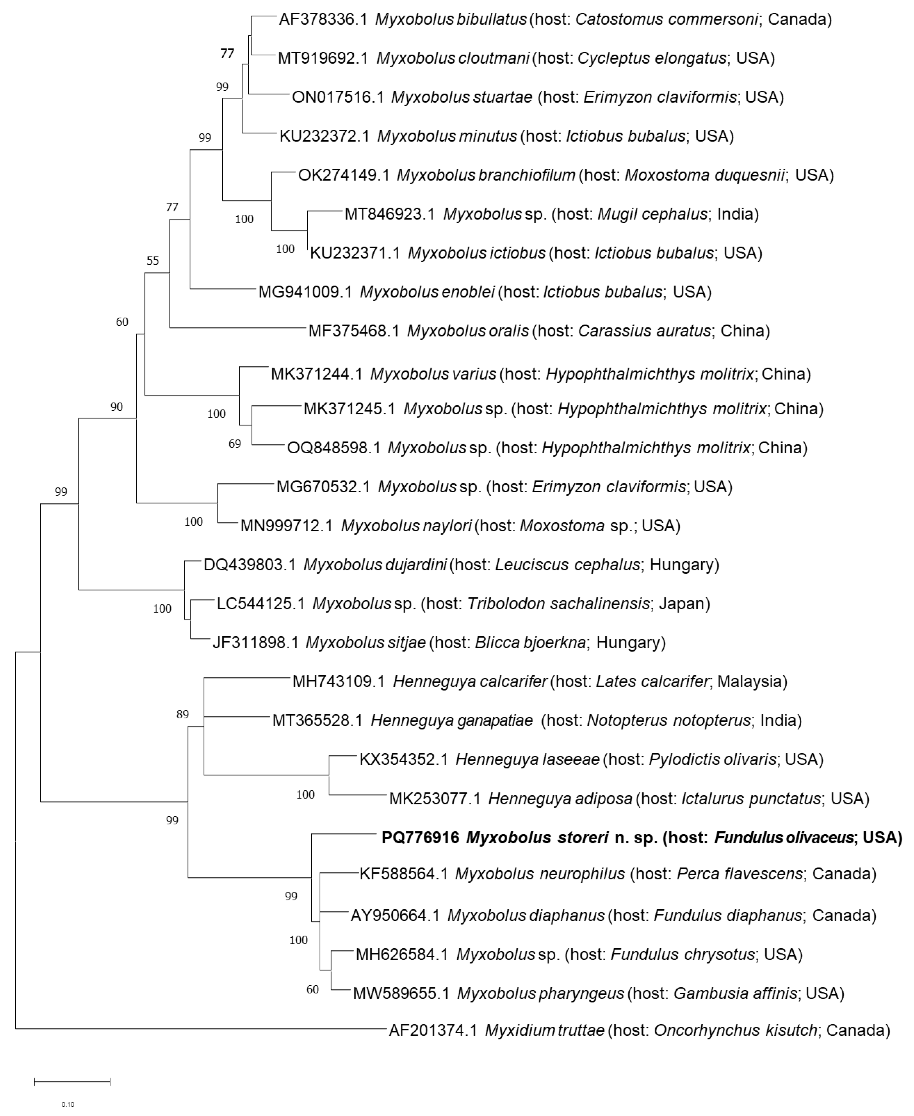
The DNA of myxospore samples preserved in molecular grade ethanol was extracted using a Qiagen DNeasy Kit. The SSU rRNA gene was amplified by placing 5 µL of extracted DNA in a Platinum Taq DNA Polymerase High Fidelity Master Mix (Thermo Fisher, Waltham, Massachusetts, MA, USA) with a set of the following primer pairs from Leis et al. [23]: ERIB1/ACT1R, Myxo1F/MyxGen3R, GenMyxo3/H2, H9/H2, or H9/ERIB10. Successful PCR products were sent for Sanger sequencing at Eurofins Genomics (Louisville, Kentucky, PA, USA). Chromatograms were edited and de novo assembled to form a contiguous sequence in Geneious Prime 23.2.1 (www.geneious.com, accessed om 29 August 2024). The contig was BLAST searched for similarity with other known sequences in GenBank. Similar sequences were downloaded and underwent phylogenetic analysis in MEGA X (https://www.megasoftware.net/, version 10.2.4.). Maximum Likelihood analysis was completed using a General Time Reversible Model with gamma distribution and invariate sites (G + I) with a partial deletion (95% cut off) option. The final dataset was 1796 bp, and Myxidium truttae Léger, 1930 (AF201374.1; host: coho salmon, Oncorhynchus kisutch [Walbaum]), was chosen to root the tree.

3. Results

3.1. Description of the New Species

Phylum Cnidaria Hatschek, 1888
Subphylum Myxozoa Grassé, 2021
Class Myxosporea Bütschli, 1881
Family Myxobolidae Thélohan, 1892
Genus Myxobolus Bütschli, 1882
Myxobolus storeri sp. n. (Figure 1A–C and Figure 2)
ZooBank: To comply with the regulations set out in article 8.5 of the amended 2012 version of the International Code of Zoological Nomenclature [24], details of the new species have been submitted to ZooBank. The Life Science Identifier (LSID) for Myxobolus storeri sp. n. is urn:lsid:zoobank.org:pub:urn:lsid:zoobank.org:act:5CA7AB5B-746C-464F-99CB-0A4D313FF217.
Material: Syntypes: proxy photographs of the myxospores according to ICZN Declaration 45 [24], HWML 219936; stained histological sections, HWML 217934; and partial SSU rRNA gene (1726 bp), GenBank accession number PQ776916.
Diagnosis: Myxospores morphologically consistent with those of the genus Myxobolus Bütschli, 1882 [2,25], parasitizing Fundulus olivaceus in Arkansas, USA, are distinguished from other species of Myxobolus by having the following combination of characteristics: round to ovoid or diffuse plasmodia infecting the mesenteries, 394 (135–699) × 270 (90–516); pyriform myxospores 15.0 (13.1–16.9) × 7.1 (6.2–8.2) × 5.5 (4.5–5.8), with distinct a sutural ridge; two narrowly pyriform polar capsules, equal to subequal in size, longer polar capsule 8.6 (7.3–9.4) × 1.9 (1.6–2.2), and shorter polar capsule 8.2 (6.9–9.4) × 1.8 (1.3–2.2); coiled polar tubule with 9 (7–10) coils; mucous envelope and intercapsular process absent; and possessing a unique partial SSU rRNA gene (GenBank accession number PQ776916). Myxobolus storeri sp. n. is similar to Myxobolus diaphanus Fantham, Porter, and Richardson, 1940, from banded killifish and Fundulus diaphanus (Lesueur, 1817) from Nova Scotia, Canada, in having pyriform myxospores. Although there is considerable overlap in measurements, the myxospores of M. storeri sp. n. (15.0 [13.1 − 16.9] × 7.1 [6.2 − 8.2]) µm are generally shorter and wider than those of M. diaphanus. Furthermore, M. storeri sp. n. has 9 (7–10) fewer coils of the polar capsule tubules than M. diaphanus (11–15) (Table 1). The gene sequences of these two species were only 93.4% similar, indicating they are clearly separate species (see Molecular characterization and phylogenetic analysis below).
The myxospores of M. storeri sp. n., Myxobolus subtecalis (Bond, 1938) Landsberg and Lom (1991) from mummichog, and Fundulus heteroclitis (L., 1766) from Maryland and New York, USA, are pyriform and have similar measurements (Table 1), but are easily distinguished by the presence of three distinct ridges on the posterior portion of the myxospore in valvular view of M. subtecalis that are lacking in M. storeri sp. n. [26].
Description (based on fresh specimens): Plasmodia round to ovoid or diffuse, 394 ± 236 (135–699) × 270 ± 177 (90–516) (n = 5). Myxospores pyriform, 15.0 ± 0.7 (13.1–16.9) (n = 30) × 7.1 ± 0.5 (6.2–8.2) (n = 30) × 5.5 ± 0.4 (4.5–5.8) (n = 9), with distinct sutural ridge. Polar capsules: two, narrowly pyriform, equal to subequal in size; longer polar capsule 8.6 ± 0.6 (7.3–9.4) × 1.9 ± 0.2 (1.6–2.2), 57 ± 0.4 (53–66)% of myxospore length (n = 30); shorter polar capsule 8.2 ± 0.7 (6.9–9.4) × 1.8 ± 0.2 (1.3–2.2), 54 ± 0.4 (49–64)% of myxospore length (n = 30). Coiled polar tubule with 9 ± 0.8 (7–10) coils (n = 30). Mucous envelope absent. Intercapsular process absent. Number of sutural markings variable, 0–6.
Type host: Blackspotted topminnow, Fundulus olivaceus (Storer, 1845), male, 75 mm TL, collected 17 March 2024.
Other host: F. olivaceus, male, 80 mm TL; same locality and date.
Prevalence: Two of six (33.3%); none of the F. catenatus or F. notatus were infected.
Site of infection: Mesenteries. No other tissues possessed visible plasmodia.
Etymology: The new species is named in honor of the describer of the host, David Humphreys Storer (1804–1891), an American physician and naturalist. He described numerous other fish species and published on the fishes of New England [27].
Geographic Distribution: Town Creek at Salem, Fulton County, Arkansas, USA (36°06′4.3848″ N, −91°05′7.9224″ W).
ZooBank registration: The Life Science Identifier (LSID) for Myxobolus girardi sp. n. is urn:lsid:zoobank.org:act:5CA7AB5B-746C-464F-99CB-0A4D313FF217.
Table 1. Morphometrics of Myxobolus of Fundulus. Mean measurements (µm) followed by range in parentheses.
SpeciesFundulus spp. HostPLSPLLPLWMXSMXLMXWMXTPCSLPCLLPCWSPCLSPCWPTCMEIVSMIPType/
Voucher Specimens
DNASiteLocalityReference(s)
M. bilineatum Bond, 1938F. heteroclitus-(250–1000)-Broadly obovoid10.5 (10–12)(9–10)(9–10)Nearly
globular
----(7–9)-P----Brain,
kidney,
urinary,
MD, NY[26]
gall bladder, gill arch
M. diaphanus (Fanthum, Porter, and Richardson, 1940), Landsberg and Lom, 1991F. diaphanus --Elong. ovoid-pyriform(15.5–20)(5.2–7.6)-Elong.
pyriform
(7.4–9.6)(1.5–2.2)(7.4–9.6)(1.5–2.2)(11–15) APP-AY950664TestesNS[16,28,29,30]
M. diaphanus (Fanthum, Porter, and Richardson, 1940), Landsberg and Lom, 1991F. diaphanusRound to elongate<500-Elong. pyriform19.1 (17.0–21.8)7.2 (6.8–7.7)5.8Elong.
pyriform
8.5 (7.4–9.8)2.3 (1.9–2.7)--(7–10)AAP-USNM 95334AY950664Numerous organsNS [31]
M. diaphanus (Fanthum, Porter, and Richardson, 1940), Landsberg and Lom, 1991F. diaphanus---Elong. oval---------------Numerous organsMD, NS, ON [32]
M. funduli (= M. musculi Hahn, 1915) Hahn, 1915, Kudo, 1920F. heteroclitus, F. majalis---Pyriform14.36.76.7Elong.
Pyriform
6.52--(10–14)-P----Muscle, gills, connective tissueMA[29,30,33,34,35,36]
M. funduli (= Myxosoma funduli Kudo, 1918) (Kudo, 1918)F. heteroclitus, F. majalisSpherical150 (?–360)-Pyriform1486Elong.
Pyriform
82----AP (7–10)---Gill lamellaeMA[29,30,36,37]
M. funduli (= Myxosoma funduli Kudo, 1918) (Kudo, 1918)F. heteroclitus, F. majalis, F. diaphanus--------------A----Gill lamellaeMD, NY[26]
M. funduli (= Myxosoma funduli Kudo, 1918) (Kudo, 1918)F. heteroclitus, F. majalis, F. diaphanus-150 (?–360)-Pyriform-----------P (7–10)---GillsMA, MD [38]
M. funduli (= Myxosoma funduli Kudo, 1918) (Kudo, 1918)F. kansae-------------------GillsNE [39,40,41]
M. funduli (= Myxosoma funduli Kudo, 1918) (Kudo, 1918)F. Diaphanus---Pyriform---------------Gill lamellaeMD, NB, NS [32]
M. hudsonius (Bond, 1938), Landsberg and Lom, 1991, two morphsF. heteroclitus-(230–305)(170–260) Scales at base of finsNY[26,29,30]
Type A Pyriform---Pyriform(4–5)(2–2.5) (7–9)-AA---
Type B Obovoid(11.5–12.5)7-Pyriform(4–5)(2–2.5) (7–9)-AA---
M. neurophilus (Guilford, 1963)F. heteroclitus, Perca flavescensDiffuse---11.84.5--5.6----------Optic
tectum,
spinal cord
NS[42]
M. storeri sp. n.F. olivaceusRound to ovoid, or diffuse394 (135–699)270 (90–516)Pyriform15.0 (13.1–16.9)7.1 (6.2–8.2)5.5 (4.5–5.8)Narrowly pyriform8.6 (7.3–9.4)1.9 (1.6–2.2)8.2 (6.9–9.4)1.8 (1.3–2.2)9 (7–10)A-P (2–6)AHWML 217936PQ776916MesenteriesARPresent study
M. subtecalis (Bond, 1938); Landsberg and Lom, 1991F. heteroclitusDiffuse(50–300) Pyriform(15–18)(6.5–8)6Elong. pyriform(7–8)2 (11–12)-A----Viscera, brain, finsMD, NY [26,29,30,43]
Myxobolus sp.F. chrysotus---Ovoidal16 (14–16)8 (7–9)-Deltoid-Narrowly pyriform9 (8–10)3 (2–3)8 (7–9)3 (2–3)------MH626584PeritoneumAR [44]
F. chrysotusSpheroidal(200–400) Ovoidal17 (15–18)8 (7–9)-Deltoid-Narrowly pyriform9 (8–10)3 (2–3)8 (7–9)3 (2–3)-----HWML139415-GillAR[44]
Myxobolus sp.F. diaphanusDiffuse mass--Round---------------Connective tissue of the brain, skullNSCone et al. [32]
PLS, plasmodium shape; PLL, plasmodium length; PLW, plasmodium width; MXS, myxospore shape; MXL, myxospore length; MXW, myxospore width; MXT, myxopore thickness; PCS, polar capsule shape; LPCL, length of longer polar capsule, LPCW, width of longer polar capsule; SPCL, length of shorter polar capsule; SPCW, width of shorter polar capsule; PTC, number of polar tubule coils; ME, mucous envelope (A = absent, P = present); IV, iodinophilus vacuole; SM, sutural markings; IP, intercapsular process; and DNA, gene sequence. Host names are according to Page et al. [1].

3.2. Molecular Characterization and Phylogenetic Analysis

A 1726 bp sequence was produced for M. storeri sp. n. and a BLAST search (Figure 3) revealed this species was 93.4% similar to M. diaphanus (AY950664) and 92.63% similar to an unnamed Myxobolus sp. identified from the peritoneum of 4 of 52 (8%) golden topminnows, Fundulus chrysotus (Günther, 1866), in Arkansas [44]. All other species in GenBank (Figure 3) shared <92% similarity with the novel species. Phylogenetic analysis revealed that M. storeri sp. n. clustered with other myxozoan species that infected various tissues (brain, peritoneum, branchial cavity) of a variety of small-bodied fish, including other Fundulus spp., yellow perch, Perca flavescens (Mitchill, 1814), and western mosquitofish, Gambusia affinis (Baird and Girard, 1853), which is native to the USA [31,45,46,47].

3.3. Histopathology

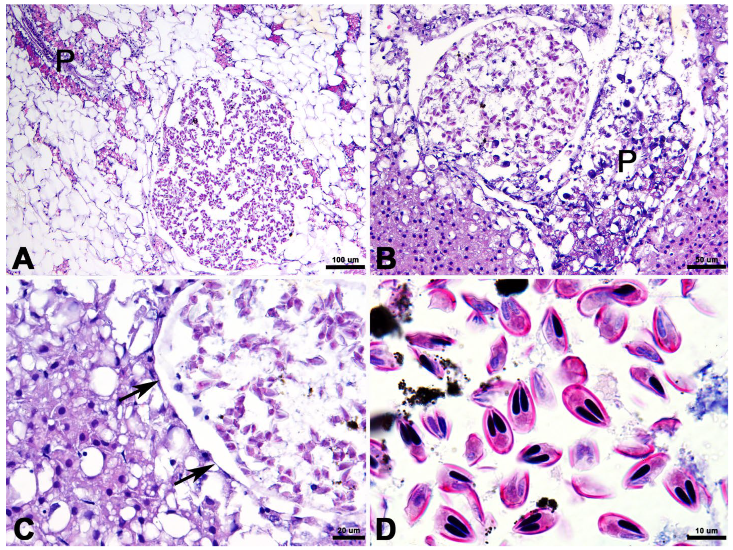
The examined tissues consisted of mesenteric adipose/adipocytes with the embedded exocrine pancreas, pancreatic islets, and spleen from one fish, and the liver and intestine from the second. Variably sized, round to ovoid plasmodia ranging from 153 × 196 to 516 × 699 µm were scattered within the mesenteric adipose (Figure 4A) and hepatic parenchyma, often immediately adjacent to exocrine pancreatic tissue (Figure 4B). Plasmodia were limited by a thin membranous wall, possibly of host origin, studded occasionally by flattened nuclei but with no additional organized morphologic structure (Figure 4C). Large numbers of elongate, pyriform myxospores with thick refractile shell valves, two elongated polar capsules, and a prominent sporoplasm filled plasmodia (Figure 4D). No developing pansporoblasts or other generative stages were observed. The expansile plasmodia slightly compressed adjacent adipose and hepatic parenchyma. Myxospores were often mixed with aggregates of melanin granules but elicited no other inflammatory response (Figure 4B).

4. Discussion

Phylogenetic analysis (Figure 3) revealed an intriguing, strongly supported connection between Myxobolus storeri sp. n. and other myxozoans that infect a variety of tissues of small-bodied North American fishes. While the similarity of the new species with Myxobolus spp. that infect Fundulus spp., including an unnamed species that similarly infects the peritoneum of F. chrysotus [44] and M. diaphanus and infects the connective tissue in various organs of F. diaphanus [32], was somewhat expected; the association with M. neurophilus Guilford, 1963, from the nervous system of P. flavescens from Saskatchewan, Canada, and Myxobolus (syn. Myxosoma) pharyngeus Parker, Spall, and Warner, 1971, from the branchial cavity epithelium of G. affinis from Payne County, Oklahoma, USA, were not expected, since they both infect different host families and different tissues. The evolution of such a lineage is interesting to consider and would possibly involve actinospore and pre-sporogonic life stages (see [48]) capable of infecting a wider range of hosts/tissues than typically seen with other myxozoan species.
Histologically, plasmodia were limited to mesenteric adipose and hepatic parenchyma (Figure 4), where they potentially acted as space occupying masses causing mild compression of adjacent tissue but eliciting no associated inflammatory response in the sections examined.

5. Conclusions

We provide a description of a new species of Myxobolus from a fundulid fish from Arkansas, USA. This is the first time F. olivaceus has been reported to harbor a myxozoan parasite and the seventh species reported from this family of fishes. Morphological, pathological, and molecular data are documented to support our description.

Author Contributions

This study was designed by C.T.M., D.G.C. and E.M.L. Field collections were performed by all authors. Laboratory procedures for processing fish and necropsy were performed by C.T.M., G.B. and M.R.R. Laboratory procedures for measurements, photomicrographs (Figure 1), and isolation of plasmodia and myxospores were performed by C.T.M., D.G.C. and E.M.L. The line drawing (Figure 2) was prepared by D.G.C. E.M.L. performed molecular analyses and prepared Figure 3. A.C.C. performed the histopathological analysis and prepared the photomicrographs in Figure 4. The manuscript was written by all authors and all of them read and approved the final manuscript. All authors have read and agreed to the published version of the manuscript.

Funding

This research received no external funding.

Institutional Review Board Statement

Not applicable.

Data Availability Statement

The data presented in this study are available within the article.

Acknowledgments

The Arkansas Game and Fish Commission issued a Scientific Collecting Permit no. 032820232 to C.T.M. Scott L. Gardner and Gabor R. Rácz (HWML) are acknowledged for expert curatorial assistance. We thank Dwight Ferguson, Black Rock, Arkansas, USA, for allowing us to process fish in his fish hut. Usage of trade names does not imply endorsement by the U.S. Government. The findings and conclusions in this article are those of the authors and do not necessarily represent the views of the U.S. Fish and Wildlife Service.

Conflicts of Interest

The authors declare there are no conflicts of interest.

References

  1. Eiras, J.C. An overview on the myxosporean parasites in amphibians and reptiles. Acta Parasitol. 2005, 50, 267–275. [Google Scholar]
  2. Fiala, I.; Bartoŝová-Sojková, P.; Whipps, C.M. Classification and Phylogenetics of Myxozoa. In Myxozoan Evolution, Ecology and Development; Okamura, B., Gruhl, A., Bartholomew, J.L., Eds.; Springer International Publishing: Cham, Switzerland, 2015. [Google Scholar]
  3. Bartholomew, J.L.; Atkinson, S.D.; Hallett, S.L.; Lowenstine, L.J.; Garner, M.M.; Gardiner, C.H.; Rideout, B.A.; Keel, M.K.; Brown, J.D. Myxozoan parasitism in waterfowl. Int. J. Parasitol. 2008, 38, 1199–1207. [Google Scholar] [CrossRef]
  4. Prunescu, C.-C.; Prunescu, P.; Lom, J. The first finding of myxosporean development from plasmodia to spores in terrestrial mammals: Soricimyxum fegati gen. et sp. n. (Myxozoa) from Sorex araneus (Soricomorpha). Folia Parasitol. 2007, 54, 159–164. [Google Scholar] [CrossRef]
  5. Székely, C.; Cech, G.; Atkinson, S.D.; Kálmán, M.; Egyed, L.; Gubányi, A. A novel myxozoan parasite of terrestrial mammals: Description of Soricimyxum minuti sp. n. (Myxosporea) in pygmy shrew Sorex minutus from Hungary. Folia Parasitol. 2015, 62, 45–49. [Google Scholar] [CrossRef]
  6. Kent, M.L.; Margolis, L.; Corliss, J.O. The demise of a class of protists: Taxonomic and nomenclatural revisions proposed for the protist phylum Myxozoa Grasse, 1970. Can. J. Zool. 1994, 72, 932–937. [Google Scholar] [CrossRef]
  7. Eiras, J.C.; Molnár, K.; Lu, Y.S. Synopsis of the species of Myxobolus Bütschli, 1882. Parasitology 2005, 88, 11–36. [Google Scholar]
  8. Eiras, J.C.; Zhang, J.; Molnár, K. Synopsis of the species of Myxobolus Bütschli, 1882 (Myxozoa: Myxosporea: Myxobolidae) described between 2005 and 2013. Syst. Parasitol. 2014, 88, 11–36. [Google Scholar] [CrossRef] [PubMed]
  9. Eiras, J.C.; Cruz, C.F.; Saraiva, A.; Adriano, E.A. Synopsis of the species of Myxobolus (Cnidaria, Myxozoa, Myxosporea) described between 2014 and 2020. Folia Parasitol. 2021, 68, 012. [Google Scholar] [CrossRef]
  10. Page, L.M.; Burr, B.M. Peterson Field Guide to Freshwater Fishes of North America North of Mexico, 2nd ed.; Houghton Mifflin Harcourt: New York, NY, USA, 2011. [Google Scholar]
  11. Shute, J.R. Fundulus olivaceus. In Atlas of North American Freshwater Fishes; Lee, D.S., Gilbert, C.R., Hocutt, C.H., Jenkins, R.E., McAllister, D.E., Stauffer, J.R., Jr., Eds.; North Carolina State Museum of Natural History: Raleigh, NC, USA, 1980. [Google Scholar]
  12. Robison, H.W.; Buchanan, T.M. Fishes of Arkansas, 2nd ed.; University of Arkansas Press: Fayetteville, AR, USA, 2020. [Google Scholar]
  13. Rice, L.A. The food of seventeen Reelfoot Lake fishes in 1941. J. Tenn. Acad. Sci. 1942, 17, 4–13. [Google Scholar]
  14. Ross, S.T. The Inland Fishes of Mississippi; University Press of Mississippi: Jackson, MS, USA, 2001. [Google Scholar]
  15. Wellborn, T.L., Jr.; Rogers, W.A. Five new species of Gyrodactylus (Trematoda: Monogenea) from the southeastern U.S. J. Parasitol. 1967, 53, 10–14. [Google Scholar] [CrossRef]
  16. Hoffman, G.L. Parasites of North American Freshwater Fishes, 2nd ed.; Comstock Publishing Associates: Ithaca, NY, USA, 1999. [Google Scholar]
  17. McAllister, C.T.; Bursey, C.R.; Fayton, T.J.; Font, W.F.; Robison, H.W.; Connior, M.B.; Cloutman, D.G. Helminth parasites of the blackspotted topminnow, Fundulus olivaceus (Cyprinodontiformes: Fundulidae), from the Interior Highlands of Arkansas. J Ark. Acad. Sci. 2015, 69, 135–138. [Google Scholar] [CrossRef]
  18. Use of Fishes in Research Committee (Joint Committee of the American Fisheries Society, the American Institute of Fishery Research Biologists, and the American Society of Ichthyologists and Herpetologists). Guidelines for the Use of Fishes in Research. 2014. Available online: https://fisheries.org/docs/wp/Guidelines-for-Use-of-Fishes.pdf (accessed on 1 July 2024).
  19. Lom, J.; Arthur, J.R. A guideline for the preparation of species description in Myxosporea. J. Fish Dis. 1989, 12, 151–156. [Google Scholar] [CrossRef]
  20. Clopton, R.E. Standard nomenclature and metrics of plane shapes for use in gregarine taxonomy. Comp. Parasitol. 2004, 71, 130–140. [Google Scholar] [CrossRef] [PubMed]
  21. Bush, A.O.; Lafferty, K.D.; Lotz, J.M.; Shostak, A.W. Parasitology meets ecology on its own terms: Margolis et al. revisited. J. Parasitol. 1997, 83, 575–583. [Google Scholar] [CrossRef]
  22. Page, L.M.; Bemis, K.E.; Dowling, T.E.; Espinosa-Pérez, H.S.; Findley, L.T.; Gilbert, C.R.; Hartel, K.E.; Lea, R.N.; Mandrak, N.E.; Neighbors, M.A.; et al. Common and Scientific Names of Fishes from the United States, Canada, and Mexico, 8th ed.; Special Publication 37; American Fisheries Society: Bethesda, MD, USA, 2023. [Google Scholar]
  23. Leis, E.M.; Rosser, T.G.; Baumgartner, W.A.; Griffin, M.J. Two novel myxozoans from pirate perch Aphredoderus sayanus (Gilliams, 1824) in the Upper Mississippi River, including the first North American species of Hennegoides Lom, Tonguthai, & Dykova, 1991. J. Parasitol. 2019, 105, 918–927. [Google Scholar] [PubMed]
  24. ICZN (International Commission on Zoological Nomenclature). Declaration 45—Addition of recommendations to Article 73 and of the term “specimen, preserved” to the glossary. Bull. Zool. Nomen. 2017, 73, 96–97. [Google Scholar] [CrossRef]
  25. Lom, J.; Dyková, I. Myxozoan genera: Definition and notes on taxonomy, life-cycle, terminology and pathogenic species. Folia Parasitol. 2006, 53, 1–36. [Google Scholar] [CrossRef]
  26. Bond, F.F. Cnidosporidia from Fundulus heteroclitus Lin. Trans. Amer. Micros. Soc. 1938, 57, 107–122. [Google Scholar] [CrossRef]
  27. Scudder, S.H. David Humphreys Storer. Proc. Amer. Acad. Arts Sci. 1891, 27, 388–391. [Google Scholar]
  28. Fantham, H.B.; Porter, A.; Richardson, L.R. Some more Myxosporidia observed in Canadian fishes. Parasitology 1940, 32, 333–353. [Google Scholar] [CrossRef]
  29. Landsberg, J.H.; Lom, J. Taxonomy of the genera of the Myxobolus/Myxosoma group (Myxobolidae: Myxosporea), current listing of species and revision of synonyms. Syst. Parasitol. 1991, 18, 165–186. [Google Scholar] [CrossRef]
  30. Cone, D.K.; Raesly, R.L. Redescription of Myxobolus rhinichthidis (Myxosporea) parasitizing Rhinichthys cataractae, with a revised taxonomic list of species of Myxobolus known from North American freshwater fishes. Can. J. Fish. Aquat. Sci. 1995, 52, 7–12. [Google Scholar] [CrossRef]
  31. Cone, D.; Easy, R. Supplemental diagnosis and molecular taxonomy of Myxobolus diaphanus (Fantham, Porter et Richardson, 1940) (Myxozoa) parasitizing Fundulus diaphanus (Cyprinodontiformes) in Nova Scotia, Canada. Folia Parasitol. 2005, 52, 217–222. [Google Scholar] [CrossRef] [PubMed]
  32. Cone, D.K.; Marcogliese, D.J.; Barse, A.M.; Burt, M.D.B. The myxozoan fauna of Fundulus diaphanus (Cyprinodontidae) from freshwater localities in eastern North America: Prevalence, community structure, and geographic distribution. J. Parasitol. 2006, 92, 52–57. [Google Scholar] [CrossRef]
  33. Hahn, C.W. Sporozoon parasites of certain fishes in the vicinity of Woods Hole, Massachusetts. Bull. U. S. Bureau Fish. 1915, 33, 191–214. [Google Scholar]
  34. Hahn, C.W. On the sporozoon parasites of the fishes of Woods Hole and Vicinity. I. Further observations on Myxobolus musculi from Fundulus. J. Parasitol. 1917, 3, 91–104. [Google Scholar] [CrossRef]
  35. Hahn, C.W. On the sporozoon parasites of the fishes of Woods Hole and Vicinity. II. Additional observations on Myxobolus musculi. J. Parasitol. 1917, 3, 150–162. [Google Scholar] [CrossRef]
  36. Kudo, R. Contributions to the study of parasitic Protozoa. IV. Note on some Myxosporidia from certain fish in the vicinity of Woods Hole. J. Parasitol. 1918, 25, 11–16. [Google Scholar]
  37. Kudo, R. Studies on Myxosporidia. A synopsis of genera and species of myxosporidia. Ill. Biol. Monogr. 1920, 5, 1–265. [Google Scholar]
  38. Hoffman, G.L.; Putz, R.E.; Dunbar, C.E. Studies on Myxosoma cartilaginis n. sp. (Protozoa: Myxosporidia) of centrarchid fish and a synopsis of the Myxosoma of North American freshwater fishes. J. Protozool. 1965, 12, 319–332. [Google Scholar] [CrossRef]
  39. Adams, A.M. Parasites on the gills of the plains killifish, Fundulus kansae, in the South Platte River, Nebraska. Trans. Am. Microscop. Soc. 1985, 104, 278–284. [Google Scholar] [CrossRef]
  40. Janovy, J., Jr.; Hardin, E.L. Population dynamics of the parasites in Fundulus zebrinus in the Platte River of Nebraska. J. Parasitol. 1987, 73, 689–696. [Google Scholar] [CrossRef]
  41. Janovy, J., Jr.; Hardin, E.L. Diversity of the parasite assemblage of Fundulus zebrinus in the Platte River of Nebraska. J. Parasitol. 1988, 74, 207–213. [Google Scholar] [CrossRef] [PubMed]
  42. Guilford, H.G. New species of Myxosporidia found in percid fishes from Green Bay (Lake Michigan). J. Parasitol. 1963, 49, 474–478.43. [Google Scholar] [CrossRef]
  43. Bond, F.F. Experimental studies on myxosporidiosis of Fundulus heteroclitus (Linn.) and F. diaphanus (Le Sueur). Trans. Am. Microsc. Soc. 1939, 58, 164–169. [Google Scholar] [CrossRef]
  44. McAllister, C.T.; Fayton, T.J.; Cloutman, D.G.; Bursey, C.R.; Robison, H.W.; Trauth, S.E.; Whipps, C.W. Parasites of the golden topminnow, Fundulus chrysotus (Cyprinodontiformes: Fundulidae), from Arkansas, U.S.A. Comp. Parasitol. 2019, 87, 19–32. [Google Scholar] [CrossRef]
  45. Parker, J.D.; Spall, R.D.; Warner, M.C. Two new species of myxosporida, Henneguya gambusi sp. n. and Myxospora pharyngeus sp. n., in the mosquitofish, Gambusia affinis (Baird and Girard). J. Parasitol. 1971, 57, 1297–1301. [Google Scholar] [CrossRef] [PubMed]
  46. Khoo, L.; Rommel, F.A.; Smith, S.A.; Griffin, M.J.; Pote, L.M. Myxobolus neurophilus: Morphologic, histopathologic and molecular characterization. Dis. Aquat. Organ. 2010, 89, 51–61. [Google Scholar] [CrossRef]
  47. Stilwell, J.M.; Rosser, T.G.; Leary, J.H.; Woodyard, E.T.; Richardson, B.; Lopez-Porras, A.; Mischke, C.C.; Camus, A.C.; Griffin, M.J. Characterisation of myxozoan fauna of western mosquitofish, Gambusia affinis (Baird and Gerard 1853) (Cyprinodontiformes: Poeciliidae), inhabiting experimental catfish ponds in Mississippi, USA. Syst. Parasitol. 2021, 98, 423–441. [Google Scholar] [CrossRef]
  48. Holzer, A.S.; Piazzon, M.C.; Barrett, D.; Bartholomew, J.L.; Sitjà-Bobadilla, A. To react or not to react: The dilemma of fish immune systems facing myxozoan infections. Front. Immunol. 2021, 12, 734238. [Google Scholar] [CrossRef]
Figure 1. Photomicrographs of Myxobolus storeri sp. n. from Fundulus olivaceus. (A) Round plasmodium (P) in mesentery tissue. (B) Diffuse plasmodium (P) among adipocytes in mesentery tissue. (C) Myxospores (HWML 219936).
Diversity 17 00192 g001
Figure 2. Composite line drawing of the myxospore of Myxobolus storeri sp. n. (A) Valvular view. (B) Sutural view.
Diversity 17 00192 g002
Figure 3. Maximum likelihood analysis of SSU rRNA genes of Myxobolus storeri sp. n. and related species in GenBank with 1000 bootstrap replications (values > 50 are shown).
Diversity 17 00192 g003
Figure 4. Photomicrographs of Fundulus olivaceus mesentery and liver with plasmodia and spores of Myxobolus storeri sp. n. (A) Mesenteric adipose with scattered exocrine pancreas (P) and a large spore filled plasmodium (H&E stain). (B) Liver with a small plasmodium adjacent to exocrine pancreatic tissue (P) (H&E stain). (C) Liver with plasmodium causing slight compression of adjacent parenchyma. Plasmodia were limited by thin membrane-like structures (arrows), presumably of host origin, with no additional morphologic features (H&E stain). (D) High magnification image of pyriform myxospores with two elongated polar capsules amongst scattered melanin granules (Ziehl–Neelsen stain).
Diversity 17 00192 g004
Disclaimer/Publisher’s Note: The statements, opinions and data contained in all publications are solely those of the individual author(s) and contributor(s) and not of MDPI and/or the editor(s). MDPI and/or the editor(s) disclaim responsibility for any injury to people or property resulting from any ideas, methods, instructions or products referred to in the content.