Article

Exploring Endophytic Fungi from Humulus lupulus L. for Biocontrol of Phytopathogenic Fungi

National Research Council (CNR), Institute of Biosciences and Bioresources (IBBR), Via della Madonna Alta, 130-06128 Perugia, Italy
*
Authors to whom correspondence should be addressed.
These authors contributed equally to this work.
Diversity 2025, 17(2), 94; https://doi.org/10.3390/d17020094
Submission received: 30 October 2024 / Revised: 13 January 2025 / Accepted: 24 January 2025 / Published: 28 January 2025
(This article belongs to the Special Issue Fungi, Ecology, and Global Change)

Abstract

:
Humulus lupulus L. (hop) is a crucial crop within the brewing industry and a rich source of bioactive compounds. Traditionally concentrated in northeast regions of Europe, hop cultivation has expanded towards southern territories such as Italy over recent decades. Managing phytosanitary threats in Mediterranean climates poses challenges due to limited knowledge and registered agrochemicals. In pursuit of eco-friendly alternatives for disease management, we isolated 262 endophytic fungal strains from wild hop roots, stems, leaves, and flowers. Through phylogenetic analyses, we identified 51 operational taxonomic units. Dominant species such as Ilyonectria macrodidyma, Penicillium sp., Diaporthe columnaris, Plectosphaerella cucumerina, and Fusarium oxysporum were exclusive to roots. In contrast, Alternaria spp. and Epicoccum spp. were prevalent in other tissues, and Botrytis cinerea was exclusively detected in female flowers. We tested seven isolates—Epicoccum sp., Aureobasidium pullulans, Plectosphaerella cucumerina, Stemphylium vesicarium, Periconia byssoides, Talaromyces wortmannii, and Nigrospora sphaerica—against the four phytopathogenic fungi Alternaria sp., Fusarium oxysporum, Botrytis cinerea, and Sclerotinia sclerotiorum. All endophytes exhibited antagonistic effects against at least one pathogen, with Plectosphaerella cucumerina showing the strongest inhibition against Alternaria sp. This study marks the first exploration of endophytic fungi from various hop tissues. All isolated strains were ex situ conserved for future bioactivity assessments and biotechnological applications. Original data with a key relevance for the environmentally friendly management of plant diseases are provided.

1. Introduction

Hop (Humulus lupulus L.) is a dioecious, perennial, and herbaceous climbing plant of the Cannabaceae family. While it naturally thrives in temperate areas, it is also cultivated for its secondary metabolites, which impart flavor, bitterness, aroma, and antimicrobial properties to beer. Moreover, certain metabolites exhibit bioactive properties with pharmaceutical potential, notably as sedatives and antimicrobial agents [1]. Hop has also garnered attention for its possible cancer chemopreventive effects [2,3]. Over 1000 chemicals have been identified in hop, primarily comprising essential oils, α- and β-acids, and prenylflavonoids, which accumulate in the resinous substance (lupulin) of the female flowers, called cones [4]. According to the 2021 FAO report, global annual hop production and harvesting areas increased by 34% and 18%, respectively, from 2011 to 2021. The United States of America and Germany stand out as the most prolific hop-producing countries. However, the European Union remains the primary contributor to global hop production, accounting for nearly 50% of the total output. While hop cultivation in Europe is traditionally concentrated in the northeast regions like Germany, the Czech Republic, and southeast England, in the last decades, the cultivation borders have moved towards southern European countries, including Italy. Despite the natural growth of hop plants across the entire peninsula, Italy relies heavily on imports, with 98% of its hop requirements are sourced from external suppliers [5]. In recent years, the growing interest in hop cultivation stems from the proliferation of microbreweries producing craft beers with diverse tastes and flavors. These breweries are increasingly inclined to use locally cultivated hops rather than relying on imports (https://www.assobirra.it/wp-content/uploads/2021/06/AssoBirra_AnnualReport_2020_giugno2021_DEF.pdf, accessed on 20 January 2024). Moreover, the Italy’s remarkable variety of pedoclimatic characteristics could influence and shape hop organoleptic qualities. However, there is a notable lack of experience and knowledge in Italy regarding hop cultivation practices, phenology, and yard management, hindering the development of this burgeoning sector. Mediterranean climatic conditions present particular challenges, including the threat of phytopathogenic fungi, insect pests, mites, viruses, and viroids, all of which pose significant risks to hop production in terms of both yield and quality [6,7]. Concerning pathogenic fungi, the most prevalent hop diseases include downy mildew caused by Pseudoperonospora humuli and powdery mildew caused by Podosphaera macularis, alongside various types of rots, wilts, and others. Current management strategies primarily involve the use of resistant cultivars and fungicides targeting P. macularis [6,8]. However, the use of fungicides and other plant protection products against the different pests is challenged by the lack of registered active substances for hops under Italian legislation [7].
Plant endophytic fungi colonize the intercellular spaces of living plant tissues without triggering disease symptoms [9]. The inner part of the plant is a protected niche containing the necessary nutrients for fungal survival and growth. Colonization can occur in tissues of one or more parts of the host plant, including roots, stems, leaves, reproductive systems, and fruits. In exchange for this safe place, endophytic fungi may improve plant fitness by different mechanisms ranging from biological control of phytopathogens to biofertilization and stress tolerance. Such benefits can occur directly and/or directly. The direct mechanisms include the increase in plant nutrient acquisition and phytohormones production, which are directly related to the increase in biomass, root system expansion, plant height, and weight. Tolerance to biotic and abiotic stresses as well as activation of systemic resistance and production of antibiotics and secondary metabolites are considered indirect aspects of such growth promotion [10]. Exploring plant–endophyte interactions across various crops is pivotal for fostering sustainable cultivation practices. Many endophytic species produce antibiotics and antifungal compounds that safeguard plants against pathogens, offering promising avenues for eco-friendly and economically sustainable agriculture [11]. The advantages of using fungi in agriculture include greater biosafety and less environmental and human health risk, specificity for the target pest and others. Indeed, the utilization of endophytic fungi in agriculture for biological control of phytopathogens has garnered increasing attention during the last decades [12], driven by the increasing demand for sustainable and environmentally friendly alternatives to chemical products [13]. However, research on endophytic microorganisms for disease control in hops remains limited [14].
The aim of this study was to isolate endophytic fungi from hop plants, characterize them taxonomically, and test their bioactivity against agriculturally significant phytopathogenic fungi. To achieve this, an ex situ collection of endophytic fungal strains was established by isolating fungi from various tissues (roots, stems, leaves, and both female and male flowers) of wild H. lupulus accessions collected from different sites in Central Italy. To the best of our knowledge, this research presents the initial comprehensive report on the endophytic fungi associated with H. lupulus and their potential application in agriculture as biocontrol agents against phytopathogenic fungi.

2. Materials and Methods

2.1. Biological Material and Study Sites

Wild, healthy plants of H. lupulus were collected between 2017 and 2019 from ten natural sites in Central Italy: seven in the Umbria region, two in the Marche region, and one in the Lazio region (Table 1). A total of 36 plants were collected, ranging from 1 to 6 per site. Different tissues, namely roots, stems, leaves, female flowers (cones), and male flowers, were collected. Due to factors such as harvesting time/life stage, sex, and accessibility to various plants parts, not all tissues could be collected from every plant or site. However, cones and leaves were obtained from nearly all plants and sites (Table 1).

2.2. Isolation of Fungi and Molecular Identification

All plant tissues were surface-sterilized basically as described by Belfiori and colleagues [15]: They were treated with 0.3% sodium hypochlorite for 3 min, 70% ethanol for 1 min, and then rinsed three times with sterile distilled water. As a control of the sterilization, the last water rinse was incubated in PDA to exclude any fungal growth. Tissues were air-dried under sterile conditions, cut into small segments (0.5–1 cm) using a sterile surgical blade, and placed on potato dextrose agar (PDA, Merck) supplemented with 100 mM ampicillin to prevent bacterial contamination. The cultures were then incubated at 25 °C and inspected every 3–4 days for the emergence of hyphae from the tissues, up to approximately 4 weeks. For each tissue sample and each sampling site, all mycelia from colonies exhibiting different morphologies were picked and re-inoculated onto fresh PDA in Petri dishes to obtain pure cultures. Finally, the single cultures were transferred into potato dextrose broth (PDB, Merck) with 50% (v/v) glycerol, frozen in liquid nitrogen, and kept at −70 °C for long-term storage. The obtained fungal isolates are deposited in the collection of the BioMemory Project (https://biomemory.cnr.it/collections/CNR-IBBR-FABI, accessed on 20 January 2024).

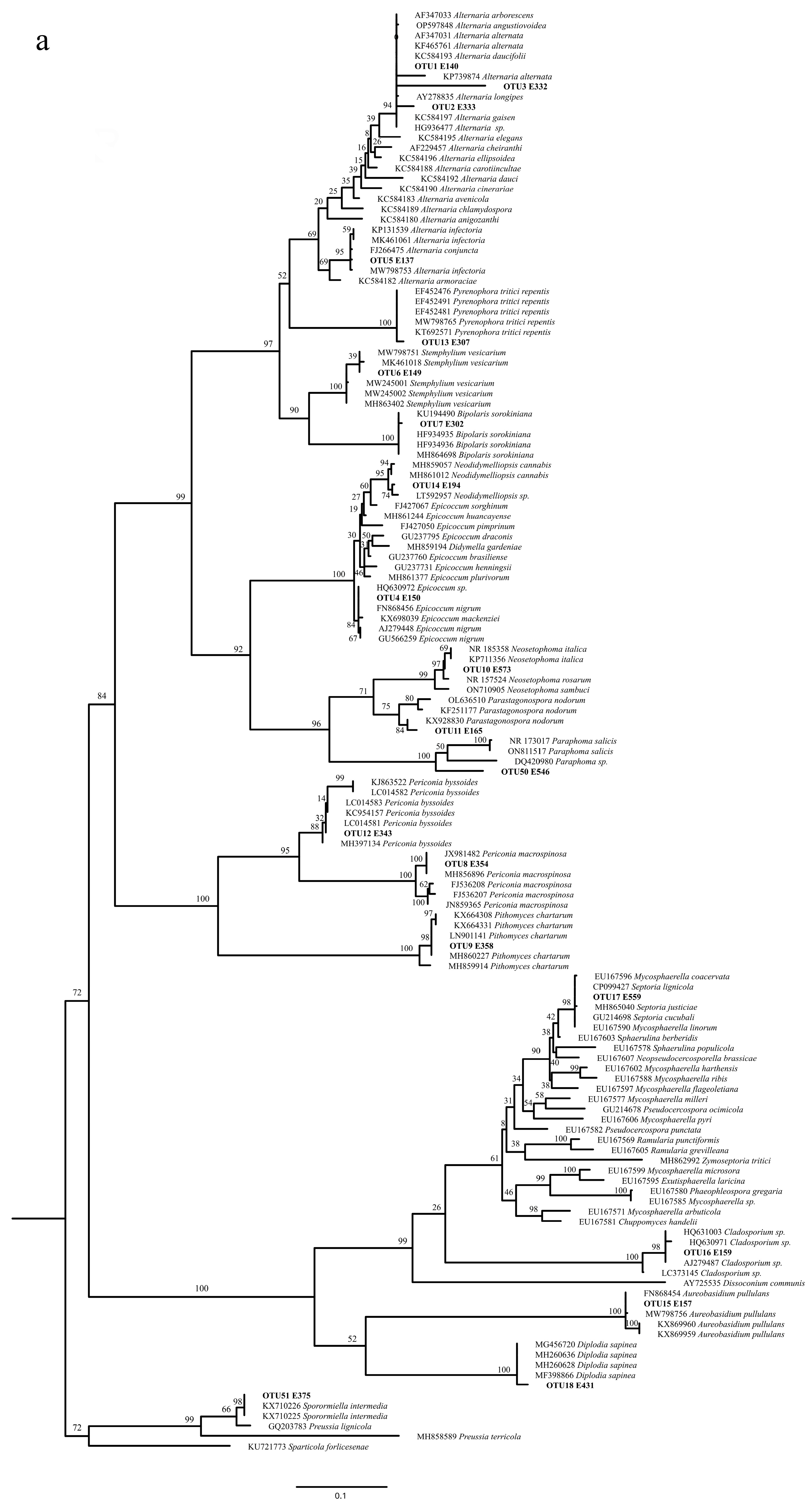
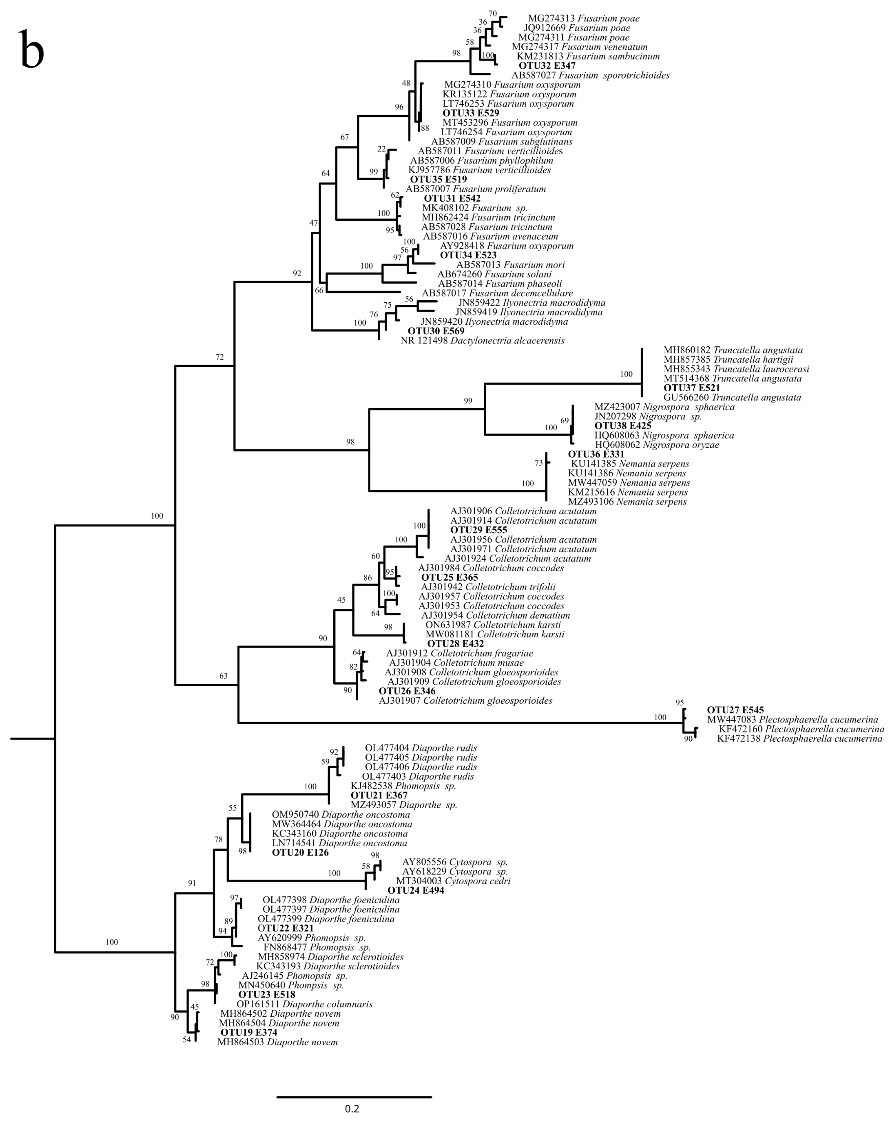
2.3. OTUs Molecular Identification and Phylogenetic Analysis

Genomic DNA was isolated from each strain as described in Arnold and Lutzoni [16]. A small amount (0.3 g) of mycelium was ground and resuspended in 300 µL of buffer containing 200 mM Tris-HCl pH 7.5, 250 mM NaCl, 25 mM EDTA, and 0.5% SDS; vortexed for 10 s; and centrifuged at 14,000 rpm for 10 min. The supernatant was precipitated in an equal volume of isopropanol for 30 min at −20 °C. The DNA was pelleted by maximum-speed centrifugation for 20 min at 4 °C, vacuum-dried, and resuspended in 50 µL of double-distilled nuclease-free water. DNA concentration was measured using a NanoDrop 2000 UV–vis Spectrophotometer (Thermo Scientific, Waltham, MA, USA). The ITS region was amplified by PCR with the primers ITS1f [17] and ITS4 [18]. PCR was carried out in a 25 µL reaction mixture containing template DNA (10 ng), 10X reaction buffer (RBC Bioscience, New Taipei City, Taiwan), 4 mM MgCl2, dNTPs (0.2 mM each), 10 µM of each primer, and 1 U of RBC Taq polymerase (RBC Bioscience). A GeneAmp® PCR System 9700 (Applied Biosystems, Foster City, CA, USA) was used under the following conditions: initial denaturation at 94 °C for 2 min, 35 cycles of denaturation at 94 °C for 15 s, annealing at 55 °C for 20 s, extension at 72 °C for 45 s, and a final extension at 72 °C for 7 min. Sequencing was performed using the primers ITS1f, ITS4, 5.8sf, and 5.8sb [18,19] and the BigDye Terminator Cycle V 3.1 Sequencing Kit (Applied Biosystems, Foster City, CA, USA) according to the supplier’s instructions. Capillary electrophoresis was carried out with an ABI 3130 Genetic Analyzer (Applied Biosystems). Assembly, editing, and alignment of sequences were conducted using GENEIOUS version 4.8.5. The resulting DNA sequences were deposited in GenBank, under accession numbers OQ257045 to OQ257306. Similarity searches were performed in GenBank database using BLASTn [20]. In order to designate operational taxonomic units (OTUs), sequences were clustered using a 97% similarity threshold using CD-HIT-EST [21] (https://github.com/weizhongli/cdhit, accessed on 27 January 2025). The functional diversity of the identified fungal genera was analyzed according to the database FungalTraits [22]. Phylogenetic analyses were performed to better identify the OTUs in the Sordariomycetes and Dothideomycetes classes that were the most prevalent. To this purpose, based on BLASTn searches, similar sequences were downloaded from GenBank and aligned with the sequenced OTUs. Multiple sequences alignments were performed using MAFFT version 7 [23] with the L-INS-I parameters. Trees were generated using RaxML version 8.2.12 software [24] using the following options: rapid bootstrapping with auto MRE, GTRGAMMA distribution model, and empirical base frequency. The species names of sequences downloaded from GenBank are reported in the trees along with the accession numbers. Alignments and trees were deposited in TreeBASE under accession no. 31340.

2.4. Diversity Analyses

Species richness was calculated for each tissue as the number of different OTUs. Since different numbers of roots, stems, leaves, and flowers were sampled and from different plants, we could not calculate the relative abundance of each OTU per plant. However, the relative abundance of each OTU was calculated for each tissue by dividing the number of isolates of an OTU in that tissue by the total number of isolates from that tissue. The dominant species were determined for each tissue according to Rivera-Orduña et al. [25] as the OTUs with Pi > 1/S, with Pi being the relative abundance and S the species richness.

2.5. Antifungal Activity of Isolated Endophytic Fungi

Among the isolated endophytes, seven strains (see results) belonging to various phylogenetic groups and with a different tissue distribution were selected for testing. Four pathogenic species were used for these tests: Botrytis cinerea (AR593) and Sclerotinia sclerotiorum (AR397) were previously isolated from the basal part of the petiole of symptomatic leaves of Vitis vinifera, whereas Alternaria sp. (strain E140) and Fusarium oxysporum (strain E529) were isolated from asymptomatic hop tissues. However, their pathogenicity was demonstrated in experiments on tobacco leaves (unpublished results).

2.5.1. Dual-Culture Assays

Endophytic fungi were tested against pathogenic fungi by dual-culture assays. Briefly, 5 mm diameter mycelial plugs from the edge of young cultures of both the endophytic and pathogen fungus were placed on the opposite sides of a 9 cm diameter Petri plate containing PDA at about 1 cm from the margin. Control plates were prepared by growing the endophytic and the pathogenic strains in single cultures in the same conditions. Three replicates were made for each treatment. Following an incubation period of approximately seven days at 25 °C, plates were inspected, and the presence of inhibition signals in the interaction zone between the two mycelia was assessed to determine bioactivity.

2.5.2. Agar Diffusion Method

We followed the procedure described by Hajieghrari et al. [26] with some modifications. The endophytic fungi to be tested were grown on PDA plates for about seven days. Subsequently, a mycelial agar disc (5 mm in diameter) was excised from the culture’s periphery and inoculated in 100 mL of sterile PDB in a 250 mL conical flask. The flasks were incubated at 25 °C on a rotary shaker at 100 rpm for 14 days. The culture was filtered with one or two layers of Miracloth (Millipore, Burlington, MA, USA) to remove mycelial parts and then sterilized using 0.2 μm pore filters (Minisart® Syringe Filters, Sartorius, Goettingen, Germany). The filtrate was added to PDA medium molten at 43 °C at a final concentration of 20% (v/v) and poured into Petri dishes. A 5 mm diameter plug of pathogenic mycelium was put on the center of the PDA plates containing the endophytic extract and incubated at 25 °C for 6–8 days. Control plates were prepared for growing the pathogenic fungus in PDA containing PDB 20% v/v. Three replicates were performed for each treatment. Radial growths of the mycelia were recorded at 6 to 8 days of incubation, and the percentage of inhibition of the pathogen growth was calculated according to Edington et al. [27] as follows:
I = [(C − T)/C] × 100
where I indicates the percentage of inhibition, C the radial growth of the pathogen in control plates, and T the radial growth of the pathogen in the presence of the endophytic broth. The percentage of inhibition was calculated for each treatment as an average among the three replicates.

3. Results

3.1. Isolation and Identification of Hop Endophytic Fungi

A total of 262 fungal isolates were recovered from roots (27), stems (24), leaves (98), cones (102), and male flowers (11) of H. lupulus. The isolates were identified through analyses of the ITS ribosomal gene sequence. Clustering of the sequences at 97% of identity allowed the detection of 51 OTUs (Table 2). The putative species names, inferred by BLASTn searches and phylogenetic analysis (Figure 1a,b), are reported in Table 2. More specifically, 45 OTUs (88.2%) belonged to Ascomycota, 5 (9.8%) to Basidiomycota, and 1 (2%) to Mucoromycota. All the Ascomycota OTUs belonged to Pezizomycotina and clustered in four classes: Sordariomycetes and Dothideomycetes were the most represented (20 OTUs each), followed by Eurotiomycetes (4 OTUs) and Leotiomycetes (1 OTU). Among Ascomycota, the order with the highest number of OTUs was Pleosporales (16 OTUs), followed by Hypocreales and Diaporthales (6 OTUs each), Glomerellales (5 OTUs), Eurotiales (4 OTUs), Xylariales (3 OTUs), and Dothideales, Cladosporiales, Mycosphaerellales, Botryosphaeriales, and Helotiales (1 OTU each). All the Basidiomycota OTUs belonged to Agaricomycotina, in the classes of Tremellomycetes (three OTUs) and Agaricomycetes (two OTUs). Basidiomycota were represented by four orders, namely Tremellales (two OTUs), Polyporales, Cantharellales, and Cystofilobasidiales (one OTU each). Mucoromycota were represented by one OTU belonging to the class Mucoromycetes, order Mucorales.

3.2. Characterization and Distribution of Endophytic Fungi in the Different Tissues and Sampling Sites

Species names were assigned to 37 out of the 51 OTUs, whereas 14 OTUs were identified at the genus level only (Table 2). The alpha diversity, measured as species richness (S), ranged from 4 to 25 in the different plant tissues. Roots, stems, and male flowers had a higher S than leaves and cones, with respect to the number of isolates obtained from each tissue (Table 2). Thirteen OTUs were shared between different tissues; in particular, Alternaria sp., Epicoccum sp., Diaporthe novem, and Fusarium were the most common taxa among tissues (Table 2).
Dominant species were identified in each tissue as those species with Pi > 1/S (see Section 2 and Table 2). In the roots, the 1/S value was 0.067, and a strong dominance of Ilyonectria macrodidyma (OTU 30) was found (Pi = 0.259), followed by Penicillium sp. (OTU 40, Pi = 0.111) and Diaporthe columnaris, Plectosphaerella cucumerina, and Fusarium oxysporum (OTUs 23, 27, 33, and 34; Pi = 0.074 each). All these dominant species were exclusively found in roots (Table 2). Conversely, Alternaria sp. (OTU 1), which was dominant in all the other tissues, was not found in roots (Table 2). In addition, Epicoccum sp. (OTU 4) was the most dominant in leaves and dominated in stems and cones, too, whereas it was absent in roots and scarcely present in male flowers. Among the other dominant species, B. cinerea (OTU 39) was found in cones only.
Considering the different localities, the highest S value was found in Piediluco, followed by Città di Castello and Belfiore; these localities also yielded the highest numbers of isolates (Table 3). Regarding the functional diversity, according to the FungalTraits database, most of the OTUs (36 out of 51, i.e., about 71%) are reported as plant pathogens, whereas the remaining 29% are saprotrophic of various substrates (Table S1). Also, most of the OTUs (29 out of 51) have endophytic interaction capability. In FungalTraits, the dominant genera are all reported as plant pathogens except Penicillium, but their endophytic capacity is also mentioned.

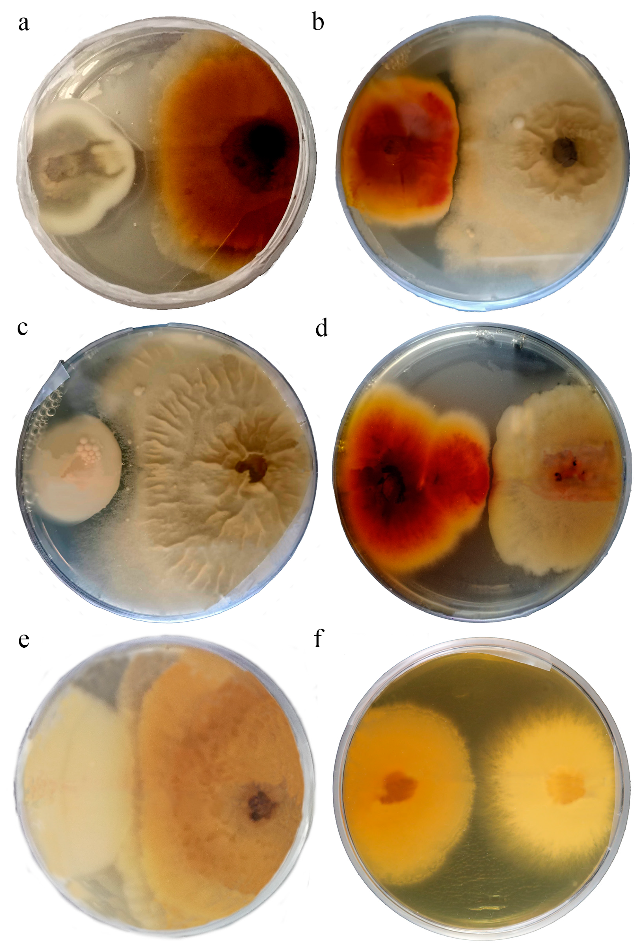
3.3. Interaction Tests

To investigate endophytic fungi as potential biocontrol agents, we assessed their bioactivity against phytopathogenic fungi using two distinct methods. First, we employed a dual-culture technique, followed by evaluating the impact of non-volatile metabolites produced by the endophytes on the mycelial growth of the pathogens through an agar diffusion method.
Seven endophytic strains, namely E150 (Epicoccum sp., OTU 4), E157 (Aureobasidium pullulans, OTU 15), E545 (Plectosphaerella cucumerina, OTU 27), E149 (Stemphylium vesicarium, OTU 6), E343 (Periconia byssoides, OTU12), E536 (Talaromyces wortmannii, OTU43), and E425 (Nigrospora sphaerica, OTU38), were tested against the four pathogenic fungi (Alternaria sp., Fusarium oxysporum, Botrytis cinerea, and Sclerotinia sclerotiorum). The endophytic strains were selected among species not widely known as plant pathogens and as belonging to different phylogenetic groups. In particular, we focused on fungi belonging to the most represented classes (Dothideomycetes, Sordariomycetes, and Eurotiomycetes; see Table 2). Also, we selected both strains occurring in all the different plant tissues and strains with a different tissue specificity. For example, some strains (OTU4 and OTU15) are essentially ubiquitous, whereas others (OTU27, 12, 43, and 38) are tissue-specific.
Results of the interactions between endophytic and pathogen mycelia using both methodologies are shown in Table 4. All the endophytic strains showed antagonistic effects towards at least one pathogen. In the dual cultures, the inhibition appeared as a non-reciprocal contact between the endophyte and the pathogen, with the pathogen mycelium curling and growing slower than the control along the interaction line with the endophyte (Figure 2a–d). In the absence of inhibition, both fungi grew into each other without any visible signs of interaction (Figure 2e,f). Concerning the agar diffusion method, the percentages of inhibition are reported in Table 4. The highest percentage of inhibition was shown by P. cucumerina against Alternaria sp. (Figure 3a,b). An inhibition signal was evidenced also in the dual culture of these mycelia.

4. Discussion

Wild relatives of crop plants are highly valued and exploited, compared to their domesticated counterparts, for genes that confer increased resistance to biotic and abiotic stresses. Wild plants host a wide range of microorganisms, including some beneficial species that are absent or under-represented in the domesticated crops [28] and that provide such special traits for adaptation and resistance to stressing environmental conditions [29]. In fact, it has been largely reported that endophytes isolated from underutilized crops or their wild relatives exhibit higher diversity and richness than those found in related cultivars [30]. Furthermore, when introduced as bioinoculants, these endophytes aid cultivars in overcoming adverse conditions [31]. In this study, we exploited Italian wild hop accessions previously characterized in a population genetics study [32] to isolate and characterize their fungal endophytic communities living in the different plant tissues (roots, stems, leaves, and flowers). The fungal diversity was compared across ten wild productive sites in Central Italy. A biocontrol potential activity against common phytopathogenic fungi was unveiled for some of the isolated fungi. An ex situ conserved collection of endophytic fungal strains was established for further bioactivity tests.

4.1. Diversity of Hop Endophytic Fungi

The ITS region employed here as a barcode for the identification of endophytic fungi does not exhibit uniform variability across all fungal groups. Insufficient variability in the ITS region can pose challenges for species-level identification, particularly within certain species-rich genera of Ascomycota, such as Alternaria, Aspergillus, Cladosporium, Penicillium, and Fusarium ([33] and references therein). This is the reason why, in this work, species names were assigned to 37 out of the 51 OTUs, whereas 14 OTUs were identified at the genus level only. The large majority of fungal strains identified among the 51 OTUs belong to Ascomycota (88%), with a predominance of the Dothideomycetes and Sordariomycetes classes, whereas Basidiomycota and Mucoromycota represented the remaining 9.8 and 2%, respectively. The prevalence of Ascomycota among fungi colonizing both aboveground and belowground plant tissues is widely documented in different species [34], including other species of the Cannabaceae family [35]. Consistent with this, Ascomycota appear to possess superior adaptations compared to Basidiomycota and other phyla for colonizing internal plant tissues [36]. Interestingly, in the present work, Basidiomycota were absent in hop flowers and stems. Additionally, Alternaria sp. emerged as the dominant taxon in all tissues except for the roots, where it was completely absent. This fungus is among the most widespread plant endophytic species [37] and was previously reported as a dominant endophyte in different plant species [38,39]. Alternaria alternata is known to produce for the host plant the growth regulator indole-acetic acid [39], a key molecule for important physiological processes such as cell division or cell elongation, tissue differentiation, phototrophic or geotropic responses, and all subsequent effects on plant growth and development [40]. Epicoccum sp. dominated in leaves slightly more than Alternaria sp. Epicoccum spp. are ubiquitous ascomycetes known to produce diverse classes of biologically active secondary metabolites holding cytotoxic, anticancer, antimicrobial, and anti-diabetic activities [41]. As evidenced by the FungalTraits characterization, Epicoccum strains may be plant pathogens, saprotrophs, or endophytes. Some endophytic species of Epicoccum have been demonstrated to have biological control activity against various plant pathogens [42,43]. Given that certain Epicoccum strains exhibit dual roles as both pathogens and biological control agents, understanding the pathogenic potential of the fungal strain is of paramount importance [44]. Ilyonectria macrodydima was the most dominant species in roots and was exclusively found in this tissue. This fungus is not commonly found at the endophytic status; rather, it can cause root rot in olive [45] and black-foot disease in grapevine [46], a serious disease in most wine- and grape-producing regions of the world. In line with our findings, I. macrodydima was previously identified as the most abundant fungus in roots, even in wild grapevines [47].
Interestingly, these and most of the other dominant species (Diaporthe spp., P. cucumerina, F. oxysporum, and B. cinerea) are well-known phytopathogenic fungi. Indeed, concerning the lifestyle and trophic modes of the identified fungal genera, we observed that, according to the FungalTraits database, most of the OTUs (71%) belong to plant pathogenic genera as for their primary lifestyle. This outcome is unsurprising, given that we isolated fungi from wild accessions, which, being surrounded by several plant species, are likely to be significantly exposed to a greater diversity of fungi compared to cultivated hops. We are currently engaged in a dedicated research project to test this hypothesis further by comparing fungal communities associated to cultivated and wild hops through metabarcoding approaches. The abundance of pathogenic fungal species could also mean that, although we isolated mycelia from healthy tissues and adopted the classical procedures for the isolation of plant endophytic fungi, these strains may not strictly be endophytic. As Schulz and Boyle [48] suggested, the endophytic condition should be viewed as a temporary status since plant–endophyte interaction may change over time depending on several factors. Endophytic fungi may in fact behave as latent pathogens that, because of physiological changes in the host, such as abiotic stress, growing stages, and interaction with other microorganisms [49], might switch from a symptomless, endophytic condition to a pathogenic stage. In addition, virulence genes can be activated or deactivated by mutations [50]. For these reasons, despite the recognized beneficial effects of many of the isolated species, the occurrence of such a large number of potentially pathogenic fungi is somewhat concerning since it may indicate the emergence of phytopathogens in wild hops in Italian and, more broadly, Mediterranean environmental conditions. This could potentially jeopardize ongoing and future hop cultivations in these regions.
We observed a tissue-specific pattern for some taxa. Dothideomycetes were exclusively found in the aerial parts of the plants, primarily represented by Pleosporales. Conversely, roots were predominantly colonized by Sordariomycetes, particularly taxa belonging to Hypocreales. Moreover, roots shared only three out of fifteen OTUs with other tissues, whereas the other tissues shared more OTUs among each other. Several species showed tissue-specificity: considering the most abundant species, notable examples include B. cinerea in the cones, I. macrodidyma in the roots, Bipolaris sorokiniana in the leaves, and Cladosporium sp. in both leaves and cones. Such a tissue-specific pattern has been observed across various plant species [15,51], likely influenced by specific ecological challenges, such as phytopathogens and other biotic/abiotic stresses, encountered by the different plant organs. We would like to point out that the main goal of this study was to establish a strain collection aimed at identifying potential biocontrol agents. Therefore, the distribution of the different taxa in hop plants presented here is preliminary. Currently, a study utilizing a metabarcoding approach is underway to comprehensively investigate the overall biodiversity of hop endophytic fungi, encompassing variations across different sites, seasons, and plant tissues.

4.2. Antifungal Activities of the Isolated Strains

A major goal outlined by the European Union (EU) is the conservation of biodiversity for the planet health. This is pursued through measures such as strengthening protected areas, restoring degraded ecosystems via the promotion of organic farming and reforestation, and ensuring sustainability in food production. Therefore, the directives of the European Green Deal aim to reduce the reliance on pesticides, antimicrobials, and fertilizers in agriculture to combat plant diseases. Instead, there is a prioritization of biological control methods over synthetic compounds. Hence, particular attention has been paid to the endophytic fungal diversity of crop plants for the potential use of these fungi as biocontrol agents for the management of plant diseases with a low impact to the environment, as they allow the reduction in agrochemicals and fertilizers [13]. In this study, a preliminary screening of the biocontrol potential of the fungal strains isolated from hop was performed using four distinct phytopathogenic fungi as target species. Alternaria sp. and F. oxysporum were chosen, as they are common pathogens affecting various plant species, including the potential threat they pose to H. lupulus, as indicated by our findings. Additionally, B. cinerea and S. sclerotiorum were included in the tests owing to their widespread presence as plant pathogens known to impact H. lupulus as well, where they can induce grey mold disease in hop cones, especially in heavy rainfall seasons. This species is also known as a primary cause of post-harvest infections of numerous agricultural commodities. Consequently, the biocontrol of B. cinerea holds significance not only during cultivation but also in the stages of harvesting and distributing the final product. Notably, we utilized strains of these species previously isolated from diseased leaves of Vitis vinifera. The fungal strains tested against the pathogens were selected from different classes and orders and from different plant tissues. In most cases, there was consistency between the results obtained from the two different inhibition assays. They both either showed positive or negative reactions of the endophyte towards the pathogen (Table 4). However, it is worth noting that in 10 out of the 28 endophyte/pathogen combinations, the two tests yielded contrasting results. In some instances, inhibition was observed with the agar diffusion method but not with the dual-culture method. This suggests that under in vitro optimal growing conditions, reciprocal contact between the mycelia may not be sufficient to elicit an inhibition reaction. Instead, achieving inhibition may require a high concentration of bioactive molecules produced by the endophyte over an extended period (two weeks in our experiments), regardless of the presence of the pathogen. On the contrary, the detection of inhibition signals exclusively in co-culture conditions suggests that the presence of the pathogen stimulates the endophyte to synthesize bioactive compounds. The most susceptible pathogenic fungi, inhibited by nearly all the tested endophytes, were S. sclerotiorum and Alternaria sp. This evidence suggests the opportunity to individually test these endophytes and their consortia, both under greenhouse and field conditions, to evaluate their potential as biocontrol agents for plants challenged with S. sclerotiorum or Alternaria spp. Such experiments will enable the identification of suitable SynComs for the management of plant diseases. Among the tested interactions, the strongest inhibition signal was evidenced by the growth medium of P. cucumerina against Alternaria sp., with a 41% growth inhibition. The dual-culture method confirmed this result. P. cucumerina is widely known as a plant pathogen capable of causing sudden death and blight disease in a variety of crops [52] rather than as an endophyte with potential protective properties against other fungal pathogens. Interestingly, nematocidal [53] and antibacterial [54] properties have been reported for P. cucumerina. Thus, our evidence is quite unexpected and points to the possible new potential of this species in the biological control of plant diseases caused by pathogenic fungi. Additionally, A. pullulans showed another significant bioactive role against B. cinerea as evidenced by both methods, although B. cinerea was the least susceptible pathogen among those tested against our panel of endophytic fungi. A. pullulans is a ubiquitous saprophytic, yeast-like fungus with a high biotechnological potential. It has been reported as an antifungal agent against post-harvest pathogens of fruit and vegetables (e.g., B. cinerea) [55] and an effective biocontrol agent against airborne plant pathogens. All these attributes are of major relevance to the vitivinicultural sector [56] and could potentially be leveraged in the emerging hop sector as well. Epicoccum is among the dominant species in the hop plants analyzed, with the 40 strains isolated from different tissues belonging to a single OTU. One of these strains exhibited bioactivity against B. cinerea and S. sclerotiorum using the dual-culture method and against Alternaria sp. using the agar diffusion method. Previous studies demonstrated that certain endophytic species of Epicoccum possess biological control activity against various plant pathogens, including B. cinerea, S. sclerotiorum, and Alternaria sp. However, in plant ecosystems, Epicoccum species can function as endophytes, saprophytes, or pathogens. Therefore, it is advisable to assess the pathogenic potential of an Epicoccum isolate before evaluating its effectiveness as a biological control agent against a specific pathogen in order to prevent disease development and minimize plant yield losses [43]. The other tested strains, including S. vesicarium, P. byssoides, T. wortmannii, and N. sphaerica, showed potential antagonisms against the tested pathogenic fungi. Although some antifungal activities were previously reported for these species [57,58,59], to the best of our knowledge, they were tested against different pathogenic fungi compared to those used in this study.
Overall, the preliminary results obtained in this work regarding the biological activities of the isolated fungi suggest that these strains could be novel resources of antifungal metabolites to be exploited in the sustainable management of crop diseases not only in the emerging hop sector in Italy but also in other agricultural sectors. Moving forward, our future endeavors will focus on exploring the biocontrol potential of additional fungal strains isolated in this study. Moreover, it will be interesting to disclose the molecular mechanisms and the specific metabolites involved in these biological activities. For example, this could involve whole-genome sequencing and transcriptome analyses of selected endophytic fungal species. As an example, these studies could be focused on Eurotiomycetes fungi, such as Talaromyces spp., which are well known to produce secondary metabolites with antibacterial activity [60,61] but scarcely known for their antifungal potential.

5. Conclusions

To our knowledge, this is the first comprehensive characterization of endophytic fungal communities of the different tissues of wild hop (H. lupulus) plants. Interaction tests allowed us to identify interesting strains with a biocontrol activity on phytopathogenic fungi. All the isolated strains were conserved ex situ for further bioactivity tests and production of metabolites of agronomical and biotechnological interest. This paper provides original data with a key relevance for the environmentally friendly management of plant diseases.

Supplementary Materials

The following supporting information can be downloaded at https://www.mdpi.com/article/10.3390/d17020094/s1. Table S1: FungalTraits.

Author Contributions

Conceptualization, C.R., A.R. and B.B.; methodology, A.R.; formal analysis, A.R. and M.C.; investigation, C.R., A.R., B.B. and M.C.; writing—original draft preparation, C.R.; writing—review and editing, A.R and B.B. All authors have read and agreed to the published version of the manuscript.

Funding

This research was partially funded by the project “BioMemory—Network of scientific collections for bio-monitoring, biodiversity conservation, agri-food and environmental sustainability, and well-being” (https://biomemory.cnr.it), funded by the Department of Biology, Agriculture, and Food Sciences (DiSBA), National Research Council of Italy (CNR), project no. SAC. AD002.173.

Institutional Review Board Statement

Not applicable.

Data Availability Statement

An ex situ conserved collection of endophytic fungal strains was established at CNR-IBBR-Perugia as part the BioMemory Project (https://biomemory.cnr.it/collections/CNR-IBBR-FABI). Sequence data were submitted to GenBank (Accession no. OQ257045 to OQ257306).

Conflicts of Interest

The authors declare no conflicts of interest.

References

  1. Zanoli, M.; Zavatti, M. Pharmacognostic and pharmacological profile of Humulus lupulus L. J. Ethnopharmacol. 2008, 116, 383–396. [Google Scholar] [CrossRef]
  2. Lamy, V.; Roussi, S.; Chaabi, M.; Gossé, F.; Schall, N.; Lobstein, A.; Raul, F. Chemopreventive effects of lupulone, a hop b-acid, on human colon cancer-derived metastatic SW620 cells and in a rat model of colon carcinogenesis. Carcinogenesis 2007, 28, 1575–1581. [Google Scholar] [CrossRef]
  3. Liu, M.; Hansen, P.E.; Wang, G.; Qiu, L.; Dong, J.; Yin, H.; Qian, Z.; Yang, M.; Miao, J. Pharmacological Profile of Xanthohumol, a Prenylated Flavonoid from Hops (Humulus lupulus). Molecules 2015, 20, 754–779. [Google Scholar] [CrossRef]
  4. Van Cleemput, M.; Cattoor, K.; De Bosscher, K.; Haegeman, G.; De Keukeleire, D.; Heyerick, A. Hop (Humulus lupulus)-derived bitter acids as multipotent bioactive compounds. J. Nat. Prod. 2009, 72, 1220–1230. [Google Scholar] [CrossRef]
  5. Licciardo, F.; Carbone, K.; Ievoli, C.; Manzo, A.; Tarangioli, S. Outlook Economico-Statistico del Comparto Luppolo. 2021. Available online: https://www.reterurale.it/flex/cm/pages/ServeBLOB.php/L/IT/IDPagina/22822 (accessed on 25 October 2024).
  6. Gargani, E.; Ferretti, L.; Faggioli, F.; Haegi, A.; Luigi, M.; Landi, S.; Simoni, S.; Benvenuti, C.; Guidi, S.; Simoncini, S.; et al. A survey on pests and diseases of Italian hop crops. Italus Hortus 2018, 24, 1–17. [Google Scholar] [CrossRef]
  7. Rossini, F.; Virga, G.; Loreti, P.; Iacuzzi, N.; Ruggeri, R.; Provenzano, M.E. Hops (Humulus lupulus L.) as a Novel Multipurpose Crop for the Mediterranean Region of Europe: Challenges and Opportunities of Their Cultivation. Agriculture 2021, 11, 484. [Google Scholar] [CrossRef]
  8. Porteous-Álvarez, A.J.; Maldonado-González, M.M.; Mayo-Prieto, S.; Lorenzana, A.; Paniagua-García, A.I.; Casquero, P.A. Green Strategies of Powdery Mildew Control in Hop: From Organic Products to Nanoscale Carriers. J. Fungi 2021, 7, 490. [Google Scholar] [CrossRef] [PubMed]
  9. Carroll, G.C. The biology of endophytism in plants. In Microbiology of the Phyllosphere; Fokkema, N.J., van den Heuvel, J., Eds.; Cambridge University Press: Cambridge, UK, 1986; pp. 205–222. [Google Scholar]
  10. Baron, N.C.; Rigobelo, E.C. Endophytic fungi: A tool for plant growth promotion and sustainable agriculture. Mycology 2022, 13, 39–55. [Google Scholar] [CrossRef] [PubMed]
  11. Deka, D.; Tayung, K.; Jha, D.K. Harnessing Fungal Endophytes for Plant and Human Health. In Endophytes: Biology and Biotechnology; Maheshwari, D.K., Ed.; Springer: Cham, Switzerland, 2017; Volume 1, pp. 59–98. [Google Scholar]
  12. Fontana, D.C.; de Paula, S.; Torres, A.G.; de Souza, V.H.M.; Pascholati, S.F.; Schmidt, D.; Dourado Neto, D. Endophytic Fungi: Biological Control and Induced Resistance to Phytopathogens and Abiotic Stresses. Pathogens 2021, 10, 570. [Google Scholar] [CrossRef]
  13. De Silva, N.I.; Brooks, I.; Lumyong, S.; Hyde, K.D. Use of endophytes as biocontrol agents. Fungal Biol. Rev. 2019, 33, 133–148. [Google Scholar] [CrossRef]
  14. Porteous-Álvarez, A.J.; Fernández-Marcos, A.; Ramírez-Lozano, D.; Mayo-Prieto, S.; Cardoza, R.E.; Gutiérrez, S.; Casquero, P.A. Native Trichoderma Isolates from Soil and Rootstock to Fusarium spp. Control and Growth Promotion of Humulus lupulus L. Plantlets. Agriculture 2023, 13, 720. [Google Scholar] [CrossRef]
  15. Belfiori, B.; Rubini, A.; Riccioni, C. Diversity of endophytic and pathogenic fungi of saffron (Crocus sativus) plants from cultivation sites in Italy. Diversity 2021, 13, 535. [Google Scholar] [CrossRef]
  16. Arnold, A.E.; Lutzoni, F. Diversity and Host Range of Foliar Fungal Endophytes: Are Tropical Leaves Biodiversity Hotspots? Ecology 2007, 88, 541–549. [Google Scholar] [CrossRef]
  17. Gardes, M.; Bruns, T.D. ITS primers with enhanced specificity for basidiomycetes application to the identification of mycorrhizae and rusts. Mol. Ecol. 1993, 2, 113–118. [Google Scholar] [CrossRef]
  18. White, T.J.; Bruns, T.; Lee, S.; Taylor, J. Amplification and direct sequencing of fungal ribosomal RNA genes for phylogenetics. In PCR Protocols. A Guide to Methods and Applications; Innis, M.A., Gelfand, D.H., Sninsky, J.J., White, T.J., Eds.; Academic Press: San Diego, CA, USA, 1990; pp. 315–322. [Google Scholar]
  19. Rubini, A.; Paolocci, F.; Granetti, B.; Arcioni, S. Single Step Molecular Characterization of Morphologically Similar Black Truffle Species. FEMS Microbiol. Lett. 1998, 164, 7–12. [Google Scholar] [CrossRef]
  20. Altschul, S.F.; Gish, W.; Miller, W.; Myers, E.W.; Lipman, D.J. Basic Local Alignment Search Tool. J. Mol. Biol. 1990, 215, 403–410. [Google Scholar] [CrossRef]
  21. Huang, Y.; Niu, B.; Gao, Y.; Fu, L.; Li, W. CD-HIT Suite: A Web Server for Clustering and Comparing Biological Sequences. Bioinformatics 2010, 26, 680–682. [Google Scholar] [CrossRef]
  22. Põlme, S.; Abarenkov, K.; Henrik Nilsson, R.; Lindahl, B.D.; Clemmensen, K.E.; Kauserud, H.; Nguyen, N.; Kjøller, R.; Bates, S.T.; Baldrian, P.; et al. FungalTraits: A user-friendly traits database of fungi and fungus-like stramenopiles. Fungal Divers. 2020, 105, 1–16. [Google Scholar] [CrossRef]
  23. Katoh, K.; Standley, D.M. MAFFT multiple sequence alignment software version 7: Improvements in performance and usability. Mol. Biol. Evol. 2013, 30, 772–780. [Google Scholar] [CrossRef]
  24. Stamatakis, A. RAxML Version 8: A Tool for Phylogenetic Analysis and Post-Analysis of Large Phylogenies. Bioinformatics 2014, 30, 1312–1313. [Google Scholar] [CrossRef]
  25. Rivera-Orduña, F.N.; Suarez-Sanchez, R.A.; Flores-Bustamante, Z.R.; Gracida-Rodriguez, J.N.; Flores-Cotera, L.B. Diversity of Endophytic Fungi of Taxus Globosa (Mexican yew). Fungal Divers. 2011, 47, 65–74. [Google Scholar] [CrossRef]
  26. Hajieghrari, B.; Torabi-Giglou, M.; Mohammadi, M.R.; Davari, M. Biological potential of some Iranian Trichoderma isolates in the control of soil borne plant pathogenic fungi. Afr. J. Biotechnol. 2008, 7, 967–972. [Google Scholar]
  27. Edington, L.V.; Khew, K.L.; Barron, G.I. Fungitoxic spectrum of benzimidazole compounds. Phytopathology 1971, 61, 42–44. [Google Scholar] [CrossRef]
  28. Ofek-Lalzar, M.; Gur, Y.; Ben-Moshe, S.; Sharon, O.; Kosman, E.; Mochli, E.; Sharon, A. Diversity of fungal endophytes in recent and ancient wheat ancestors Triticum dicoccoides and Aegilops sharonensis. FEMS Microbiol. Ecol. 2016, 92, 1–11. [Google Scholar] [CrossRef]
  29. Mili, C.; Roy, S.; Tayung, K. Endophytic Fungi of Wild and Domesticated Crop Plants and Their Prospect for Applications in Sustainable Agriculture. In Endophytes; Patil, R.H., Maheshwari, V.L., Eds.; Springer: Singapore, 2021; pp. 21–35. [Google Scholar] [CrossRef]
  30. Kernaghan, G.; Mayerhofer, M.; Griffin, A. Fungal endophytes of wild and hybrid Vitis leaves and their potential for vineyard biocontrol. Can. J. Microbiol. 2017, 63, 583–595. [Google Scholar] [CrossRef]
  31. Murphy, B.R.; Doohan, F.M.; Hodkinson, T.R. From concept to commerce: Developing a successful fungal endophyte inoculant for agricultural crops. J. Fungi 2018, 4, 24. [Google Scholar] [CrossRef]
  32. Riccioni, C.; Belfiori, B.; Sileoni, V.; Marconi, O.; Perretti, G.; Bellucci, M.; Rubini, A. High genetic and chemical diversity of wild hop populations from Central Italy with signals of a genetic structure influenced by both sexual and asexual reproduction. Plant Sci. 2021, 304, 110794. [Google Scholar] [CrossRef]
  33. Baturo-Ciesniewska, A.; Pusz, W.; Patejuk, K. Problems, limitations, and challenges in species identifcation of Ascomycota members on the basis of ITS regions. Acta Mycol. 2020, 55, 1–18. [Google Scholar] [CrossRef]
  34. Trivedi, P.; Leach, J.E.; Tringe, S.G.; Sa, T.; Singh, B.K. Plant–microbiome interactions: From community assembly to plant health. Nat. Rev. Microbiol. 2020, 18, 607–621. [Google Scholar] [CrossRef]
  35. Kusari, P.; Kusari, S.; Spiteller, M.; Kayser, O. Endophytic fungi harbored in Cannabis sativa L.: Diversity and potential as biocontrol agents against host plant-specific phytopathogens. Fungal Divers. 2013, 60, 137–151. [Google Scholar] [CrossRef]
  36. Rashmi, M.; Kushveer, J.S.; Sarma, V.V. A worldwide list of endophytic fungi with notes on ecology and diversity. Mycosphere 2019, 10, 798–1079. [Google Scholar] [CrossRef]
  37. Shipunov, A.; Newcombe, G.; Raghavendra, A.K.; Anderson, C.L. Hidden diversity of endophytic fungi in an invasive plant. Am. J. Bot. 2008, 95, 1096–1108. [Google Scholar] [CrossRef] [PubMed]
  38. Khalmuratova, I.; Choi, D.H.; Woo, J.R.; Jeong, M.J.; Oh, Y.; Kim, Y.G.; Kim, J.G. Diversity and plant growth-promoting effects of fungal endophytes isolated from salt-tolerant plants. J. Microbiol. Biotechnol. 2020, 30, 1680. [Google Scholar] [CrossRef] [PubMed]
  39. Khan, A.R.; Ullah, I.; Waqas, M.; Shahzad, R.; Hong, S.J.; Park, G.S.; Jung, B.K.; Lee, I.J.; Shin, J.H. Plant growth-promoting potential of endophytic fungi isolated from Solanum nigrum leaves. World J. Microbiol. Biotechnol. 2015, 31, 1461–1466. [Google Scholar] [CrossRef]
  40. Teale, W.D.; Paponov, I.A.; Palme, K. Auxin in action: Signalling, transport and the control of plant growth and development. Nat. Rev. Mol. Cell Biol. 2006, 7, 847–859. [Google Scholar] [CrossRef]
  41. Fatima, N.; Ismail, T.; Muhammad, S.A.; Jadoon, M.; Ahmed, S.; Azhar, S.; Mumtaz, A. Epicoccum sp., an emerging source of unique bioactive metabolites. Acta Pol. Pharm. 2016, 73, 13–21. [Google Scholar]
  42. Braga, R.M.; Padilla, G.; Araújo, W.L. The biotechnological potential of Epicoccum spp.: Diversity of secondary metabolites. Crit. Rev. Microbiol. 2018, 44, 759–778. [Google Scholar] [CrossRef]
  43. Bagy, H.M.K.; Hassan, E.A.; Nafady, N.A.; Dawood, M.F. Efficacy of arbuscular mycorrhizal fungi and endophytic strain Epicoccum nigrum ASU11 as biocontrol agents against blackleg disease of potato caused by bacterial strain Pectobacterium carotovora subsp. atrosepticum PHY7. Biol. Control 2019, 134, 103–113. [Google Scholar] [CrossRef]
  44. Taguiam, J.D.; Evallo, E.; Balendres, M.A. Epicoccum species: Ubiquitous plant pathogens and effective biological control agents. Eur. J. Plant Pathol. 2021, 159, 713–725. [Google Scholar] [CrossRef]
  45. Úrbez-Torres, J.R.; Peduto, F.; Gubler, W.D. First report of Ilyonectria macrodidyma causing root rot of olive trees (Olea europaea) in California. Plant Dis. 2012, 96, 1378. [Google Scholar] [CrossRef]
  46. Dos Santos, R.F.; Blume, E.; Muniz, M.F.B.; Heckler, L.I.; Finger, G.; Maciel, C.G.; Garrido, L.D.R. First report of Ilyonectria macrodidyma associated with black foot disease of grapevine in Brazil. Plant Dis. 2014, 98, 156. [Google Scholar] [CrossRef] [PubMed]
  47. Radić, T.; Likar, M.; Hančević, K.; Regvar, M.; Čarija, M.; Zdunić, G. Root-associated community composition and co-occurrence patterns of fungi in wild grapevine. Fungal Ecol. 2021, 50, 101034. [Google Scholar] [CrossRef]
  48. Schulz, B.; Boyle, C. What Are Endophytes? In Microbial Root Endophytes; Schulz, B.J.E., Boyle, C.J.C., Sieber, T.N., Eds.; Springer: Berlin, Germany, 2006; pp. 1–13. [Google Scholar]
  49. Schulz, B.; Römmert, A.K.; Dammann, U.; Aust, H.J.; Strack, D. The Endophyte-Host Interaction: A Balanced Antagonism? Mycol. Res. 1999, 103, 1275–1283. [Google Scholar] [CrossRef]
  50. Wilson, D. Endophyte: The Evolution of a Term, and Clarification of Its Use and Definition. Oikos 1995, 73, 274–276. [Google Scholar] [CrossRef]
  51. Wearn, J.A.; Sutton, B.C.; Morley, N.J.; Gange, A.C. Species and organ specificity of fungal endophytes in herbaceous grassland plants. J. Ecol. 2012, 100, 1085–1092. [Google Scholar] [CrossRef]
  52. Pétriacq, P.; Stassen, J.H.M.; Ton, J. Spore Density Determines Infection Strategy by the Plant Pathogenic Fungus Plectosphaerella cucumerina. Plant Physiol. 2016, 170, 2325–2339. [Google Scholar] [CrossRef]
  53. Girardi, N.; Sosa, A.L.; Loyola García, J.; Pellegrino, M.; Passone, M.A. Ecophysiological characteristics of the nematophagous fungus, Plectosphaerella plurivora, with biocontrol potential on Nacobbus aberrans sl in tomato. Eur. J. Plant Pathol. 2023, 167, 867–881. [Google Scholar] [CrossRef]
  54. Zhou, J.; Bi, S.; Chen, H.; Chen, T.; Yang, R.; Li, M.; Fu, Y.; Jia, A.Q. Anti-biofilm and antivirulence activities of metabolites from Plectosphaerella cucumerina against Pseudomonas aeruginosa. Front. Microbiol. 2017, 8, 769. [Google Scholar] [CrossRef]
  55. Prasongsuk, S.; Lotrakul, P.; Ali, I.; Bankeeree, W.; Punnapayak, H. The current status of Aureobasidium pullulans in biotechnology. Folia Microbiol. 2018, 63, 129–140. [Google Scholar] [CrossRef]
  56. Bozoudi, D.; Tsaltas, D. The multiple and versatile roles of Aureobasidium pullulans in the vitivinicultural sector. Fermentation 2018, 4, 85. [Google Scholar] [CrossRef]
  57. Azhari, A.; Supratman, U. The chemistry and pharmacology of fungal genus Periconia: A review. Sci. Pharm. 2021, 89, 34. [Google Scholar] [CrossRef]
  58. Manoch, L.; Dethoup, T. A potential use of Talaromyces species as biological agents against plant pathogenic fungi. Thai J. Agric. Sci. 2011, 44, 81–91. [Google Scholar]
  59. Wu, Z.; Xie, Z.; Wu, M.; Li, X.; Li, W.; Ding, W.; Li, C. New antimicrobial cyclopentenones from Nigrospora sphaerica ZMT05, a fungus derived from Oxya chinensis Thunber. J. Agric. Food Chem. 2018, 66, 5368–5372. [Google Scholar] [CrossRef] [PubMed]
  60. Yilmaz, N.; Visagie, C.M.; Houbraken, J.; Frisvad, J.C.; Samson, R.A. Polyphasic taxonomy of the genus Talaromyces. Stud. Mycol. 2014, 78, 175–341. [Google Scholar] [CrossRef]
  61. Lan, D.; Wu, B. Chemistry and bioactivities of secondary metabolites from the genus Talaromyces. Chem. Biodivers. 2020, 17, e2000229. [Google Scholar] [CrossRef]
Figure 1. ITS based phylogenetic trees of Sordariomycetes (a) and Dothideomycetes (b), showing the position of the OTUs identified in this study (in bold). GenBank accession numbers of sequences downloaded from GenBank are reported near the taxa names. Numbers near the branches indicate bootstrap values (percentage over 1000 replicates).
Diversity 17 00094 g001aDiversity 17 00094 g001b
Figure 2. Dual-culture interaction tests between selected endophytic strains (left side of the plates) and pathogenic fungi (right side of the plates). (a) Periconia byssoides (E343) vs. S. sclerotiorum (AR397s2); (b) Epicoccum sp. (E150) vs. B. cinerea (AR593); (c) A. pullulans (E157) vs. B. cinerea (AR593); (d) Epicoccum sp. (E150) vs. S. sclerotiorum (AR397s2); (e) A. pullulans (E157) vs. S. sclerotiorum (AR397s2); (f) Epicoccum sp. (E150) vs. F. oxysporum (E529).
Diversity 17 00094 g002
Figure 3. Agar diffusion method interaction test of the pathogenic strain Alternaria sp. (E140) growing in media added (a) or not (b) with culture broth of the endophyte E545.
Diversity 17 00094 g003
Table 1. Sampling sites and number of isolates from the different sites and tissues.
Sampling
Site
LocalityRegionLatitudeLongitudeNo. Isolates per Tissue
RootsStemsLeavesConesMale
Flowers
Total
1Ponte San GiovanniUmbria43.09205612.461544 4225 31
2ValfabbricaUmbria43.32752012.718487 1211 23
3Città di CastelloUmbria43.45761512.2304731061623762
4BelfioreUmbria42.98165112.734363551014 34
5Nera RiverUmbria42.73492112.833146 419 23
6PiedilucoUmbria42.53072712.733482129255 51
7OrvietoUmbria42.69795912.211644 23 5
8Bolsena LakeLazio42.63857311.889101 73414
9Potenza RiverMarche43.24661113.219093 5 5
10Penna San GiovanniMarche43.04982213.464626 14 14
Total no. isolates27249810211262
Table 2. Description of the isolated OTUs, their distribution in the different tissues of Humulus lupulus, relative abundance, and dominant species.
IsolatesClosest Match in GenBankNo. of IsolatesPi
OTUStrainAccession
No.
ClassificationAccession
No.
Similarity
(%)
RootsStemsLeavesConesMale
Flowers
TotalRootsStemsLeavesConesMale Flowers
ASCOMYCOTA
DOTHIDEOMYCETES
Pleosporales
1E140OQ257068Alternaria sp.AF347031100 72345782 0.304 *0.235 *0.447 *0.636 *
2E333OQ257172Alternaria sp.HG93647796 1 1 0.010
3E332OQ257171Alternaria sp.AF34703194.8 1 1 0.010
4E150OQ257077Epicoccum sp.HQ630972100 5249240 0.174 *0.245 *0.087 *0.182
5E137OQ257065Alternaria infectoriaMK46106199.69 2215 0.0200.0190.091
6E149OQ257076Stemphylium vesicariumMK46101899.68 22 4 0.087 *0.020
7E302OQ257143Bipolaris sorokinianaKU194490100 3 3 0.031
8E354OQ257187Periconia macrospinosaJX98148299.8 2 2 0.019
9E358OQ257191Pithomyces chartarumMH860227100 1 1 0.010
10E573OQ257304Neosetophoma italicaKP71135699.84 1 1 0.043
11E165OQ257091Parastagonospora nodorumKX92883098.89 1 1 0.010
12E343OQ257177Periconia byssoidesKC95415799.8 1 1 0.010
13E307OQ257148Pyrenophora tritici-repentisKT69257199.6 1 1 0.010
14E194OQ257100Neodidymelliopsis cannabisMH85905799.08 1 1 0.010
50E546OQ257285Paraphoma sp.DQ42098091 1 1 0.043
51E375OQ257207Sporormiella intermediaJX13624999.10 1 1 0.010
Dothideales
15E157OQ257083Aureobasidium pullulansFN868454100 172 10 0.0430.0710.019
Cladosporiales
16E159OQ257085Cladosporium sp.HQ631003100 1112124 0.112 *0.117 *0.091
Mycosphaerellales
17E559OQ257293Mycosphaerella sp.EU167596100 1 1 0.010
Botryosphaeriales
18E431OQ257234Diplodia sapineaMF39886698.1 1 1 0.010
SORDARIOMYCETES
Diaporthales
19E374OQ257206Diaporthe novemMH8645041001135 100.0370.0430.0310.049 *
20E126OQ257055Diaporthe oncostomaLN71454199.83 14 5 0.0430.041 *
21E367OQ257200Diaporthe sp.KJ482538100 31 4 0. 0310.010
22E321OQ257161Diaporthe foeniculinaAY620999100 1 2 3 0.043 0.019
23E518OQ257262Diaporthe columnarisMN45064099.82 20.074 *
24E494OQ257241Cytospora sp.AY61822998.4 1 1 0.043
Glomerellales
25E365OQ257198Colletotrichum coccodesAJ30198499.50 12 3 0.0100.019
26E346OQ257180Colletotrichum gloeosporioidesAJ301907100 11 2 0.0100.010
27E545OQ257284Plectosphaerella cucumerinaKF47213898.82 20.074 *
28E432OQ257235Colletotrichum karstiMW08118199.19 1 1 0.010
29E555OQ257291Colletotrichum acutatumAJ301971100 1 1 0.043
Hypocreales
30E569OQ257300Ilyonectria macrodidymaJN8594221007 70.259 *
31E542OQ257282Fusarium sp.MK40810299.81121 50.0370.0430.0200.010
32E347OQ257181Fusarium sambucinumKM23181399.81112 50.0370.0430.0100.019
33E529OQ257272Fusarium oxysporumMT45329699.822 1 30.074 * 0.010
34E523OQ257267Fusarium oxysporumAY9284181002 20.074 *
35E519OQ257263Fusarium verticillioidesKJ95778699.81 10.037
Xylariales
36E331OQ257170Nemania serpensKU141386100 1 1 0.010
37E521OQ257265Truncatella angustataMT51436899.61 10.037
38E425OQ257228Nigrospora sphaericaHQ608063100 1 1 0.010
LEOTIOMYCETES
Helotiales
39E235OQ257130Botrytis cinereaMH860108100 8 8 0.078 *
EUROTIOMYCETES
Eurotiales
40E554OQ257290Penicillium sp.KF3674971003 30.111 *
41E490OQ257238Penicillium sp.MN86127898.041 10.037
42E161OQ257087Aspergillus sp.MK46102299 1 1 0.010
43E536OQ257278Talaromyces wortmanniiNR_17203999.81 10.037
BASIDIOMYCOTA
AGARICOMYCETES
Polyporales
44E148OQ257075Hyphodermella rosaeMF47598399.84 1 1 0.010
Cantharellales
45E544OQ257283Rhizoctonia solaniMH86255799.691 10.037
TREMELLOMYCETES
Cystofilobasidiales
46E557OQ257292Itersonilia perplexansMH86189099.69 1 1 0.010
Tremellales
47E142OQ257070Vishniacozyma heimaeyensisKY105824100 1 1 0.010
48E143OQ257071Cryptococcus sp.EU85235999.8 1 1 0.010
MUCOROMYCOTA
MUCOROMYCETES
Mucorales
49E520OQ257264Mucor fragilisGU56627599.81 10.037
Total no. isolates27249810211262
Species richness14132520451
1/S 0.0670.0770.0400.0450.250
Pi = ratio number of isolates of one species/total isolates. * Dominant species (Pi > 1/S).
Table 3. Diversity of fungal strains in H. lupulus at different collection sites.
OTU No.TaxonSampling Sites
PVCBNPBoOPsPr
1Alternaria sp.67171571012 44
2Alternaria sp.1
3Alternaria sp.1
4Epicoccum sp.9313438
5Alternaria infectoria 121 1
6Stemphylium vesicarium2 2
7Bipolaris sorokiniana 2 1
8Periconia macrospinosa 2
9Pithomyces chartarum 1
10Neosetophoma italica 1
11Parastagonospora nodorum 1
12Periconia byssoides 1
13Pyrenophora tritici-repentis 1
14Neodidymelliopsis cannabis 1
39Botrytis cinerea 2 5 1
15Aureobasidium pullulans1 225
16Cladosporium sp.427 24 131
17Mycosphaerella sp. 1
18Diplodia sapinea 1
19Diaporthe novem24 121
20Diaporthe oncostoma3 2
21Diaporthe sp. 1 2 1
22Diaporthe foeniculina1 2
23Diaporthe columnaris 1 1
24Cytospora sp. 1
25Colletotrichum coccodes 11 1
26Colletotrichum gloeosporioides 1 1
27Plectosphaerella cucumerina 2
28Colletotrichum karsti 1
29Colletotrichum acutatum 1
30Ilyonectria macrodidyma 4 3
31Fusarium sp. 1 2 2
32Fusarium sambucinum 3 2
33Fusarium oxysporum 12
34Fusarium oxysporum 11
35Fusarium verticillioides 1
36Nemania serpens1
37Truncatella angustata 1
38Nigrospora sphaerica 1
40Penicillium sp. 3
41Penicillium sp. 1
42Aspergillus sp. 1
43Talaromyces wortmannii 1
44Hyphodermella rosae 1
45Rhizoctonia solani 1
46Itersonilia perplexans 1
47Vishniacozyma heimaeyensis 1
48Cryptococcus sp. 1
49Mucor fragilis 1
50Paraphoma sp. 1
51Sporormiella intermedia 1
Total no. isolates312362342351145145
Species richness111018158203482
P = Ponte S. Giovanni; V = Valfabbrica; C = Città di Castello; B = Belfiore; N = Nera River; Pi = Piediluco lake; Bo = Bolsena Lake; O = Orvieto; Ps = Penna S. Giovanni; Pr = Potenza River.
Table 4. In vitro antagonism of seven endophytic strains against four pathogenic fungi using dual-culture and crude extracts assays.
Isolate NamePATHOGENIC FUNGUS
E140_OTU1_Alternaria sp.E529_OTU33_F. oxysporumAR593_B. cinereaAR397s2_S. sclerotiorum
ENDOPHYTIC FUNGUS
E150_OTU4_Epicoccum sp.−/15.3−/0+/0+/0
E157_OTU15_Aureobasidium pullulans−/0−/0+/27.3−/12.5
E545_OTU27_Plectosphaerella cucumerina+/40.9−/0−/0−/0
E149_OTU6_Stemphylium vesicarium+/7.5−/12.0−/0−/10.9
E343_OTU12_Periconia byssoides+/0−/6.5−/0+/8.97
E536_ OTU43_Talaromyces wortmannii−/0+/5.6−/0−/8.97
E425_OTU38_Nigrospora sphaerica+/15.6−/3.2−/0+/14.5
Results of the dual cultures are reported as “+” (endophytic fungus inhibition) or “−” (no inhibition), followed by percentages of pathogen growth inhibition in the crude extract assays.
Disclaimer/Publisher’s Note: The statements, opinions and data contained in all publications are solely those of the individual author(s) and contributor(s) and not of MDPI and/or the editor(s). MDPI and/or the editor(s) disclaim responsibility for any injury to people or property resulting from any ideas, methods, instructions or products referred to in the content.

