Article

Recruitment and Controlled Growth of Juveniles of the Critically Endangered Fan Mussel Pinna nobilis in the Northern Adriatic

1
Marine Biology Station Piran, National Institute of Biology, Fornače 41, SI-6330 Piran, Slovenia
2
ARPAL, Regional Agency for the Protection of the Ligurian Environment, Via Bombrini 8, 16149 Genova, Italy
3
SHORELINE Soc. Coop, AREA Science Park, Padriciano, 99, 34149 Trieste, Italy
4
Aquarium Piran, University of Primorska, Kidričevo nabrežje 4, 6330 Piran, Slovenia
5
Department of Veterinary Medicine, University of Sassari, Via Vienna 2, 07100 Sassari, Italy
6
Department of Biomedical Sciences, University of Sassari, Viale San Pietro 43b, 07100 Sassari, Italy
7
Department of Life Sciences, University of Trieste, Via Licio Giorgieri 5, 34127 Trieste, Italy
*
Author to whom correspondence should be addressed.
Diversity 2025, 17(10), 666; https://doi.org/10.3390/d17100666
Submission received: 21 July 2025 / Revised: 17 September 2025 / Accepted: 17 September 2025 / Published: 23 September 2025

Abstract

The fan mussel Pinna nobilis is the largest bivalve species in the Mediterranean Sea and provides numerous ecosystem services. It is classified as critically endangered by IUCN (International Union for Conservation of Nature) due to severe mass mortality events throughout the Mediterranean. The aims of this work are as follows: (i) to assess the current recruitment potential of the species, (ii) to enhance recruitment by keeping juveniles in controlled conditions before releasing them back into the sea, and (iii) to assess the health status of recruits. In the period 2022–2023, larval collectors were set up in the Gulf of Trieste as part of the LIFE Pinna project. The collected individuals were kept in aquaria in two different facilities under different conditions: (a) a closed system with constant water temperature, live phytoplankton, and commercial food and (b) an open system with ambient seawater temperature and commercial food. A clear temporal and spatial variability in recruitment was observed: 13 recruits were found in 2022 and 50 recruits in 2023. The live specimens were between 0.5 and 8 cm in size upon collection and larger in 2023. The growth and survival rate did not differ significantly between the two systems, but the average monthly growth and survival rate were related to the initial size of the juveniles.

1. Introduction

The Mediterranean endemic fan mussel Pinna nobilis Linnaeus, 1758, is the largest bivalve in the Mediterranean Sea and one of the largest in the world. It provides many ecosystem services. It filters large quantities of seawater and retains a high percentage of organic matter [1], thus contributing to water clarity and quality. In addition, it provides hard substrate in soft-bottom areas, increasing habitat variability and providing a surface that can be colonised by other benthic invertebrates [2,3] and fish [2,4,5,6,7,8,9,10]. It also plays a key role in the food web, serving as a host for symbionts like the crustaceans Pontonia pinnophylax and Nepinnotheres pinnotheres [11,12] and as prey for other species, e.g., Octopus vulgaris [13] and Marthasterias glacialis [14].
P. nobilis is on the list of endangered species and is protected by the EU Habitats Directive (92/43/EEC, Annex IV), the Protocol for Specially Protected Areas and Biological Diversity in the Mediterranean of the Barcelona Convention (Annex II), and the national legislation of most Mediterranean countries. In autumn 2016, the first mass mortality event (hereafter MME) of P. nobilis was observed along the southeast coasts of the Iberian Peninsula, with the mortality rate exceeding 90% within a few weeks. By June 2017, the MMEs had spread northwards, affecting all age classes of fan mussel populations at all depths and habitat types, with mortality rates of up to 100% in some locations [15]. These events have spread rapidly eastwards, and now the majority of populations in the whole Mediterranean are in sharp decline [16]. In 2018, MME reached the eastern Mediterranean (Malta, Greece, Cyprus, and Turkey) [17] and southern Mediterranean (Algeria, Tunisia, and Morocco), with mortality rates often reaching 100% [16,18,19]. In 2019 the mass mortality extended throughout the Adriatic Sea, with reports along the coast of Albania and Croatia [20]. Due to the sharp decline in the global population and the high risk of extinction, P. nobilis was classified as Critically Endangered in the IUCN (International Union for Conservation of Nature) Red List of Threatened Species in 2019 [16].
The first histological examinations in 2017 revealed the presence of a haplosporidian-like parasite in the digestive gland of affected fan mussels [15,21] and in a subsequent study, the haplosporidian parasite was described as a new species, Haplosporidium pinnae [22]. This parasite has been described in several sites affected by the MME [18,23,24], but other pathogens, such as Mycobacteria, Vibrio sp., and viruses, have also been observed in affected Pinna nobilis individuals [20,25,26,27,28]. In particular, a previously undescribed picornavirus (P. nobilis Picornavirus-PnPV), which infects the immune cells of P. nobilis was discovered and linked with MME [29], as it causes immunosuppression in both natural and captive specimens [30].
The Gulf of Trieste, located in the northernmost part of the Adriatic Sea, has historically harboured one of the densest populations of P. nobilis [8,31,32] and was one of the last areas to be affected by the MME, with the first detection in 2019 [33]. For these reasons, the Gulf was selected as a pilot area for restoration actions within the LIFE Pinna project “Conservation and restocking of the Pinna nobilis in the western Mediterranean and Adriatic Sea” (LIFE20NAT/IT/001122 LIFE PINNA).
The aims of the present work are as follows: (i) to investigate the recruitment potential of P. nobilis following MME in the Gulf of Trieste through the installation of larval collectors; (ii) to enhance recruitment of the species by maintaining juveniles under controlled conditions both in situ and ex situ; and (iii) to assess the health status of the juveniles.

2. Materials and Methods

2.1. Study Area

The Gulf of Trieste is a shallow, semi-enclosed embayment located in the northernmost part of the Adriatic Sea (Mediterranean Sea). It extends from Cape Savudrija (Croatia) to Grado (Italy) and includes the entire Slovenian coast. The maximum depth (approximately 40 m) is reached in the waters off Piran (Slovenia). The area is characterised by the lowest winter temperatures in the Mediterranean Sea, which can fall below 10 °C [34]. The average salinity is 37, but it is influenced near the coast by freshwater inputs, mainly from the Isonzo River [35]. The hydrodynamism is mainly related to the ascending eastern current from the Istrian coast. The general circulation pattern is predominantly counterclockwise in the lower layer and clockwise in the surface layer. This circulation, especially in the surface layer, can be modulated by prevailing winds from eastern quadrants such as the Bora [36]. During summer, a typical thermal stratification of the water column is formed due to surface heating and freshwater inflow [34]. In winter, the water column is characterised by considerable vertical homogeneity due to autumnal cooling processes and wind mixing [35]. The embayed nature of the Gulf, in combination with dominant winds blowing in an offshore direction (from the northeast) and very shallow waters, creates a quite sheltered condition [34].
For this study, the monthly average temperatures for the years 2022 and 2023 were taken from the monitoring performed by national environmental protection authorities in Italy and Slovenia. In particular, for the northern sites (Italy), the surface water temperatures (<6 m depth) were obtained from the monthly reports of ARPA FVG [37]. For the southern sites (Slovenia), the surface water temperatures measured by the Mareograph Station in Koper were acquired from the monthly reports of ARSO [38].

2.2. Field Work

Larval collectors were constructed following the IUCN guidelines using polypropylene mesh bags designed for vegetable storage and filled with polypropylene mesh material [39]. These bags were attached to a main rope and either anchored to the seafloor or suspended from mussel farm longlines, as described in more detail below.
At the southern sites (Slovenia), the bags were tied to a rope with a weight on the bottom acting as an anchor and an empty plastic bottle on the surface acting as a float so that the rope with the bags remained in a vertical position. The collectors were deployed at 5 sites, with 3 lines of collectors per site (Figure 1, Table S1). In 2022, the number of bags per collector ranged from 3 to 7 depending on the depth (the first bags at 3.1 m, the last at 5.1–9.7 m), while in 2023, 6 bags per collector were deployed spread over 3 depths (2, 3, and 5 m), which were the most suitable for larval recruitment based on the previous year’s findings. The complete sampling scheme is provided as (supplementary material Table S1).
At the northern sites (Italy), the collectors were of 3 types: (1) bags tied to a rope with a weight on the bottom that serves as an anchor and a buoy to keep the rope in a vertical position, (2) bags attached with a rope to the longline of mussel farms, and (3) horizontal collectors (see Figure S1, Table S1). Collectors of the first type were deployed at 3 sites, each featuring 2 lines of collectors distributed across 3 depths (2, 3, and 6 m) within the Miramare Marine Protected Area (MPA) and (MIR 1-3, Figure 1). Collectors of the second type were installed at 1 site on the longlines in the Santa Croce mussel farm (MF B5, Figure 1) and comprised 6 lines, with a total of 18 bags distributed over 3 depths (2, 3, and 6 m). Finally, the collectors of the third type were placed at a site in the shallow lagoon waters of the Bay of Panzano (PAN, Figure 1) and consisted of 2 lines for a total of 18 bags.
The main reproduction period of P. nobilis is from May to August, and the main settlement period is estimated to be from July to September [39]. Larval collectors were placed in summer (June/July 2022, June 2023) and subsequently removed in autumn and winter (November to February) in both areas. The collectors from Sečovlje (SEC) in 2022 and from the Bay of Panzano (PAN) in 2023 were lost, so no data are available for these areas in that year. Upon removal, the collectors were immediately placed in plastic boxes filled with seawater and quickly brought to the laboratory. In the laboratory the bags were immediately opened, and the P. nobilis recruits were carefully extracted from the nets. Initial measurements of the total antero–posterior height (Htot) and maximum length (Lmax) of each juvenile shell (whether living or dead) were recorded.
Live specimens collected from the southern sites were transferred to the Marine Biology Station Piran (MBSP NIB), while specimens from the northern part were brought to the Miramare MPA. In both areas, the animals were kept ex situ in aquaria or in situ in protective sea cages (lantern nets). At MBSP NIB, the specimens were kept in aquaria in winter and transferred to sea cages in spring. At Miramare MPA, 15 specimens were always maintained ex situ and 9 in situ. The ex situ maintenance procedures are described in detail in Section 2.3, and the in situ setup is described in Section 2.4.
At the end of the experiment, the animals kept in the ex situ facilities were moved to the sea. If they measured less than 6 cm (Htot), they were kept in lantern nets; otherwise, they were transplanted to the sediment bottom. Animals kept in lantern nets were transplanted to the sediment bottom when they reached a size of 6–10 cm.

2.3. Ex Situ Controlled Maintenance

In the MBSP NIB facilities, juveniles were kept in an open system equipped with UV sterilisation and fed three times a week with an aliquot of Easy Reefs (El Puerto de Sta María, Cádiz, Spain) artificial feed (a mixture of frozen dried shrimp, microalgae, and blue fish oil). The temperature was the same as ambient seawater temperature (9.2–14.5 °C in 2022; 11.9–19.3 °C in 2023), and salinity ranged from 35 to 37.5.
In the Miramare MPA facilities, juveniles were kept in a closed system and fed three times a week with a mix of the following live microalgae cultures: Isochrysis galbana at an average concentration of 3.25 × 10⁶ cells/mL; Chaetoceros calcitrans at an average concentration of 3.75 × 10⁶ cells/mL; and Diacronema lutheri at an average concentration of 2.33 × 10⁶ cells/mL. The microalgae were grown in the laboratory inside 40 L bioreactors at 31.5–32. The density of the cultured algal cells was checked using the Bürker hemocytometer. The dosage of microalgae to be supplied to Pinna nobilis juveniles was calculated based on information from different studies [40,41,42]. P. nobilis juveniles were maintained in aquaria at a controlled temperature of 18–20 °C. Salinity ranged from 32 to 35, while the pH levels ranged from 7.9 to 8.5.
Young pinnids were monitored daily, and Htot was measured approximately every three months by the NIB and Shoreline staff. They spent a maximum of 8 months in ex situ facilities.

2.4. In Situ Controlled Maintenance

Lantern nets, which are traditionally used in shellfish aquaculture, served as nurseries for the growth of P. nobilis specimens in the present study. At the MBSP NIB, the specimens were placed in lantern nets in a shallow area in front of the MBSP, where they were monitored approximately twice a month and measured (Htot) by the NIB staff. They remained in lantern nets for the period necessary for growth (maximum 7 months). In 2022, each animal was secured in a jute basket tied to the cage shortly before the transport from ex situ facilities to the sea. The lantern net was anchored by a weight at the bottom and buoyed to keep the rope in a vertical position. In 2023, each animal was put in jute baskets filled with sediment one month prior to the transport to the sea, and the lantern nets were tied to a fixed structure elevated from the sea bottom at a depth of around 3 m.
Specimens kept in Miramare MPA facilities were placed in lantern nets attached to the mussel farm longlines, remaining suspended in the aquatic medium for a period necessary for the growth (maximum 8 months). Each individual of P. nobilis placed in the sea was situated in protected areas under shoreline concessions. Cleaning, monitoring, and measurements (Htot) were always undertaken by SCUBA divers, ensuring that both cages and juveniles remained submerged to minimise stress.

2.5. Taxonomic Identification of P. nobilis and Molecular Diagnostic Analyses of Pathogens of P. nobilis

Molecular analyses were performed to assess the presence of pathogens infecting the tissues of pinnids and to confirm the taxonomic identity of small individuals. When the pinnids are very small, it is not possible to distinguish P. nobilis from the closely related Pinna rudis and Atrina fragilis by morphological examination alone.
At the University of Sassari, genomic DNA extraction was performed on fragments of mantle tissue obtained from 7 dead juvenile individuals found in the collectors in the southern sites by NIB staff (Table 1). The DNA was extracted using the NucleoSpin Tissue Kit (Macherey-Nagel GmbH & Co. KG, Düren, Germany), following the manufacturer’s protocol. The concentration of the extracted DNA was measured using a NanoDrop™ Lite Spectrophotometer (Thermo Scientific, Waltham, MA, USA), yielding an average of 18 ng/μL. A fragment of the mitochondrial cytochrome c oxidase subunit I (COI) gene was then amplified through qualitative PCR using COI-specific primers (L: 5′-GGTTGAACTATHTATCCNCC-3′ and H: 5′-GAAATCATYCCAAAAGC-3′) designed by the authors [43], which allowed us to obtain a COI fragment that was 338 base pairs long and useful for the taxonomic identification of individuals. All specimens were further tested for the presence of pathogens using PCR primers designed to target protozoan and bacterial rRNA regions and viral nucleic acid segments (see references [22,28,44,45,46,47,48,49,50,51,52,53] and references therein for primer sequences). Reactions were performed in 25 µL volumes, each comprising 10 ng of genomic DNA, 0.6 µM of each primer, and a single PuReTaq Ready-To-Go PCR bead (GE Healthcare, Wauwatosa, WI, USA). The bead formulation includes stabilisers, 4 ng of bovine serum albumin, deoxynucleotide triphosphates, 2.5 U of PuReTaq DNA polymerase, and optimised reaction buffer. Upon bead dissolution to the final volume, deoxynucleotide triphosphates and MgCl2 achieved concentrations of 200 µM and 1.5 mM, respectively. Thermal cycling was conducted on an Applied Biosystems GeneAmp PCR System 9700 (Waltham, MA, USA) under the following conditions: 1 cycle of 4 min at 94 °C, 35 cycles of 30 s at 94 °C, 30 s at the annealing temperature (i.e., 46 °C for the pinnids COI gene and a range from 45 °C to 60 °C for protozoan, bacterial, and viral nucleic acids), and 30 s at 72 °C. At the end, a post-treatment of 10 min at 72 °C and a final cooling at 4 °C were carried out. PCR assays included high-quality DNA from conspecific specimens as positive controls and no-template reactions as negative controls to validate amplification specificity and rule out contamination. Electrophoresis was carried out on 2% agarose gels in 1× TAE buffer (Tris–acetate–EDTA, pH 8.3) and stained with GelRed™ (Biotium Inc., Fremont, CA, USA). Following confirmation of target fragment size, PCR products were enzymatically purified using ExoSAP-IT™ (USB Corporation, Cleveland, OH, USA) and bidirectionally sequenced with the original PCR primers via Sanger sequencing at Macrogen Europe (Milan, Italy).
All generated sequences were queried against the NCBI GenBank nucleotide collection using BLAST + v. 2.16.0, Bethesda, MD, USA (https://blast.ncbi.nlm.nih.gov/Blast.cgi?CMD=Web&PAGETYPE=BLASTHome, accessed on 15 January 2025) to search for significant matches with sequences already deposited in the database and proceed with the taxonomic identification of organisms.
At the University of Trieste, genomic DNA extraction was performed on portions of mantle tissue from 2 deceased individuals, from the gills of 1 deceased individual, and from the faeces of 9 individuals (Table 1). From the extracted samples, the presence of the protozoan Haplosporidium pinnae was checked by qPCR using the species-specific assay designed on a portion of 102 bp of the 18S rDNA [33].

2.6. Data Analysis

The total number of juvenile pinnids found at different sites in the two years was calculated and compared. The number was also normalised on the basis of sampling effort, measured by the number of collector bags checked (lost collectors were not considered). To test whether the initial pinnids’ size (Htot) found in the collectors varied among years, month of collection, and sampling sites, Chi-square test applied to Kruskal–Wallis (KW) ranks [54] was run, and a Wilcoxon rank sum test (W) post hoc comparison test was also performed when necessary. The same tests were used to check whether the average growth, survival rate, and initial size (Htot) of the pinnids varied between the three maintenance systems (ex situ open, ex situ closed, and lantern net). Spearman’s coefficients for nonparametric distributions [55] were used to test the relationship between initial size (Htot) and growth and survival. A significance level of p < 0.05 was chosen. Analyses were performed with R software package v4.0.2 (Vienna, Austria) [56].

3. Results

3.1. Sea Temperature

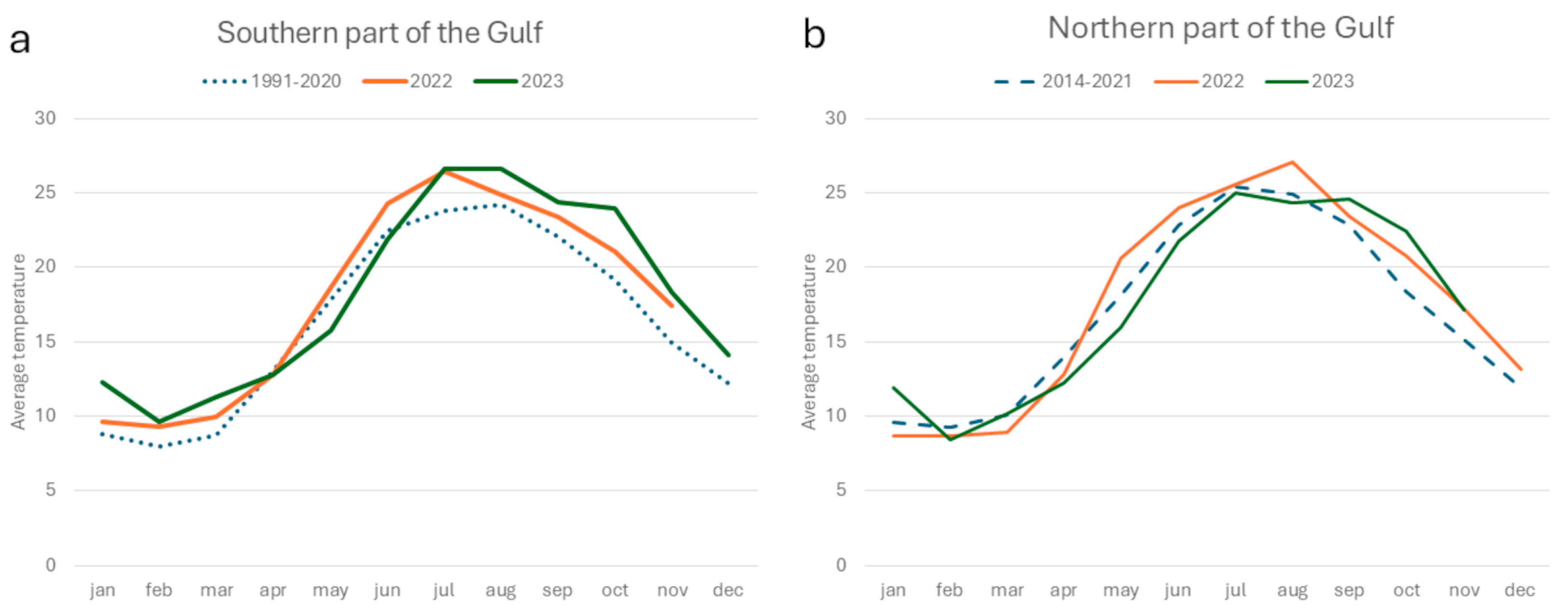
The sea temperature in Slovenia during 2002 and 2023 was above the average of the previous 30 years (1991–2020), with the only exception of May 2023, when the values were below the average of the previous period (Figure 2a). In both years, July was the warmest month (26.6 °C). In 2023, the August–October period was warmer (from 26.6 °C in August to 24 °C in October) than in 2022 (from 24.9 °C in August to 21.1 °C in October), while the temperatures from May to June were higher in 2022 (18.7 °C in May and 24.3 °C in June) than in 2023 (15.8 °C in May and 21.9 °C in June) (Figure 2a). Sea temperatures at the northern sites (Italy) showed a similar trend, with spring temperature higher in 2022 and autumn temperatures higher in 2023 (Figure 2b). For this part of the Gulf, only the monthly average temperatures of the last ten years (2014–2021) were available, which is why the winter-spring temperatures aligned more closely with the historical data. However, the temperatures from August onwards were above the average of the previous period in both 2022 and 2023 (Figure 2b).

3.2. Pinna nobilis Recruitment

A total of 63 live juvenile pinnids were found: 47 in the larval collectors located in shallow coastal waters and 16 in collectors situated within mussel farms. In addition, 87 empty shells of pinnids were found in the collectors.
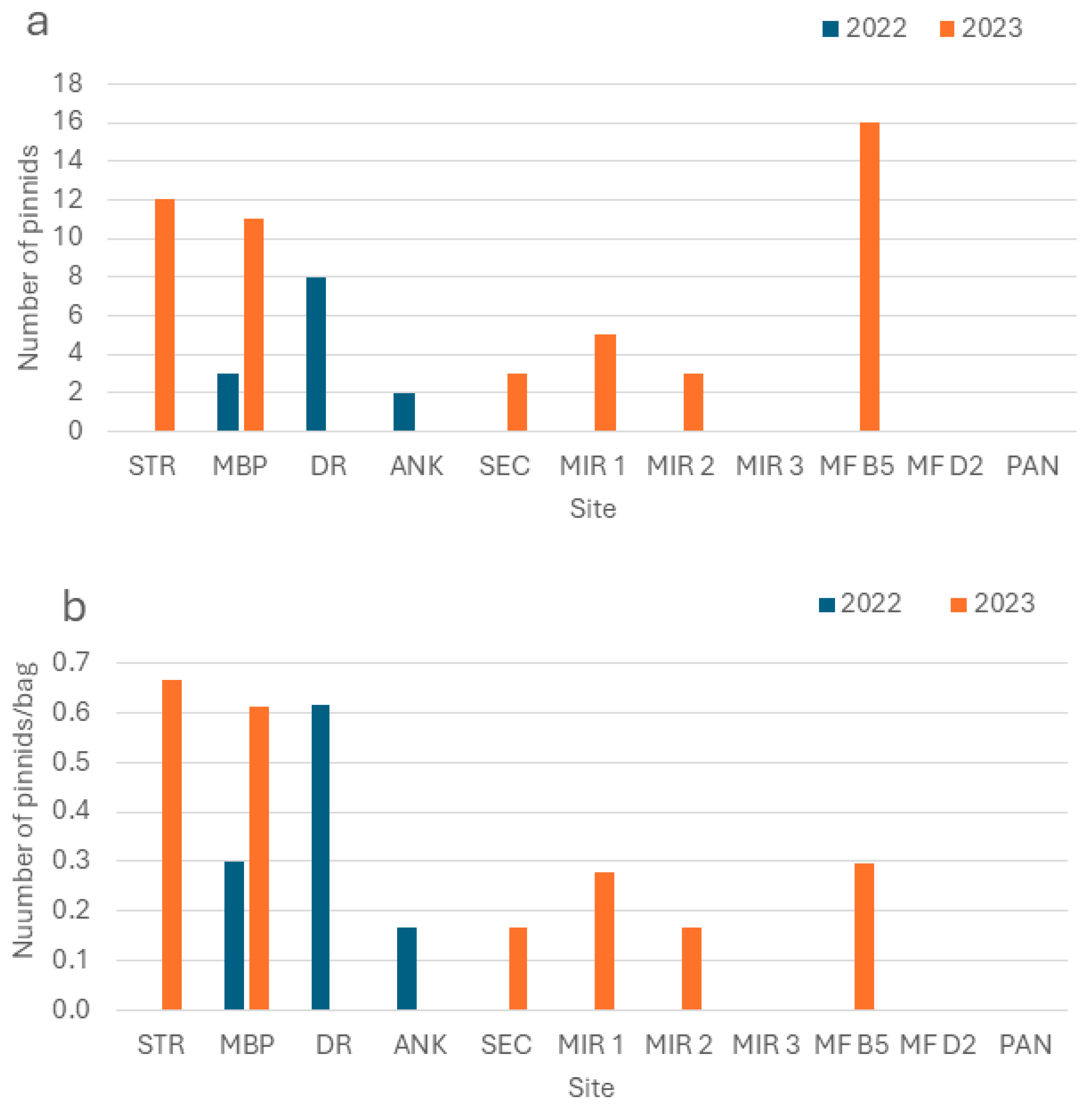
In 2022, only 13 live pinnids were found, all in collectors at 3 southern sites (Figure 3a). In contrast, 50 juveniles were discovered at 6 sites in 2023, evenly distributed between the southern and northern parts (3 each; Figure 3a). The highest numbers were found at the Santa Croce mussel farm B5 (MF B5) with 16 individuals, followed by Strunjan (STR) with 12 individuals and Piran (MBP) with 11 individuals. When normalising these numbers per sampling effort, we found the highest density of living pinnids in 2022 at site DR, with 0.6 pinnids/bag, while in 2023 the highest values were observed at MBP and STR sites, with 0.6 and 0.7 pinnids/bag, respectively (Figure 3b). All collectors at site SEC were lost in 2022, and those at site PAN were lost in 2023. Notably, while the highest density in 2022 was found at DR, no living pinnids were found there in 2023. Conversely, STR had the highest density of living pinnids in 2023, but no living pinnids were found in 2022 at the same site. Site MBP was the only one where living pinnids were found both in 2022 and in 2023. At site ANK, living pinnids were only found in 2022, while sites SEC, MIR1, MIR2, and MF B5 had living pinnids only in 2023.
The living specimens ranged from 0.5 to 8 cm in total height (Htot) and were mainly found in the first 6 m of depth.
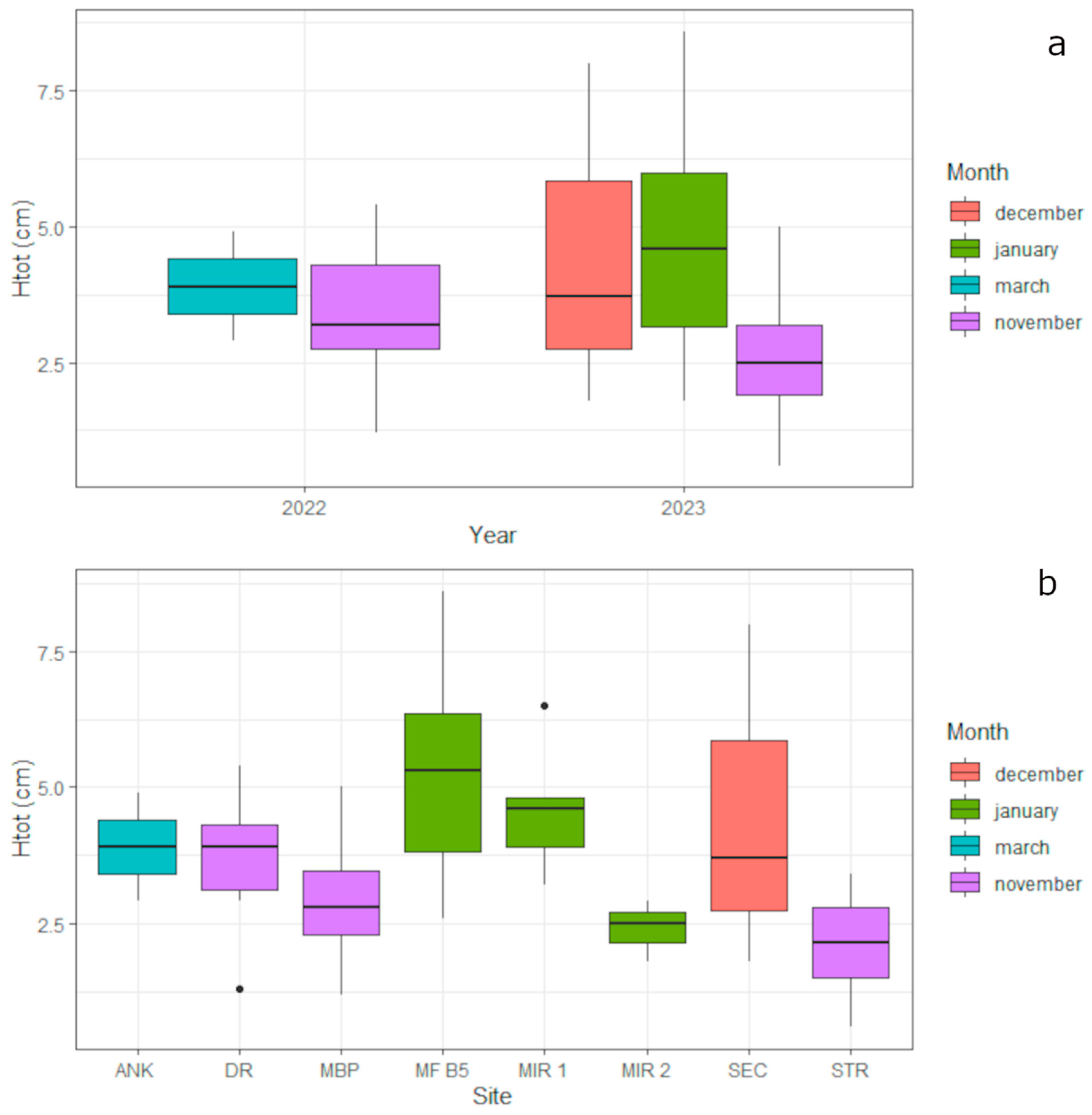
The size of pinnids (Htot) did not significantly differ from 2022 to 2023 (KW, p > 0.05, Figure 4a), even when considering only pinnids collected in the same month (November, KW, p > 0.05), but it differed significantly among sites (KW, p < 0.05). In particular, the size was significantly higher in collectors installed on mussel farms than in other collectors (post hoc, p < 0.05), in particular at sites MBP and STR (post hoc, p < 0.05), where the individuals were smaller (Figure 4b). This variation could be related to the month of collection; in fact, collectors at MF B5 were checked in January, while those at sites MBP and STR were collected in November (Figure 4b). Nevertheless, some differences among sites were observed also for collectors checked in the same month. The recruits found in collectors at site MF B5 were larger than at site MIR 2, both of which were checked in January (post hoc, p < 0.05, Figure 4b), and the recruits at site DR were bigger than at site STR, both of which were checked in November (post hoc, p < 0.05, Figure 4b).

3.3. Pinna nobilis Controlled Growth

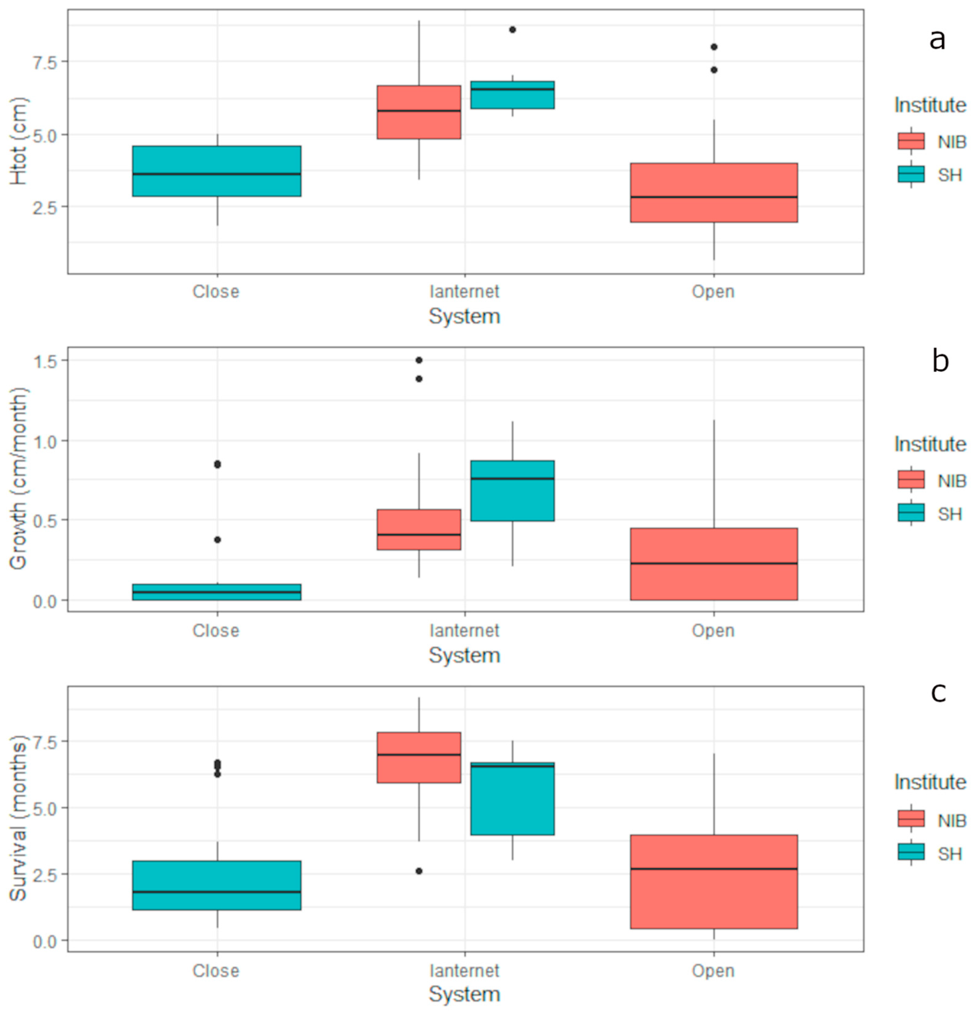
When put in lanternets, the young pinnids were significantly bigger (Htot) than when they were put in ex situ facilities (W, p < 0.05), but the initial size of pinnids (Htot) did not differ between the open system in the facilities of the MBP and the closed system in the facilities of Miramare MPA (KW, p > 0.05, Figure 5). The average monthly growth and survival rates were not significantly different between the two facilities (open and closed systems) (W, p > 0.05), but they were both significantly higher for pinnids held in lantern nets compared to those held in ex situ facilities (KW, W p < 0.05). The initial size, average growth, and survival of pinnids in lanternet did not differ between those located in Piran and those located in Miramare MPA (KW, p > 0.05, Figure 5).
Overall, the survival rate improved with increasing initial size of the animal (rs = 0.31, p < 0.05). This correlation was particularly strong for the juveniles kept at the Miramare MPA facility (rs = 0.68, p < 0.05) but was not significant for the juvenile kept at the MBP facility (p > 0.05) and for those kept in lantern nets (p > 0.05). The same was observed for the average monthly growth, which generally increased with an increasing initial size (rs = 0.37, p < 0.05). This correlation was significant for the juveniles kept in the Miramare MPA facility (rs = 0.54, p < 0.05) but was not significant for those kept at the facility of the MBP, nor in lantern nets (p > 0.05).
At the end of the experiment, the size of surviving juveniles kept in ex situ facilities at MBSP ranged from 3.5 to 8.9 cm; juveniles kept in ex situ facilities in Miramare MPA ranged from 10.3 to 10.4 cm, juveniles kept in in situ facilities in Piran ranged from 6.4 to 8.9 cm and juveniles kept in situ facilities at Miramare MPA ranged from 9.6 to 14 cm.

3.4. Taxonomic Identification of P. nobilis and Molecular Diagnostic Analyses of Pathogens

Pinnids collected in the southern sites were not big enough to enable a determination through the morphological approach. From six specimens (L9, 12,13, 15, 17, and 19), high-quality sequences were obtained among the COI fragments analysed at the University of Sassari, and all exhibited 100% identity with the species P. nobilis (Table 1). Pinnids collected in the northern sites were big enough to be determined as P. nobilis through the morphological approach (Table 1). No other species were detected.
In the sample of P. nobilis collected at the southern sites, subjected to molecular diagnostics for pathogen detection, the presence of haplosporidian species, Mycobactirium spp, Vibrio sp., and Perkinsus sp. as potential environmental pathogens was confirmed. No significant tissue infections emerged, with the sole exception of Haplosporidium pinnae for two samples (results obtained from quantitative PCR). The two fan mussels infected with H. pinnae (L17, L18) showed signs of poor condition shortly after collection, including slow shell closure, and died in quarantine aquaria within a month.
Among the 12 juvenile pinnids collected at the northern sites analysed for pathogen detection, only 4 tested positive for H. pinnae (SH3, 6, 10, and 22, Table 1). In one case the pathogen was identified from the analysis of feaces, while in the others it was detected from the analyses of the mantle and gills.

4. Discussion

4.1. Pinna nobilis Recruitment

The number of recruits found in the studied areas aligns with post-MME numbers reported in the literature for the Croatian coast [57], reflecting the low number of breeding adults in the wild. This result confirms recent investigations that pointed out a general disruption in recruitment of P. nobilis after the MME [57,58]. Our results indicate a marked temporal and spatial variability in P. nobilis recruitment, which is likely due to natural fluctuations in environmental parameters. A high variability of recruitment across years was already reported for Spain during the pre-MME period from 2003 to 2011, with the number of recruits per year varying between 2 and 164 with the same sampling effort [59]. In this context, the authors found a significant positive correlation between total recruitment and average water temperature in June at 25 m depth. In our case, the opposite trend was observed. Water temperatures recorded in the Gulf in May and June 2023 were lower than in May and June 2022. In May 2022, temperatures increased from 13 °C to 24.4 °C, followed by an abrupt decrease of 7 °C in only 2 days at the end of the month [37,38]. Conversely, the sea temperature remained below 20 °C at the end of May 2023 and only reached 24 °C in June 2023 [37,38]. However, recruitment was higher in 2023.
Under experimental conditions, is known that an increase in temperatures of 5–10 °C [60] triggers spawning in P. nobilis [61]. These data indicate the possibility of early spawning in May 2022. Larvae of P. nobilis are estimated to spend ten to twenty days in plankton [38], but to date, spawning experiments in the laboratory have not reached the settlement phase even after 22 days [60,61], and recent studies suggest that the larval phase could even last a month [60]. At the same time, larvae reared in the laboratory showed faster development at higher temperatures [62], suggesting that the higher temperatures in spring 2022 may have accelerated the whole process, including settlement, which could have taken place before the installation of the collector.
Another possible explanation for the lower recruitment observed in 2022 is that the abrupt cooling of waters after a short period at the end of May 2022 could have prevented the maturation of female gametes, which normally take longer to mature [61]. In fact, P. nobilis is a successive hermaphrodite with asynchronous maturation [16]. Nonetheless, there are known cases of synchronous release of both female and male gametes under experimental conditions [60].
A marked variability between years was also observed in the settlement sites. One possible explanation for this is the presence of competing fouling organisms that compete with P. nobilis for space and food. In the central Adriatic, a massive occurrence of the tunicate Styela plicata (Lesueur, 1823) was observed on larval collectors that had no juveniles of the fan mussel [57]. This same species was also heavily covering collectors at the DR site in 2023, potentially explaining the absence of recruits. Conversely, in 2022, when collectors at DR were installed later, they were not massively overgrown by S. plicata juveniles and P. nobilis juveniles, were found.
The dispersion of larvae of the fan mussel is dependent on current movements [58]. In the Western Mediterranean, P. nobilis populations are known to show high genetic connectivity and to function as a metapopulation with a source-sink dynamic [63], and models were implemented to identify the potential source sites for larvae [58]. In the Gulf of Trieste, single adults of Pinna nobilis can still be found at some sites [64,65], but the closest known healthy population is located in the lagoon of Venice, making it a potential source of larvae [66]. The presence of P. nobilis recruits in larval collectors, in fact, is not necessarily indicative of a nearby adult living populations as reported in other areas [57,58,59].
The size of the juveniles collected did not vary in the two years studied, but significant differences were observed among sites. In some cases, these discrepancies were attributed to the collection period, with larger specimens found in collectors retrieved in January. This month may be the optimal time for collection, as those collected later were often more damaged, likely due to the competition with other animals living on/in the collectors [64,65].
However, these considerations alone are not sufficient to explain the temporal and spatial variability of P. nobilis recruitment. A longer time series would improve our understanding of the factors contributing to this variability.

4.2. Pinna nobilis Controlled Growth

The two ex situ systems, open and closed, yielded comparable results in terms of growth and survival, but the most favourable outcomes were achieved with the in situ systems, both in terms of survival and growth metrics. These results were predictable, as natural conditions in the laboratory can hardly be matched, especially in terms of diet, and they are consistent with previous experiments conducted in Spain, where juveniles of Pinna nobilis kept in the laboratory showed lower growth and survival rates than their conspecifics of the same age in the field [41]. Similar in situ facilities have been shown to be effective in other areas of the Mediterranean, namely South-Eastern Adriatic [67], the Aegean Sea [68], and North-Western Mediterranean [59], prior to the MMEs. Nevertheless, in situ controlled growth would be challenging for the smallest pinnids due to the mesh size of the lantern nets. Cages with small mesh sizes could pose a problem for water flow, so controlled growth of the smallest recruits with an initial phase in ex situ facilities could be more efficient.
The growth and survival of the juveniles were correlated to their initial size. It is known that natural mortality decreases with age and size [59,69]. But the growth of juveniles of Pinna nobilis is known to be related to various factors, such as water temperature and food availability. Research indicates that the growth rates of pinnids kept in open systems increase with increasing seawater temperature [70], as has been observed for in situ controlled growth in other areas [69]. Conversely, juveniles kept in in situ cages in the Aegean Sea exhibited growth patterns more closely related to food availability (in terms of Chl a) than to temperature [68]. Food is critical also for juveniles of P. noblis kept in aquaria. Laboratory experiments have further suggested that diet quality is an important factor mediating host condition and disease resistance [41].
The pre-MME investigation of population size structure in Spain [59] suggests that P. nobilis smaller than 45 cm in shell height were vulnerable to predation. The use of protective cages during the vulnerable period can significantly increase survival rates and accelerate the recovery of the species. Even if signs of predation by small animals, such as juveniles of Hexaplex trunculus, have been observed also inside the cages in Spain [59], mortality from predation increases significantly without protection, which is exacerbated by the presence of many other predators, e.g., cephalopods, fishes, and starfish [13,14,71,72]. Cages require regular monitoring, cleaning, and maintenance; otherwise, they may accumulate sediments and fouling, which can reduce light, oxygen levels, and food availability inside the cages. Therefore, they should only be installed in locations that allow for regular checking and cleaning.

4.3. Taxonomic Identification of P. nobilis and Molecular Diagnostic Analyses of Pathogens

For pinnids of small size, the morphological approach alone is insufficient to discriminate among the closely related species P. nobilis, Pinna rudis, and Atrina fragilis.
P. rudis used to occur exclusively in the warmest areas of the Atlantic and Western Mediterranean, with low population density [73]. The first confirmed record for the Adriatic dates back to 2018 in Kotor Bay [74]. Following the decline of P. nobilis and the rise in water temperatures due to global warming, new records and locations of P. rudis have been identified across the Mediterranean, indicating that the species is expanding its range northwards and eastwards [75,76,77,78]. To our knowledge, P. rudis remains so far confined to the central part of the Adriatic, with no confirmed record of P. rudis in the Gulf of Trieste.
Atrina fragilis is instead common in the Gulf. It lives in muddy sediments at greater depths compared to the shallow sediment bottom where P. nobilis used to thrive (Authors’ unpublished data). In 2022, juveniles of A. fragilis were found on the longlines of a mussel farm near the DR site (where larval collectors were installed) and brought to the facilities of the aquarium of Piran (University of Primorska). In 2023, a juvenile P. nobilis was also found on the same mussel farm and brought to the facilities of the Marine Biology Station Piran [70]. A. fragilis has never been found in larval collectors to date.
The parasite Haplosporidium pinnae was first detected in the Gulf of Trieste in 2019 [33]. It remains present, as genetic diagnostic testing has confirmed that some dead individuals were positive for H. pinnae. Despite its presence, in situ controlled growth has yielded better results than ex situ controlled growth. Even if the results may be biased by the size of the animals (with larger juveniles placed in lantern nets), they support the decision to return juveniles found in collectors back to the sea. In the frame of the severe mortality this species has experienced in the field, likely mainly related to the parasite H. pinnae, captive growth in a closed system at low temperatures is one of the measures undertaken to protect this species. However, captive growth in closed systems faces different constraints, mainly related to diet and health [41]. Mortality in captive conditions has also been associated with other pathogens, including Vibrio mediterranei [41]. Recent studies investigating the impact of the infection of P. nobilis with Picornavirus (PnPV) on the immune system of this bivalve have found that wild animals in nature showed a more effective immune response than those kept in aquaria [30]. These considerations could help to explain the mortality rate of juveniles in captivity that are not infected with H. pinnae. Nevertheless, further investigation should be carried out to assess the impact of pathogens on the mortality of P. nobilis. Following the outbreak of mass mortality events (MMEs) in P. nobilis, various non-destructive sampling methods for genetic analysis have been developed to minimise stress on the bivalves, including the analysis of faeces [71] and surrounding seawater [72]. Nonetheless, most of these techniques are ineffective or yield poor results in very small recruits, emphasising the need to develop new diagnostic techniques to increase our knowledge of the role of pathogens during early growth stages, particularly in relation to Pn Picornavirus.

5. Conclusions

Our results confirm the presence of P. nobilis larvae in the Gulf of Trieste, indicating a source population of spawning individuals in the northern Adriatic Sea. The recruitment of P. nobilis showed a clear temporal and spatial variability.
Growth and survival rates in controlled conditions did not differ significantly between the two ex situ systems (open and closed), but the most favourable outcomes were achieved with in situ systems, both in terms of survival and growth metrics. The average monthly growth and survival rate were correlated to the initial size of the juveniles, and this could have influenced our results (specimens in in situ facilities were bigger than those kept ex situ). Despite the presence of the parasite in some dead specimens, our results support the decision to return juveniles found in collectors back to the sea.

Supplementary Materials

The following supporting information can be downloaded at: https://www.mdpi.com/article/10.3390/d17100666/s1, Table S1: Details of the installation of collectors (site, coordinates, depth, installation and retrieval dates, number of liner and number of bags). Figure S1: Juvenile collection: (a) preparation of collectors, (b) collectors at sea, (c) checking collectors, (d) juvenile pinnid in collectors. Figure S2: Ex Situ and in situ maintenance. (a) ex situ maintenance in Miramare MPA facilities, (b) ex situ maintenance in MBS Piran facilities, (c) lantern net attached to the mussel farm’s longline in the Miramare MPA, (d) lantern net tied on a fixed structure close to MBS in Piran.

Author Contributions

Conceptualization, V.P., A.F., D.T., and S.C.; methodology, V.P., A.F., D.T., S.C., M.S., E.B., and D.S.; validation, V.P., A.F., S.C., and D.S.; formal analysis, V.P.; investigation, V.P., D.T., A.F., S.C., M.S., E.B., M.O.-B., L.L., B.M., M.R., F.S., D.S., I.A., C.L., M.C., C.,M., and A.P.; resources, V.P., A.F., and D.T.; data curation, V.P., A.F., D.T., and E.B.; writing—original draft preparation, V.P.; writing—review and editing, V.P., D.T., S.C., M.S., E.B., M.O.-B., L.L., B.M., M.R., F.S., D.S., I.A., C.L., M.C., D.C., C.M., A.P., and A.F.; supervision, V.P., A.F., S.C., M.S., and D.S.; project administration, D.C.; funding acquisition, D.C. All authors have read and agreed to the published version of the manuscript.

Funding

This research was financed mainly by the European Union under the Life project LIFE PINNA, LIFE20NAT/IT/001122. It was co-financed by the Ministry of Natural Resources and Spatial Planning of Slovenia, with financial support from the Slovenian Research Agency (research core funding n. P1-0237). Part of this project was also funded by the Italian Ministry of University and Research, PNRR, Missione 4, Componente 2 “Dalla ricerca all’impresa”, Investimento 1.4 Project CN00000033 which supported the National Biodiversity Future Centre of the University of Sassari.

Institutional Review Board Statement

The Ministry of the Environment of Italy (protocol number:_03-1140_2023-0027, released on 30.3.2025), following a positive opinion from ISPRA (Higher Institute for Environmental Protection and Research) released on 23 March 2023 (protocol number 0015399-2023), and the Ministry of Environment and Spatial Planning of Slovenia (protocol code 35606-9/2022-2550-5, released on 22 February 2022) has issued the specific authorizations required for the collection and the manipulation of the species protected by the Habitat Directive.

Data Availability Statement

Dataset available on request from the authors.

Acknowledgments

Special thanks go to the staff of Marine Biology Station Piran and Aquarium Piran (Slovenia), in particular Aljoša Gračner, Ana Lokovšek, Leon Lojze Zamuda, Milijan Šiško, and Tihomir Makovec and the students—Melina Tadić and Urška Kajtna—who helped with the preparation, collection, and checking of the collectors and with the care of the small pinnids in aquaria.

Conflicts of Interest

The authors declare no conflicts of interest. The funders had no role in the design of the study; in the collection, analyses, or interpretation of data; in the writing of the manuscript; or in the decision to publish the results. Authors Saul Ciriaco, Marco Segarich and Edoardo Batistini were employed by the company SHORELINE Soc. Coop. The remaining authors declare that the research was conducted in the absence of any commercial or financial relationships that could be construed as a potential conflict of interest.

Abbreviations

The following abbreviations are used in this manuscript:
IUCNInternational Union for Conservation of Nature
MMEsMass Mortality Events
MPAMarine protected Area
PnPVPinna nobilis Picornavirus

References

  1. Trigos, S.; García-March, J.; Vicente, N.; Tena, J.; Torres, J. Utilization of muddy detritus as organic matter source by the fan mussel Pinna nobilis. Mediterr. Mar. Sci. 2014, 15, 667–674. [Google Scholar] [CrossRef]
  2. Rabaoui, L.; Belgacem, W.; Ismail, D.B.; Mansour, L.; Tlig-Zouari, S. Engineering effect of Pinna nobilis shells on benthic communities. Oceanologia 2015, 57, 271–279. [Google Scholar] [CrossRef]
  3. Iannucci, S.; Auriemma, R.; Davanzo, A.; Ciriaco, S.; Segarich, M.; Del Negro, P. Can the Empty Shells of Pinna nobilis Maintain the Ecological Role of the Species? A Structural and Functional Analysis of the Associated Mollusc Fauna. Diversity 2023, 15, 956. [Google Scholar] [CrossRef]
  4. Trkov, D.; Ivajnšič, D.; Kovačić, M.; Lipej, L. Factors influencing habitat selection of three cryptobenthic clingfish species in the shallow North Adriatic Sea. J. Mar. Sci. Eng. 2021, 9, 789. [Google Scholar] [CrossRef]
  5. Trkov, D.; Fortič, A.; Pitacco, V.; Lipej, L. Importance of dead fan mussel (Pinna nobilis) shells for fish community. In Proceedings of the 43rd CIESM Congress Proceedings, Palermo, Italy, 14–18 October 2024; p. 12. [Google Scholar]
  6. Trkov, D.; Mavrič, B.; Orlando-Bonaca, M.; Lipej, L. Marine cryptobenthic fish fauna of Slovenia (northern Adriatic Sea). Ann. Anal. Za Istrske Mediter. študije. Ser. Hist. Nat. 2019, 29, 59–72. [Google Scholar]
  7. Addis, P.; Secci, M.; Brundu, G.; Manunza, A.; Corrias, S.; Cau, A. Density, size structure, shell orientation and epibiontic colonization of the fan mussel Pinna nobilis L. 1758 (Mollusca: Bivalvia) in three contrasting habitats in an estuarine area of Sardinia (W Mediterranean). Sci. Mar. 2009, 73, 143–152. [Google Scholar] [CrossRef]
  8. Lipej, L.; Mavrič, B.; Orlando Bonaca, M. Opredelitev Stanja Populacij Leščurja in Morskega Datlja ter Habitatnih Tipov Morski Travniki in Podmorski Grebeni v Naravnem Rezervatu Strunjan in Priporočila za Usmerjanje Obiska Morskega dela Rezervata: Zaključno Poročilo o Projektu; Nacionalni Inštitut za Biologijo: Morska Biološka Postaja: Piran, Slovenia, 2012; 49p. [Google Scholar]
  9. Plećaš, D. Epibionti na Plemenitoj Periski (Pinna nobilis Linnaeus, 1758; Mollusca: Bivalvia). Master’s Thesis, University of Zagreb, Zagreb, Croatia, 2017. [Google Scholar]
  10. Giacobbe, S. Epibiontic mollusc communities on Pinna nobilis L.(Bivalvia, Mollusca). J. Nat. Hist. 2002, 36, 1385–1396. [Google Scholar] [CrossRef]
  11. Rabaoui, L.; Zouari, S.T.; Hassine, O.K.B. Two species of Crustacea (Decapoda) associated with the fan mussel, Pinna nobilis Linnaeus, 1758 (Mollusca, Bivalvia). Crustaceana 2008, 81, 433–446. [Google Scholar] [CrossRef]
  12. Acarli, S.; Vural, P.; Öktener, A. Association between the crab, Nepinnotheres pinnotheres (Linnaeus, 1758), and the endangered species fan mussel, Pinna nobilis (Linnaeus, 1758), from the Aegean Sea, Turkey. Alinteri J. Agric. Sci. 2019, 34, 169–174. [Google Scholar] [CrossRef]
  13. Fiorito, G.; Gherardi, F. Prey-handling behaviour of Octopus vulgaris (Mollusca, Cephalopoda) on bivalve preys. Behav. Processes 1999, 46, 75–88. [Google Scholar] [CrossRef]
  14. Çinar, M.E.; Bilecenoglu, M. The Predation of Pinna nobilis (Mollusca) Juveniles by the Spiny Sea Star Marthasterias glacialis (Echinodermata) in the Sea of Marmara. Sustainability 2023, 15, 15719. [Google Scholar] [CrossRef]
  15. Vázquez-Luis, M.; Álvarez, E.; Barrajón, A.; García-March, J.R.; Grau, A.; Hendriks, I.E.; Jiménez, S.; Kersting, D.; Moreno, D.; Pérez, M. SOS Pinna nobilis: A mass mortality event in western Mediterranean Sea. Front. Mar. Sci. 2017, 4, 220. [Google Scholar] [CrossRef]
  16. Kersting, D.; Benabdi, M.; Čižmek, H.; Grau, A.; Jimenez, C.; Katsanevakis, S.; Öztürk, B.; Tuncer, S.; Tunesi, L.; Vázquez-Luis, M. Pinna nobilis. In The IUCN Red List of Threatened Species 2019 e.T160075998A160081499; 2019; p. 24. Available online: https://www.iucnredlist.org/species/160075998/160081499 (accessed on 9 May 2025).
  17. IUCN. Pinna nobilis Mass Mortality Outbreak in the Mediterranean—Call to Action for Central and Western Mediterranean Countries. Available online: https://www.iucn.org/news/mediterranean/201805/pinna-nobilis-mass-mortality-outbreak-mediterranean-%E2%80%93-call-action-central-and-western-mediterranean-countries (accessed on 10 April 2025).
  18. Katsanevakis, S.; Tsirintanis, K.; Tsaparis, D.; Doukas, D.; Sini, M.; Athanassopoulou, F.; Κolygas, M.N.; Tontis, D.; Koutsoubas, D.; Bakopoulos, V. The cryptogenic parasite Haplosporidium pinnae invades the Aegean Sea and causes the collapse of Pinna nobilis populations. Aquat. Invasions 2019, 14, 150–164. [Google Scholar] [CrossRef]
  19. IUCN. Mediterranean Noble Pen Shell Crisis (Pinna nobilis)—June 2019 Update. Available online: https://www.iucn.org/news/mediterranean/201907/mediterranean-noble-pen-shell-crisis-pinna-nobilis-june-2019-update. (accessed on 10 April 2025).
  20. Čižmek, H.; Čolić, B.; Gračan, R.; Grau, A.; Catanese, G. An emergency situation for pen shells in the Mediterranean: The Adriatic Sea, one of the last Pinna nobilis shelters, is now affected by a mass mortality event. J. Invertebr. Pathol. 2020, 173, 107388. [Google Scholar] [CrossRef]
  21. Darriba, S. First haplosporidan parasite reported infecting a member of the Superfamily Pinnoidea (Pinna nobilis) during a mortality event in Alicante (Spain, Western Mediterranean). J. Invertebr. Pathol. 2017, 148, 14–19. [Google Scholar] [CrossRef]
  22. Catanese, G.; Grau, A.; Valencia, J.M.; Garcia-March, J.R.; Vázquez-Luis, M.; Alvarez, E.; Deudero, S.; Darriba, S.; Carballal, M.J.; Villalba, A. Haplosporidium pinnae sp. nov., a haplosporidan parasite associated with mass mortalities of the fan mussel, Pinna nobilis, in the Western Mediterranean Sea. J. Invertebr. Pathol. 2018, 157, 9–24. [Google Scholar] [CrossRef]
  23. Panarese, R.; Tedesco, P.; Chimienti, G.; Latrofa, M.S.; Quaglio, F.; Passantino, G.; Buonavoglia, C.; Gustinelli, A.; Tursi, A.; Otranto, D. Haplosporidium pinnae associated with mass mortality in endangered Pinna nobilis (Linnaeus 1758) fan mussels. J. Invertebr. Pathol. 2019, 164, 32–37. [Google Scholar] [CrossRef]
  24. Scarpa, F.; Sanna, D.; Azzena, I.; Mugetti, D.; Cerruti, F.; Hosseini, S.; Cossu, P.; Pinna, S.; Grech, D.; Cabana, D. Multiple non-species-specific pathogens possibly triggered the mass mortality in Pinna nobilis. Life 2020, 10, 238. [Google Scholar] [CrossRef]
  25. Carella, F.; Antuofermo, E.; Farina, S.; Salati, F.; Mandas, D.; Prado, P.; Panarese, R.; Marino, F.; Fiocchi, E.; Pretto, T. Corrigendum: In the Wake of the Ongoing Mass Mortality Events: Co-occurrence of Mycobacterium, Haplosporidium and Other Pathogens in Pinna nobilis Collected in Italy and Spain (Mediterranean Sea). Front. Mar. Sci. 2020, 7, 382. [Google Scholar] [CrossRef]
  26. Lattos, A.; Bitchava, K.; Giantsis, I.A.; Theodorou, J.A.; Batargias, C.; Michaelidis, B. The implication of vibrio bacteria in the winter mortalities of the critically endangered Pinna nobilis. Microorganisms 2021, 9, 922. [Google Scholar] [CrossRef]
  27. Lattos, A.; Giantsis, I.A.; Karagiannis, D.; Theodorou, J.A.; Michaelidis, B. Gut symbiotic microbial communities in the IUCN critically endangered Pinna nobilis suffering from mass mortalities, revealed by 16S rRNA amplicon NGS. Pathogens 2020, 9, 1002. [Google Scholar] [CrossRef]
  28. Prado, P.; Carrasco, N.; Catanese, G.; Grau, A.; Cabanes, P.; Carella, F.; García-March, J.R.; Tena, J.; Roque, A.; Bertomeu, E. Presence of Vibrio mediterranei associated to major mortality in stabled individuals of Pinna nobilis L. Aquaculture 2020, 519, 734899. [Google Scholar] [CrossRef]
  29. Carella, F.; Prado, P.; De Vico, G.; Palic, D.; Villari, G.; Garcia March, J.R.; Medialdea, J.T.; Cortés Melendreras, E.; Casalduero, F.G.; Sigovini, M.; et al. A widespread picornavirus affects the hemocytes of the noble pen shell (Pinna nobilis) leading to immunosuppression. Front. Vet. Sci. 2023, 10, 1273521. [Google Scholar] [CrossRef]
  30. Carella, F.; Prado, P.; García-March, J.R.; Tena-Medialdea, J.; Melendreras, E.C.; Porcellini, A.; Feola, A. Measuring immunocompetence in the natural population and captive individuals of noble pen shell Pinna nobilis affected by Pinna nobilis Picornavirus (PnPV). Fish Shellfish. Immunol. 2024, 151, 109664. [Google Scholar] [CrossRef]
  31. Tempesta, M.; Del Piero, D.; Ciriaco, S. Definition of a new formula for the calculation of the total height of the fan shell Pinna nobilis in the Miramare Marine Protected Area (Trieste, Italy). Ann. Ser. Hist. Nat. 2013, 23, 17–24. [Google Scholar]
  32. Lipej, L.; Mavrič, B.; Šiško, M.; Orlando Bonaca, M. Pregled Morske Biodiverzitete v Občini Ankaran; National Institute of Biology, Marine Biology Station Piran: Piran, Slovenia, 2016; p. 146. [Google Scholar]
  33. Manfrin, C.; Ciriaco, S.; Segarich, M.; Aiello, A.; Florian, F.; Avian, M.; Terlizzi, A.; Giulianini, P.G.; Spoto, M.; Pallavicini, A. Haplosporidium pinnae Detection from the Faeces of Pinna nobilis: A Quick and Noninvasive Tool to Monitor the Presence of Pathogen in Early-Stage or during Fan Mussel Mass Mortalities. Diversity 2023, 15, 477. [Google Scholar] [CrossRef]
  34. Boicourt, W.C.; Kuzmić, M.; Hopkins, T.S. The inland sea: Circulation of Chesapeake Bay and the Northern Adriatic. In Ecosystems at the Land-Sea Margin: Drainage Basin to Coastal Sea, 1st ed.; Malone, T.C., Malej, A., Harding, L.W.J., Smodlaka, N., Turner, R.E., Eds.; Coastal and Estuarine Studies, American Geophysical Union: Washington, DC, USA, 1999; Volume 55, pp. 81–129. [Google Scholar]
  35. Mozetič, P.; Umani, S.F.; Cataletto, B.; Malej, A. Seasonal and inter-annual plankton variability in the Gulf of Trieste (northern Adriatic). ICES J. Mar. Sci. 1998, 55, 711–722. [Google Scholar] [CrossRef]
  36. Stravisi, F. The vertical structure annual cycle of the mass field parameters in the Gulf of Trieste. Boll Oceanol. Teor. Appl. 1983, 1, 239–250. [Google Scholar]
  37. ARPAFVG. Stato Oceanografico ed Ecologico del Golfo di Trieste. 2022–2023. Available online: https://arpa.fvg.it/temi/temi/acqua/sezioni-principali/acque-marino-costiere-e-di-transizione/bollettino-oceanografico/ (accessed on 22 April 2025).
  38. ARSO. Mesečni Bilten ARSO (Slovenian Environment Agency). 2022–2023. Available online: http://www.arso.gov.si/o%20agenciji/knji%C5%BEnica/mese%C4%8Dni%20bilten/bilten2023.htm (accessed on 18 March 2024).
  39. Kersting, D.K.; Hendriks, I.E. Short Guidance for the Construction, Installation and Removal of Pinna nobilis Larval Collectors. IUCN 2019, 6p. Available online: https://iucn.org/sites/default/files/content/documents/2019/larval_colectors_p._nobilis_guidance_-_pinna_nobilis.pdf (accessed on 10 April 2025).
  40. Trigos, S.; García-March, J.R.; Vicente, N.; Tena, J.; Torres, J. Respiration rates of the fan mussel Pinna nobilis at different temperatures. J. Molluscan Stud. 2015, 81, 217–222. [Google Scholar] [CrossRef]
  41. Prado, P.; Cabanes, P.; Catanese, G.; Carella, F.; Carrasco, N.; Grau, A.; Hernandis, S.; García-March, J.R.; Tena, J.; Caiola, N. Growth of juvenile Pinna nobilis in captivity conditions: Dietary and pathological constraints. Aquaculture 2020, 522, 735167. [Google Scholar] [CrossRef]
  42. Hernandis, S.; Ibarrola, I.; Tena-Medialdea, J.; Vázquez-Luis, M.; García-March, J.; Prado, P.; Albentosa, M. Scope for growth and dietary needs of Mediteranean Pinnids maintained in captivity. BMC Zool. 2022, 7, 43. [Google Scholar] [CrossRef]
  43. Sanna, D.; Cossu, P.; Dedola, G.L.; Scarpa, F.; Maltagliati, F.; Castelli, A.; Franzoi, P.; Lai, T.; Cristo, B.; Curini-Galletti, M.; et al. Mitochondrial DNA Reveals Genetic Structuring of Pinna nobilis across the Mediterranean Sea. PLoS ONE 2013, 8, e67372. [Google Scholar] [CrossRef]
  44. Carella, F.; Aceto, S.; Pollaro, F.; Miccio, A.; Iaria, C.; Carrasco, N.; Prado, P.; De Vico, G. A mycobacterial disease is associated with the silent mass mortality of the pen shell Pinna nobilis along the Tyrrhenian coastline of Italy. Sci. Rep. 2019, 9, 2725. [Google Scholar] [CrossRef]
  45. Weisburg, W.G.; Barns, S.M.; Pelletier, D.A.; Lane, D.J. 16S ribosomal DNA amplification for phylogenetic study. J. Bacteriol. 1991, 173, 697–703. [Google Scholar] [CrossRef]
  46. Webb, S.C.; Fidler, A.; Renault, T. Primers for PCR-based detection of ostreid herpes virus-1 (OsHV-1): Application in a survey of New Zealand molluscs. Aquaculture 2007, 272, 126–139. [Google Scholar] [CrossRef]
  47. Sato, M.; Nanami, A.; Bayne, C.J.; Makino, M.; Hori, M. Changes in the potential stocks of coral reef ecosystem services following coral bleaching in Sekisei Lagoon, southern Japan: Implications for the future under global warming. Sustain. Sci. 2020, 15, 863–883. [Google Scholar] [CrossRef]
  48. Mor, S.K.; Phelps, N.B.; Barbknecht, M.; Hoffman, M.A.; Goyal, S.M. A multiplex RT-PCR assay for the detection of fish picornaviruses. J. Virol. Methods 2015, 221, 131–134. [Google Scholar] [CrossRef]
  49. Füstös, Z.; Belák, Á.; Maráz, A. Colonization ability of Escherichia coli and Listeria monocytogenes in the endosphere of sweet pepper (Capsicum annuum var. grossum). Acta Aliment. 2017, 46, 481–491. [Google Scholar] [CrossRef]
  50. Ramilo, A.; Navas, J.I.; Villalba, A.; Abollo, E. Species-specific diagnostic assays for Bonamia ostreae and B. exitiosa in European flat oyster Ostrea edulis: Conventional, real-time and multiplex PCR. Dis. Aquat. Org. 2013, 104, 149–161. [Google Scholar] [CrossRef]
  51. Protasov, E.S.; Axenov-Gribanov, D.V.; Rebets, Y.V.; Voytsekhovskaya, I.V.; Tokovenko, B.T.; Shatilina, Z.M.; Luzhetskyy, A.N.; Timofeyev, M.A. The diversity and antibiotic properties of actinobacteria associated with endemic deepwater amphipods of Lake Baikal. Antonie Van Leeuwenhoek 2017, 110, 1593–1611. [Google Scholar] [CrossRef]
  52. Vaulot, D.; Geisen, S.; Mahé, F.; Bass, D. pr2-primers: An 18S rRNA primer database for protists. Mol. Ecol. Resour. 2022, 22, 168–179. [Google Scholar] [CrossRef]
  53. Bearham, D.; Spiers, Z.; Raidal, S.; Jones, J.B.; Nicholls, P.K. Detection of Minchinia sp., in rock oysters Saccostrea cuccullata (Born, 1778) using DNA probes. J. Invertebr. Pathol. 2008, 97, 50–60. [Google Scholar] [CrossRef]
  54. Kruskal, W.H.; Wallis, W.A. Use of ranks in one-criterion variance analysis. J. Am. Stat. Assoc. 1952, 47, 583–621. [Google Scholar] [CrossRef]
  55. Spearman, C. Demonstration of formulae for true measurement of correlation. Am. J. Psychol. 1907, 18, 161–169. [Google Scholar] [CrossRef]
  56. RDevelopmentCoreTeam. R: A Language and Environment for Statistical Computing; R Foundation for Statistical Computing: Vienna, Austria, 2023; Available online: https://www.R-project.org/ (accessed on 10 March 2025).
  57. Bakran-Petricioli, T.; Kujundžić, D.; Naranđa, M.; Petricioli, D.; Petricioli, L.; Kipson, S. Fouling Community on Pinna nobilis Larval Collectors in the Adriatic—Impact of Invasive Species. J. Mar. Sci. Eng. 2023, 11, 618. [Google Scholar] [CrossRef]
  58. Kersting, D.K.; Vázquez-Luis, M.; Mourre, B.; Belkhamssa, F.Z.; Álvarez, E.; Bakran-Petricioli, T.; Barberá, C.; Barrajón, A.; Cortés, E.; Deudero, S.; et al. Recruitment disruption and the role of unaffected populations for potential recovery after the Pinna nobilis mass mortality event. Front. Mar. Sci. 2020, 7, 594378. [Google Scholar] [CrossRef]
  59. Kersting, D.K.; García-March, J.R. Long-term assessment of recruitment, early stages and population dynamics of the endangered Mediterranean fan mussel Pinna nobilis in the Columbretes Islands (NW Mediterranean). Mar. Environ. Res. 2017, 130, 282–292. [Google Scholar] [CrossRef]
  60. Trigos, S.; Vicente, N.; Prado, P.; Espinós, F.J. Adult spawning and early larval development of the endangered bivalve Pinna nobilis. Aquaculture 2018, 483, 102–110. [Google Scholar] [CrossRef]
  61. Ferranti, M.P. (Department of Earth, Environment and Llife Science—DISTAV, University of Genova, Corso Europa 26, Genova, Italy). Personal communication, 2025.
  62. Ferranti, M.P.; Azzena, I.; Batistini, E.; Caracciolo, D.; Casu, M.; Chiantore, M.; Ciriaco, S.; Firpo, V.; Intini, L.; Locci, C. Handling of the Bivalve Pinna nobilis, Endangered and Pathogen-Affected Species, for Controlled Reproduction: Precautions Taken. Ecol. Evol. 2024, 14, e70565. [Google Scholar] [CrossRef]
  63. Wesselmann, M.; González-Wangüemert, M.; Serrão, E.A.; Engelen, A.H.; Renault, L.; García-March, J.R.; Duarte, C.M.; Hendriks, I.E. Genetic and oceanographic tools reveal high population connectivity and diversity in the endangered pen shell Pinna nobilis. Sci. Rep. 2018, 8, 4770. [Google Scholar] [CrossRef]
  64. Fortič, A. (Marine Biology Station Piran, National Institute of Biology, Fornače 41, SI-6330 Piran, Slovenia); Trkov, D. (Marine Biology Station Piran, National Institute of Biology, Fornače 41, SI-6330 Piran, Slovenia); Lipej, L. (Marine Biology Station Piran, National Institute of Biology, Fornače 41, SI-6330 Piran, Slovenia); Mavrič, B. (Marine Biology Station Piran, National Institute of Biology, Fornače 41, SI-6330 Piran, Slovenia). Unpublished data. 2025. [Google Scholar]
  65. Ciriaco, S. (SHORELINE Soc. Coop, E3, Località Padriciano, 99, 34149 Area di Ricerca, Trieste, Italy); Segarich, M. (SHORELINE Soc. Coop, E3, Località Padriciano, 99, 34149 Area di Ricerca, Trieste, Italy); Batistini, E. (SHORELINE Soc. Coop, E3, Località Padriciano, 99, 34149 Area di Ricerca, Trieste, Italy). Unpublished data. 2025. [Google Scholar]
  66. Sigovini, M.; Sabino, A.; Manea, E.; Carella, F.; Bergamasco, A. The last stronghold in the Adriatic: Distribution and status of Pinna nobilis in the Venice lagoon. In Proceedings of the 43rd CIESM Congress Proceedings, Palermo, Italy, 14–18 October 2024; p. 11. [Google Scholar]
  67. Kožul, V.; Glavić, N.; Bolotin, J.; Antolović, N. The experimental rearing of fan mussel Pinna nobilis (Linnaeus, 1758). In Proceedings of the 46th Croatian and 6th International Symposium on Agriculture, Opatija, Croatia, 14–18 February 2011; p. 806. [Google Scholar]
  68. Acarli, S.; Lok, A.; Yigitkurt, S.; Palaz, M. Culture of fan mussel (Pinna nobilis, Linnaeus 1758) in relation to size on suspended culture system in Izmir Bay, Aegean Sea, Turkey. Kafkas Univ. Vet. Fakültesi Derg. 2011, 17, 995–1002. [Google Scholar]
  69. Kožul, V.; Glavić, N.; Bolotin, J.; Antolović, N. Growth of the fan mussel Pinna nobilis (L innaeus, 1758)(Mollusca: Bivalvia) in experimental cages in the South Adriatic Sea. Aquacult. Res. 2012, 44, 31–40. [Google Scholar] [CrossRef]
  70. Pitacco, V.; Fortič, A.; Trkov, D.; Mavrič, B.; Mlinar, C.; Lipej, L. Growth rates of the critically endangered fan mussel Pinna nobilis in the Slovenian Sea (Northern Adriatic). Endanger Spec. Res. 2025, in press. [Google Scholar] [CrossRef]
  71. Katsanevakis, S. Growth and mortality rates of the fan mussel Pinna nobilis in Lake Vouliagmeni (Korinthiakos Gulf, Greece): A generalized additive modelling approach. Mar. Biol. 2007, 152, 1319–1331. [Google Scholar] [CrossRef]
  72. García-March, J.R.; García-Carrascosa, A.; Peña Cantero, A.; Wang, Y.-G. Population structure, mortality and growth of Pinna nobilis Linnaeus, 1758 (Mollusca, Bivalvia) at different depths in Moraira bay (Alicante, Western Mediterranean). Mar. Biol. 2007, 150, 861–871. [Google Scholar] [CrossRef]
  73. Gvozdenović, S.; Mačić, V.; Pešić, V.; Nikolić, M.; Peraš, I.; Mandić, M. Review on Pinna rudis (Linnaeus, 1758)(Bivalvia: Pinnidae) presence in the Mediterranean. Poljopr. Sumar. 2019, 65, 115–126. [Google Scholar] [CrossRef]
  74. Petović, S. Additions to the checklist of the malacofauna of the Boka Kotorska Bay (south-east Adriatic Sea). Stud. Mar. 2018, 31, 23–36. [Google Scholar]
  75. Kersting, D.K.; Ballesteros, E. Is the local extinction of Pinna nobilis facilitating Pinna rudis recruitment? Mediterr. Mar. Sci. 2021, 2, 623–626. [Google Scholar] [CrossRef]
  76. Oprandi, A.; Aicardi, S.; Azzola, A.; Benelli, F.; Bertolino, M.; Bianchi, C.N.; Chiantore, M.; Ferranti, M.P.; Mancini, I.; Molinari, A. A tale of two sisters: The southerner Pinna rudis is getting north after the regional extinction of the congeneric P. nobilis (Mollusca: Bivalvia). Diversity 2024, 16, 120. [Google Scholar] [CrossRef]
  77. Zotou, M.; Papadakis, O.; Catanese, G.; Stranga, Y.; Ragkousis, M.; Kampouris, T.; Aga-Spyridopoulou, R.N.; Papadimitriou, E.; Koutsoubas, D.; Katsanevakis, S. New kid in town: Pinna rudis spreads in the eastern Mediterranean. Mediterr. Mar. Sci. 2023, 24, 709–721. [Google Scholar] [CrossRef]
  78. Denti, G.; Fanelli, G.; Tiscar, P.G.; Paoletti, B.; Hattab, J.; Mastrototaro, F.; Carella, F.; Villari, G.; Montesanto, F.; Chimienti, G.; et al. Mass mortality of Pinna nobilis Linnaeus, 1758 (MOLLUSCA, BIVALVIA) in the Taranto and Tremiti Islands populations: Citizen Science to help scientists. Biol. Mar. Mediterr. 2023, 27, 21–24. [Google Scholar]
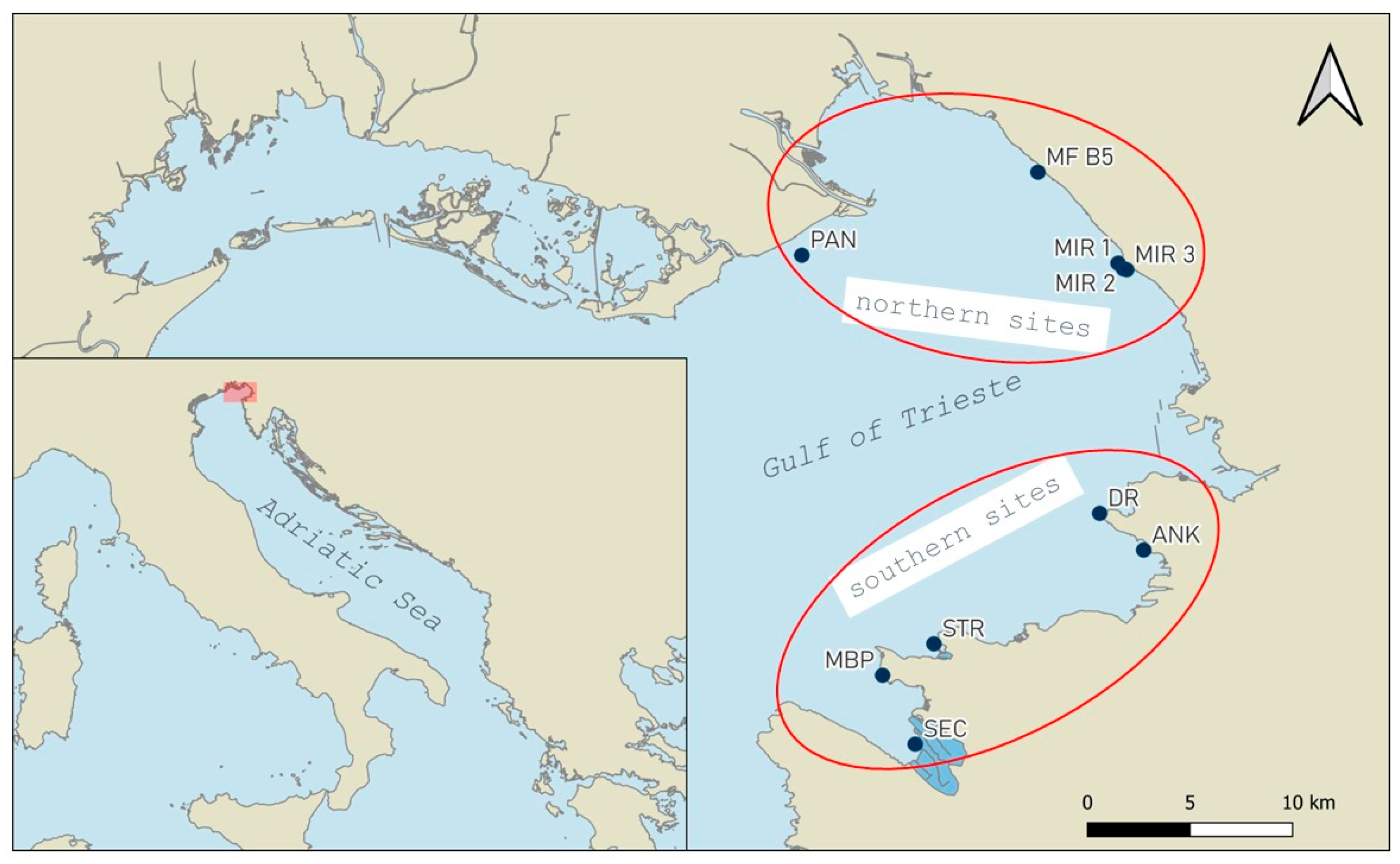
Figure 1. Map of the study area, with northern sites (MIR1-3, MF B5, and PAN) and southern sites (SEC, MBP, STR, ANK, and DR).
Diversity 17 00666 g001
Figure 2. (a) Average monthly temperature (°C) in the southern sites (Slovenia): data of surface waters from Mareographic Station in Koper from ARSO 2022−2023. (b) Average monthly temperature in the northern sites (Italy): data of surface waters from ARPA FVG 2022−2023.
Diversity 17 00666 g002
Figure 3. Total number of pinnids (a) and number of live pinnids per bag (b) found in collectors at different sites in 2022 and 2023.
Diversity 17 00666 g003
Figure 4. Boxplots showing variability of (a) pinnids size (Htot) among years and (b) and pinnids size (Htot) among sites. Colours represent the month of collection.
Diversity 17 00666 g004
Figure 5. Boxplots showing variability of (a) pinnids’ size (Htot), (b) pinnids’ average monthly growth and (c) pinnids’ survival in different controlled growth system. Colours represent the institute in change in the activity (red = NIB, Piran, blue = Shoreline, Miramare MPA).
Diversity 17 00666 g005
Table 1. Results of genetic analyses. Htot = total height of specimens in cm, Haplo = Haplosporidium pinnae, * morphologically confirmed, ** confirmed with genetic analysis. ND = no data obtained from genetic analysis.
CodeSiteDateHtotSpeciesHaploSample TypeGenetic Analyses
L9DR23 November 20221.3Pinna nobilis **negativetissue (dead specimen)UNISS
L12ANK23 March 20234.9Pinna nobilis **negativetissue (dead specimen)UNISS
L13ANK23 March 20232.9Pinna nobilis **negativetissue (dead specimen)UNISS
L15MBP20 November 20232.3Pinna nobilis **negativetissue (dead specimen)UNISS
L17MBP20 November 20233.6Pinna nobilis **positivetissue (dead specimen)UNISS
L19MBP20 November 20232.6Pinna nobilis **positivetissue (dead specimen)UNISS
L32STR21 November 20230.9NDnegativetissue (dead specimen)UNISS
SH2MF B530 January 20245.9Pinna nobilis *negativefeacesUNITS
SH3MF B530 January 20246.8Pinna nobilis *positivefeacesUNITS
SH4MF B530 January 20244.6Pinna nobilis *negativefeacesUNITS
SH5MF B530 January 20246.5Pinna nobilis *negativefeacesUNITS
SH6MF B530 January 20246.3Pinna nobilis *positivemantleUNITS
SH8MF B530 January 20245.6Pinna nobilis *negativefeacesUNITS
SH9MF B530 January 20247Pinna nobilis *negativefeacesUNITS
SH10MF B530 January 20243.6Pinna nobilis *positivegillUNITS
SH16MIR 130 January 20244.8Pinna nobilis *negativefeacesUNITS
SH22MF B530 January 20245.9Pinna nobilis *positivemantleUNITS
SH23MF B530 January 20248.6Pinna nobilis *negativefeacesUNITS
SH24MF B530 January 20243.9Pinna nobilis *negativefeacesUNITS
Disclaimer/Publisher’s Note: The statements, opinions and data contained in all publications are solely those of the individual author(s) and contributor(s) and not of MDPI and/or the editor(s). MDPI and/or the editor(s) disclaim responsibility for any injury to people or property resulting from any ideas, methods, instructions or products referred to in the content.