Article

Morphological and Phylogenetic Evidences Reveal Lasiodiplodia chonburiensis and L. theobromae Associated with Leaf Blight in Hevea brasiliensis in Southern Thailand

Agricultural Innovation and Management Division, Faculty of Natural Resources, Prince of Songkla University, Hat Yai 90110, Songkhla, Thailand
*
Author to whom correspondence should be addressed.
Diversity 2023, 15(9), 961; https://doi.org/10.3390/d15090961
Submission received: 12 July 2023 / Revised: 19 August 2023 / Accepted: 24 August 2023 / Published: 25 August 2023
(This article belongs to the Special Issue Fungi, Ecology, and Global Change)

Abstract

:
The rubber tree is an important economic tree in Thailand. Recently, the cultivation of rubber trees in Thailand has suffered from a novel leaf fall disease with diverse symptoms, including leaf spot and leaf blight, resulting in severe leaf defoliation. Fungi from the Lasiodiplodia genus, which causes leaf disease in rubber trees, have not been reported in Thailand. Our research aimed to identify Lasiodiplodia associated with leaf blight disease in Thailand by examining morphological characteristics and completing a multi-gene sequence analysis and pathogenicity test to fulfill Koch’s postulates. The internal transcribed spacer regions, translation elongation factor 1-α, and β tubulin 2 were sequenced for the multi-gene sequence analysis. In total, we recovered 14 isolates with 6 of those isolates. Of the six pathogenetic isolates, LST001, LST002, LYT003, LSrt001, and LSrt002 were determined to be Lasiodiplodia chonburiensis, and isolate LYL005 was determined to be L. theobromae. To the best of our knowledge, this is the first report of L. chonburiensis and L. theobromae being associated with leaf blight disease in rubber trees in Thailand or elsewhere.

1. Introduction

Fungal diseases of economically important crops occur worldwide and have been implicated as a significant problem for rubber trees (Hevea brasiliensis) [1,2]. In particular, fungal species causing abnormal rubber tree defoliation appear more frequently than in the past and are now considered the most destructive diseases to affect rubber plantations [3]. To this day, the phenomenon of leaf fall has extended beyond the normal ‘wintering’ of the annual dry season. One case report described a diagnosis of leaf fall disease in Papua New Guinea and Vietnam in 2021, which resulted in a reduction of thousands of hectares in rubber plantations worldwide, with an average reduction of 380,000 ha in Indonesia, 5000 ha in Malaysia, and 52,000 ha in Thailand [3]. Of interest is a novel leaf disease first observed in Thailand rubber trees in 2019 [4]; however, rubber trees demonstrating similar symptoms were previously reported in other regions including North Sumatra in 2016, South Sumatra and Malaysia in late 2017 [5], and Cameroon in 2018 [1].
The rubber tree or rubber plant, belonging to the family Euphobiaceae, is an angiosperm plant and is a fast growing, medium to tall tree. Rubber trees are commonly grown in tropical areas as the climate is suitable for their growth and development [6,7]. The cultivation of rubber trees in Thailand suffers from a variety of fungal diseases. Fungal disease outbreaks in rubber plantations occur due to a variety of reasons including temperature, humidity, and rainfall [7]. Increased fluctuations in extreme climate change have a major potential to lead to a higher pest risk and disease severity in rubber plantations [8]. Several reports showed that during 2019–2022 there was a prolonged monsoon season from September to January in southern Thailand [4,9]. During this period, there was an increase in overall fungal outbreaks on rubber plants and a novel leaf disease was also reported to appear during this time [4,9]. Other factors favorable to a fungal disease outbreak were also reported, including a lower-than-average temperature (24–26 °C), higher rainfall averages (>100 mm), and a higher average relative humidity (>90% RH).
In addition to the unidentified novel leaf disease, incidences of Phytophthora leaf fall disease caused by Phyotophthora citrophthora [10] and leaf fall caused by Neopestalotiopsis cubana and N. formicarium were also detected during this time frame in Thailand [4]. Lastly, another fungus that causes leaf fall in rubber trees, Calonectria foliicola, was also documented in the southern part of Thailand [9]. In addition, both fungal genera Neopestalotiopsis and Lasiodiplodia are species that can cause leaf disease in a wide variety of commercial plant species. Lasiodiplodia spp. have been known to infect almond (Prunus amygdalus), blueberry (Vaccinium corymbosum), castor (Ricinus communis), Chinese hackberry (Celtis sinensis), citrus (Citrus spp.), cocoa (Theobroma cacao), coconut (Cocos nucifera), grapevine (Vitis spp.), groundnut (Arachis hypogaea), jackfruit (Artocarpus heterophyllus), mango (Mangifera indica), mulberry (Morus spp.), melon (Cucumis spp.), olive (Olea europaea), pine (Pinus spp.), strawberry (Fragaria spp.), rice (Oryza spp.) [5,11,12,13,14,15,16,17,18], and durian (Durio spp.) crops [19]. It is generally accepted that Lasiodiplodia spp. have an endophytic lifestyle in addition to causing dieback disease. Host symptoms include trunk canker, yellow leaves, and wilting due to stem and root infection [5,17,18]. The severity of host symptoms has been shown to be influenced by climatic factors [20]. Dianda et al. [20] reported isolating six different Lasiodiplodia species from mango seedlings, including L. euphorbicola that accounted for 36 of the 47 isolates. The occurrence and incidence of this species were widely found in the drier and warmer regions in Burkina Faso, Africa. It is difficult to distinguish between Lasiodiplodia spp. based on morphological features alone; therefore, a DNA-sequence-based approach has been recommended [14] since there is a clear phylogenetic boundary [21] between currently known species. For example, Meng et al. [22] confirmed a new species, Lasiodiplodia syzygii, from post-harvest water-soaked brown lesions on Syzygium samarangense using the DNA sequencing method. Although Lasiodiplodia spp. have been studied in numerous plant species, there are limited studies of Lasiodiplodia-associated leaf blight disease in rubber trees in Thailand.
Hence, our study aimed to identify the fungal pathogen (Lasiodiplodia sp.) that causes the novel leaf blight disease in Thailand using both morphological and molecular approaches and verify its pathogenicity using healthy rubber tree leaves. This is important because this pathogen could represent a new Lasiodiplodia sp. and identifying the etiological agent is an important step in treating this disease in rubber trees.

2. Materials and Methods

2.1. Sample Collection and Fungal Isolation

A total of 25 symptomatic leaf samples were collected from infected areas in southern Thailand. The codes LST, LSrt, and LYL indicate that the samples were from the Satun, Surattani, and Yala provinces, respectively. Symptomatic leaf samples were kept in plastic bags and in a cool box and brought to the Plant Pathology Laboratory, Faculty of Natural Resources, Prince of Songkla University, Thailand, where the isolation was subsequently conducted. Fungal isolation was conducted using the tissue transplantation method. Pieces of symptom-containing healthy parts of 3 × 3 mm were cut and surface disinfected using 70% ethanol and 0.1% sodium hypochlorite (NaOCl). Excess NaOCl was removed by sterile distilled water (DW) 3 times, and the samples were hung to dry on sterile filter paper. The samples were directly placed on water agar (WA) and incubated at ambient temperature (28 ± 2 °C) for 3 days. Hyphal tips recovered from the infected tissues were cut and directly placed on potato dextrose agar (PDA; HiMedia, Mumbai, India) for purification. The fungal samples were transferred to a PDA slant and stored at 10 °C for further analyses.

2.2. Pathogenicity Test

A pathogenicity test was conducted to fulfill Koch’s postulates. Healthy rubber leaves were surface disinfected using 70% ethanol. Inoculation was then performed using the agar plug method, as previously described by Thaochan et al. [9]. Each fungal isolate was cultured on PDA and incubated at an ambient temperature for 3 days. A mycelial plug (5 mm) was cut from the edge of the 3-day-old incubated colony. We compared inoculations on rubber leaves with and without wounding. For the wounded samples, the rubber leaves were wounded using fine needles. Mycelial plugs were directly placed onto the rubber leaves. Only plugs that consisted of the PDA medium were used as the control. Each treatment was composed of 5 leaves (replicates) and the experiment was repeated twice. The inoculated samples were incubated in a humid chamber to maintain humidity (approximately 80–90%) for 7 days. The symptoms that developed on the inoculated leaves were observed and photographed daily. Symptomatic leaf samples were re-isolated using the tissue transplantation method indicated in Section 2.1. The morphology was observed using a compound microscope.

2.3. Morphology Study

Each fungal isolate was cultured on PDA and incubated at a temperature of 28.0 ± 2.0 °C. The colony diameter was measured daily to observe the growth rate. The macroscopic characteristics of pycnidia formation were observed using a stereo microscope (Leica Microsystems, Wetzlar, Germany). The microscopic features of the conidia were observed using a compound microscope (Leica Microsystems). The dimensions of the conidia (n = 20) were measured and photographed. The fungal cultures were deposited in the Culture Collection of the Pest Management Division, Faculty of Natural Resources, Prince of Songkla University.

2.4. Molecular Study

Each fungal isolate was cultured on PDA for 2 days to obtain young mycelia, which were subjected to DNA extraction using the mini-preparation method [23]. Portions of internal-transcribed spacer (ITS), translation elongation factor 1-α (tef1-α), and β-tubulin 2 (tub2) regions were amplified using the primer pairs ITS1/ITS4 [24], EF1-728F/EF1-986R [25], and Bt2a/Bt2b [26], respectively. A PCR mixture containing a DNA template, 10 pmol of each primer, 2× Green PCR Master Mix (Thermo Scientific, Waltham, MA, USA), and DNase/RNase free distilled water was placed in a 50 μL microtube. PCR amplification was performed using a BIO-RAD T100TM Thermal Cycler (Hercules, CA, USA). The PCR profile was denatured at 95 °C for 3 min, followed by 35 cycles of denaturing at 95 °C for 30 s, annealing at 50 °C for 30 s, elongation at 72 °C for 1 min, and a final extension at 72 °C for 10 min. The PCR products were observed via 1% agarose gel electrophoresis using novel juice DNA staining.
A portion of the PCR product was sequenced at the Macrogen Sequencing Service (Seoul, Republic of Korea) using the same primers as the PCR reaction. The DNA sequences were aligned with the known sequences in the NCBI (National Center for Biotechnology Information) database using the MAFFT v. 7 online servers (http://mafft.cbrc.jp/alignment/server/index.html) (accessed on 21 March 2022); these were then manually adjusted using MEGA X [27]. The phylogenetic tree estimation for each alignment was performed using maximum likelihood (ML), maximum parsimony (MP), and Bayesian inference (BI) methods. The ML tree was constructed using MEGA X, based on the T92 + G + I evolutionary model. The MP tree was obtained using the heuristic search option, with 1000 random additions of sequences and using Tree Bisection and Reconnection (TBR) as the branch-swapping algorithm of MEGA X. The Bayesian tree was generated using MrBayes ver. 3.2.7 [28]. Two parallel Markov chain Monte Carlo (MCMC) runs were performed for 2,000,000 generations; these were sampled every 100 generations. The initial 1000 generations were discarded as burn-in. The remaining trees were used to calculate the Bayesian inference posterior probability (BIPP) values. The phylogenetic trees were visualized using FigTree v1.4.0 (http://tree.bio.ed.ac.uk/software/figtree/) (accessed on 29 March 2022).

3. Results

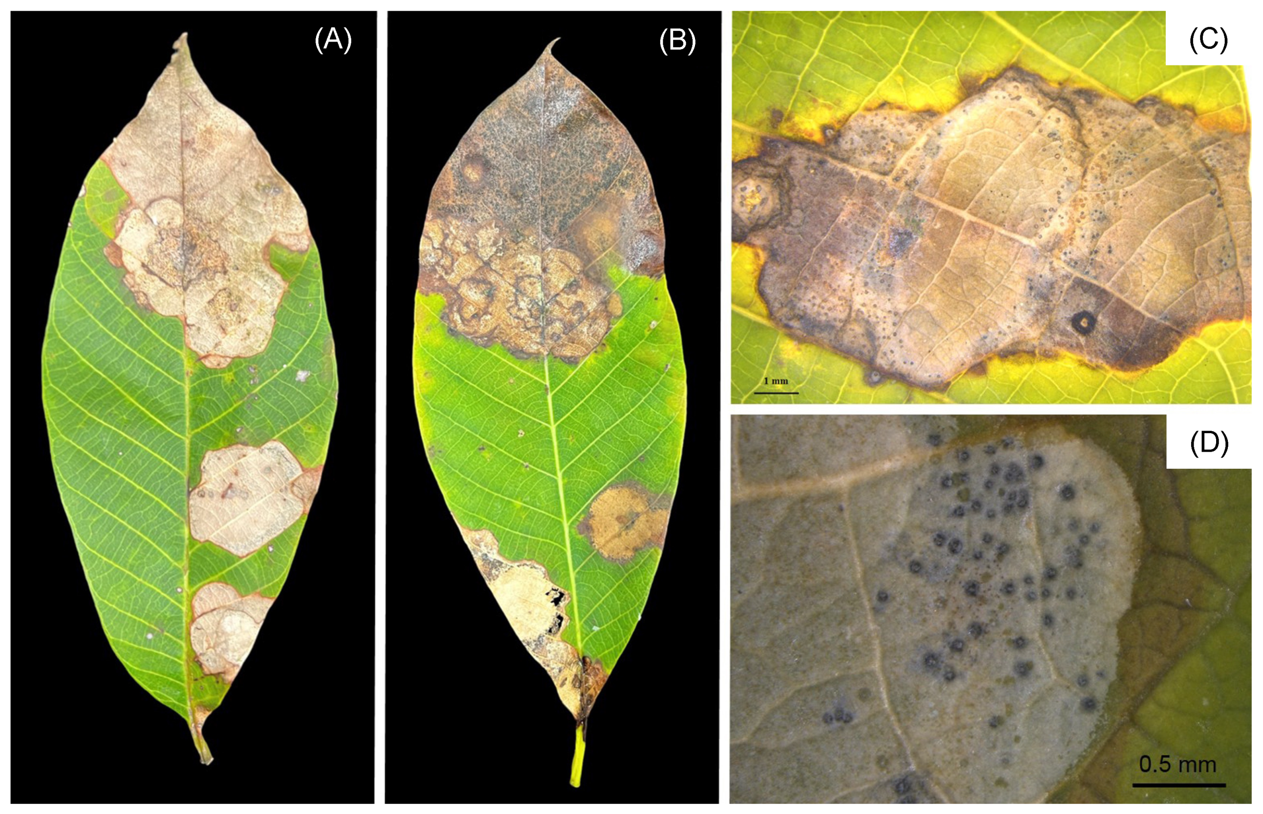
3.1. Symptom Recognition and Fungal Isolates

Disease incidence was about 10–15% in the observed field. The disease appeared as small orange circular or irregular spots on the rubber leaves. Subsequently, the spots enlarged and coalesced into regular or irregular brown necrotic lesions with a dark-brown margin (Figure 1). Dark grey to black pycnidia were observed within the brown spots (Figure 1C,D). In total, we isolated 14 isolates from necrotic lesions.

3.2. Pathogenicity of Lasiodiplodia

During the pathogenicity analysis of the 14 fungal isolates on healthy rubber leaves, we observed that only 6 isolates resulted in the production of the necrotic lesions observed in the field. The fungal isolates LST001, LST002, LST003, LSrt001, LSrt002, and LYL005 showed dark-brown necrotic lesions on both wounded and unwounded samples (Figure 2). The wounded samples displayed more rapid and severe lesion development than the unwounded samples. There were no visible symptoms produced on the control leaves with and without wounding (Figure 2). The same fungal isolates were re-isolated from symptomatic inoculated leaves to confirm Koch’s postulates.

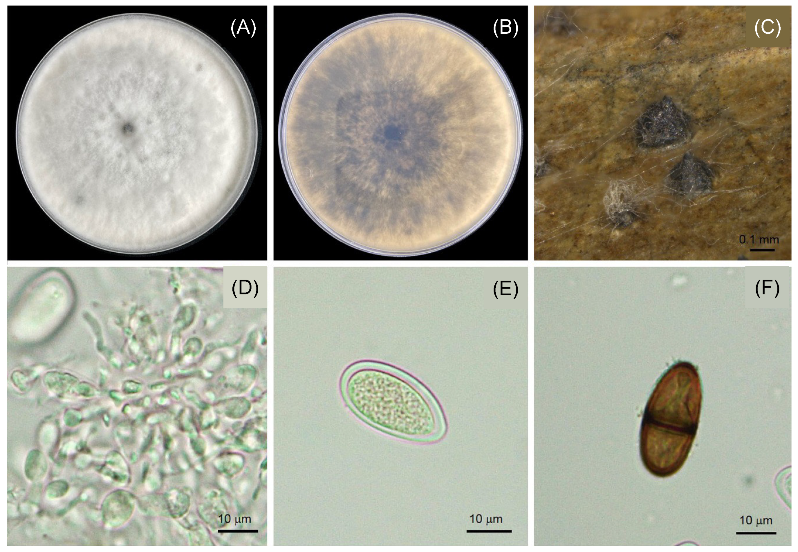
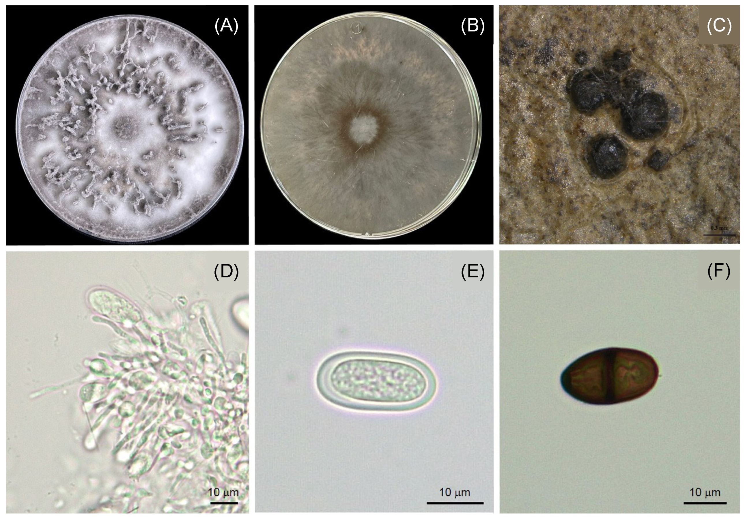
3.3. Morphological Characteristics of Lasiodiplodia

All six fungal isolates were fast growing in PDA medium and covered the 8.5 cm Petri dishes within 7 days. This indicated a growth rate of 1.17 ± 0.1 cm/day. Colonies were white to pale greenish-gray and gradually became dark-grayish with age (Figure 3 and Figure 4). The pycnidia developed on the substrate were solitary, globose to subglobose, black, and L × W (avr ± SD; n = 20). Immature and mature conidia were observed in this study. Both immature and mature conidia were subovoid to ellipsoid in shape, with a rounded apex and tapering to a truncated base. The conidiogenous cells were hyaline and cylindrical with a thin wall. The paraphyses were aseptate and hyaline. The immature conidia were aseptate, double-layered, hyaline, unicellular, and 21.10–24.69 × 10.73–13.79 μm (avr ± SD; n = 20). The mature conidia were dark-brown, septate, and 21.41–25.06 × 10.79–13.26 (avr ± SD; n = 20). Based on the morphological characteristics, we tentatively identified the six fungal isolates as Lasiodiplodia sp. [29]. The morphologies of the fungal isolates were compared with known species, as demonstrated in Table 1.

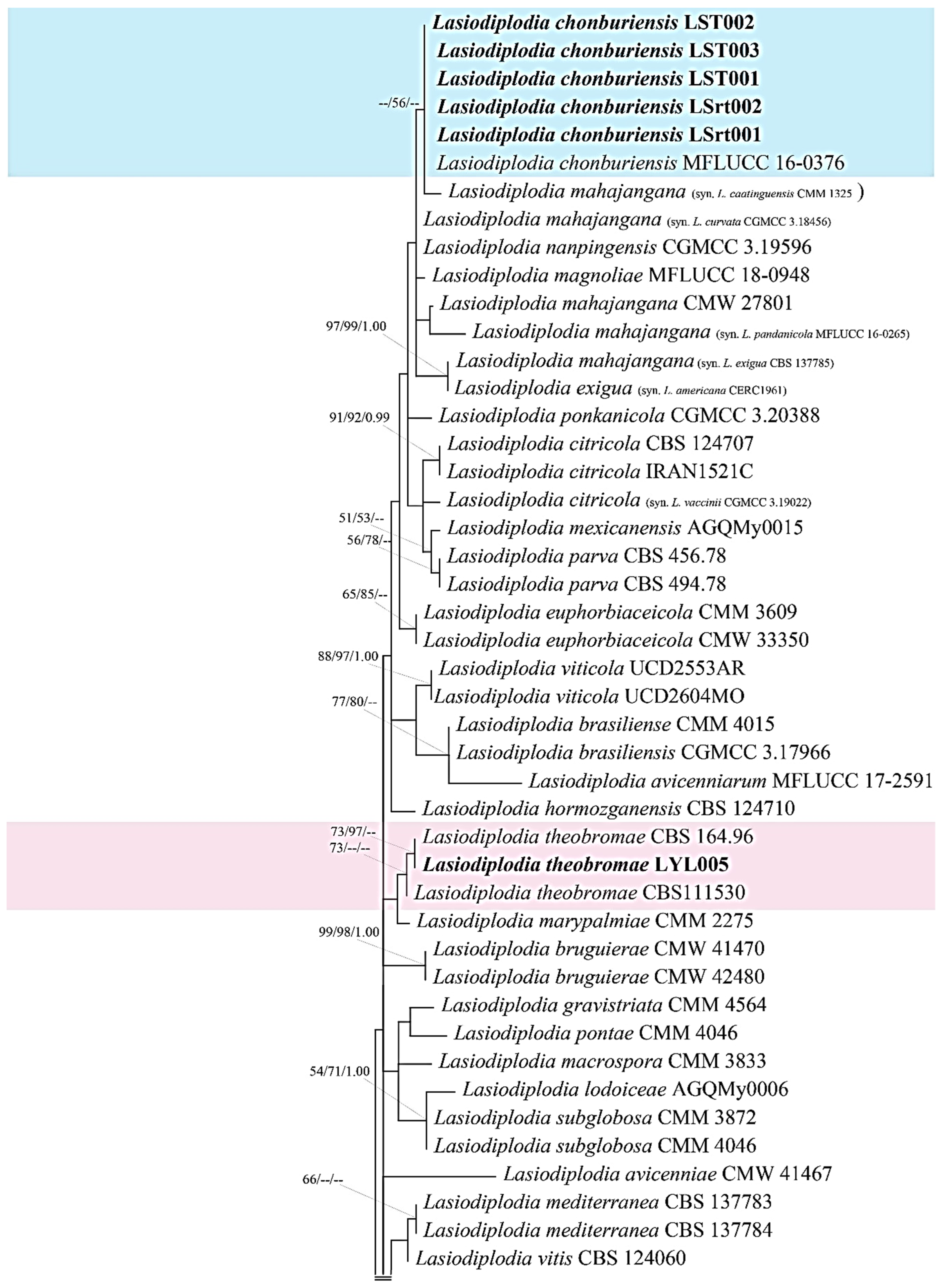
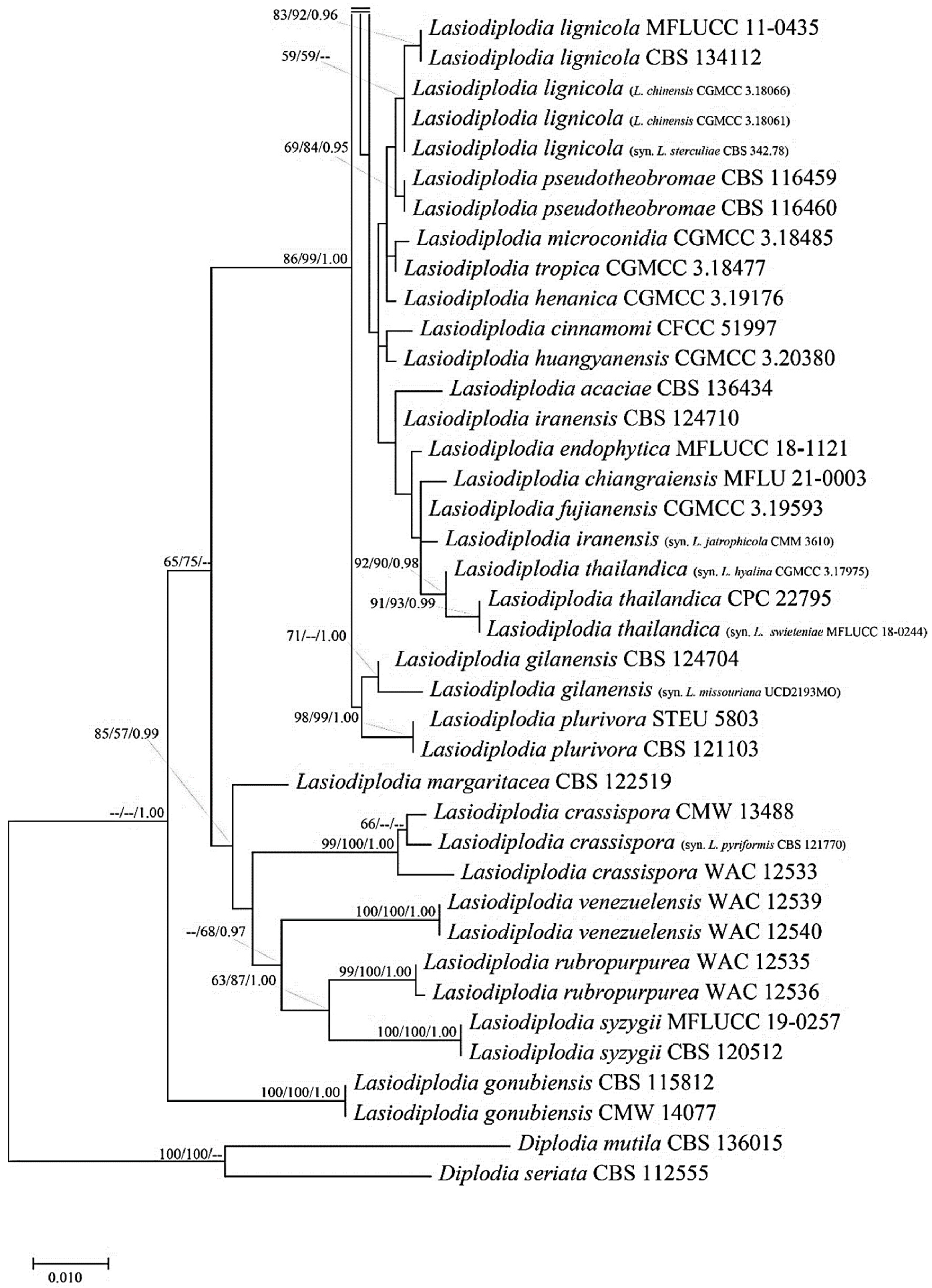
3.4. Molecular Properties of Lasiodiplodia

A BLAST search in the GenBank database revealed that five isolates (LST001, LST002, LST003, LSrt001, and LSrt002) had a 99–100% sequences similarity to L. caatingensis, L. citricola, L. exigua, L. mahajangana, L. pandanicola, and L. theobromae. Another isolate (LYL005) showed 100% sequence similarity to L. theobromae. To construct the phylogenetic tree of the combined DNA sequences of ITS, tef1-α, and tub2, the sequences of the six fungal isolates from the present study were aligned with the sequences of 76 reference isolates of Lasiodiplodia and two outgroup taxa (Diplodia mutila and D. seriata). The gene boundaries in the alignment were ITS 1–486, tef1-α 487–702, and tub2 704–1019. The maximum likelihood (ML) tree revealed that the phylogenetic position of the five isolates from the rubber leaves clustered with the ex-type isolate of L. chonburiensis (-/56/-; ML/MP/BPP), whereas another isolate clustered with the ex-type isolate of L. theobromae (73/97/-; ML/MP/BPP) (Figure 5). Based on the results from this study, fungal isolates LST001, LST0 02, LST003, LSrt001, and LSrt002 were identified as L. chonburiensis and isolate LYL005 was verified as L. theobromae.

4. Discussion

In the present study, we identified the pathogens causing leaf blight on rubber leaves as the fungi L. chonburiensis and L. theobromae. The identification was based on the morphological characteristics and molecular similarity was based on multiple DNA loci (ITS, tef1-α, and tub2). Fungi of the genus Lasiodiplodia belong to the family Botryosphaeriaceae and are commonly found in tropical and subtropical areas [29], causing several diseases in many plant species [35,36,37]. In Thailand, Lasiodiplodia species have been associated with various diseases, for instance, canker in rubber trees [38], spadix rot of Anthurium andraeanum [39], and fruit rot in longan [33].
Fungi of the family Botryosphaeriaceae display an abundant morphology that is different from other fungi; they are easily recognized and can be rapidly identified [32]. Certain fungi in the Lasiodiplodia species have shown overlapping morphological characteristics (such as growth rate, colony morphology, and conidia size and shape [40]) that could not be identified on a species level, as also observed in this study. The fungus L. chonburiensis could not be distinguished from L. theobromae based on morphology identification alone (Figure 3 and Figure 4). The use of a combination of morphologic and molecular tools can be sufficient to identify Lasiodiplodia at a species level, as previously described by several researchers [5,39,40]. In the present study, the use of multiple DNA sequences (ITS, tef1-α, and tub2) successfully identified Lasiodiplodia obtained from rubber leaves to a species level as L. chonburiensis and L. theobromae with different clades (Figure 5). Our results are in agreement with de Silva et al. [41], who used DNA sequences of ITS, tef1-α, and tub2 to study Lasiodiplodia species associated with Magnolia.
A pathogenicity test using the agar plug method fulfilled Koch’s postulates in this study. We did not use conidia for inoculation on the rubber leaves for three reasons: (i) the formation of pycnidia required a significant amount of time (at least 4 months); (ii) pycnidia rarely developed on PDA; and (iii) we could not obtain conidia with a concentration of ×106 conidia/mL. Therefore, we used the mycelial plug method for the pathogenicity test on rubber leaves, as previously described by Xia et al. [42]. The pathogenicity test revealed that both L. chonburiensis and L. theobromae could initiate infections and colonization on plant tissues with wounding. The wounding method appeared to be more effective at inoculating the inoculum (mycelia or conidia) into the plant tissues earlier than the unwounded method. This phenomenon may have been due to the wounded tissues helping the fungal mycelia to penetrate the plant, causing colonization and infection [43]. Several fungal pathogens use appressoria to form a penetration peg in host plants to cause infection [44,45]. Lasiodiplodia species do not form appressoria or other structures to penetrate plant tissues, but are pathogenic to host plants. Several publications have revealed that fungi in Botryophaeriaceae can invade host plants through endophytic ability, injuries, soil contamination, or insect infestation [5,35,36,37,38,39,40,41,42,43,44,45,46]. To date, the infection mechanism of Lasiodiplodia species and how they become pathogenic is still unclear.
Lasiodiplodia chonburiensis was first described from the dead leaves of Pandanus sp. in Thailand as a saprophyte [30]. There is no other report indicating that L. chonburiensis can cause disease in plants. In the present study, L. chonburiensis was confirmed as a causal pathogen of leaf blight in rubber leaves in Thailand. Lasiodiplodia theobromae was first described as causing charcoal rot of cocoa in Ecuador [29]. Lasiodiplodia was then recorded as causing diseases in several plant species in different regions, including dieback and gummosis in mangoes in Pakistan [47], bot canker and gummosis in nectarines in Turkey [48], and panicle blight in grapes in India [49]. Our result is the first report of L. theobromae causing leaf blight in rubber leaves. Trakunyingcharoen et al. [38] reported that only L. pseudotheobromae caused canker in rubber trees in northern Thailand. There has been no report prior to this study that L. theobromae also causes leaf blight in rubber leaves in Thailand. Our study revealed that both L. chonburiensis and L. theobromae act as plant pathogens associated with leaf-blight disease in rubber trees.
Although, the primary cause of leaf disease outbreaks remains unknown, the multiple fungal infections in plants belonging to the genus Pestalotiopsis [1], Neopestalotiopsis [8], or Calonectria foliicola [4] cause serious damage to rubber production and other plants [2]. A long-established report showed that canopy reductions from rubber leaf fall disease have been increasingly impacted, already resulting in severely affected observations of tree health and latex yield [50]. The above-mentioned phenomenon can reduce the tree canopy density by up to 90% in severely affected areas [51]. Similarly, it should be noted that rubber harvesting and production in Thailand and other countries result in an estimated yield loss of between 15% and 50% [4]. In order to determine a fundamental method to reduce leaf fall disease and a reduced disease risk policy, additional research should be performed.

5. Conclusions

In the current study, pathogens causing leaf blight of rubber trees in Thailand were identified as L. chonburiensis and L. theobromae based on morphological and molecular studies of multiple DNA sequences. This study has expanded the knowledge of Lasiodiplodia species by providing a record of two species in a new host (H. brasiliensis), creating a new record of fungal distribution and host ranges. The precise identification of fungal pathogens is the first step in disease management. Studies of the management of Lasiodiplodia diseases need to be conducted in the near future.

Author Contributions

Conceptualization, C.P. and A.S.; methodology, C.P. and N.T.; software, C.P.; validation, C.P., T.C. and A.S.; formal analysis, C.P.; investigation, N.T.; resources, N.T.; data curation, C.P.; writing—original draft preparation, N.T.; writing—review and editing, C.P., T.C. and A.S.; visualization, A.S.; supervision, A.S.; project administration, A.S.; funding acquisition, N.T. and A.S. All authors have read and agreed to the published version of the manuscript.

Funding

This work was supported by the National Science, Research and Innovation Fund (NSRF) and Prince of Songkla University (grant nos. NAT6601012M and NAT6601012a).

Institutional Review Board Statement

Not applicable.

Data Availability Statement

The DNA sequence data obtained from this study were deposited in GenBank under accession numbers ITS (LC762191–LC762196), tef1-α (LC762197–LC762202), and tub2 (LC762203–LC762208).

Acknowledgments

This work was partially supported by the Center of Excellence in Agricultural and Natural Resources Biotechnology (CoE-ANRB), Phase 3, Prince of Songkla University.

Conflicts of Interest

The authors declare no conflict of interest.

References

  1. Nyaka Ngobisa, A.I.C.; Owona, N.P.A.; Doungous, O.; Godswill, N.N.; Njonje, S.W.; Ehabe, E.E. Characterization of Pestalotiopsis microspora, the causal agent of rubber leaf blight disease in Cameroon. Rubber Sci. 2018, 31, 112–120. [Google Scholar]
  2. Gazis, R.; Chaverri, P. Diversity of fungal endophytes in leaves and stems of wild rubber trees (Hevea brasiliensis) in Peru. Fungal Ecol. 2010, 3, 240–254. [Google Scholar] [CrossRef]
  3. Fernando, S.; Silva, K.; Tennakoon, I.; Siriwardena, D.; Wijayaratne, C.; Nishantha, N. Re-Emergence of the Abnormal Leaf Fall Disease Condition in Sri Lankan Rubber; Plantations Rubber Research Institute of Sri Lanka (RRISL): Matugama, Sri Lanka, 2020.
  4. Pornsuriya, C.; Chairin, T.; Thaochan, N.; Sunpapao, A. Identification and characterization of Neopestalotiopsis fungi associated with a novel leaf fall disease of rubber trees (Hevea brasiliensis) in Thailand. J. Phytopathol. 2020, 168, 416–427. [Google Scholar] [CrossRef]
  5. Huda-Shakirah, A.R.; Mohamed Nor, N.M.I.; Zakaria, L.; Leong, Y.H.; Mohd, M.H. Lasiodiplodia theobromae as a causal pathogen of leaf blight, stem canker, and pod rot of Theobroma cacao in Malaysia. Sci. Rep. 2022, 12, 8966. [Google Scholar] [CrossRef] [PubMed]
  6. Singh, D.; Slik, J.W.F.; Jeon, Y.S.; Tomlinson, K.W.; Yang, X.; Wang, J.; Kerfahi, D.; Porazinska, D.L.; Adams, J.M. Tropical forest conversion to rubber plantation affects soil micro- & mesofaunal community & diversity. Sci. Rep. 2019, 9, 5893. [Google Scholar] [PubMed]
  7. Thithuan, N.; Bunjonsiri, P.; Sunpapao, A. Morphology and behavior of gametes and zoospores from the plant-parasitic green algae, Cephaleuros (Chlorophyta, Ulvophyceae). Pac. Sci. 2019, 73, 403–410. [Google Scholar] [CrossRef]
  8. Jacob, J.; Gitz, V.; Gohet, E.; Kadir, A.B.; Nair, L.; Salvatore, P.; Nghia, N.A.; Sergey, B.; Michael, B.; Omar, C.P.; et al. Natural rubber contributions to adaptation to climate change. In Proceedings of the XV World Forestry Congress, Coex, Seoul, Republic of Korea, 2–6 May 2022. [Google Scholar]
  9. Thaochan, N.; Pornsuriya, C.; Chairin, T.; Chomnunti, P.; Sunpapao, A. Morphological and molecular characterization of Calonectria foliicola associated with leaf blight on rubber tree (Hevea brasiliensis) in Thailand. J. Fungi 2022, 8, 986. [Google Scholar] [CrossRef]
  10. Laohasakul, B.; Boonyapipat, P.; Plodpai, P. First Report of Phytophthora citrophthora Causing Leaf Fall of Para Rubber Tree (Hevea brasiliensis) in Thailand. Plant Dis. 2017, 101, 1057. [Google Scholar] [CrossRef]
  11. Nam, M.H.; Park, M.S.; Kim, H.S.; Kim, T.I.; Lee, E.M.; Park, J.D.; Kim, H.G. 2016. First report of dieback caused by Lasiodiplodia theobromae in strawberry plants in Korea. Mycobiology 2016, 44, 319–324. [Google Scholar] [CrossRef]
  12. Rosado, A.W.C.; Machado, A.R.; Freire, F.D.C.O.; Pereira, O.L. Phylogeny, identification, and pathogenicity of Lasiodiplodia associated with postharvest stem-end rot of coconut in Brazil. Plant Dis. 2016, 100, 561–568. [Google Scholar] [CrossRef]
  13. Saeed, E.E.; Sham, A.; AbuZarqa, A.; Al Shurafa, K.A.; Al Naqbi, T.S.; Iratni, R.; El-Tarabily, K.; AbuQamar, S.F. Detection and management of mango dieback disease in the United Arab Emirates. Int. J. Mol. Sci. 2017, 18, 2086. [Google Scholar] [CrossRef] [PubMed]
  14. Bautista-Cruz, M.A.; Almaguer-Vargas, G.; Leyva-Mir, S.G.; Colinas-León, M.T.; Correia, K.C.; Camacho-Tapia, M.; Robles-Yerena, L.; Michereff, S.J.; Tovar-Pedraza, J.M. Phylogeny, distribution and pathogenicity of Lasiodiplodia species associated with cankers and dieback symptoms of Persian lime in Mexico. Plant Dis. 2019, 103, 1156–1165. [Google Scholar] [CrossRef] [PubMed]
  15. Liang, L.; Li, H.; Zhou, L.; Chen, F. Lasiodiplodia pseudotheobromae causes stem canker of Chinese hackberry in China. J. For. Res. 2019, 31, 2571–2580. [Google Scholar] [CrossRef]
  16. Chen, J.; Zhu, Z.; Fu, Y.; Cheng, J.; Xie, J.; Lin, Y. Identification of Lasiodiplodia pseudotheobromae causing fruit rot of citrus in China. Plants 2021, 10, 202. [Google Scholar] [CrossRef]
  17. Pečenka, J.; Tekielska, D.A.; Kocanová, M.; Peňázová, E.; Berraf-Tebbal, A.; Eichmeier, A. First report of Lasiodiplodia theobromae causing decline of blueberry (Vaccinium corymbosum L.) in the Czech Republic. Plant Dis. 2021, 105, 215. [Google Scholar] [CrossRef]
  18. Gnanesh, B.N.; Arunakumar, G.S.; Tejaswi, A.; Supriya, M.; Manojkumar, H.B.; Devi, S.S. Characterization and pathogenicity of Lasiodiplodia theobromae causing black root rot and identification of novel sources of resistance in mulberry collections. Plant Pathol. J. 2022, 38, 272–286. [Google Scholar] [CrossRef]
  19. Chantarasiri, A.; Boontanom, P. Fusarium solani and Lasiodiplodia pseudotheobromae, fungal pathogens causing stem rot disease on durian trees (Durio zibethinus) in Eastern Thailand. New Dis. Rep. 2021, 44, 1–3. [Google Scholar] [CrossRef]
  20. Dianda, O.Z.; Wonni, I.; Ouédraogo, L.; Sankara, P.; Tollenaere, C.; Del Ponte, E.M.; Fernandez, D. Lasiodiplodia species associated with mango (Mangifera indica L.) decline in Burkina Faso and influence of climatic factors on the disease distribution. Physiol. Mol. Plant Pathol. 2003, 126, 102041. [Google Scholar] [CrossRef]
  21. Burgess, T.I.; Barber, P.A.; Mohali, S.; Pegg, G.; de Beer, W.; Wingfield, M.J. Three new Lasiodiplodia spp. from the tropics, recognized based on DNA sequence comparisons and morphology. Mycologia 2006, 98, 423–435. [Google Scholar] [CrossRef]
  22. Meng, C.R.; Zhang, Q.; Yang, Z.F.; Geng, K.; Zeng, X.Y.; Chethana, K.T.; Wang, Y. Lasiodiplodia syzygii sp. nov. (Botryosphaeriaceae) causing post-harvest water-soaked brown lesions on Syzygium samarangense in Chiang Rai, Thailand. Biodivers. Data J. 2021, 9, e60604. [Google Scholar] [CrossRef]
  23. Saitoh, K.; Togashi, K.; Arie, T.; Teraoka, T. A simple method for a mini-preparation of fungal DNA. J. Gen. Plant Pathol. 2006, 72, 348–350. [Google Scholar] [CrossRef]
  24. White, T.J.; Bruns, T.; Lee, S.; Taylor, J. Amplification and direct sequencing of fungal ribosomal RNA genes for phylogenetic. In PCR Protocols: A Guide to Methods and Applications; Innis, M.A., Gelfand, D.H., Sninsky, J.J., White, T.J., Eds.; Academic: San Diego, CA, USA, 1990; pp. 315–322. [Google Scholar]
  25. Carbone, I.; Kohn, L.M. A method for designing primer sets for speciation studies in filamentous ascomycetes. Mycologia 1999, 91, 553–556. [Google Scholar] [CrossRef]
  26. Glass, N.L.; Donaldson, G.C. Development of primer sets designed for use with the PCR to amplify conserved genes from filamentous ascomycetes. Appl. Environ. Microbiol. 1995, 61, 1323–1330. [Google Scholar] [CrossRef]
  27. Kumar, S.; Stecher, G.; Li, M.; Knyaz, C.; Tamura, K. MEGA X: Molecular Evolutionary Genetics Analysis across computing platforms. Mol. Biol. Evol. 2018, 35, 1547–1549. [Google Scholar] [CrossRef]
  28. Rongquist, F.; Teslenko, M.; van der Mark, P.; Ayres, D.L.; Darling, A.; Höhna, S.; Larget, B.; Liu, L.; Suchard, M.A.; Huelsenbeck, J.P. MrBayes 3.2: Efficient Bayesian phylogenetic inference and model choice across a large model space. Syst. Biol. 2012, 61, 539–542. [Google Scholar] [CrossRef]
  29. Phillips, A.J.; Alves, A.; Abdollahzadeh, J.; Slippers, B.; Wingfield, M.J.; Groenewald, J.Z.; Crous, P.W. The Botryosphaeriaceae: Genera and species known from culture. Stud Mycol. 2013, 76, 51–167. [Google Scholar] [CrossRef]
  30. Tibpromma, S.; Hyde, K.D.; McKenzie, E.H.; Bhat, D.J.; Phillips, A.J.; Wanasinghe, D.N.; Samarakoon, M.C.; Jayawardena, R.S.; Dissanayake, A.J.; Tennakoon, D.S.; et al. Fungal diversity notes 840–928: Micro-fungi associated with Pandanaceae. Fungal Divers. 2018, 93, 1–160. [Google Scholar] [CrossRef]
  31. Alves, A.; Crous, P.W.; Correia, A.; Phillips, A.J.L. Morphological and molecular data reveal cryptic speciation in Lasiodiplodia theobromae. Fungal Divers. 2008, 28, 1–13. [Google Scholar]
  32. Munirah, M.S.; Azmi, A.R.; Yong, S.Y.C.; Nur, A.I.M.Z. Characterization of Lasiodiplodia theobromae and L. pseudotheobromae causing fruit rot on pre-harvest mango in Malaysia. Plant Pathol. Quar. 2017, 7, 202–213. [Google Scholar] [CrossRef]
  33. Pipattanapuckdee, A.; Boonyakait, D.; Tiyayon, C.; Seehanam, P.; Ruangwong, O.-U. Lasiodiplodia pseudotheobromae causes postharvest fruit rot of longan in Thailand. Australas. Plant Dis. Notes 2019, 14, 21. [Google Scholar] [CrossRef]
  34. Coutinho, I.B.L.; Freire, F.C.O.; Lima, C.S.; Lima, J.S.; Goncalves, F.J.T.; Machado, A.R.; Silva, A.M.S.; Cardoso, J.E. Diversity of genus Lasiodiplodia associated with perennial tropical fruit plants in north-eastern Brazil. Plant Pathol. 2017, 66, 90–104. [Google Scholar] [CrossRef]
  35. Slippers, B.; Wingfield, M.J. Botryosphaeriaceae as endophytes and latent pathogens of woody plants: Diversity, ecology, and impact. Fungal Biol. Rev. 2007, 21, 90–106. [Google Scholar] [CrossRef]
  36. Mehmood, N.; Riaz, A.; Hassan, I.; Ghuffar, S.; Sattar, A.; Aurangzeb, W.; Azhar, S. First report of Lasiodiplodia theobromae as causal agent of strawberry dieback in Punjab, Pakistan. J. Plant Pathol. 2021, 103, 1355–1356. [Google Scholar] [CrossRef]
  37. Aguilera-Cogley, V.; Atencio, R.; Jaén, M. First report of Lasiodiplodia theobromae causing branch canker and dieback of cashew in Panama. Plant Dis. 2022, 45, e12063. [Google Scholar] [CrossRef]
  38. Trakunyingcharoen, T.; Cheewangkoon, R.; To-anun, C. Phylogenetic study of the Botryosphaeriaceae species associated with avocado and Pará rubber in Thailand. Chiang Mai J. Sci. 2015, 42, 104–116. [Google Scholar]
  39. Daengsuwan, W.; Wonglom, P.; Sunpapao, A. First report of Lasiodiplodia theobromae causing spadix rot in Anthurium andraeanum. J. Phytopathol. 2019, 168, 129–133. [Google Scholar] [CrossRef]
  40. Slippers, B.; Roux, J.; Wingfield, M.J.; van der Walt, F.J.; Jami, F.; Mehl, J.W.; Marais, G.J. Confronting the constraints of morphological taxonomy in the Botryosphaeriales. Persoonia 2014, 33, 155–168. [Google Scholar] [CrossRef]
  41. de Silva, N.I.; Phillips, A.J.L.; Liu, J.K.; Lumyong, S.; Hyde, K.D. Phylogeny and morphology of Lasiodiplodia species associated with Magnolia forest plants. Sci. Rep. 2019, 9, 14355. [Google Scholar] [CrossRef]
  42. Xia, G.; Manawasinghe, I.S.; Phillips, A.J.L.; You, C.; Jayawardena, R.S.; Luo, M.; Hyde, K.D. Lasiodiplodia fici sp. nov., Causing Leaf Spot on Ficus altissima in China. Pathogens 2022, 11. [Google Scholar] [CrossRef]
  43. Fukada, F.; Kodama, S.; Nishiuchi, T.; Kajikawa, N.; Kubo, Y. Plant pathogenic fungi Colletotrichum and Magnaporthe share a common G1 phase monitoring strategy for proper appressorium development. New Phytol. 2019, 222, 1909–1923. [Google Scholar] [CrossRef]
  44. de Jong, J.C.; McCormack, B.J.; Smirnoff, N.; Talbot, N.J. Glycerol generates turgor in rice blast. Nature 1997, 389, 244–245. [Google Scholar] [CrossRef]
  45. Perfect, S.E.; Hughes, H.B.; O’Connell, R.J.; Green, J.R. Colletotrichum—A model genus for studies on pathology and fungal–plant interactions. Fungal Genet. Biol. 1999, 27, 186–198. [Google Scholar] [CrossRef] [PubMed]
  46. Sakalidis, M.L.; Ray, J.D.; Lanoiselet, V.; Hardy, G.E.S.; Burgess, T.I. Pathogenic Botryosphaeriaceae associated with Mangifera indica in the Kimberley region of Western Australia. Eur. J. Plant Pathol. 2011, 130, 379–391. [Google Scholar] [CrossRef]
  47. Khanzada, M.A.; Lodhi, A.M.; Shahzad, S. Mango dieback and gummosis in Sindh, Pakistan caused by Lasiodiplodia theobromae. Plant Health Prog. 2004, 15, 13. [Google Scholar] [CrossRef]
  48. Endes, A.; Kayim, M.; Eskalen, A. First report of Lasiodiplodia theobromae, L. pseudotheobromae, and Diplodia seriata causing bot canker and gummosis of nectarines in Turkey. Plant Dis. 2016, 100, 2321. [Google Scholar] [CrossRef]
  49. Mahadevakumar, S.; Joy, J.; Mamatha Bhanu, L.S.; Niranjan Raj, S.; Sharvani, K.A.; Sowmya, R.; Chandranayaka, S. First report of Lasiodiplodia theobromae associated with panicle blight of grapes (Vitis vinifera L.) in India. Plant Dis. 2022, 107, 225. [Google Scholar] [CrossRef]
  50. Pinizzotto, S.; Kadir, A.A.S.A.; Gitz, V.; Sainte-Beuve, J.; Naire, L.; Gohet, E.; Penot, E.; Meybeck, A. Natural Rubber and Climate Change: A Policy Paper; FTA Brief; CIFOR: Bogor, Indonesia, 2021; Volume 6, pp. 1–24. [Google Scholar]
  51. Ismail, T.; Gohet, E. Impact of climate change on latex harvesting Tajuddin. Nat. Rubber Syst. Clim. Change (FTA Work. Rep.) 2020, 150, 01008. [Google Scholar]
Figure 1. Symptomatic leaf samples observed in the rubber field: brown lesions expanded with dark-brown margins (A); dark-brown to green lesions (B); zoomed in view of lesions with black pycnidia (C,D).
Diversity 15 00961 g001
Figure 2. Pathogenicity test of fungal isolates on healthy rubber leaves: control uninoculated (A) and leaves inoculated with LST (B), LSRT (C), and LYL (D). The isolates shown were treated with wounded (left part of leaf) and unwounded (right part of leaf) methods.
Diversity 15 00961 g002
Figure 3. Morphological characteristics of Lasiodiplodia sp. Isolate LST001: top view of colony on PDA (A); bottom view of colony on PDA (B); pycnidia (C); conidiogenous cells and paraphyses (D); immature conidium (E); mature conidium (F).
Diversity 15 00961 g003
Figure 4. Morphological characteristics of Lasiodiplodia sp. isolate LYL005: top view of colony on PDA (A); bottom view of colony on PDA (B); pycnidia (C); conidiogenous cells and paraphyses (D); immature conidium (E); mature conidium (F).
Diversity 15 00961 g004
Figure 5. The maximum likelihood (ML) tree of combined DNA sequences of ITS, tef1-α, and tub2. Isolates in bold represent isolates in the present study. Bootstrap support values for MP and ML equal to or greater than 50% and Bayesian inference (BI) equal to or greater than 0.90 are defined as ML/MP/BPP above or below the nodes. Lasiodiplodia chonburiensis and L. theobromae are highlighted in blue and pink color, respectively. Diplodia mutila and D. seriata were used as outgroups.
Diversity 15 00961 g005aDiversity 15 00961 g005b
Table 1. Morphology dimension of representative Lasiodiplodia spp.
SpeciesConidiaParaphysesSources
SizeSeptation
Lasiodiplodia chonburiensis15.0–30.0 × 10.0–15.0--[30]
19.4–25.2 × 10.3–13.41SeptateThis study (LSrt001)
20.7–25.4 × 10.5–13.61SeptateThis study (LSrt002)
17.7–24.9 × 11.2–12.41SeptateThis study (LST001)
18.2–24.6 × 10.0–11.41SeptateThis study (LST002)
19.3–26.9 × 11.1–14.61SeptateThis study (LST003)
L. pseudotheobromae25.5–30.5 × 14.8–17.21Septate[31]
25.5–27.3 × 12.7–14.61Septate[32]
23.7–28.2 × 12.4–14.91Septate[33]
L. theobromae26.2–27.0 × 14.0–14.41Septate[31]
19.7–26.7 × 10.9–15.31Septate[34]
23.0–27.4 × 12.2–14.71SeptateThis study (LYL005)
Disclaimer/Publisher’s Note: The statements, opinions and data contained in all publications are solely those of the individual author(s) and contributor(s) and not of MDPI and/or the editor(s). MDPI and/or the editor(s) disclaim responsibility for any injury to people or property resulting from any ideas, methods, instructions or products referred to in the content.

