Article

Prevalence, Molecular Characterization, and Ecological Associations of Filarioid Helminths in a Wild Population of Blue Tits (Cyanistes caeruleus)

1
Department of Zoology, Faculty of Sciences, University of Granada, Av. Fuente Nueva s/n, 18071 Granada, Spain
2
Department of Interdisciplinary Life Sciences, Konrad Lorenz Institute of Ethology, University of Veterinary Medicine Vienna, Savoyenstrasse 1, 1160 Vienna, Austria
3
Institute of Pathology, Department of Pathobiology, University of Veterinary Medicine Vienna, Veterinaerplatz 1, 1210 Vienna, Austria
4
Institute of Parasitology, Department of Pathobiology, University of Veterinary Medicine Vienna, Veterinaerplatz 1, 1210 Vienna, Austria
5
Department of Biological Sciences, Dartmouth College, Hannover, NH 03755, USA
*
Author to whom correspondence should be addressed.
Diversity 2023, 15(5), 609; https://doi.org/10.3390/d15050609
Submission received: 4 April 2023 / Revised: 25 April 2023 / Accepted: 27 April 2023 / Published: 29 April 2023
(This article belongs to the Special Issue Diversity of Wildlife Pathogens)

Abstract

:
Filarioid nematodes (commonly known as filarial worms) are known to impact human and domestic animal health, but studies examining their ecological relevance and impacts on wildlife are still underrepresented. In the case of birds, microfilariae are typically found at low prevalence, but they may negatively affect some fitness-related traits. Here, we study the prevalence and associations of microfilariae in a wild population of blue tits (Cyanistes caeruleus) inhabiting a woodland comprising different forestry formations. In addition, we characterize the filarioid lineages through the cytochrome c oxidase subunit I (COI) gene sequence. We found a moderate prevalence of microfilariae in the blue tit population (9.4%) and that the presence of such parasites was negatively associated with host body mass. Neither forest type nor host sex influenced microfilariae presence. Phylogenetic analyses revealed the presence of five filarioid lineages clustered in the Onchocercidae family—four out of five lineages clustered in the Splendidofilaria clade, while the remaining lineage could not be clearly assigned to a genus. In addition, this is the first study examining the filarioid lineages infecting the blue tit. Our results suggest that hosts in poorer body condition, in terms of lower body mass, are more susceptible to be parasitized by filarioid nematodes and call for further genetic studies of these parasites.

1. Introduction

Helminth infections are widespread [1] and part of a global health concern affecting over a billion people, especially in tropical and subtropical regions [2]. Lymphatic filariasis and onchocerciasis are vector-borne helminth diseases caused by filarioid nematodes, which affect millions of humans worldwide [3]. During the last few decades, new advances in vaccine development [4], bacteria-based treatments [5], and diagnostic methods [6,7] have been developed because of the epidemiological and public health relevance of filariasis. Moreover, diseases caused by some filarioid species represent emerging zoonosis for humans and domestic animals nowadays [8,9]. Adult filarioids dwell in specific vertebrate–host tissues and cavities (depending on the filarioid group). They produce microfilariae as the first-stage larvae, typically present in the host bloodstream, which then develop into the infective third-stage larvae in the blood-sucking arthropod vectors (e.g., mosquitoes, biting midges, blackflies) [10,11,12]. Thus, most ecological studies using host populations utilize microfilariae as a non-lethal filaremia indicator, as they are the most readily accessible stages of filarioids in wild animals.
Although most research on filarioid nematodes focuses on humans and domestic animals, studies examining their ecological relevance and impacts on wildlife are still underrepresented. Filarioid nematodes can infect a wide variety of wild vertebrates, including mammals [13,14], reptiles [15], amphibians [16], and birds [11]. In wild populations of birds, the prevalence of filarioids across the globe is usually low, although some geographical and species-dependent variation exists. For example, studies examining bird communities report the presence of microfilariae in 3.6–11.0% of birds from African [17,18,19], 0.3–0.6% from Asian [20,21], 1.0–6.6% from South American [22,23,24,25,26,27], 1.0–8.1% from Central American [28,29,30,31], and 0.4–3.2% from European [32,33] regions, but the prevalence may reach up to 30–40% in bird populations inhabiting insular habitats [34,35,36,37]. In addition, some bird species appear to be more susceptible to be infected by filarioids than others, such as the song thrush (Turdus philomenos) in Europe [38], the rufous-crowned sparrow (Aimophila ruficeps) in North America [39], or alethes (Alethe spp.) in Africa [17,19]. Additionally, the prevalence of filarioids and their impacts on bird hosts (see below) may be driven by environmental factors, such as habitat characteristics, that can limit vector abundance or distribution [40,41]. This habitat-dependent vector abundance may be patent even at smaller spatial scales (e.g., biting midges [42]), which could explain the local variations in susceptibility to filarial infection among bird species [36]. In fact, some studies report that filarioid nematodes infecting wild birds are less frequent in highlands compared to lowlands in mountain systems [43,44] and also that the abiotic conditions of a particular habitat, such as the temperature, precipitation, or insular environmental particularities, may affect the prevalence of these blood parasites in wild birds [36,43].
However, most of the aforementioned studies conducted at the community level typically reported a low sample size per bird species or population, making the analysis of filarial effects on the health or condition of birds unfeasible. Although infections by filarioids are traditionally considered non-pathogenic with negligible impacts on wild bird hosts [11,45], recent evidence suggests that these parasites may affect body condition, blood physiology, or even some life-history traits, with potential negative impacts on host fitness. For instance, in several bird species, infection by microfilariae is associated with a decrease in body mass [46,47], reduced feather growth [48], or multiple impacts on blood protein and immune-cell physiology [35,36,49]. Altogether, filarioids may alter some life-history traits of birds, such as diminishing the migration return rate [50], thus potentially decreasing survival prospects, especially when the host is heavily parasitized ([51], but see [48]).
On the other hand, the diversity of Filarioidea species infecting wildlife remains insufficiently explored. Most ecological studies examining blood smears have reported on avian filarioid species or genera exhibiting microfilaria’s morphological characters (e.g., Splendidofilaria spp., Eufilaria delicata or Paronchocerca spp.; [20,32,33,49]), but this methodology is limited due to the similarities in the morphology of microfilaria parasites or differences in blood film preparations [52]. In this sense, molecular-based techniques have proven to be useful in the detection of filarioids, species identification [53,54], and for making inferences regarding their phylogenetic relationships [54,55]. Yet, few DNA sequences have been published from avian filarioids ([52] and references therein), but recent studies have developed suitable techniques and methods to molecularly characterize avian filarioids, which mainly target the mitochondrial cytochrome c oxidase subunit I (COI) and the nuclear 18S rRNA gene [52,56,57].
The aim of this study is to examine the prevalence of microfilariae and the effects of these blood parasites in a wild population of blue tits (Cyanistes caeruleus), examining the variation with laying date, host sex and habitat, but also the relationship with host body condition. Moreover, we molecularly characterize the filarioid species infecting this host species to better understand the diversity and the ecological role of these parasites infecting wildlife.

2. Materials and Methods

2.1. Study Area and Blue Tit Sampling

The study was conducted during the springs of 2017, 2018, and 2019 in the Sierra Nevada National Park (southeastern Spain, 36°57′ N, 3°24′ W, 1700–1800 m a.s.l.), in a continuum woodland separated by a river (Río Chico). Because of the different levels of exposure to the sun, the western part of the woodland, which is composed of Holm oak (Quercus ilex) forest and Pyrenean oak (Q. pyrenaica) forest, was drier than the eastern part, which is composed by a Scot pine (Pinus sylvestris) forest and a mixed forest of Holm oaks and Pyrenean oaks. These forests differed in several other abiotic environmental factors, as well as ectoparasites (blowflies, fleas, etc.) and vectors (biting midges, blackflies, etc.) prevalence. A detailed description of the study area can be found in [58,59].
The blue tit population bred in nest boxes, all of the same type (ICONA C model; [60]), which were hung from tree branches using metal hooks. The nest boxes were placed at a 3–4 m height in 100 m intervals and inspected regularly during all breeding seasons to determine laying date (standardized as Julian date) and hatching date. When the nestlings were between 8 and 11 days old (day 0 = hatching date), we captured adult blue tits in their nest boxes using scuttles which closed the nest box entry when entered to feed the nestlings. This age range was chosen in order to ensure that nestlings were not harmed because the capture of parents provokes a delay in their return to the nest box [61], but blue tit nestlings can self-thermoregulate from day 8 [62]. Once captured, adults were banded with aluminum rings and sexed, checking for the presence of brood patches in females. Tarsus length was measured with a digital caliper (accuracy: 0.01 mm; always by the same researcher, G.M.R.) and body mass with a digital portable scale (accuracy: 0.1 g).
Before releasing blue tits (at maximum 10 m away from their nest boxes), we took a 100 µL blood sample from their jugular vein using heparinized insulin syringes in sterile conditions, following the actions taken in [63]. This procedure was performed by the same researcher (G.M.R.). When collecting the morphometric measurements and blood samples, the handling time was kept at minimum to reduce bird stress [64]. Microfilariae are present in the bird bloodstream permanently, normally showing a circadian rhythm with peaks of intensity in the evening and night [56]. To avoid bias in microfilariae detection, the blue tits were typically sampled at afternoon (16:00 to 18:00 h GMT +2). Blood was preserved in 1.5 mL tubes with absolute ethanol and then transported to the laboratory, where they were stored at −20 °C until further genetic analyses. In total, we sampled 171 adult blue tits (sample size per year: 2017—48, 2018—64, 2019—59).

2.2. DNA Extraction, Filarioid PCR Screening and Sequencing

Approximately 10 µL of blood per blue tit was used for the DNA isolation procedure. Genomic DNA was extracted with the DNeasy Blood & Tissue kit (Qiagen, Hilden, Germany) following the manufacturer’s instructions, except for the sample heating time, which was increased to 1 h.
DNA was stored at −20 °C until further procedure. The DNA was screened for the presence of filarioid nematodes targeting sections of both the nuclear 18S rRNA gene and the mitochondrial COI gene. All samples were screened with the primers ChandFO (5′-GAG ACC GTT CTC TTT GAG GCC-3′) and ChandRO (5′-GTC AAG GCG TAN NTT TAC CGC CGA-3′) [57] to obtain a 560 pb fragment of the 18S rRNA gene. All positive samples were additionally screened with the primers COIint-F (5′-TGA TTG GTG GTT TTG GTA A-3′) and COIint-R (5′-ATA AGT ACG AGT ATC AAT ATC-3′) [65] to obtain a 689 pb fragment of the COI gene. Since all PCRs using the latter primer set were negative, we designed primers based on all complete COI sequences of Onchocercidae available on NCBI Genbank. The new primers OnchoCOI_F1 (5′-TTG TGG AAT GAC TTT TGG YAA T-3′)/OnchoCOI_R1 (5′-AAT CTT AAC AGC TCT AGG AAT AGC-3′) and OnchoCOI_F2 (5′-CTG TTA ATC ATA AGA CTA TTG GTA CT-3′)/OnchoCOI_R2 (5′-CAG CAC TAA AAT AAG TAC GAG TAT C-3′) allowed for the amplification of a 900 bp section of the COI in a nested PCR. The PCR protocol for each nested step was as follows: initial denaturation (95 °C for 2 min), 35 cycles of denaturation (95 °C for 1 min), annealing (nested step 1: 53 °C for 1 min; nested step 2: 50 °C for 1 min) and extension (72 °C for 1 min), before a final extension at 72 °C for 5 min. Each 1 µL of the first PCRs was used as a template for the nested PCRs.
The PCRs were performed in 25 µL volumes using the GoTaq® G2 DNA polymerase (Promega Biotech, Madison, WI, USA). The master mixes contained 14.375 µL nuclease-free water, 5 µL 5× Green Reaction Buffer, 2 µL MgCl2 (25 mM), 0.5 dNTPs (10 mM), 0.125 µL GoTaq G2 Polymerase (5 u/µL), 1 µL primer (10 pmol/µL) each, and 1 µL DNA. In all PCR runs, a negative control (nuclease-free water) and a known positive control were included. PCR products were separated by electrophoresis on 2% agarose gels stained with Midori Green Advance DNA stain (Nippon Genetics Europe, Düren, Germany). When conducting the PCRs, we followed the recommendations outlined in [66] to avoid potential cases of cross-contamination.
All PCR-positive samples were sent for purification and sequencing (in both directions) to LGC Genomics (Berlin, Germany). The raw sequences were analyzed and aligned using BioEdit v.7.0.5.3 [67]. The 18S and COI sequences obtained in the present study were deposited in NCBI GenBank under the accession numbers: OQ859189 to OQ859204 (18S gene), and OQ848453 to OQ848460 (COI gene).

2.3. Filarioid Phylogenetic Analysis

Phylogenetic trees were calculated based on a 600 bp section of the COI gene. The COI sequence was used for calculating the phylogenetic tree because the 560 bp section of the 18S gene was too conserved and sequences were available from few Onchocercidae taxa only. GenBank sequences were retrieved by performing a BLAST search targeting Onchocercidae. The BLAST search retrieved 466 sequences, which covered the entire 600 bp section. The sequences were aligned with MAFFT v.7.311 [68] with the default options applied and collapsed to haplotypes with DAMBE v.7.0.51 [69], resulting in 296 unique sequences (including sequences of the 5 lineages found in the present study). Based on this alignment, one sequence per species/lineage (in the case of Splendidofilaria and Eufilaria, for all sequences) was selected, resulting in a final alignment of 114 sequences. A sequence of Oswaldofilaria chabaudi (KP760204), taking a basal position in the Onchocercidae phylogeny, was used as outgroup. The best-fit substitution model, according to the corrected Akaike Information Criterion (AICc), was evaluated using IQ-TREE v.1.6.12 [70], resulting in the model GTR+I+G. A Maximum Likelihood ‘majority rule consensus’ tree was calculated using IQ-TREE v.1.6.12 [70] by performing 1000 bootstrap replicates each. A Bayesian Inference tree was calculated using MrBayes v.3.2 [71]; the analyses were run for 5 million generations (2 runs each with 4 chains, one of which was heated) and every thousandth tree was sampled. The first 25% of trees were discarded as burn-in and 50% majority rule consensus trees were calculated from the remaining 37,500 trees each.

2.4. Statistical Analysis

Before the calculation of body condition, body mass and tarsus length of the blue tits were log-transformed. The log body mass of male and female blue tits was compared in a linear model using log tarsus length as a covariate, sex as a fixed factor, and the interaction between both variables. There was a significant positive correlation between body mass and tarsus length (F1, 165 = 15.24, p < 0.001), but neither sex (F1, 165 = 0.60, p = 0.44) nor the interaction between tarsus length and sex (F1, 165 = 0.57, p = 0.45) were significant. Thereby, males and females were pooled when estimating the body condition. We calculated the body condition index as the residual of the ordinary least squares (OLS) regression of log body mass on log tarsus length [72]. For the subsequent statistical analyses, we considered both residuals of such regression and body mass itself as proxies of body condition in separate models as they has been proved to adequately reflect the fat content in birds [72].
During the spring of 2019, we performed a cross-fostering experiment which involved the exchange of whole broods between two out of the four forest types (i.e., between the western and eastern part of the woodland). This cross-fostering study performed in our study population was previously described in [42]. Although the experiment was developed to identify the potential genetic (or maternal) and environmental components of nestling physiology variance, rearing a non-own brood could affect the probability of infection by filarioids in blue tit parents. Because of this, we first explored whether the cross-fostering experiment influenced the probability of infection using a generalized linear model (GLM) with a binomial distribution and linked to a logit function. The prevalence of filarioids was the dependent variable and forest of origin, forest of fostering, and their interaction were included as fixed factors. The probability of infection did not show significant variations with any of these factors (forest of origin: z = −0.004, p = 0.99; forest of fostering: z = 1.78, p = 0.074; interaction forest of origin*forest of fostering: z = 0.00, p = 0.99); thus, adults sampled in 2019 were pooled with those from 2017 and 2018 for further statistical analyses.
To test for the variation in probability of infection by filarioids, we constructed two different full GLM with binomial distribution and linked to a logit function. The two full GLM included sex (two levels), forest (two levels: western and eastern), year (three levels), and laying date as independent variables, but the first one also included body condition index as independent variable, while the second one included body mass in the place of body condition index. Interactions between independent variables were removed from the two full GLM because none proved significant. To choose the best models of all possibilities, we applied a model-selection approach independently for each aforementioned full GLM. We used the Akaike’s information criterion (AIC) and selected those models with a ΔAIC under 2 units [73]. The parameters were estimated by model averaging all models with a ΔAIC under 2 units [74]. We tested the normality and homoscedasticity of model residuals by following the methodology in [75]. As sequencing was more accurate than PCR screening when describing the prevalence of filarioids (see Results), we used the prevalence data obtained by sequences for the aforementioned models. All analyses were performed using the software R 4.0.0. [76], with the package ‘MuMIn’ [77].

3. Results

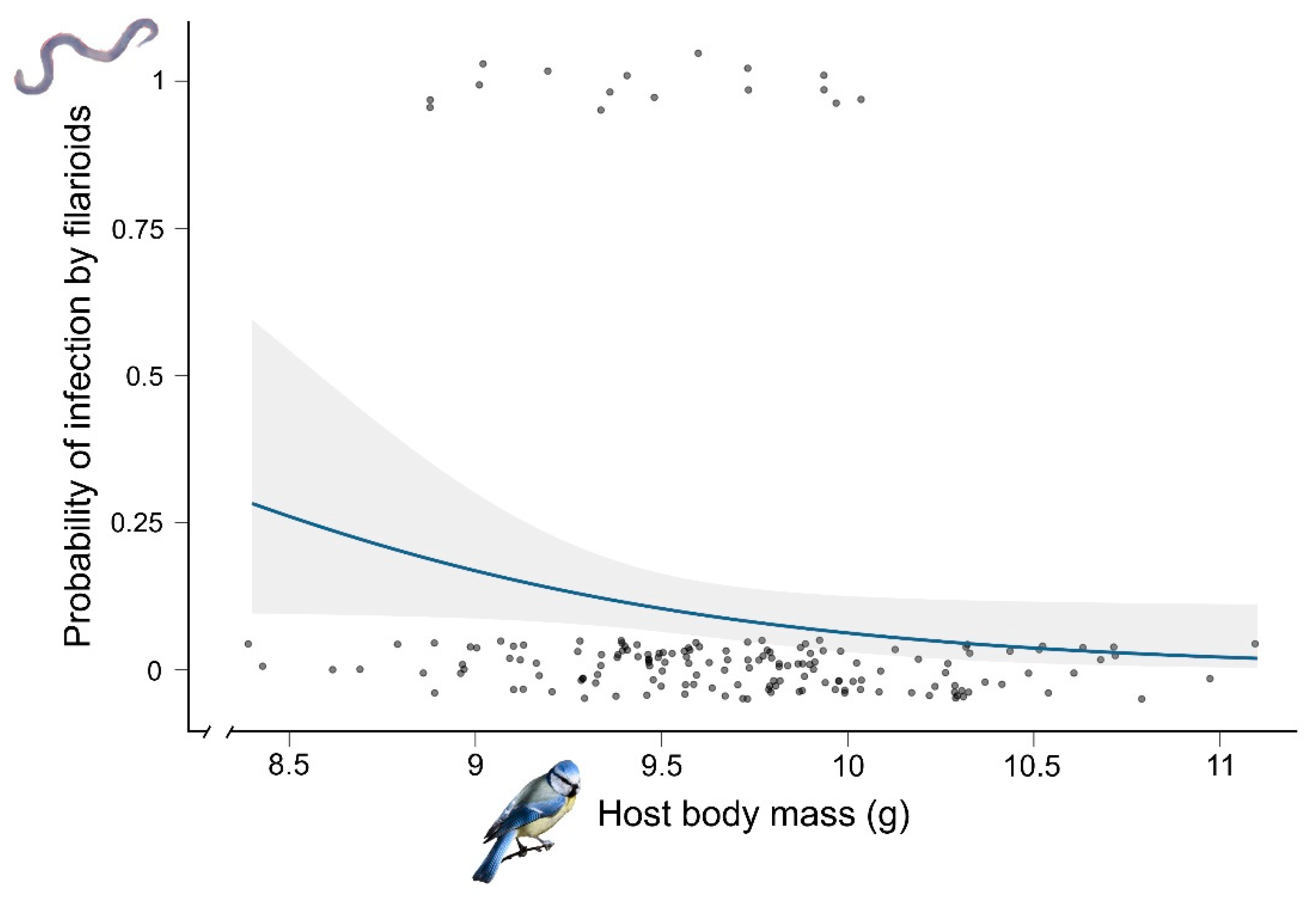
3.1. Prevalence and Probability of Infection by Filarioids

Overall, using the 18S PCR screening, 18 out of 171 (10.53%) blue tits were positive for filarioid helminths. However, only 16 of the 18 PCR-positive samples were confirmed by sequencing, slightly reducing the prevalence to 9.36%.
The results of the model selection from the first full GLM showed that the best model for explaining the probability of infection by filarioids included only the body condition index as the predictor variable (Table 1). Blue tits in a poorer condition were more susceptible to be infected by filarial nematodes than those in a better condition, although the relationship was marginally non-significant (estimate = −24.24, z = −1.75, p = 0.078). However, none of the selected models differed significantly from the null model (Table 1); thus, the independent variables did not have any significant effect on the probability of infection by filarioids.
The results of the model selection from the second full GLM revealed that the best model explaining the probability of infection included only body mass as the predictor variable (Table 1). The probability of infection by filarioids increased significantly with decreasing body mass of adult blue tits (estimate = −1.11, z = −1.96, p = 0.049; Figure 1). The second and third best models included also laying date and forest, respectively, as predictors, but these variables did not significantly affect the probability of infection (laying date: estimate = 0.04, z = 1.16, p = 0.24; forest: estimate = −0.32, z = 0.58, p = 0.56). The best model significantly differed from the null model (null model: AIC = 107.9, ΔAIC = 2.04).

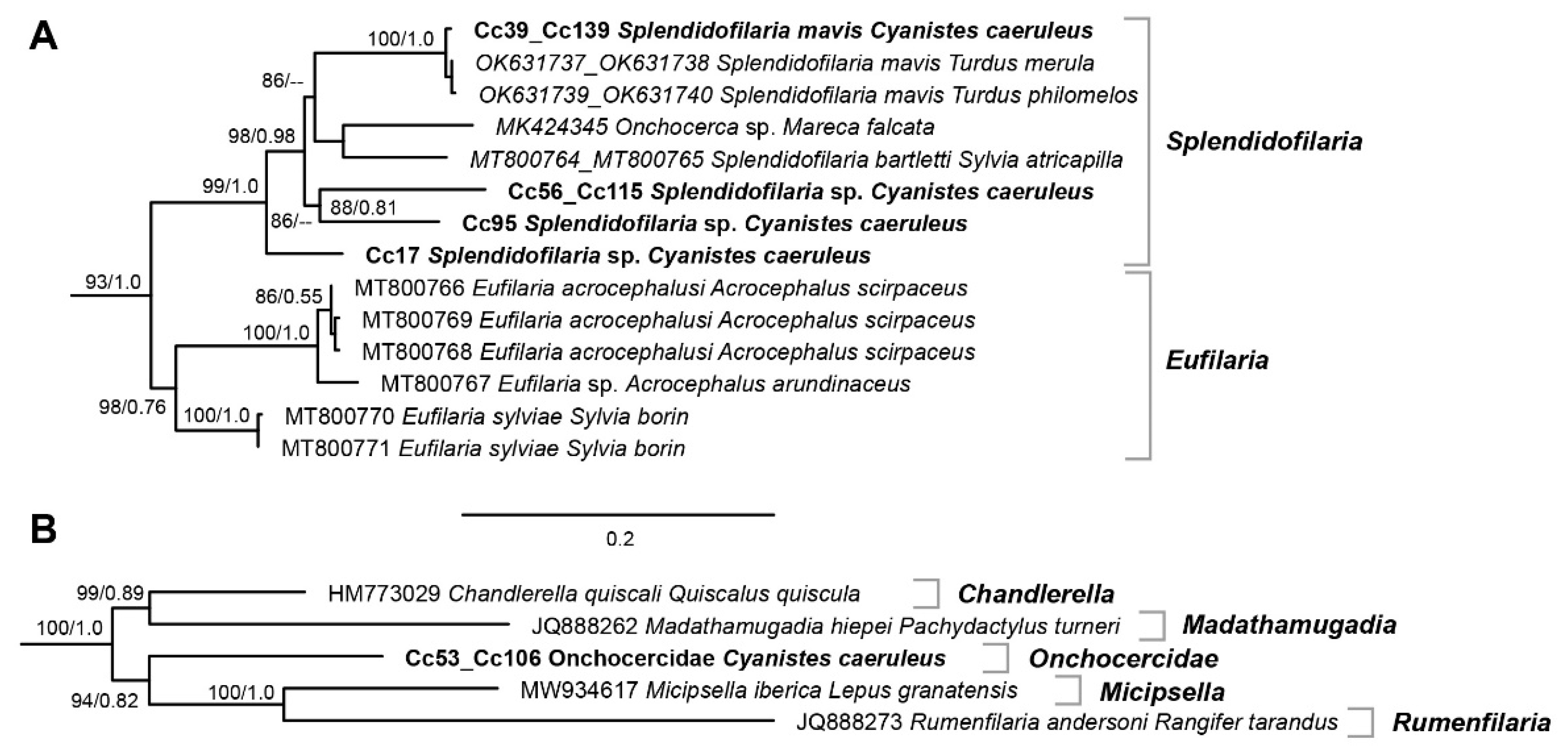
3.2. Filarioid Sequences and Phylogenetic Tree

In total, sixteen individuals were confirmed positive for Onchocercidae by PCR and sequencing the 560 pb section of the 18S gene. Five samples (Cc017, Cc056, Cc095, Cc139, and Cc150) featured two lineages differing by few bp from those of Onchocercidae detected in the American robin (Turdus migratorius) (JQ867037, JQ867035, JQ867026) and the common grackle (Quiscalus quiscula) (JQ867040). Eleven samples (Cc023, Cc030, Cc038, Cc039, Cc053, Cc058, Cc070, Cc106, Cc115, Cc166, Cc169) featured a new lineage separated by at least five bp from other Onchocercidae.
The 900 pb COI section was successfully sequenced in eight samples only. A phylogenetic tree was calculated based on a 600 pb section of the COI, including representatives of all Onchocercidae species (Supplementary Material, Figure S1). The two clades featuring lineages detected in the present study are shown in Figure 2. The lineage detected in samples Cc39 and Cc139 differed by 0.5% to 0.7% from Splendidofilaria mavis isolated from the blackbird (Turdus merula) (OK631737, OK631738) and song thrush (OK631739, OK631740) in Lithuania [56] and likely belongs to the same parasite species. The samples Cc56 and Cc115, Cc95, and Cc17 featured three additional lineages, which also clustered in the Splendidofilaria clade and likely belong to three separate Splendidofilaria species. The lineage of samples Cc56 and Cc115 differed by 7.7% to 8.3% from already known Splendidofilaria lineages, Cc95 by 7.2% to 7.8%, and Cc17 by 6.7% to 7.8%. The samples Cc53 and Cc106 featured an Onchocercidae lineage that could not be clearly assigned to any genus of filarioid parasites. It clustered in a well-supported clade with representatives of four Onchocercidae genera, forming the sister clade to Micipsella iberica from the Iberian hare (Lepus granatensis) (MW934617) and Rumenfilaria andersoni from the reindeer (Rangifer tarandus) (Q888273). Moreover, the clade also contained sequences of Chandlerella quiscali from the common grackle (HM773029) and Madathamugadia hiepei from the gecko Pachydactylus turneri (JQ888262). The sequences of samples Cc53 and Cc106 differed from the latter four lineages by 10.5% to 14.5%.

4. Discussion

Our results showed that blue tits in a poorer body condition, in terms of lower body mass, are more susceptible to be parasitized by filarioid nematodes. Although a strong relationship was not apparent, the findings of the present study are in accordance with other studies which found a negative association between microfilarial infection status and bird body mass [46,47]. Furthermore, we molecularly characterized the microfilariae infecting such bird hosts, revealing that blue tits are parasitized by several lineages of Onchocercidae in our study area. Our molecular data thus added new useful information to better understand the diversity of avian filarioids, calling for further genetic studies of this understudied nematode group [52,56]. In contrast, although habitat type has been shown to affect filarioid prevalence or distribution in other bird populations [18,36,43], we did not find evidence to suggest that forest type at smaller scales can alter the prevalence of these blood parasites infecting blue tits. Lastly, host sex did not alter the probability of infection by filarioids, suggesting the absence of sex differences in the infection susceptibility to these parasites in avian hosts. Other studies have observed the same pattern in other bird species [50,78]. These aspects are discussed in more detail below.

4.1. Host Body Condition and Infection by Filarioid Nematodes

In our study area, blue tits with a lower body mass appear to be more likely to be parasitized by filarioid nematodes. We found a negative association between microfilariae infection status and host body mass—a proxy for body condition [72]—although the relationship was not too pronounced (the body condition index models did not significantly differ from the null model, whereas body mass models did; Table 1). This same pattern has been observed in some wild bird populations, but not in others. For example, the intensity of infection by microfilaria is negatively correlated with host body mass in the white-necked thrush (Turdus albicollis) [46], while village weavers (Ploceus cucullatus) infected by microfilariae show a lower body mass compared to uninfected individuals [47]. In contrast, no association between host body mass or condition index and microfilarial infection status has been reported in Aimophila sparrows [39], nor in some insular birds, such as Galápagos penguins (Spheniscus mendiculus) or flightless cormorants (Phalacrocorax harrisi) [37]. Even a positive relationship between microfilariae infection status and host body mass has been observed in the fire-crested alethe (Alethe diademata) [19]. Moreover, other studies have revealed complex relationships between filarial infections and host immune system and blood physiology. For example, New Caledonian Zosterops spp. individuals exhibit elevated heterophil to lymphocyte ratios when infected by microfilariae, mainly because these parasites provoked a proliferation of heterophils in the bloodstream [36], but no alterations of the heterophil to lymphocyte ratio have been observed in the white-necked thrush [46]. Furthermore, microfilariae may diminish the erythrocytic sedimentation rate (an indirect indicator of immunocompetence [49]) and increase the blood packed cell volume [35], but by contrast, these parasites may not alter other measures of blood physiology, such as hematocrit [47,49], serum biochemistry [34], or polychromasia levels [78]. Thus, the potential alteration of the blood homeostasis could enhance a mobilization of host nutrient stores, causing microfilariae to parasitize and successfully develop in birds with a lower body mass or poorer condition. However, further experimental studies are necessary to clarify the observed associations between filarial infection and variation in host body mass, as positive and negative correlations have been reported in several filarioid–bird systems (see above).
Altogether, the aforementioned studies suggest that filarioid nematodes commonly establish complex relationships with their hosts, not always negatively affecting host energy stores or blood physiology. Nevertheless, these parasites may sometimes exert a cost to wild birds in the same way that other well-studied blood parasites do (e.g., avian malaria and avian malaria-like parasites; [79,80,81]), especially to those individuals with a poorer body condition, which may be more likely to be parasitized by filarioids ([47]; this study) or harbor a greater number of microfilariae in their bloodstream [46]. Thereby, parasitized birds in poor body condition could be impaired, not only because of the direct impacts of filarioids, but also in terms of the increased risk of co-infection, as nematode-induced immune modulation may facilitate malaria co-infections [36,82]. This could have negative consequences for such birds relative to blood-protein physiology [83] or future survival prospects [50] when being co-infected with avian malaria or avian malaria-like parasites, but further studies are necessary to further understand this possibility.

4.2. Molecular Characterization of Filairoids Infecting Blue Tits

The sequencing of a 900 bp COI section of eight individuals revealed the presence of five different Onchocercidae lineages. Four of the lineages clustered in the Splendidofilaria clade, but only one lineage could be attributed to a known species, Splendidofilaria mavis. The remaining lineage clustered in an Onchocercidae lineage that could not be clearly assigned to any genus. Although, to date, few studies have addressed the molecular characterization of filarioid nematodes infecting wild birds, as the lineages they found (based on 18S rRNA, 28S rRNA, and COI fragments) are in line with the results of the present study. For example, the authors of [57] found two major clades of filarioids, one belonging to the putative Chandlerella quiscali and the second to Splendidofilaria spp., infecting several passerine species. In fact, four out of five lineages found here clustered in the Splendidofilaria clade (Figure 2A), while the remaining lineage was genetically close to C. quiscali (Figure 2B). In addition, S. mavis has been detected in blackbirds and song thrushes from Lithuania [56], and indeed we found S. mavis in two of the infected blue tits. Apart from S. mavis, the only other molecularly known species from this genus is S. bartletti, which was detected in Eurasian blackcaps (Sylvia atricapilla) [52]. This study also identified and discovered new filarioid species (Eufilaria acrocephalusi, E. sylviae and S. bartletti) infecting common reed warblers (Acrocephalus scirpaceus), garden warblers (Sylvia borin), and Eurasian blackcaps [52]. Overall, the results suggest that blue tits harbor a relatively large diversity of filarioid nematode lineages, and, to our knowledge, this is the first study examining and identifying the filarioid nematodes infecting blue tits. Still, lineages need to be linked to morphospecies, and our study call for further genetic studies of this group of parasites.

4.3. Prevalence of Filarioids Unaffected by Habitat Type and Host Sex

We also found that the probability of infection by microfilariae did not vary with the forest type. Other studies have shown that habitat characteristics may have important implications for the prevalence of filarioids in bird populations. For example, the occurrence of microfilariae is greater in dry forests than in humid forests [18], and in insular systems, the presence of microfilaria typically vary according to several environmental factors, such as ambient temperature, precipitation, or vegetation quality [43], leading to an island-dependent mosaic distribution [36]. A forest-dependent probability of infection by microfilaria was expected in our study area, as blood parasites (especially vector-borne parasites) are strongly driven by environmental factors, which can limit vector distribution [40,41]. Both parts of the woodland (eastern and western) from our study area differed in several abiotic and biotic factors [58,59], and in fact, we have previously shown that biting midge (Culicoides spp.) abundance within blue tit nest boxes was higher in the dry, western woodland, than in the humid, eastern woodland [42]. Additionally, the prevalence of black flies (Simulium spp.) in our study area tended to be higher in nest boxes from the humid woodland than in the dry woodland (unpublished data). As both haematophagous arthropod groups are competent vectors for avian filarioids [11], we expected some forest-dependent variation in the occurrence of microfilariae infecting adult blue tits. The obtained results are attributable to two possibilities: (1) filarioid occurrence developing in each vector group (biting midges and black flies) did not show any forest-dependent variation, or (2) the prevalence of filarioids was similar in biting midges and in black flies, leading to the observed prevalence in the present study, as biting midges were more abundant in the dry forests, while black flies tended to be more abundant in the humid forests (see above). However, both possibilities assume a similar vector competence or transmission efficiency across the woodland. Further studies should be conducted in order to identify the presence of filarioid nematodes developing in both vector groups and whether biting midges and black flies could potentially transmit nematode immature stages to blue tits in a similar manner.
On the other hand, several studies have reported a clear altitudinal pattern in the presence of microfilaria infecting wild birds across the globe, with birds from highland zones having the lowest prevalences. For example, in the Neotropical region, the probability of filarioid infection is higher in birds from lowland humid forests than in highland forests [27], with microfilariae being absent from elevations of 3.000 m [44]. In Central Africa and Madagascar, the filarioid prevalence is also negatively correlated with elevation, with microfilariae not being present in highland mountain forests [18,19]. Lastly, in the Galápagos islands, a negative association between microfilariae prevalence and elevation has also been reported for some avian species [43]. These studies are in accordance with the general assumption that haemoparasite prevalence decreases with elevation [84], but interestingly, we found a relatively high microfilariae prevalence (9.4%) in a Mediterranean mountain woodland located at ca. 1.800 m a.s.l.—an altitude point in which the prevalence of filarioids is typically less than 3% or even zero [18,44]. However, elevational patterns of parasites infecting birds may be complex depending on the parasite group [85] or the mountain system studied [86]. Concretely, in the Sierra Nevada mountain range, the abundance and prevalence of ectoparasites, vectors, and haemoparasites may decrease, increase, or be stable with elevation depending on the taxa [86,87,88]. Thus, in Sierra Nevada, the relatively high microfilariae prevalence is likely brought about by Mediterranean climatic particularities.
Finally, we did not find evidence to suggest that host sex affected the probability of infection by microfilariae, contrasting with the general acceptance that males are more susceptible to parasites and diseases than females, mainly due to physiological causes (e.g., interaction between immune system and testosterone; reviewed in [89,90]). However, in parasite–host systems formed by filarioids and birds, no differences in the parasite’s susceptibility between sexes have been reported in a wide variety of bird species, such as the purple martin (Progne subis) [50], the Northern cardinal (Cardinalis cardinalis) [78], the Galápagos cormorant (Phalacrocorax harrisi) [37], the village weaver (Ploceus cucullatus) [47], or the blue tit (in this study). Moreover, most of these bird species exhibit moderate to high degrees of sexual dichromatism, with males being typically brighter and more colored than females. Because the expression of secondary sexual characters is related to infection susceptibility and immune function [89], males are expected to suffer more from parasitism [91]. However, this scenario has not been observed in the present study, nor in other filarioid–bird systems worldwide. The non-generalized immune costs or immune-cell overproduction associated with filarioid parasitization (see above) may explain why most of studies did not find sex differences in microfilariae infection rates.

Supplementary Materials

The following supporting information can be downloaded at: https://www.mdpi.com/article/10.3390/d15050609/s1, Figure S1: Clades of maximum likelihood tree based on a 600 pb section of the COI gene, including representatives of all Onchocercidae species. Maximum likelihood bootstrap values and Bayesian posterior probabilities are indicated at all nodes. The scale bar indicates the expected mean number of substitutions per site according to the model of sequence evolution applied.

Author Contributions

Conceptualization, G.M.-R., H.-P.F. and J.H.; methodology, G.M.-R., J.G.-B., M.C., H.-P.F. and J.H.; software, J.G.-B., H.-P.F. and J.H.; validation, J.G.-B., H.-P.F., J.H. and M.C.; formal analysis, J.G.-B., H.-P.F. and J.H.; investigation, G.M.-R., J.G.-B., M.C., H.-P.F. and J.H.; resources, H.-P.F., J.H., S.S., G.M.-R. and D.J.P.; data curation, J.G.-B., H.-P.F. and J.H.; writing—original draft preparation, J.G.-B.; writing—review and editing, J.G.-B., G.M.-R., H.-P.F. and J.H.; visualization, J.G.-B., H.-P.F. and J.H.; supervision, S.S., D.J.P. and G.M.-R.; project administration, H.-P.F., J.H., S.S., G.M.-R. and D.J.P.; funding acquisition, G.M.-R. All authors have read and agreed to the published version of the manuscript.

Funding

This research was funded by National Plan of the Spanish Ministry of Economy and Competition (CGL2014-55969-P and CGL2017-84938-P) and the Andalusian government (A.RNM.48.UGR20), co-funded with FEDER funds from the European Union. J.G.-B. was supported by an FPU predoctoral contract from the Spanish Ministry of Education (FPU18/03034) and by an FPU mobility grant from the Spanish Ministry of Education (EST21/00626). M.C. was supported with a postdoctoral contract from the Spanish Ministry of Universities, funded with Next Generation funds.

Institutional Review Board Statement

The animal study protocol was approved by the Ethics Committee of the University of Granada and the Andalusian Regional Government (protocol code 03-06-15-259 of 3 June 2015).

Data Availability Statement

Data will be provided by the authors under request.

Acknowledgments

José Luis Ros-Santaella, Eliana Pintus, Abelardo Requena Blanco, Nicola Bernardo, and Paula Lopezosa supported us during the fieldwork. Mohammed Bakkali advised us in genetic analyses, and Gopi Munimanda helped us during the laboratory work. We are grateful to the staff of the National Park of Sierra Nevada for their constant support. Two anonymous reviewers improved the actual version of manuscript.

Conflicts of Interest

The authors declare no conflict of interest.

References

  1. McCarthy, J.; Moore, T.A. Emerging Helminth Zoonoses. Int. J. Parasitol. 2000, 30, 1351–1360. [Google Scholar] [CrossRef]
  2. de Silva, N.R.; Brooker, S.; Hotez, P.J.; Montresor, A.; Engels, D.; Savioli, L. Soil-Transmitted Helminth Infections: Updating the Global Picture. Trends Parasitol. 2003, 19, 547–551. [Google Scholar] [CrossRef]
  3. Taylor, M.J.; Hoerauf, A.; Bockarie, M. Lymphatic Filariasis and Onchocerciasis. Lancet 2010, 376, 1175–1185. [Google Scholar] [CrossRef] [PubMed]
  4. Kalyanasundaram, R.; Khatri, V.; Chauhan, N. Advances in Vaccine Development for Human Lymphatic Filariasis. Trends Parasitol. 2020, 36, 195–205. [Google Scholar] [CrossRef] [PubMed]
  5. Allen, J.E.; Adjei, O.; Bain, O.; Hoerauf, A.; Hoffmann, W.H.; Makepeace, B.L.; Schulz-Key, H.; Tanya, V.N.; Trees, A.J.; Wanji, S.; et al. Of Mice, Cattle, and Humans: The Immunology and Treatment of River Blindness. PLoS Negl. Trop. Dis. 2008, 2, e217. [Google Scholar] [CrossRef] [PubMed]
  6. Molyneux, D.H. Filaria Control and Elimination: Diagnostic, Monitoring and Surveillance Needs. Trans. Roy. Soc. Trop. Med. Hyg. 2009, 103, 338–341. [Google Scholar] [CrossRef]
  7. Dieki, R.; Nsi-Emvo, E.; Akue, J.P. The Human Filaria Loa loa: Update on Diagnostics and Immune Response. Res. Rep. Trop. Med. 2022, 13, 41–54. [Google Scholar] [CrossRef]
  8. Simón, F.; Siles-Lucas, M.; Morchón, R.; González-Miguel, J.; Mellado, I.; Carretón, E.; Montoya-Alonso, J.A. Human and Animal Dirofilariasis: The Emergence of a Zoonotic Mosaic. Clin. Microbiol. Rev. 2012, 25, 507–544. [Google Scholar] [CrossRef]
  9. Otranto, D.; Dantas-Torres, F.; Brianti, E.; Traversa, D.; Petrić, D.; Genchi, C.; Capelli, G. Vector-Borne Helminths of Dogs and Humans in Europe. Parasites Vectors 2013, 6, 16. [Google Scholar] [CrossRef] [PubMed]
  10. Orihel, T.C.; Eberhard, M.L. Zoonotic Filariasis. Clin. Microbiol. Rev. 1998, 11, 366–381. [Google Scholar] [CrossRef]
  11. Bartlett, C.M. Filarioid Nematodes. In Parasitic Diseases of Wild Birds; Atkinson, C.T., Thomas, N.J., Hunter, D.B., Eds.; Wiley Blackwell: Oxford, UK, 2008; pp. 439–462. ISBN 978-0-8138-0462-0. [Google Scholar]
  12. Bain, O.; Babayan, S. Behaviour of Filariae: Morphological and Anatomical Signatures of Their Life Style within the Arthropod and Vertebrate Hosts. Filaria J. 2003, 2, 16. [Google Scholar] [CrossRef]
  13. Mutinda, M.; Otiende, M.; Gakuya, F.; Kariuki, L.; Obanda, V.; Ndeere, D.; Ndambiri, E.; Kariuki, E.; Lekolool, I.; Soriguer, R.C.; et al. Putative Filariosis Outbreak in White and Black Rhinoceros at Meru National Park in Kenya. Parasites Vectors 2012, 5, 206. [Google Scholar] [CrossRef]
  14. Laaksonen, S.; Solismaa, M.; Orro, T.; Kuusela, J.; Saari, S.; Kortet, R.; Nikander, S.; Oksanen, A.; Sukura, A. Setaria tundra Microfilariae in Reindeer and Other Cervids in Finland. Parasitol. Res. 2009, 104, 257–265. [Google Scholar] [CrossRef]
  15. Bain, O. Evolutionary Relationships among Filarial Nematodes. In The Filaria; Klei, T.R., Rajan, T.V., Eds.; World Class Parasites; Springer: New York, NY, USA, 2002; Volume 5, pp. 21–29. ISBN 978-0-306-47661-7. [Google Scholar]
  16. Readel, A.M.; Goldberg, T.L. Blood Parasites of Frogs from an Equatorial African Montane Forest in Western Uganda. J. Parasitol. 2010, 96, 448–450. [Google Scholar] [CrossRef]
  17. Sehgal, R.N.M.; Jones, H.I.; Smith, T.B. Blood Parasites of Some West African Rainforest Birds. J. Vet. Med. Sci. 2005, 67, 295–301. [Google Scholar] [CrossRef]
  18. Savage, A.F.; Robert, V.; Goodman, S.M.; Raharimanga, V.; Raherilalao, M.J.; Andrianarimisa, A.; Ariey, F.; Greiner, E.C. Blood Parasites in Birds from Madagascar. J. Wildl. Dis. 2009, 45, 907–920. [Google Scholar] [CrossRef]
  19. Sehgal, R.N.M.; Jones, H.I.; Smith, T.B. Molecular Evidence for Host Specificity of Parasitic Nematode Microfilariae in Some African Rainforest Birds. Mol. Ecol. 2005, 14, 3977–3988. [Google Scholar] [CrossRef] [PubMed]
  20. Elahi, R.; Islam, A.; Hossain, M.S.; Mohiuddin, K.; Mikolon, A.; Paul, S.K.; Hosseini, P.R.; Daszak, P.; Alam, M.S. Prevalence and Diversity of Avian Haematozoan Parasites in Wetlands of Bangladesh. J. Parasitol. Res. 2014, 2014, 493754. [Google Scholar] [CrossRef] [PubMed]
  21. Valkiūnas, G.; Iezhova, T.A. A Comparison of the Blood Parasites in Three Subspecies of the Yellow Wagtail Motacilla flava. J. Parasitol. 2001, 87, 930–934. [Google Scholar] [CrossRef]
  22. Silveira, P.; Belo, N.O.; Rodello, D.; Pinheiro, R.T.; Braga, É.M. Microfilariae Infection in Wild Birds from the Brazilian Cerrado. J. Wildl. Dis. 2010, 46, 1305–1309. [Google Scholar] [CrossRef] [PubMed]
  23. Sebaio, F.; Braga, É.M.; Branquinho, F.; Fecchio, A.; Marini, M.Â. Blood Parasites in Passerine Birds from the Brazilian Atlantic Forest. Rev. Bras. Parasitol. Vet. 2012, 21, 7–15. [Google Scholar] [CrossRef]
  24. Bennett, G.F.; Garvin, M.; Bates, J.M. Avian Hematozoa from West-Central Bolivia. J. Parasitol. 1991, 77, 207–211. [Google Scholar] [CrossRef] [PubMed]
  25. Matta, N.E.; Basto, N.; Gutierrez, R.; Rodríguez, O.A.; Greiner, E.C. Prevalence of Blood Parasites in Tyrannidae (Flycatchers) in the Eastern Plains of Colombia. Mem. Inst. Oswaldo Cruz 2004, 99, 271–274. [Google Scholar] [CrossRef]
  26. Londoño, A.; Pulgarin-R, P.C.; Blair, S. Blood Parasites in Birds from the Lowlands of Northern Colombia. Caribb. J. Sci. 2007, 43, 87–93. [Google Scholar] [CrossRef]
  27. De La Torre, G.M.; Campião, K.M. Bird Habitat Preferences Drive Hemoparasite Infection in the Neotropical Region. Integr. Zool. 2021, 16, 755–768. [Google Scholar] [CrossRef] [PubMed]
  28. Villalva-Pasillas, D.; Medina, J.P.; Soriano-Vargas, E.; Martínez-Hernández, D.A.; García-Conejo, M.; Galindo-Sánchez, K.P.; Sánchez-Jasso, J.M.; Talavera-Rojas, M.; Salgado-Miranda, C. Haemoparasites in Endemic and Non-Endemic Passerine Birds from Central Mexico Highlands. Int. J. Parasitol. 2020, 11, 88–92. [Google Scholar] [CrossRef]
  29. Benedikt, V.; Barus, V.; Capek, M.; Havlicek, M.; Literak, I. Blood Parasites (Haemoproteus and Microfilariae) in Birds from the Caribbean Slope of Costa Rica. Acta Parasitol. 2009, 54, 197–204. [Google Scholar] [CrossRef]
  30. Young, B.E.; Garvin, M.C.; McDonald, D.B. Blood Parasites in Birds from Monteverde, Costa Rica. J. Wildl. Dis. 1993, 29, 555–560. [Google Scholar] [CrossRef]
  31. Valkiūnas, G.; Iezhova, T.A.; Brooks, D.R.; Hanelt, B.; Brant, S.V.; Sutherlin, M.E.; Causey, D. Additional Observations on Blood Parasites of Birds in Costa Rica. J. Wildl. Dis. 2004, 40, 555–561. [Google Scholar] [CrossRef] [PubMed]
  32. Haas, M.; Baruš, V.; Benedikt, V.; Literák, I. Microfilariae in Birds in the Czech Republic, Including a Note on Adult Nematodes Eufilaria delicata in a Song Thrush Turdus philomelos. Parasitol. Res. 2011, 109, 645–655. [Google Scholar] [CrossRef]
  33. Merino, S.; Potti, J.; Fargallo, J.A. Blood Parasites of Passerine Birds from Central Spain. J. Wildl. Dis. 1997, 33, 638–641. [Google Scholar] [CrossRef]
  34. Travis, E.K.; Vargas, F.H.; Merkel, J.; Gottdenker, N.; Miller, R.E.; Parker, P.G. Hematology, Serum Chemistry, and Serology of Galápagos Penguins (Spheniscus mendiculus) in the Galápagos Islands, Ecuador. J. Wildl. Dis. 2006, 42, 625–632. [Google Scholar] [CrossRef] [PubMed]
  35. Travis, E.K.; Vargas, F.H.; Merkel, J.; Gottdenker, N.; Miller, R.E.; Parker, P.G. Hematology, Plasma Chemistry, and Serology of the Flightless Cormorant (Phalacrocorax harrisi) in the Galápagos Islands, Ecuador. J. Wildl. Dis. 2006, 42, 133–141. [Google Scholar] [CrossRef]
  36. Clark, N.J.; Wells, K.; Dimitrov, D.; Clegg, S.M. Co-Infections and Environmental Conditions Drive the Distributions of Blood Parasites in Wild Birds. J. Anim. Ecol. 2016, 85, 1461–1470. [Google Scholar] [CrossRef] [PubMed]
  37. Merkel, J.; Jones, H.I.; Whiteman, N.K.; Gottdenker, N.; Vargas, H.; Travis, E.K.; Miller, R.E.; Parker, P.G. Microfilariae in Galápagos Penguins (Spheniscus mendiculus) and Flightless Cormorants (Phalacrocorax harrisi): Genetics, Morphology, and Prevalence. J. Parasitol. 2007, 93, 495–503. [Google Scholar] [CrossRef]
  38. Cardells-Peris, J.; Gonzálvez, M.; Ortega-Porcel, J.; Ruiz de Ybáñez, M.R.; Martínez-Herrero, M.C.; Garijo-Toledo, M.M. Parasitofauna Survey of Song Thrushes (Turdus philomelos) from the Eastern Part of Spain. Parasitol. Int. 2020, 79, 102176. [Google Scholar] [CrossRef]
  39. Deviche, P.; McGraw, K.; Greiner, E.C. Interspecific Differences in Hematozoan Infection in Sonoran Desert Aimophila Sparrows. J. Wildl. Dis. 2005, 41, 532–541. [Google Scholar] [CrossRef]
  40. Sehgal, R.N.M. Manifold Habitat Effects on the Prevalence and Diversity of Avian Blood Parasites. Int. J. Parasitol. 2015, 4, 421–430. [Google Scholar] [CrossRef] [PubMed]
  41. Ferraguti, M.; Martínez-de la Puente, J.; Bensch, S.; Roiz, D.; Ruiz, S.; Viana, D.S.; Soriguer, R.C.; Figuerola, J. Ecological Determinants of Avian Malaria Infections: An Integrative Analysis at Landscape, Mosquito and Vertebrate Community Levels. J. Anim. Ecol. 2018, 87, 727–740. [Google Scholar] [CrossRef] [PubMed]
  42. Garrido-Bautista, J.; Martínez-de la Puente, J.; Ros-Santaella, J.L.; Pintus, E.; Lopezosa, P.; Bernardo, N.; Comas, M.; Moreno-Rueda, G. Habitat-Dependent Culicoides Species Composition and Abundance in Blue Tit (Cyanistes caeruleus) Nests. Parasitology 2022, 149, 1119–1128. [Google Scholar] [CrossRef]
  43. Siers, S.; Merkel, J.; Bataille, A.; Vargas, F.H.; Parker, P.G. Ecological Correlates of Microfilariae Prevalence in Endangered Galápagos Birds. J. Parasitol. 2010, 96, 259–272. [Google Scholar] [CrossRef]
  44. González, A.D.; Matta, N.E.; Ellis, V.A.; Miller, E.T.; Ricklefs, R.E.; Gutiérrez, H.R. Mixed Species Flock, Nest Height, and Elevation Partially Explain Avian Haemoparasite Prevalence in Colombia. PLoS ONE 2014, 9, e100695. [Google Scholar] [CrossRef]
  45. Campbell, T.W.; Ellis, C.K. Avian and Exotic Animal Hematology and Citology, 3rd ed.; Blackwell Publishing Ltd.: Oxford, UK, 2007. [Google Scholar]
  46. De La Torre, G.M.; Freitas, F.F.; Fratoni, R.D.O.; Guaraldo, A.D.C.; Dutra, D.D.A.; Braga, M.; Manica, L.T. Hemoparasites and Their Relation to Body Condition and Plumage Coloration of the White-Necked Thrush (Turdus albicollis). Ethol. Ecol. Evol. 2020, 32, 509–526. [Google Scholar] [CrossRef]
  47. Atawal, A.F.; Mgbeahuruike, A.C.; Hammers, M. Microfilarial Infections Associated with Body Mass Loss of Village Weavers Ploceus cucullatus. Ostrich 2019, 90, 41–44. [Google Scholar] [CrossRef]
  48. Höglund, J.; Alatalo, R.V.; Lundberg, A. The Effects of Parasites on Male Ornaments and Female Choice in the Lek-Breeding Black Grouse (Tetrao tetrix). Behav. Ecol. Sociobiol. 1992, 30, 71–76. [Google Scholar] [CrossRef]
  49. Valera, F.; Hoi, H.; Krištín, A. Parasite Pressure and Its Effects on Blood Parameters in a Stable and Dense Population of the Endangered Lesser Grey Shrike. Biodivers. Conserv. 2006, 15, 2187–2195. [Google Scholar] [CrossRef]
  50. Davidar, P.; Morton, E.S. Are Multiple Infections More Severe for Purple Martins (Progne subis) than Single Infections? Auk 2006, 123, 141–147. [Google Scholar] [CrossRef]
  51. Larrat, S.; Dallaire, A.D.; Lair, S. Emaciation and Larval Filarioid Nematode Infection in Boreal Owls (Aegolius funereus). Avian Pathol. 2012, 41, 345–349. [Google Scholar] [CrossRef] [PubMed]
  52. Binkienė, R.; Chagas, C.R.F.; Bernotienė, R.; Valkiūnas, G. Molecular and Morphological Characterization of Three New Species of Avian Onchocercidae (Nematoda) with Emphasis on Circulating Microfilariae. Parasites Vectors 2021, 14, 137. [Google Scholar] [CrossRef] [PubMed]
  53. Ferri, E.; Barbuto, M.; Bain, O.; Galimberti, A.; Uni, S.; Guerrero, R.; Ferté, H.; Bandi, C.; Martin, C.; Casiraghi, M. Integrated Taxonomy: Traditional Approach and DNA Barcoding for the Identification of Filarioid Worms and Related Parasites (Nematoda). Front. Zool. 2009, 6, 1. [Google Scholar] [CrossRef]
  54. Bain, O.; Casiraghi, M.; Martin, C.; Uni, S. The Nematoda Filarioidea: Critical Analysis Linking Molecular and Traditional Approaches. Parasite 2008, 15, 342–348. [Google Scholar] [CrossRef]
  55. Eamsobhana, P.; Lim, P.-E.; Yong, H.S. Molecular Phylogeny of Filarial Worms (Nematoda: Filarioidea). Raffles Bull. Zool. 2013, 29, 99–109. [Google Scholar]
  56. Chagas, C.R.F.; Binkienė, R.; Valkiūnas, G. Description and Molecular Characterization of Two Species of Avian Blood Parasites, with Remarks on Circadian Rhythms of Avian Haematozoa Infections. Animals 2021, 11, 3490. [Google Scholar] [CrossRef] [PubMed]
  57. Hamer, G.L.; Anderson, T.K.; Berry, G.E.; Makohon-Moore, A.P.; Crafton, J.C.; Brawn, J.D.; Dolinski, A.C.; Krebs, B.L.; Ruiz, M.O.; Muzzall, P.M.; et al. Prevalence of Filarioid Nematodes and Trypanosomes in American Robins and House Sparrows, Chicago USA. Int. J. Parasitol. 2013, 2, 42–49. [Google Scholar] [CrossRef]
  58. Garrido-Bautista, J.; Soria, A.; Trenzado, C.E.; Pérez-Jiménez, A.; Ros-Santaella, J.L.; Pintus, E.; Bernardo, N.; Comas, M.; Moreno-Rueda, G. Oxidative Status of Blue Tit Nestlings Varies with Habitat and Nestling Size. Comp. Biochem. Physiol. A 2021, 258, 110986. [Google Scholar] [CrossRef] [PubMed]
  59. Garrido-Bautista, J.; Soria, A.; Trenzado, C.E.; Pérez-Jiménez, A.; Pintus, E.; Ros-Santaella, J.L.; Bernardo, N.; Comas, M.; Kolenčík, S.; Moreno-Rueda, G. Within-Brood Body Size and Immunological Differences in Blue Tit (Cyanistes caeruleus) Nestlings Relative to Ectoparasitism. Avian Res. 2022, 13, 100038. [Google Scholar] [CrossRef]
  60. Moreno-Rueda, G. Selección de Cajas-Nido Por Aves Insectívoras En Sierra Nevada. Zool. Baetica 2003, 13/14, 131–138. [Google Scholar]
  61. Schlicht, E.; Kempenaers, B. Immediate Effects of Capture on Nest Visits of Breeding Blue Tits, Cyanistes caeruleus, Are Substantial. Anim. Behav. 2015, 105, 63–78. [Google Scholar] [CrossRef]
  62. Perrins, C.M. British Tits; Collins: Glasgow, UK, 1979. [Google Scholar]
  63. Owen, J.C. Collecting, Processing, and Storing Avian Blood: A Review. J. Field Ornithol. 2011, 82, 339–354. [Google Scholar] [CrossRef]
  64. de Jong, A. Less Is Better. Avoiding Redundant Measurements in Studies on Wild Birds in Accordance to the Principles of the 3Rs. Front. Vet. Sci. 2019, 6, 195. [Google Scholar] [CrossRef]
  65. Casiraghi, M.; Anderson, T.J.C.; Bandi, C.; Bazzocchi, C.; Genchi, C. A Phylogenetic Analysis of Filarial Nematodes: Comparison with the Phylogeny of Wolbachia Endosymbionts. Parasitology 2001, 122, 93–103. [Google Scholar] [CrossRef] [PubMed]
  66. Bensch, S.; Inumaru, M.; Sato, Y.; Lee Cruz, L.; Cunningham, A.A.; Goodman, S.J.; Levin, I.I.; Parker, P.G.; Casanueva, P.; Hernández, M.; et al. Contaminations Contaminate Common Databases. Mol. Ecol. Resour. 2021, 21, 355–362. [Google Scholar] [CrossRef]
  67. Hall, T.A. BioEdit: A User-Friendly Biological Sequence Alignment Editor and Analysis Program for Windows 95/98/NT. Nucl. Acid. Sym. Ser. 1999, 41, 95–98. [Google Scholar]
  68. Kumar, S.; Stecher, G.; Tamura, K. MEGA7: Molecular Evolutionary Genetics Analysis Version 7.0 for Bigger Datasets. Mol. Biol. Evol. 2016, 33, 1870–1874. [Google Scholar] [CrossRef] [PubMed]
  69. Xia, X. DAMBE7: New and Improved Tools for Data Analysis in Molecular Biology and Evolution. Mol. Biol. Evol. 2018, 35, 1550–1552. [Google Scholar] [CrossRef]
  70. Nguyen, L.-T.; Schmidt, H.A.; von Haeseler, A.; Minh, B.Q. IQ-TREE: A Fast and Effective Stochastic Algorithm for Estimating Maximum-Likelihood Phylogenies. Mol. Biol. Evol. 2015, 32, 268–274. [Google Scholar] [CrossRef]
  71. Ronquist, F.; Teslenko, M.; van der Mark, P.; Ayres, D.L.; Darling, A.; Höhna, S.; Larget, B.; Liu, L.; Suchard, M.A.; Huelsenbeck, J.P. MrBayes 3.2: Efficient Bayesian Phylogenetic Inference and Model Choice across a Large Model Space. Syst. Biol. 2012, 61, 539–542. [Google Scholar] [CrossRef]
  72. Labocha, M.K.; Hayes, J.P. Morphometric Indices of Body Condition in Birds: A Review. J. Ornithol. 2012, 153, 1–22. [Google Scholar] [CrossRef]
  73. Quinn, G.P.; Keough, M.J. Experimental Design and Data Analysis for Biologists; Cambridge University Press: Cambridge, UK, 2002. [Google Scholar]
  74. Symonds, M.R.E.; Moussalli, A. A Brief Guide to Model Selection, Multimodel Inference and Model Averaging in Behavioural Ecology Using Akaike’s Information Criterion. Behav. Ecol. Sociobiol. 2011, 65, 13–21. [Google Scholar] [CrossRef]
  75. Zuur, A.F.; Ieno, E.N.; Elphick, C.S. A Protocol for Data Exploration to Avoid Common Statistical Problems: Data Exploration. Methods Ecol. Evol. 2010, 1, 3–14. [Google Scholar] [CrossRef]
  76. R Core Team. Development Core Team R: A Language and Environment for Statistical Computing 2020; R Foundation for Statistical Computing: Vienna, Austria, 2020. [Google Scholar]
  77. Bartoń, K. MuMIn: Multi-Model Inference; R Package Version 1.43.17; R Foundation for Statistical Computing: Vienna, Austria, 2020. [Google Scholar]
  78. Astudillo, V.G.; Hernández, S.M.; Kistler, W.M.; Boone, S.L.; Lipp, E.K.; Shrestha, S.; Yabsley, M.J. Spatial, Temporal, Molecular, and Intraspecific Differences of Haemoparasite Infection and Relevant Selected Physiological Parameters of Wild Birds in Georgia, USA. Int. J. Parasitol. 2013, 2, 178–189. [Google Scholar] [CrossRef]
  79. Merino, S.; Moreno, J.; Sanz, J.J.; Arriero, E. Are Avian Blood Parasites Pathogenic in the Wild? A Medication Experiment in Blue Tits (Parus caeruleus). Proc. R. Soc. B Biol. Sci. 2000, 267, 2507–2510. [Google Scholar] [CrossRef]
  80. Marzal, A.; Lope, F.D.; Navarro, C.; Møller, A.P. Malarial Parasites Decrease Reproductive Success: An Experimental Study in a Passerine Bird. Oecologia 2005, 142, 541–545. [Google Scholar] [CrossRef]
  81. Christe, P.; Glaizot, O.; Strepparava, N.; Devevey, G.; Fumagalli, L. Twofold Cost of Reproduction: An Increase in Parental Effort Leads to Higher Malarial Parasitaemia and to a Decrease in Resistance to Oxidative Stress. Proc. R. Soc. B Biol. Sci. 2012, 279, 1142–1149. [Google Scholar] [CrossRef] [PubMed]
  82. Druilhe, P.; Tall, A.; Sokhna, C. Worms Can Worsen Malaria: Towards a New Means to Roll Back Malaria? Trends Parasitol. 2005, 21, 359–362. [Google Scholar] [CrossRef]
  83. Merino, S.; Martínez, J.; Møller, A.P.; Barbosa, A.; De Lope, F.; Rodríguez-Caabeiro, F. Blood Stress Protein Levels in Relation to Sex and Parasitism of Barn Swallows (Hirundo rustica). Écoscience 2002, 9, 300–305. [Google Scholar] [CrossRef]
  84. Zamora-Vilchis, I.; Williams, S.E.; Johnson, C.N. Environmental Temperature Affects Prevalence of Blood Parasites of Birds on an Elevation Gradient: Implications for Disease in a Warming Climate. PLoS ONE 2012, 7, e39208. [Google Scholar] [CrossRef]
  85. Rooyen, J.V.; Lalubin, F.; Glaizot, O.; Christe, P. Altitudinal Variation in Haemosporidian Parasite Distribution in Great Tit Populations. Parasites Vectors 2013, 6, 139. [Google Scholar] [CrossRef] [PubMed]
  86. Illera, J.C.; López, G.; García-Padilla, L.; Moreno, Á. Factors Governing the Prevalence and Richness of Avian Haemosporidian Communities within and between Temperate Mountains. PLoS ONE 2017, 12, e0184587. [Google Scholar] [CrossRef]
  87. Álvarez-Ruiz, L.; Megía-Palma, R.; Reguera, S.; Ruiz, S.; Zamora-Camacho, F.J.; Figuerola, J.; Moreno-Rueda, G. Opposed Elevational Variation in Prevalence and Intensity of Endoparasites and Their Vectors in a Lizard. Curr. Zool. 2018, 64, 197–204. [Google Scholar] [CrossRef]
  88. Moreno-Rueda, G. Elevational Patterns of Blowfly Parasitism in Two Hole Nesting Avian Species. Diversity 2021, 13, 591. [Google Scholar] [CrossRef]
  89. Møller, A.P.; Christe, P.; Lux, E. Parasitism, Host Immune Function, and Sexual Selection. Q. Rev. Biol. 1999, 74, 3–20. [Google Scholar] [CrossRef]
  90. Zuk, M.; McKean, K.A. Sex Differences in Parasite Infections: Patterns and Processes. Int. J. Parasitol. 1996, 26, 1009–1024. [Google Scholar] [CrossRef] [PubMed]
  91. Hamilton, W.; Zuk, M. Heritable True Fitness and Bright Birds: A Role for Parasites? Science 1982, 218, 384–387. [Google Scholar] [CrossRef] [PubMed]
Figure 1. Probability of infection by filarioid nematodes with fitted values of blue tit (Cyanistes caeruleus) body mass, obtained by the binomial GLM. The shade corresponds to the 95% confidence interval.
Diversity 15 00609 g001
Figure 2. Clades of Maximum Likelihood tree (Supporting Information, Figure S1) calculated based on 600 bp sections of the COI gene. Maximum likelihood bootstrap values and Bayesian posterior probabilities are indicated at all nodes. The scale bar indicates the expected mean number of substitutions per site according to the model of sequence evolution applied. (A) Clade containing Splendidofilaria and Eufilaria sequences; (B) Clade containing unknown Onchocercidae lineage.
Diversity 15 00609 g002
Table 1. AIC values and ΔAIC of the models for probability of infection by filarioids with the variables included in the models indicated. The variables that were significant at p < 0.05 are shown in bold. See the Section 2 for the structure of the full models relative to body condition index and body mass.
VariableAICΔAIC
Body condition index models
 Body condition index106.70.00
 Body condition index, laying date107.50.87
 Null model 1107.91.23
 Body condition index, forest108.41.75
 Body condition index, sex108.61.94
Body mass models
Body mass105.90.00
Body mass, laying date106.60.76
Body mass, forest107.61.73
1 Null model included only the intercept.
Disclaimer/Publisher’s Note: The statements, opinions and data contained in all publications are solely those of the individual author(s) and contributor(s) and not of MDPI and/or the editor(s). MDPI and/or the editor(s) disclaim responsibility for any injury to people or property resulting from any ideas, methods, instructions or products referred to in the content.

