Article

Museomics Provides Insights into Conservation and Education: The Instance of an African Lion Specimen from the Museum of Zoology “Pietro Doderlein”

1
Laboratory of Ancient DNA, Department of Cultural Heritage (DBC), University of Bologna, 48121 Ravenna, Italy
2
Center for Evolutionary Hologenomics, Globe Institute, Faculty of Health and Medical Sciences, University of Copenhagen, DK-1353 Copenhagen, Denmark
3
FabLab Palermo APS, 90135 Palermo, Italy
4
IRCCS Istituto Delle Scienze Neurologiche di Bologna, 40139 Bologna, Italy
5
Laboratory of Forensic Genetics, Department of Medical and Surgical Sciences, Section of Legal Medicine, University of Bologna, 40126 Bologna, Italy
6
Museo Della Fauna, Department of Veterinary Science, University of Messina, 98122 Messina, Italy
7
Department of Biological, Chemical and Pharmaceutical Sciences and Technologies (STeBiCeF), University of Palermo, Via Archirafi 18, 90123 Palermo, Italy
8
Museum of Zoology “P. Doderlein”, Sistema Museale d’Ateneo (SIMUA), University of Palermo, Via Archirafi 16, 90123 Palermo, Italy
9
NBFC, National Biodiversity Future Center, 90133 Palermo, Italy
*
Author to whom correspondence should be addressed.
These authors contributed equally to this work.
Diversity 2023, 15(1), 87; https://doi.org/10.3390/d15010087
Submission received: 23 November 2022 / Revised: 24 December 2022 / Accepted: 2 January 2023 / Published: 10 January 2023
(This article belongs to the Special Issue Genetic Diversity, Ecology and Conservation of Endangered Species)

Abstract

:
Innovative technological approaches are crucial to enhance naturalistic museum collections and develop information repositories of relevant interest to science, such as threatened animal taxa. In this context, museomics is an emerging discipline that provides a novel approach to the enhancement and exploitation of these collections. In the present study, the discovery of a neglected lion skeleton in the Museum of Zoology “Pietro Doderlein” of the University of Palermo (Italy) offered the opportunity to undertake a multidisciplinary project. The aims of the study consisted of the following: (i) adding useful information for museographic strategies, (ii) obtaining a new genetic data repository from a vulnerable species, (iii) strengthening public awareness of wildlife conservation, and (iv) sharing new learning material. The remains of the lion were examined with a preliminary osteological survey, then they were restored by means of 3D printing of missing skeletal fragments. Phylogenetic analyses based on cytochrome b sequence clearly indicate that the specimen belongs to the Central Africa mitochondrial clade. At the end of the study, the complete and restored skeleton was exhibited, along with all of the information and data available from this project. This study shows a useful approach for the restoration and enhancement of a museum specimen, with important opportunities for preserving biodiversity and driving specific conservation policies, but also for providing Life Science learning material.

1. Introduction

Historical collections from natural history museums represent an important reservoir of biological materials that could be used to trace biodiversity changes through time. Collections can be important to resolve taxonomic issues and reconstruct distribution patterns of species and evolutionary processes across different time frames and geographical areas [1,2,3]. The investigation of such materials is of particular importance for rare or threatened species, such as large marine or terrestrial carnivores, often stored in old collections [4,5,6].
These mammals have been demonstrated to be charismatic species that can be used as fundamental tools for raising awareness among wider audiences regarding conservation challenges [7]. The purpose of the project presented here was aimed at the planned exhibition of a skeleton of a lion, which is considered to be a globally vulnerable and critically endangered species in Western Africa according to the IUCN [8]. Historical data indicate that lions were formerly distributed throughout West Africa, except for coastal rainforests and the interior of the Sahara Desert. Recent studies suggest that lions have lost almost 99% of their former range in West Africa [4,9].
In recent years, methodological advances [10,11,12,13,14] have allowed for useful genetic and genomic data to be obtained from degraded samples, such as the ones from museums and archives. Indeed, historical and ancient specimens from museum collections have been widely analyzed in order to reconstruct the evolutionary history, ecology, and phylogeography of many species and populations [15,16,17,18,19,20,21,22]. The results of such published studies underline the importance of museum specimens in research and orient the management of natural resources, further improving and driving advancements in museomics. This recent research field involves the application of -omics techniques (genomics, palaeogenomics, and palaeoproteomics) to historical and archival samples for the study of extinct or endangered species [23].
The analysis of ancient and/or historical DNA was used in several studies where it provided important conservation insights, e.g., tigers in Asia (Panthera tigris) [24], the African elephant (Lodoxonta spp.) [25], tuna (Thunnus thynnus) [26], or Atlantic cod (Gadus morhua) [27]. Historical DNA analyses are particularly valuable when the aim is to identify the geographical origin, especially when the sampling location is missing or when a sample comes from a private collection that has not been properly catalogued. Furthermore, historical DNA studies have also highlighted the possibility that some museum samples could have been misidentified or mislabeled [16,28,29]. In this particular context, the use of ancient DNA techniques as a tool to support the study of museum specimens could achieve the correct identification of individuals that have been taken from certain locations in the wild and smuggled into Western countries.
In this study, we analyzed the skeletal remains of a lion (Panthera leo—register number AN-1420), from which all the information is missing, which presumably was acquired by the Museum of Zoology “Pietro Doderlein” of the University of Palermo, in a period around the 19th century.
In this project, the specimen was completely reconstructed and finally given back to the community in its full magnificence (Figure 1). The restoration and exhibition of the skeleton were planned and performed in several phases: (a) the cleaning and the study of the remains, (b) the 3D printing of the missing skeletal elements and the restoration of the skeleton, and (c) the phylogenetic characterization of the specimen through ancient DNA investigation in order to find out its geographical origin.
Any scientific information on the historical lion and its exhibition was intended to improve education regarding ecological issues and public awareness of biodiversity and conservation, but also for the phylogenetic placement of the specimen and the preservation of genetic material for future studies.

2. Materials and Methods

2.1. The History of the Museum Collection and the Lion Sample

In 1862, the unification of Italy had just occurred, leading to a thorough reassessment of Italian academia. It was in this period that there was a separation of the Life and Earth Sciences. Thus, the Chairs of Zoology and Comparative Anatomy were also established in Italy. Given this new academic context, the University of Palermo saw the need to replace the old Zootomic Cabinet with a proper Museum of Zoology. Of the greatest importance were zoological specimens for comparative study and the development of the disciplines of zoology and comparative anatomy. Many of the collections from the initial “Doderlein phase” of the Museum are still well preserved today [30].
As Sicilian mammalian fauna (and particularly larger animals) were scarce and not unique compared to the continental mammalian fauna, Doderlein collected only some species from Sicily (Sicilian wolf, Canis lupus; fox, Vulpes vulpes; martens, Martes martes and M. foina; the weasel, Mustela nivalis; and bats and various rodents) and bought exotic species captured in overseas expeditions and from private collections or public parks (Doderlein’s historical notes). The Mammalogy Collection today comprises almost 200 mounted specimens.
The recent discovery of a neglected skeleton of a lion (Panthera leo Linnaeus, 1758—register number AN-1420) in the Museum of Zoology “P. Doderlein” provided an opportunity for its restoration and molecular analysis as the history of the specimen was unknown, as well as its origin. No information or documentation about it was found. The lion was presumably collected in the second half of the 19th century, and the skeleton had been mounted and displayed in the Doderlein museum but was then disassembled on a date unknown to us.

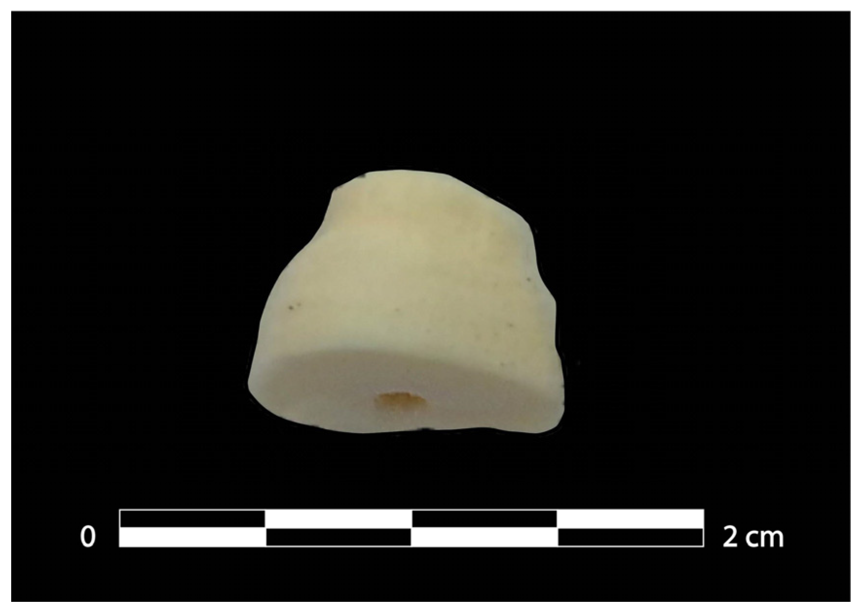
2.2. 3D Printing of Missing Skeletal Fragments and Restoration

A preliminary assessment of the state of conservation of the skeleton and the missing parts was carried out. For the reconstruction of the missing bone elements, two strategies were adopted. Taking advantage of the bilaterality of the skeleton, it was possible to generate the missing elements from mirror copies of the skeletal elements that were present through 3D printing. Where bones were completely missing, another lion skeleton (ID: 547) from the Faunal Museum of the Department of Veterinary Science of the University of Messina (Messina, Italy) was used as a source model with a metric adjustment of the osteometric parameters before proceeding to the creation of the missing elements so that the 3D-printed elements would fit the skeleton (AN-1420).
For the reconstruction of bones and teeth, the structured-light 3D scans for the subsequent filament 3D printing (fused filament fabrication—FFF) were used. This technology was appropriate for the creation of replicas suitable for being assembled with original skeletal elements and forming the whole skeleton for museum exhibition purposes within the protection of a transparent display case.
The structured light scanner used for this project works using the projection method and is a scan in a box (Open Technologies, Rezzato, Italy) that comprises a 200 lumens 800 × 600 pixels LED projector and two 1.3 MPx cameras. The scanner was calibrated based on the size of the skeletal remains to be scanned and the exposure conditions. Subsequently, based on the geometry of the findings, the positions of the various objects were chosen with respect to the rotating table (i.e., choice of the rotation axis).
Therefore, we proceeded, for each skeletal element, with the acquisition of one or more sets of “depth images” of the object placed on the rotating table (at least 20 images for a 360° rotation). The alignment of the various point clouds was manually performed and then a mesh was created and improved. The meshes were saved in STL and OBJ format. The STL files, obtained from the scanner’s software, could be directly processed by slicing software to obtain the file for 3D FFF printing. However, in the case of these skeletal remains, these files were further processed by using the freeware Autodesk Meshmixer (https://www.autodesk.com/research/projects/meshmixer (accessed on 03 February 2022)) to perform localized “smoothing” or reduce the number of polygons. For the printing of the reproductions of the bones, we used the Ultimaker CURA slicing software v. 5.2.1 (https://ultimaker.com/software/ultimaker-cura (accessed on 10 March 2022)), setting the height of the layers at 160 µm, the printing speed at 50 mm/s, and the temperature at 200 °C. The Easyfil™ PLA Light Gray filament from FormFutura (Nijmegen, The Netherlands) was used to print the reproductions of the skeletal remains (See Appendix A for a detailed explanation of the methodology).

2.3. Sampling and DNA Extraction

The sampling of the material necessary for DNA extraction was performed in a room of the Doderlein Museum, following strict criteria for decontamination [31]. As ancient DNA analysis is a destructive technique, the sampling was carried out to maximize the yield of DNA, but at the same time to minimize the visual impact of the sampled area. We decided to cut the apex part of the root of a tooth (Figure 2), which, once re-inserted into the alveolus, would not create aesthetic damage for exhibition. A tooth root was chosen as previous studies demonstrated that teeth are an optimal source for the retrieval of endogenous DNA [32,33]. Thus, for exhibition in museums, but also to preserve the specimen for future research, the choice of sampling area is very important, in order to both avoid/minimize external damage to the sample while not frustrating efforts at genotyping.
The sample was processed in the Ancient DNA (aDNA) laboratory at the Department of Cultural Heritage (University of Bologna), following appropriate and strict criteria [31,34,35], to avoid contamination and to authenticate the data obtained. The tooth was first photographed, then decontaminated and drilled in a designated room.
The DNA extraction and pre-PCR setup were performed in physically separated areas reserved for ancient DNA analysis. In order to minimize the contamination from modern DNA, the working area and the equipment used for sampling, DNA extraction, and PCR were decontaminated and sterilized with bleach and ethanol or DNA-ExitusPlus washing and UV irradiation before use. Bodysuits, two pairs of gloves, hair caps, face masks, and arm covers were worn during the analysis in the pre-PCR areas, and all the reagents used during the extraction and PCR setup were exposed to UV irradiation except DNA polymerase, primers, and dNTPs. Negative controls were processed along the sample to monitor the occurrence of contamination.
The fragment of the tooth was first cleaned on its surface with sodium hypochlorite solution (2%) using a sterile brush and then decontaminated with UV irradiation for 20 min. Dentine was removed along the root by powdering the internal surface with a 1.2 mm spherical drilling bit, and discarding the powder from the pulp chamber.
During the DNA extraction protocol [36], which was slightly modified from a previous paper [37], 300 mg of dentine powder was decalcified overnight at 37 °C under constant rotation in a 3 mL extraction buffer (0.45 M EDTA, 0.25 mg/mL proteinase K). Then, the solution was concentrated and purified using a silica-based method with High Pure Viral Nucleic Acid kit and finally eluted twice with a total of 50 μL of EB buffer (Qiagen, Hilden, Germany). DNA concentration was measured with the QuBit® fluorometer and the extract was stored at −20 °C.

2.4. DNA Amplification and Sequencing

According to Barnett and colleagues [38], we selected a set of 14 pairs of primers to amplify 981 bp of the cytochrome b (cytb) gene using short overlapping fragments. In this study, a portion of the cytb was amplified as, despite its limited length, it includes a high number of polymorphic sites and is commonly used for determining phylogenetic relationships between organisms [38,39]. In addition, the choice of the marker was driven by the fact that mitochondrial DNA is easily recovered from ancient remains [40]. We set the PCR with the pairs of primers, previously designed and successfully described by Burger et al. [41] and Barnett and colleagues [38] (Table S1 and Figure S1).
In order to avoid contamination by amplicons, PCR set-up was performed in the ancient DNA laboratory, but PCR runs were conducted in physically separated rooms designated for post-PCR analysis. The PCR reaction final volume was 25 μL, composed of 2.5 mM of MgCl2, 0.25 mM of dNTPs, 0.2 μM of forward and reverse primers, 0.8 mg/mL of Bovine Serum Albumin, and 2.5 units of AmpliTaq Gold (Applied Biosystems, Waltham, MA, USA). Then, 2–4 μL of DNA were added to the amplification solution, and the PCR was set with an initial denaturation at 95 °C for 5 min, then 40 cycles at 95 °C for 45 s, followed by specific annealing temperature for 45 s and the extension at 72 °C for 45 s, with the final step at 72 °C for 7 min. Amplifications were conducted in triplicate in order to avoid miscalling of bases during consensus sequence reconstruction, due to potential damage to DNA sequences. We performed electrophoresis on a 2% agarose gel to observe positive amplifications, and then PCR products were purified through the MinElute® PCR Purification kit (Qiagen, Hilden, Germany) and eluted in a 15 ul EB buffer (Qiagen, Hilden, Germany).
The Sanger sequencing reaction was performed using the BigDye™ Terminator v1.1 Cycle Sequencing Kit (Applied Biosystems, Waltham, MA, USA). Then, the products were purified with ethanol and sodium acetate and resuspended in water. DNA sequencing fragments were separated and detected by capillary electrophoresis on the SeqStudioTM Genetic Analyzer (Applied Biosystems, Waltham, MA, USA) at the Laboratory of Forensic Genetics in the Department of Medical and Surgical Sciences, Section of Legal Medicine of the University of Bologna.

2.5. Data Analysis

The chromatograms obtained from the sequencing were visualized through FinchTV v 1.5.0 (Geospiza, Seattle, WA, USA). All of the amplified fragments were checked and aligned using BioEdit v. 7.2.5 (Hall, 1999) to a Panthera leo cytochrome b gene sequence (GenBank accession number MG677922.1).
A dataset of 963 bp Panthera leo mtDNA cytb region sequences [38,42] was used for comparison with modern and ancient genetic diversity (Table S2), representative of most common lion cytb haplotypes from Africa and Asia regions (Panthera leo, sensu stricto). Only sequences of a known origin were selected, while data from samples of an uncertain provenance were discarded.
A Median Joining (MJ) network was created through PopArt software v. 1.7 [43] using this database (ε = 0). Moreover, a maximum likelihood (ML) phylogenetic tree was reconstructed with the software MEGA v. 10.2 [44] using this dataset, by setting the substitution model HKY + I + G, assessed by JModelTest2 v.2.1.10 [45], running 1000 bootstrap repetitions, and using a Panthera pardus cytb sequence (GenBank acc n. KP001507.1) as an outgroup.

3. Results

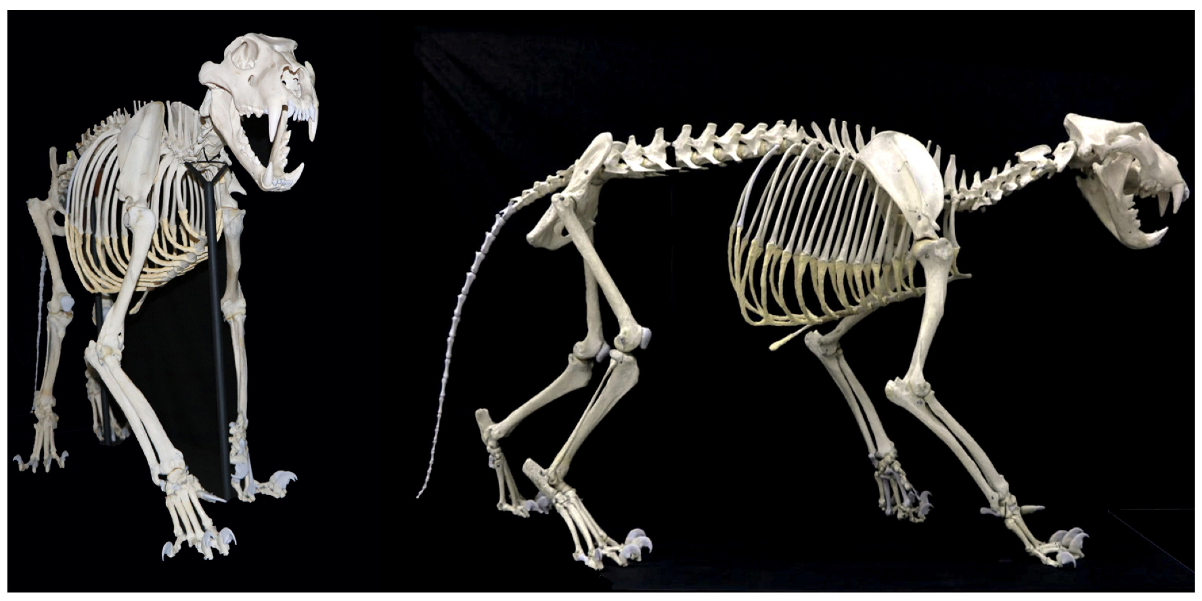
3.1. Skeletal Reconstruction and Exhibition

The skeletal tissue was in excellent condition. The various skeletal districts were well cleaned and, in general, all the bones were well preserved. The skeleton had been disassembled, and all bones, except some from the distal limbs, were not articulated. There were holes and iron wires on the surface of several bones, which were evidence of the previous assembly, as well as two wooden sternebrae. Moreover, in the sternum, the distal-caudal cartilage had been made from cardboard. From this assessment, it emerged that the skeleton of AN-1420 was incomplete: 69 anatomical parts were absent, including bones, teeth, and 18 claws (Figures S2–S5).
All epiphyses were fused and the dental eruption was complete, so the specimen was identified as a young adult, following White and Belant [46]. Following Naples and Rothschild [47], the sex of AN-1420 was determined as being female.
All of the missing skeletal elements were reconstructed (Figures S6–S8) and the skeleton was articulated and mounted (Figure 1). The 3D models of all of the skeletal elements are available for scientific, educational and exhibition purposes on the Sketchfab repository (https://skfb.ly/oAA6D (accessed on 3 November 2022)) or upon request to the authors.

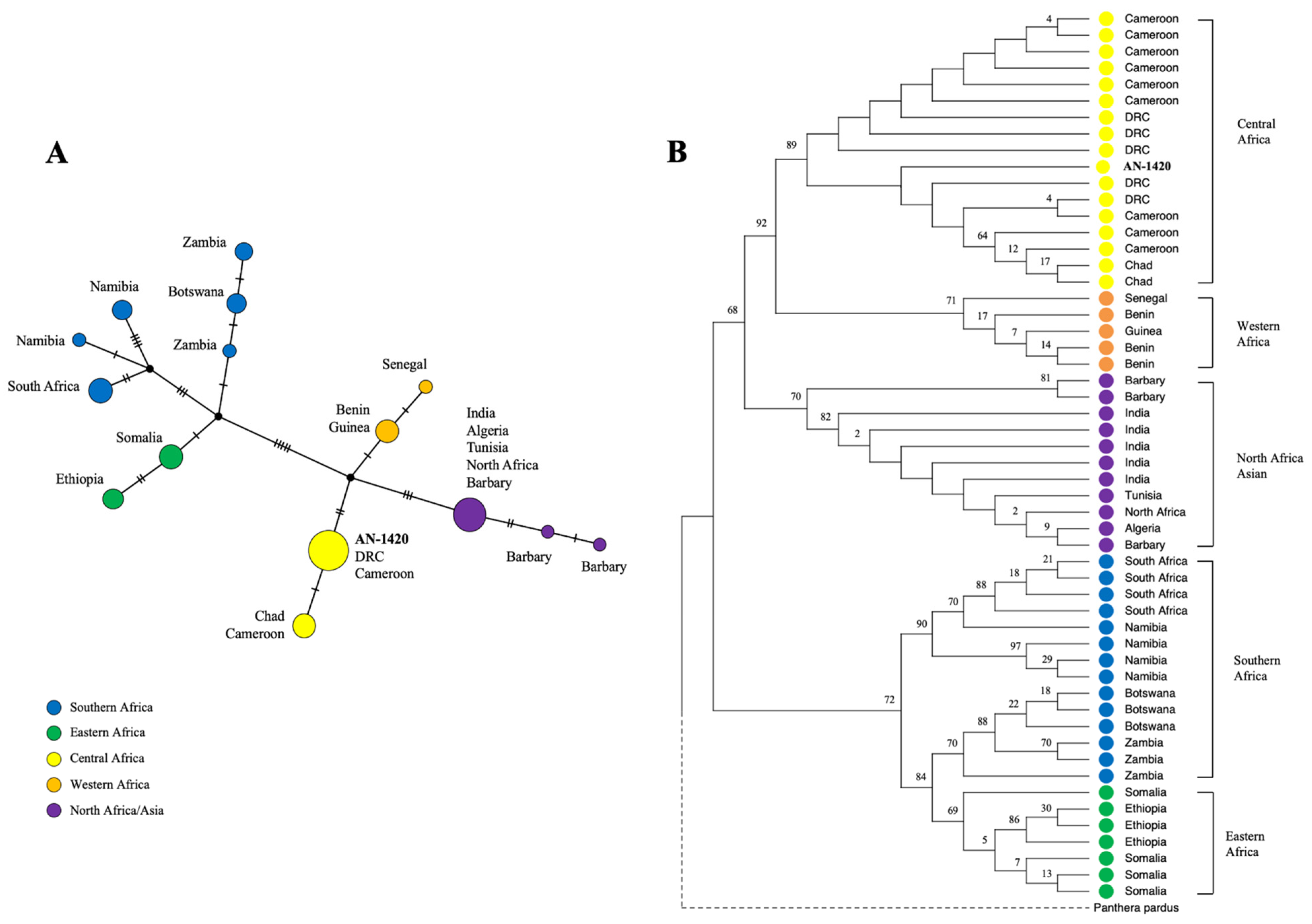
3.2. Phylogeographical Analyses

One sample from the AN-1420 specimen was co-extracted and amplified along with the negative controls. A total of 0.267 ng/µL of DNA was quantified using a Qubit fluorometer, and no contaminations were detected in the negative controls. Moreover, the authenticity of the data was confirmed through agreement in the sequence of nucleotides present in the overlapping regions and in the replicate amplifications. We successfully amplified 981 bp of cytb sequence from 11 of the 14 pairs of primers used. No amplifications were obtained for the following primer pairs: CB12RU–CB12RL, CB14RU–CB14BRL, and CB22RU–CB22RL.
The 981 bp consensus sequence from specimen AN-1420 (GenBank acc. n. OP837954) was then compared with those deposited in the GenBank database [48], using the Basic Local Alignment Search Tool (BLAST) [49]. The relevant alignments on GenBank with 100% identity were found with 19 sequences from Panthera leo specimens, geographically limited to the Central African region (Table S3).
Then, we analyzed the phylogenetic relationships between the cytb region of specimen AN-1420 and the ones included in the dataset created for this study. We first performed a Median Joining network (Figure 3A), where specimen AN-1420 was placed within the lineage of the Central African lions (in yellow), and more specifically in a pool that contained samples from the Democratic Republic of the Congo (DRC) and Cameroon. From the analyses of the data and a comparison with the published sequences, it was also possible to assign the sample to haplotype A [38], identified as haplotype 9 in another study [42]. The Maximum Likelihood tree (Figure 3B), although with limited support for the nodes due to the length of the sequences here analyzed, shows a clear clustering of the haplotypes into five main clades: (I) Central Africa, (II) Western Africa, (III) Asia/Northern Africa, (IV) Southern Africa, and (V) Eastern Africa, as previously highlighted [38]. In the phylogenetic tree, the sample from the Museum Doderlein was placed in the Central Africa clade, as suggested by the network analysis.

4. Discussion

In this study, a neglected skeleton of a lion, retrieved from the Museum of Zoology “P. Doderlein” of the University of Palermo, was chosen for restoration and molecular analysis, as the history of the specimen was unknown, as well as its origin. The specimen underwent an osteological survey, then it was restored by means of the reproduction of missing elements from the digitization and 3D printing of these bones. An animated morphological rendering of several bones of the lion skeleton, using 3D models, was obtained. The cytochrome b gene of the mitochondrial DNA was amplified and sequenced to assess the geographical origin of the specimen.
This project aimed to achieve results that enable applications in different fields, including the following: (1) add useful information for museographic strategies, (2) obtain genetic resources from a vulnerable species, (3) strengthen public awareness of wildlife conservation, and (4) share new learning biological material.
The lion, which represents one of the world’s most iconic animals, was once one of the most widespread terrestrial large carnivores, which had a wide species distribution range that included most of non-Saharan Africa, the Middle East, and southwest Asia into the Eastern Balkans in Europe and Western India during human history [4].
Recent surveys have estimated that only about 23,000–39,000 lions are living in the wild in Africa, compared with the already critically shrinking populations of Asian lions, consisting of only 411 wild individuals [8,50]. Since 1977, the African lion has been listed in CITES Appendix II, although researchers have suggested that the African lion might soon become endangered as well [8]. Particularly, under Appendix II, the trade of bones is considered legal if they are from captive sources. Since the 19th century, lion populations have begun to disappear from southwestern Eurasia and North Africa [4]. Nowadays, the lion population is experiencing a dramatic decline and fragmentation of remaining populations, caused by anthropogenic factors such as hunting, poaching, habitat loss, and prey base depletion.
Two extant subspecies of lions are recognized by most taxonomic authorities, namely, Panthera leo leo (Linnaeus, 1758) distributed in Asia, West Africa, and Central Africa, and Panthera leo melanochaita (Hamilton Smith, 1842), spread in East and South Africa [51]. However, recent whole genome sequencing data [52] have suggested that, even though the Central African lions cluster with P. leo leo in mtDNA-based phylogenies, the nuclear data show a higher affinity with P. leo melanochaita, distributed in East and South Africa. Thus, the population dynamics of this species are not yet fully understood, and more data are needed to clarify the taxonomic position of the Central African lions.
The phylogenetic analyses conducted (tree and network) here showed that specimen AN-1420 from the Museum “P. Doderlein” belongs to the Central African mtDNA clade, and in particular from haplotype A [38], or haplotype 9, following the nomenclature proposed by Bertola and colleagues [42]. The results and the data obtained were useful for providing the correct phylogenetic and phylogeographical assignment of this sample for museographic purposes, but at the same time represent an invaluable resource for conservation and educational aims. In light of the picture outlined by the genomic analyses [52], from a conservation point of view, any ancient and/or historical genetic material that could be useful to better clarify the population dynamics and connectivity of lions in Africa and, in particular, for Central and Western African lions, is important. The study of historical samples is fundamental to the conservation strategy of vulnerable species because it can add a temporal dimension to the genetic diversity characterization.
At the same time, from an educational perspective, the lion is one of the most charismatic species, which can be viewed as a tool to promote actions and initiatives addressed to wider audiences. The role of charisma is important in public communication, as people are naturally attracted to specific species and may understand biodiversity loss better via storytelling about endangered large felids [7,53].
The animated morphological rendering of the lion skeleton can be used to attract people towards topics related to evolution, the adaptability of animals, and the risk of extinction. Thanks to the digital materials available on the Sketchfab repository, consisting of 3D reconstructions with rendering and animated videos, people can observe animal life with curiosity. Moreover, the approach is useful for the preservation of historical, museum, and archaeological remains, also representing a useful database that may meet the needs or interests of academics.
The essential roles of a museum institution include the preservation of findings (e.g., [54,55]), the dissemination of knowledge, and the collection of new material. The scientific collections in museums are an essential vehicle for informing visitors about the actual threats to nature, as well as an indirect means of study and research, and a source of economic and social benefits [2,56,57]. Museums have a fundamental role in such a view as they can invest efforts in exhibitions of exotic animal collections and digitizing material to adopt for education purposes, but also with potential scientific benefits. Natural history collections, often forgotten, are a crucial cornerstone for systematics, natural history, ecology, and conservation biology, and represent important sources of biological data for scientists [3,54,55,58]. Many authors have highlighted the primary benefits that natural history collections provide to science and society [59].
Information and data gained from ancient/historical museum samples have the potential to allow the temporal study of genetic erosion in endangered populations; to analyze genomic data from extinct species; and to obtain information about phylogeny, population dynamics, taxonomy, and genetic diversity [54,60]. The results from ancient and historical specimens, as here obtained, can play a pivotal role in preserving biodiversity and driving specific conservation policies [1,61].

Supplementary Materials

The following supporting information can be downloaded at: https://www.mdpi.com/article/10.3390/d15010087/s1. Figure S1: Graphical representation of the portion of the cytochrome b analyzed in this study. Figure S2: Schematic view of the missing skeletal elements of AN-1420 on the entire skeleton. Figure S3: Missing elements from the cranium (left side view). Figure S4: Missing skeletal elements of the cranium and the mandible (dorsal, frontal and ventral view). Figure S5: Missing elements of the distal region of the skeleton—autopodium. Figure S6: The copies of the carpals created for specimen AN-1420. Figure S7: The mounted left forelimb of specimen AN-1420. Figure S8: A particular of the restored skull of Panthera leo AN-1420. Table S1: List of the primers used in this study. Table S2: Database of the 55 sequences used for phylogenetic analyses. Table S3: Sequences present in Genbank that match the one obtained in this study [62,63,64,65,66,67,68].

Author Contributions

Conceptualization, S.L.B.; methodology, E.C., D.L. and S.L.B.; software, E.C., F.F., R.I., M.P., and P.D.B.; validation, S.L.B., E.C., and F.F.; genetic analyses, F.F., T.M., S.D.F., and C.B.; digital restoration, M.P. and P.D.B.; sample collection, S.L.B. and F.S.; resources, S.L.B., D.L., and S.P.; data curation, F.F. and S.L.B.; writing—original draft preparation, E.C., F.F., and M.M.C.; writing—review and editing, S.L.B., and D.L.; visualization, F.F., E.C., and R.I.; supervision, S.L.B.; funding acquisition, S.L.B. All authors have read and agreed to the published version of the manuscript.

Funding

The authors acknowledge the support of the Sistema Museale di Ateneo (SiMuA) of the University of Palermo and of the NBFC to University of Palermo, funded by the Italian Ministry of University and Research, PNRR, Missione 4 Componente 2, “Dalla ricerca all’impresa”, Investimento 1.4, Project CN00000033.

Institutional Review Board Statement

Not applicable.

Data Availability Statement

The consensus sequence from the AN-1420 specimen can be downloaded from GenBank, acc. no. OP837954. The 3D rendering of the skeletal elements is available on the Sketchfab repository (https://skfb.ly/oAA6D (accessed on 3 November 2022)) or upon request to the authors.

Acknowledgments

The authors are grateful to Enrico Bellia and Daniele Di Lorenzo for their technical support.

Conflicts of Interest

The authors declare no conflict of interest.

Appendix A

Details and explanations of the techniques used for the 3D digital restoration of the skeleton of the specimen AN-1420.

References

  1. Lo Brutto, S.; Puleio, R.; Vicari, D.; Calascibetta, A.; Bellia, E.; Bellante, A.; Giacalone, V.; Buffa, G. Dal Mare Al Museo: Il Recupero Di Uno Scheletro Di Stenella coeruleoalba (Meyen 1833). Museol. Sci. 2022, 16, 62–69. [Google Scholar]
  2. Movalli, P.; Koschorreck, J.; Treu, G.; Slobodnik, J.; Alygizakis, N.; Androulakakis, A.; Badry, A.; Baltag, E.; Barbagli, F.; Bauer, K.; et al. The Role of Natural Science Collections in the Biomonitoring of Environmental Contaminants in Apex Predators in Support of the EU’s Zero Pollution Ambition. Environ. Sci. Eur. 2022, 34, 1–7. [Google Scholar] [CrossRef]
  3. Tiralongo, F.; Badalamenti, R.; Arizza, V.; Prieto, L.; Lo Brutto, S. The Portuguese Man-of-War Has Always Entered the Mediterranean Sea—Strandings, Sightings, and Museum Collections. Front. Mar. Sci. 2022, 9, 377. [Google Scholar] [CrossRef]
  4. Curry, C.J.; Davis, B.W.; Bertola, L.D.; White, P.A.; Murphy, W.J.; Derr, J.N. Spatiotemporal Genetic Diversity of Lions Reveals the Influence of Habitat Fragmentation across Africa. Mol. Biol. Evol. 2021, 38, 48–57. [Google Scholar] [CrossRef] [PubMed]
  5. Lo Brutto, S.; Calascibetta, A.; Pavan, G.; Buffa, G. Cetacean Strandings and Museum Collections: A Focus on Sicily Island Crossroads for Mediterranean Species. Diversity 2021, 13, 104. [Google Scholar] [CrossRef]
  6. Yeates, D.K.; Zwick, A.; Mikheyev, A.S. Museums Are Biobanks: Unlocking the Genetic Potential of the Three Billion Specimens in the World’s Biological Collections. Curr. Opin. Insect Sci. 2016, 18, 83–88. [Google Scholar] [CrossRef]
  7. Albert, C.; Luque, G.M.; Courchamp, F. The Twenty Most Charismatic Species. PLoS ONE 2018, 13, e0199149. [Google Scholar] [CrossRef] [Green Version]
  8. Bauer, H.; Packer, C.; Funston, P.F.; Henschel, P.; Nowell, K. Panthera leo. IUCN Red List Threat. Species 2016, e. T15951A115130419. [Google Scholar] [CrossRef]
  9. Henschel, P.; Coad, L.; Burton, C.; Chataigner, B.; Dunn, A.; MacDonald, D.; Saidu, Y.; Hunter, L.T.B. The Lion in West Africa Is Critically Endangered. PLoS ONE 2014, 9, e83500. [Google Scholar] [CrossRef] [Green Version]
  10. Straube, N.; Lyra, M.L.; Paijmans, J.L.A.; Preick, M.; Basler, N.; Penner, J.; Rödel, M.O.; Westbury, M.V.; Haddad, C.F.B.; Barlow, A.; et al. Successful Application of Ancient DNA Extraction and Library Construction Protocols to Museum Wet Collection Specimens. Mol. Ecol. Resour. 2021, 21, 2299–2315. [Google Scholar] [CrossRef]
  11. Roycroft, E.; Moritz, C.; Rowe, K.C.; Moussalli, A.; Eldridge, M.D.B.; Portela Miguez, R.; Piggott, M.P.; Potter, S. Sequence Capture From Historical Museum Specimens: Maximizing Value for Population and Phylogenomic Studies. Front. Ecol. Evol. 2022, 10, 667. [Google Scholar] [CrossRef]
  12. Kapp, J.D.; Green, R.E.; Shapiro, B. A Fast and Efficient Single-Stranded Genomic Library Preparation Method Optimized for Ancient DNA. J. Hered. 2021, 112, 241–249. [Google Scholar] [CrossRef] [PubMed]
  13. Rohland, N.; Glocke, I.; Aximu-Petri, A.; Meyer, M. Extraction of Highly Degraded DNA from Ancient Bones, Teeth and Sediments for High-Throughput Sequencing. Nat. Protoc. 2018, 13, 2447–2461. [Google Scholar] [CrossRef] [PubMed]
  14. Puncher, G.N.; Cariani, A.; Cilli, E.; Massari, F.; Leone, A.; Morales-Muñiz, A.; Onar, V.; Toker, N.Y.; Bernal Casasola, D.; Moens, T.; et al. Comparison and Optimization of Genetic Tools Used for the Identification of Ancient Fish Remains Recovered from Archaeological Excavations and Museum Collections in the Mediterranean Region. Int. J. Osteoarchaeol. 2019, 29, 365–376. [Google Scholar] [CrossRef]
  15. Agne, S.; Naylor, G.J.P.; Preick, M.; Yang, L.; Thiel, R.; Weigmann, S.; Paijmans, J.L.A.; Barlow, A.; Hofreiter, M.; Straube, N. Taxonomic Identification of Two Poorly Known Lantern Shark Species Based on Mitochondrial DNA From Wet-Collection Paratypes. Front. Ecol. Evol. 2022, 10, 569. [Google Scholar] [CrossRef]
  16. Angelici, F.; Ciucani, M.; Angelini, S.; Annesi, F.; Caniglia, R.; Castiglia, R.; Fabbri, E.; Galaverni, M.; Palumbo, D.; Ravegnini, G.; et al. The Sicilian Wolf: Genetic Identity of a Recently Extinct Insular Population. Zoolog. Sci. 2019, 36, 189–197. [Google Scholar] [CrossRef]
  17. Leone, A.; Puncher, G.N.; Ferretti, F.; Sperone, E.; Tripepi, S.; Micarelli, P.; Gambarelli, A.; Sarà, M.; Arculeo, M.; Doria, G.; et al. Pliocene Colonization of the Mediterranean by Great White Shark Inferred from Fossil Records, Historical Jaws, Phylogeographic and Divergence Time Analyses. J. Biogeogr. 2020, 47, 1119–1129. [Google Scholar] [CrossRef]
  18. Koupadi, K.; Fontani, F.; Ciucani, M.M.; Maini, E.; De Fanti, S.; Cattani, M.; Curci, A.; Nenzioni, G.; Reggiani, P.; Andrews, A.J.; et al. Population Dynamics in Italian Canids between the Late Pleistocene and Bronze Age. Genes 2020, 11, 1409. [Google Scholar] [CrossRef]
  19. Straube, N.; Preick, M.; Naylor, G.J.P.; Hofreiter, M. Mitochondrial DNA Sequencing of a Wet-Collection Syntype Demonstrates the Importance of Type Material as Genetic Resource for Lantern Shark Taxonomy (Chondrichthyes: Etmopteridae). R. Soc. Open Sci. 2021, 8, 210474. [Google Scholar] [CrossRef]
  20. Ciucani, M.M.; Palumbo, D.; Galaverni, M.; Serventi, P.; Fabbri, E.; Ravegnini, G.; Angelini, S.; Maini, E.; Persico, D.; Caniglia, R.; et al. Old Wild Wolves: Ancient DNA Survey Unveils Population Dynamics in Late Pleistocene and Holocene Italian Remains. PeerJ 2019, 7, e6424. [Google Scholar] [CrossRef]
  21. Ciucani, M.M.; Jensen, J.K.; Sinding, M.H.S.; Smith, O.; Lucenti, S.B.; Rosengren, E.; Rook, L.; Tuveri, C.; Arca, M.; Cappellini, E.; et al. Evolutionary History of the Extinct Sardinian Dhole. Curr. Biol. 2021, 31, 5571–5579.e6. [Google Scholar] [CrossRef] [PubMed]
  22. Andrews, A.J.; Di Natale, A.; Bernal-Casasola, D.; Aniceti, V.; Onar, V.; Oueslati, T.; Theodropoulou, T.; Morales-Muñiz, A.; Cilli, E.; Tinti, F. Exploitation History of Atlantic Bluefin Tuna in the Eastern Atlantic and Mediterranean—Insights from Ancient Bones. ICES J. Mar. Sci. 2022, 79, 247–262. [Google Scholar] [CrossRef]
  23. Lalueza-Fox, C. Museomics. Curr. Biol. 2022, 32, R1214–R1215. [Google Scholar] [CrossRef] [PubMed]
  24. Mondol, S.; Bruford, M.W.; Ramakrishnan, U. Demographic Loss, Genetic Structure and the Conservation Implications for Indian Tigers. Proc. R. Soc. B Biol. Sci. 2013, 280, 20130496. [Google Scholar] [CrossRef] [PubMed] [Green Version]
  25. Palkopoulou, E.; Lipson, M.; Mallick, S.; Nielsen, S.; Rohland, N.; Baleka, S.; Karpinski, E.; Ivancevic, A.M.; To, T.H.; Daniel Kortschak, R.; et al. A Comprehensive Genomic History of Extinct and Living Elephants. Proc. Natl. Acad. Sci. USA 2018, 115, E2566–E2574. [Google Scholar] [CrossRef] [Green Version]
  26. Andrews, A.J.; Puncher, G.N.; Bernal-Casasola, D.; Di Natale, A.; Massari, F.; Onar, V.; Toker, N.Y.; Hanke, A.; Pavey, S.A.; Savojardo, C.; et al. Ancient DNA SNP-Panel Data Suggests Stability in Bluefin Tuna Genetic Diversity despite Centuries of Fluctuating Catches in the Eastern Atlantic and Mediterranean. Sci. Rep. 2021, 11, 20744. [Google Scholar] [CrossRef] [PubMed]
  27. Pinsky, M.L.; Eikeset, A.M.; Helmerson, C.; Bradbury, I.R.; Bentzen, P.; Morris, C.; Gondek-Wyrozemska, A.T.; Baalsrud, H.T.; Brieuc, M.S.O.; Kjesbu, O.S.; et al. Genomic Stability through Time despite Decades of Exploitation in Cod on Both Sides of the Atlantic. Proc. Natl. Acad. Sci. USA 2021, 118, e2025453118. [Google Scholar] [CrossRef]
  28. Ciucani, M.M.; Ramos-Madrigal, J.; Hernández-Alonso, G.; Carmagnini, A.; Aninta, S.G.; Scharff-Olsen, C.H.; Lanigan, L.T.; Fracasso, I.; Clausen, C.G.; Aspi, J.; et al. Genomes of the Extinct Sicilian Wolf Reveal a Complex History of Isolation and Admixture with Ancient Dogs. bioRxiv 2022, 21, 477289. [Google Scholar] [CrossRef]
  29. Hempel, E.; Bibi, F.; Faith, J.T.; Koepfli, K.-P.; Klittich, A.M.; Duchêne, D.A.; Brink, J.S.; Kalthoff, D.C.; Dalén, L.; Hofreiter, M.; et al. Blue Turns to Grey—Palaeogenomic Insights into the Evolutionary History and Extinction of the Blue Antelope (Hippotragus Leucophaeus). Mol. Biol. Evol. 2022, 39, msac241. [Google Scholar] [CrossRef]
  30. Tedesco, M.R.; Mandalà, G.; Consentino, M.C.; Ienti, R.; Lo Valvo, F.; Movalli, P.; Sabella, G.; Spadola, F.; Spinnato, A.; Toscano, F.; et al. The First Inventory of Birds of Prey in Sicilian Museum Collections (Italy). Museol. Sci. 2020, 14, 67–80. [Google Scholar] [CrossRef]
  31. Llamas, B.; Valverde, G.; Fehren-Schmitz, L.; Weyrich, L.S.; Cooper, A.; Haak, W. From the Field to the Laboratory: Controlling DNA Contamination in Human Ancient DNA Research in the High-Throughput Sequencing Era. STAR Sci. Technol. Archaeol. Res. 2017, 3, 1–14. [Google Scholar] [CrossRef]
  32. Higgins, D.; Austin, J.J. Teeth as a Source of DNA for Forensic Identification of Human Remains: A Review. Sci. Justice 2013, 53, 433–441. [Google Scholar] [CrossRef] [PubMed]
  33. Allentoft, M.E.; Sikora, M.; Sjogren, K.-G.; Rasmussen, S.; Rasmussen, M.; Stenderup, J.; Damgaard, P.B.; Schroeder, H.; Ahlstrom, T.; Vinner, L.; et al. Population Genomics of Bronze Age Eurasia. Nature 2015, 522, 167–172. [Google Scholar] [CrossRef] [PubMed] [Green Version]
  34. Cooper, A.; Poinar, H.N. Ancient DNA: Do It Right or Not at All. Science 2000, 289, 1139. [Google Scholar] [CrossRef] [PubMed]
  35. Fulton, T.L.; Shapiro, B. Setting Up an Ancient DNA Laboratory. Methods Mol. Biol. 2019, 1963, 1–13. [Google Scholar] [CrossRef] [PubMed]
  36. Cilli, E.; Gabanini, G.; Ciucani, M.M.; De Fanti, S.; Serventi, P.; Bazaj, A.; Sarno, S.; Ferri, G.; Fregnani, A.; Cornaglia, G.; et al. A Multifaceted Approach towards Investigating Childbirth Deaths in Double Burials: Anthropology, Paleopathology and Ancient DNA. J. Archaeol. Sci. 2020, 122, 105219. [Google Scholar] [CrossRef]
  37. Dabney, J.; Knapp, M.; Glocke, I.; Gansauge, M.-T.; Weihmann, A.; Nickel, B.; Valdiosera, C.; Garcia, N.; Paabo, S.; Arsuaga, J.-L.; et al. Complete Mitochondrial Genome Sequence of a Middle Pleistocene Cave Bear Reconstructed from Ultrashort DNA Fragments. Proc. Natl. Acad. Sci. USA 2013, 110, 15758–15763. [Google Scholar] [CrossRef] [Green Version]
  38. Barnett, R.; Yamaguchi, N.; Shapiro, B.; Ho, S.Y.; Barnes, I.; Sabin, R.; Werdelin, L.; Cuisin, J.; Larson, G. Revealing the Maternal Demographic History of Panthera leo Using Ancient DNA and a Spatially Explicit Genealogical Analysis. BMC Evol. Biol. 2014, 14, 70. [Google Scholar] [CrossRef] [Green Version]
  39. Castresana, J. Cytochrome b Phylogeny and the Taxonomy of Great Apes and Mammals. Mol. Biol. Evol. 2001, 18, 465–471. [Google Scholar] [CrossRef] [Green Version]
  40. Merheb, M.; Matar, R.; Hodeify, R.; Siddiqui, S.S.; Vazhappilly, C.G.; Marton, J.; Azharuddin, S.; Zouabi, H.A.L. Mitochondrial DNA, a Powerful Tool to Decipher Ancient Human Civilization from Domestication to Music, and to Uncover Historical Murder Cases. Cells 2019, 8, 433. [Google Scholar] [CrossRef] [Green Version]
  41. Burger, J.; Rosendahl, W.; Loreille, O.; Hemmer, H.; Eriksson, T.; Götherström, A.; Hiller, J.; Collins, M.J.; Wess, T.; Alt, K.W. Molecular Phylogeny of the Extinct Cave Lion Panthera leo Spelaea. Mol. Phylogenet. Evol. 2004, 30, 841–849. [Google Scholar] [CrossRef]
  42. Bertola, L.D.; Jongbloed, H.; Van Der Gaag, K.J.; De Knijff, P.; Yamaguchi, N.; Hooghiemstra, H.; Bauer, H.; Henschel, P.; White, P.A.; Driscoll, C.A.; et al. Phylogeographic Patterns in Africa and High Resolution Delineation of Genetic Clades in the Lion (Panthera leo). Sci. Rep. 2016, 6, 30807. [Google Scholar] [CrossRef] [Green Version]
  43. Leigh, J.W.; Bryant, D. Popart: Full-Feature Software for Haplotype Network Construction. Methods Ecol. Evol. 2015, 6, 1110–1116. [Google Scholar] [CrossRef]
  44. Kumar, S.; Stecher, G.; Li, M.; Knyaz, C.; Tamura, K. MEGA X: Molecular Evolutionary Genetics Analysis across Computing Platforms. Mol. Biol. Evol. 2018, 35, 1547. [Google Scholar] [CrossRef] [PubMed]
  45. Darriba, D.; Taboada, G.L.; Doallo, R.; Posada, D. JModelTest 2: More Models, New Heuristics and High-Performance Computing. Nat. Methods 2012, 9, 772. [Google Scholar] [CrossRef] [Green Version]
  46. White, P.A.; Belant, J.L. Individual Variation in Dental Characteristics for Estimating Age of African Lions. Wildlife Biol. 2016, 22, 71–77. [Google Scholar] [CrossRef] [Green Version]
  47. Naples, V.L.; Rothschild, B.M. Sex Determination in Lions (Panthera leo, Felidae): A Novel Method of Distinguishing Male and Female Skulls. Mammalia 2012, 76, 99–103. [Google Scholar] [CrossRef]
  48. Benson, D.A.; Cavanaugh, M.; Clark, K.; Karsch-Mizrachi, I.; Lipman, D.J.; Ostell, J.; Sayers, E.W. GenBank. Nucleic Acids Res. 2013, 41, D36–D42. [Google Scholar] [CrossRef] [Green Version]
  49. Altschul, S.F.; Gish, W.; Miller, W.; Myers, E.W.; Lipman, D.J. Basic Local Alignment Search Tool. J. Mol. Biol. 1990, 215, 403–410. [Google Scholar] [CrossRef]
  50. Singh, H.S.; Gibson, L. A Conservation Success Story in the Otherwise Dire Megafauna Extinction Crisis: The Asiatic Lion (Panthera leo Persica) of Gir Forest. Biol. Conserv. 2011, 144, 1753–1757. [Google Scholar] [CrossRef]
  51. Kitchener, A.C.; Breitenmoser-Würsten, C.; Eizirik, E.; Gentry, A.; Werdelin, L.; Wilting, A.; Yamaguchi, N.; Abramov, A.V.; Christiansen, P.; Driscoll, C.; et al. A Revised Taxonomy of the Felidae: The Final Report of the Cat Classification Task Force of the IUCN Cat Specialist GroupCAT News Spec. Issue 2017, 11, 71–73. [Google Scholar]
  52. de Manuel, M.; Barnett, R.; Sandoval-Velasco, M.; Yamaguchi, N.; Vieira, F.G.; Lisandra Zepeda Mendoza, M.; Liu, S.; Martin, M.D.; Sinding, M.H.S.; Mak, S.S.T.; et al. The Evolutionary History of Extinct and Living Lions. Proc. Natl. Acad. Sci. USA 2020, 117, 10927–10934. [Google Scholar] [CrossRef] [PubMed]
  53. Lo Brutto, S.; Battaglia, F.; Biundo, G.; Calascibetta, A.; Conti, F.; Nuccio, A.; Pellerito, F. Un’esperienza Di Divulgazione Condotta Da Studenti Universitari. Museol. Sci. 2020, 14, 133–138. [Google Scholar] [CrossRef]
  54. Lo Brutto, S. The Case of a Rudderfish Highlights the Role of Natural History Museums as Sentinels of Bio-Invasions. Zootaxa 2017, 4254, 382–386. [Google Scholar] [CrossRef] [PubMed]
  55. Lo Brutto, S. A Finding at the Natural History Museum of Florence Affords the Holotype Designation of Orchestia stephenseni Cecchini, 1928 (Crustacea: Amphipoda: Talitridae). Zootaxa 2017, 4231, 569–572. [Google Scholar] [CrossRef] [PubMed]
  56. Movalli, P.; Duke, G.; Ramello, G.; Dekker, R.; Vrezec, A.; Shore, R.F.; García-Fernández, A.; Wernham, C.; Krone, O.; Alygizakis, N.; et al. Progress on Bringing Together Raptor Collections in Europe for Contaminant Research and Monitoring in Relation to Chemicals Regulation. Environ. Sci. Pollut. Res. 2019, 26, 20132–20136. [Google Scholar] [CrossRef] [Green Version]
  57. Suarez, A.V.; Tsutsui, N.D. The Value of Museum Collections for Research and Society. Bioscience 2004, 54, 66–74. [Google Scholar] [CrossRef]
  58. Cook, J.A.; Light, J.E. The Emerging Role of Mammal Collections in 21st Century Mammalogy. J. Mammal. 2019, 100, 733–750. [Google Scholar] [CrossRef]
  59. Bradley, R.D.; Bradley, L.C.; Garner, H.J.; Baker, R.J. Assessing the Value of Natural History Collections and Addressing Issues Regarding Long-Term Growth and Care. Bioscience 2014, 64, 1150–1158. [Google Scholar] [CrossRef] [Green Version]
  60. Mannino, A.M.; Balistreri, P.; Iaciofano, D.; Galil, B.S.; Lo Brutto, S. An Additional Record of Kyphosus vaigiensis (Quoy & Gaimard, 1825) (Osteichthyes, Kyphosidae) from Sicily Clarifies the Confused Situation of the Mediterranean Kyphosids. Zootaxa 2015, 3963, 45–54. [Google Scholar] [CrossRef]
  61. Jensen, E.L.; Díez-del-Molino, D.; Gilbert, M.T.P.; Bertola, L.D.; Borges, F.; Cubric-Curik, V.; de Navascués, M.; Frandsen, P.; Heuertz, M.; Hvilsom, C.; et al. Ancient and Historical DNA in Conservation Policy. Trends Ecol. Evol. 2022, 37, 420–429. [Google Scholar] [CrossRef] [PubMed]
  62. Pales, L.; Lambert, C. Atlas Ostéologique Pour Servir à l’identification Des Mammifères Du Quaternaire; Editions du CNRS: Paris, France, 1971. [Google Scholar]
  63. Bertola, L.D.; van Hooft, W.F.; Vrieling, K.; Uit de Weerd, D.R.; York, D.S.; Bauer, H.; Prins, H.H.T.; Funston, P.J.; Udo de Haes, H.A.; Leirs, H.; et al. Genetic Diversity, Evolutionary History and Implications for Conservation of the Lion (Panthera leo) in West and Central Africa. J. Biogeogr. 2011, 38, 1356–1367. [Google Scholar] [CrossRef]
  64. Bertola, L.D.; Tensen, L.; Van Hooft, P.; White, P.A.; Driscoll, C.A.; Henschel, P.; Caragiulo, A.; Dias-Freedman, I.; Sogbohossou, E.A.; Tumenta, P.N.; et al. Autosomal and MtDNA Markers Affirm the Distinctiveness of Lions in West and Central Africa. PLoS ONE 2015, 10, e0137975. [Google Scholar] [CrossRef] [PubMed] [Green Version]
  65. Li, G.; Davis, B.W.; Eizirik, E.; Murphy, W.J. Phylogenomic Evidence for Ancient Hybridization in the Genomes of Living Cats (Felidae). Genome Res. 2016, 26, 1–11. [Google Scholar] [CrossRef] [PubMed] [Green Version]
  66. Paijmans, J.L.A.; Fickel, J.; Courtiol, A.; Hofreiter, M.; Förster, D.W. Impact of Enrichment Conditions on Cross-Species Capture of Fresh and Degraded DNA. Mol. Ecol. Resour. 2016, 16, 42–55. [Google Scholar] [CrossRef]
  67. Ma, Y.P.; Wang, S. Mitochondrial Genome of the African Lion Panthera Leo Leo. Mitochondrial DNA 2015, 26, 951–952. [Google Scholar] [CrossRef]
  68. Bruche, S.; Gusset, M.; Lippold, S.; Barnett, R.; Eulenberger, K.; Junhold, J.; Driscoll, C.A.; Hofreiter, M. A Genetically Distinct Lion (Panthera Leo) Population from Ethiopia. Eur. J. Wildl. Res. 2013, 59, 215–225. [Google Scholar] [CrossRef]
Figure 1. The skeleton of a lion, Panthera leo, AN-1420, restored and mounted for exhibition. Frontal (left) and lateral (right) views. Photos credit: Andrea Calascibetta, Daniele Di Lorenzo, kind concession of the Museum of Zoology "P. Doderlein", Sistema Museale d’Ateneo, Università degli Studi di Palermo.
Diversity 15 00087 g001
Figure 2. The apex part of the tooth root collected for the DNA extraction.
Diversity 15 00087 g002
Figure 3. Phylogenetic analyses of the lion data based on the alignment created for this study. (A) Median Joining network for the alignment of the cytb haplotypes. (B) Phylogenetic tree reconstruction with the Maximum Likelihood method. Sample AN-1420 is placed in the Central African clade.
Diversity 15 00087 g003
Disclaimer/Publisher’s Note: The statements, opinions and data contained in all publications are solely those of the individual author(s) and contributor(s) and not of MDPI and/or the editor(s). MDPI and/or the editor(s) disclaim responsibility for any injury to people or property resulting from any ideas, methods, instructions or products referred to in the content.

