Article

Review of the Cyanobacterial Genus Phormidesmis (Leptolyngbyaceae) with the Description of Apatinema gen. nov.

1
Polar Alpine Botanic Garden Institute Kola Science Center of RAS, Kirovsk 184256, Russia
2
Institute of the Industrial Ecology Problems of the North of the Kola Science Center of RAS, Apatity 184209, Russia
*
Author to whom correspondence should be addressed.
Diversity 2022, 14(9), 731; https://doi.org/10.3390/d14090731
Submission received: 5 July 2022 / Revised: 24 August 2022 / Accepted: 1 September 2022 / Published: 5 September 2022
(This article belongs to the Special Issue Biodiversity and Biogeography of Terrestrial Algae and Cyanobacteria)

Abstract

:
Cyanobacteria are crucial components of biological soil crusts of polar landscapes and carry out many functions in subaerial environments. Simple untapered filamentous cyanobacteria are typically in the terrestrial biotopes. They appear to be a group with an abundance of cryptic taxa. We isolated 23 strains of cyanobacteria from the different habitats of the Arctic and temperate zone, from 10 locations in order to characterize their morphological and genotypic diversity. Phylogenetic analyses were conducted on the 16S and 16S–23S ITS rRNA gene regions using Bayesian inference and maximum likelihood. A morphological comparison of the isolated strains with similar known species, as well as its phylogenetic analyses, revealed that they belong to three species of the genus Phormidesmis (P. nigrescens, P. pristley, and P. communis)—and to the previously unknown genus of Leptolyngbyaceae. Using an integrative approach, we provide here a description of a new taxon Apatinema gen. nov.

1. Introduction

Cyanobacteria are a ubiquitous and widely distributed group of phototrophic microorganisms. Acquired nutritional strategies and great metabolic diversity allow cyanobacteria to colonize many terrestrial and aquatic ecosystems. They are one of the most abundant microphototrophs in polar landscapes and are adequately adapted to harsh environmental conditions. The study of cyanobacterial diversity is important to consider different microbial distribution patterns.
In spite of the progress in the cyanobacterial phylogeny, their taxonomy is still undergoing revision. Simple filamentous forms of Cyanobacteria such as Leptolyngbya s.l. with narrow trichomes thrive in most types of terrestrial habitats [1]. High adaptive capacity, immense dispersal abilities, and relatively fast growth rate [2] facilitate their frequent occurrence in the biological soil crusts and subaerophytic wet wall assemblages of algae.
Correct identification of thin untapered filamentous organisms without heterocytes and akinetes is particularly difficult because they are phenotypically plastic and have a restricted number of morphological features. Moreover, some features may be environmentally plastic and do not have a resolution for species differentiation.
According to recent studies based on comprehensive approaches, several genera with similar morphology from the order Synechococcales were revised: Alkalinema [3], Cartusia, Drouetiella [4], Elainella [5], Haloleptolyngbya [6], Kaiparowitsia, Komarkovaea [4], Kovacikia [7], Limnolyngbya [8], Myxacorys [9], Nodosilinea [10,11], Oculatella [12,13], Onodrimia [14], Pantanalinema [3], Pegethrix [4], Phormidesmis [15,16], Pinocchia [17], Plectolyngbya [18], Scytolyngbya [19], Stenomitos [7,20], Thermoleptolyngbya [21], Tildeniella [4], and Timaviella [22].
The isolation and characterization of cyanobacterial strains from diverse biotopes remains extremely important for the study of cyanobacterial distribution. The determination of environmental cyanobacterial samples and the purification of cultured strains allowed the discovery of a high diversity in the Phormidesmis genus.
Phormidesmis is a recently established cyanobacterial genus, some species of which appear to be vague. The genus was described by Turicchia et al. [15] by a comprehensive approach based on 16S rRNA gene phylogeny and defined as a separate genus phylogenetically allied to Leptolyngbya s. str.
Raabová et al. [16] described two species of Phosmidesmis (P. arctica Raabová et al., reference strain CCALA 1101 and P. communis Raabová et al., AB 11-10) and Leptolyngbya nigrescens was classified by Komárek as Phormidesmis nigrescens (Komárek) Raabová et al. Their descriptions of P. arctica and P. communis are invalid due to the absence of holotypes citation and a description of unpreserved cultures in a metabolically active state in accordance with the requirements of the International Code for Algae, Fungi and Plants [23]. This fact adds confusion to the understanding of Phormidesmis taxonomy.
The analysis of nucleotide sequences named as “Phormidesmis strains” deposited in the National Center for Biotechnology Information (NCBI) GenBank (GB) database [24], based on phylogenetic traits, even placed them in different genera of two families [25]. Thus, there is no common acceptance of the genus and species limits and the level of infraspecific variability remained unresolved within a genus.
The aim of the present study is to reveal a diversity of the genus Phormidesmis in different localities of the Arctic and temperate zone, predominantly in European Russia, deposited in the collection of Polar Alpine Botanical Garden Institute Kola SC RAS (KPABG), and provide a description of the new genus Apatinema gen. nov.

2. Materials and Methods
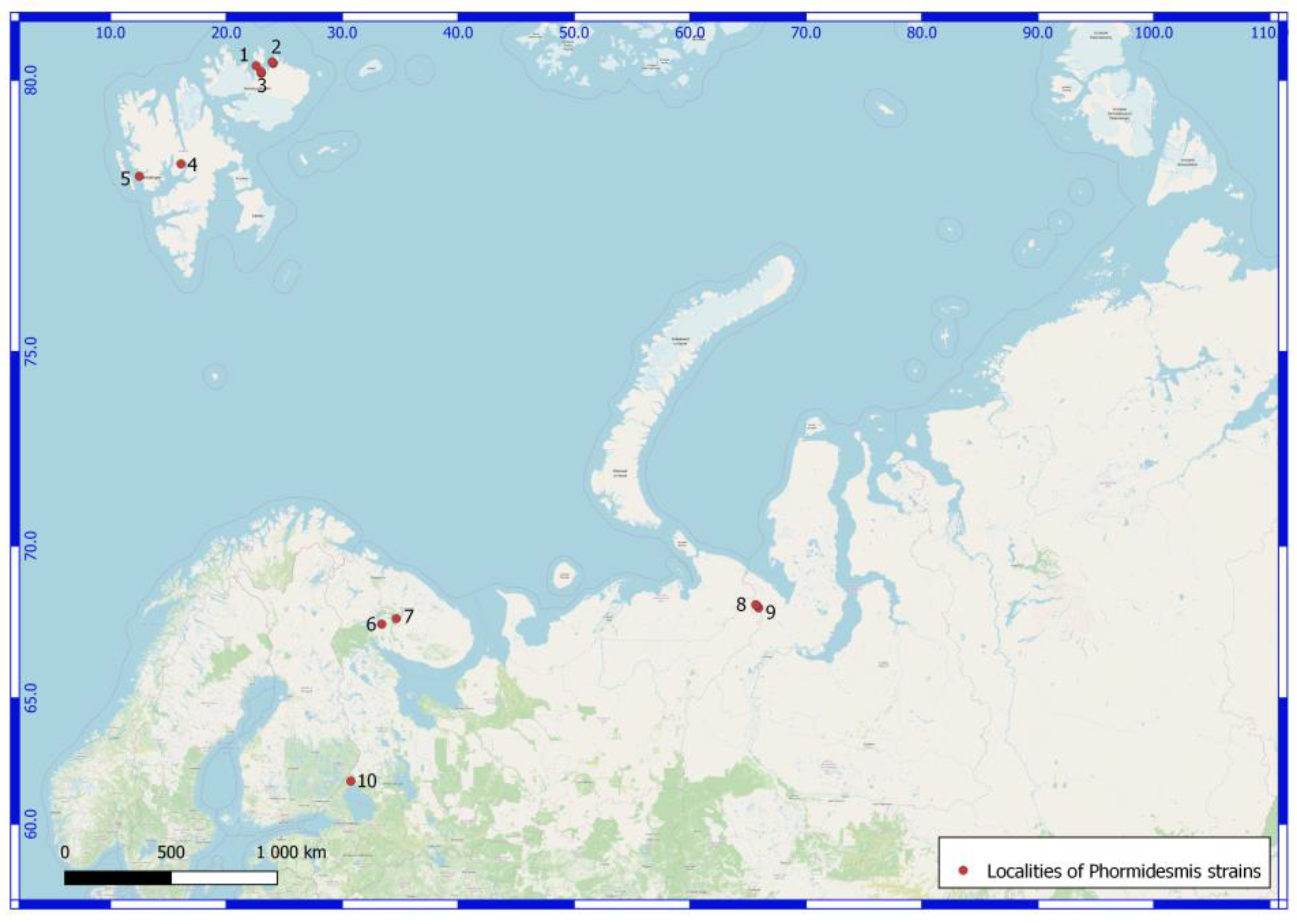
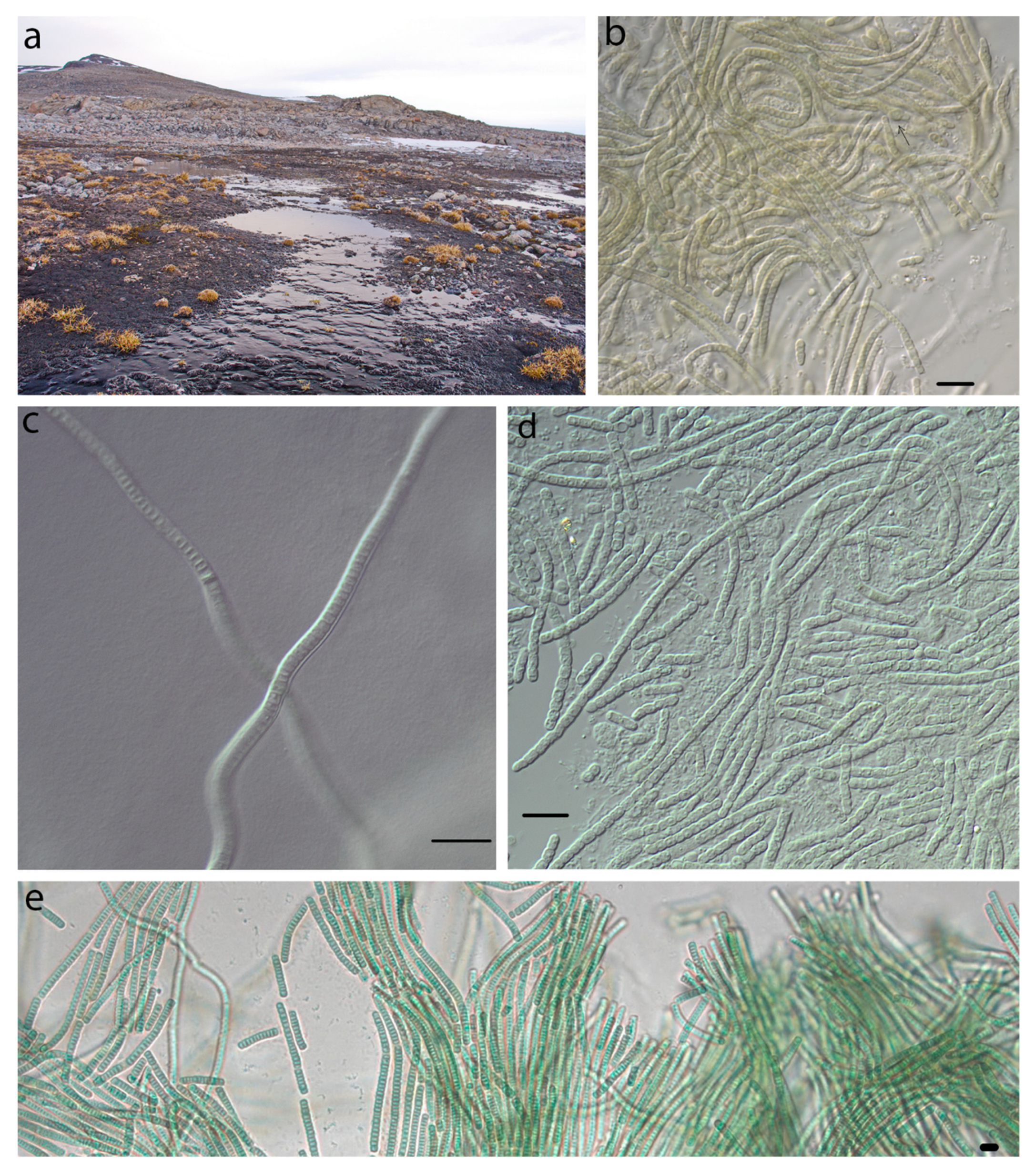
2.1. Sampling

Twenty-three cyanobacterial samples were collected during the summers of 2006–2019 from 10 locations in the different habitats of the Arctic and temperate zone (Table 1, Figure 1). Majority of the samples were collected by the senior author. Sample from site #6 was collected by V. Redkina, #10 by O. Rodina. The collected cyanobacterial samples were taken and dried in sterile paper bags then stored until needed for enrichment cultivation. Samples were stored air-dry in the laboratory until enrichment cultivation during.
Five areas were investigated in Svalbard Archipelago (Figure 1, #1–5). The North East Land Island region (#1–3) located in the Polar desert zone, constitutes a stony desert with a local mountains ridge. The main part of the territory has relatively flat relief. Typically, it is a plateau, rising by an average of 300 m above sea level (m a.s.l.). There are rocks of different geological origins (granites, gneisses, granitoid, quartzites, siltstones, and shales).
The area of West Spitsbergen Island (Figure 1, #4, 5) is formed by rugged mountains reaching the highest peaks of up to 935 m a.s.l. The rocks are presented by conglomerates, sandstones, and limestones. These territories belong to the Arctic tundra zone. The area of the Svalbard Archipelago is covered by numerous lakes, rivers, streams, and seepages. In general, water supply is provided by the melting snow and glaciers.
Two samples were collected in Murmansk Region. The habitat of the natural population of a strain of 6 plot was an upper layer of anthropogenic soil under a footpath in Apatity town. The second sample was found in Lovozerskie Mountains on a high cliff of a wall in a tundra belt.
Six samples were collected in the northern part of the Polar Urals (Figure 1, #8, 9). The landscape of the Polar Urals is characterized by glacial landforms with high peaks (up to 1400 m a.s.l.) and ridges. The northern part of the Polar Urals is situated within a field of limestones with shale and trachybasalts. Moreover, the Svalbard Archipelago and Murmansk Region are located north of the Arctic Circle.
Three specimens were obtained from the wet granitic rock of Ristijärvi park and Filina Mountain in the temperature zone of the Karelia Republic (Figure 1, #10).
Svalbard’s cyanobacterial flora has been characterized in previous studies [26,27,28,29]. Samples (Figure 1) were collected at the different territory of the polar deserts of the North East Island have been conducted cyanobacterial diversity: (east coast of Rijpfjorden bay, (#1) [26], Innvika cove coast (#3) [27], Pyramiden (#4) [28] and the west part of Oscar II Land (#5) [29] of West Spitsbergen Island were investigated. The data on the cyanobacterial diversity of the Polar Ural Mountains (Figure 1, #8, 9) are given in a recent were reported in previous study [30]. The main characteristics of sampling areas are listed in cited articles.

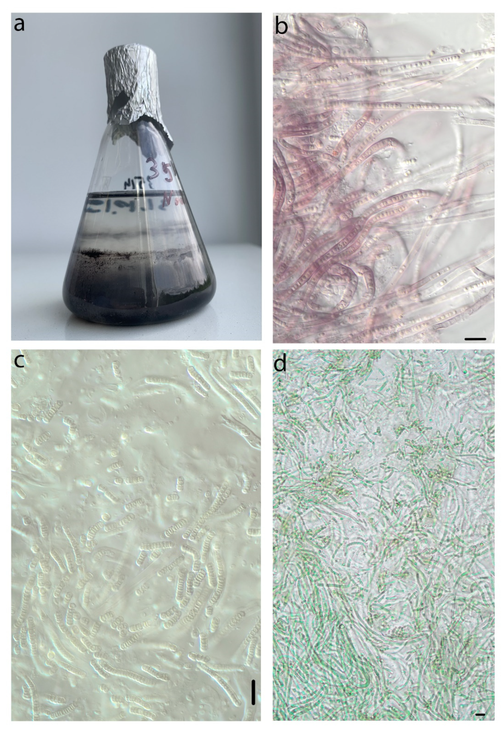
2.2. Isolation of Strains

In the laboratory, the sample was placed into a liquid Z8 medium [31,32]. Unialgal cultures were obtained by picking material from the edge of discrete colonies that had been growing for about 3 weeks on solid BG11 media [32].
Cultures were maintained under an artificial illumination of 16 h light 35 μmol photons m−2 s−1/8 h dark photoperiod at 22 °C. The strains were deposited in the Collection of Cyanoprokaryotes of Polar-Alpine Botanical Garden-Institute (KPABG). A portion of growing material was dried and deposited in the herbarium of KPABG. The label information was included in the “L.” information system [33]. It is a web service cataloguing the biodiversity of cryptogams (https://isling.org/, accessed on 1 May 2022).

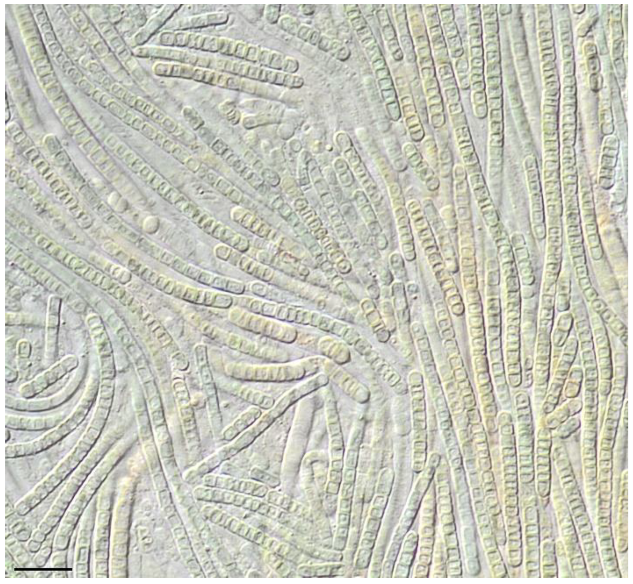
2.3. Morphological Characterization

The strains were observed with an AxioScope A1 (Carl Zeiss, Jena, Germany) microscope equipped with a Nomarski interference contrast and Olympus DP23 camera (Olympus, Tokyo, Japan). Observations were made in the second and fourth week of exponential growth phase as well as in the mature phase after 2 months of incubation. The diacritical morphological traits used in species descriptions were considered, including width and length of cells, the shape of cells, presence or absence of constrictions at the cross-wall, presence of necridic cells, and color of the sheath, and presence or absence of false branching. The cell measurements were based on at least 50 individuals and expressed as minimum-average-maximum values in the taxonomic description.

2.4. DNA Extraction and Sequencing

DNA was extracted from the unialgal culture with the DNeasy Plant Mini Kit (Qiagen, Hilden, Germany) according to the manufacturer’s protocol. Amplification of partial 16S rRNA gene and 16S-23S ITS rRNA region was performed with pair of primers 1 (5′-CTC TGT GTG CCT AGG TAT CC-3′) suggested by [34] and 27F (5′-AGA GTT TGA TCC TGG CTC AG-3′) suggested by [35]. PCR was carried out in 20 µL volumes with MasDDTaqMIX (Dialat Ltd., Moscow, Russia), 10 pmol of each oligonucleotide primer, and 1 ng of DNA with the following amplification cycles: 3 min at 94 °C, 40 cycles (30 s 94 °C, 40 s 56 °C, 60 s 72 °C) and 2 min of final extension time at 72 °C. Amplified fragments were visualized on 1% agarose TAE (mixture of Tris base, acetic acid and EDTA) gels by ethidium bromide staining, purified with the QIAquick Gel Extraction Kit (Qiagen, Hilden, Germany), and then used as a template in sequencing reactions with the ABI PRISM® BigDye™ Terminator v. 3.1 (Applied Biosystems, Foster City, CA, USA) Sequencing Ready Reaction Kit following the standard protocol provided for Applied Biosystems 3730 DNA Analyzer (Applied Biosystems, Foster City, CA, USA). The internal primer 2 (5′-GGG GGA TTT TCC GCA ATG GG-3′) by [36] was additionally used for sequencing.

2.5. Molecular Analyses
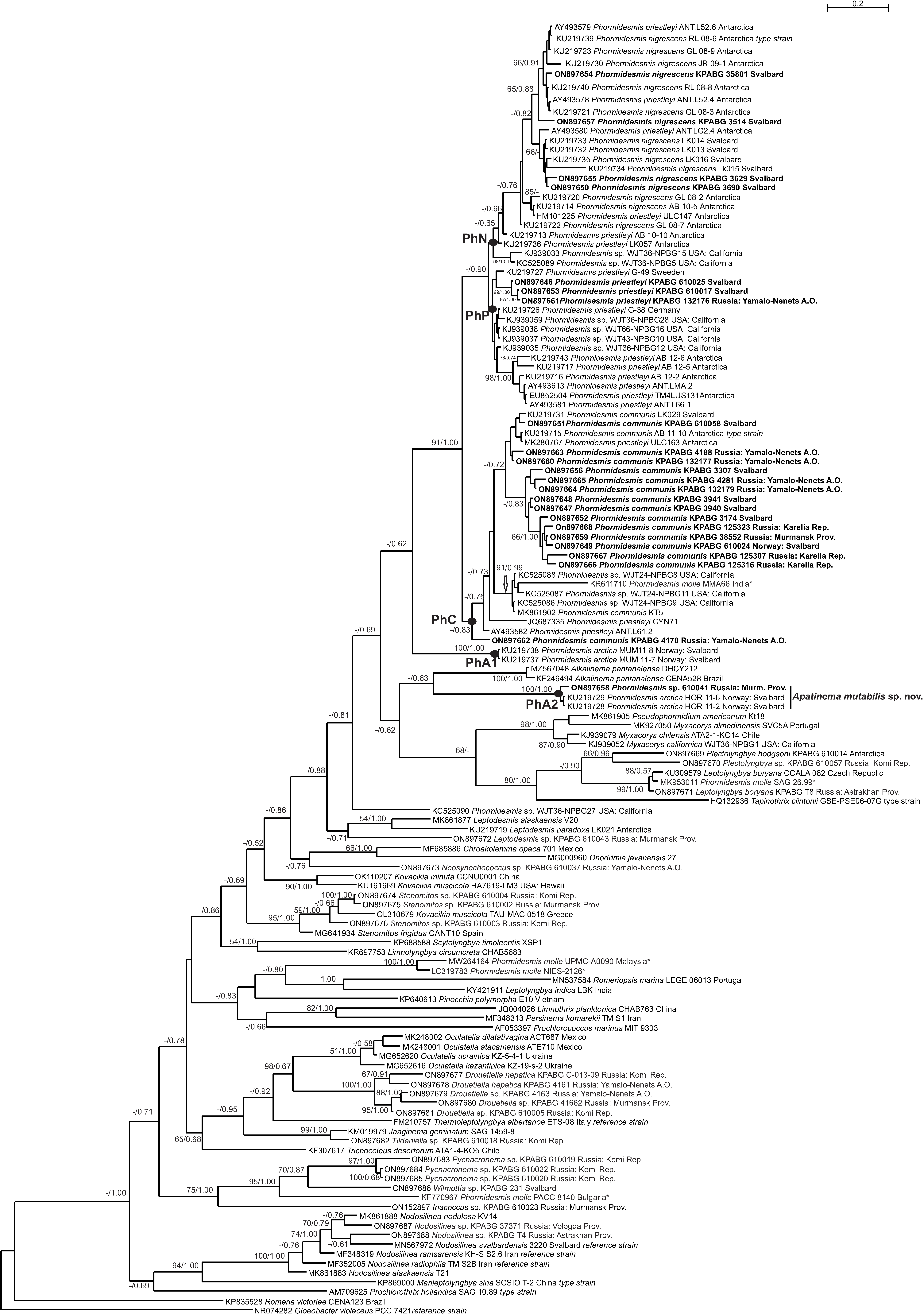
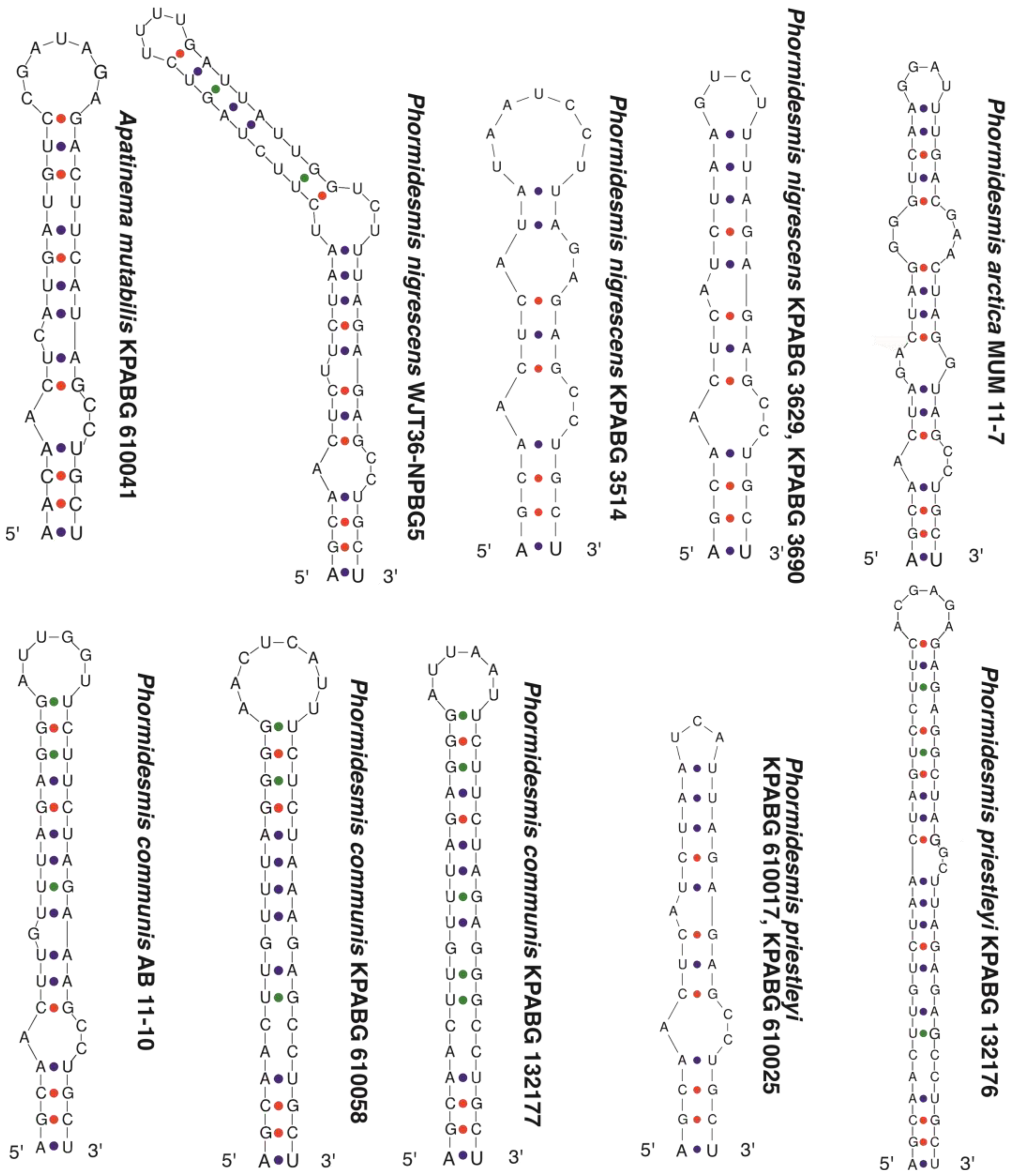
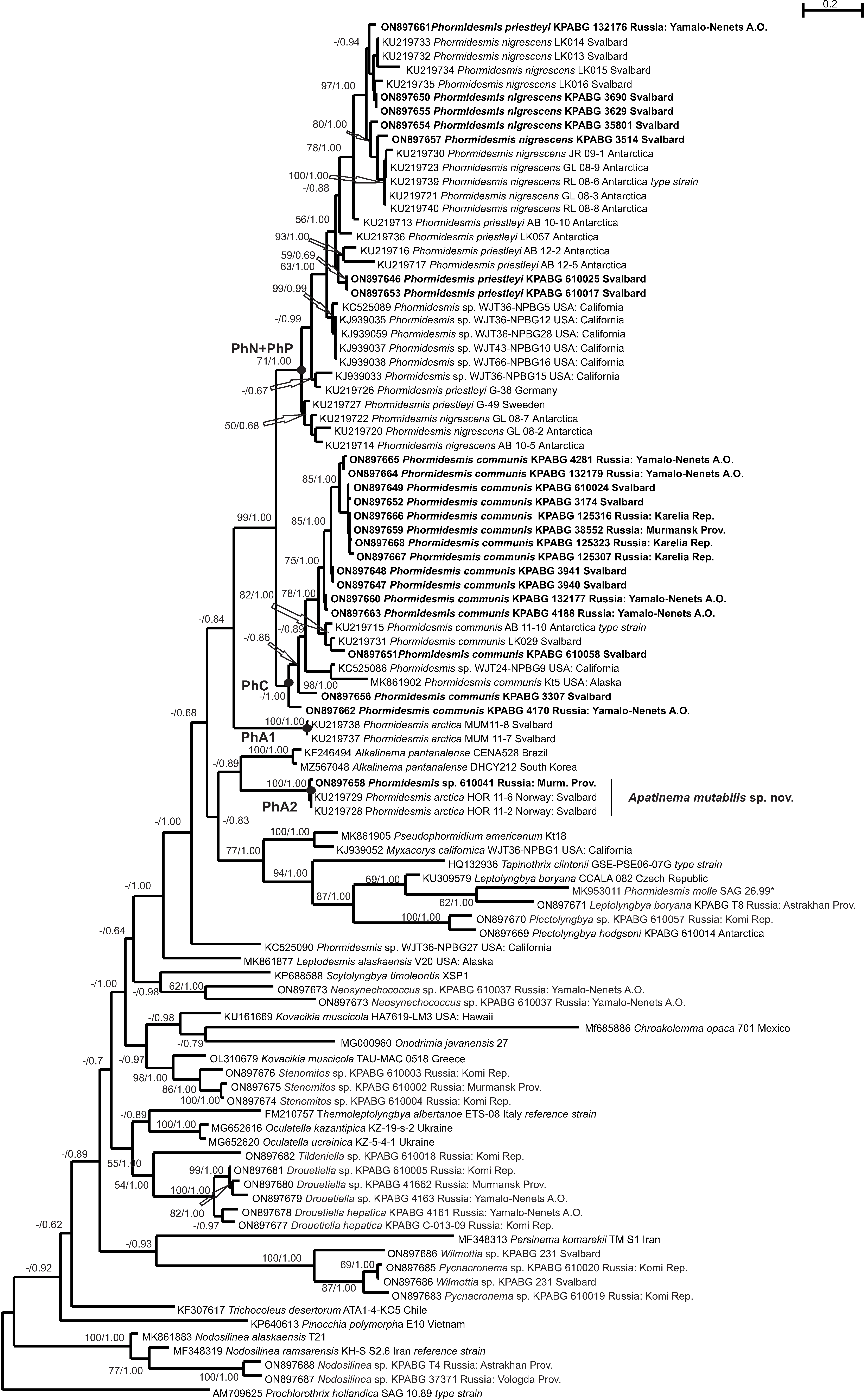
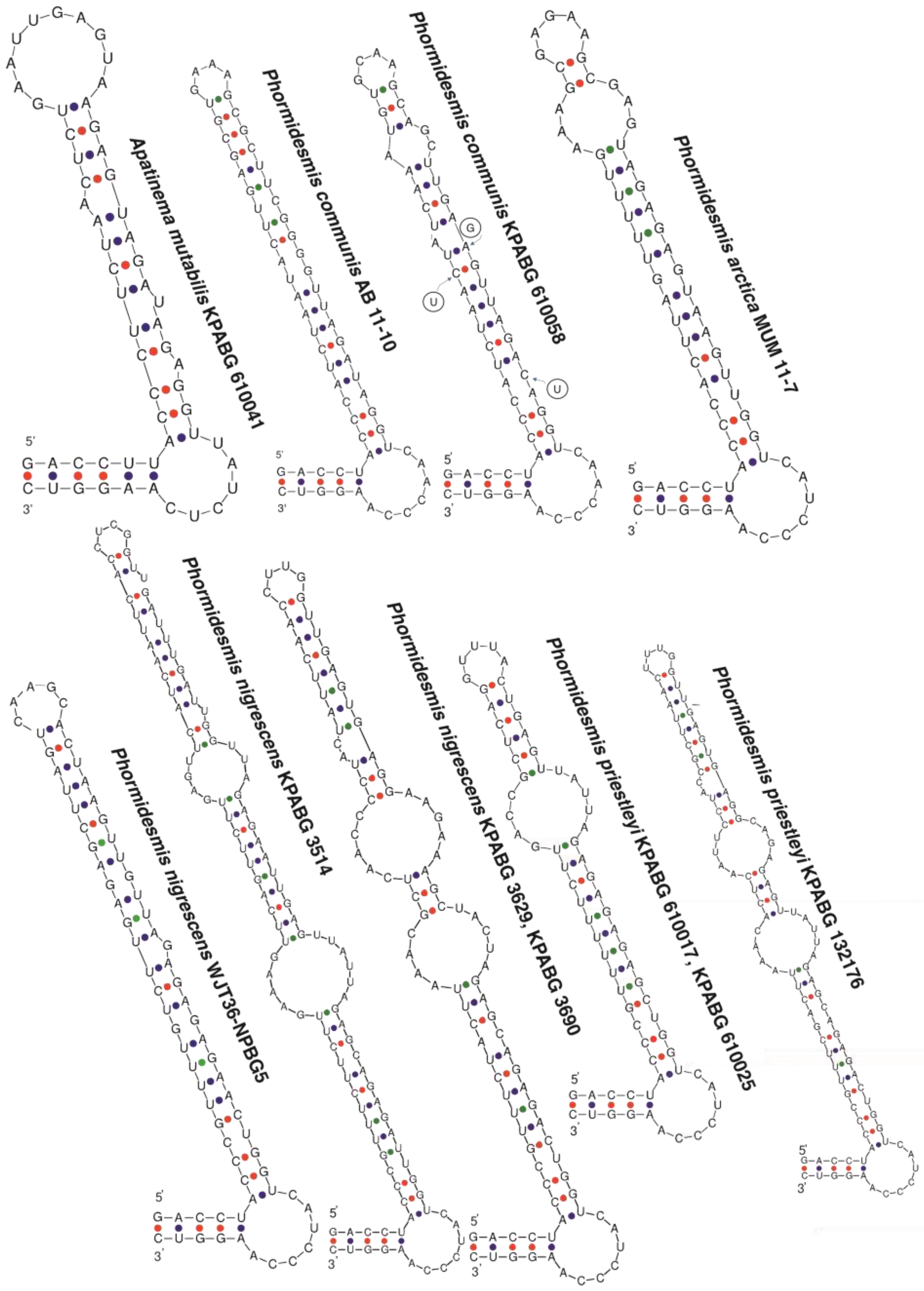
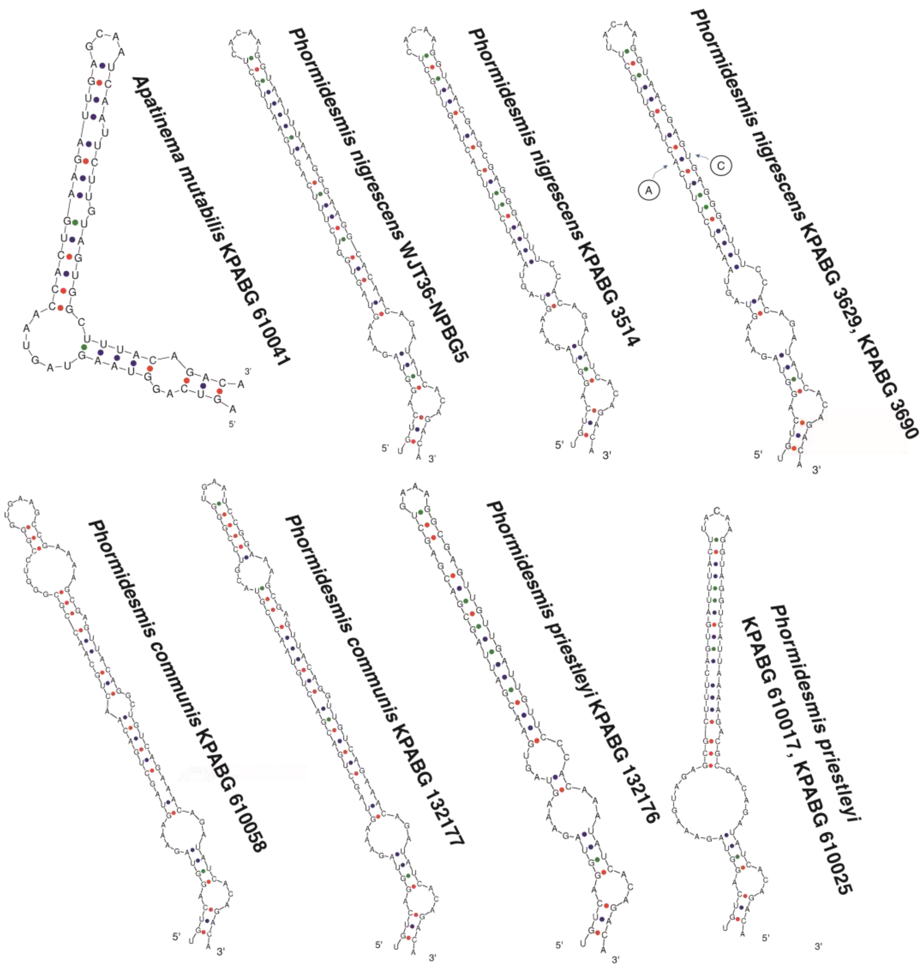
The single amplicon for each strain was obtained and the nucleotide sequence of partial 16S rRNA gene and 16S-23S ITS rRNA region were assembled and edited using BioEdit 7.0.1 [37]. The strain’s accession numbers deposited in GB are given in Table 2. Additionally, molecular data from 20 cultivated in our collection strains morphologically assigned to Synechococcales were used to phylogenetic reconstructions (Appendix A) as well as nucleotide sequences from 94 accessions downloaded from GB and suggested as allied by the phylogeny of [25].
Two datasets were produced for phylogenetic estimation. The first dataset included sequences of 16S rRNA gene for 137 accessions with Gloeobacter violaceus Rippka et al., PCC 7421 as an outgroup, a total length of alignment counts 1141 characters sites. The second dataset presented by 99 accessions from the first dataset that contained 16S rRNA gene and 16S-23S ITS with Prochlorothrix hollandica Burger-Wiersma et al. SAG 10.89 as an outgroup, total length of alignment counts 1836 characters sites.
Both datasets were estimated by two analytical procedures: maximum likelihood (ML) with RaxML v. 1.0.0 [38] and Bayesian approach with MrBayes v. 3.2.1 [39]. The ModelGenerator [40] determined GTR+I+G as the best fit evolutionary model of nucleotide substitutions for both datasets. This model was used in ML analysis together with four rate categories of the gamma distribution to evaluate the rate of heterogeneity among sites and resampling procedure with 200 replicates and automatic bootstopping (cutoff: 0.03) to obtain a bootstrap support for individual nodes.
The Bayesian analysis of both datasets included the GTR + I + G model and gamma distributions with four rate categories, partitions of 16S rRNA gene and 16S-23S ITS in the second dataset were separately assigned the GTR + I + G model. Two independent runs of the Metropolis-coupled ΜCMC were used to sample parameter values in proportion to their posterior probability. Each run included three heated chains and one unheated, and two starting trees were chosen randomly. Chains were run for ten million generations and trees were sampled every 100th generation for16S rRNA gene dataset. The 16S rRNA gene and 16S–23S ITS dataset were analyzed with five million generations and trees were sampled every 100th generation. For the 16S rRNA gene dataset the software tool Tracer [41] revealed the effective sample size (EES) as 3189.3304 and auto-correlation time (ACT) as 5643.8806. The first 25,000 trees in each run were discarded as burn-in. Thereafter 150,000 trees were sampled from both runs. The average standard deviation of split frequencies between two runs was 0.009267. For the 16S rRNA gene and 16S–23S ITS dataset Tracer suggested ESS as 2775.4259 and auto-correlation time as 3242.8177. The first 12,500 trees in each run were discarded as burn-in. Thereafter 75,000 trees were sampled from both runs. The average standard deviation of split frequencies between two runs was 0.004776. Bayesian posterior probabilities in both estimations were calculated from trees sampled after burn-in. Majority rule (MJ) consensus tree for both datasets was calculated after combining the runs minus burnin of 25% and the topology was illustrated with FigTree v. 1.4.4 [42].
The infrageneric and infraspecific variability of 16S rRNA gene and 16S-23S ITS of tested Phormidesmis species and related taxa were calculated as the average pairwise p-distances in Mega 11 [43] using the pairwise deletion option for counting gaps with the following formula 100 × (1 − p).
The hypothetical secondary structures of conserved helices (D1-D1′, BoxB, and V3) in the 16S–23S ITS region were estimated using Mfold [44] including strains sampling of different Phormidesmis nigrescens (type strain RL 08-6 did not provide appropriate sequence data) and Phormidesmis priestleyi (type strain not sequenced) subclades and data for known type strain of Phormidesmis communis AB 11-1.

3. Results

The ML analysis of the 16S rRNA gene resulted in a single tree with the arithmetic mean of Log likelihood −13,603.071670. In Bayesian analysis of the 16S rRNA gene the arithmetic means of Log likelihoods for each sampling run were −13,672.64 and −13,678.14. Both trees possess similar topology, thus Figure 2 demonstrated the topology of the MJ consensus tree after Bayesian calculation with an indication of bootstrap support values (BS) calculated in ML analyses and Bayesian posterior probabilities (PP). The ML analysis of the 16S rRNA gene and 16S–23S ITS resulted in a single tree with the arithmetic mean of Log likelihood −27,707.794017, the arithmetic means of Log likelihoods for each sampling run in BA analyses were −27,005.68 and −27,006.79. The topologies from both calculations are similar, thus Figure 3 presents the MJ consensus tree after BA with values of BS supports and PP.
The trees topologies reconstructed from both datasets were congruent and resembled the phylogeny of Synechococcales as published previously [4]. Here we concentrated on species identification and affinity concerning 23 newly sequenced strains from our collection.
Based on 16S estimation, our Phormidesmis strains were found in four clades intermingled with accessions based on Raabová et al. from [16] and other GB data, whereas on topologies from the whole region of 16S rRNA gene and 16S–23S ITS only three clades could be recognized. In both estimations, clades containing 15 Phormidesmis communis strains could be robustly recognized (PhC) with PP = 0.83 and PP = 1.00 (Figure 2 and Figure 3). Five specimens determined by us as Phormidesmis nigrescens and two as Phormidesmis priestleyi (F.E. Fritsch) Komárek et al. were placed in intermingled terminal clade that slightly subdivided (PP = 0.9) on two subclades (PhN and PhP) in 16S calculation and partially corresponded with subdivision obtained by Raabová et al. in [16] due to the transfer of two accessions (KU219713 and KU219736) from PhP to PhN clade. The presence in analyses of ITS data influenced the producing of the common intermingled PhP+PhN clade with support BS = 71% and PP = 1.00 in Figure 3. The sequence analysis by eye allows us to determine three positions in 16S that could differentiate Phormidesmis priestleyi and P. nigrescens strains, whereas the whole alignment of strains from both species resembled something of a puzzle with intermingled motifs across the dataset. The result of 16S gene phylogenetic estimation showed sample distribution without these differentiated substitutions, strains of both species are marked on Figure 2 and Figure 3.
Three strains (Phormidesmis arctica HOR11-2 (KU219728), P. arctica HOR11-6 (KU219729), and KPABG 610041) formed a clade in sister position to Alkalinema pantanalense (Figure 2, PhA2 clade) with the highest supports in all calculations. Raabová et al. [16] tested four specimens named as Phormidesmis arctica that were placed in two sister-clades. Both clades are characterized by quite long and slight branches, providing little support, however, the authors consider this as a single species. The addition in the current analysis of the members of the recently described genus, Alkalinema [3], revealed the relationship of the Phormidesmis arctica (PhA2)-clade to this genus (PP = 0.89, PP = 0.63), whereas other Phormidesmis arctica (PhA1)-clade kept their basal position in the Phormidesmis (PP = 0.84, PP = 0.62)-clade.
The p-distances for Phormidesmis nigrescens and P. priestleyi were counted twice according to distribution among two clades in 16S gene analysis and according to alignment (Table 2). Infraspecific divergence in Phormidesmis priestleyi in both types of calculations was stable, whereas P. nigrescens became more variable according to tree topology (99.16% in 16S gene and 92.95% in ITS) than according to alignment (99.61% and 95.30%, consequently). Nevertheless, a divergence between both species revealed similar levels (98.58/91.54 and 98.50/91.01), which exceed the values of infraspecific variation and suggested both species as separate taxa.
Phormidesmis communis appear to be more variable in 16S gene loci then two discussed species (98.88%) and more stable in ITS (93.32%). It is robustly divergent from both Phormidesmis nigrescens and P. priestleyi and differs from them in a similar level. The specimens of Phormidesmis arctica from PhA1-clade are identical by both loci, the specimens of PhA2-clade varied only in the 16S gene (99.82%). The level of differences between both Phormidesmis arctica clades counts 94.49% in the 16S gene and 83.43% in ITS, which is higher than the divergence between Phormidesmis nigrescens, P. priestleyi, and P. communis.
The specimens of Phormidesmis arctica from PhA1–clade are closer to other Phormidesmis species (approximately 94% in 16S gene and 85–87% in ITS) than specimens of PhA2-clade (approximately 92% in 16S gene and 83–84% in ITS). Alkalinema pantanalense differs from PhA1-clade in 93.18% and 83.82%, from PhA2—in 93.11% and 84.55%. One of the nearest relatives to the ingroup taxa, the genus Myxacorys, is well-differentiated from both Phormidesmis arctica clades and Alkalinema (Table 2). Based on the studies by Yarza et al. [45], the similarity cut-off which is considered clear evidence of genera delineation is 94.5% 16S rRNA sequence identity.
Phormidesmis strains have variable secondary structures for the 16S–23S ITS region, which is distinctive compared to the helices of Apatinema (Figure 4, Figure 5 and Figure 6). The D1-D1’ helices for Phormidesmis were distinctly longer (64–114 nucleotides) than the D1-D1’ helix of Apatinema, with the 57 nucleotides. Phormidesmis had a similar basal loop (5’-CAAUCCCA-3’) in contradistinction to Apatinema (5’-UAUCUC-3’). Apatinema strain also differed from the rest by having a longer terminal loop (Figure 4).
The great diversity of the D1-D1’ helix secondary structure was suggested in the genus Phormidesmis. The secondary structures of reference strains of species types Phormidesmis molle 3CC04S05, Phormidesmis nigrescens RL 08-6, Phormidesmis priestleyi are not comparable. The strains of Phormidesmis communis possess shorter D1-D1’ regions compared with the other three species that are characterized by absent or slightly marked loops during the stem. This region among Phormidesmis nigrescens presented in two loops that differed in nucleotide composition and position on the stem. The D1-D1’ helices of strains Phormidesmis nigrescens KPABG 3629, and KPABG 3690, were identified. The Phormidesmis priestleyi strains KPABG 610017, KPABG 610025, tested here, are identical in secondary structures and characterized by a single loop in the stem length. An unexpected result was obtained for strain Phormidesmis priestleyi KPABG 132176 that placed it with the strains of Phormidesmis priestleyi KPABG 610017, KPABG 610025 by 16S sequence data but revealed a D1-D1’ helices structure typical of Phormidesmis nigrescens.
The Box-B structures differed consistently in species Phormidesmis, and Apatinema (Figure 5), and greatly varied in length within the Phormidesmis species. Nevertheless, some strains, Phormidesmis priestleyi (KPABG 610017, KPABG 610025) and P. nigrescens (KPABG 3629, KPABG 3690), had a similar the Box-B region, but the typical structure for each species was not reconstructed due to the variable position of loops among strains of a single species. Phormidesmis communis strains are characterized by a long stem with a terminal loop. The Box-B helices did not provide as clear separation of the species in the Phormidesmis as did the D1-D1’ helices.
The structure of V3 helices clearly diverged Apatinema from Phormidesmis (Figure 6). The V3 helices of Phormidesmis species were similar in the basal portion of the helix, but divergent in the apical regions. Between Phormidesmis nigrescens KPABG 3514, KPABG 3629, KPABG 3690, the V3 helices were almost invariant in structure and similar in sequence. Phormidesmis communis revealed a similar structure to them, but with a loop remote to terminal one. Phormidesmis priestleyi strains KPABG 610017, KPABG 610025 are characterized by an enlarged loop near the stem base.
Taking into account phylogenetic affinity and DNA sequence variability, we suggested strains from PhA2-clade be considered as a new genus that is described here.

Taxonomic Description

  • Class Cyanophyceae
  • Subclass Synechococcophycidae
  • Order Synechococcales
  • Family Leptolyngbyaceae
  • Apatinema Davydov gen. nov.
Diagnosis: Apatinema is distinguished morphologically from all other closely related sister clades in the Leptolyngbyaceae by its propensity to have trichomes of variable morphology with short or long cells, and also by its phylogenetic position based on 16S rRNA and 16S–23S ITS rRNA gene phylogenies, p-distance analysis, and differences in secondary structures of D1-D1’, and Box-B helices. Filaments with single trichomes without sheath in young cultures and hormogonia. Filaments are cylindrical, straight or flexuous, sometimes bent and coiled, not attenuated to the ends.
Etymology. Apati (ἀπάτη) (Greek, fem. n.)—to deceive, confuse, and refers to the appearance of the organism in Apatity town; nema (N.L. neut. n.) thread, filament.
Type species: Apatinema mutabilis Davydov sp. nov. (Figure 7).
Diagnosis: filaments long, cylindrical, ±straight or flexuous, sometimes bent and coiled, not attenuated to the ends. Sheaths contain one trichome, colorless, fine thin, sometimes confluent. Trichomes are pale blue-green or olive-green, slightly or distinctly constricted at the cross-walls with variable cell sizes within trichomes. End cells rounded, not calyptrate. Cells are shorter or longer rather than wide, 1.7–2.5(4.7) μm long, (1.7)2–3.5(4) μm wide.
Etymology: A. mutabilis N.L. fem. Adj. = liable to change, variable.
Type Locality: Murmansk Region of Russia, Apatity town, Bredova st. between 17 and 19 buildings. The habitat of the natural populations of species was typically an upper layer of anthropogenic soil under a footpath. 67°561394″ N, 33°410578″ E, elevation 197 m a.s.l., collected by V. Redkina on 1 September 2019.
Habitat: In the upper layer of podzol soil (AY, 5 cm) under a footpath.
Holotype: KPABG 4504! in the herbarium of Polar-Alpine Botanical Garden-Institute of Kola Science Centre of RAS, Kirovsk, Murmansk region, Russia.
Reference strain: LID-610041 in the Collection of Cyanoprokaryotes of Polar-Alpine Botanical Garden-Institute of Kola Science Centre of RAS, Kirovsk, Murmansk region, Russia.
In a publication by Raabová et al. [16], a holotype depository was not indicated for the Phormidesmis communis species. We provide the missing type depository with a diagnosis of Phormidesmis communis in order to validate the name under the conditions required by the ICN [23].
Phormidesmis communis Raabová, L. Kováčik, Elster et Strunecký sp. nov.
Based on: Phormidesmis communis Raabová, L. Kováčik, Elster et Strunecký, Phytotaxa. 395:1, 2019 [16] nom. inval. (Art. 8.4 Art. 40.8; a holotype depository was not indicated and cultures of cyanobacterium did not preserve in a metabolically inactive state).
Note: The reference to the description in [16] (p. 12), together with the reference to the holotype here, validates the taxon.
Type Locality: Svalbard Archipelago. North East Land Island. Orvin Land. Duvefjorden bay. Sætherbukta cove. Damflya plain. The habitat of the natural populations of species was on pebbles in a slow stream. The location is 80°24648″ N, 24°05033″ E, elevation 100 m a.s.l., collected by D. Davydov on 29 July 2012.
Holotype: KPABG 3679! in the herbarium of Polar-Alpine Botanical Garden-Institute of Kola Science Centre of RAS, Kirovsk, Murmansk region, Russia.
Reference strain: LID-610024 in the Collection of Cyanoprokaryotes of Polar-Alpine Botanical Garden-Institute of Kola Science Centre of RAS, Kirovsk, Murmansk region, Russia.
The morphological traits of Phormidesmis, and Apatinema species are similar (Table 3).
Investigated strains of Phormidesmis priestleyi are characterized by wavy constricted trichomes with colorless envelopes, cells pale blue-green or olive-green, short or isodiametric which are (1.8)2.3–3.3 μm wide. One sheath usually contains one trichome, in rare cases 2–3 trichomes. Macroscopically, mats have an agar with a characteristic rusty color (Figure 8).
Morphologically, the studied strains of Phormidesmis nigrescens (KPABG 3514, KPABG 35801, KPABG 3629, KPABG 3690) are quite similar. In old cultures, sheaths are violet or blackish (Figure 9). Trichomes are distinctly constricted, occasionally with necridic cells. Cells are 1.6–3.1 μm wide and 1.2–2.5 μm long. Apical cells are rounded, without calyptra.
Filaments of Phormidesmis communis have thin colorless sheaths, bright, pale blue-green, or olive-green, trichomes, 1.5–3.6 μm wide, the cells are short or, less frequently, long 1.0–4.3 μm with constrictions at cross-walls. In cultures, the trichomes form wide and long cells (Figure 10b), the rare filaments are falsely pseudobranched. Theapical cells are rounded, and without calyptra. They are subaerophytic on wet rocks and on boulders in a slow stream (Figure 10a) or pools, forming mats on seepages.

4. Discussion

Phormidesmis strains sequences deposited in the GB database are not from the loner clade. The results suggested Phormidesmis to be a polyphyletic taxon. The studied strains analyzed here form at least four different lineages in the Phormidesmis and one clade in a sister position to Alkalinema, Myxacorys, Plectolyngbya, Tapinothrix (Figure 2 and Figure 3). They could be assigned to Phormidesmis priestleyi, Phormidesmis communis, Phormidesmis nigrescens, Phormidesmis arctica and Apatinema mutabilis.
The 16S phylogeny more clearly separated Phormidesmis priestleyi and Phormidesmis nigrescens clades, whereas addition of 16S–23S ITS region brings confusion in species delimitation. Each species is characterized by several types of secondary structures of three stem-loop regions, moreover, a single strain—Phormidesmis priestleyi KPABG 132176—possesses 16S of Phormidesmis priestleyi and 16S–23S ITS region of Phormidesmis nigrescens. On the one hand, each of the obtained subclades within Phormidesmis priestleyi or Phormidesmis nigrescens could be described as a distinct taxon according to their secondary structure, but on the other hand, the strain sampling shown here suggests the presence of great nucleotide sequence diversity in the ITS region between natural populations of single species. That fact should be taken into account in taxonomical studies and prevent the description of new species, which perhaps appear to be a member of widely distributed variable species.
Several “Phormidesmis” strains are related to the different Leptolyngbyacae genera (Figure 2). For example, Phormidesmis molle SAG 26.99 (MK953011) clustered with the Leptolyngbya s.str and should be assigned to Leptolyngbya boryana. The clade including Phormidesmis molle UPMC-A0090 (MW264164) and Phormidesmis molle NIES-2126 (LC319783) is in the sister position to Romeriopsis.

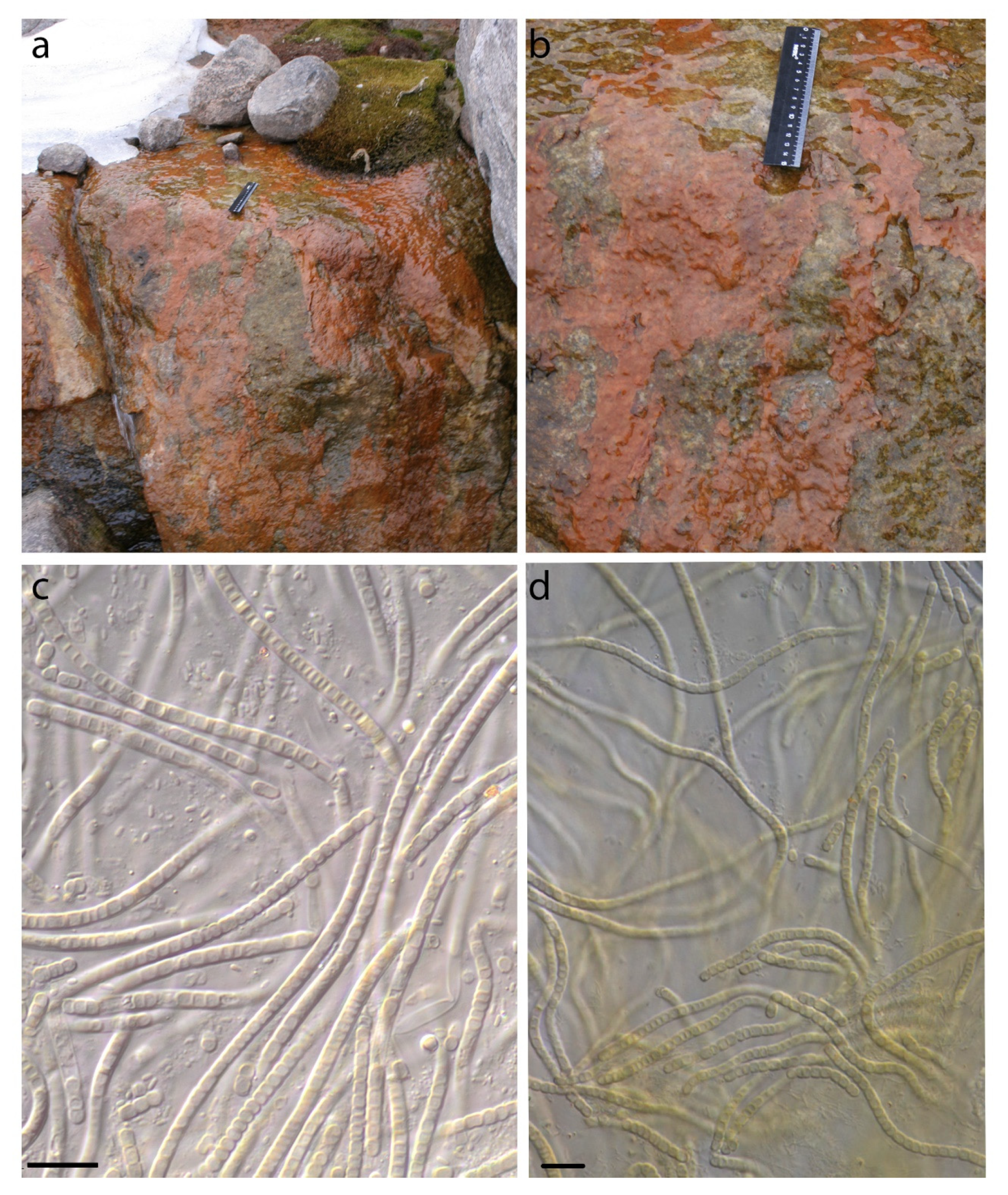
4.1. Phormidesmis molle

The reference strain, of Phormidesmis molle (Gomont) Turicchia, Ventura, Komárková et Komárek [15] was characterized as a solitary or irregularly clustered filament, slightly coiled, and not attenuated at the ends. The moniliform trichomes cells are composed of short to ±isodiametric, barrel-shaped cells (2.5-)3-4(-6) µm wide, constricted at cross-walls, with homogeneous blue-green content. Sheaths are facultatively developed, fine or firm, thin, colorless. End cells are rounded and do not differ from other vegetative cells, rounded at the end.
The reference strain of Phormidesmis molle (3CC04S05) was isolated from the alkaline marshes (Chan Chen) in northern Belize. The rRNA sequence of the strain type was not deposited in public databases and cannot be used for comparison.
One of the strains, Phormidesmis molle MMA66 (KR611710), bearing this name from India, falls into Phormidesmis communis clade. Another of the strains, Phormidesmis mole, is affiliated with Leptolyngbya or different genera in Leptolyngbyaceae. Strain Phormidesmis molle PACC 8140 (KF770967) from Bulgaria does not belong to the Synechococcales.

4.2. Phormidesmis priestleyi

The basionym of Phormidesmis priestleyi (F.E. Fritsch) Komárek, Kastovský, Ventura, Turicchia et Smarda is Phormidium priestleyi F.E. Fritsch. The article by Taton et al. [46] adduces nine different strains (ANT.L52.4, ANT.LG2.4, ANT.L52.6, ANT.LPR.5, ANT.LPR.6, ANT.L66.1, ANT.LMA.2, ANT.LACV5.1, ANT.L61.2) isolated from Cape Adare of James Ross Island, Antarctica, which belong to the Phormidesmis priestleyi.
As shown previously [16], strains ANT.LG2.4, ANT.L52.4, ANT.L52.6 fall into Phormidesmis nigrescens clade. This topology was also confirmed in our study. We consider strains by [46] ANT.L66.1, ANT.LMA.2 as reference strains of Phormidesmis priestleyi. Strains AB 12-2, AB 12-5, AB 12-6 from Antarctica, and G-49 from Sweden should be assigned to that taxon.
Some Phormidesmis strains were submitted to NCBI as unidentified. In the current study, we included such sequences (KJ939035, KJ939037, KJ939038, and KJ939059) in our analyses to determine their phylogenetic placement and confirmed that they belong to Phormidesmis priestleyi.
Several strains in our collection follow a perfectly matched criteria set for Phormidesmis priestleyi KPABG 610017 (ON897653) (Figure 8), and KPABG 610025 (ON897646) from Svalbard archipelago, KPABG 132176 (ON897661) from other Arctic area in the Polar Urals. However, the position of P. priestley KPABG 132176 on phylogenetic trees and distinguished differences in the secondary structures of helices 16S-23S ITS region may indicate that the strain belongs to a separate species.
In Svalbard ecosystems, Phormidesmis priestleyi strains usually grow in melting fields and slow streaming water attached to stones and form macroscopic, rusty reddish-brown, thick mats during the vegetation peak. It is less frequently found in pools and in seepages under similar conditions. In the Polar Urals the species was found on a wet wall of rock, near a water line of a stream.
Phormidesmis priestleyi is distributed in polar regions, but the occurrences in Germany (G-38, KU219726) and California (WJT36-NPBG12, WJT43-NPBG10) provided evidence that the species is widespread.

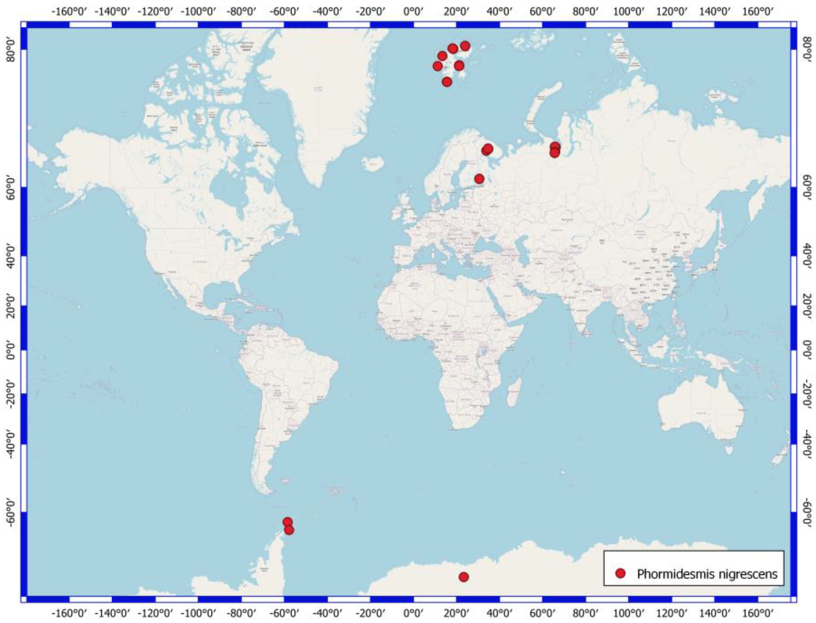
4.3. Phormidesmis nigrescens

The basionym of Phormidesmis nigrescens (Komárek) Raabová, Kovacik, Elster et Strunecký is Leptolyngbya nigrescens Komárek. The morphotype was described as being from the Antarctic. Taxon is characterized by firm, thin, blackish sheaths, and isodiametric cells, 0.8–2.5 µm wide. Trichomes are slightly (indistinctly) or clearly constricted at cross-walls. The reference strain by [16] is RL 08-6. The unidentified Phormidesmis strain (sequence KC525089) we consider to be part of Phormidesmis nigrescens.
The ecological properties of Phormidesmis nigrescens showed that it is a typical subaerophytic. The species inhabits on wet rock and outcrops, less frequently on soil and the surface of mats in seepages, and on littoral of lakes. It is widely distributed in the Polar Regions of the Arctic and Antarctic (Figure 11).
The strains of Phormidesmis communis do not have clearly morphological traits to separate that taxon from other species of the genus. Species occurrences are known in the Arctic, Antarctic, and temperature zone.

4.4. Phormidesmis arctica

The specimens of Phormidesmis arctica (MUM 11-7, MUM 11-8) have shown a high percentage of dissimilarity in genetic distance analysis. This lineage may be considered as a loner genus in Leptolyngbyaceae, but formal relocation of the taxon is not possible because the name of the species is invalidly published.

4.5. Apatinema mutabilis

The species is characterized by variable cell size within trichomes and a specific habitat—it has occurred in the soil. The cell length is variable from shorter (1.7 μm) up to twice as long as the width (4.7 μm).
Obviously, the morphological data (Table 3) are not reliably able to distinguish the species in Phormidesmis and relative genera. Diverse morphological autapomorphy is one of the greatest restrictions within Leptolyngbyaceae. Different Phormidesmis lineages are distinctly separable by molecular traits. Without the study of multiple populations, this difference would likely have gone unnoticed, and its significance would not have been recognized.
In agreement with other cyanobacterial taxonomists [4,10,13,47,48], we consider that the ITS region analysis allows us to separate phylogenetically close species, and the p-distance similarity of the 16S and 16S-ITS alignments also supported the recognition of Phormidesmis species.
The populations of distinct species such as Phormidesmis nigrescens KPABG 3690 and Phormidesmis communis KPABG 3940, or Phormidesmis communis KPABG 132177 and Phormidesmis priestleyi KPABG 132176 were found in close spatial proximity within ten meters of each other. This is not a unique observation, as noticed in Myxacoris, lineages were pointed to a likewise case [9]. The presence of two related taxa in nearby habitats suggests their niche differentiation.

5. Conclusions

We investigated the diversity of cyanobacteria in similar terrestrial and near-water habitats. The use of unialgal cultures with subsequent molecular analyses allowed us to reveal great nucleotide sequence diversity in ITS region in widely distributed species that should be carefully treated in taxonomical studies, especially in the course of description of new species. Notwithstanding that Phormidesmis, Alkalinema, Apatinema species possess overlapping morphological traits, they could be identified in base with a comprehensive approach using microscopic evaluation of morphologies, cultivation of strains, and study of sequencing data.

Author Contributions

Conceptualization, D.D.; methodology, D.D. and A.V.; formal analysis, D.D. and A.V.; investigation, D.D. and A.V.; resources, D.D.; data curation, D.D. and A.V.; writing—original draft preparation, D.D. and A.V.; writing—review and editing, D.D. and A.V.; visualization, D.D. and A.V.; supervision, D.D.; project administration, D.D.; funding acquisition, D.D. and A.V. All authors have read and agreed to the published version of the manuscript.

Funding

This research was funded by the Russian Science Foundation, grant number 21-14-00029 (https://rscf.ru/project/21-14-00029/). The research was performed using large-scale research facilities at the herbarium at the Polar-Alpine Botanical Garden-Institute (KPABG; Kirovsk, Russia) reg. No. 499397.

Institutional Review Board Statement

Not applicable.

Informed Consent Statement

Not applicable.

Data Availability Statement

The data supporting reported results can be found in a publicly archived dataset of “L” Information system https://isling.org/ (accessed on 10 May 2022).

Acknowledgments

The authors are grateful to Vera Redkina and Oksana Rodina for sampling of Cyanobacteria in Apatity and Karelia Republic.

Conflicts of Interest

The authors declare no conflict of interest. The funders had no role in the design of the study; in the collection, analyses, or interpretation of data; in the writing of the manuscript; or in the decision to publish the results.

Appendix A

The sequenced strains additionally used in current study with voucher details and GB accession numbers for 16S rRNA gene and 16S-23S ITS rRNA.
  • Drouetiella hepatica, KPABG 4161 Russia: Yamalo-Nenets A.O., ON897678;
  • Drouetiella hepatica, KPABG C-013-09, Russia: Komi Rep., ON897677;
  • Drouetiella sp., KPABG 4163, Russia: Yamalo-Nenets A.O., ON897679;
  • Drouetiella sp., KPABG 41662, Russia: Murmansk Prov., ON897680;
  • Drouetiella sp., KPABG 610005, Russia: Komi Rep., ON897681;
  • Leptodesmis sp., KPABG 610043, Russia: Murmansk Prov., ON897672;
  • Leptolyngbya boryana, KPABG T8, Russia: Astrakhan Prov., ON897671;
  • Neosynechococcus sp., KPABG 610037, Russia: Yamalo-Nenets A.O., ON897673;
  • Nodosilinea sp., KPABG 37371, Russia: Vologda Prov., ON897687;
  • Nodosilinea sp., KPABG T4, Russia: Astrakhan Prov., ON897688;
  • Plectolyngbya hodgsoni, KPABG610014, Antarctica, ON897669;
  • Plectolyngbya sp., KPABG 610057, Russia: Komi Rep., ON897670;
  • Pycnacronema sp., KPABG 610019, Russia: Komi Rep., ON897683;
  • Pycnacronema sp., KPABG 610020, Russia: Komi Rep., ON897685;
  • Pycnacronema sp., KPABG 610022, Russia: Komi Rep., ON897684;
  • Stenomitos sp., KPABG 610002, Russia: Murmansk Prov., ON897675;
  • Stenomitos sp., KPABG 610003, Russia: Komi Rep., ON897676;
  • Stenomitos sp., KPABG 610004, Russia: Komi Rep., ON897674;
  • Tildeniella sp., KPABG 610018, Russia: Komi Rep., ON897682;
  • Wilmottia sp., KPABG 231, Norway: Svalbard, ON897686.

References

  1. Davydov, D.; Patova, E. The diversity of Cyanoprokaryota from freshwater and terrestrial habitats in the Eurasian Arctic and Hypoarctic. Hydrobiologia 2018, 811, 119–138. [Google Scholar] [CrossRef]
  2. Achtman, M.; Wagner, M. Microbial diversity and the genetic nature of microbial species. Nat. Rev. Microbiol. 2008, 6, 431–440. [Google Scholar] [CrossRef] [PubMed]
  3. Vaz, M.G.M.V.; Genuáro, D.B.; Andreote, A.P.D.; Malone, C.F.S.; Sant’Anna, C.L.; Barbiero, L.; Fiore, M.F. Pantanalinema gen. nov. and Alkalinema gen. nov.: Novel pseudanabaenacean genera (Cyanobacteria) isolated from saline-alkaline lakes. Int. J. Syst. Evol. Microbiol. 2015, 65, 298–308. [Google Scholar] [CrossRef] [PubMed]
  4. Mai, T.; Johansen, J.R.; Pietrasiak, N.; Bohunicka, M.; Martin, M.P. Revision of the Synechococcales (Cyanobacteria) through recognition of four families including Oculatellaceae fam. nov. and Trichocoleaceae fam. nov. and six new genera containing 14 species. Phytotaxa 2018, 365, 16. [Google Scholar] [CrossRef]
  5. Jahodářová, E.; Dvořák, P.; Hašler, P.; Holušová, K.; Poulíčková, A. Elainella gen. nov.: A new tropical cyanobacterium characterized using a complex genomic approach. Eur. J. Phycol. 2017, 53, 39–51. [Google Scholar] [CrossRef]
  6. Dadheech, P.W.; Mahmoud, H.; Kotut, K.; Krienitz, L. Haloleptolyngbya alcalis gen. et sp. nov., a new filamentous cyanobacterium from the soda lake Nakuru, Kenya. Hydrobiologia 2012, 637, 269–283. [Google Scholar] [CrossRef]
  7. Miscoe, L.H.; Johansen, J.R.; Kociolek, J.P.; Lowe, R.L.; Vaccarino, M.A.; Pietrasiak, N.; Sherwood, A.R. Novel cyanobacteria from caves on Kauai, Hawaii. Bibl. Phycol. 2016, 120, 75–152. [Google Scholar]
  8. Li, X.; Li, R. Limnolyngbya circumcreta gen & comb. nov. (Synechococcales, Cyanobacteria) with three geographical (provincial) genoptypes in China. Phycologia 2016, 55, 478–491. [Google Scholar] [CrossRef]
  9. Pietrasiak, N.; Osorio-Santos, K.; Shalygin, S.; Martin, M.P.; Johansen, J.R. When Is A Lineage A Species? A Case Study in Myxacorys Gen. Nov. (Synechococcales: Cyanobacteria) with the Description of Two New Species from The Americas. J. Phycol. 2019, 55, 976–996. [Google Scholar] [CrossRef]
  10. Perkerson, R.B., III; Johansen, J.R.; Kovácik, L.; Brand, J.; Kastovsky, J.; Casamatta, D.A. A unique pseudanabaenalean (cyanobacteria) genus Nodosilinea gen. nov. based on morphological and molecular data. J. Phycol. 2011, 47, 1397–1412. [Google Scholar] [CrossRef]
  11. Davydov, D.; Shalygin, S.; Vilnet, A. New cyanobacterium Nodosilinea svalbardensis sp. nov. (Prochlorotrichaceae, Synechococcales) isolated from alluvium in Mimer river valley of the Svalbard archipelago. Phytotaxa 2020, 442, 61–79. [Google Scholar] [CrossRef]
  12. Zammit, G.; Billi, D.; Albertano, P. The subaerophytic cyanobacterium Oculatella subterranea (Oscillatoriales, Cyanophyceae) gen. et sp. nov.: A cytomorphological and molecular description. Eur. J. Phycol. 2012, 47, 341–354. [Google Scholar] [CrossRef]
  13. Osorio-Santos, K.; Pietrasiak, N.; Bohunicka, M.; Laura, H.; Kovačik, L.; Martin, M.P. Seven new species of Oculatella (Pseudanabaenales, Cyanobacteria): Taxonomically recognizing cryptic diversification. Eur. J. Phycol. 2014, 49, 450–470. [Google Scholar] [CrossRef]
  14. Jahodářová, E.; Dvořák, P.; Hašler, P.; Poulíčková, A. Revealing hidden diversity among tropical cyanobacteria: The new genus Onodrimia (Synechococcales, Cyanobacteria) described using the polyphasic approach. Phytotaxa 2017, 329, 28–40. [Google Scholar] [CrossRef]
  15. Turicchia, S.; Ventura, S.; Komárková, J.; Komárek, J. Taxonomic evaluation of cyanobacterial microflora from alkaline marshes of northern Belize. 2. Diversity of oscillatorialean genera. Nova Hedwig. 2009, 89, 165–200. [Google Scholar] [CrossRef]
  16. Raabová, L.; Kovacik, L.; Elster, J.; Strunecký, O. Review of the genus Phormidesmis (Cyanobacteria) based on environmental, morphological, and molecular data with description of a new genus Leptodesmis. Phytotaxa 2019, 395, 1–16. [Google Scholar] [CrossRef]
  17. Dvořák, P.; Jahodářová, E.; Hašler, P.; Gusev, E.; Pouličková, A. A new tropical cyanobacterium Pinocchia polymorpha gen. et sp. nov. derived from the genus Pseudanabaena. Fottea 2015, 15, 113–120. [Google Scholar] [CrossRef]
  18. Taton, A.; Wilmotte, A.; Smarda, J.; Elster, J.; Komárek, J. Plectolyngbya hodgsonii: A novel filamentous cyanobacterium from Antarctic lakes. Polar Biol. 2011, 34, 181–191. [Google Scholar] [CrossRef]
  19. Song, G.; Jiang, Y.; Li, R. Scytolyngbya timoleontis gen et sp. nov. (Leptolyngbyaceae, Cyanobacteria): A novel false branching Cyanobacteria from China. Phytotaxa 2015, 224, 72–84. [Google Scholar] [CrossRef]
  20. Shalygin, S.; Shalygina, R.R.; Redkina, V.V.; Gargas, C.B.; Johansen, J.R. Description of Stenomitos kolaenensis and S. hiloensis sp. nov. (Leptolyngbyaceae, Cyanobacteria) with an emendation of the genus. Phytotaxa 2020, 440, 108–128. [Google Scholar] [CrossRef]
  21. Sciuto, K.; Moro, I. Detection of the new cosmopolitan genus Thermoleptolyngbya (Cyanobacteria, Leptolyngbyaceae) using the 16S rRNA gene and 16S-23S ITS region. Mol. Phylogenet. Evol. 2016, 105, 15–35. [Google Scholar] [CrossRef] [PubMed]
  22. Sciuto, K.; Moschin, E.; Moro, I. Cryptic cyanobacterial diversity in the Giant Cave (Trieste, Italy): The new genus Timaviella (Leptolyngbyaceae). Cryptogam. Algol. 2017, 38, 285–323. [Google Scholar] [CrossRef]
  23. Turland, N.J.; Wiersema, J.H.; Barrie, F.R.; Greuter, W.; Hawksworth, D.L.; Herendeen, P.S.; Knapp, S.; Kusber, W.-H.; Li, D.-Z.; Marhold, K.; et al. (Eds.) International Code of Nomenclature for Algae, Fungi, and Plants (Shenzhen Code) Adopted by the Nineteenth International Botanical Congress Shenzhen, China, July 2017; Koeltz Botanical Books: Glashütten, Germany, 2018; 254p. [Google Scholar] [CrossRef]
  24. National Center for Biotechnology Information. Available online: https://www.ncbi.nlm.nih.gov (accessed on 10 May 2022).
  25. Herdman, M.; Rippka, R. The Cyanobacterial Phylogeny and Taxonomy Reference Website, Release 13.1. Available online: http://cyanophylogeny.scienceontheweb.net/ (accessed on 10 May 2022).
  26. Davydov, D. Diversity of the Cyanoprokaryota in polar deserts of Rijpfjorden east coast, North-East Land (Nordaustlandet) Island, Spitsbergen. Algol. Stud. 2013, 142, 29–44. [Google Scholar] [CrossRef]
  27. Davydov, D. Diversity of the Cyanoprokaryota in polar deserts of Innvika cove North-East Land (Nordaustlandet) Island, Spitsbergen. Czech Polar Rep. 2016, 6, 66–79. [Google Scholar] [CrossRef]
  28. Davydov, D. Diversity of the Cyanoprokaryota of the area of settlement Pyramiden, West Spitsbergen Island, Spitsbergen archipelago. Folia Cryptogam. Est. 2014, 51, 13–23. [Google Scholar] [CrossRef]
  29. Davydov, D. Cyanoprokaryotes of the west part of Oscar II Land, West Spitsbergen Island, Spitsbergen archipelago. Czech Polar Rep. 2017, 7, 94–108. [Google Scholar] [CrossRef]
  30. Davydov, D. Cyanobacterial Diversity of the Northern Polar Ural Mountains. Diversity 2021, 13, 607. [Google Scholar] [CrossRef]
  31. Kotai, J. Instructions for preparation of modified nutrient solution Z8 for algae. In Norwegian Institute for Water Research; 11/69; Blindern: Oslo, Norway, 1972; pp. 1–5. [Google Scholar]
  32. Waterbury, J.B. The Cyanobacteria—Isolation, purification and identification of major groups of Cyanobacteria. In The Prokaryotes; Dworkin, M., Falkow, S., Rosenberg, E., Schleifer, K.-H., Stackebrandt, E.H., Eds.; Springer: New York, NY, USA, 2006; pp. 1053–1073. [Google Scholar] [CrossRef]
  33. Melekhin, A.V.; Davydov, D.A.; Borovichev, E.A.; Shalygin, S.S.; Konstantinova, N.A. CRIS—Service for input, storage and analysis of the biodiversity data of the cryptogams. Folia Cryptogam. Est. 2019, 56, 99–108. [Google Scholar] [CrossRef]
  34. Wilmotte, A.; Van Der Auwera, C.; De Wachter, R. Structure of the 16S ribosomal RNA of the thermophilic cyanobacteria Chlorogloeopsis HTF (‘Mastigocladus laminosus HTF’) strain PCC7518 and phylogenetic analysis. FEBS Lett. 1993, 317, 96–100. [Google Scholar] [CrossRef] [Green Version]
  35. Neilan, B.A.; Jacobs, D.; Therese, D.D.; Blackall, L.L.; Hawkins, P.R.; Cox, P.T.; Goodman, A.E. rRNA Sequences and Evolutionary Relationships among Toxic and Nontoxic Cyanobacteria of the Genus Microcystis. Int. J. Syst. Bacteriol. 1997, 47, 693–697. [Google Scholar] [CrossRef]
  36. Nübel, U.; Garcia-Pichel, F.; Muyzer, G. PCR primers to amplify 16S rRNA genes from cyanobacteria. Appl. Environ. Microbiol. 1997, 63, 3327–3332. [Google Scholar] [CrossRef]
  37. Hall, T.A. BioEdit: A user-friendly biological sequence alignment editor and analysis program for Windows 95/98/NT. Nucleic Acids Symp. Ser. 1999, 41, 95–98. [Google Scholar]
  38. Kozlov, A.M.; Darriba, D.; Flouri, T.; Morel, B.; Stamatakis, A. RAxML-NG: A fast, scalable, and user-friendly tool for maximum likelihood phylogenetic inference. Bioinformatics 2019, 35, btz305. [Google Scholar] [CrossRef]
  39. Ronquist, F.; Teslenko, M.; Mark, P.; van der Ayres, D.L.; Darling, A.; Höhna, S.; Larget, B.; Liu, L.; Suchard, M.A.; Hülsenbeck, J.P. MrBayes 3.2: Efficient Bayesian phylogenetic inference and model choice across a large model space. Syst. Biol. 2012, 61, 539–542. [Google Scholar] [CrossRef]
  40. Keane, T.M.; Creevey, C.J.; Pentony, M.M.; Naughton, T.J.; Mclnerney, J.O. Assessment of methods for amino acid matrix selection and their use on empirical data shows that ad hoc assumptions for choice of matrix are not justified. BMC Evol. Biol. 2006, 6, 29. [Google Scholar] [CrossRef]
  41. Rambaut, A.; Drummond, A.J. Tracer v1.4. 2007. Available online: http://beast.community/ (accessed on 10 November 2021).
  42. Rambaut, A. FigTree Version 1.4.4. 2020. (Default Version). Available online: http://tree.bio.ed.ac.uk/software/figtree/ (accessed on 10 May 2022).
  43. Tamura, K.; Stecher, G.; Kumar, S. MEGA11: Molecular Evolutionary Genetics Analysis version 11. Mol. Biol. Evol. 2021, 38, 3022–3027. [Google Scholar] [CrossRef]
  44. Zuker, M. Mfold web server for nucleic acid folding and hybridization prediction. Nucleic Acids Res. 2003, 31, 3406–3415. [Google Scholar] [CrossRef]
  45. Yarza, P.; Yilmaz, P.; Pruesse, E.; Glöckner, L.W.; Schleifer, K.H.; Witman, W.B.; Euzéby, J.; Amann, R.; Rosselló-Móra, R. Uniting the classification of cultured and uncultured bacteria and archaea using 16S rRNA gene sequences. Nat. Rev. Microbiol. 2014, 12, 635–645. [Google Scholar] [CrossRef]
  46. Taton, A.; Grubisic, S.; Ertz, D.; Hodgson, D.A.; Piccardi, R.; Biondi, N.; Tredici, M.R.; Mainini, M.; Losi, D.; Marinelli, F.; et al. Polyphasic study of Antarctic cyanobacterial strains. J. Phycol. 2006, 42, 1257–1270. [Google Scholar] [CrossRef]
  47. Boyer, S.L.; Flechtner, V.R.; Johansen, J.R. Is the 16S-23S rRNA Internal Transcribed Spacer Region a Good Tool for Use in Molecular Systematics and Population Genetics? A Case Study in Cyanobacteria. Mol. Biol. Evol. 2001, 18, 1057–1069. [Google Scholar] [CrossRef] [Green Version]
  48. Johansen, J.R.; Kováčik, L.; Casamatta, D.A.; Fucikova, K.; Kaštovský, J. Utility of 16S-23S ITS sequence and secondary structure for recognition of intrageneric and intergeneric limits within cyanobacterial taxa: Leptolyngbya corticola sp. nov. (Pseudanabaenaceae, Cyanobacteria). Nova Hedwig. 2011, 92, 283–302. [Google Scholar] [CrossRef]
Figure 1. Position of sample plots of Phormidesmis strains, numbers of sample plots as in the Table 1.
Diversity 14 00731 g001
Figure 2. Phylogram obtained under the Bayesian approach for 137 accessions of Leptolyngbyaceae and related taxa based on 16S rRNA gene. Bootstrap support values from maximum likelihood and Bayesian posterior probabilities more than 50% (0.50) are indicated. The strains sequenced in the current study are in bold, accessions of Phormidesmis molle are marked by asterisk. PhA1—Phormidesmis arctica clade 1, PhA2—Phormidesmis arctica clade 2, PhC—Phormidesmis communis, PhN—Phormidesmis nigrescens, PhP—Phormidesmis priestleyi.
Diversity 14 00731 g002
Figure 3. Phylogram obtained under the Bayesian approach for 99 accessions of Leptolyngbyaceae and related taxa based on 16S rRNA gene and 16S–23S ITS. Bootstrap support values from maximum likelihood and Bayesian posterior probabilities more than 50% (0.50) are indicated. The strains sequenced in the current study are in bold, accessions of Phormidesmis molle are marked by an asterisk. PhA1—Phormidesmis arctica clade 1, PhA2—Phormidesmis arctica clade 2, PhC—Phormidesmis communis, PhN—Phormidesmis nigrescens, PhP—Phormidesmis priestleyi.
Diversity 14 00731 g003
Figure 4. Secondary structure of the D1-D1’ helix of the 16S–23S ITS region.
Diversity 14 00731 g004
Figure 5. Secondary structure of the Box-B helix of the 16S–23S ITS region.
Diversity 14 00731 g005
Figure 6. Secondary structure of the V3 helix of the 16S–23S ITS region. The difference between strains Phormidesmis nigrescens KPABG 3514 and KPABG 3629, KPABG 3690 marked by arrows.
Diversity 14 00731 g006
Figure 7. DIC micro-photographs of Apatinema mutabilis. Scale 10 μm.
Diversity 14 00731 g007
Figure 8. Habitat where Phormidesmis priestleyi was found. (a,b) a cyanobacterial mat, (c,d) DIC micro-photographs of Phormidesmis priestleyi KPABG 610017. Scale 10 μm.
Diversity 14 00731 g008
Figure 9. Morphology of cyanobacterial strains Phormidesmis nigrescens. (a) a liquid culture medium of KPABG 3514 strain colored by sheath production, (b) DIC micro-photographs of strain KPABG 3514, (c,d) micro-photographs of strain KPABG 3629. Scale 10 μm.
Diversity 14 00731 g009
Figure 10. Morphology of cyanobacterial strains Phormidesmis communis: (a) habitat where strain KPABG 610024 was found; (be) DIC micro-photographs, (b) strain KPABG 610024, (c) KPABG 38552, (d) KPABG 132177, (e) KPABG 125316. Scale 10 μm.
Diversity 14 00731 g010
Figure 11. Distribution of Phormidesmis nigrescens according to information system “L” data.
Diversity 14 00731 g011
Table 1. Description of sampling sites.
Number of LocationNumber of a Strain in Collection (GB
Accession Number)
LocalityHabitatsLatitude
N
Longitude
E
Elev. (m)
Svalbard Archipelago. North East Land Island.
1610025
(ON897646)
Phormidesmis priestleyi
Prins Oscars Land. East coast of Rijpfjorden bay.Stony desert with spots of lichens and mosses. On the upper side of a boulder.80.21148422.584801100
23940
(ON897647)
Phormidesmis communis
Orvin Land. Duvefjorden bay. Sætherbukta cove. Damflya plain.On the shore of lake.80.2683923.964813
23941
(ON897648)
Phormidesmis communis
-//--//-80.2683923.964813
2610024
(ON897649)
Phormidesmis communis
-//-On pebbles in a slow stream.80.2464824.05033100
23690
(ON897650)
Phormidesmis nigrescens
-//-Seashore at 3 m from the water line. In the sand.80.263723.98641
3610058
(ON897651)
Phormidesmis communis
Prins Oscars Land. Duvefjorden bay. Innvika cove. The eastern shore Ringgåsvatnet lake. Slope of w. exposure Innvikhøgda mount.On the horizontal surface of a rock.80.1029723.0211770
33174
(ON897652)
Phormidesmis communis
Prins Oscars Land. Duvefjorden day. Innvika cove.Seepage, mats on the bottom of local deepening.80.109723.0296920
3610017
(ON897653)
Phormidesmis priestleyi
Prins Oscars Land. Duvefjorden bay. Innvika cove. The east shore of the Ringgåsvatnet lake. Vikvaktaren mountain western exposure slope.Boulder on which the water flows melting snow. Red-brown mats.80.104223.02434
335801
(ON897654)
Phormidesmis nigrescens
Prins Oscars Land. Duvefjorden bay. Innvika cove. The north shore of Ringgåsvatnet lake, littoral.At the bottom of the lake.80.109423.019922
33629
(ON897655)
Phormidesmis nigrescens
Orvin Land. Duvefjorden bay. Innvika cove. Slope of a hill northern exposure.Under the overhanging rock.80.1259822.9858971
Norway. Spitsbergen Archipelago. West Spitsbergen Island.
43307
(ON897656)
Phormidesmis communis
Billefjorden bay. The neighborhood of settlement Pyramiden. Planteryggen Mountain of S exposition.Cyanobacterial mats on a seepage.78.66516.08917369
53514
(ON897657)
Phormidesmis nigrescens
Oscar II Land. Slope W exposure of the Svartfjella mountain.Overhanging boulder. Wet rock outcrops.78.4528212.50317250
Russia. Murmansk Region.
6610041
(ON897658)
Apatinema mutabilis
Apatity town. Bredova st., between 17 and 19 buildings.In the upper layer of podzol soil (AY, 5 cm) under a footpath67.56139433.410578197
738552 (ON897659)
Phormidesmis communis
Lovozerskie Mountains. Valley of the river Kitkuay.A cliff in tundra. On the wet rock.67.7452234.65973663
Russia. Yamalo-Nenets Autonomous Okrug. The Polar Urals Mountains.
8132177
(ON897660)
Phormidesmis communis
The left shore of the Ochetyvis river.A stagnant pool.68.1645965.75465224
8132176
(ON897661)
Phormidesmis priestleyi
-//-On a wet wall of rock, near a water line.68.1645965.67815154
84170 (ON897662)
Phormidesmis communis
-//-On boulders underwater.68.1899565.67754167
84188 (ON897663)
Phormidesmis communis
-//-A stagnant pool.68.1645965.75466225
9132179
(ON897664)
Phormidesmis communis
Unnamed mountain, 832.5 m alt. North exposure slope.A slow stream on a wet rock68.0918765.91724669
94281 (ON897665)
Phormidesmis communis
-//-A slow stream on a wet rock.-//--//--//-
Russia. The Karelia Republic.
10125316
(ON897666)
Phormidesmis communis
Ristijärvi park.On the wet granitic rock.61.798314330.74168830
10125307 (ON897667)
Phormidesmis communis
-//--//--//--//--//-
10125323 (ON897668)
Phormidesmis communis
Filina Mts museum.On the wet granitic rock.61.548341830.198932330
-//- Same as above.
Table 2. The level of infraspecific and infrageneric variation for the genus Phormidesmis, based on 16S gene and ITS sequence data, % 1.
TaxonInfraspecific Variation, 16S/ITS, %Infrageneric Variation, 16S/ITS, %
123456
1. P. nigrescens99.16/92.95
99.61/95.3 *
2. P. priestley99.18/92.71
99.06/92.38 *
98.58/91.54
98.50/91.01 *
3. P. communis98.88/93.3297.84/87.93
97.94/87.22 *
97.59/88.02
97.59/88.57 *
4. P. arctica 1100/10094.31/86.43
94.25/86.11 *
94.45/87.27
94.45/87.23 *
94.34/85.88
5. Apatinema mutabilis (P. arctica 2)99.82/10092.46/84.34
92.45/84.26 *
92.44/84.52
92.45/84.52 *
92.45/83.2594.49/83.43
6. Alkalinema pantanalense99.73/97.9693.33/82.87
93.19/82.53 *
93.55/84.01
93.60/83.88 *
93.27/83.4493.18/83.8293.11/84.55
7. Myxacorys californican/c/n/c92.12/79.70
92.04/79.24 *
92.57/78.91
92.50/79.57 *
93.17/80.2191.73/79.0690.39/78.7592.5/82.6
1 The species sampling corresponds to the clades determined on the BA tree for 16S gene sequence data. * For Phormidesmis nigrescens and P. priestley values of the level of sequence divergence counted according to sequence alignment are marked by an asterisk.
Table 3. Morphological traits of Phormidesmis, and Apatinema species.
Strain
Number
Cell Width, μmCell Length, μmNecri-DiaSheathsGranu-LesCells Color
Phormidesmis priestleyi
6100172.0–2.71.3–2.7+colorless+olive-green
6100252.7–3.32.4–3.5+colorless+olive-green
1321761.8–2.91.7–3.2(4)+colorless blue-green
Phormidesmis nigrescens
35142.0–2.51.2–2.1?blackish, layered pale blue-green
358011.7–2.21.5–2.5+colorless brownish
36291.8–3.11–2.1+colorless/blackish +pale blue-green
36901.6–2.31.3–2.3?colorless blue-green
Phormidesmis communis
31741.6–2.21.6–2.6+colorless pale blue-green
33072.0–3.62.6–4+colorless olive-green
385521.8–2.31.3–2+colorless olive-green
3940(1.9)2.2–3.21.3–2.3(3)+colorless pale blue-green
39412.1–3.21.4–2.3+colorless olive-green
41701.7–2.51.1–2.7+colorless
41882.5–3.41.1–2.3+colorless brownish
42811.7–2.71.5–4.3+colorless brownish
6100241.9–2.91.9–2.4+colorless+olive-green/brownish
6100581.5–1.71.3–1.8+colorless olive-green
1253072.0–2.91.0–2.0+colorless+olive-green
1253162.1–3.21.2–1.9+colorless+blue-green
1253231.7–2.51.1–1.7+colorless+olive-green
1321772.5–3.41.1–2.3+colorless+blue-green
1321792.5–3.31.0–2.3+colorless+brownish
Apatinema mutabilis
6100411.7–3.5(4)1.7–2.5(4.7)rarerare+blue-green/olive green
Publisher’s Note: MDPI stays neutral with regard to jurisdictional claims in published maps and institutional affiliations.

