Article

Coprinus leucostictus Rediscovered after a Century, Epitypified, and Its Generic Position in Hausknechtia Resolved by Multigene Phylogenetic Analysis of Psathyrellaceae

1
Jiangxi Key Laboratory for Conservation and Utilization of Fungal Resources, Jiangxi Agricultural University, Nanchang 330045, China
2
Laboratory for Biological Diversity, Ruđer Bošković Institute, 10000 Zagreb, Croatia
3
Institute of Edible Mushroom, Fujian Academy of Agricultural Sciences: National and Local Joint Engineering Research Center for Breeding & Cultivation of Features Edible Mushroom, Fuzhou 350011, China
4
Nanjing Institute of Environmental Sciences, Ministry of Ecology and Environment/State Environmental Protection Scientific Observation and Research Station for Ecological Environment of Wuyi Mountains, Nanjing 210042, China
5
Shandong Key Laboratory of Edible Mushroom Technology, School of Agriculture, Ludong University, Yantai 264025, China
6
Microbiology Division, Jawaharlal Nehru Tropical Botanic Garden & Research Institute, Palode, Thiruvananthapuram 695562, Kerala, India
*
Authors to whom correspondence should be addressed.
These authors contributed equally to this work.
Diversity 2022, 14(9), 699; https://doi.org/10.3390/d14090699
Submission received: 24 July 2022 / Revised: 17 August 2022 / Accepted: 19 August 2022 / Published: 24 August 2022
(This article belongs to the Topic Fungal Diversity)

Abstract

:
About a century after the first finding in northern Vietnam (1908), Coprinus leucostictus is rediscovered on 12 localities in southern India and southern to southeastern China, growing in evergreen subtropical or tropical forests. It is morphologically a rather unique species with coprinoid basidiomata, strongly branched and diverticulate veil hyphae, and a hymeniderm pileipellis. The BLAST search of ITS and tef-1α sequences showed its close relationship to Hausknechtia floriformis, which is not clear based on morphological characters. Multigene phylogenetic analysis of a concatenated dataset of ITS, LSU, tef-1α, and β-tubulin sequences, revealed C. leucostictus and H. floriformis as separate, but sister species. Molecular phylogenetic relationships within the family Psathyrellaceae (including 17 genera) are presented in the phylogram. The genera Hausknechtia and Candolleomyces formed two well-supported lineages and were recovered as a monophyletic group. A total of 27 sequences from the genus Hausknechtia were newly generated in this study. Coprinus leucostictus is combined as Hausknechtia leucosticta, its epitype is designated, and the hitherto monotypic genus Hausknechtia is emended. A detailed morphological description of H. leucosticta supplemented with colour photographs and line drawings is provided.

1. Introduction

From 2004 to 2022, 30 collections of the same coprinoid species (dry, plicate-sulcate pileus and dark basidiospores) were found at 12 localities in southern India and southern to eastern China. Its strongly branched and diverticulate veil hyphae (Alachuani-type) indicated that the species belongs to the genus Coprinopsis P. Karst. However, it was in contrast to its hymeniderm pileipellis, as all Coprinopsis species are characterized by having a cutis unlike all other Psathyrellaceae genera characterized by a hymeniderm [1]. The examination of the morphological characters of all known coprinoid species with this type of veil revealed that our collections match only Coprinus leucostictus, despite scarce original description, and revision of holotype which lacks data on pileipellis and cystidia.
Coprinus leucostictus Pat. [2] was described from northern Vietnam (Hanoi) in Southeast Asia, based on a collection by V. Demange from 1908. Until this study, the species has not been recorded again. Yang [3] revised the holotype, made a description, and illustrated basidiomata (mainly based on Demange’s colour plate), basidiospores, veil hyphae, and clamp-connections in line drawings. He was unable to observe the other microcharacters because of poor condition of the material (pers. comm.). Obviously based on the branched and diverticulate veil hyphae, he concluded that the species could be placed in the subsection Alachuani Singer of the section Coprinus. After the split of the large, polyphyletic genus Coprinus Pers. in four genera [4], species of the subsection Alachuani were transferred to the genus Coprinopsis, but C. leucostictus was never recombined. The part of the holotype of C. leucostictus we received on loan for morphological study confirmed Yang’s [3] observations. The material was in very poor condition and only basidiospores and veil hyphae were preserved. Nevertheless, these elements, as well as the macromorphological characters of the basidiomata, agree well with those in our collections.
However, the morphology alone is insufficient to reveal the taxonomic position of Coprinus leucostictus. GenBank BLAST analysis of ITS and tef-1α sequences from our collections showed their close relationship only with Galerella floriformis Hauskn. [5] (known only from the type locality in Vanuatu, South Pacific). That species settled among members of the family Psathyrellaceae Vilgalys, Moncalvo & Redhead [4] (where most coprinoid species belong), instead in the family Bolbitiaceae Singer as originally expected [6]. Until the study by Wächter and Melzer [1], a considerable number of unresolved phylogenetic and related taxonomic problems existed in Psathyrellaceae, including the status of G. floriformis. These authors carried out a taxon-rich multigene phylogenetic analysis which resulted in description of seven new genera and allowed a more natural taxonomic position for many species. They transferred G. floriformis in the newly described monotypic genus Hausknechtia D. Wächt. & A. Melzer, which formed a sister clade to the new genus Candolleomyces D. Wächt. & A. Melzer (accommodating Psathyrella candolleana (Fr.) Maire and related species from the section Spintrigerae (Fr.) Konrad & Maubl.).
Although the morphological characters of Coprinus leucostictus and Hausknechtia floriformis (Hauskn.) D. Wächt. & A. Melzer (especially the basidiospores) did not suggest their close relationship, four-gene phylogenetic study performed here confirmed that these two species are congeneric. Therefore, C. leucostictus was transferred to the genus Hausknechtia and its generic concept was emended. Also, an epitype of C. leucostictus was designated because of the very poor condition of the holotype (some important micro-characters were collapsed, e.g., pileipellis, cystidia, and basidia) and in this case the extraction of DNA from the holotype was not allowed by Farlow Herbarium, although the chance of success would be very low due to the age and condition of the sample.

2. Materials and Methods

2.1. Description of Sites

The Chinese specimens were collected in five provinces of southern and southeastern China: Anhui, Fujian, Jiangxi, Zhejiang, and Hainan. The first four mentioned provinces belong to the subtropical monsoon climate region with an average annual temperature of 13–20 °C and an average annual rainfall of (generally) 800–1500 mm. The vegetation type is subtropical evergreen broadleaf forest, mainly of Fagaceae trees (e.g., Castanopsis spp. and Quercus spp.). Hainan Province has a tropical monsoon climate with an average annual temperature of 22.5–25.6 °C and an annual precipitation of 1500–2500 mm, characterized by tropical vegetation composed mainly of Rhizophoraceae and Lauraceae trees (e.g., Rhizophora spp. and Cinnamomum spp.). The Indian specimens were collected in the southern part of Kerala State (southern India) in evergreen tropical forests, with an average rainfall of 3000 mm and an average temperature of 25–32 °C.

2.2. Sampling and Morphological Study

Thirty collections of Coprinus leucostictus were found between 2004 and 2022 on 12 localities, 17 collections in southern India (from two localities), and 13 collections in southern and southeastern China (from 10 localities). Collected basidiomata were preserved by drying. The Chinese specimens, including the epitype (designated below), were deposited in the Herbarium of Jiangxi Agricultural University (HFJAU), Nanchang, China, and in the Croatian National Fungarium (CNF), Zagreb, Croatia (isoepitype). The Indian specimens were deposited in the Mycological Herbarium of Tropical Botanic Garden and Research Institute (TBGT), Thiruvananthapuram, Kerala, India, and in the Croatian National Fungarium (one collection). Holotype of C. leucostictus is deposited in Farlow Herbarium (FH) of Harvard University, Cambridge, MA, USA. The holotype and paratype collections of Hausknechtia floriformis are deposited in Herbarium Universität Wien (WU), Vienna, Austria.
The morphological description of H. leucosticta is based on Indian and Chinese collections. The macrocharacters were noted from fresh samples and photographs of basidiomata. The microcharacters were observed in 2.5–5% aqueous potassium hydroxide solution (KOH), water, 5% NH4OH and/or Congo Red, using optical microscope under the magnification up to 1500×. Basidiospores from dry, mature lamellae rehydrated in KOH were randomly selected and measured. Their length/width ratio is expressed as “Q” value (min.—av.—max.). The average basidiospore length, width, and Q value are shown in italics. Numbers in square brackets [X/Y/Z] denote X basidiospores measured in Y basidiomata from Z collections. Amyloid and dextrinoid reactions were tested in Melzer’s reagent [7].

2.3. DNA Extraction and Sequencing

Genomic DNA was extracted from dried specimens using the NuClean Plant Genomic DNA kit (CWBIO, Taizhou, China) (for Hausknechtia leucosticta specimens from China), and with EZNA® HP Fungal DNA Kit (Omega Bio-tek, Norcross, GA, USA) (for H. leucosticta specimen from India and H. floriformis) following the manufacturer’s protocols.
The sequences of internal transcribed spacer region (ITS), nuclear 28S rRNA gene (LSU), translation elongation factor 1α (tef-1α), and β-tubulin gene regions were amplified with ITS1F/ITS4 [8,9], LR0R/LR7 [10], EF983F/EF2218R [11] and B12r-psa/B36f-psa [12] primers, respectively. PCR amplification of ITS, LSU, and tef-1α was performed using a touchdown program: 5 min at 95 °C; 1 min at 95 °C; 30 s at 65 °C (add −1 °C per cycle); 1 min at 72 °C; cycle 15 times; 1 min at 95 °C; 30 s at 50 °C; 1 min at 72 °C; cycle 20 times; 10 min at 72 °C [13]. PCR amplification of β-tubulin was performed under the following conditions: 5 min for 95 °C; 30 s at 95 °C, 30 s at 52 °C, 45 s at 72 °C, repeated for 35 cycles; 5 min at 72 °C (adapted from [12]). Successful PCR products were purified using ExoSAP-IT™ (Thermo Fisher Scientific, Waltham, MA, USA) cleanup reagent and sent to Macrogen Europe (Amsterdam, The Netherlands) for Sanger sequencing in both directions using the same primers as for amplification.
A holotype of H. floriformis (WU22832) was newly sequenced and H. floriformis (WU22833) was resequenced for ITS, LSU, and tef-1α marker genes and newly sequenced for β-tubulin.

2.4. Data Analyses

Sequence reads were assembled and edited using Geneious Prime 2021.2.2. (https://www.geneious.com, Biomatters, Auckland, New Zealand, accessed on 6 July 2022) and sequences were deposited at the National Center for Biotechnology Information (NCBI) GenBank database. Newly obtained ITS sequences of Hausknechtia leucosticta were BLAST searched against NCBI GenBank’s nucleotide database (https://blast.ncbi.nlm.nih.gov/Blast.cgi (accessed on 14 June 2022)) resulting in H. floriformis (Accession number JX968254, [6]) as the only close hit (97.09–97.47% identity), taking in consideration sequences from published sources. Phylogenetic dataset comprised a total of 248 sequences of four gene regions (ITS, LSU, tef-1α, and β-tubulin) from 64 taxa covering generic diversity of Psathyrellaceae, including 27 newly generated Hausknechtia sequences (Table 1). Sequences were aligned by each locus using MAFFT v7.450 [14,15] available as Geneious Prime plugin. Concatenation of ITS, LSU, tef-1α, and β-tubulin alignments was done using Geneious Prime 2021.2.2. After being aligned and trimmed, the combined dataset contained 3490 characters including gaps, with 786 characters for ITS, 1251 characters for LSU, 1034 characters for tef-1α, and 419 characters for β-tubulin alignment.
Phylogenetic analyses of concatenated four-gene (ITS, LSU, tef-1α, β-tubulin) sequence alignment were conducted using Maximum Likelihood (ML) analysis in IQTREE v1.6.12 [37,38] and a Bayesian Inference (BI) analysis in MrBayes 3.2.6 (Geneious plugin, [39]). The best model (GTR + F + I + G4) was selected by ModelFinder implemented in IQ-TREE considering separately the corrected Akaike, and Bayesian Information Criterion (cAIC, BIC). ML analysis was executed by applying the ultrafast bootstrap approximation with 1000 replicates. BI analysis was executed for 10,000,000 generations, sampling trees and other parameters every 10,000 generations. The default number of chains (four) and heating parameters were used. Posterior probabilities (BPP) were calculated after burning the first 25% of the posterior sample. The outgroup taxa (Bolbitius subvolvatus (WU28379) and B. excoriatus (LO23-10)) were selected following [1]. Phylogenetic tree was visualized and annotated using iTOL v6.5.4 [40] and FigTree 1.4.3 (http://tree.bio.ed.ac.uk/software/figtree/ (accessed on 6 July 2022)).

3. Results

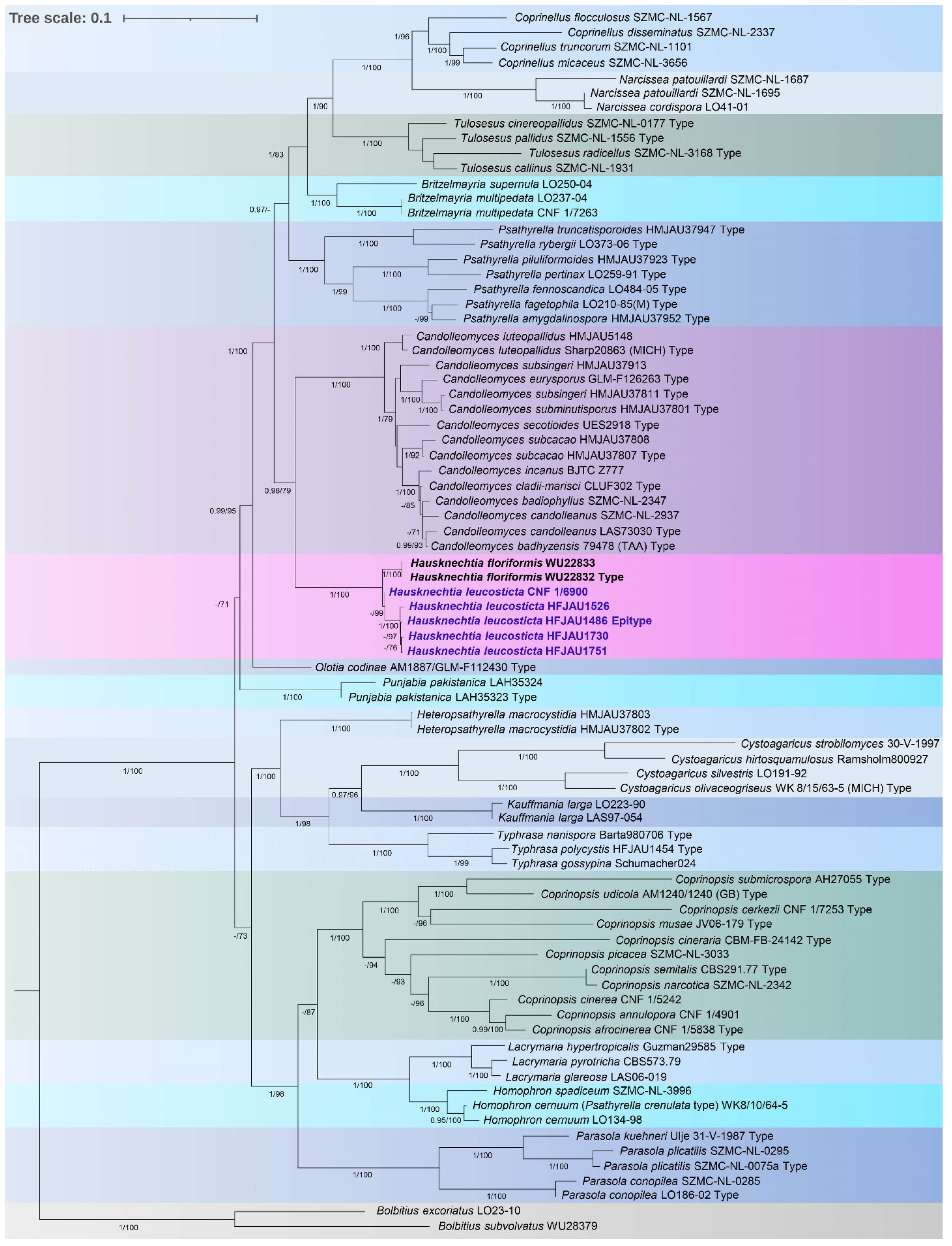
3.1. Molecular Phylogenetic Analysis
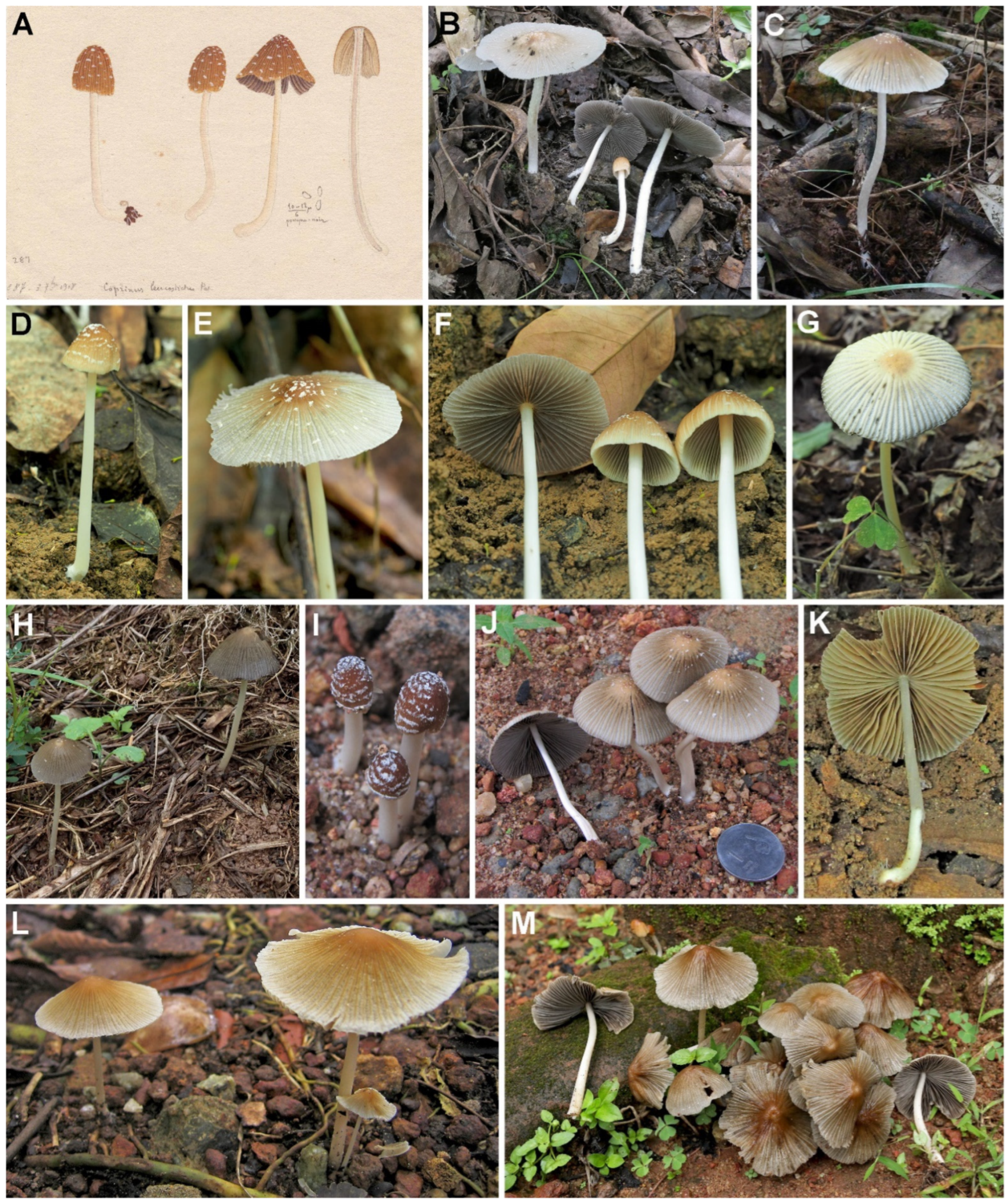
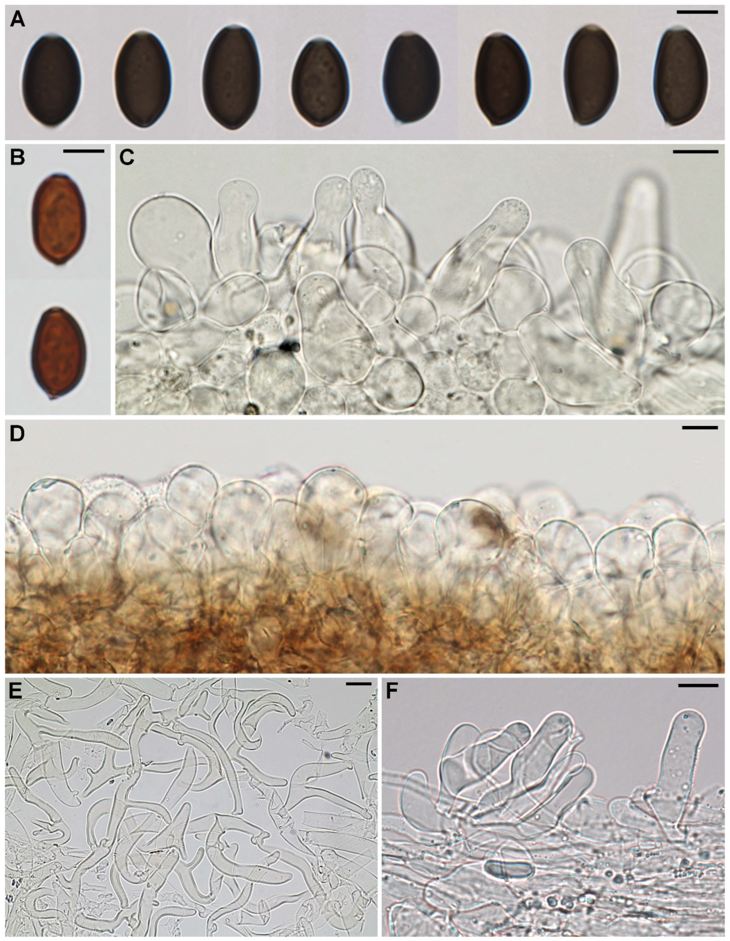
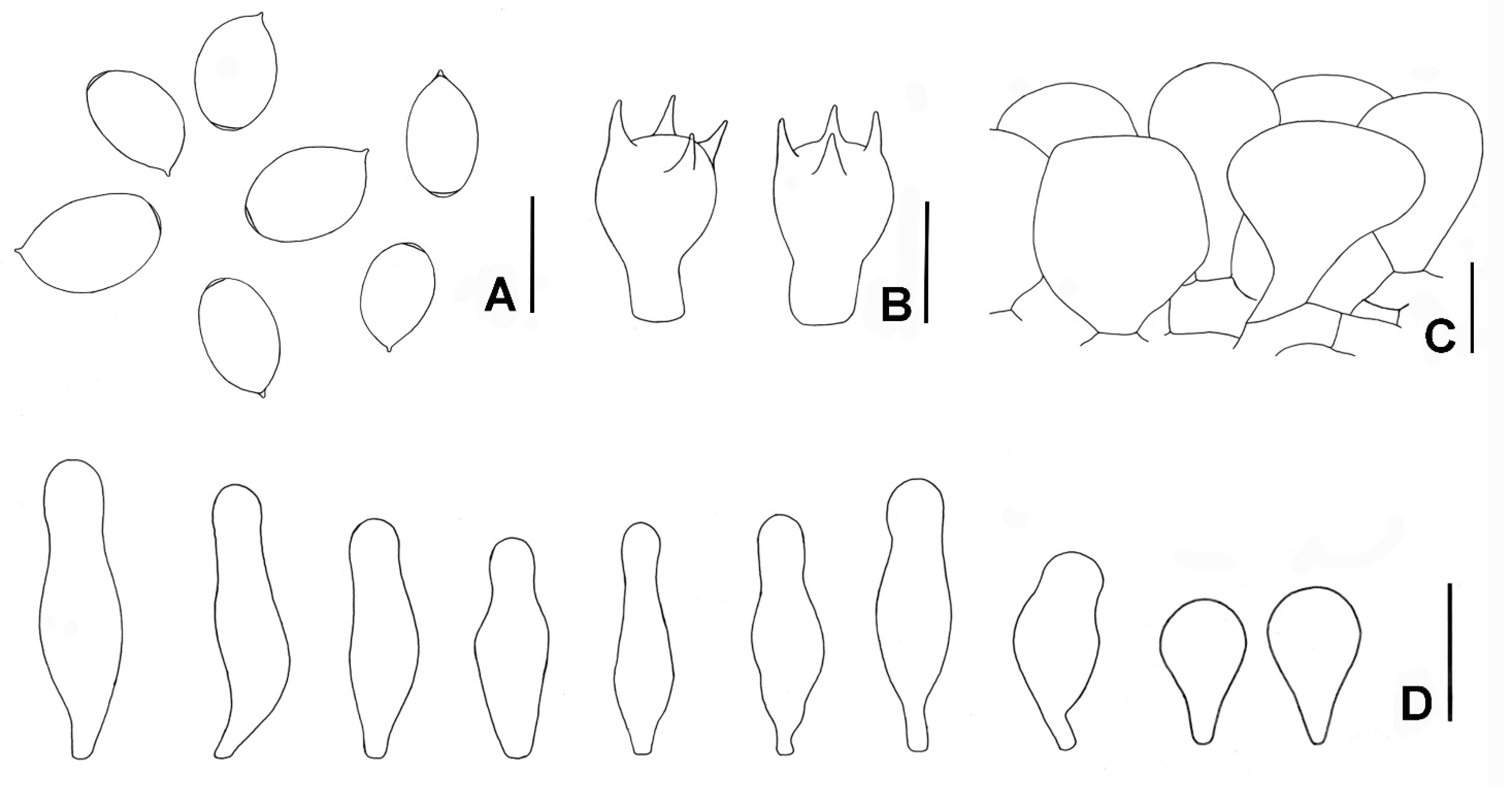
A total of 27 sequences from seven Hausknechtia specimens (five H. leucosticta and two H. floriformis) were newly generated in this study. Sequence identity analysis between H. floriformis WU22832 (holotype) and H. floriformis WU22833 (paratype) revealed 100% identity for each of the four gene regions. Based on the BLAST results, H. leucosticta (five collections) shared identity with H. floriformis (newly generated sequences) of 97.67–97.95% for ITS, 99.40–99.78% for LSU, 95.22–97.29% for tef-1α, and 95.83–96.69% for β-tubulin gene region. A total of 17 branches (BI-PP ≥ 0.95, ML-BP ≥ 70), representing 17 genera within the family Psathyrellaceae, are formed in the phylogenetic tree (Figure 1). The two well supported lineages (BI-PP = 0.98, ML-BP = 79), corresponding to the genera Hausknechtia and Candolleomyces, were recovered as a monophyletic group. At the species level, H. leucosticta formed a distinct lineage separated from H. floriformis with high support (BI-PP = 1, ML-BP = 100).

3.2. Taxonomy

Hausknechtia D. Wächt. & A. Melzer [1], emend. Tkalčec, J.-Q. Yan, C. Nie & C.K. Pradeep
Type species: Hausknechtia floriformis (Hauskn.) D. Wächt. & A. Melzer, Mycol. Progr. 19(11): 1234 (2020)
Transfer of Coprinus leucostictus in hitherto monotypic genus Hausknechtia required emendation of the concept of the genus, which is given below.
Emended description: Basidiomata thin-fleshed and fragile. Pileus dry, plicate-sulcate, later deeply, strongly radially splitted and ragged or not, with veil (at least when young). Lamellae adnexed to adnate, deliquescent (easily collapsing) when old or not. Stipe slender. Basidiospores smooth, ellipsoid, ovoid or cylindrical (in frontal view), yellowish-hyaline or brown in maturity, thin- to moderately thick-walled; germ-pore present or absent. Hymenophysalides (pseudoparaphyses) present or not. Cheilocystidia absent or present. Pleurocystidia absent. Pileipellis a hymeniderm. Veil hyphae septate, occasionally to strongly branched, diverticulate or not. Clamp-connections present.
Hausknechtia leucosticta (Pat.) Tkalčec, J.-Q. Yan, C. Nie & C.K. Pradeep, comb. nov. (Figure 2, Figure 3 and Figure 4)
MycoBank MB 844448
Basionym: Coprinus leucostictus Pat., Bull. Soc. Mycol. Fr. 33: 61 (1917).
Holotype: Vietnam, Hanoi, on soil, 1908, leg. V. Demange (287), FH 4457 (barcode 00258859).
Epitype (designated here): China. Anhui Province: Chizhou Guliujiang Nature Reserve, leg. Jun-Qing Yan, on soil, 8 June 2020, HFJAU1486, MBT 10007586; CNF 1/8787 isoepitype.
Description: Pileus (10–)15–60 mm broad, ellipsoid to paraboloid when young, later ± convex, conical, obtusely conical, broadly conical, plano-conical, plano-convex or applanate to plano-concave with a broad obtuse umbo; margin mostly straight, sometimes moderately deflexed or reflexed (when fully mature); surface dry, distinctly to strongly radially plicate-sulcate up to 85% of the radius (except when young), central disc smooth; at first light to medium orangish- to orange-brown or ± dark reddish-brown, uniformly coloured or paler towards the margin, later light to medium orangish-brown, orange-brown or brown in the central part and on the ribs, gradually paler towards the margin, between the ribs and sometimes throughout the whole narrower or wider outer zone pale grey(ish) to pale brownish-greyish, non-deliquescent. Veil white, thin, felty, covering the whole pileus in primordial stage only, very soon splitting up in small, flat, ± concentrically arranged patches, becoming scattered over the pileus at maturity and can fall off during the rain or on handling. Lamellae adnexed to adnate, moderately crowded to crowded, L = ca. 30, l = 1–7, up to 6 mm wide, white at first, becoming light yellowish-brown, greyish-brown, then brownish-grey, grey or dark grey-brown, with whitish, entire edge, non-deliquescent. Stipe 23–132 × 1.4–5 mm, central, ± cylindric with slightly broadened base (up to 6 mm) or gradually thickening downwards, white to cream, finely pruinose in the upper part, below smooth or mottled with small white fibrils, often white strigose at the very base, dry, hollow. Context thin, fragile, whitish, greyish or orangish-brownish. Odour mild, not characteristic. Spore print black.
Basidiospores [540/7/6] (8.8–)9.2–10.8–13.0(–13.7) × (4.9–)5.2–6.2–7.3(–7.7) µm, averages of different collections 10.5–11.2 × 5.8–6.5 μm, Q = (1.46–)1.53–1.74–2.00(–2.23), Q av. = 1.65–1.81, ellipsoid, narrowly ellipsoid or ovoid in frontal view, ellipsoid, narrowly ellipsoid or subamygdaliform in side view, rarely ovoid, not or only insignificantly flattened, smooth, moderately thick-walled (up to 0.8(–1) µm), with distinct and central germ-pore (0.8–1.5 µm wide inner diameter, 1.4–2.4 µm wide outer diameter), rusty brown to dark red brown in H2O and NH4OH, medium to dark brown in KOH (become more greyish over time), non-amyloid and non-dextrinoid. Basidia (13–)16–25(–32) × (9.5–)10–13 μm, clavate to broadly clavate, 4-spored, occasionally 2- or 3-spored, thin-walled, hyaline, mostly with refractive guttules, surrounded by 3–6 hymenophysalides (pseudoparaphyses). Hymenophysalides 10–20 × 6–20 μm, subglobose, broadly clavate, ellipsoid, broadly cylindrical or cuboid, thin-walled, hyaline; fully developed only in mature basidiomata. Lamellar edge sterile with rather crowded cheilocystidia. Cheilocystidia 12–50 × 8–30(–40) μm, develop to the full maturity of the lamellae; versiform, predominantly ± utriform (with ± broadened apex or not), (broadly) clavate, subglobose, spheropedunculate, obovoid or ± ellipsoid; thin-walled, hyaline. Pleurocystidia absent. Hymenophoral trama regular to subregular, composed of 1.5–15(–20) μm broad, thin-walled, hyaline hyphae. Pileipellis a hymeniderm, composed of clavate, broadly clavate, ellipsoid, spheropedunculate or subglobose elements, 18–70 × 11–45 μm, thin- to moderately thick-walled (up to 1 µm), hyaline or with pale brown, intracellular pigment. Pileal trama composed of 3–25 μm broad, thin- to thick-walled (up to 1.2 µm), hyaline or brown pigmented hyphae; pigment partially encrusted, mostly present in upper part. Veil composed of 2–12 μm broad, septate, strongly branched and diverticulate (excrescences with rounded tip, 1–30 μm long), thin- to moderately thick-walled (up to 1 µm), hyaline to pale brown hyphae (pigment intracellular). Stipitipellis a cutis of repent hyphae, 1.5–10 μm broad, thin- to moderately thick-walled (up to 0.8 µm), hyaline. Stipe trama composed of parallel and longitudinal hyphae, often somewhat swollen and constricted at septae, 1.5–24 μm broad, thin- to moderately thick-walled (up to 0.8 µm), hyaline. Caulocystidia present in the upper part of the stipe, 20–105 × 6–25 µm, variously shaped: ± utriform, narrowly to broadly clavate, ellipsoid, subcylindric or irregular; thin- to thick-walled (up to 2.8 µm), hyaline. Clamp-connections present, abundant.
The macro- and micromorphological characters of the epitype (designated above) fit well with the above comprehensive description of the Indian and Chinese collections. The particular measures of the epitype are as follows: pileus 16–43 mm broad; stipe 25–53 × 1.4–3.2 mm (1.7–5 mm at the base); basidiospores (8.8–)9.3–10.8–12.5(–13.5) × (5.0–)5.2–6.0–7.0 µm, Q = 1.50–1.80–2.00(–2.14).
Habit, habitat, and phenology: Solitary, scattered or in groups on soil, soil litter or decaying wood, in evergreen subtropical or tropical forests; basidiomata occurred from April to October.
Distribution: Known so far from Northern Vietnam (Hanoi Province), southern India (southern part of Kerala State), southern and southeastern China (Anhui, Fujian, Hainan, Jiangxi, and Zhejiang provinces). Based on BLAST search of ITS sequences data against GenBank (0.44–1.10% difference to our H. leucosticta sequences), the species was also found in Thailand (MN483260) and Brazil (MK881745).
Specimens examined: Vietnam. Hanoi Province: leg. V. Demange, on soil, 1908, FH 4457 (barcode 00258859; holotype).
China. Anhui Province: Chizhou Guliujiang Nature Reserve, ca. 30.1008° N, 117.4975° E, leg. J.Q. Yan, on soil, 8 June 2020, HFJAU1486 (epitype, designated above), CNF 1/8787 (isoepitype); Fujian Province: Wuyishan Reserve, ca. 27.6181° N, 117.9939° E, leg. J.Q. Yan, 13 June 2020, HFJAU1526; Hainan Province: Parrot Ridge Nature Reserve, ca. 18.9136° N, 109.6881° E, leg. Y. Fan, 4 July 2020, HFJAU2349; 5 July 2020, HFJAU2350.
NW from Changfeng town, ca. 18.8478° N, 110.2550° E, leg. Y. Fan, on soil, 1 May 2021, HFJAU3631; Jianxi Province: Jiu Lian Shan National Nature Reserve, ca. 24.6772° N, 114.5775° E, leg. J.Q. Yan, 22 August 2020, HFJAU1730; HFJAU1751; Jianxi Agricultural University, ca. 50 m a.s.l., ca. 28.7614° N, 115.8325° E, leg. Z. Yi, on soil/litter, 18 May 2019, HFJAU1200; leg. J.Q. Yan, 26 June 2019, HFJAU1295; Zhejiang Province: Suichang County Ramishang village, ca. 28.6689° N, 119.0444° E, leg. J.Q. Yan & Y. Chen, on soil litter, 14 July 2020, HFJAU2076; vicinity of Lishui city, ca. 28.4933° N, 119.6917° E, leg. J.Q. Yan & Z. Liu, on soil, 3 August 2021, HFJAU2683; ca. 220 m a.s.l., ca. 28.4300° N, 120.0535° E, leg. Q. Na, B. Ke & Z. Zeng, on decaying wood, 5 August 2021, HFJAU2836; vicinity of Qingtian County Grid Rossi village, ca. 28.3797° N, 119.9733° E, leg. Q. Na, B. Ke & Z. Zeng, on soil, 5 August 2021, HFJAU2822.
India. Kerala State: Kollam district, Thenmala, 190 m a.s.l., 8.9632° N, 77.0651° E, leg. C.K. Pradeep, on soil, 16 September 2004, TBGT7860; Thiruvananthapuram district, Palode, JNTBGRI campus, ca. 120 m a.s.l., ca. 8.7244° N, 77.0248° E, leg. C.K. Pradeep, on soil, 14 May 2009, TBGT12452; 11 May 2010, TBGT13214; 24 May 2010, TBGT13261; 27 April 2011, TBGT13573; 4 May 2011, TBGT13589, CNF 1/6900; 5 May 2011, TBGT13592; 18 May 2011, TBGT 13595; 23 May 2011, TBGT13599; 30 May 2011, TBGT13607; 31 May 2011, TBGT13608; 13 June 2011, TBGT13646; 10 May 2013, TBGT14472; 3 October 2013, TBGT14823; 5 May 2014, TBGT14983; 11 July 2016 TBGT16390; 6 April 2022, TBGT18769.
Specimens of Hausknechtia floriformis examined: Vanuatu. Efate island, Blue Water beach, leg. I. Hausknecht, on sandy soil near Pandanus sp., 8 April 2003, WU 22832 (holotype); leg. A. Hausknecht, 10 April 2003, WU 22833 (paratype).

4. Discussion

Although Hausknechtia leucosticta (Coprinus leucostictus) is rediscovered after almost a century, the number of its collections and localities found within this study showed that it is not a rare species. For example, during the study of Chinese psathyrelloid species, H. leucosticta was frequently collected during field research in the eastern and southern regions of the country. An insufficient level of fungal diversity research in southern and southeastern Asia is the most likely reason that the species has not been found again for so long.
Based on only one collection, a scarce original description, and a redescription of the holotype in very poor condition, knowledge of the morphology of H. leucosticta (especially microcharacters) was limited. The present study revealed morphological characters of H. leucosticta in detail. It is a rather unique taxon. The combination of coprinoid basidiomata, a veil consisting exclusively of strongly branched and diverticulate hyphae (Alachuani-type), and a hymeniderm pileipellis is not known in other species. The morphological characters of H. leucosticta collections studied (including the holotype) were not very variable. The same is true for ITS and β-tubulin gene regions. Based on the BLAST results, the identities among our five ITS sequences are 99.35–100%, and among our β-tubulin sequences are 99.37–100%. The homogeneity among four Chinese sequences is even higher: identities of 99.84–100% for ITS and 99.79–100% for β-tubulin. However, the identities between the Indian tef-1α sequence on the one side and the Chinese tef-1α sequences on the other are 96.66–96.93%. The homogenity among Chinese tef-1α sequences is much higher: identities of 99.40–99.90%. Despite that, due to the high identity of ITS and β-tubulin gene regions, as well as the concordance of morphological characters, we consider that the Indian and Chinese collections belong to the same species characterized by somewhat greater tef-1α variability.
Phylogenetic analysis showed that H. leucosticta and H. floriformis are closely related species. However, their morphological characters do not indicate such close relationship. Although both possess a dry, plicate-sulcate pileus with veil (at least when young), hymeniderm pileipellis, and lack pleurocystidia, there are many morphological differences. Among others, H. floriformis differs from H. leucosticta by strongly radially splitted and ragged pileus at maturity, deliquescent (easily collapsing) lamellae when old, elongated ellipsoid to cylindrical and yellowish-hyaline basidiospores without germ-pore, the absence of cheilocystidia and hymenophysalides, and by only occasionally branched, non-diverticulate veil hyphae [3,5].
The genus Psathyrella was treated in a broader sense by Voto [41]. He disagreed with Wächter & Melzer [1] who transferred members of Psathyrella section Spintrigerae in the new genus Candolleomyces and Galerella floriformis in the new genus Hausknechtia. Accordingly, he proposed a new combination Psathyrella floriformis (Hauskn.) Voto [41]. However, our multigene phylogenetic analysis recovered the two well supported monophyletic lineages (Hausknechtia and Candolleomyces) distant from the genus Psathyrella s. str., in agreement with study by Wächter & Melzer [1].

Author Contributions

Conceptualization, J.-Q.Y. and Z.T.; methodology, J.-Q.Y., Z.T., C.N., A.M. and A.P.; formal analysis, J.-Q.Y., Z.T., C.N., S.-N.W. and A.P.; investigation, J.-Q.Y., Z.T., C.N., A.P. and C.K.P.; resources, J.-Q.Y., Q.N., H.Z., Y.H., H.D., Y.G., G.H. and C.K.P.; data curation, Z.T., C.N., A.P. and C.K.P.; writing—original draft preparation, Z.T., J.-Q.Y., C.N., A.M. and A.P.; writing—review and editing, J.-Q.Y., Z.T., C.N. and A.M.; visualization, Z.T., J.-Q.Y. and A.P.; supervision, J.-Q.Y., Z.T., C.N. and A.M.; project administration, J.-Q.Y. and Z.T.; funding acquisition, A.M. and C.N. All authors have read and agreed to the published version of the manuscript.

Funding

This work was supported in part by the National Natural Science Foundation of China (31960008), Jiangxi Provincial Natural Science Foundation (20202BABL213041), and by the Croatian Science Foundation under project grants HRZZ-IP-2018-01-1736 (to A.M., Z.T. and A.P.) and HRZZ-2018-09-7081 (to A.P.). The project was also supported by the Biodiversity Investigation, Observation and Assessment Programme (2019–2023) of the Ministry of Ecology and Environment of China (2110404); Central Public-Interest Scientific Institution Basal Research Fund (GYZX200203); and by the Project of Biological Resources Survey in Wuyishan National Park (HXQT2020120701).

Institutional Review Board Statement

Not applicable.

Informed Consent Statement

Not applicable.

Data Availability Statement

The data generated in this study are available on request from the corresponding authors. Sequences obtained in this study are deposited at the GenBank database of National Center for Biotechnology Information (NCBI).

Acknowledgments

We are very grateful to Danny Haelewaters (Ghent University, Belgium) for his generous help with phylogenetic analysis and to Margita Jadan for help with sequencing. Donald H. Pfister and Michaela Schmull from the Farlow Herbarium (FH) of Harvard University (Cambridge, MA, USA) are greatly acknowledged for providing us with the holotype material of Coprinus leucostictus for morphological analysis and for granting us permission to reproduce the original painting of the holotype basdiomata. We are very thankful to Irmgard Krisai-Greilhuber from Herbarium Universität Wien (WU) for loan of Hausknechtia floriformis type specimens.

Conflicts of Interest

The authors declare no conflict of interest.

References

  1. Wächter, D.; Melzer, A. Proposal for a subdivision of the family Psathyrellaceae based on a taxon-rich phylogenetic analysis with iterative multigene guide tree. Mycol. Prog. 2020, 19, 1151–1265. [Google Scholar] [CrossRef]
  2. Patouillard, N. Quelques champignons du Tonkin. Bull. Trimest. Soc. Mycol. Fr. 1917, 33, 50–63. [Google Scholar]
  3. Yang, Z.-L. Type studies on agarics described by N. Patouillard (and his co-authors) from Vietnam. Mycotaxon 2000, 75, 431–476. [Google Scholar]
  4. Redhead, S.A.; Vilgalys, R.; Moncalvo, J.M.; Johnson, J.; Hopple, J.S., Jr. Coprinus Pers. and the disposition of Coprinus species sensu lato. Taxon 2001, 50, 203–241. [Google Scholar] [CrossRef]
  5. Hausknecht, A.; Contu, M. The genus Galerella. A world-wide survey. Osterr. Z. Pilzkd. 2003, 12, 31–40. [Google Scholar]
  6. Tóth, A.; Hausknecht, A.; Krisai-Greilhuber, I.; Papp, T.; Vágvölgyi, C.; Nagy, L.G. Iteratively refined guide trees help improving alignment and phylogenetic inference in the mushroom family Bolbitiaceae. PLoS ONE 2013, 8, e56143. [Google Scholar] [CrossRef]
  7. Erb, B.; Matheis, W. Pilzmikroskopie, 1st ed.; Kosmos: Stuttgart, West Germany, 1983; pp. 1–166. [Google Scholar]
  8. White, T.J.; Bruns, T.; Lee, S.; Taylor, J. Amplification and direct sequencing of fungal ribosomal RNA genes for phylogenetics. In PCR Protocols: A Guide to Methods and Applications; Innis, M.A., Gelfand, D.H., Sninsky, J.J., White, T.J., Eds.; Academic Press: San Diego, CA, USA, 1990; pp. 315–322. [Google Scholar] [CrossRef]
  9. Gardes, M.; Bruns, T.D. ITS primers with enhanced specificity for basidiomycetes—application to the identification of mycorrhizae and rusts. Mol. Ecol. 1993, 2, 113–118. [Google Scholar] [CrossRef]
  10. Hopple, J.S., Jr.; Vilgalys, R. Phylogenetic relationships in the mushroom genus Coprinus and dark-spored allies based on sequence data from the nuclear gene coding for the large ribosomal subunit RNA: Divergent domains, outgroups, and monophyly. Mol. Phylogen. Evolut. 1999, 13, 1–19. [Google Scholar] [CrossRef]
  11. Matheny, P.B.; Wang, Z.; Binder, M.; Curtis, J.M.; Lim, Y.W.; Nilsson, R.H.; Hughes, K.W.; Hofstetter, V.; Ammirati, J.F.; Schoch, C.L.; et al. Contributions of rpb2 and tef1 to the phylogeny of mushrooms and allies (Basidiomycota, Fungi). Mol. Phylogen. Evol. 2007, 43, 430–451. [Google Scholar] [CrossRef]
  12. Nagy, L.G.; Walther, G.; Házi, J.; Vágvölgyi, C.; Papp, T. Understanding the evolutionary processes of fungal fruiting bodies: Correlated evolution and divergence times in the Psathyrellaceae. Syst. Biol. 2011, 60, 303–317. [Google Scholar] [CrossRef]
  13. Bau, T.; Yan, J.-Q. A new genus and four new species in the /Psathyrella s.l. clade from China. MycoKeys 2021, 80, 115–131. [Google Scholar] [CrossRef] [PubMed]
  14. Katoh, K.; Misawa, K.; Kuma, K.; Miyata, T. MAFFT: A novel method for rapid multiple sequence alignment based on fast Fourier transform. Nucleic Acids Res. 2002, 30, 3059–3066. [Google Scholar] [CrossRef] [PubMed]
  15. Katoh, K.; Standley, D.M. MAFFT multiple sequence alignment software version 7: Improvements in performance and usability. Mol. Biol. Evol. 2013, 30, 772–780. [Google Scholar] [CrossRef] [PubMed]
  16. Örstadius, L.; Larsson, E. Bolbitius excoriatus, flagnande guldskivling, funnen på spillning i Sverige. Sven. Mykol. Tidskr. 2013, 34, 2–6. [Google Scholar]
  17. Örstadius, L.; Ryberg, M.; Larsson, E. Molecular phylogenetics and taxonomy in Psathyrellaceae (Agaricales) with focus on psathyrelloid species: Introduction of three new genera and 18 new species. Mycol. Prog. 2015, 14, 25. [Google Scholar] [CrossRef]
  18. Mešić, A.; Šamec, D.; Jadan, M.; Bahun, V.; Tkalčec, Z. Integrated morphological with molecular identification and bioactive compounds of 23 Croatian wild mushrooms samples. Food Biosci. 2020, 37, 100720. [Google Scholar] [CrossRef]
  19. Sicoli, G.; Passalacqua, N.G.; De Giuseppe, A.B.; Palermo, A.M.; Pellegrino, G. A new species of Psathyrella (Psathyrellaceae, Agaricales) from Italy. MycoKeys 2019, 52, 89–102. [Google Scholar] [CrossRef]
  20. Büttner, E.; Karich, A.; Nghi, D.H.; Lange, M.; Liers, C.; Kellner, H.; Hofrichter, M.; Ullrich, R. Candolleomyces eurysporus—A new Psathyrellaceae (Agaricales) species from the tropical Cúc Phương National Park, Vietnam. Osterr. Z. Pilzkd. 2021, 28, 79–92. [Google Scholar]
  21. Zhou, H.; Cheng, G.Q.; Sun, X.M.; Cheng, R.Y.; Zhang, H.L.; Dong, Y.M.; Hou, C.L. Three new species of Candolleomyces (Agaricomycetes, Agaricales, Psathyrellaceae) from the Yanshan Mountains in China. MycoKeys 2022, 88, 109–121. [Google Scholar] [CrossRef]
  22. Yan, J.-Q.; Bau, T. The Northeast Chinese species of Psathyrella (Agaricales, Psathyrellaceae). MycoKeys 2018, 33, 85–102. [Google Scholar] [CrossRef]
  23. Moreno, G.; Heykoop, M.; Esqueda, M.; Olariaga, I. Another lineage of secotioid fungi is discovered: Psathyrella secotioides sp. nov. from Mexico. Mycol. Prog. 2015, 14, 34. [Google Scholar] [CrossRef]
  24. Bau, T.; Yan, J.-Q. Two new rare species of Candolleomyces with pale spores from China. MycoKeys 2021, 80, 149–161. [Google Scholar] [CrossRef]
  25. Nagy, L.G.; Házi, J.; Vágvölgyi, C.; Papp, T. Phylogeny and species delimitation in the genus Coprinellus with special emphasis on the haired species. Mycologia 2012, 104, 254–275. [Google Scholar] [CrossRef] [PubMed]
  26. Nagy, L.G.; Urban, A.; Örstadius, L.; Papp, T.; Larsson, E.; Vágvölgyi, C. The evolution of autodigestion in the mushroom family Psathyrellaceae (Agaricales) inferred from Maximum Likelihood and Bayesian methods. Mol. Phylogen. Evol. 2010, 57, 1037–1048. [Google Scholar] [CrossRef]
  27. Crous, P.W.; Wingfield, M.J.; Burgess, T.I.; Hardy, G.E.S.J.; Gené, J.; Guarro, J.; Baseia, I.G.; García, D.; Gusmão, L.F.P.; Souza-Motta, C.M.; et al. Fungal Planet description sheets: 716–784. Persoonia 2018, 40, 239–392. [Google Scholar] [CrossRef] [PubMed]
  28. Tibpromma, S.; Hyde, K.D.; Jeewon, R.; Maharachchikumbura, S.S.N.; Liu, J.K.; Bhat, D.J.; Jones, E.B.G.; McKenzie, E.H.C.; Camporesi, E.; Bulgakov, T.S.; et al. Fungal diversity notes 491–602: Taxonomic and phylogenetic contributions to fungal taxa. Fungal Divers. 2017, 83, 1–261. [Google Scholar] [CrossRef]
  29. Nagy, L.G.; Kocsubé, S.; Papp, T.; Vágvölgyi, C. Phylogeny and character evolution of the coprinoid mushroom genus Parasola as inferred from LSU and ITS nrDNA sequence data. Persoonia 2009, 22, 28–37. [Google Scholar] [CrossRef]
  30. Vellinga, E.C. Genera in the family Agaricaceae: Evidence from nrITS and nrLSU sequences. Mycol. Res. 2004, 108, 354–377. [Google Scholar] [CrossRef]
  31. Larsson, E.; Örstadius, L. Fourteen coprophilous species of Psathyrella identified in the Nordic countries using morphology and nuclear rDNA sequence data. Mycol. Res. 2008, 112, 1165–1185. [Google Scholar] [CrossRef]
  32. Deschuyteneer, D.; Melzer, A.; Pérez-De-Gregorio, M.À. Psathyrella codinae, a new species from Spain. Bull. Assoc. Mycol. Francoph. Belg. 2018, 11, 4–8. [Google Scholar]
  33. Szarkándi, J.G.; Schmidt-Stohn, G.; Dima, B.; Hussain, S.; Kocsubé, S.; Papp, T.; Vágvölgyi, C.; Nagy, L.G. The genus Parasola: Phylogeny and the description of three new species. Mycologia 2017, 109, 620–629. [Google Scholar] [CrossRef] [PubMed]
  34. Hussain, S.; Usman, M.; Afshan, N.-ul-S.; Ahmad, H.; Khan, J.; Khalid, A.N. The genus Coprinellus (Basidiomycota; Agaricales) in Pakistan with the description of four new species. MycoKeys 2018, 39, 111–126. [Google Scholar] [CrossRef] [PubMed]
  35. Házi, J.; Nagy, L.G.; Vágvölgyi, C.; Papp, T. Coprinellus radicellus, a new species with northern distribution. Mycol. Prog. 2010, 10, 363–371. [Google Scholar] [CrossRef]
  36. Wang, S.-N.; Hu, Y.-P.; Chen, J.-L.; Qi, L.-L.; Zeng, H.; Ding, H.; Huo, G.-H.; Zhang, L.-P.; Chen, F.-S.; Yan, J.-Q. First record of the rare genus Typhrasa (Psathyrellaceae, Agaricales) from China with description of two new species. MycoKeys 2021, 79, 119–128. [Google Scholar] [CrossRef]
  37. Nguyen, L.-T.; Schmidt, H.A.; von Haeseler, A.; Minh, B.Q. IQ-TREE: A fast and effective stochastic algorithm for estimating maximum-likelihood phylogenies. Mol. Biol. Evol. 2015, 32, 268–274. [Google Scholar] [CrossRef] [PubMed]
  38. Trifinopoulos, J.; Nguyen, L.T.; von Haeseler, A.; Minh, B.Q. W-IQ-TREE: A fast online phylogenetic tool for maximum likelihood analysis. Nucleic Acids Res. 2016, 44, W232–W235. [Google Scholar] [CrossRef]
  39. Huelsenbeck, J.P.; Ronquist, F. MrBayes: Bayesian inference of phylogeny. Bioinformatics 2001, 17, 754–755. [Google Scholar] [CrossRef]
  40. Letunic, I.; Bork, P. Interactive Tree Of Life (iTOL) v5: An online tool for phylogenetic tree display and annotation. Nucleic Acids Res. 2021, 49, W293–W296. [Google Scholar] [CrossRef] [PubMed]
  41. Voto, P. Novelties in the Family Psathyrellaceae. Part VI. Mycol. Obs. 2022, 2, 9. [Google Scholar]
Figure 1. Concatenated phylogenetic tree of Psathyrellaceae species based on Bayesian interference (BI) and Maximum Likelihood (ML) analyses of four-gene (ITS, LSU, tef-1α, β-tub) sequence alignment. BI and ML analyses were performed using MrBayes v. 3.2.6 and IQ-TREE 1.6.12, respectively. Significant branch support values, Bayesian posterior probability (BI-PP ≥ 0.95) and ultrafast bootstrap support (ML-BP ≥ 70%), are presented at the nodes. Species with newly generated sequences are marked in bold and recombined species is marked in colour and bold.
Diversity 14 00699 g001
Figure 2. Hausknechtia leucosticta; basidiomata. (A) Original painting of the holotype by V. Demange (1908), from the Farlow Reference Library of Cryptogamic Botany at Harvard University. (B) HFJAU1486 (epitype). (C) HFJAU1200. (DF) HFJAU3631. (G) HFJAU2076. (H) HFJAU2822. (IK) TBGT13589 & CNF 1/6900. (L) TBGT14823. (M) TBGT16390.
Diversity 14 00699 g002
Figure 3. Hausknechtia leucosticta, TBGT13589 & CNF 1/6900. (A) Basidiospores in KOH. (B) Basidiospores in H2O. (C) Cheilocystidia. (D) Pileipellis. (E) Veil on the pileus. (F) Caulocystidia. Bars: (A,B) = 5 µm, (CF) = 10 μm.
Diversity 14 00699 g003
Figure 4. Hausknechtia leucosticta. (A) Basidiospores. (B) Basidia. (C) Pileipellis. (D) Cheilocystidia. Bars: (AC) = 10 µm, (D) = 20 μm.
Diversity 14 00699 g004
Table 1. Species included in this study, voucher numbers, countries of origin, and GenBank accession numbers. Newly generated sequences are in bold.
TaxonVoucherCountryITSLSUtef-1αβ-TubulinRefs.
Bolbitius excoriatusLO23-10SwedenKC456419KC456419KJ732834-[1,16]
Bolbitius subvolvatusWU28379HungaryJX968248JX968365JX968454-[6]
Britzelmayria multipedataLO237-04SwedenKC992888KC992888KJ732777KJ664867.1[17]
Britzelmayria multipedataCNF 1/7263CroatiaMK169241---[18]
Britzelmayria supernulaLO250-04SwedenKC992867KC992867KJ732763KJ664849.1[17]
Candolleomyces badhyzensis (type)79478 (TAA) TurkmenistanKC992883KC992883--[17]
Candolleomyces badiophyllusSZMC-NL-2347-FN430699FM876268FN396261FM897252[12]
Candolleomyces candolleanus (type)LAS73030 HungaryKM030175KM030175--[17]
Candolleomyces candolleanusSZMC-NL-2937HungaryFN396114FN396165FN396220FN396307[12]
Candolleomyces cladii-marisci (type)CLUF302 ItalyMK080112---[19]
Candolleomyces eurysporus (type)GLM-F126263 GermanyMT651560MT651560--[20]
Candolleomyces incanusBJTC Z777 China, BeijingON042759ON042766ON098508ON098513[21]
Candolleomyces luteopallidus (type)Sharp20863 (MICH) USAKC992885KC992885KJ732775KJ664865[17]
Candolleomyces luteopallidusHMJAU5148China, JilinMG734736MW301084MW314056MW314073[22]
Candolleomyces secotioides (type)UES2918 MexicoKR003281KR003282-KR003283[23]
Candolleomyces subcacao (type)HMJAU37807 China, HenanNR_173317NG_079683MW314081MW314063[24]
Candolleomyces subcacaoHMJAU37808China, HenanMW301065MW301093MW314082MW314064[24]
Candolleomyces subminutisporusHMJAU37916China, HenanMW301067MW301095MW314084MW314066[24]
Candolleomyces subsingeri (type)HMJAU37811 China, JilinMG734715MW301097MW314085MW314067[22,24]
Candolleomyces subsingeriHMJAU37913China, JilinMG734725MW301098MW314086MW314067[22,24]
Coprinellus disseminatusSZMC-NL-2337HungaryFM878017FM876274-FN396282[12]
Coprinellus flocculosusSZMC-NL-1567-FN430683JN159593-FN396345[12]
Coprinellus micaceusSZMC-NL-3656-JN159567JN159588-JN159644[25]
Coprinellus truncorumSZMC-NL-1101SwedenJN159562FM876262FM897225JN159629[12,26]
Coprinopsis afrocinerea (type)CNF 1/5838NigeriaMG662162MG662158--[27]
Coprinopsis annuloporaCNF 1/4901CroatiaMG662170---[27]
Coprinopsis cerkezii (type)CNF 1/7253 CroatiaNR_173361NG_068782--[28]
Coprinopsis cineraria (type)CBM-FB-24142 JapanKC992962---[17]
Coprinopsis cinereaCNF 1/5242CroatiaMG662167---[27]
Coprinopsis musae (type)JV06-179 DenmarkNR_148070KC992965-KJ664920[17]
Coprinopsis narcoticaSZMC-NL-2342HungaryFM163180FM160729FN396244FN396290[29]
Coprinopsis picaceaSZMC-NL-3033-FN396119FN396168FN396223FN396312[12]
Coprinopsis semitalis (type)CBS291.77 -GQ249278GQ249287GQ249270GQ249262[12]
Coprinopsis submicrospora (type)AH27055 SpainKC992959KC992959-KJ664918[17]
Coprinopsis udicola (type)AM1240GermanyNR_148071KC992967KJ732831KJ664922[17]
Cystoagaricus hirtosquamulosusRamsholm800927FinlandKC992945KC992945--[17]
Cystoagaricus olivaceogriseus (type)WK 8/15/63-5USAKC992948KC992948--[17]
Cystoagaricus silvestrisLO191-92SwedenKC992949KC992949--[17]
Cystoagaricus strobilomyces30-V-1997JapanAY176347AY176348--[30]
Hausknechtia floriformis (type)WU22832VanuatuON745613ON745616ON746007ON746008This study
Hausknechtia floriformisWU22833VanuatuON745619ON745615ON746009ON746010This study
Hausknechtia leucostictaCNF 1/6900IndiaON745618ON745617ON746005ON746006This study
Hausknechtia leucosticta (epitype)HFJAU1486China, AnhuiOL435561OL435565OL439896ON677539This study
Hausknechtia leucostictaHFJAU1526China, FujianOL4355630L435566OL439897ON677541This study
Hausknechtia leucostictaHFJAU1730China, JiangxiOL435562-OL439898ON677540This study
Hausknechtia leucostictaHFJAU1751China, JiangxiOL435564OL435567OL439899ON677542This study
Heteropsathyrella macrocystidia (type)HMJAU37802China, FujianMW405102MW413359MW411004-[24]
Heteropsathyrella macrocystidiaHMJAU37803China, FujianMW405101MW413358MW411003-[24]
Homophron cernuumLO134-98SwedenDQ389726DQ389726KJ732828KJ664915[17,31]
Homophron cernuum (Psathyrella crenulata type)WK8/10/64-5USAKC992957--.[17]
Homophron spadiceumSZMC-NL-3996HungaryFN396132FN396180FN396231FN396333.1[12]
Kauffmania largaLO223-90SwedenDQ389694DQ389694KJ732824KJ664912.1[17]
Kauffmania largaLAS97-054SwedenDQ389695DQ389695- [17]
Lacrymaria glareosaLAS06-019SwedenKC992954KC992954KJ732827KJ664914[17]
Lacrymaria hypertropicalis (type)Guzman29585 MexicoKC992958KC992958-KJ664916[17]
Lacrymaria pyrotrichaCBS573.79-GQ249280GQ249289GQ249272GQ249264[12]
Narcissea cordisporaLO41-01SwedenDQ389723DQ389723-KJ664910[17,31]
Narcissea patouillardiSZMC-NL-1687HungaryFM878009FM876265FM897238FN396257[26]
Narcissea patouillardiSZMC-NL-1695HungaryFN430685FN396196-FN396258[26]
Olotia codinae (type)AM1887/GLM-F112430 SpainMG696611MG674714--[32]
Parasola conopilea (type)LO186-02 SwedenDQ389725DQ389725--[31]
Parasola conopileaSZMC-NL-0285HungaryFM163225FM160684FM897237FN396247[29]
Parasola kuehneri (type)Ulje 31-V-1987NetherlandsKY928608KY928633--[33]
Parasola plicatilisSZMC-NL-0295HungaryFM163216FM160693FM897242FN396253[29]
Parasola plicatilis (type)SZMC-NL-0075a HungaryFM163213FM160696--[29]
Psathyrella amygdalinospora (type)HMJAU37952 China, SichuanNR_173320MW413361MW410999MW410991[24]
Psathyrella fagetophila (type)LO210-85 (M) SwedenNR_167955KC992902-KJ664879[17]
Psathyrella fennoscandica (type)LO484-05 SwedenKC992903KC992903KJ732790KJ664881[17]
Psathyrella pertinax (type)LO259-91 SwedenDQ389701DQ389701KJ732809-[17]
Psathyrella piluliformoides (type)HMJAU37923China, ZhejiangMW405106MW413362MW411002-[24]
Psathyrella rybergii (type)LÖ373-06SwedenKC992893KC992893KJ732781KJ664872[17]
Psathyrella truncatisporoides (type)HMJAU37947 China, ZhejiangMW405107MW413363MW410990MW410998[24]
Punjabia pakistanica (type)LAH35323PakistanMH366736---[34]
Punjabia pakistanicaLAH35324PakistanMH366737---[34]
Tulosesus callinusSZMC-NL-1931HungaryFN396105FN396158FN396213FN396299[12]
Tulosesus cinereopallidus (type)SZMC-NL-0177 HungaryHQ847001HQ847090-HQ847149[25]
Tulosesus pallidus (type)SZMC-NL-1556 HungaryHQ846989HQ847076-HQ847155[25]
Tulosesus radicellus (type)SZMC-NL-3168 SwedenGU227719HQ847077-GU227737[25,35]
Typhrasa gossypinaSchumacher024GermanyKC992946KC992946KJ732825-[17]
Typhrasa nanispora (type)Barta980706AustriaKC992947KC992947--[17]
Typhrasa polycystis (type)HFJAU1454China, ZhejiangMW466538MW466544MW475280-[36]
Publisher’s Note: MDPI stays neutral with regard to jurisdictional claims in published maps and institutional affiliations.

