Article

Stripes Matter: Integrative Systematics of Coryphellina rubrolineata Species Complex (Gastropoda: Nudibranchia) from Vietnam

1
Invertebrate Zoology Department, Lomonosov Moscow State University, Leninskie gori 1-12, 119234 Moscow, Russia
2
A.N. Severtsov Institute of Ecology and Evolution, Leninskiy prosp. 33, 119071 Moscow, Russia
3
N.A. Pertsov White Sea Biological Station, Faculty of Biology, Lomonosov Moscow State University, Leninskie gori 1-12, 119234 Moscow, Russia
*
Author to whom correspondence should be addressed.
Diversity 2022, 14(4), 294; https://doi.org/10.3390/d14040294
Submission received: 19 March 2022 / Revised: 8 April 2022 / Accepted: 11 April 2022 / Published: 13 April 2022
(This article belongs to the Special Issue Systematics and Evolution of Gastropods)

Abstract

:
Coryphellina rubrolineata (Gastropoda: Nudibranchia: Flabellinidae) was believed to be a widespread tropical species demonstrating high diversity in external and internal morphological traits. In this paper, we perform an integrative analysis of the C. rubrolineata species complex based on samples collected in Vietnam waters, combined with available data from other localities of the Indo-West Pacific. The methods of the study include morphological analysis of external and internal traits using light and scanning electron microscopy and the molecular analysis of four markers (COI, 16S, H3, and 28S). The phylogenetic hypothesis was performed using Bayesian and maximum likelihood approaches, and the species delimitation analyses included ASAP, GMYC, and bPTP. Our results support the validity of the genus Coryphellina as a distinct taxon and confirm that Coryphellina rubrolineata is restricted to the type locality and adjacent waters, while in the Indo-West Pacific, it represents a complex of pseudocryptic species. Based on our integrative analysis, we describe four new species: Coryphellina pseudolotos sp. nov., Coryphellina pannae sp. nov., Coryphellina flamma sp. nov., and Coryphellina aurora sp. nov. For the first time, Coryphellina lotos is reported in Vietnam waters. All five species differ in combination of coloration and other external traits and show minor differences in internal morphology.

1. Introduction

The advancement of DNA sequencing methods in taxonomical studies dramatically increased discovery rates of cryptic or pseudocryptic species complexes [1,2,3]. In morphology-based taxonomical studies, the presence of morphologically and ecologically diverse species has been widely accepted [4]. However, molecular systematics often demonstrates breaks in genetic diversity either across distant populations of putative widespread species [5,6,7,8,9,10] or within a single sympatric “population” demonstrating different ecological traits [11,12,13,14]. The existence of geographical barriers and allopatric speciation was believed to be the dominant driver for the diversification of widely distributed species; however, most recent studies highlighted the importance of ecological speciation for species living in sympatry and occupying different ecological niches [15,16]. In some cases, the cryptic or pseudocryptic diversity may be explained by a combination of different speciation scenarios [9].
The nudibranch Coryphellina rubrolineata O’Donoghue, 1929 (Gastropoda: Nudibranchia: Flabellinidae) was believed to have a very wide distributional range, being described from the Suez region in the Red Sea [17] and further reported from the Mediterranean Sea (non-native population, see Gat [18], Yokes, Rudman [19]), the Red Sea [20,21], Indian Ocean [22,23,24,25,26], the Indo-West Pacific [27,28,29,30,31], subtropical waters of Korea [32], Japan [33], and Australia [34,35,36]. The main identification trait for this species was the presence of three red (commonly from pink to red-violet) lines continued along the dorsum and lateral sides of molluscs. However, in most regions, this species demonstrated several chromatic variations of body color (from white to purple) and cerata color (from translucent to orange, red, or violet) and different densities of dorsolateral red lines [37]. In 2017, a new species, Coryphellina lotos Korshunova et al., 2017, was described from the Pacific coast of Japan based on the integrative morphological and molecular analysis [38]. This species also possesses three red lines on dorsal and lateral sides, but those lines are discontinuous, suggesting that Coryphellina rubrolineata represents a species complex, and its chromatic variations in other regions require an additional study [38]. This view was confirmed in a subsequent work on the Coryphellina rubrolineata specimens collected from the type locality [39] (under the name Flabellina rubrolineata). It showed a clear separation of C. rubrolineata specimens collected in the Red Sea and the Arabian Sea from other specimens collected from the Indo-West Pacific and Australia. The authors highlighted that these results support the suggestion by Gosliner et al. [40] that “true” C. rubrolineata is restricted to the Red and Arabian seas and also non-natively occurs in the Mediterranean Sea, while in other regions it represents a complex of cryptic or pseudocryptic species [39].
The genus Coryphellina was recently re-established for flabellinids with papillate rhinophores and bilobed seminal receptaculum, following the integrative revision of the traditional family Flabellinidae [38]. This work showed polyphyly of traditionally defined genus Flabellina and suggested the major reclassification of this group with distinct families Apataidae, Samlidae, Unidentiidae, Flabellinopsidae, Paracoryphellidae, Coryphellidae, and Flabellinidae s.str and 27 genera. Although the validity of the families Apataidae, Samlidae, and Unidentiidae was widely accepted [40], the taxonomical changes within the rest of flabellinid diversity were taken with caution by some researchers [41,42,43,44]. Several subsequent works highlighted the suggested taxonomical scheme is excessively splitting and suggested synonymization of several flabellinid taxa (Calmella with Flabellina, Furfaro et al. [45]; all coryphellid genera with Coryphella, see Ekimova [46]; Ekimova et al. [44]). The identity of the genus Coryphellina also remains questionable, as few species have been studied to date, and it is not clear whether suggested synapomorphic traits are characteristic for all putative members of this genus.
Coryphellina rubrolineata was registered in Vietnam waters in 2012 (Martynov, Korshunova, 2012). However, consequent studies indicated that it was genetically distinct from the C. rubrolineata from the type locality, and differences in coloration suggested the presence of pseudocryptic species in this area [39]. Contemporary data on cladobranch diversity in Vietnam’s coastal waters remain incomplete and require de-dicated integrative studies [29,43]. From 2016 to 2021, 28 specimens of the genus Coryphellina were collected in different localities of Vietnam, 8 of them were identified as Coryphellina exoptata (Gosliner, Willan, 1991), and the rest belonged to the Coryphellina rubrolineata species complex. Some specimens of the latter species demonstrate similar external morphological traits (discontinuous dorsolateral lines) to C. lotos from Japan, and all specimens show high variation in coloration. The main goal of this study is to observe the potential cryptic diversity within the Coryphellina rubrolineata species complex in Vietnam based on integrative morphological and molecular analysis.

2. Material and Methods

2.1. Collection Data and Community Descriptions

A total of 28 specimens belonging to the genus Coryphellina were collected in 2016–2021 in various localities in the Southern (Phu Quoc, Tho Chu islands) and Central (Nha Trang Bay) Vietnam (Figure 1) during expeditions of the Vietnamese-Russian Tropical Centre. Most specimens were found in Nha Trang Bay, around Hon Tre, Hon Mot, and Hon Nok Islands. All specimens were collected during SCUBA diving at depths of 5–25 m. The specimens were photographed and then fixed in either 96% ethanol or in 4% formaldehyde, in the latter case a piece of tissue was cut off and fixed in 96% ethanol for molecular analysis. Voucher specimens and DNA samples are stored in the collections of the Invertebrate Zoology department, Lomonosov Moscow State University (IZ). Type material is deposited in the collections of the Zoological Museum, Russian Academy of Science (ZIN). The collection of ZIN has policies to ensure compliance with laws governing the collection and sampling of wildlife from the country of origin and confirms that samples were legally imported to the museum repository where they are listed. Detailed sampling information and voucher numbers for each specimen are given in Table S1.

2.2. DNA Extraction, Amplification, and Sequencing

Total genomic DNA was extracted from tissue samples preserved in 96% EtOH (Table S1) following the invertebrate protocol of the Canadian Center for DNA Barcoding [47]. Extracted DNA was used as a template for amplification of partial mitochondrial cytochrome c oxidase subunit I (COI) and 16S rRNA, and nuclear histone H3 and 28S rRNA. Reaction conditions and primers are shown in Table 1. Polymerase chain reactions were conducted with the “HS Taq” kit (Eurogen Lab, Moscow, Russia), following the manufacturer’s protocol. For sequencing, 1 to 2 μL of amplicons were purified by ammonium acetate precipitation [48] and used for the sequencing reactions with the BigDye Terminator v3.1 sequencing kit by Applied Biosystems (Waltham, MA, USA). The reactions were analyzed using an ABI 3500 Genetic Analyser (Applied Biosystems) at N.K. Koltzov Institute of Developmental Biology RAS. All novel sequences were submitted to NCBI GenBank (Table S2).

2.3. Data Processing and Phylogenetic Reconstruction

All raw reads for each gene were assembled and checked for ambiguities and low-quality data in Geneious R10 [54]. All edited sequences were checked for contamination using the BLAST-n algorithm run over the GenBank nr/nt database [55]. For phylogenetic reconstruction, molecular data from previous studies were added to the analyses [38,39]. All available sequences were aligned with the MUSCLE [56] algorithm in MEGA 7 [57]. For verification of reading frames and the presence of stop-codons, the COI and H3 sequences were translated into amino acids. Saturation was checked by plotting for all spe-cimens, including the outgroup of the total number of pairwise differences (transitions and transversions), against uncorrected p-distances. In the case of protein-coding markers, saturation was further examined separately for the first, second, and third codon positions. Sequences were concatenated by a simple biopython script following [58]. Phylogenetic reconstructions were conducted for the concatenated multi-gene partitioned data sets. The best-fit nucleotide evolution model for MrBayes phylogeny reconstruction method was selected in ModelTest-NG v0.1.7 [59,60] as follows: HKY+G+I for mitochondrial markers, K2+G+I for nuclear markers. Multi-gene analyses were performed by applying evolutionary models separately to partitions representing single markers. The Bayesian phylogenetic analyses and estimation of posterior probabilities were performed in MrBayes 3.2 [61]. The analysis was initiated with a random starting tree and ran for 107 generations. Maximum likelihood phylogeny inference was performed in the HPC-PTHREADS-AVX option of RaxML HPC-PTHREADS 8.2.12 [62] with 1000 pseudoreplicates under the GTRCAT model of nucleotide evolution. Bootstrap values were placed on the best tree found with SumTrees 3.3.1 from DendroPy Phylogenetic Computing Library 3.12.0. Final phylogenetic tree images were rendered in FigTree 1.4.0 and further modified in Adobe Illustrator CS 2015.

2.4. Species Delimitation

The COI alignment was used for computational species delimitations methods. P-distances were calculated using MEGA7 software [57]. To confirm the status of the clades recovered in our analysis as putative species, we used the Assemble Species by Automatic Partitioning (ASAP) method [63] to detect breaks in the distribution of intra- and interspecific distances without any prior species hypothesis, referred to as the “barcode gap” [64]. The ASAP analysis was run on the online version of the program (https://bioinfo.mnhn.fr/abi/public/asap/asapweb.html) (accessed on 1 March 2022) with the Jukes-Cantor (JC69) model. It was complemented by Poisson tree processes (PTP) [65]. The test was run using the bPTP Server http://species.h-its.org/ptp/ (accessed on 1 March 2022) with 500,000 generations and with other settings set as default and with a COI-based maximum likelihood tree as an input. Additionally, we performed a GMYC test [66] and implemented it by the work of [67]. The COI-based ultrametric tree was calculated using BEAST 2.6.4 [68] with 107 generations and then analyzed in the R environment (package splits), following instructions by the work of [67]. Uncorrected p-distances in COI, H3, and 28S alignments were calculated in MEGA 7 [57].

2.5. Morphological Studies

All collected specimens were examined under a stereomicroscope. The internal morphology of 13 specimens was also studied, with closer attention to the digestive and reproductive systems. The buccal mass of each specimen was extracted and incubated overnight in proteinase K solution at 60 °C for dissolving the connective and muscle tissues, and then additionally soaked for 5 min in sodium hypochlorite. The radula and the jaws were rinsed in distilled water, air-dried, mounted on an aluminum stub, and sputter-coated with gold for visualization under a JEOL JSM 6380 scanning electron microscope (SEM, Jeol Ltd., Tokyo, Japan) and Tescan MIRA 3 LMH (Tescan, Brno, Czech Republic). General morphology of jaws and denticulation of masticatory border were examined by optical stereomicroscopy and SEM. For the study of the reproductive system, specimens were dissected from the dorsal side along the midline and examined under a stereomicroscope.

2.6. Nomenclatural Acts

The electronic edition of this article conforms to the requirements of the amended International Code of Zoological Nomenclature (ICZN), and hence the new names contained herein are available under that code from the electronic edition of this article. This published work and the nomenclatural acts it contains have been registered in ZooBank, the online registration system for the ICZN. The LSID for this publication is: urn:lsid:zoobank.org:pub:0A201DEA-A909-42EB-85F2-8294E7690170.

3. Results

3.1. Phylogenetic Analysis

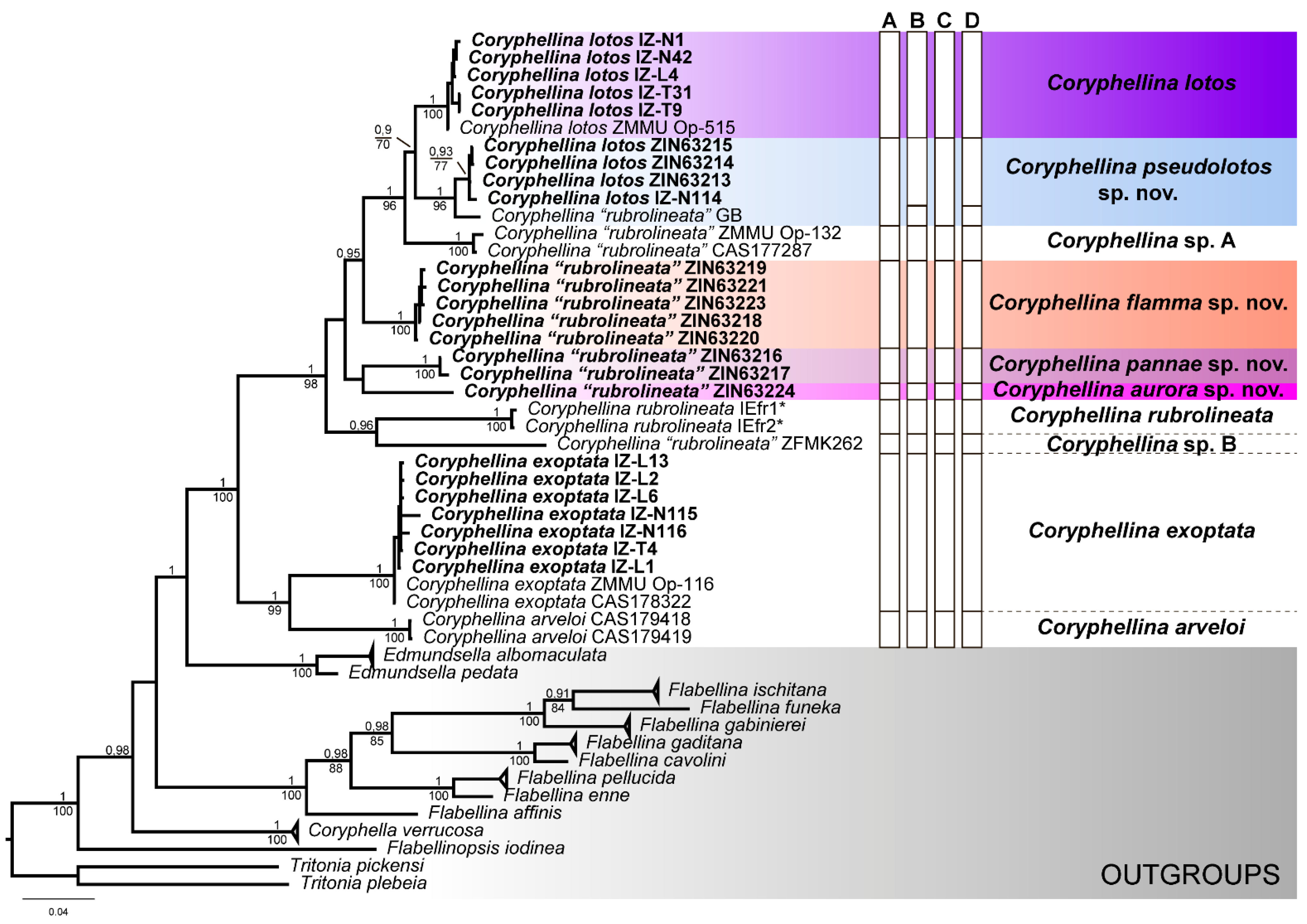
Trees based on the single-gene analyses were poorly resolved (Data S1); however, the concatenated tree provided suitable resolution for most clades (Figure 2). The topology of the concatenated trees generated with the Bayesian inference (BI) and the maximum likelihood (ML) analyses was congruent (Figure 2, Data S1). In our analysis, the genus Coryphellina was recovered as monophyletic, with high statistical support (PP (posterior probability from the Bayesian inference) = 1; BS (bootstrap support from maximum likelihood) = 100). On the tree, a total of eight distinct clades containing putative Coryphellina species were recovered, but only two of them strictly corresponded to the initial species hypothesis: Coryphellina arveloi (Ortea and Espinosa, 1998) (PP = 1; BS = 100) and C. exoptata (PP = 1; BS = 100). Specimens of C. lotos were grouped into two distinct and highly supported clades: one clade was represented by the type specimen (ZMMU Op-515) and five specimens from Vietnam (PP = 1; BS = 100). Smaller specimens from Vietnam were grouped with “C. rubrolineata” from GenBank collected in Queensland, Australia (PP = 1; BS = 96). Coryphellina rubrolineata was represented by four highly supported groups and two singletons. Specimens collected close to the type locality (Coryphellina rubrolineata IEfr1, IEfr2) represented a separate clade (PP = 1; BS = 100), which was sister to a sample from Sulawesi (ZFMK262). Specimens from Vietnam formed two distinct clades (PP = 1; ML = 100 in both cases) and one singleton. The deep relationships within “rubrolineata” and “lotos” species complexes are mostly unresolved, but all species together form a highly supported clade (PP = 1; BS = 98). In summary, our analysis indicates the presence of eight putative species of C. rubrolineata species complex (including C. lotos), represented by highly supported clades or derived singletons. Same results were received in the analysis of trees based on single-gene data sets (Data S1).

3.2. Species Delimitation

The initial species hypothesis implied the presence of 10 monophyletic units recovered in the phylogenetic analysis: Coryphellina rubrolineata (from the type locality), C. lotos (clade comprising the type material), C. exoptata, C. arveloi, and 6 candidate species from C. rubrolineata species complex (Figure 2). The ASAP analysis (Figure S1) showed the lowest ASAP score (1.5) for the two hypotheses: one corresponded to the initial species hypothesis with 10 candidate species, and in the second hypothesis, the single specimen from GenBank (COI accession KJ001316) represented a separated species, implying 11 candidate species. The same 11 candidate species were identified in the bPTP (Figure S2) and GMYC (Figure S3) analyses. This result was also supported by the calculated uncorrected p-distances in COI, H3, and 28S markers. The intraspecific variability of COI markers within candidate species did not exceed 4.2%, and the interspecific distances varied between 9.38% and 16.79% (Table S3). It should also be noted that in most cases, the H3 and 28S data set showed high genetic variation, as interspecific pairwise distances between putative species of C. rubrolineata species complex varied from 0% to 4.29% in the case of the H3 data set and from 0.37% to 13.28% in the case of 28S.
To sum up, we can conclude that the diversity of Coryphellina rubrolineata species complex in Southern Vietnam represents a complex of cryptic and pseudocryptic species, including C. lotos and four new distinct species, which are formally described below. We also identified two other new species, but since their material was not available for morphological study (only sequences from GenBank), their formal taxonomical description is a prospect for future studies.

3.3. Systematics

Order Nudibranchia Blainville, 1814
Suborder Cladobranchia William and Morton, 1984
Superfamily Fionoidea Gray, 1857
Family Flabellinidae Bergh, 1889
Genus Coryphellina O’Donoghue, 1929
Diagnosis (from Korshunova et al. [38] with modifications): Body narrow. Notal ridge present, reduced, discontinuous, in several indistinct pieces. Cerata on low elevations, in several groups. Rhinophores similar in length or shorter than oral tentacles, densely papillated. Anterior foot corners present. Anus pleuroproctic. Distinct oral glands. Rachidian teeth with narrow compressed cusp and distinct denticles. Lateral teeth denticulated with attenuated process basally. Proximal receptaculum seminis bilobed. Distal receptaculum seminis present. Short prostatic vas deferens. Penis conical to bulbous.
Type species: Coryphellina rubrolineata O’Donoghue, 1929

3.3.1. Coryphellina lotos Korshunova, Martynov, Bakken, Evertsen, Fletcher, Mudianta, Saito, Lundin, Schrödl, and Picton, 2017

Material studied:IZ-T31, 1 specimen, dissected, 17 mm in length preserved, Vietnam, Nha Trang, Hon Tre, coordinates and depth unknown, 29 October 2015 coll. E.S. Mekhova. IZ-N1, 1 specimen, 12 mm in length preserved, Vietnam, Nha Trang, Hon Mot, N 12°10′27.56″ E 109°16′19.61″, 16 m in depth, on rocks, 30 March 2016, coll. Y.V. Deart. IZ-N42, 1 specimen, dissected, 17 mm in length preserved, Vietnam, Nha Trang, Dambay, N 12°11′39.84″ E 109°17′26.74″, 5–6 m in depth, on net in sand, 9 April 2016 coll. Y.V. Deart. IZ-T9, 1 specimen, dissected, 19 mm in length preserved, Vietnam, Nha Trang, Hon Mun, N 12°10′026″ E 109°17′725″, 10–18 m in depth, stone on silty bottom, 25 October 2016 coll. V.O. Barkalova. IZ-L4, 1 specimen, dissected, 23 mm in length preserved, Vietnam, Nha Trang, Hon Tre, N 12°10′54.95″ E 109°17′37.94″, 10 m in depth, 2 April 2019, coll. E.S. Mekhova.
External morphology (Figure 3): Length up to 23 mm (preserved). Body slender, foot slender with long anterior corners. Oral tentacles 1.5–2 times longer than rhinophores. Rhinophores highly papillated, bearing up to 70 papillae on inner side. Cerata cylindrical, pointed distally, elongated, arranged in distinct groups. Up to six groups of cerata per row. First group largest, with 9–13 cerata in group. Second group with 4–5 cerata. Cnido-sacs on top of cerata. Digestive gland diverticula cylindrical, fill about ¼–1/3 of ceratal volume. Well-defined discontinued notal edge under ceratal groups. Anus pleuroproctic. Reproductive openings lateral, below first group of cerata.
Color (Figure 3 and Figure 4): Background from translucent white to violet and purple. Digestive gland mass and diverticula in cerata from peachy to light brown. Cerata peachy to purple, cnidosac area covered by peachy pigment with intense red to violet subapical rings. Underneath subapical rings, cerata covered by sparse white opalescent speckling. Prominent thick pink line beginning between oral tentacles, line on body indistinct, discontinuous. Pink to lilac discontinuous lines located laterally in ceratal intergroup space under notal edge, continuing from head to tail. Tip of tail same color as lines. Rhinophores same color as body or apricot, with translucent tips and pink to purple subapical rings. Oral tentacles white to purple, covered by sparse white opalescent powder in middle, with intensive pink to purple subapical rings and translucent tip.
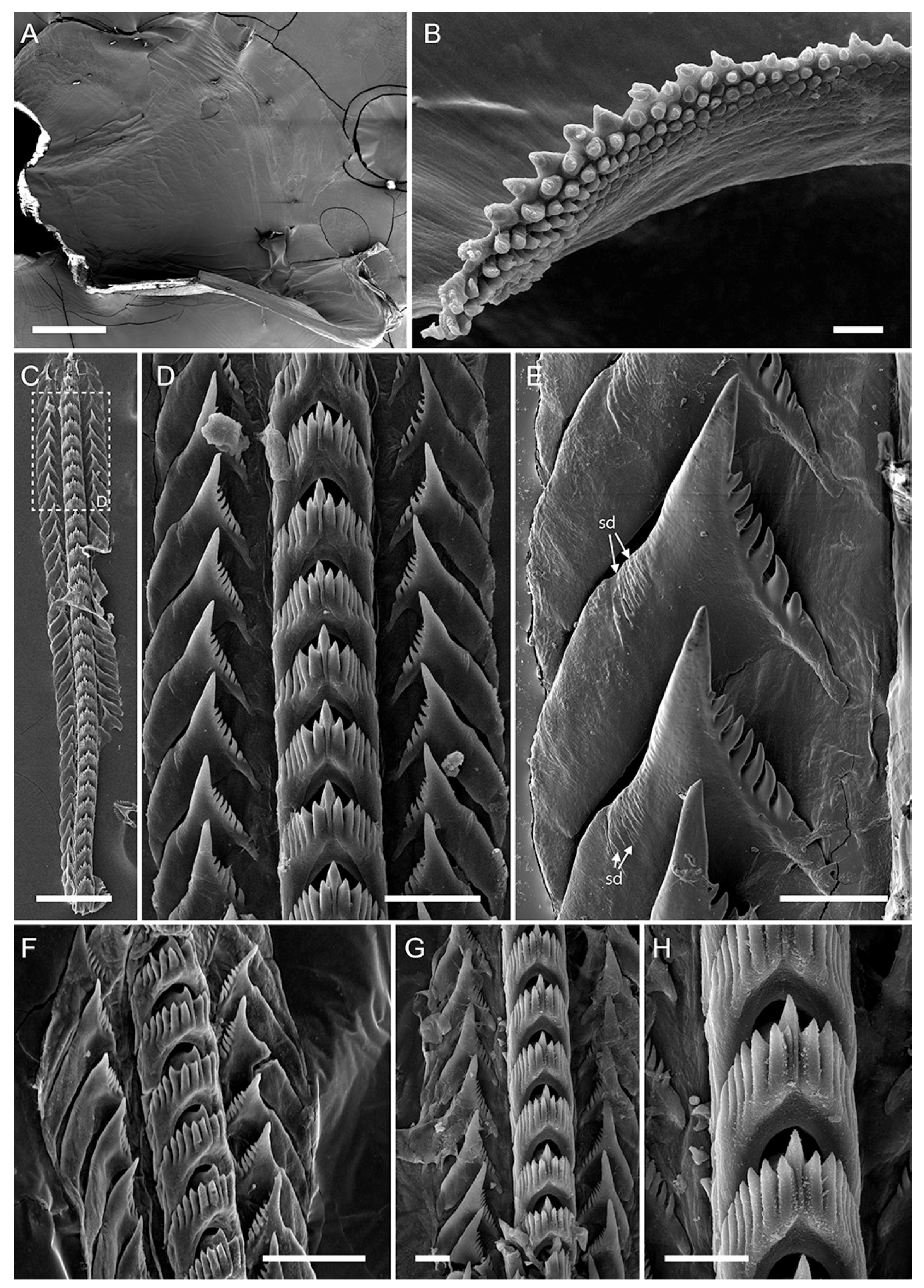
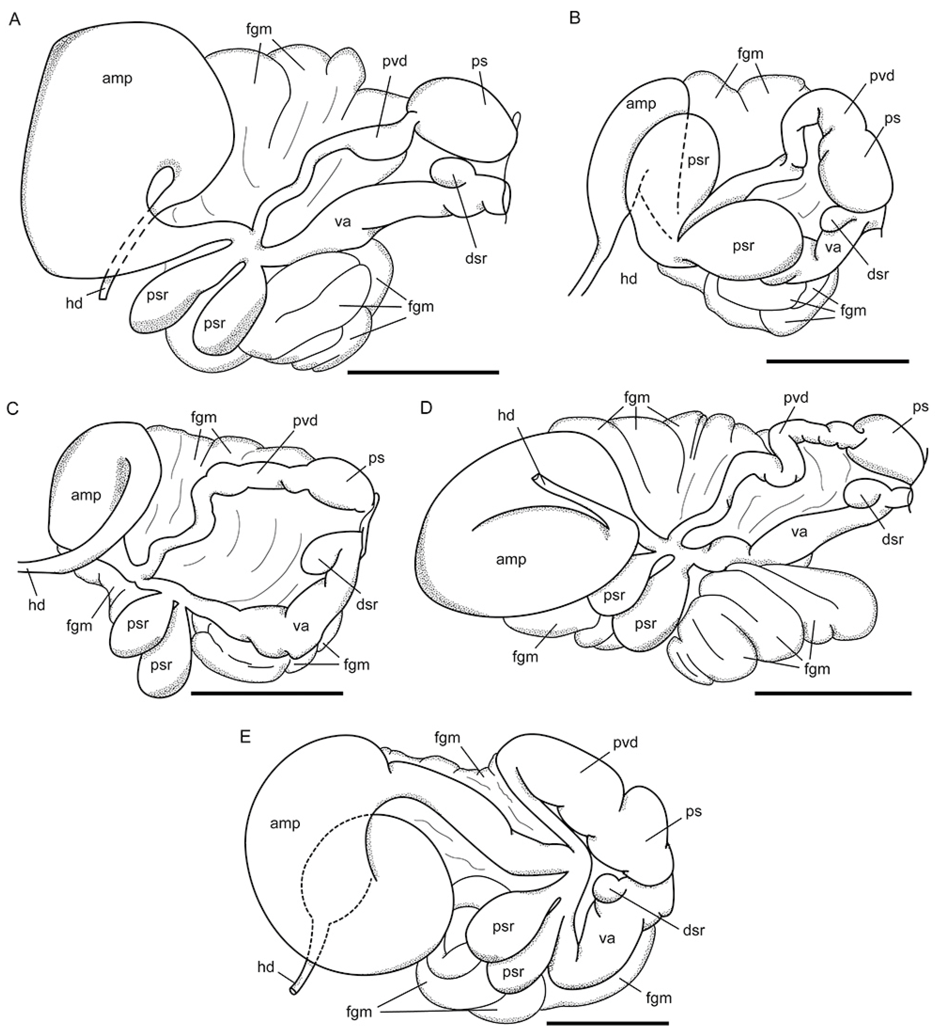
Internal morphology (Figure 5, Figure 6B and Figure 7A): Jaws composed of two triangular plates with triangular masticatory process (Figure 5A). Masticatory process with numerous sharp denticles, arranged in up to five rows, outermost denticles largest with secondary dentitions, innermost blunt (Figure 5B). Radula triseriate, radular formula: 27–35 × 1.1.1 (Figure 5C–H). Rachidian tooth elongated-triangular with short conical central cusp, bearing 5–7 large denticles with deep furrows on both sides. Cusp sharp, small, slightly compressed by adjacent denticles. Lateral teeth of widened triangular shape with elongated cusp and 7–11 denticles on inner edge. Base of teeth almost right angled proximally, oblong distally, with long attenuated processes. Small dentitions on outer tooth side (Figure 5E). Reproductive system diaulic (Figure 7A). Ampulla large, sausage-shaped, widened in middle. Proximal seminal receptacle bilobed. Vas deferens slightly widens distally before entering penial sac, presenting prostatic area. Mucous gland lays distally into vagina, albumen, and membrane glands next to proximal seminal receptacle. Their connection to vagina is not clear. Distal seminal receptacle small, opened into vagina distally. Penis small, conical, unarmed.
Distribution: Records of this species are confirmed in two localities: Pacific coast of Honshu, Japan (the type locality, Korshunova et al. [38]) and Central Vietnam (Nha Trang Bay: Hon Mun, Hon Mot, Hon Tre) (this study). It is likely that this species has a wide distribution range in the Indo-West Pacific [40]; however, it must be verified by testing DNA samples.
Ecology: Found on stones and rocks on sandy bottom at 5–20 m in depth.
Genetic barcode: ON040918-ON040922.
Remarks: This species shows high variability in coloration, having white, pink, or violet chromatic variations (Figure 3). Discontinuous dorsal and dorsolateral pigmental lines are the main diagnostic traits. The dorsal line is often thick in the head region and poorly visible along the dorsum. In this trait, C. lotos differs from the rest of the genus diversity except for C. pseudolotos sp. nov. (see below). In addition, C. lotos has specific morphology of the lateral teeth, possessing large and long attenuated processes at their basal outer side, while in other species, these processes are much shorter (Figure 5 and Figure 6B).

3.3.2. Coryphellina pseudolotos sp. nov.

Zoobank lsid: urn:lsid:zoobank.org:act:D3502637-84EC-4CB9-8D89-4DE13C3B49F7
Type material:Holotype: ZIN63213, 15 mm in length preserved, Vietnam, Phu Quoc, st. 1, N 9°55′20.40″ E 103°59′50.58″, 10 m in depth, 27 April 2018, coll. E.S. Mekhova. Paratypes: ZIN63214, 1 specimen, dissected, 7 mm in length preserved, Vietnam, Nha Trang, Hon Mot, N 12°10′27.56″ E 109°16′19.61″, 16–20 m in depth, on hydroids, 21 October 2016, coll. T.I. Antokhina. ZIN63215, 1 specimen, dissected, 12 mm in length preserved, Phu Quoc, st. 1, N 9°55′20.40″ E 103°59′50.58″, 10 m in depth, 27 April 2018, coll. E.S. Mekhova.
Type locality: Vietnam, Phu Quoc, N 9°55′20.40″ E 103°59′50.58″, 10 m in depth.
Additional material studied:IZ-N114, small damaged specimen, only DNA sample remained, Vietnam, Nha Trang, Hon Tre, N 12°10′54.95″ E 109°17′37.94″, 8 m in depth, on hydroids, 12 April 2017, coll. Y.V. Deart.
Etymology: The species name refers to the similarities of C. pseudolotos sp. nov. to C. lotos in external and internal morphology.
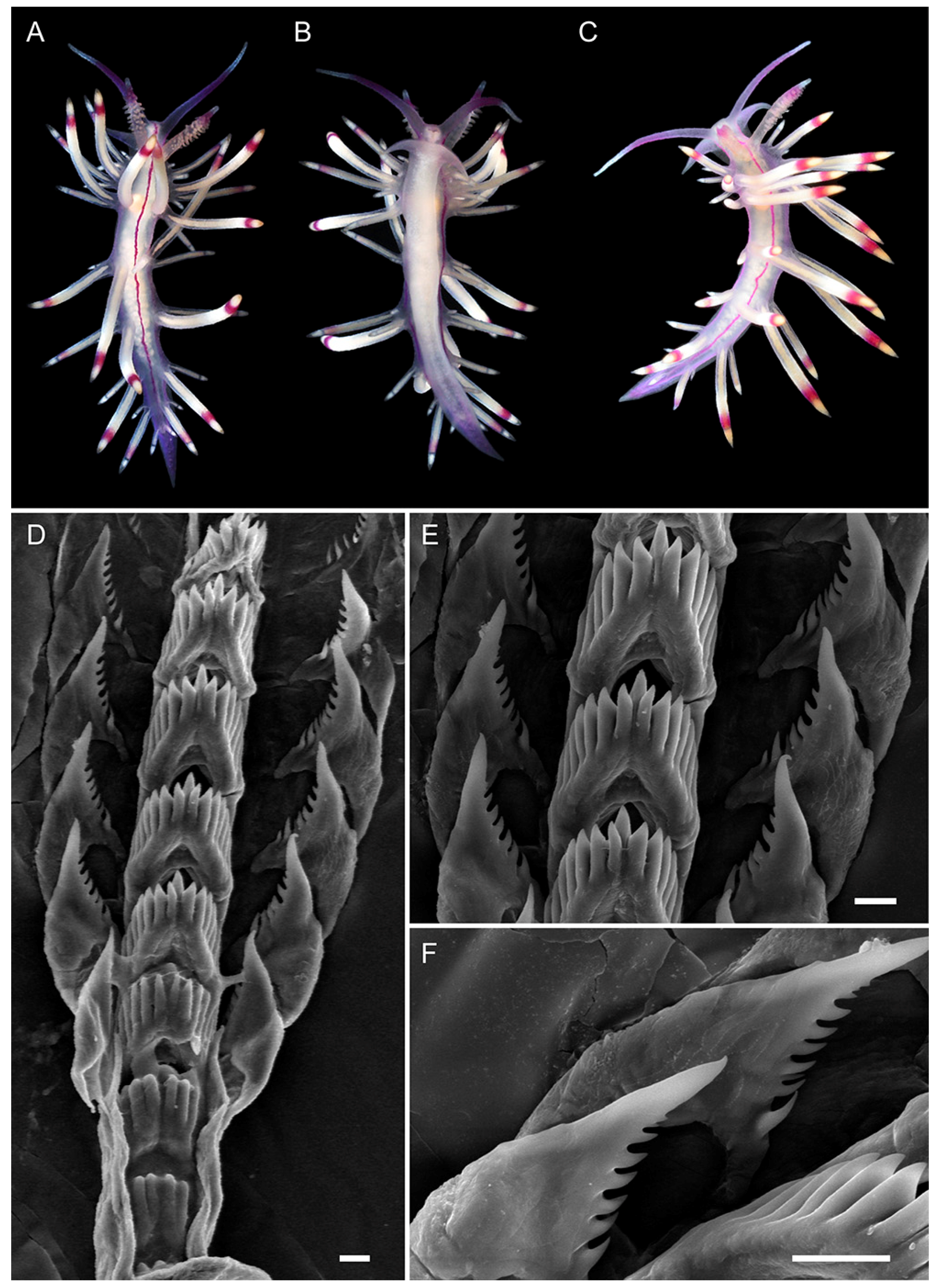
External morphology (Figure 8A–C): Length up to 15 mm (preserved). Body slender, foot slender with long anterior corners. Oral tentacles two times longer than rhinophores. Rhinophores highly papillated, bearing up to 50 papillae on inner side. Cerata finger-shaped, pointed distally, elongated, arranged in distinct groups on low elevations. Up to six groups of cerata per row. First group largest, with 9–12 cerata in group. Second group with 3–5 cerata. Cnidosacs on top of cerata. Digestive gland diverticula cylindrical, fill about 1/3–1/2 of ceratal volume. Well-defined discontinued notal edge under ceratal groups. Anus pleuroproctic. Reproductive openings lateral, below first group of cerata.
Figure 8. Coryphellina pseudolotos sp. nov., external morphology and buccal armature. (A)—holotype ZIN63213, specimen 15 mm in length (preserved). (B)—paratype ZIN63215, specimen 12 mm in length (preserved). (C)—paratype ZIN63214, specimen 7 mm in length (preserved). (D)—paratype ZIN63215, masticatory border of jaws. (E)—paratype ZIN63215, posterior radular portion. Scale bars: (D) = 20 µm; (E) = 10 µm. Photo credits: (A,B): Elena Mekhova; (C): Tatiana Antokhina.
Diversity 14 00294 g008
Color (Figure 4 and Figure 8A–C): Background color translucent. Digestive gland diverticula in cerata from peachy to light brown. Cerata translucent white, red to violet subapical rings with sparse white opalescent speckling underneath, white to peachy cnidosac area. Short pink to violet line between oral tentacles, line on body discontinuous in some specimens indistinct. Pink to violet discontinuous lines located laterally in ceratal intergroup space under notal edge, continuing from head to tail. Tail pink to violet. Rhinophores same color as body or light yellow, violet, or purple subapical rings. Oral tentacles pink to violet, more intensive in middle, white or translucent tip.
Internal morphology (Figure 6C, Figure 7B and Figure 8D,E): Jaws composed of two triangular plates with triangular masticatory process. Masticatory process with numerous sharp denticles, arranged in up to six rows, outermost denticles hamate with secondary dentitions at base, innermost sharp (Figure 8D). Radula triseriate, radular formula: 17–22 × 1.1.1 (Figure 8E). Rachidian tooth elongated-triangular with short conical central cusp, bearing six large denticles with deep furrows on both sides. Cusp sharp, small, slightly compressed by adjacent denticles. Lateral teeth narrow triangular with elongated cusp and 4–7 denticles on inner edge. Base of teeth with short attenuated processes. Small dentitions on outer tooth side absent. Reproductive system diaulic (Figure 7B). Ampulla large, sausage-shaped. Proximal seminal receptacle bilobed. Vas deferens slightly widens distally before entering penial sac, presenting prostatic area. Mucous gland lays distally into vagina, albumen and membrane glands next to proximal seminal receptacle. Their connection to vagina is not clear. Distal seminal receptacle small, opened into vagina distally. Penis small, conical, unarmed.
Distribution: The confirmed distributional range includes Southern (Phu Quoc Island) and Central (Nha Trang Bay: Hon Mot, Hon Tre) Vietnam. A specimen that is genetically similar to this species was collected in Queensland, Australia (GenBank COI accession: KJ001316), suggesting this species may have wider distribution.
Ecology: Found on stones and rocks on soft bottom at 5–20 m in depth.
Genetic barcode: ON040923-ON040925.
Remarks: This species closely match the morphology of its sibling species C. lotos (Figure 3, Figure 4 and Figure 8). Both species are found sympatrically in Central Vietnam (Nha Trang Bay: Hon Mot, Hon Tre). Nevertheless, the differences in nuclear marker 28S (one substitution in all studied specimens, 0.37%) and relatively high interspecific p-distance (9.63%) in mitochondrial COI indicate the existence of genetic barriers in these lineages. Therefore, we conclude that C. pseudolotos sp. nov. represents a distinct cryptic species. In morphological characters, the C. pseudolotos sp. nov. differs from C. lotos by the translucent coloration of the body that remains in an adult state (in specimens ~20 mm in length) (Figure 8A–C), while C. lotos is usually brightly colored with iridescent white to lilac background color (Figure 3). In addition, C. pseudolotos sp. nov. has a shorter process in lateral teeth and much fewer denticles than in C. lotos (Figure 6B,C). From other species of the C. rubrolineata species complex, the new species differs in the presence of three discontinuous red dorsal and dorsolateral lines (Table 2). Genetically it differs from them by 10.62–13.09% in COI, 0.43–4.29% in H3, and 1.48–12.18% in 28S (Table S3). We also detected a 4.20% p-distance in the COI marker between Vietnamese specimens and samples from Australia (GenBank data). It may be explained by either intraspecific variability across distant populations of a single species or by the presence of hidden cryptic diversity. To clarify this issue, more DNA-based studies of this complex in the Indo-West Pacific should be conducted.

3.3.3. Coryphellina pannae sp. nov.

Zoobank lsid: urn:lsid:zoobank.org:act:F42B143B-B8F1-4659-A1BD-412DAFC98C8F
Type material: Holotype: ZIN63216, 6 mm in length preserved, Vietnam, Nha Trang, Dambay, N 12°11′44.20″ E 109°17′28.03″, 5 m in depth, 16 April 2017, coll. Y.V. Deart. Para-type: ZIN63217, dissected, 5 mm in length preserved, Vietnam, Nha Trang, Dambay, N 12°11′39.84″ E 109°17′26.74″, 5 m in depth, 25 April 2018, coll. Y.V. Deart.
Type locality: Vietnam, Nha Trang, Tre Is., Dambay station, N 12°11′44.20″ E 109°17′28.03″, 5 m depth.
Etymology: Named after Panna I. Ekimova, mother of the first author.
External morphology (Figure 9A–C): Length up to 6 mm (preserved). Body slender, foot slender with long anterior corners. Oral tentacles 1.5–2 times longer than rhinophores. Rhinophores highly papillated, bearing up to 40 papillae on inner side. Cerata finger-shaped, pointed distally, elongated, arranged in distinct groups. Up to five groups of cerata per row. First group largest, with 8–9 cerata in group. Second group with three cerata. Cnidosacs on top of cerata. Digestive gland diverticula cylindrical, fill about ½ of ceratal volume. Well-defined discontinued notal edge under ceratal groups. Anus pleuroproctic. Reproductive openings lateral, below first group of cerata.
Color (Figure 4 and Figure 9A–C): Background from translucent white to light lilac. Digestive gland diverticula in cerata from white to light yellow. Cerata milky-white, white to yellow cnidosac area with red subapical rings. Prominent dorsal pink line beginning between oral tentacles and continuing to tail. Two pink pigmental lines located laterally under notal edge, continuing from head to tail. All three lines merging on dorsal side of tail. Rhinophores light pink, purple subapical rings, white tips. Oral tentacles translucent violet, more intensive to tip, tips white.
Internal morphology (Figure 6D, Figure 7C and Figure 9D–F): Jaws composed of two triangular plates with triangular masticatory process. Masticatory process with numerous sharp denticles, arranged in up to four rows. Radula triseriate, radular formula: 15 × 1.1.1 (Figure 9D–F). Rachidian tooth elongated-triangular with short conical central cusp, bearing 6–7 large denticles with deep furrows on both sides. Cusp sharp, small, slightly compressed by adjacent denticles. Lateral teeth triangular, slightly curved, with elongated cusp and 7–9 denticles on inner edge. Base of teeth with short attenuated processes. Small dentitions on outer tooth side absent. Reproductive system diaulic (Figure 7C). Ampulla large, sausage-shaped. Proximal seminal receptacle bilobed. Vas deferens loops and widens distally before entering penial sac, presenting prostatic area. Mucous gland lays distally into vagina, albumen, and membrane glands next to proximal seminal receptacle. Their connection to vagina is not clear. Distal seminal receptacle small, opened into vagina distally. Penis small, conical, unarmed.
Distribution: This species is known only from the type locality.
Ecology: Both specimens were found on artificial substrate (old net) on sandy bottom at 5 m in depth. This species likely prefers sheltered environment.
Genetic barcode: ON040926, ON040927.
Figure 9. Coryphellina pannae sp. nov., external morphology and buccal armature. (A)—paratype ZIN63217, specimen 5 mm in length (preserved), dorsal view. (B)—paratype ZIN63217, ventral view. (C)—holotype ZIN63216, specimen 6 mm in length (preserved), dorsal view. (D)—paratype ZIN63217, posterior radular portion. (E)—paratype ZIN63217, rachidian and lateral teeth. (F)—paratype ZIN63217, lateral teeth. Scale bars: 10 µm. Photo credit: (AC): Yury Deart.
Diversity 14 00294 g009
Remarks: The results of morphological and molecular analyses support C. pannae sp. nov. to be a new distinct species. Genetically it differs from the rest of the C. rubrolineata species complex diversity by 10.62–15.31% in COI, 2.15–3.86% in H3, and 4.80–12.55% in 28S (Table S3). Morphologically it differs from the rest diversity of the C. rubrolineata species complex by smaller body size (up to 6 mm in adult state, preserved specimen), presence of three continuous lines, white coloration of body, low number of cerata, and triangular curved form of the lateral teeth (Table 2). In coloration pattern, the new species is most similar to the C. rubrolineata (Figure 4 and Figure 9); however, the latter species is usually covered by white opalescent powder [39], while C. pannae sp. nov. is translucent white to light lilac. In addition, C. rubrolineata has more cerata in each group, and more groups of cerata (up to 7 in C. rubrolineata and 5 in C. pannae sp. nov.) (Table 2).

3.3.4. Coryphellina flamma sp. nov.
Zoobank lsid: urn:lsid:zoobank.org:act:4A89825D-ED98-40B9-9AE2-5C4CDC44060C
Type material: Holotype: ZIN63218, 12 mm in length preserved, Vietnam, Nha Trang, Hon Nok, N 12°11′23.17″ E 109°20′40.87″, 10 m in depth, 12 July 2019, coll. Y.V. Deart. Paratypes: ZIN63219, 1 specimen, dissected, 10 mm in length preserved, Vietnam, Tho Chu, st. 5, N 9°15′25.74″ E 103°28′10.08″, 8–10 m in depth, 10 May 2018, coll. E.S. Mekhova. ZIN63220, 1 specimen, damaged during collection, 13 mm in length preserved, Vietnam, Nha Trang, Hon Nok, N 12°11′23.17″ E 109°20′40.87″, 10 m in depth, 12 July 2019, coll. Y.V. Deart. ZIN63221, 1 specimen, dissected, 12 mm in length preserved, Vietnam, Nha Trang, Hon Nok, N 12°11′23.17″ E 109°20′40.87″, 10 m in depth, 12 July 2019, coll. Y.V. Deart. ZIN63222, 3 specimens, 2 dissected, 7–12 mm in length preserved, Vietnam, Nha Trang, Hon Nok, N 12°11′ E109°20′, depth unknown, 22 April 2019, coll. E.S. Mekhova. ZIN63223, 1 specimen, dissected, 10 mm in length preserved, Vietnam, Nha Trang, Hon Nok, N 12°11′27.74″ E 109°20′31.16″, 12 m in depth, on rocks, 10 June 2021, coll. Y.V. Deart.
Type locality: Vietnam, Nha Trang, Hon Nok, N 12°11′22.29″ E 109°20′43.14″, 10 m depth.
Etymology: from flamma (= flame, fire, Latin), referring to reddish-orange coloration of this species.
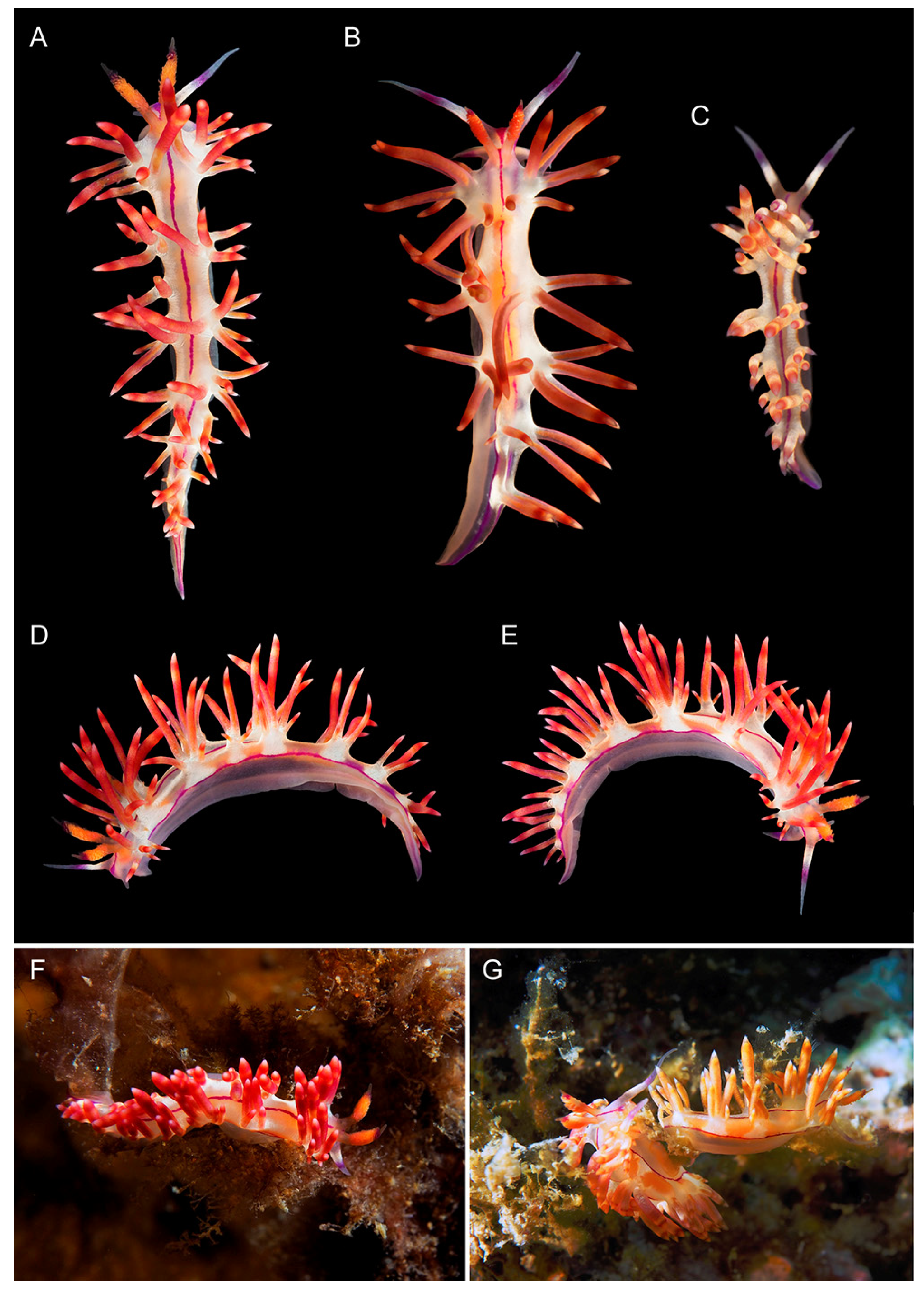
External morphology (Figure 10): Length up to 13 mm (preserved). Body slender, foot slender with long anterior corners. Oral tentacles 1.5 times longer than rhinophores. Rhinophores highly papillated, bearing up to 50 papillae on inner side. Cerata finger-shaped, pointed distally, elongated, arranged in distinct groups. Up to seven groups of cerata per row. First group largest, with 8–10 cerata in group. Second group with five cerata. Cnido-sacs on top of cerata. Digestive gland diverticula cylindrical, fill about 1/2–1/3 of ceratal volume. Well-defined discontinued notal edge under ceratal groups. Anus pleuroproctic. Reproductive openings lateral, below first group of cerata.
Color (Figure 4 and Figure 10): Background translucent white with opaque-white opalescent patches below ceratal groups on dorsum and lateral sides. Digestive gland diverticula in cerata brownish. Cerata with two rings of salmon pink pigmentation alternating with peachy orange or reddish-brown, tips light pink. Prominent thick pink line on dorsal midline, thickened between oral tentacles, narrowing and becoming discontinuous at posterior dorsum part. Two other pink pigment lines located laterally under notal edge, continuing from head to tail. All three lines merging on dorsal side of tail. Rhinophores translucent white with orange papillae, violet-red subapical rings and translucent tips. Oral tentacles covered by sparse white opalescent powder with lilac subapical rings and translucent tips.
Figure 10. Coryphellina flamma sp. nov., external morphology. (A)—holotype ZIN63218, specimen 12 mm in length (preserved), dorsal view. (B)—paratype ZIN63219, specimen 10 mm in length (preserved). (C)—paratype ZIN63222, specimen 7 mm in length (preserved). (D)—holotype ZIN63218, lateral view from left. (E)—holotype ZIN63218, lateral view from right. (F)—holotype ZIN63218 in natural environment. (G)—paratypes ZIN63220, ZIN63221 in natural environment. Photo credits: (A,DG): Yury Deart; (B,C): Elena Mekhova.
Diversity 14 00294 g010
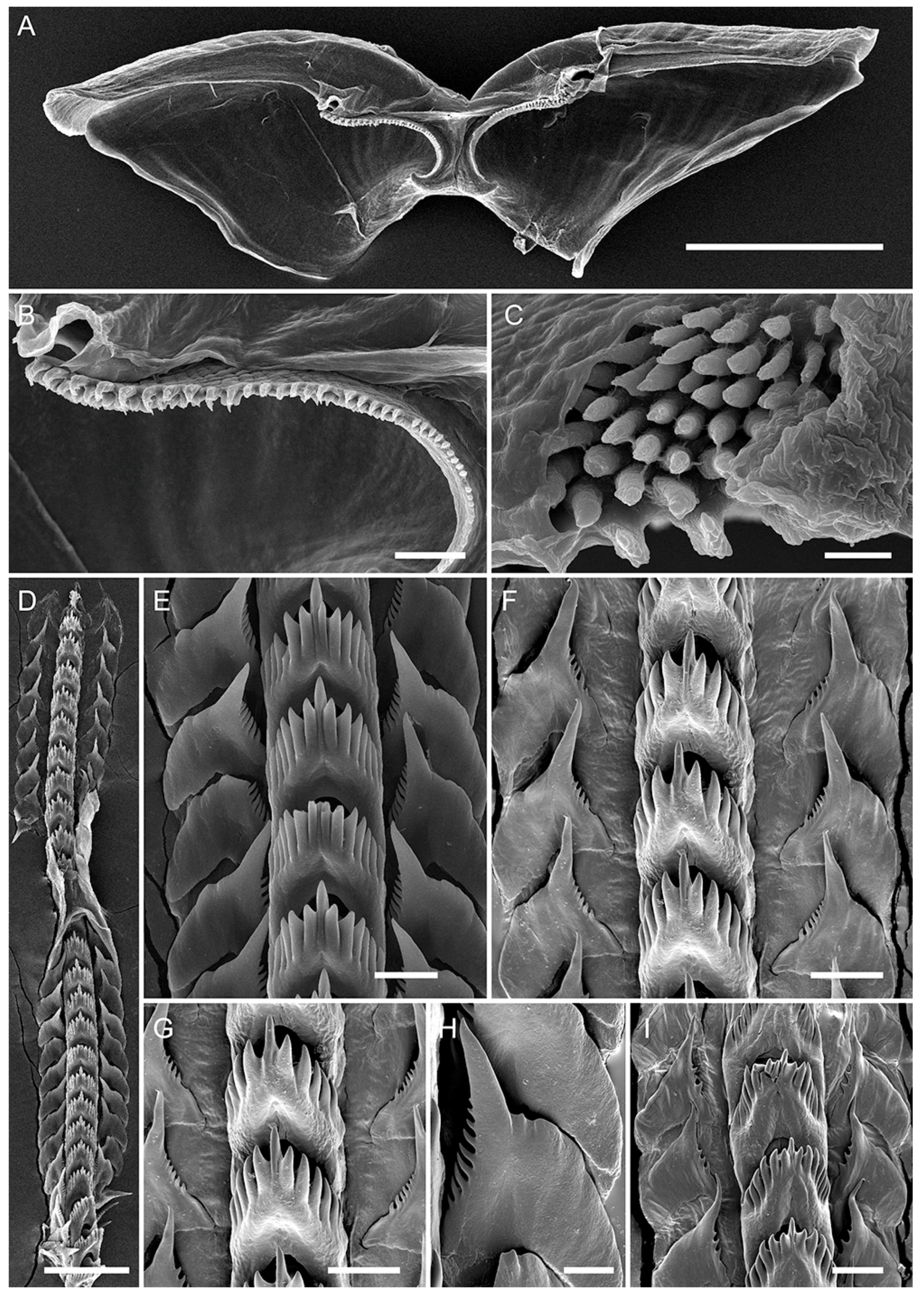
Internal morphology (Figure 6E, Figure 7D and Figure 11): Jaws composed of two triangular plates with triangular masticatory process (Figure 11A). Masticatory process with numerous sharp denticles, arranged in up to six rows (Figure 11B,C). Radula triseriate, radular formula: 23–25 × 1.1.1 (Figure 11D–I). Rachidian tooth triangular with short conical central cusp, bearing 6–9 large denticles with deep furrows on both sides. Cusp sharp, small, slightly compressed by adjacent denticles. Lateral teeth triangular with elongated cusp and 8–12 denticles on inner edge. Teeth slightly curved distally, with short attenuated processes. Dentitions on outer tooth side absent. Reproductive system diaulic (Figure 7D). Ampulla large, sausage-shaped, bent in midline. Proximal seminal receptacle bilobed. Vas deferens gradually widens distally before entering penial sac, presenting prostatic area. Mucous gland lays distally into vagina, albumen, and membrane glands next to proximal seminal receptacle. Their connection to vagina is not clear. Distal seminal receptacle small, opened into vagina distally. Penis small, conical, unarmed.
Distribution: For now, this species is known only from Central (Nha Trang: Hon Nok), and Southern (Tho Chu Is.) Vietnam. Probably it has a wider distribution in the Indo-West Pacific.
Ecology: Found on hydroids on rocks and vertical walls in exposed conditions on 5–15 m in depth.
Genetic barcode: ON040929-ON040933.
Remarks: The results of morphological and molecular analyses support C. flamma sp. nov. to be a new distinct species. Genetically it differs from the rest of the C. rubrolineata species complex diversity by 9.38–15.31% in COI, 1.72–4.29% in H3, and 1.85–11.44% in 28S (Table S3). In external characters, this species differs from other representatives of the C. rubrolineata species complex in the presence of three continuous red lines on dorsal and dorsolateral sides and a peculiar orange-red to bright-red coloration pattern with orange rhinophores and opaque-white pigmental patches on the notal edge below cerata (Table 2). Coryphellina pannae sp. nov. and C. rubrolineata also have three continuous lines but differ in coloration pattern (see above and Ekimova et al. [39]) by overall whitish background color and white to lilac cerata.
Figure 11. Coryphellina flamma sp. nov., buccal armature. (A)—Paratype ZIN63219, jaw plates. (B)—paratype ZIN63219, masticatory process. (C)—paratype ZIN63222, denticulation of masticatory border. (D)—paratype ZIN63222 (specimen 12 mm in length), radula. (E)—paratype ZIN63222(specimen 12 mm in length), anterior radular portion. (F)—paratype ZIN63222 (specimen 12 mm in length), posterior radular portion. (G)—paratype ZIN63222 (specimen 12 mm in length), rachidian teeth. (H)—paratype ZIN63222 (specimen 12 mm in length), lateral tooth. (I)—paratype ZIN63222 (specimen 7 mm in length), posterior radular portion. Scale bars: (A) = 500 µm; (B) = 50 µm; (C,H) = 10 µm; (D) = 100 µm; (EG), (I) = 20 µm.
Diversity 14 00294 g011

3.3.5. Coryphellina aurora sp. nov.
Zoobank lsid: urn:lsid:zoobank.org:act:FCAB9741-416D-4994-83EF-5DF2A9BB5AC6
Type material: Holotype ZIN63224, 27 mm in length preserved, specimen dissected, radula and jaws mounted on aluminum stub, Vietnam, Nha Trang, Hon Nok, N 12°11′22.29″ E 109°20′43.14″, 25 m depth, collected 25 July 2019, coll. Y.V. Deart.
Type locality: Vietnam, Nha Trang, Hon Nok, N 12°11′22.29″ E 109°20′43.14″, 25 m depth.
Etymology: From aurora (= dawn, Latin) referred to the beautiful pinkish coloration of this species, resembling the color patterns of the sunrise along the Vietnamese coast.
Figure 12. Coryphellina aurora sp. nov., holotype ZIN63224, specimen 27 mm in length (preserved), external morphology. (AC)—dorsal view. (D,E)—lateral view from left. (F)—lateral view from right. (G)—ventral view on anterior body part. Photo credit: Yury Deart.
Diversity 14 00294 g012
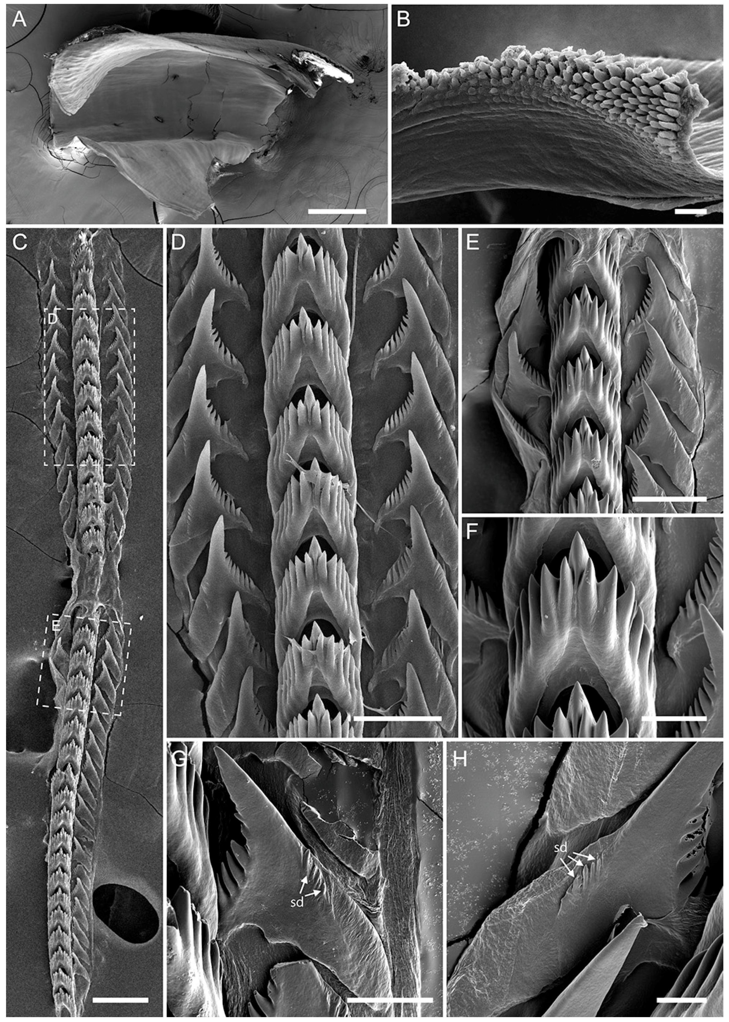
Figure 13. Coryphellina aurora sp. nov., holotype ZIN63224, buccal armature. (A)—left jaw plate. (B)—denticulation of masticatory process. (C)—radula. (D)—posterior radular portion. (E)—middle radular portion. (F)—rachidian tooth. (G)—lateral tooth. (H)—lateral tooth. Abbreviations: sd—secondary denticles on outer side of lateral tooth. Scale bars: (A,C) = 200 µm; (B,E) = 20 µm; (D,F) = 50 µm; (G,H) = 30 µm.
Diversity 14 00294 g013
External morphology (Figure 12): Length up to 27 mm (preserved). Body slender, foot slender with long anterior corners. Oral tentacles 1.2–1.5 times longer than rhinophores. Rhinophores highly papillated, bearing up to 70 papillae on inner side. Cerata cylindrical, pointed distally, elongated, arranged in distinct groups. Up to nine groups of cerata per row. First group largest, with up to 23 cerata in group. Second group with seven cerata. Cnidosacs on top of cerata. Digestive gland diverticula cylindrical, fill about 1/4–1/5 of ceratal volume. Well-defined discontinued notal edge under ceratal groups. Anus pleuroproctic. Reproductive openings lateral, below first group of cerata.
Color (Figure 4 and Figure 12): Background translucent violet. Digestive gland diverticula in cerata brownish. Cerata pink to lilac, violet-red subapical rings with sparse white opalescent speckling underneath and orange tips. Indistinct short pink dorsal line on head. Rhinophores intensive pink with orange papillae and translucent tips. Oral tentacles translucent violet to pink more intensive to tip, tips covered by sparse white opalescent powder.
Internal morphology (Figure 6F, Figure 7E and Figure 13): Jaws composed of two triangular plates with triangular masticatory process (Figure 13A). Masticatory process with numerous sharp denticles, arranged in up to 10 rows, outermost denticles hamate with secondary dentitions at base, innermost sharp (Figure 13B). Radula triseriate, radular formula: 36 × 1.1.1. Rachidian tooth elongated-triangular with short conical central cusp, bearing 6–7 large denticles with deep furrows on both sides. Cusp sharp, small, slightly compressed by adjacent denticles. Lateral teeth triangular with elongated cusp and 7–8 denticles on inner edge. Base of teeth slightly curved proximally, oblong distally, with short attenuated processes. Small dentitions on outer tooth side (Figure 13G,H). Reproductive system diaulic (Figure 7E). Ampulla large and muscular, sausage-shaped, coiled. Proximal seminal receptacle bilobed. Vas deferens narrow proximally than bent and expanded into widened prostatic part. Mucous gland of amorphous structure, laying distally to vagina, albumen, and membrane glands next to proximal seminal receptacle, well developed. Their connection to vagina is not clear. Distal seminal receptacle small, opened into vagina dorsally. Penis small, conical, unarmed.
Distribution: For now, this species is known only from the type locality. Possibly this species is the same as Flabellina sp. 2 sensu Gosliner et al. [40], and therefore, its distribution may also include at least the Philippines.
Ecology: A single large specimen was found at 25 m in depth on rock.
Genetic barcode: ON040934.
Remarks: According to morphological and molecular data, C. aurora sp. nov. clearly represents a new distinct species. Genetically it differs from the rest of the C. rubrolineata species complex diversity by 11.60–16.30% in COI, 2.15–4.72% in H3, and 3.32–11.81% in 28S (Table S3). Morphologically its main diagnostic trait is the almost complete absence of dorsal and dorsolateral pigmental lines on the body (Figure 4 and Figure 12). A single short and thin pink line presents only on the head between rhinophores. In addition, its peculiar pink to violet and lilac coloration serves as a suitable diagnostic trait for further identification of this species. In other external characteristics, this species is unique in the high number of ceratal groups (9) and in the high number of cerata in the first group (up to 23) (Table 2). In internal characters, C. aurora sp. nov. possesses up to 10 rows of denticles on the jaw masticatory border and has the longest radula among other species of the complex (36 rows of teeth) (Figure 13).

4. Discussion

Our results clearly show a much higher diversity in the genus Coryphellina than was previously suggested. All studied species conform to the diagnosis of the genus Coryphellina having papillate rhinophores, discontinuous notal edge, cerata arranged in groups, and bilobed proximal seminal receptaculum. These features clearly distinguish the genus Coryphellina from the representatives of the most closely related genus Edmundsella (includes E. pedata (Montagu, 1816) and E. albomaculata (Pola et al., 2014) see Korshunova et al. [38]) and representatives of the genus Flabellina (includes at least Flabellina affinis (Gmelin, 1791)), but clearly needs further revision, see Furfaro et al. [45]). The genus Edmundsella possesses smooth rhinophores, non-stalked cerata on elevations, and non-bilobed seminal receptaculum [38], while in Flabellina s.str., rhinophores are annulated, cerata are placed on peduncles, and the seminal receptaculum is also simple [38,45,69]. It should also be mentioned that other genera (e.g., Caronella, Calmella, etc., see Korshunova et al. [38]) from the same clade as the Flabellina s.str. also have cerata on peduncles and rhinophores of different morphology, and thus they do not conform to the diagnosis of Coryphellina as well [38,45,69]. Therefore, our results strongly correspond to the taxonomical decision suggested by Korshunova et al. [38] and support the genus Coryphellina as a valid distinct taxon.
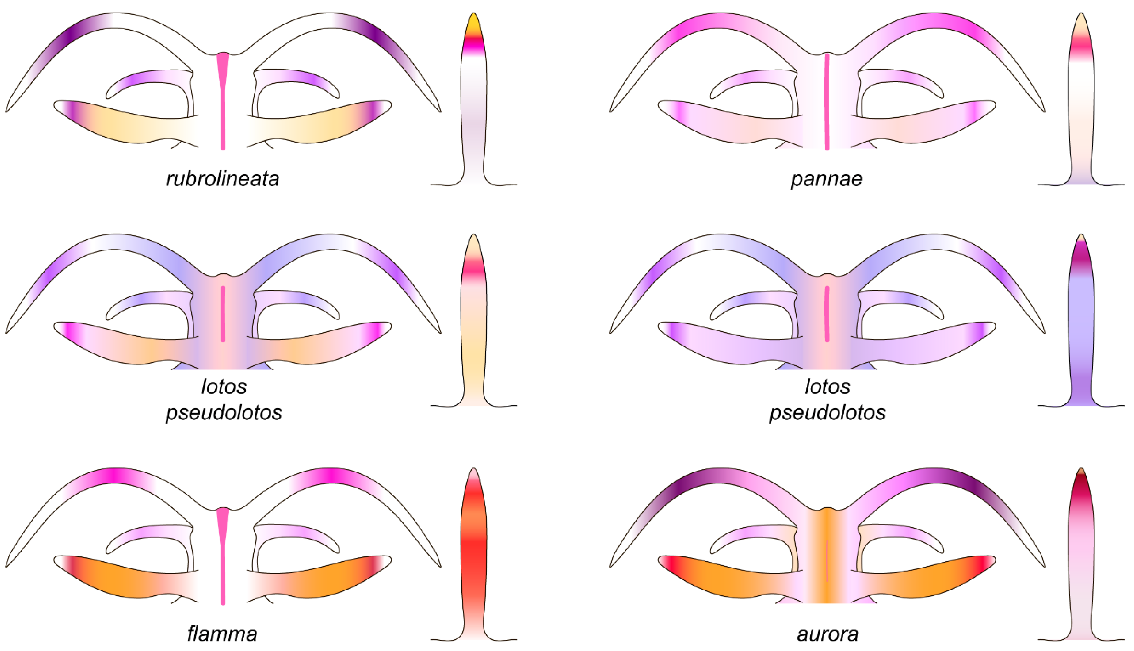
Our results indicate that the cryptic diversity found within the Coryphellina rubrolineata species complex is a pseudocryptic one in most cases, which means that morphological differences could be found with known genetic species boundaries. In this case, the main differences between species were found in the external morphology, i.e., coloration pattern (Figure 4), number of ceratal groups, and number of cerata per group (Table 2). In the coloration pattern, the most important trait is the extent of pigmental dorsal and dorsolateral lines on the body. Only a thin dorsal midline remains between the rhinophores of C. aurora sp. nov. (Figure 12). In C. lotos and C. pseudolotos sp. nov., all three lines are usually present, but they are discontinuous, and the dorsal line is well developed only between the rhinophores (Figure 3 and Figure 8A–C). Only in the C. flamma sp. nov., C. pannae sp. nov., and the “true” C. rubrolineata all three lines are well developed and continue along the whole body (Figure 9 and Figure 10). The general coloration pattern is also an important distinctive trait, as C. flamma sp. nov. differs well from C. pannae sp. nov. by orange to red coloration of cerata and distinct white patches underneath groups of cerata, while C. pannae has translucent-white coloration (Figure 9 and Figure 10). Coryphellina lotos usually demonstrates iridescent pinkish to violet coloration (Figure 3), while in its sister species, C. pseudolotos sp. nov., the general color of the body is translucent even in adult specimens (Figure 8A–C). Several differences may be found in radular characters and the morphology of the reproductive system (Table 2, Figure 7), but it is not clear whether these differences relate to animals’ size or maturity. Species complexes in which pseudocryptic species are differentiated mainly at a base of coloration pattern are well known in nudibranch research: the same was shown for the representatives of the genera Pteraeolidia [70], Unidentia [71], Glossodoris, Doriprismatica [72], and Goniobranchus [73].
From the biogeographical viewpoint, for now, three species are endemics to Vietnam waters: C. pannae sp. nov., C. flamma sp. nov., and C. aurora sp. nov. At the same time, specimens with similar coloration to C. flamma sp. nov. were recorded in other regions of the Indo-West Pacific, including the Philippines [28], Great Barrier Reef, Australia [74] and New Caledonia [75]. The same is true for the C. aurora sp. nov., as specimens of the same coloration pattern were listed as Flabellina sp. 2 collected in the Philippines [40]. Additional molecular studies are needed to confirm the wide distribution range of these species. In the case of C. lotos and C. pseudolotos sp. nov., the biogeographical implications are much more complex. Coryphellina lotos was initially described in temperate waters of Japan (Hokkaido) [38], and its specimens from Vietnam are very similar genetically but differ in general coloration of the body as most of the studied samples have a non-typical violet iridescent coloration. At the same time, its sister species, C. pseudolotos sp. nov., also possesses similar external morphological traits, while found differences in internal morphology are not obvious since they could be a result of ontogenetic variability. Coryphellina pseudolotos sp. nov. demonstrates low but stable differences in nuclear loci (the p-distance in the 28S marker is 0.37%), which suggests the restriction of gene flow between this species and sympatric C. lotos and supports the distinct status of the two species. In addition, a specimen, which is genetically similar to C. pseudolotos sp. nov., was collected in Australia [76]. However its taxonomical status is unclear due to the results of the species delimitation analyses (Figure 2), and since the morphological data are unavailable. Possibly, both C. lotos and C. pseudolotos sp. nov. have a wide distribution in the tropical Indo-West Pacific either in its northern (C. lotos) or southern (C. pseudolotos sp. nov.) parts, and the Vietnamese waters are an area of their secondary sympatry. This hypothesis, as well as the identity of Australian specimens, should be tested in further integrative works on this species complex.

Supplementary Materials

The following are available online at https://www.mdpi.com/article/10.3390/d14040294/s1, Data S1: Unedited maximum likelihood trees based on singe-gene data sets and the partitioned data set in NEWICK format; File Table S1: List of specimens used in this study, including voucher and field numbers, collection data, and collectors; File Table S2: Specimens used for molecular analysis. Voucher numbers, collection localities, and GenBank accession. Figure S1: The ASAP analysis; Figure S2: The same 11 candidate species were identified in the bPTP; Figure S3: GMYC analyses.

Author Contributions

Conceptualization, I.E.; methodology, I.E. and D.S.; software, D.S.; validation, I.E. and D.S.; formal analysis, I.E., Y.D., T.A., A.M. and D.S.; resources, I.E.; data curation, I.E. and Y.D.; writing—original draft preparation, I.E.; writing—review and editing, I.E., Y.D., T.A., A.M. and D.S.; visualization, I.E., Y.D., T.A. and A.M.; supervision, I.E.; project administration, I.E.; funding acquisition, I.E. All authors have read and agreed to the published version of the manuscript.

Funding

This study was carried out in the frame of a scientific project of the State Order of the Russian Federation Government to Lomonosov Moscow State University no. 122012100155-8 with the financial support of the Russian Science Foundation grant no. 20-74-10012.

Institutional Review Board Statement

Not applicable.

Informed Consent Statement

Not applicable.

Data Availability Statement

Unedited phylogenetic trees and results of species delimitation analyses are provided as Supplementary Material files. All sequences were deposited to GenBank.

Acknowledgments

We are deeply grateful to Elena Mekhova and Varvara Barkalova, who kindly collected and supplied additional specimens and their photos for this study. The authors gratefully acknowledge the Vietnamese-Russian Tropical Centre for the organization of field trips in Vietnam. We thank Maria Stanovova and Valentina Tambovtseva for their assistance with Sanger sequencing. Two anonymous reviewers and Academic Editors Giulia Furfaro and Paolo Mariottini are thanked for all corrections and suggestions, which helped to improve the initial version manuscript. The light microscopy and molecular studies were conducted using equipment of the Invertebrate Zoology Department MSU, the electron microscopy studies using equipment of the Electron Microscopy Laboratory of the Shared Facilities Center of Lomonosov Moscow State University sponsored by the Reuter Foundation Ministry of Education and Science and Joint Usage Center Instrumental methods in ecology at the IEE RAS. Sanger sequencing was conducted using equipment of the Core Centrum of the Institute of Developmental Biology RAS.

Conflicts of Interest

The authors declare no conflict of interest. The funders had no role in the design of the study, in the collection, analyses, or interpretation of data, in the writing of the manuscript, or in the decision to publish the results.

References

  1. Knowlton, N. Sibling species in the sea. Annu. Rev. Ecol. Syst. 1993, 24, 189–216. [Google Scholar] [CrossRef]
  2. Bickford, D.; Lohman, D.J.; Sodhi, N.S.; Ng, P.K.L.; Meier, R.; Winker, K.; Ingram, K.K.; Das, I. Cryptic species as a window on diversity and conservation. Trends Ecol. Evol. 2007, 22, 148–155. [Google Scholar] [CrossRef] [PubMed]
  3. Ratnasingham, S.; Hebert, P.D.N. BOLD: The barcode of life data system (http://www.barcodinglife.org). Mol. Ecol. Notes 2007, 7, 355–364. [Google Scholar] [CrossRef] [PubMed] [Green Version]
  4. Fauvel, P. Classes des annélides polychètes. Distribution géographique. Trait. de Zool. 1959, 5, 163–165. [Google Scholar]
  5. Laakkonen, H.M.; Strelkov, P.; Väinölä, R. Molecular lineage diversity and inter oceanic biogeographical history in Hiatella (Mollusca, Bivalvia). Zool. Scr. 2015, 44, 383–402. [Google Scholar] [CrossRef]
  6. Kienberger, K.; Carmona, L.; Pola, M.; Padula, V.; Gosliner, T.M.; Cervera, J.L. Aeolidia papillosa (Linnaeus, 1761) (Mollusca: Heterobranchia: Nudibranchia), single species or a cryptic species complex? A morphological and molecular study. Zool. J. Linn. Soc. Lond. 2016, 177, 481–506. [Google Scholar] [CrossRef] [Green Version]
  7. Layton, K.K.; Martel, A.L.; Hebert, P.D. Geographic patterns of genetic diversity in two species complexes of Canadian marine bivalves. J. Mollus. Stud. 2016, 82, 282–291. [Google Scholar] [CrossRef] [Green Version]
  8. McCarthy, J.B.; Krug, P.J.; Valdés, Á. Integrative systematics of Placida cremoniana (Trinchese, 1892) (Gastropoda, Heterobranchia, Sacoglossa) reveals multiple pseudocryptic species. Mar. Biodivers. 2019, 49, 357–371. [Google Scholar] [CrossRef]
  9. Ekimova, I.; Valdés, Á.; Chichvarkhin, A.; Antokhina, T.; Lindsay, T.; Schepetov, D. Diet–driven ecological radiation and allopatric speciation result in high species diversity in a temperate-cold water marine genus Dendronotus (Gastropoda: Nudibranchia). Mol. Phylogenet. Evol. 2019, 141, 106609. [Google Scholar] [CrossRef]
  10. Ekimova, I.A.; Mikhlina, A.L.; Vorobyeva, O.A.; Antokhina, T.I.; Tambovtseva, V.G.; Schepetov, D.M. Young but distinct: Description of Eubranchus malakhovi sp. n. a new, recently diverged nudibranch species (Gastropoda: Heterobranchia) from the Sea of Japan. Invertbr. Zool. 2021, 18, 197–222. [Google Scholar] [CrossRef]
  11. Duran, S.; Rützler, K. Ecological speciation in a Caribbean marine sponge. Mol. Phylogenet. Evol. 2006, 40, 292–297. [Google Scholar] [CrossRef] [PubMed]
  12. Churchill, C.K.; Valdés, Á.; Foighil, D.Ó. Molecular and morphological systematics of neustonic nudibranchs (Mollusca: Gastropoda: Glaucidae: Glaucus), with descriptions of three new cryptic species. Invertebr. Syst. 2014, 28, 174–195. [Google Scholar] [CrossRef]
  13. Krug, P.J.; Vendetti, J.E.; Valdés, A. Molecular and morphological systematics of Elysia Risso, 1818 (Heterobranchia: Sacoglossa) from the Caribbean region. Zootaxa 2016, 4148, 1–137. [Google Scholar] [CrossRef] [PubMed]
  14. Nygren, A.; Parapar, J.; Pons, J.; Meißner, K.; Bakken, T.; Kongsrud, J.A.; Oug, E.; Gaeva, D.; Sikorski, A.; Johansen, R.A.; et al. A mega-cryptic species complex hidden among one of the most common annelids in the North East Atlantic. PLoS ONE 2018, 13, e0198356. [Google Scholar] [CrossRef] [PubMed]
  15. Krug, P.J. Patterns of speciation in marine gastropods: A review of the phylogenetic evidence for localized radiations in the sea. Am. Malacol. Bull. 2011, 29, 169–186. [Google Scholar] [CrossRef]
  16. Fritts-Penniman, A.L.; Gosliner, T.M.; Mahardika, G.N.; Barber, P.H. Cryptic ecological and geographic diversification in coral-associated nudibranchs. Mol. Phylogenet. Evol. 2020, 144, 106698. [Google Scholar] [CrossRef]
  17. O’Donoghue, C.H. Report on the Opisthobranchia. XXXVIII. Zoological results of the Cambridge Expedition to the Suez Canal. Trans. Zool. Soc. Lon. 1929, 22, 713–841. [Google Scholar]
  18. Gat, G. Flabellina rubrolineata (O’Donoghue) and Phidiana indica (Bergh) (Nudibranchia: Aeolidoidea), two new Lessepsian immigrants in the Eastern Mediterranean. J. Mollus. Stud. 1993, 59, 120. [Google Scholar] [CrossRef]
  19. Yokes, B.; Rudman, W.B. Lessepsian opisthobranchs from southwestern coast of Turkey: Five new records for Mediterranean. Rapp. Comm. Int. Explor. Mer Méditerranée 2004, 37, 557. [Google Scholar]
  20. Yonow, N. Red Sea Opisthobranchia 4: The orders Cephalaspidea, Anaspidea, Notaspidea and Nudibranchia: Dendronotacea and Aeolidacea. Fauna Arab. 2000, 18, 87–132. [Google Scholar]
  21. Yonow, N. Opisthobranchs of the Gulf of Eilat and the Red Sea: An Account of Similarities and Differences. In Aqaba–Eilat, the Improbable Gulf: Environment, Biodiversity and Preservation; Por, F.D., Ed.; Hebrew University Magnes Press: Jerusalem, Israel, 2008; pp. 177–188. [Google Scholar]
  22. Gul, S. New records of nudibranchs (Gastropoda: Heterobranchia) from the coast of Pakistan (Northern Arabian Sea). Festivus 2019, 51, 114–124. [Google Scholar] [CrossRef]
  23. Sreeraj, C.R.; Sivaperuman, C.; Raghunathan, C. Addition to the opisthobranchiate (Opisthobranchia, Mollusca) fauna of Andaman and Nicobar Islands, India. Galaxea 2012, 14, 105–113. [Google Scholar] [CrossRef] [Green Version]
  24. Sreeraj, C.R.; Sivaperuman, C.; Raghunathan, C. An annotated checklist of opisthobranch fauna (Gastropoda: Opisthobranchia) of the Nicobar Islands, India. J. Threat. Tax 2012, 4, 2499–2509. [Google Scholar] [CrossRef] [Green Version]
  25. Sreeraj, C.R.; Sivaperuman, C.; Raghunathan, C. Species Diversity and Abundance of Opisthobranch Molluscs (Gastropoda: Opisthobranchia) in the Coral Reef Environments of Andaman and Nicobar Islands, India. In Ecology and Conservation of Tropical Marine Faunal Communities; Venkataraman, K., Sivaperuman, C., Raghunathan, C., Eds.; Springer: Berlin/Heidelberg, Germany, 2013; pp. 81–106. [Google Scholar]
  26. Tibiriçá, Y.; Pola, M.; Cervera, J.L. Astonishing diversity revealed: An annotated and illustrated inventory of Nudipleura (Gastropoda: Heterobranchia) from Mozambique. Zootaxa 2017, 4359, 1–133. [Google Scholar] [CrossRef] [Green Version]
  27. Gosliner, T.M.; Kuzirian, A. Two new species of Flabellinidae (Opisthobranchia: Aeolidacea) from Baja California. Proc. Cal. Acad. Sci. 1990, 47, 1–15. [Google Scholar]
  28. Gosliner, T.M.; Willan, R.C. Review of the Flabellinidae (Nudibranchia: Aeolidacea) from the tropical Indo–Pacific, with descriptions of five new species. Veliger 1991, 34, 97–133. [Google Scholar]
  29. Martynov, A.V.; Korshunova, T.A. Opisthobranch molluscs of Vietnam (Gastropoda: Opisthobranchia). In Benthic Fauna of the Bay of Nhatrang, Southern Vietnam; Britayev, T.A., Pavlov, D.S., Eds.; KMK Scientific Press: Moscow, Russia, 2012; Volume 2, pp. 142–257. [Google Scholar]
  30. Yonow, N. Results of the Rumphius Biohistorical Expedition to Ambon (1990). Part 16. The Nudibranchia—Dendronotina, Arminina, Aeolidina, and Doridina (Mollusca: Gastropoda: Heterobranchia). Arch. Für Molluskenkd. 2017, 146, 135–172. [Google Scholar] [CrossRef]
  31. Papu, A.; Undap, N.; Martinez, N.A.; Segre, M.R.; Kuada, R.R.; Perin, M.; Yonow, N.; Wägele, H. First Study on Marine Heterobranchia (Gastropoda, Mollusca) in Bangka Archipelago, North Sulawesi, Indonesia. Diversity 2020, 12, 52. [Google Scholar] [CrossRef] [Green Version]
  32. Jung, D.; Park, J.K. First report of an aeolid nudibranch Flabellina athadona and an identification key for the genus Flabellina from Korea. Korean J. Malacol. 2015, 31, 249–252. [Google Scholar] [CrossRef] [Green Version]
  33. Baba, K. Opisthobranchia of Sagami Bay; Iwanami Shoten: Tokyo, Japan, 1955; p. 59. [Google Scholar]
  34. Wells, F.E.; Bryce, C.W. Sea Slugs of Western Australia; Western Australian Museum: Perth, Australia, 1993; p. 184. [Google Scholar]
  35. Burn, R. A checklist and bibliography of the Opisthobranchia (Mollusca: Gastropoda) of Victoria and the Bass Strait area, south–eastern Australia. Mus. Vict. Sci. Rep. 2006, 10, 7–13. [Google Scholar] [CrossRef] [Green Version]
  36. Larkin, M.F.; Smith, S.D.; Willan, R.C.; Davis, T.R. Diel and seasonal variation in heterobranch sea slug assemblages within an embayment in temperate eastern Australia. Mar. Biodivers. 2018, 48, 1541–1550. [Google Scholar] [CrossRef]
  37. Gosliner, T.M.; Behrens, D.W.; Valdés, Á. Indo–Pacific Nudibranchs and Sea Slugs: A Field Guide to The World’s Most Diverse Fauna; Sea Challengers Natural History Books; California Academy of Sciences: San Francisco, CA, USA, 2008; p. 426. [Google Scholar]
  38. Korshunova, T.; Martynov, A.; Bakken, T.; Evertsen, J.; Fletcher, K.; Mudianta, I.W.; Saito, H.; Lundin, K.; Schroedl, M.; Picton, B. Polyphyly of the traditional family Flabellinidae affects a major group of Nudibranchia: Aeolidacean taxonomic reassessment with descriptions of several new families, genera, and species (Mollusca, Gastropoda). ZooKeys 2017, 717, 1. [Google Scholar] [CrossRef] [PubMed] [Green Version]
  39. Ekimova, I.A.; Antokhina, T.I.; Schepetov, D.M. Molecular data and updated morphological description of Flabellina rubrolineata (Nudibranchia: Flabellinidae) from the Red and Arabian seas. Ruthenica 2020, 30, 183–194. [Google Scholar] [CrossRef]
  40. Gosliner, T.M.; Valdés, Á.; Behrens, D.W. Nudibranch & Sea Slug Identification: Indo–Pacific, 2nd ed.; New World Publications: Jacksonville, FL, USA, 2018; p. 452. [Google Scholar]
  41. Goodheart, J.A.; Bazinet, A.L.; Valdés, Á.; Collins, A.G.; Cummings, M.P. Prey preference follows phylogeny: Evolutionary dietary patterns within the marine gastropod group Cladobranchia (Gastropoda: Heterobranchia: Nudibranchia). BMC Evol. Biol. 2017, 17, 1–14. [Google Scholar] [CrossRef]
  42. Valdés, Á.; Lundsten, L.; Wilson, N.G. Five new deep-sea species of nudibranchs (Gastropoda: Heterobranchia: Cladobranchia) from the Northeast Pacific. Zootaxa 2018, 4526, 401–433. [Google Scholar] [CrossRef]
  43. Ekimova, I.A.; Antokhina, T.I.; Schepetov, D.M. “Invasion” in the Russian Arctic: Is global Climate Change a real driver? A remarkable case of two nudibranch species. Ruthenica 2019, 29, 103–113. [Google Scholar]
  44. Ekimova, I.; Valdés, Á.; Malaquias, M.A.E.; Rauch, C.; Chichvarkhin, A.; Mikhlina, A.; Antokhina, T.; Chichvarkhina, O.; Schepetov, D. High-level taxonomic splitting in allopatric taxa causes confusion downstream: A revision of the nudibranch family Coryphellidae. Zool. J. Linn. Soc. Lond. 2022, 1–35. [Google Scholar] [CrossRef]
  45. Furfaro, G.; Salvi, D.; Trainito, E.; Vitale, F.; Mariottini, P. When morphology does not match phylogeny: The puzzling case of two sibling nudibranchs (Gastropoda). Zool. Scr. 2021, 50, 439–454. [Google Scholar] [CrossRef]
  46. Ekimova, I.A. A new species of the genus Coryphella (Gastropoda: Nudibranchia) from the Kuril Islands. Ruthenica 2022, 32, 41–48. [Google Scholar] [CrossRef]
  47. Ivanova, N.V.; Dewaard, J.R.; Hebert, P.D.N. An inexpensive, automation-friendly proto-col for recovering high-quality DNA. Mol. Ecol. Notes 2006, 6, 998–1002. [Google Scholar] [CrossRef]
  48. Osterburg, H.H.; Allen, J.K.; Finch, C.E. The use of ammonium acetate in the precipitation of ribonucleic acid. Biochem. J. 1975, 147, 367–368. [Google Scholar] [CrossRef] [PubMed] [Green Version]
  49. Folmer, O.; Black, M.; Hoeh, W.; Lutz, R.; Vrijenhoek, R. DNA primers for amplification of mitochondrial cytochrome c oxidase subunit I from diverse metazoan invertebrates. Mol. Mar. Biol. Biotechnol. 1994, 3, 294–299. [Google Scholar] [PubMed]
  50. Palumbi, S.; Martin, A.; Romano, S.; McMillan, W.O.; Stice, L.; Grabowski, G. The Simple Fool’s Guide to PCR; Version 2.0; University of Hawaii: Honolulu, HI, USA, 1991. [Google Scholar]
  51. Puslednik, L.; Serb, J.M. Molecular phylogenetics of the Pectinidae (Mollusca: Bivalvia) and effect of increased taxon sampling and outgroup selection on tree topology. Mol. Phylogenet. Evol. 2008, 48, 1178–1188. [Google Scholar] [CrossRef]
  52. Colgan, D.J.; McLauchlan, A.; Wilson, G.D.F.; Livingston, S.P.; Edgecombe, G.D.; Macaranas, J.; Gray, M.R. Histone H3 and U2 snRNA DNA sequences and arthropod molecular evolution. Aust. J. Zool. 1998, 46, 419–437. [Google Scholar] [CrossRef]
  53. Lê, H.L.; Lecointre, G.; Perasso, R. A 28S rRNA-based phylogeny of the gnathostomes: First steps in the analysis of conflict and congruence with morphologically based cladograms. Mol. Phylogenet. Evol. 1993, 2, 31–51. [Google Scholar] [CrossRef] [PubMed]
  54. Kearse, M.; Moir, R.; Wilson, A.; Stones-Havas, S.; Cheung, M.; Sturrock, S.; Drummond, A. Geneious Basic: An integrated and extendable desktop software platform for the organization and analysis of sequence data. Bioinformatics 2012, 28, 1647–1649. [Google Scholar] [CrossRef] [PubMed]
  55. Altschul, S.F.; Gish, W.; Miller, W.; Myers, E.W.; Lipman, D.J. Basic local alignment search tool. J. Mol. Biol. 1990, 215, 403–410. [Google Scholar] [CrossRef]
  56. Edgar, R.C. MUSCLE: Multiple sequence alignment with high accuracy and high throughput. Nucleic Acids Res. 2004, 32, 1792–1797. [Google Scholar] [CrossRef] [PubMed] [Green Version]
  57. Kumar, S.; Stecher, G.; Tamura, K. MEGA7: Molecular evolutionary genetics analysis version 7.0 for bigger datasets. Mol. Biol. Evol. 2016, 33, 1870–1874. [Google Scholar] [CrossRef] [Green Version]
  58. Chaban, E.M.; Ekimova, I.A.; Schepetov, D.M.; Chernyshev, A.V. Meloscaphander grandis (Heterobranchia: Cephalaspidea), a deep-water species from the North Pacific: Redescription and taxonomic remarks. Zootaxa 2019, 4646, 385–400. [Google Scholar] [CrossRef]
  59. Darriba, D.; Posada, D.; Kozlov, A.M.; Stamatakis, A.; Morel, B.; Flouri, T. ModelTest-NG: A new and scalable tool for the selection of DNA and protein evolutionary models. Mol. Biol. Evol. 2020, 37, 291–294. [Google Scholar] [CrossRef] [PubMed] [Green Version]
  60. Flouri, T.; Izquierdo-Carrasco, F.; Darriba, D.; Aberer, A.J.; Nguyen, L.T.; Minh, B.Q.; von Haeseler, A.; Stamatakis, A. The Phylogenetic Likelihood Library. Syst. Biol. 2014, 64, 356–362. [Google Scholar] [CrossRef] [PubMed] [Green Version]
  61. Ronquist, F.; Huelsenbeck, J.P. MrBayes 3: Bayesian phylogenetic inference under mixed models. Bioinformatics 2003, 19, 1572–1574. [Google Scholar] [CrossRef] [PubMed] [Green Version]
  62. Stamatakis, A. RAxML version 8: A tool for phylogenetic analysis and post-analysis of large phylogenies. Bioinformatics 2014, 30, 1312–1313. [Google Scholar] [CrossRef] [PubMed]
  63. Puillandre, N.; Brouillet, S.; Achaz, G. ASAP: Assemble species by automatic partitioning. Mol. Ecol. Resour. 2021, 21, 609–620. [Google Scholar] [CrossRef]
  64. Herbert, P.D.N.; Cywinska, A.; Ball, S.L.; DeWaard, J.R. Biological identifications through DNA barcodes. Proc. R. Soc. Ser. B Bio. 2003, 270, 313–321. [Google Scholar] [CrossRef] [Green Version]
  65. Zhang, J.; Kapli, P.; Pavlidis, P.; Stamatakis, A. A general species delimitation method with applications to phylogenetic placements. Bioinformatics 2013, 29, 2869–2876. [Google Scholar] [CrossRef] [Green Version]
  66. Pons, J.; Barraclough, T.G.; Gomez-Zurita, J.; Cardoso, A.; Duran, D.P.; Hazell, S.; Kamoun, S.; Sumlin, W.D.; Vogler, A.P. Sequence-based species delimitation for the DNA taxonomy of undescribed insects. Syst. Biol. 2006, 55, 595–609. [Google Scholar] [CrossRef] [Green Version]
  67. Fujisawa, T.; Barraclough, T.G. Delimiting species using single-locus data and the Generalized Mixed Yule Coalescent approach: A revised method and evaluation on simulated data sets. Syst. Biol. 2013, 62, 707–724. [Google Scholar] [CrossRef] [Green Version]
  68. Bouckaert, R.; Vaughan, T.G.; Barido-Sottani, J.; Duchêne, S.; Fourment, M.; Gavryushkina, A.; Heled, J.; Jones, G.; Kühnert, D.; De Maio, N.; et al. BEAST 2.5: An advanced software platform for Bayesian evolutionary analysis. PLoS Comput. Biol. 2019, 15, e1006650. [Google Scholar]
  69. Furfaro, G.; Salvi, D.; Mancini, E.; Mariottini, P. A multilocus view on Mediterranean aeolid nudibranchs (Mollusca): Systematics and cryptic diversity of Flabellinidae and Piseinotecidae. Mol. Phylogenet. Evol. 2018, 118, 13–22. [Google Scholar] [CrossRef] [PubMed]
  70. Wilson, N.G.; Burghardt, I. Here be dragons-phylogeography of Pteraeolidia ianthina (Angas, 1864) reveals multiple species of photosynthetic nudibranchs (Aeolidina: Nudibranchia). Zool. J. Linn. Soc. Lond. 2015, 175, 119–133. [Google Scholar] [CrossRef] [Green Version]
  71. Korshunova, T.; Mehrotra, R.; Arnold, S.; Lundin, K.; Picton, B.; Martynov, A. The formerly enigmatic Unidentiidae in the limelight again: A new species of the genus Unidentia from Thailand (Gastropoda: Nudibranchia). Zootaxa 2019, 4551, 556–570. [Google Scholar] [CrossRef] [PubMed]
  72. Matsuda, S.B.; Gosliner, T.M. Glossing over cryptic species: Descriptions of four new species of Glossodoris and three new species of Doriprismatica (Nudibranchia: Chromodorididae). Zootaxa 2018, 4444, 501–529. [Google Scholar] [CrossRef] [PubMed] [Green Version]
  73. Soong, G.Y.; Bonomo, L.J.; Reimer, J.D.; Gosliner, T.M. Battle of the bands: Systematics and phylogeny of the white Goniobranchus nudibranchs with marginal bands (Nudibranchia, Chromodorididae). ZooKeys 2022, 1083, 169. [Google Scholar] [CrossRef] [PubMed]
  74. Marshall, J.G.; Willan, R.C. Nudibranchs of Heron Island, Great Barrier Reef; Backhuys Publishers: Leiden, The Netherlands, 1999; p. 257. [Google Scholar]
  75. Herve, J.-F. Guide des Nudibranches de Nouvelle-Caledonie et autres Opisthobranches; Editions Catherine Ledru: Noumea, France, 2010; p. 403. [Google Scholar]
  76. Cheney, K.L.; Cortesi, F.; How, M.J.; Wilson, N.G.; Blomberg, S.P.; Winters, A.E.; Marshall, N.J. Conspicuous visual signals do not coevolve with increased body size in marine sea slugs. J. Evolut. Biol. 2014, 27, 676–687. [Google Scholar] [CrossRef] [PubMed]
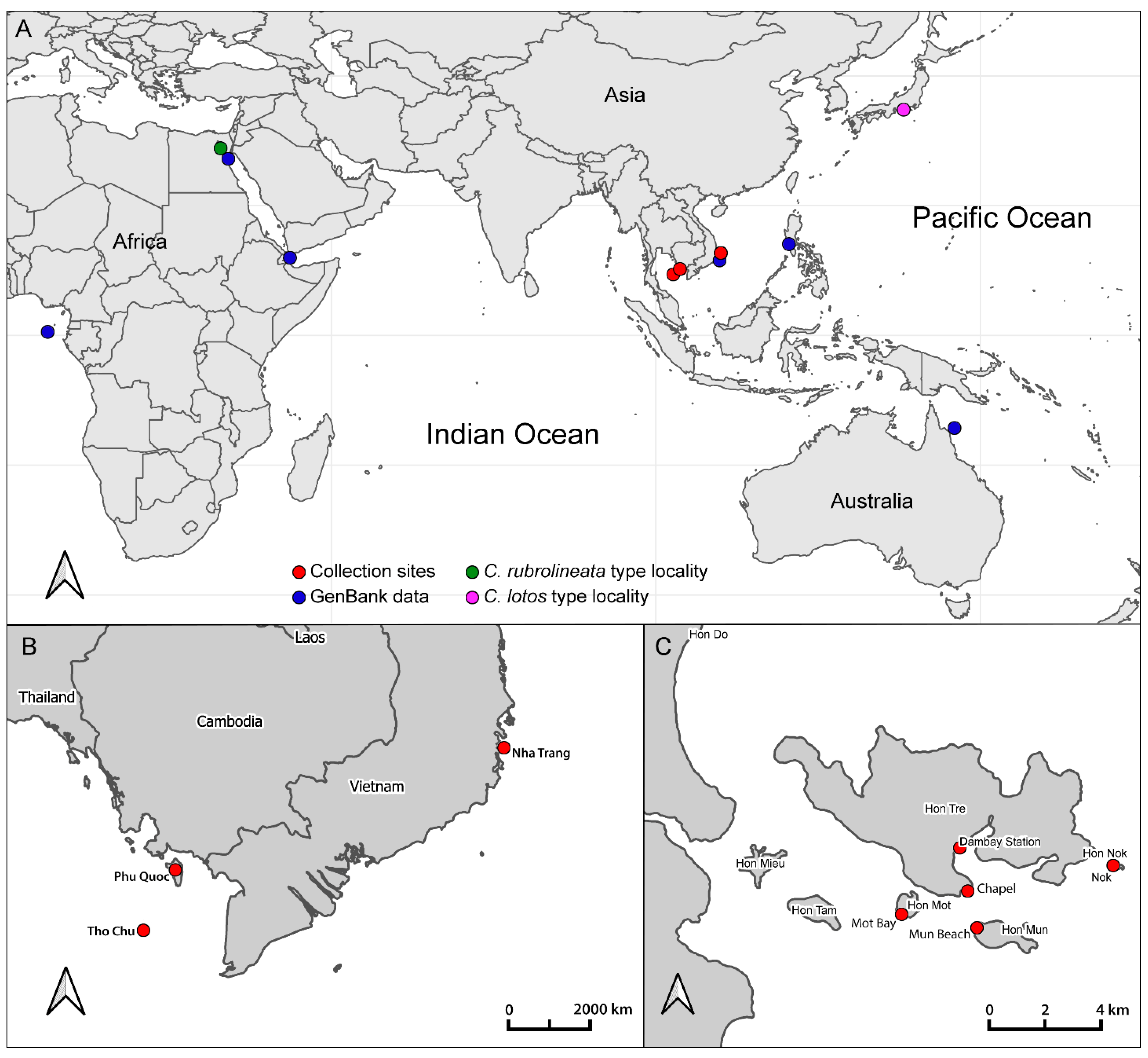
Figure 1. Collection sites of the studied material. (A)—Global map of the Pacific and Indian oceans with collection sites (red circles) and collection sites of samples from GenBank (blue circles). Green and magenta circles indicate the type localities of Coryphellina rubrolineata and C. lotos, respectively. (B)—Map of Southern and Central Vietnam. (C)—Map of collection sites in Nha Trang Bay.
Diversity 14 00294 g001
Figure 2. The Bayesian phylogenetic tree of the genus Coryphellina based on the concatenated data set of four markers (COI+16S+H3+28S). Specimens in bold are studied in this work. The taxa labels on the tree indicate the initial identification of the specimens, and species names on the right indicate the revised species hypothesis. White blocks correspond to results of species delimitation analyses: (A)—ASAP (p = 1.06, score 1.5); (B)—ASAP (p = 2.12, score 1.5); (C)—bPTP; (D)—GMYC. Numbers above branches indicate posterior probabilities of the Bayesian inference, numbers below—bootstrap support from the maximum likelihood. A star (*) indicates the nominative species Coryphellina rubrolineata, collected near the type locality.
Diversity 14 00294 g002
Figure 3. Coryphellina lotos, external morphology and variability of coloration. (A)—IZ-T9, specimen 19 mm in length (preserved). (B)—IZ-T31, specimen 17 mm in length (preserved). (C)—IZ-N1, spe-cimen 12 mm in length (preserved). (D)—IZ-N42, specimen 17 mm in length (preserved), dorsal view. (E)—IZ-N42, ventral view. (F)—IZ-N42, lateral view. (G)—IZ-L4, specimen 23 mm in length (preserved). Photo credits: (A,G): Elena Mekhova; (B): Tatiana Antokhina; (CF): Yury Deart.
Diversity 14 00294 g003
Figure 4. Diagrams of the color pattern of head region and cerata in Coryphellina rubrolineata species complex.
Diversity 14 00294 g004
Figure 5. Coryphellina lotos, buccal armature. (A)—IZ-L4, specimen 23 mm in length (preserved), right jaw plate. (B)—IZ-L4, masticatory process denticulation. (C)—IZ-L4, radula. (D)—IZ-L4, posterior radular portion. (E)—IZ-L4, details of lateral tooth denticulation. (F)—IZ-T9, specimen 19 mm in length (preserved), posterior radular portion. (G)—IZ-N42, specimen 17 mm in length (preserved), posterior radular portion. (H)—IZ-N42, rachidian tooth. Abbreviations: sd—secondary denticles on outer side of lateral tooth. Scale bars: (A,C) = 200 µm; (B,E) = 20 µm; (D,F) = 50 µm; (G,H) = 30 µm.
Diversity 14 00294 g005
Figure 6. Diagrams of transverse radular row within Coryphellina rubrolineata species complex. (A)—Coryphellina rubrolineata. (B)—Coryphellina lotos. (C)—Coryphellina pseudolotos sp. nov. (D)—Coryphellina pannae sp. nov. (E)—Coryphellina flamma sp. nov. (F)—Coryphellina aurora sp. nov.
Diversity 14 00294 g006
Figure 7. The reproductive system morphology in Coryphellina rubrolineata species complex. (A)—Coryphellina lotos. (B)—Coryphellina pseudolotos sp. nov. (C)—Coryphellina pannae sp. nov. (D)—Coryphellina flamma sp. nov. (E)—Coryphellina aurora sp. nov. Abbreviations: amp—ampulla; dsr—distal seminal receptaculum; fgm—female gland mass; hd—hermaphroditic duct; ps—penial sac; psr—proximal seminar receptaculum; pvd—prostatic vas deferens; va—vagina. Scale bars: (A,D,E) = 1 mm; B, C = 0.5 mm.
Diversity 14 00294 g007
Table 1. Amplification and sequencing primers and PCR conditions.
Marker Primers PCR Conditions Reference
Cytochrome c oxidase subunit ILCO1490
GGT CAA CAA ATC ATA AAG ATA TTG G
HCO2198
TAA ACT TCA GGG TGA CCA AAA AAT CA
5 min—94 °C, 35 × [15 s—95 °C, 45 s—45 °C, 1 min—72 °C], 7 min—72 °C[49]
16S rRNA16Sar-L
CGC CTG TTT ATC AAA AAC AT
16S R
CCG RTY TGA ACT CAG CTC ACG
5 min—94 °C, 35 × [20 s—95 °C, 30 s—52 °C, 45 s—72 °C], 7 min—72 °C [50,51]
Histone H3H3AF
ATG GCT CGT ACC AAG CAG ACV GC
H3AR
ATA TCC TTR GGC ATR ATR GTG AC
5 min—94 °C, 35 × [15 s—94 °C, 30 s—50 °C, 45 s—72 °C], 7 min—72 °C [52]
28S rRNA28SC1
ACC CGC TGA ATT TAA GCA T
28SC2
TGA ACT CTC TCT TCA AAG TTC TTT TC
5 min—94 °C, 35 × [15 s—94 °C, 30 s—50 °C, 45 s—72 °C], 7 min—72 °C [53]
Table 2. Comparative morphology of species within Coryphellina rubrolineata species complex. Abbreviations: OT = oral tentacles; RH—rhinophores; DG—digestive gland.
TraitC. rubrolineata O’Donoghue, 1929C. lotos Korshunova et al., 2017C. pseudolotos sp. nov.C. pannae sp. nov.C. flamma sp. nov.C. aurora sp. nov.
Maximal length12 mm23 mm (preserved)15 mm (preserved)6 mm (preserved)13 mm (preserved)27 mm (preserved)
OT vs. RH length2–3 times longer1.5–2 21.5–21.51.2–1.5
Groups of cerataUp to 76655–79
Cerata in first group8–129–139–128–98–10Up to 23
Cerata in second group54–53–5357
DG volume in cerata1/2–1/31/3–1/41/3–1/21/21/2–1/31/4–1/5
Background colorTranslucent to milky-whiteTranslucent white to purpleTranslucentTranslucent white to light lilacTranslucent white with opaque white patches on ceratal groupsTranslucent violet
OT colorWhite opalescent
powder with lilac subapical rings
White to purple with intensive pink to purple subapical ring, translucent tipPink to violet, more intensive in middle, white or translucent tip Translucent violet more intensive to tip, white tipsWhite opalescent
powder with lilac subapical rings and translucent tips
Translucent violet more intensive to tip, white tips
Rhinophores colorLilac tips and light-orange patches underneath themSame color as body or apricot, purple subapical ringsSame color as body or light yellow, violet or purple subapical ringsLight pink, purple subapical rings, white tipsTranslucent white, orange papillae, violet-red subapical ringsIntensive pink with orange papillae
Cerata colorWhite to violet, orange pigment on cnidosac area, purple subapical ringsPeachy to purple, red to violet subapical rings, peachy cnidosac areaTranslucent white to peachy, red to violet subapical rings, white to peachy cnidosac areaMilky-white, white to yellow cnidosac area, red subapical ringsTwo rings of salmon pink and peachy orange or brown, light-pink tipsPink to lilac, violet-red subapical rings, peachy tips
Dorsal lineContinuous, purple, thickened on head and tailDiscontinuous, pink, thickened on head, on body indistinctDiscontinuous, pink, on body indistinctContinuous, pinkContinuous to 2/3 of body length, then discontinuous, pinkOnly on head, indistinct
Dorsolateral linesContinuous, purpleDiscontinuous, lilacDiscontinuous, lilacContinuous, pinkContinuous, pinkAbsent
Jaw masticatory border8 rows5 rows6 rows4 rows7 rows10 rows
Rows of teeth (radula)27–3427–3517–221523–2536
Denticles on rachidian tooth (one side)5–85–766–76–96–7
Denticles on laterals8–118–114–77–98–127–8
AmpullaSausage-shapedSausage-shaped, widened in middleLarge, sausage-shapedLarge, sausage-shapedSausage-shaped, bent in midlineSausage-shaped, coiled
Prostatic vas deferenceWith loopsSlightly widensSlightly widensWith loopsGradually widensnarrow Proximally, bent and expanded distally
Publisher’s Note: MDPI stays neutral with regard to jurisdictional claims in published maps and institutional affiliations.