Article

Funneliformis mosseae Inoculation Enhances Cucurbita pepo L. Plant Growth and Fruit Yield by Reshaping Rhizosphere Microbial Community Structure

1
Yunnan Key Laboratory of Pollution Process and Management of Plateau Lake-Watershed, Yunnan Research Academy of Eco-Environmental Science, Kunming 650034, China
2
College of Natural Resources and Environment, Northwest A&F University, Yangling 712100, China
3
Changdao Enhancement and Experiment Station, Chinese Academy of Fishery Sciences, Changdao 265800, China
*
Author to whom correspondence should be addressed.
These authors contributed equally to this work.
Diversity 2022, 14(11), 932; https://doi.org/10.3390/d14110932
Submission received: 30 August 2022 / Revised: 6 October 2022 / Accepted: 27 October 2022 / Published: 30 October 2022
(This article belongs to the Special Issue Recent Advances in Agricultural Microbiology and Biotechnology)

Abstract

:
Arbuscular mycorrhizal fungi (AMF) are essential components of the soil microbiome that can facilitate plant growth and enhance abiotic and biotic stress resistance. However, the mechanisms via which AMF inoculation influences Cucurbita pepo L. plant growth and fruit yield remain unclear. Here, we conducted pot experiments to investigate bacterial and fungal community structure in the rhizosphere of C. pepo plants inoculated with Funneliformis mosseae (Nicoll. & Gerd.) Gerd. & Trappe based on 16S ribosomal RNA and internal transcribed spacer gene sequencing. The α-diversity of bacteria increased significantly following F. mosseae inoculation, whereas the α-diversity of fungi exhibited an opposite trend (p < 0.01). The relative abundances of major bacterial phyla, Actinobacteria, Acidobacteria, and Chloroflexi, together with the fungal phylum Ascomycota, were all higher in inoculated samples than in uninoculated controls. F. mosseae inoculation led to remarkable enrichment of potentially beneficial taxa (e.g., Streptomyces, Sphingomonas, Lysobacter, and Trichoderma), in stark contrast to depletion of fungal pathogens (e.g., Botryotrichum, Acremonium, Fusarium, and Plectosphaerella). Pathways related to amino acid metabolism and antibiotic biosynthesis were upregulated by F. mosseae inoculation, whereas pathways involved in infectious diseases were downregulated. The results suggest that F. mosseae inoculation reshapes the rhizosphere microbiome, thereby augmenting C. pepo plant growth and fruit yield.

1. Introduction

Cucurbita pepo L. is an annual herb in the family Cucurbitaceae, and it can be cultivated in diverse ecological regions because of its high adaptability. The edible fruit of C. pepo is consumed widely given its richness in nutrients such as vitamin C, carbohydrates, and calcium [1]. Additionally, oil from C. pepo seeds has antidiabetic, antihypertensive, antioxidant, antibacterial, and antitumor properties [2], with clinical applications in benign prostatic hyperplasia treatment [3]. Consequently, it is essential to investigate potential strategies that can promote C. pepo plant growth and fruit yield during agricultural production.
The soil microbiome is a key factor influencing soil health [4]. Soils that harbor greater microbial diversity can perform more ecological functions, exhibit higher environmental stress resistance, and achieve greater crop productivity [5]. The rhizosphere acts as the interface between plants and soil. Numerous beneficial microbial taxa organisms in the rhizosphere modulate the mechanisms underpinning plant growth, health, defense, and nutrient acquisition [6,7]. Furthermore, many antagonistic microorganisms are enriched in the rhizosphere relative to in bulk soil, and outcompete soil-borne pathogens, providing a healthy microenvironment for plant development [8]. Consequently, manipulation of the rhizosphere microbiome is one of the strategies for plant health and productivity management.
Arbuscular mycorrhizal fungi (AMF) are key components of the soil microbial system, and are the most widely distributed mycorrhizal fungi globally. AMF can establish mutualistic associations with 80% of terrestrial plants [9], thereby facilitating plant uptake of mineral nutrients such as nitrogen (N) and phosphorus (P) [10,11]. Moreover, AMF can augment plant physiological functions under disease and stress conditions [12], accelerate plant growth and development [13], and enhance plant yield [14]. For instance, Glomus versiforme inoculation into the plant rhizosphere modifies microbial community structure and enhances microbial activity [15]. Furthermore, inoculation with G. macrocarpum or G. fasciculatum leads to the enrichment of potentially beneficial microbial taxa (e.g., Trichoderma) and plant growth-promoting rhizobacteria (PGPRs) in the rhizosphere of Sorghum vulgare Pers [16]. The resulting increase in the diversity and abundance of potentially beneficial microbial taxa in the rhizosphere can minimize the risk of plant infection by pathogens, in addition to benefiting plant growth [17]. However, the effects of AMF inoculation on microbial community structure in the rhizosphere of C. pepo has not been investigated, and the potential importance of altered rhizosphere microbiota in plant growth and fruit yield remain unclear.
Funneliformis mosseae (Nicoll. & Gerd.) Gerd. & Trappe is a member of AMF in the family Glomeraceae, and it plays a vital role in the rhizosphere to improve plant biomass [18,19]. For example, F. mosseae is able to recruit N-fixing bacteria (e.g., Azospirillum and Bacillus) in the rhizosphere, which can promote plant uptake of N and thereby enhance the growth performance of host plants [20]. Both Azospirillum and Bacillus also secrete auxin, and thereby increase the biomass of host plants [18]. Moreover, F. mosseae exhibits beneficial effects on soil structure and aggregate stability through secretion of glomalin-related soil protein, and the modified rhizosphere environment is conducive to plant growth [21]. Additionally, F. mosseae inoculation enhances photosynthesis and induces the accumulation of phenolic compounds and flavonoids in plants, leading to better plant growth and lower susceptibility to diseases [18,22,23].
In the present study, we investigated (1) the effects of F. mosseae inoculation on C. pepo growth and yield and (2) shifts in rhizosphere microbial community structure and potential functions following F. mosseae inoculation. We hypothesized that F. mosseae inoculation could reshape rhizosphere microbial community structure, and thereby enhance C. pepo growth and yield.

2. Materials and Methods

2.1. Biological Materials and Growth Conditions

Pot experiments with Cucurbita pepo L. cv. ‘Dongyu’ were conducted at Yunnan Research Academy of Eco-environmental Science, Kunming, China. Funneliformis mosseae (BGC ID: NM02A 1511C0001BGCAM0045) was provided by the Bank of Glomeromycota in China (BGC), Plant Nutrition and Resources Institute of Beijing Academy of Agriculture and Forestry Sciences, China. The F. mosseae inoculum contained 40 spores per gram.
The experimental soil was collected from the upper layer (ca. 0–20 cm depth) of cultivated land in Kunming (N 25°42′, E 102°46′), China [24]. The soil was air-dried and passed through a sieve (<5 mm) to remove stones and debris [25]. Soil solution was prepared by shaking 200 g of dry soil in 1 L of deionized water for 30 min and filtering through qualitative filter paper with 10 µm pore size [26]. The dry soil was sterilized by γ-irradiation (10 kGy, 10 MeV γ ray) to eliminate indigenous AMF [27]. The soil had a pH in solution (1 g:2.5 mL) of 6.8 and contained 22.8 g of organic matter kg−1, 25.3 mg of N-alkali kg−1, and 7.1 mg of available P kg−1.
Each 10-L pot was sterilized by γ-irradiation (10 kGy, 10 MeV γ ray) [27] and then filled with a mixture of soil (10 kg) and F. mosseae inoculum (200 g) [25]. Uninoculated controls were prepared by adding 10 kg of soil and 200 g of irradiation-sterilized (10 kGy, 10 MeV γ ray) inoculum. Furthermore, a 100-mL soil solution containing indigenous microbiota was added to each pot of the inoculated and uninoculated control treatments [26]. Each treatment had 20 replicates, yielding a total of 40 pots. All procedures were completed on a clean bench.
C. pepo seeds were disinfected by soaking with 10% (v/v) hydrogen peroxide for 10 min and pre-germinated on wet filter paper at 22 °C in the dark for 5 days [25], with three seeds sown in each pot. Pots were randomized into blocks and placed in a greenhouse. Soil was watered to 60% field capacity and watered daily on demand to replace evapotranspiration loss [28]. After emergence, the seedlings were thinned to one per pot and grown at 25 °C/20 °C (day/night), under a light intensity of 800 μmol m−2 s−1 and a relative humidity of ~60%.

2.2. Plant Growth, Root Morphology, and Root Colonization Analysis

After 113 days of growth, the stem diameter and plant height of C. pepo in each pot were measured using a vernier caliper and a measuring tape, respectively. The leaf area per plant was measured using a LI-3000A leaf area meter (LI-COR Bio-sciences, Lincoln, NE, USA) using the default parameters in its built-in software [29]. Subsequently, whole plants were harvested to determine fresh plant weight and fruit weight per plant.
Rhizosphere soil (5 g each, sieved <0.85 mm) was collected by gently shaking the roots and kept in a freezer at −80 °C until use for DNA extraction. Afterward, the whole plants were washed three times with deionized water and divided into two parts (shoots and roots). The roots were scanned with an Expression 11000xl root scanning system (Epson, Suwa, Japan). The obtained images were analyzed using the WinRHIZO 2007 software (Epson, Suwa, Japan) to measure root morphological parameters (surface area, length, volume, and diameter). Additionally, the shoots and roots were oven-dried at 70 °C for 48 h, and their dry weights were determined [25]. The relative water content of plant samples was obtained following the method of Zafari et al. [30].
Root colonization by F. mosseae was assessed using the method described by Phillips and Hayman [31]. Briefly, 30 subsamples of root segments (1 cm each) from each treatment were stained with trypan blue (0.05%) in lactophenol. The stained samples were observed under a light microscope (WUMO Optical Instrument Co., Ltd., Shanghai, China) at 40× magnification and the roots with AMF structures were considered as infected. Colonization rate was estimated as the proportion of infected root segments out of the total number of root segments examined.

2.3. Microbial Community Analysis

The community composition and potential functions of rhizosphere microbiota were compared between soils with and without F. mosseae inoculation based on high-throughput sequencing of the bacterial 16S ribosomal RNA (rRNA) gene and fungal internal transcribed spacer (ITS) region [32]. Twenty replicates of rhizosphere soil samples in each treatment were mixed and then equally divide into three parts as three repetitions of each treatment for microbial community analysis. The samples (0.5 g each) were extracted for genomic DNA using an E.Z.N.A.® Soil DNA Kit (Omega Biotek, USA) according to the manufacturer’s protocol. The resulting DNA samples were used as template for PCR amplification in triplicate.
The V4–V5 region of the bacterial 16S rRNA gene was amplified with the oligonucleotide primer pair F515 (5′ GTGCCAGCMGCCGCGGTAA 3′) and R806 (5′ GGACTACHVGGGTWTCTAAT 3′) [33]. The first ITS region of the fungal rRNA gene was amplified using the primer pair F1 (5′ CTTGGTCATTTAGAGGAAGTAA 3′) and R2 (5′ GCTGCGTTCTTCATCGATGC 3′) [33]. The PCR mixture (30 μL) comprised 15 μL Phusion High-Fidelity PCR Master Mix (2×; New England Biolabs, Ipswich, MA, USA), 3 μL each of forward and reverse primers (0.2 μM), 10 μL template DNA (1 ng μL−1), and 2 μL double-deionized H2O [34]. PCR amplification was conducted using a Bio-Rad T100 thermal cycler (Bio-Rad Laboratories, Hercules, CA, USA). The amplification program was as follows: initial denaturation at 98 °C for 1 min, followed by 30 cycles of denaturation at 98 °C for 10 s, annealing at 50 °C for 30 s, and elongation at 72 °C for 30 s, with a final elongation at 72 °C for 5 min [34]. The amplicons were pooled in equimolar quantities and paired-end sequenced (2 × 300 bp) on an Illumina MiSeq platform (Majorbio Co., Ltd., Shanghai, China).
The raw sequences were processed with the open-source software pipeline QIIME v1.17 (http://qiime.org/ (accessed on 12 July 2020)) [35]. After quality filtering, high-quality sequences were clustered into operational taxonomic units (OTUs) with a 97% similarity cutoff using UPARSE v7.1 (http://drive5.com/uparse/ (accessed on 15 July 2020)). Venn diagrams of common and unique QTUs between the inoculated and uninoculated control treatments was drawn for bacterial and fungal communities (http://www.informationisbeautiful.net/2012/7-way-venn (accessed on 19 July 2020)). The rarefaction analysis was conducted using Mothur v.1.21.1 to reveal α-diversity of microbial communities based on Chao1, Shannon index, Simpson index, and observed OTUs (Sobs) [36]. Student’s t-test was performed to determine the differences in microbial α-diversity indices and taxa abundance between the two treatments. Principal co-ordinates analysis (PCoA) based on unweighted UniFrac distance was used to analyze the differences in microbial β-diversity at the OTU level [37]. Differentially abundant taxa (genera) between treatments were identified using the linear discriminant analysis effect size (LEfSe) that combines the nonparametric Kruskal–Wallis and Wilcoxon rank-sum tests with the effect size of the linear discriminant analysis [38].
To investigate the potential functions of microbial communities, Kyoto Encyclopedia of Genes and Genomes (KEGG) enrichment analysis of OTUs was carried out using Phylogenetic Investigation of Communities by Reconstruction of Unobserved States (PICRUSt v2.1.4) [39]. Root colonization, plant growth, and fruit yield parameters that had significant positive or negative correlations with microbial taxa were selected and incorporated into a co-expression network. Cytoscape v3.7.1 [40] was used to visualize the Pearson correlation network.

2.4. Statistical Analysis

Statistical data analyses were carried out in R v4.1.0 program (http://www.r-project.org/ (accessed on 15 July 2020)). The Student’s t-test was used to determine significant differences between two treatments for root colonization, plant growth and yield, and root morphological parameters (p < 0.05 or 0.01).

3. Results

3.1. Effects of F. mosseae Inoculation on C. pepo Growth and Yield

After 113 days of plant growth, the root colonization by F. mosseae was not observed in the uninoculated controls. The colonization rate in the inoculated plants was 23.4%, indicating that F. mosseae established a symbiotic association with C. pepo. F. mosseae inoculation significantly promoted plant growth and increased fruit yield. Plant height, leaf area, fresh weight, dry weight, relative water content, and fruit yield were 8.1%, 8.3%, 18.6, 10.5%, 7.1, and 7.2% higher, respectively, in the inoculated plants than in the uninoculated controls (p < 0.01); however, no significant difference was observed in stem diameter between the two treatments (Table 1). Furthermore, F. mosseae inoculation modified the root morphology of C. pepo plants (Table 2). Compared with the uninoculated controls, the inoculated plants showed significantly increased root length, root volume, root surface area, and root diameter, by 30.8%, 24.2%, 23.9%, and 25.0%, respectively (p < 0.01).

3.2. F. mosseae-Induced Changes in Rhizosphere Microbial Community Diversity

The changes in rhizosphere microbial community diversity following F. mosseae inoculation were analyzed using 16S rRNA and ITS sequencing. A total of 2934 bacterial OTUs were obtained, with 231 unique to the uninoculated controls, 906 unique to the inoculated samples, and 1797 common between the two treatments (Figure 1a). Additionally, 332 fungal OTUs were obtained, with 109 unique to the uninoculated controls, 72 unique to the inoculated samples, and 151 common between the two treatments (Figure 1b).
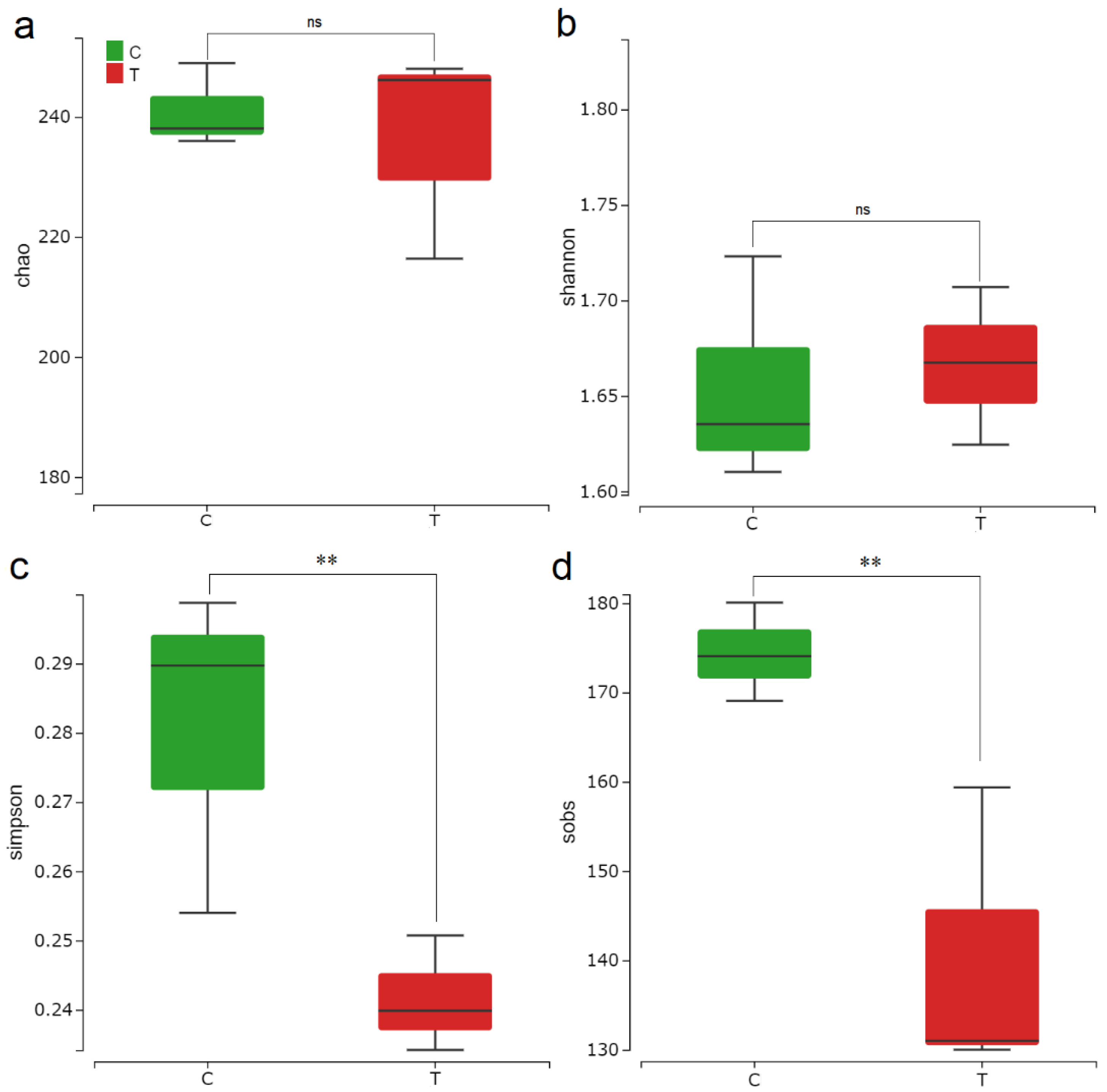
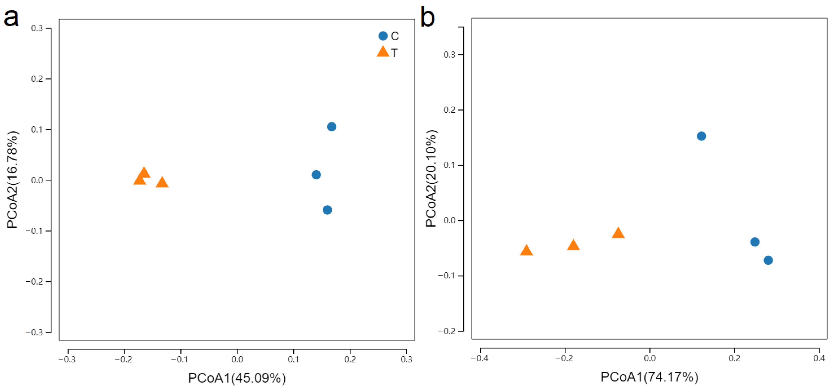
Inoculation with F. mosseae increased the α-diversity of rhizosphere bacterial community associated with C. pepo considerably (Figure 2). Compared with those in the uninoculated controls, both the Chao1 and Shannon indices of bacteria in the inoculated samples were significantly higher, indicating that F. mosseae inoculation increased bacterial diversity. The Simpson and Sobs indices of bacteria were also significantly higher in the inoculated samples than the uninoculated controls, suggesting that F. mosseae inoculation enhanced bacterial richness. Despite no significant difference in fungal diversity indices between treatments, F. mosseae inoculation diminished fungal richness, in the forms of lower Simpson and Sobs indices (Figure 3). According to the results of PCoA analysis, there was distinct variation in the β-diversity of bacterial (Figure 4a) and fungal (Figure 4b) communities between the two treatments. The results suggest that F. mosseae inoculation altered bacterial and fungal community composition prominently in the rhizosphere of C. pepo.

3.3. F. mosseae-Mediated Regulation of Microbial Community Structure in the Rhizosphere

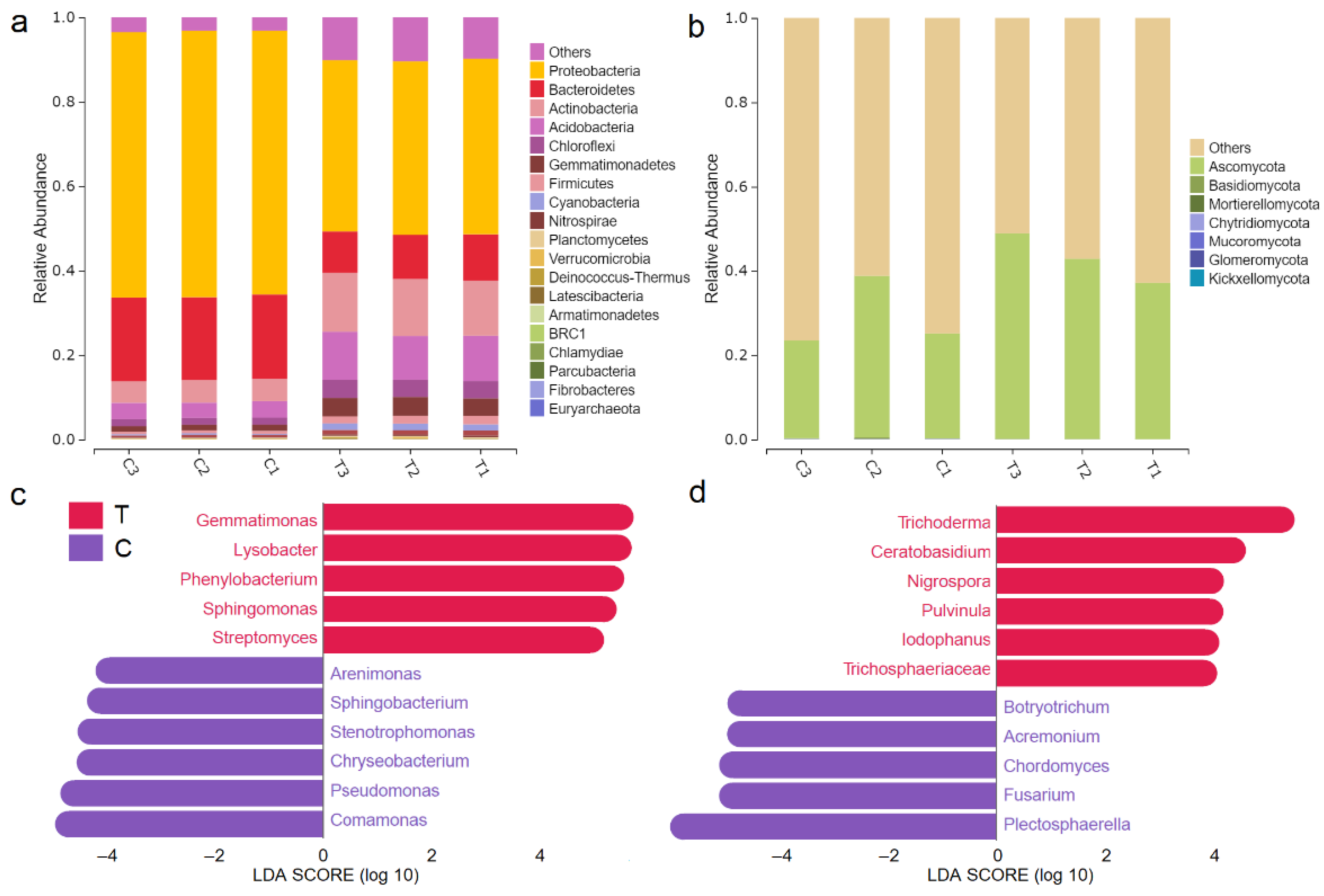
At the phylum level, the total bacterial community in the rhizosphere of C. pepo mainly consisted of Proteobacteria, Bacteroidetes, Actinobacteria, Acidobacteria, Chloroflexi, Gemmatimonadetes, Firmicutes, Cyanobacteria, Nitrospirae, and Planctomycetes (Figure 5a). Compared with the uninoculated controls, the relative abundances of Proteobacteria and Bacteroidetes in the inoculated samples decreased by 21.70% and 9.40%, respectively, whereas those of Actinobacteria, Acidobacteria, Chloroflexi, Gemmatimonadetes, Firmicutes, Cyanobacteria, and Nitrospirae increased by 8.23%, 7.07%, 2.50%, 2.94%, 1.20%, and 1.20%, respectively. The total fungal community mainly comprised Ascomycota, Basidiomycota, and Mortierellomycota. Compared with the uninoculated controls, the relative abundance of Ascomycota in the inoculated samples increased by 14.09%, whereas the relative abundances of Basidiomycota and Mortierellomycota decreased by 0.09% and 0.06%, respectively (Figure 5b).
LEfSe analysis was carried out to identify microbial taxa with significant differences in their relative abundance following F. mosseae inoculation (Figure 5c,d). At total of 11 differentially abundant taxa were identified in the inoculated samples, including five bacterial genera (i.e., Streptomyces, Sphingomonas, Phenylobacterium, Lysobacter, and Gemmatimonas) and six fungal genera (i.e., Trichosphaeriaceae, Iodophanus, Ceratobasidium, Trichoderma, Pulvinula, and Nigrospora). In the uninoculated controls, 11 differentially abundant taxa were identified, including six bacterial genera (i.e., Arenimonas, Sphingobacterium, Stenotrophomonas, Chryseobacterium, Pseudomonas, and Comamonas) and five fungal genera (i.e., Botryotrichum, Acremonium, Chordomyces, Fusarium, and Plectosphaerella).

3.4. Microbial Function Prediction

To explore the effect of F. mosseae inoculation on microbial functional pathways in the rhizosphere, the functional categories of rhizosphere microbial communities in the samples were predicted based on the KEGG database. A total of 10 metabolic pathways with significant changes in their relative abundance were identified in the inoculated samples (Figure 6). Carbohydrate metabolism accounted for the largest proportion of all the 10 pathways, followed by valine, leucine, and isoleucine biosynthesis; biosynthesis of vancomycin group antibiotics; glutamine and glutamate metabolism; and streptomycin biosynthesis. Among them, eight metabolic pathways were significantly upregulated, namely, arginine and ornithine metabolism; glutamine and glutamate metabolism; valine, leucine, and isoleucine biosynthesis; biosynthesis of vancomycin group antibiotics; streptomycin biosynthesis; oxidative phosphorylation; carbohydrate metabolism; and biosynthesis of secondary metabolites (p < 0.05). Among them, the upregulation of arginine and ornithine metabolism was the most prominent. In contrast, two metabolic pathways, namely, infectious diseases: bacterial and infectious diseases: parasitic, were significantly downregulated (p < 0.05).

4. Discussion

The present study showed that inoculation with F. mosseae effectively enhanced C. pepo plant growth and fruit yield under greenhouse conditions (Table 1). Specifically, plant height, leaf area, total biomass, and relative water content all increased substantially following F. mosseae inoculation, consistent with the results of previous studies. For example, Wu et al. [41] found that Zea mays plant height was increased following F. mosseae inoculation in a greenhouse study. In addition, F. mosseae inoculation reportedly increased Lycopersicon esculentum leaf area [42] and enhanced Glycyrrhiza uralensis plant biomass [43] under greenhouse conditions. Because leaves are a major organ of plant photosynthesis, increasing leaf area could provoke dry matter accumulation and yield formation in crops [44,45]. Furthermore, C. pepo root morphology was modified following F. mosseae inoculation, which could contribute to plant growth and fruit yield by enhancing nutrient and water uptake [19,25,44].
We hypothesized that the growth promotion and yield increase of C. pepo by F. mosseae are associated with F. mosseae-induced reshaping of rhizosphere microbial community structure. Based on LEfSe results, we found that F. mosseae induced a recruitment of plant growth-promoting microorganisms in the rhizosphere. For example, the relative abundances of Streptomyces, Sphingomonas, Lysobacter, and Trichoderma were all increased in the inoculated samples (Figure 5c,d), and positively correlated with plant growth and fruit yield parameters (Figure 7). Streptomyces can facilitate plant growth in T. aestivum [46] and Z. mays [47]. In addition, Sphingomonas and Lysobacter exhibit plant growth-promoting effects on Oryza sativa and Citrus sinensis, respectively [48,49], whereas Trichoderma enhances tomato plant growth and biomass [50]. Such beneficial effects of plant growth-promoting microorganisms are attributable to their intrinsic functions. For instance, both Streptomyces and Sphingomonas exhibit an excellent capacity to dissolve P [51,52]. Additionally, Streptomyces [53] and Sphingomonas can secrete indoleacetic acid to facilitate plant growth [52,54]. Furthermore, Trichoderma not only promotes plant growth by producing indoleacetic acid [55], but also enhances crop yield by altering rhizosphere microbial community composition [56].
As a major part of the rhizosphere microbial community, AMF play a vital role in the maintenance of rhizosphere microecological health. AMF inoculation can modify microbial community structure in the rhizosphere [57], thereby contributing to soil health maintenance [58]. In the present study, some microbial taxa potentially beneficial to plants, including the bacterial genera Streptomyces, Sphingomonas, and Lysobacter, and the fungal genus Trichoderma, were enriched in the rhizosphere of F. mosseae-inoculated plants. Among them, Streptomyces can produce numerous antibiotics to enhance plant disease and stress resistance [54]. In addition, Lysobacter exhibits broad-spectrum antimicrobial activity [59] and secretes antibiotics [60], both of which inhibit pathogen growth. Furthermore, Trichoderma, an important group of green biocontrol fungi, can constrain the growth of numerous fungal pathogens; following the colonization of the rhizosphere, Trichoderma can enhance plant stress and disease resistance [60]. In contrast, some fungal pathogens, such as Botryotrichum, Acremonium, Fusarium, and Plectosphaerella, were depleted in the rhizosphere of F. mosseae-inoculated plants. The relative abundances of these fungal pathogens were negatively correlated with C. pepo growth and yield parameters (Figure 7), possibly due to the antagonistic activity of Streptomyces, Aminomonas, Lysobacter, and Trichoderma [54,59,60,61]. Such antagonistic effects led to pronounced depletion of fungal pathogens, whereas many potentially beneficial microbial taxa were enriched in the rhizosphere of C. pepo. Consequently, a healthy micro-ecological environment of the rhizosphere was established for the plants, leading to considerable enhancement of C. pepo growth and yield.
Based on functional predictions using PICRUSt, we observed that pathways related to antibiotic biosynthesis (e.g., vancomycin group and streptomycin) were enriched following F. mosseae inoculation (Figure 6). As a protective barrier to disease infection, antibiotics play essential roles in plant health. We also observed that pathways associated with infectious diseases (infectious diseases: bacterial and infectious diseases: parasitic) were depleted following F. mosseae inoculation (Figure 6), which might be associated with the upregulation of antibiotic biosynthesis. The enrichment of antibiotic biosynthesis-related pathways can lead to the depletion of disease-related pathways [62]. In the present study, both Streptomyces and Lysobacter were enriched in the rhizosphere of C. pepo following F. mosseae inoculation, which could facilitate the production of numerous antibiotics [54,60] to enhance plant defense capacity against infectious diseases. Additionally, amino-acid metabolic pathways were enriched in the rhizosphere microbial communities after F. mosseae inoculation (Figure 6), which might be related to the enrichment of microbial taxa associated with N metabolism, such as Nitrospirae (increased by 1.20%; Figure 5). As the core microorganisms of soil N metabolism [63], Nitrospirae species can convert ammonia into nitrate, and then synthesize ammonia-like N through ammoniation, and further produce amino acids. In summary, the findings indicate that F. mosseae inoculation can enhance C. pepo plant growth and yield by regulating specific metabolic pathways of rhizosphere microbial communities, such as enriching the pathways associated with amino acid metabolism and antibiotic biosynthesis, while depleting the pathways associated with infectious diseases.

5. Conclusions

In the present study, pots with C. pepo seedlings were inoculated with F. mosseae, which promoted plant growth and increased fruit yield considerably. Additionally, the diversity and composition of rhizosphere microbiota were altered following F. mosseae inoculation. In particular, bacterial diversity and richness in the inoculated samples were distinctly enhanced. Furthermore, some potentially beneficial microbial taxa, such as Streptomyces, Sphingomonas, Lysobacter, and Trichoderma, were prominently enriched in the inoculated samples, whereas fungal pathogens, such as Botryotrichum, Acremonium, Fusarium, and Plectosphaerella, were depleted remarkably. F. mosseae inoculation also regulated the potential functions of rhizosphere microbiota, establishing more favorable conditions for plant growth and development. Overall, F. mosseae inoculation established a healthy micro-ecological environment in the rhizosphere of C. pepo, in turn enhancing plant growth and fruit yield.

Author Contributions

Investigation, J.W.; data curation, W.F.; writing—original draft preparation, C.S.; writing—review and editing, S.C.; visualization, C.T.; supervision, C.T.; funding acquisition, J.W. All authors have read and agreed to the published version of the manuscript.

Funding

This research was funded by the Key Research and Development Program of Yunnan Province, grant number 202203AC100002.

Institutional Review Board Statement

Not applicable.

Informed Consent Statement

Not applicable.

Data Availability Statement

The data presented in this study are available on request from the corresponding author.

Acknowledgments

We thank Z. Zhao and Y. Gao for assistance with sequencing data analysis.

Conflicts of Interest

The authors declare no conflict of interest.

References

  1. Santosh, K.B.; Prianka, H. Melatonin plays multifunctional role in horticultural crops against environmental stresses: A review. Environ. Exp. Bot. 2020, 176, 104063. [Google Scholar]
  2. Fu, C.; Shi, H.; Li, Q. A review on pharmacological activities and utilization technologies of pumpkin. Plant Food Hum. Nutr. 2006, 61, 70–77. [Google Scholar]
  3. Younis, Y.M.; Ghirmay, S.; Al-Shihry, S.S. African Cucurbita pepo L.: Properties of seed and variability in fatty acid composition of seed oil. Phytochemistry 2000, 54, 71–75. [Google Scholar] [CrossRef]
  4. Doran, J.W.; Zeiss, M.R. Soil health and sustainability: Managing the biotic component of soil quality. Appl. Soil. Ecol. 2000, 15, 3–11. [Google Scholar] [CrossRef] [Green Version]
  5. Chen, Q.L.; Cui, H.L.; Su, J.Q. Antibiotic resistomes in plant microbiomes. Trends Plant Sci. 2019, 24, 530–541. [Google Scholar] [CrossRef] [PubMed]
  6. Hiruma, K.; Gerlach, N.; Sacristán, S. Root endophyte Colletotrichum tofieldiae confers plant fitness benefits that are phosphate status dependent. Cell 2016, 165, 464–474. [Google Scholar] [CrossRef] [PubMed] [Green Version]
  7. Wang, X.; Wei, Z.; Yang, K. Phage combination therapies for bacterial wilt disease in tomato. Nat. Biotechnol. 2019, 37, 1513–1520. [Google Scholar] [CrossRef]
  8. Zhang, C.L.; Zhang, Y.M.; Ding, Z.J. Contribution of microbial inter-kingdom balance to plant health. Mol. Plant 2019, 12, 148–149. [Google Scholar] [CrossRef] [Green Version]
  9. Smith, F.A.; Smith, S.E. What is the significance of the arbuscular mycorrhizal colonisation of many economically important crop plants? Plant Soil 2011, 348, 63–79. [Google Scholar] [CrossRef]
  10. Hodge, A.; Fitter, A.H. Substantial nitrogen acquisition by arbuscular mycorrhizal fungi from organic material has implications for N cycling. Proc. Natl. Acad. Sci. USA 2010, 107, 13754–13759. [Google Scholar] [CrossRef] [Green Version]
  11. Hélène, J. A Medicago truncatula phosphate transporter indispensable for the arbuscular mycorrhizal symbiosis. Proc. Natl. Acad. Sci. USA 2007, 104, 1720–1725. [Google Scholar]
  12. Akhtar, M.S.; Siddiqui, Z.A. Biocontrol of a chickpea root-rot disease complex with Glomus intraradices, Pseudomonas putida and Paenibacillus polymyxa. Australas Plant Path. 2007, 36, 175–180. [Google Scholar] [CrossRef]
  13. Aroca, R.; Porcel, R. How does arbuscular mycorrhizal symbiosis regulate root hydraulic properties and plasma membrane aquaporins in Phaseolus vulgaris under drought, cold or salinity stresses? New Phytol. 2007, 173, 808–816. [Google Scholar] [CrossRef]
  14. Johnson, N.C.; Graham, J.H.; Smith, F.A. Functioning of mycorrhizal associations along the mutualism parasitism continuum. New Phytol. 1997, 135, 575–586. [Google Scholar] [CrossRef]
  15. Ahmad, A.; Imran, G.M.; Ding, H.Y.; Iqbal, M.; Cheng, Z.H.; Cai, Z.C. Arbuscular mycorrhizal inoculum coupled with organic substrate induces synergistic effects for soil quality changes, and rhizosphere microbiome structure in long-term monocropped cucumber planted soil. Rhizosphere 2021, 20, 100428. [Google Scholar]
  16. Linderman, R.G. Mycorrhizae and Plant Health; Pfleger, F.L., Linderman, R.G., Eds.; American Physics Society Press: St. Paul, MN, USA, 1994; pp. 1–25. [Google Scholar]
  17. Martin, B.N.; Joan, L.; Damase, P.K. Mycorrhizae and rhizobacteria on Precambrian rocky gold mine tailings: I. mine-adapted symbionts promote white spruce health and growth. Front. Plant Sci. 2018, 9, 1267. [Google Scholar]
  18. Bizos, G.; Efimia, M.; Papatheodorou, T.C.; Ntalli, N.; Vassilis, G.; Aschonitis, N.K. The role of microbial inoculants on plant protection, growth stimulation, and crop productivity of the olive tree (Olea europea L.). Plants 2020, 9, 743. [Google Scholar] [CrossRef]
  19. Chatzistathis, T.; Orfanoudakis, M.; Alifragis, D.; Therios, I. Colonization of Greek olive cultivars’ root system by arbuscular mycorrhiza fungus: Root morphology, growth, and mineral nutrition of olive plants. Sci. Agric. 2013, 70, 185–194. [Google Scholar] [CrossRef] [Green Version]
  20. Abd-Alhamid, N.; Hassan, H.S.A.; Haggag, L.F.; Hassan, A.M. Effect of mineral and bio-fertilization on vegetative growth, leaf mineral contents and flowering of manzanillo olive trees. Int. J. ChemTech. Res. 2015, 8, 51–61. [Google Scholar]
  21. Meng, L.L.; Srivastava, A.K.; Kuca, K.; Wu, Q.S. Earthworm (Pheretima guillelmi)-mycorrhizal fungi (Funneliformis mosseae) association mediates rhizosphere responses in white clover. Appl. Soil. Ecol. 2022, 172, 104371. [Google Scholar] [CrossRef]
  22. Seifi, E.; Teymoor, Y.S.; Alizadeh, M.; Fereydooni, H. Olive mycorrhization: Influences of genotype, mycorrhiza, and growing periods. Sci. Hortic. Amst. 2014, 180, 214–219. [Google Scholar] [CrossRef]
  23. Kapulnik, Y.; Tsror, L.; Zipori, I.; Hazanovsky, M.; Wininger, S.; Dag, A. Effect of AMF application on growth, productivity and susceptibility to Verticillium wilt of olives grown under desert conditions. Symbiosis 2010, 52, 103–111. [Google Scholar] [CrossRef]
  24. Giovannini, C.; Garcia-Mina, J.M.; Ciavatta, C.; Marzadori, C. Ureic nitrogen transformation in multi-layer soil columns treated with urease and nitrification inhibitors. J. Agric. Food Chem. 2009, 57, 4883–4887. [Google Scholar] [CrossRef] [PubMed]
  25. Sun, C.; Yang, Y.; Zeeshan, M.; Qin, S.; Ma, J.; Liu, L.; Yang, J.; Zhou, X.; Huang, J. Arbuscular mycorrhizal fungi reverse selenium stress in Zea mays seedlings by improving plant and soil characteristics. Ecotoxicol. Environ. Saf. 2021, 228, 113000. [Google Scholar] [CrossRef] [PubMed]
  26. Puschel, D.; Rydlova, J.; Sudova, R.; Gryndler, M.; Vosatka, M. The potential of mycorrhizal inoculation and organic amendment to increase yields of Galega orientalis and Helianthus tuberosus in a spoil-bank substrate. J. Plant Nutr. Soil. Sci. 2011, 174, 664–672. [Google Scholar] [CrossRef]
  27. Yu, Y.; Zhang, S.; Huang, H.; Luo, L.; Wen, B. Arsenic accumulation and speciation in maize as affected by inoculation with arbuscular mycorrhizal fungus Glomus mosseae. J. Agric. Food Chem. 2009, 57, 3695–3701. [Google Scholar] [CrossRef]
  28. Escrig, P.V.; Iglesias, D.J.; Corma, A.; Primo, J.; Primo-Millo, E.; Cabedo, N. Euphorbia characias as bioenergy crop: A study of variations in energy value components according to phenology and water status. J. Agric. Food Chem. 2013, 61, 10096–10109. [Google Scholar] [CrossRef]
  29. Zhao, L.; Sun, Y.; Hernandez-Viezcas, J.A.; Servin, A.D.; Hong, J.; Niu, G.; Peralta-Videa, J.R.; Duarte-Gardea, M.; Gardea-Torresdey, J.L. Influence of CeO2 and ZnO nanoparticles on cucumber physiological markers and bioaccumulation of Ce and Zn: A life cycle study. J. Agric. Food Chem. 2013, 61, 11945–11951. [Google Scholar] [CrossRef]
  30. Zafari, M.; Ebadi, A.; Jahanbakhsh, S.; Sedghi, M. Safflower (Carthamus tinctorius) biochemical properties, yield, and oil content affected by 24-epibrassinosteroid and genotype under drought stress. J. Agric. Food Chem. 2020, 68, 6040–6047. [Google Scholar] [CrossRef]
  31. Phillips, J.M.; Hayman, D.S. Improved procedures for clearing roots and staining parasitic and vesicular-arbuscular mycorrhizal fungi for rapid assessment of infection. Trans. Br. Mycol. Soc. 1970, 55, 158–161. [Google Scholar] [CrossRef]
  32. Lear, G.; Dickie, I.; Banks, J.; Boyer, S.; Buckley, H.L.; Buckley, T.R.; Cruickshank, R.; Dopheide, A.; Handley, K.M.; Hermans, S.; et al. Methods for the extraction, storage, amplification and sequencing of DNA from environmental samples. N. Zldn. J. Ecol. 2018, 4, 10. [Google Scholar] [CrossRef] [Green Version]
  33. Yu, P.; He, X.; Baer, M.; Beirinckx, S.; Tian, T.; Moya, Y.; Zhang, X.; Deichmann, M.; Frey, F.P.; Bresgen, V.; et al. Plant flavones enrich rhizosphere Oxalobacteraceae to improve maize performance under nitrogen deprivation. Nat. Plants 2021, 7, 481–499. [Google Scholar] [CrossRef] [PubMed]
  34. Duan, S.; Hu, X.; Li, M.; Miao, J.; Du, J.; Wu, R. Composition and metabolic activities of the bacterial community in shrimp sauce at the flavor-forming stage of fermentation as revealed by metatranscriptome and 16S rRNA gene sequencings. J. Agric. Food Chem. 2016, 64, 2591–2603. [Google Scholar] [CrossRef] [PubMed]
  35. Caporaso, J.G.; Kuczynski, J.; Stombaugh, J.; Bittinger, K.; Bushman, F.D.; Costello, E.K.; Fierer, N.; Peña, A.G.; Goodrich, J.K.; Gordon, J.I.; et al. QIIME allows analysis of high-throughput community sequencing data. Nat. Methods 2010, 7, 335–336. [Google Scholar] [CrossRef] [PubMed] [Green Version]
  36. Schloss, P.D.; Westcott, S.L.; Ryabin, T.; Hall, J.R.; Hartmann, M.; Hollister, E.B.; Lesniewski, R.A.; Oakley, B.B.; Parks, D.H.; Robinson, C.J.; et al. Introducing Mothur: Open-source, platform-independent, community-supported software for describing and comparing microbial communities. Appl. Environ. Microbiol. 2009, 75, 7537–7541. [Google Scholar] [CrossRef] [PubMed] [Green Version]
  37. Lozupone, C.; Lladser, M.E.; Knights, D.; Stombaugh, J.; Knight, R. UniFrac: An effective distance metric for microbial community comparison. ISME J. 2011, 5, 169–172. [Google Scholar] [CrossRef] [PubMed]
  38. Segata, N.; Huttenhower, C. Toward an efficient method of identifying core genes for evolutionary and functional microbial phylogenies. PLoS ONE 2011, 6, e24704. [Google Scholar] [CrossRef] [PubMed]
  39. Langille, M.G.I.; Zaneveld, J.; Caporaso, J.G.; McDonald, D.; Knights, D.; Reyes, J.A.; Clemente, J.C.; Burkepile, D.E.; VegaThurber, R.L.; Knight, R.; et al. Predictive functional profiling of microbial communities using 16S rRNA marker gene sequences. Nat. Biotechnol. 2013, 31, 814–821. [Google Scholar] [CrossRef]
  40. Shannon, P.; Markiel, A.; Ozier, O.; Baliga, N.S.; Wang, J.T.; Ramage, D.; Amin, N.; Schwikowski, B.; Ideker, T. Cytoscape: A software environment for integrated models of biomolecular interaction networks. Genome Res. 2003, 13, 2498–2504. [Google Scholar] [CrossRef]
  41. Wu, S.C.; Cao, Z.H.; Li, Z.G.; Cheung, K.C.; Wong, M.H. Effects of biofertilizer containing N-fixer, P and K solubilizers and AM fungi on maize growth: A greenhouse trial. Geoderma 2004, 125, 155–166. [Google Scholar] [CrossRef]
  42. Al-Karaki, G.N. Growth of mycorrhizal tomato and mineral acquisition under salt stress. Mycorrhiza 2000, 10, 51–54. [Google Scholar] [CrossRef]
  43. Chen, M.L.; Yang, G.; Sheng, Y.; Li, P.Y.; Qiu, H.Y.; Zhou, X.T.; Huang, L.Q.; Chao, Z. Glomus mosseae inoculation improves the root system architecture, photosynthetic efficiency and flavonoids accumulation of liquor ice under nutrient stress. Front. Plant Sci. 2017, 8, 931. [Google Scholar] [CrossRef] [PubMed] [Green Version]
  44. Wu, Q.S.; Xia, R.X. Arbuscular mycorrhizal fungi influence growth, osmotic adjustment and photosynthesis of citrus under well-watered and water stress conditions. J. Plant Physiol. 2006, 163, 417–425. [Google Scholar] [CrossRef] [PubMed]
  45. Al-Karaki, G.N.; Hammad, R.; Rusan, M. Response of two tomato cultivars differing in salt tolerance to inoculation with mycorrhizal fungi under salt stress. Mycorrhiza 2001, 11, 43–47. [Google Scholar] [CrossRef]
  46. Jog, R.; Pandya, M.; Nareshkumar, G.; Rajkumar, S. Mechanism of phosphate solubilization and antifungal activity of Streptomyces spp. isolated from wheat roots and rhizosphere and their application in improving plant growth. Microbiology 2014, 160 Pt 4, 778–788. [Google Scholar] [CrossRef] [Green Version]
  47. Ma, J.; Liu, Y.; Li, Y.; Sun, Y.; Yang, B.; Lai, H.; Xue, Q. Effects and mechanism of two Streptomyces strains on promoting plant growth and increasing grain yield of maize. Chin. J. Appl. Ecol. 2017, 28, 315–326. [Google Scholar]
  48. Adikari, T.B.; Joseph, C.M.; Yang, G.P. Evaluation of bacteria isolated from rice for plant growth promotion and biological control of seedling disease of rice. Can. J. Microbiol. 2001, 47, 916–924. [Google Scholar] [CrossRef]
  49. Trivedi, P.; Duan, Y.P.; Wang, N. Huanglongbing, a systemic disease, restructures the bacterial community associated with citrus roots. Appl. Environ. Microbiol. 2010, 76, 3427–3436. [Google Scholar] [CrossRef] [Green Version]
  50. Lee, S.; Yap, M.; Behringer, G. Volatile organic compounds emitted by Trichoderma species mediate plant growth. Fungal Biol. Biotechnol. 2016, 3, 7. [Google Scholar] [CrossRef] [Green Version]
  51. Marcela, F.C.; Angelica, Q.; Christian, D.; Christian, S.; Maria, X. Evaluation of actinomycete strains for key traits related with plant growth promotion and mycorrhiza helping activities. Appl. Soil. Ecol. 2010, 45, 209–217. [Google Scholar]
  52. Asaf, S.; Numan, M.; Khan, A.L.; Al-Harrasi, A. Sphingomonas: From diversity and genomics to functional role in environmental remediation and plant growth. Crit. Rev. Biotechnol. 2020, 40, 138–152. [Google Scholar] [CrossRef] [PubMed]
  53. El-Tarabily, K.A. Promotion of tomato (Lycopersicon esculentum Mill.) plant growth by rhizosphere competent 1-aminocyclopropane-1-carboxylic acid deaminase-producing streptomycete actinomycetes. Plant Soil. 2008, 308, 161–174. [Google Scholar] [CrossRef]
  54. Vilaene, T.; Langendries, S.; Beirinckx, S.; Maes, M.; Goormactig, S. Streptomyces as a plant’s best friend? FEMS Microbiol. Ecol. 2016, 92, fiw119. [Google Scholar] [CrossRef] [Green Version]
  55. Zhang, S.W.; Gan, Y.T.; Xu, B.L. Mechanisms of the IAA and ACC-deaminase producing strain of Trichoderma longibrachiatum T6 in enhancing wheat seedling tolerance to NaCl stress. BMC Plant Biol. 2019, 19, 22. [Google Scholar] [CrossRef] [PubMed] [Green Version]
  56. Zhang, F.G.; Xu, X.X.; Huo, Y.Q. Trichoderma-inoculation and mowing synergistically altered soil available nutrients, rhizosphere chemical compounds and soil microbial community, potentially driving alfalfa growth. Front. Microbiol. 2018, 9, 3241. [Google Scholar] [CrossRef]
  57. Miransari, M. Interactions between arbuscular mycorrhizal fungi and soil bacteria. Appl. Microbiol. 2011, 89, 917–930. [Google Scholar] [CrossRef]
  58. Sharmah, D.; Jha, D.K. Diversity of arbuscular mycorrhizal fungi in undisturbed forest, slash-and-burn field, and monoculture forest of Indo-Burma megadiverse region. Braz. J. Bot. 2014, 37, 339–351. [Google Scholar] [CrossRef]
  59. Postma, J.; Stevens, L.H.; Wiegers, G.L. Biological control of Pythium aphanidermatum in cucumber with a combined application of Lysobacter enzymogenes strain 3.1T8 and chitosan. Biol. Control 2009, 48, 301–309. [Google Scholar] [CrossRef]
  60. Wang, Y.; Dai, J.; Zhang, L. Lysobacter ximonensis sp. nov., isolated from soil. Int. J. Syst. Evol. Microbiol. 2009, 59, 786–789. [Google Scholar] [CrossRef] [Green Version]
  61. Wang, Y.F.; Hou, X.Y.; Jiang, C.Y.; Zhai, T.T.; Miao, R.; Deng, J.J.; Yao, Z.H.; Zhang, R.S. A native Trichoderma harzianum strain Th62 displays antagonistic activities against phytopathogenic fungi and promotes the growth of Celosia cristata. Horticult. Environ. Biotechnol. 2021, 62, 169–179. [Google Scholar]
  62. Pierson, L.S.; Thomashow, L.S. Cloning and hetero logous expression of the phenazine biosynthetic locus from Pseudomonas aureofaciens 30–84. Mol. Plant Microbe Interact. 1992, 5, 330–339. [Google Scholar] [CrossRef] [PubMed]
  63. Xun, W.; Liu, Y.; Li, W.; Li, W.; Ren, Y.; Xiong, W.; Xu, Z.H.; Zhang, N.; Miao, Y.Z.; Shen, Q.R.; et al. Specialized metabolic functions of keystone taxa sustain soil microbiome stability. Microbiome 2021, 9, 35. [Google Scholar] [CrossRef] [PubMed]
Figure 1. Venn diagrams showing the unique and common operational taxonomic units (OTUs) of rhizosphere bacterial (a) and fungal (b) communities between rhizosphere soil samples inoculated with Funneliformis mosseae (T) and without inoculation (C).
Diversity 14 00932 g001
Figure 2. α-Diversity of rhizosphere bacterial community associated with Cucurbita pepo plants grown in Funneliformis mosseae-inoculated soil (T) and uninoculated control soil (C). (a), (b), (c), and (d) represent the Chao1, Shannon, Simpson, and Sobs indices, respectively. In boxplots, the middle line represents median, the upper and bottom horizontal lines represent the third and first quartiles, respectively, and the upper and lower whiskers represent the inter-quartile range. Asterisks (**) indicate significant difference between the two treatments, at the level of p < 0.01 (n = 3).
Diversity 14 00932 g002
Figure 3. α-Diversity of rhizosphere fungal community associated with Cucurbita pepo plants grown in Funneliformis mosseae-inoculated soil (T) and uninoculated control soil (C). (a), (b), (c), and (d) represent the Chao1, Shannon, Simpson, and Sobs indices, respectively. In boxplots, the middle line represents the median, the upper and bottom horizontal lines represent the third and first quartiles, respectively, and the upper and lower whiskers represent the inter-quartile range. Asterisks (**) indicate significant difference between the two treatments, at the level of p < 0.01 (n = 3). Ns indicates no significant difference between the two treatments.
Diversity 14 00932 g003
Figure 4. Principal coordinate analysis (PCoA) of bacterial (a) and fungal (b) β-diversity based on unweighted UniFrac distance between all rhizosphere soil samples inoculated with Funneliformis mosseae (T) and without inoculation (C) (n = 3).
Diversity 14 00932 g004
Figure 5. The relative abundances of major bacterial (a) and fungal (b) phyla, and the graphical summary of the top 11 bacterial (c) and fungal (d) biomarkers identified by LEfSe, in the rhizosphere of Cucurbita pepo plants inoculated with Funneliformis mosseae (T) and without inoculation (C) (n = 3).
Diversity 14 00932 g005
Figure 6. Functional prediction of rhizosphere microbial communities associated with Cucurbita pepo based on PICRUSt between all rhizosphere soil samples inoculated with Funneliformis mosseae (T) and without inoculation (C), at the level of p < 0.01 (n = 3).
Diversity 14 00932 g006
Figure 7. Pearson correlation network of F. mosseae colonization, plant growth, fruit yield, and microbial taxa. The green and red lines represent positive and negative correlations, respectively.
Diversity 14 00932 g007
Table 1. Effect of inoculation with Funneliformis mosseae on Cucurbita pepo growth and yield. Asterisks (**) indicate significant difference between the two treatments, at the level of p < 0.01 (Student’s t-test; n = 20).
TreatmentRoot Colonization Rate
(%)
Plant Height
(cm)
Stem Diameter
(cm)
Leaf Area
(cm2)
Plant Fresh Weight
(g plant−1)
Plant Dry Weight
(g plant−1)
Relative Water Content (%)Fruit Yield
(kg plant−1)
Uninoculated control60.22.0657732780.775.24.31
Inoculation23.465.1 **2.01625 **388 **89.2 **82.3 **4.62 **
Coefficient of variation (%)10.416.22.620.415.514.49.212.8
Table 2. Effect of inoculation with Funneliformis mosseae on Cucurbita pepo root morphology. Asterisks (**) indicate significant difference between the two treatments, at the level of p < 0.01 (Student’s t-test; n = 20).
TreatmentRoot Length
(m)
Root Volume
(cm3)
Root Surface Area
(cm2)
Root Diameter
(mm)
Uninoculated control9.46.226.80.8
Inoculation12.3 **7.7 **33.2 **1.0 **
Coefficient of variation (%)14.213.920.112.9
Publisher’s Note: MDPI stays neutral with regard to jurisdictional claims in published maps and institutional affiliations.