Article

Micractinium tetrahymenae (Trebouxiophyceae, Chlorophyta), a New Endosymbiont Isolated from Ciliates

1
Research Department for Limnology, Leopold-Franzens-University of Innsbruck, Mondsee, A-5310 Mondsee, Austria
2
Limnological Station, Department of Plant and Microbial Biology, University of Zürich, CH-8802 Kilchberg, Switzerland
3
Albrecht-von-Haller-Institute of Plant Sciences, Experimental Phycology and Culture Collection of Algae, Georg-August-University of Göttingen, D-37073 Göttingen, Germany
*
Author to whom correspondence should be addressed.
Diversity 2020, 12(5), 200; https://doi.org/10.3390/d12050200
Submission received: 28 April 2020 / Revised: 13 May 2020 / Accepted: 13 May 2020 / Published: 15 May 2020
(This article belongs to the Special Issue Biodiversity of Ciliates and their Symbionts)

Abstract

:
Endosymbiosis between coccoid green algae and ciliates are widely distributed and occur in various phylogenetic lineages among the Ciliophora. Most mixotrophic ciliates live in symbiosis with different species and genera of the so-called Chlorella clade (Trebouxiophyceae). The mixotrophic ciliates can be differentiated into two groups: (i) obligate, which always live in symbiosis with such green algae and are rarely algae-free and (ii) facultative, which formed under certain circumstances such as in anoxic environments an association with algae. A case of the facultative endosymbiosis is found in the recently described species of Tetrahymena, T. utriculariae, which lives in the bladder traps of the carnivorous aquatic plant Utricularia reflexa. The green endosymbiont of this ciliate belonged to the genus Micractinium. We characterized the isolated algal strain using an integrative approach and compared it to all described species of this genus. The phylogenetic analyses using complex evolutionary secondary structure-based models revealed that this endosymbiont represents a new species of Micractinium, M. tetrahymenae sp. nov., which was further confirmed by the ITS2/CBC approach.

Graphical Abstract

1. Introduction

The genus Micractinium with its type species, M. pusillum, was described by Fresenius [1] for a coccoid green alga, which formed colonies of 2–4 cells and produced bristles. Since the first description, several species of Micractinium were established based on cell shape, number of bristles, and arrangement of cells into colonies [2]. All species occurred in all kinds of freshwater habitats, such as lakes and small ponds, and were typical planktonic species. Phylogenetic analyses of Micractinium surprisingly showed that M. pusillum is closely related to the genus Chlorella, a unicellular green alga without any cell appendices. Luo et al. [3,4] have demonstrated that the colony and bristle formation was a response on grazing through the rotifer Brachionus calyciflorus. The SSU and ITS rDNA sequences revealed that M. pusillum represented a cryptic species complex [4,5]. In addition, Pröschold et al. [6] transferred the genus Diacanthos with its type species D. belenophorus to the genus Micractinium. Apart from these free-living species of Micractinium, Pröschold et al. [7] indicated that a green algal endosymbiont of the ciliate Paramecium bursaria also belonged to Micractinium. Brandt [8] was the first who discovered that “chlorophyll-bearing bodies” in Paramecium bursaria and Stentor polymorphus were independent organisms and not plastids. Since then, endosymbiotic algae in ciliates, heliozoa, amoeba, or other invertebrates have been of special interests in phycology as well as in zoology, microbiology, and virology. Within ciliates, green algal endosymbionts are widely distributed. Around 40 species of ciliates and other protists live in symbiosis with green algae [9]. For most of these endosymbionts, the origin and phylogenetic position are unknown. The majority of the investigated green algae belong to the Chlorella clade of the Trebouxiophyceae ([7] and references therein). Interestingly, the endosymbionts do not form a single lineage within the Chlorella clade, but are closely related to free-living species of Chlorella, Micractinium [7], and Meyerella [10], and sometimes formed an own genus like Carolibrandtia [11,12].
Symbiotic interactions between green algae and ciliates are known to be of different nature. Some mixotrophic ciliates always bear zoochlorellae in their cells and rarely occur algae-free. Such obligate endosymbiosis is found for example in Paramecium bursaria, one of best investigated ciliate species [13]. In contrast, several ciliates live only facultatively in symbiosis with green algae. One of these ciliates is the recently described Tetrahymena utriculariae, which lives in symbiosis with the alga Micractinium [14]. T. utriculariae lives inside bladder traps of Utricularia reflexa, a carnivorous aquatic plant. The ciliate survives the typically anoxic and nutrient-rich milieu inside traps, most likely because of its green algal endosymbionts. Cultivated outside the traps under oxygenic conditions, the ciliates lose their endosymbionts and switch to a heterotrophic way of life. This clearly indicated that the green algal endosymbiont Micractinium has a special function by providing oxygen to its hosts [15].
The aim of this study was to clarify the phylogenetic position and the taxonomic status within Micractinium. We isolated the strain from its host Tetrahymena utriculariae and deposited it under the number SAG 2587 in the Culture Collection of Algae at the University of Göttingen. We used an integrative approach (morphology and phenotypic plasticity, SSU, and ITS rDNA sequences including their secondary structures) for comparing this strain with existing described species of Micractinium.

2. Material and Methods

The strain SAG 2587 was isolated from the host as described in Pitsch et al. [14] and cultivated on agarized basal medium with peptone (ESP; medium 1b in [16]). For morphological investigations, we cultivated the strain at 18 °C, with 50 µmol photons/m2s1 provided by daylight fluorescent tubes (Osram L36W/954 Lumilux de lux daylight, Munich, Germany), and light:dark cycle of 16:8 hrs for two to three weeks. The light microscopic investigations were conducted using an Olympus BX-60 microscope (Olympus, Tokyo, Japan) and the micrographs were taken with a ProgRes C14plus camera using the ProgRes CapturePro imaging system (version 2.9.0.1, both from Jenoptik, Jena, Germany).
The genomic DNA of the strain was extracted using the DNeasy Plant Mini Kit (Qiagen, Hilden, Germany) following the instructions provided by the manufacturer. The SSU and ITS rDNA was amplified in PCR reactions using the Taq PCR MasterMix Kit (Qiagen, Hilden, Germany) with the primers EAF3 and ITS055R [17]. The PCR product was purified and sequenced as described by Darienko et al. [18]. The SSU and ITS rDNA sequence is available in the EMBL, GenBank, and DDBJ sequence databases under the accession number MT359915. This sequence was aligned and included into a data set of a total of 40 sequences (2602 bp) of representatives of the Chlorellaceae (Trebouxiophyceae). The data set was aligned according to the secondary structures. The secondary structures were folded using the software mfold [19], which uses the thermodynamic model (minimal energy) for RNA folding. GenBank accession numbers of all sequences used are given in the figure. For the phylogenetic analyses, the dataset with unambiguously aligned base positions was used. To test which evolutionary model fitted best for the data set, we calculated the log-likelihood values of 56 models using the automated model selection tool implemented in PAUP, version 4.0b167 [20], and the best model according to the Akaike criterion by PAUP was chosen for the analyses. The setting of the best model is given in the figure legend. The following methods were used for the phylogenetic analyses: distance, maximum parsimony, maximum likelihood, and Bayesian inference. Programs used included PAUP version 4.0b167 [20], RAxML version 8.2.12 [21], MrBayes version 3.2.7a [22], and the PHASE package 2.0 [23,24,25,26,27]. For the Bayesian calculations, the secondary structure models of SSU and ITS (doublet in MrBayes and RNA7D in PHASE) were also taken into account.

3. Results

Micractinium tetrahymenae Pröschold, Pitsch, & Darienko sp. nov. (Figure 1)
Description: Young cells are solitary, ellipsoidal up to broadly ellipsoidal; 3.1–4.2 µm in size. Mature vegetative cells are broadly ellipsoidal up to spherical, 4.8 × 4.9 µm up to 7.1 × 7.6 µm in size; rarely pyriform under suboptimal condition, 8.5 × 5.3 µm. Old cells are spherical up to 9.3 µm in diameter. Chloroplast is parietal cup-shaped possessing a single pyrenoid surrounded by starch grains. Cytoplasm is vacuolized. Asexual reproduction by autosporulation. The autospores are produced by 2–4 per cell. Autosporangia are 4.6 × 6.2 µm up to 6.3 × 7.4 µm. Release of autospores occurs after rupture of the mother cell wall. Bristle formation was not observed.
Diagnosis: Differs from morphologically similar M. conductrix and other free-living species of Micractinium through genetic signatures in SSU and ITS-2 rDNA sequences as well as in ITS-2 Barcode (see Section 4.2).
Holotype (designated here): The authentic strain SAG 2587 is cryopreserved in a metabolically inactive state at SAG under the number Z000694542.
Type locality: Facultative endosymbiont of Tetrahymena utriculariae (Oligohymenophorea, Ciliophora).
Etymology: The name reflected the appearance in the host organism.
Phylogenetic position and genetic signatures of the endosymbiont of Tetrahymena utriculariae: The SSU and ITS rDNA sequences of strain SAG 2587 (MT359915) were completely identical with those deposited in GenBank by Pitsch et al. [14] under the number LT605003. This endosymbiont clearly is the sister of Micractinium pusillum, based on the phylogenetic analyses of SSU and ITS rDNA sequences (Figure 2). The genus Micractinium is only highly supported in Bayesian analyses using the complex evolutionary models, which included the doublet and RNA7D functions (secondary structure models implemented in MrBayes and PHASE, respectively; see details in Material and Methods). The maximum likelihood analyses using bootstrapping resulted in a high to moderate support for the genus Micractinium. In contrast, the common branch of the genus Chlorella was not supported in Bayesian analyses and only got moderate values in bootstrap calculations. All analyses showed that the separation of Micractinium and Chlorella is not supported using simple evolutionary models and distance or parsimony methods (data not shown). However, both genera together were highly supported in all analyses questioning the separation into two genera. The other genera belonging to the Chlorella clade were highly supported in all of our analyses.
Within Micractinium, M. tetrahymenae sp. nov. is closely related to M. pusillum. The genetic variability of SSU rDNA among the species of Micractinium was very low (only 28 variable positions of 1783 bp = 1.6%). Even variable regions such as V4 showed only little changes (5 bases). Only the V9 region was partly diagnostic (Figure 3), being unique for both, M. conductrix and M. pusillum. In contrast, The V9 of M. tetrahymenae/M. belenophorum and M. inermum/M. simplicissimum/M. singulare/M.variabile were identical, respectively. The variability among the species was higher in the ITS-1 and ITS-2. The general structures of M. tetrahymenae are presented in Figure 4 and were similar to those of the members of Micractinium and other genera of the Chlorella clade. The ITS-1 and ITS-2 showed the typical four helices called helices 1–4 of ITS-1 and helices I-IV for ITS-2 according to Coleman and Mai [28]. The differences among the species in ITS-1 and ITS-2 showed that all species could be distinguished by characteristic compensatory base changes (CBCs and HCBCs) and loops (highlighted in white boxes in Figure 5 and Figure 6). The base pair differences of V9 (SSU) and the conserved region of ITS-2 among the Micractinium species are summarized in Figure 7. In total, ten CBCs, seven HCBCs, and six insertion/deletions could be discovered (highlighted with an asterisk in Figure 7). By replacing base pairs with a number code, representatives of Micractinium received a unique barcode based on which species could be clearly recognized.

4. Discussion

4.1. Green Algae in Endosymbiosis Belonging to the Chlorellaceae

Zoochlorellae or Chlorella-like algae living as endosymbionts in ciliates and other protozoa are known for a long time ([7] and references therein). Interestingly, most of these green algae belonged to the Chlorella clade of the Trebouxiophyceae. Within this clade, six out of the seven species (highlighted with green circles in Figure 2) exclusively occurred in endosymbiotic associations. Only Chlorella vulgaris could be found free-living in various habitats (see details in reference [18]). C. vulgaris, C. variabilis, and Micractinium conductrix formed an obligate endosymbiont in Paramecium bursaria [7,29]. Meyerella planctonica is the endosymbiont of another green Paramecium (P. chlorelligerum [10]). The genus Carolibrandtia was discovered to be the endosymbionts of the ciliates Pelagodileptus trachelioides, Cyclotrichium viride, and Stokesia vernalis [11,12].
Micractinium tetrahymenae sp. nov. represented a second species within this genus that lives in symbiosis with a ciliate. In contrast to M. conductrix, a species which is the obligate endosymbiont of Paramecium bursaria [7,29] and Coleps primhirtus [30], M. tetrahymenae formed only under anoxic or microaerobic conditions a symbiotic association with Tetrahymena utriculariae [14]. This demonstrated that M. tetrahymenae is a facultative endosymbiont. However, whether this species can also occur free-living needs further investigations. No entry in GenBank could be found in the BLASTn search (100% coverage, 97% identity) using our SSU and ITS sequence (2452 bp). It is also unknown if Tetrahymena utriculariae would be able to live in symbiosis with other green algae belonging to the Chlorellaceae.

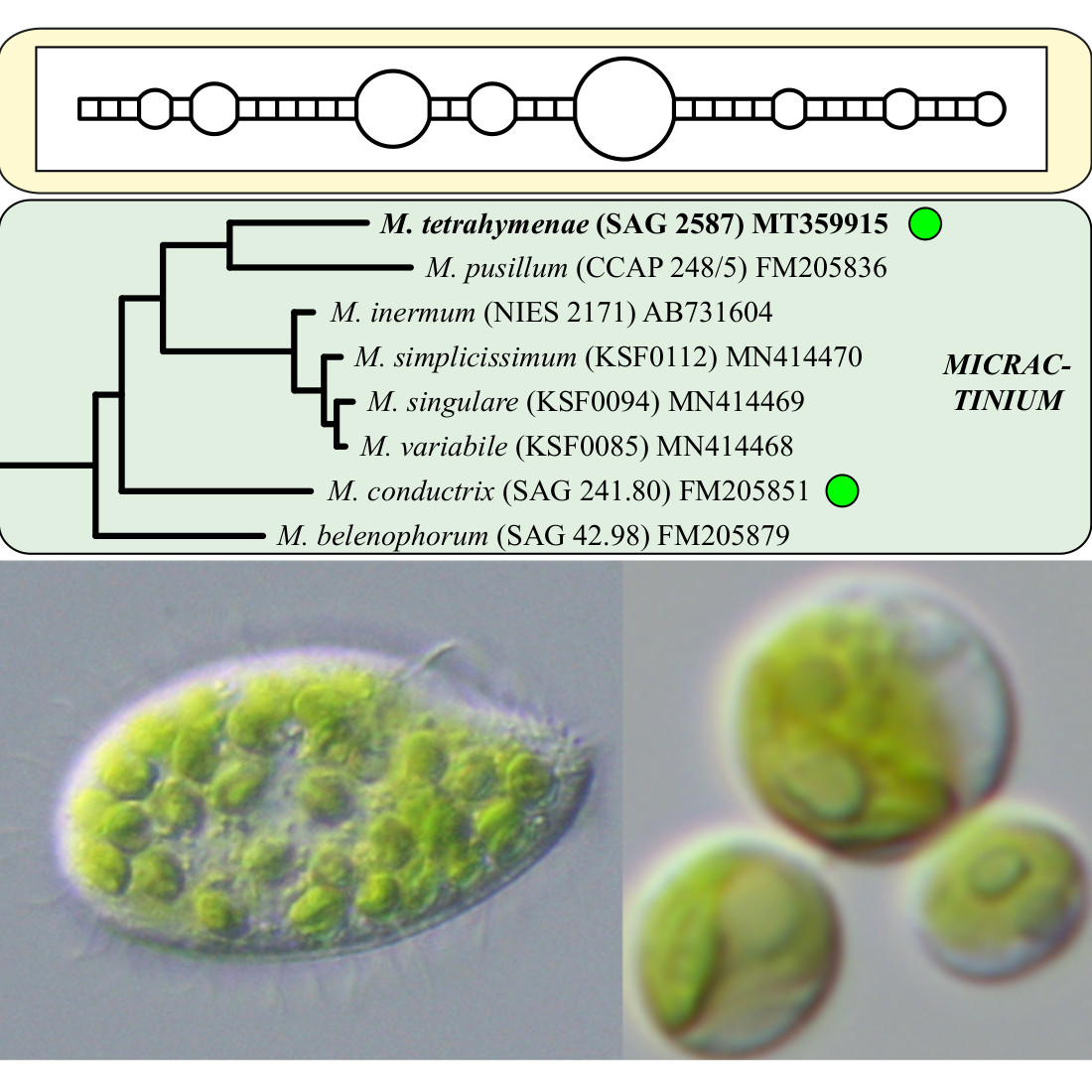
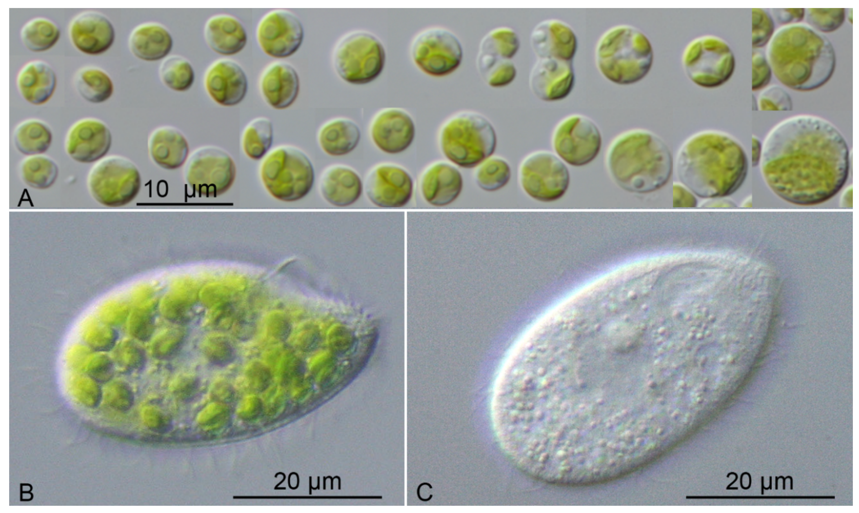
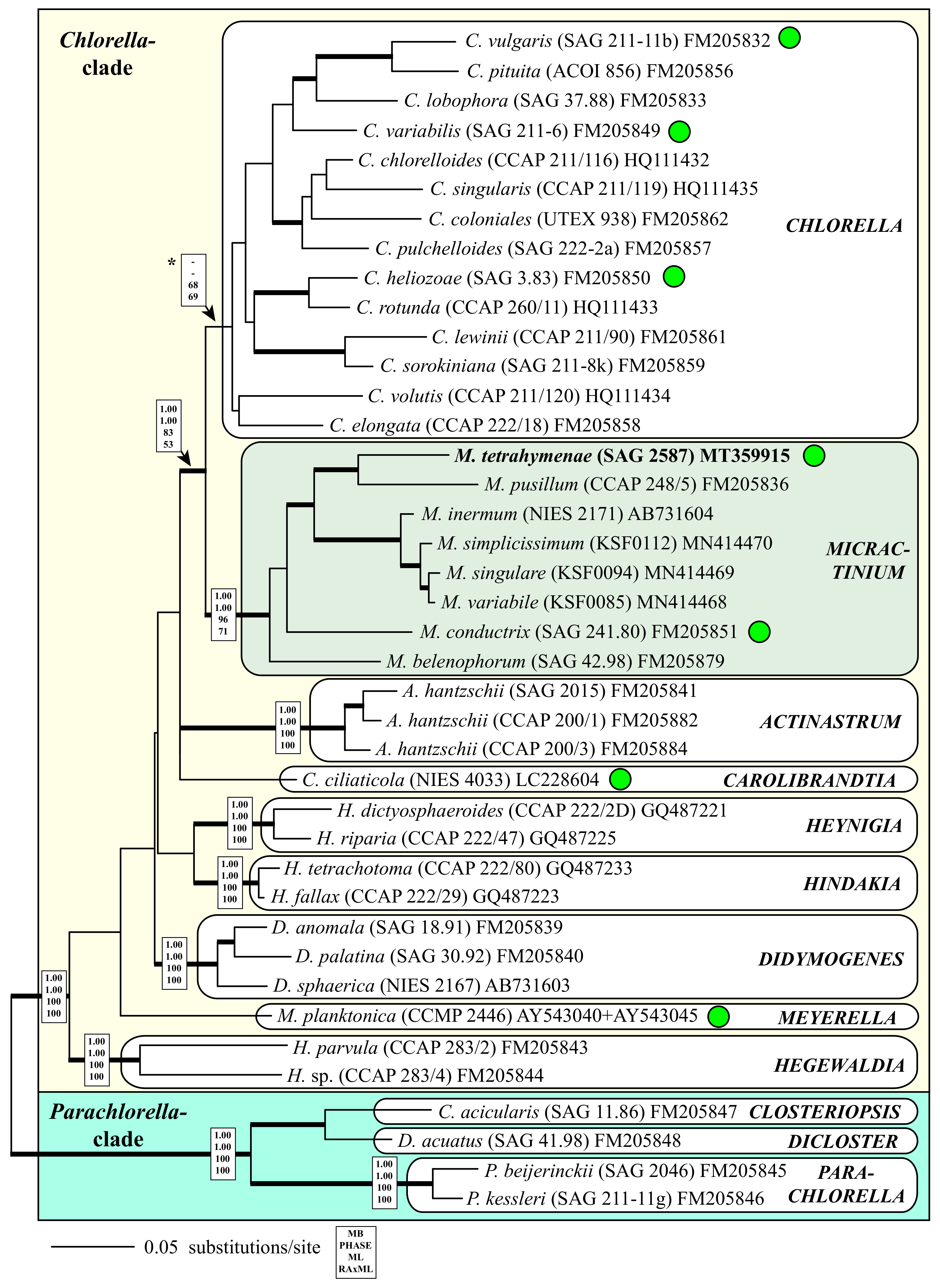
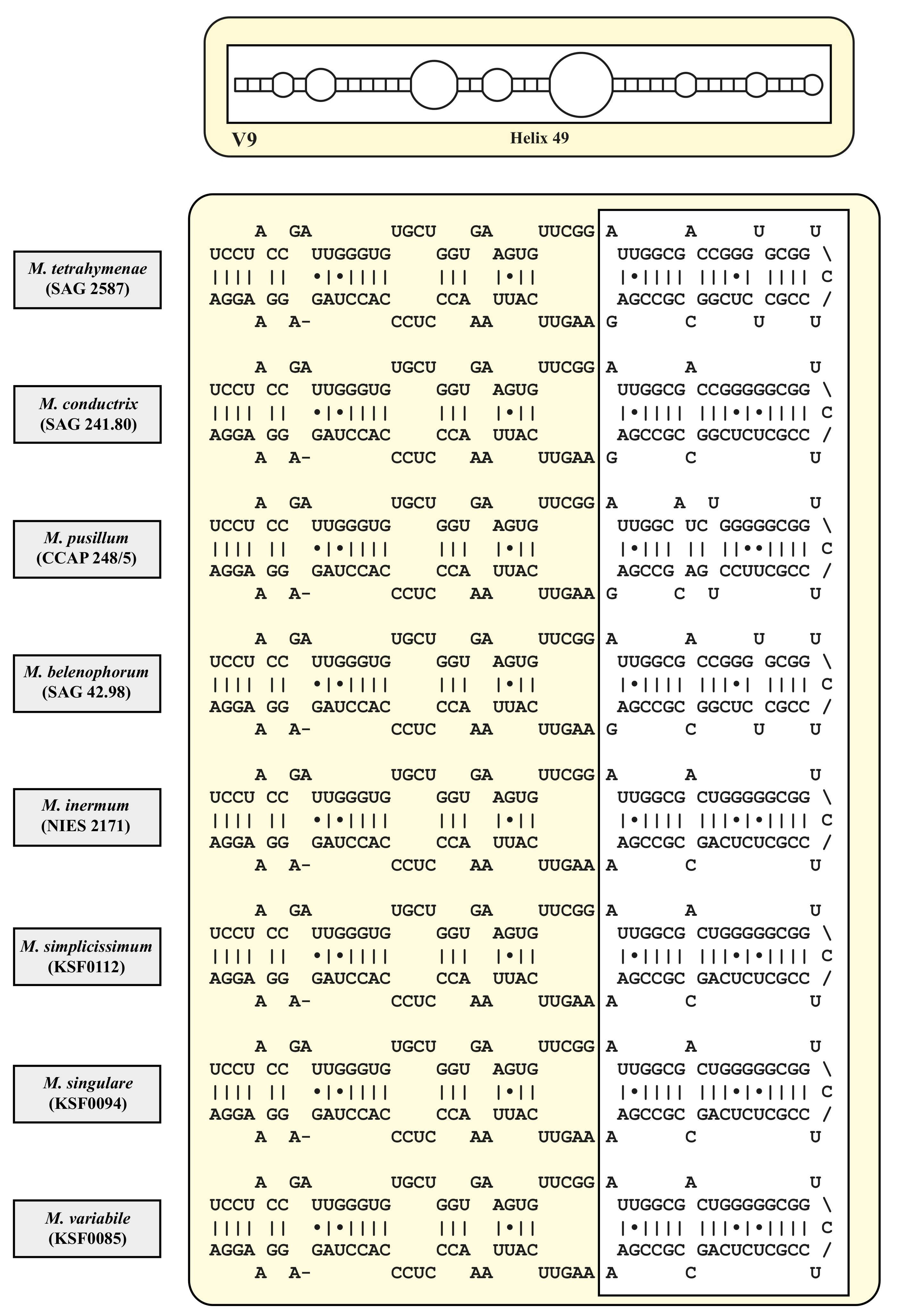
4.2. Taxonomy and Systematics of the Genus Micractinium

Morphologically, both endosymbiotic Micractinium species were difficult to distinguish from each other. M. tetrahymenae sp. nov. was slightly smaller than M. conductrix (3–8 vs. 4–10 µm). Both species showed no bristle formation under the chosen culture conditions. Three other species of Micractinium, all occurring free-living, were known to be bristle-less (M. inermum, M. simplicissimum, and M. singulare [31,32]). Colony formation among Micractinium species was not always observed. The morphological features of all currently accepted species are summarized in Table 1.The taxonomy and systematics of spiny coccoid green algae is very confusing and unclear for two major reasons: (i) Most species were described based on field samples and no type material of these species is available for comparative studies; often only pictures were presented as holotypes [2]; (ii) cultured material such as strains of Micractinium were unicellular and without any bristles, which made it almost impossible to distinguish them from members of the genus Chlorella. Luo et al. [3,4] demonstrated that bristle and colony formation is an inducible defense mechanism against grazing of the rotifer Brachionus calcyflorum. Phylogenetic analyses such as those presented in Figure 2 revealed the close relationship between Chlorella and Micractinium. In contrast to Micractinium, the monophyly of Chlorella was not supported in our analyses. However, the molecular signature described by Pröschold et al. [7], the CBC at the end of helix III in ITS-2 (G-C in Chlorella vs. C-G in Micractinium), remained.
Traditionally both genera belonged to two different families. The family Chlorellaceae comprised algae reproducing exceptionally by autospores without sexual reproduction or zoosporogenesis. Other important criteria for separation of Chlorellaceae was composition of cell wall, which consisted of 2–3 layers containing obligatory cellulose and an outside layer of sporopollenin [2]. Unfortunately, this feature was based on the investigation of Chlorella fusca (now Scenedesmus abundans, Chlorophyceae) and the cell wall of “true” Chlorella species did not contain sporopollenin [33]. In contrast, the family Micractiniaceae contains algae, in which sexual reproduction, but no production of zoospores, is known. The cells are arranged in colonies consisting out of 2 up to 256, and were covered with bristles. The cell walls contain cellulose, without sporopollenin [34,35]. In summary, the differences between both families were the presence of sexual reproduction and bristles in Micractiniaceae. However, phylogenetic analyses have revealed that both families were polyphyletic (see [36] and references therein).
Hegewald and Schnepf [34,37] revised the representatives of the family Micractiniaceae based on morphological, ultrastructural investigations using SEM and TEM. They studied living cultures and some formaldehyde-fixed type material to explore the nature of spines and bristles used for the differentiation at generic level within this family. By definition, bristles contained, in contrast to spines, no cellulose and only proteins in their appendices. In addition, the formation of both is different. Whereas spines were formed before the cell walls were produced, bristles were exhibited after the cells are covered by the rigid cell wall. Considering these features, they revised the genus Micractinium by transferring several species to this genus, which were originally as species of other genera, such as Golenkinia and Golenkiniopsis. The genus Micractinium comprised four species, M. pusillum, M. appendiculatum, M. elongatum, and M. parvulum, according to Hegewald and Schnepf [34,37], and the complicated synonymy were provided therein. However, the validation of these taxonomical combinations needs to be proven.
The latter species was transferred to another genus, Hegewaldia, based on phylogenetic analyses of SSU and ITS rDNA sequences [6]. In addition, they also transferred Diacanthos belenophorus to Micractinium, which was assigned to the Micractiniaceae by Hegewald and Schnepf [38].
Interestingly, it is the occasional occurrence of the sexual reproduction in the family Micractiniaceae. The oogamy was observed in Micractinium pusillum by Nygaard [39], Lund [40], Korschikov [41], and Hegewald [42], and in Hegewaldia parvula by Iyengar and Balakrishnan [43], Starr [44], and Ellis and Machlis [45], originally assigned as Golenkinia minutissima. The ultrastructure of the spermatozoid was investigated by Moestrup [46], who showed that the spermatozoid had an untypical structure of the flagella (9 + 1). The presence of sexual reproduction in Micractinium and Hegewaldia and its absence in Chlorella could be potential criteria for distinguishing the genera. However, Fucikova et al. [47] found in Chlorella meiotic genes and genes that were transcribed during sexual reproduction, in only asexually reproducing trebouxiophytes. This questioned the traditional concept of genera.
As already pointed out Hegewald and Schnepf [34], even the formation of bristles considered as a good morphological feature, is not a stable feature. The morphology and length of bristles is polymorphic and dependent on temperature and media. For example, they observed that Micractinium strigonense Hortobagyi sometimes have different bristles (thick and delicate) and occurred sometimes without bristles. Considering these observations, they proposed to synonymize several species, which is unfortunately illegitimate.
The high phenotypic plasticity and the lack of stable morphological and ultrastructural characters requested a new generic and species concept within the Chlorellaceae and Micractiniaceae. These traditional families should be rejected according the phylogenetic analyses of molecular marker genes. Considering the SSU and ITS sequences, new species were described from Japan [31] and Antarctica [32]. The integrative approach used in this study clearly demonstrated that Micractinium contained eight species (Figure 2). The morphological features of those species as well as the remaining species of Hegewald and Schnepf [34,37] were compared in Table 1. The comparison and judgement of traditional features and molecular data is quite difficult. For example, both endosymbiotic species showed only small morphological differences and were not considered as members of Micractinium without phylogenetic analyses. However, our study showed that both are separate species based on the CBC approach, as demonstrated in Figure 7. On the other hand, molecular data provided an inflation of new species descriptions, when the traditional literature was not considered and no strains are available in public culture collections. As an example, Chae et al. [32] described Micractinium variabile based on SSU and ITS rDNA sequences. Morphologically, this species is very similar to M. quadrisetum, which is unfortunately not available in culture. Therefore, it is possible that both species represent only one species. According to the ICN, M. quadrisetum would have priority against M. variabile. As described in the results, only little genetic differences among Micractinium species could be discovered. In particular, M. inermum and the three species described by Chae et al. [32] had identical V9 regions and little differences in ITS-1 and ITS-2, but they differed by two CBCs and three HCBs (Figure 5, Figure 6 and Figure 7). Considering the ITS-2/CBC approach, we do not propose any taxonomic changes without further investigations.

4.3. Ecology and Distribution of Micractinium

The genera Chlorella and Micractinium have different ecological patterns and are distributed in various habitats. Whereas Chlorella has a worldwide distribution in almost all kinds of habitats, it seems that Micractinium is restricted to freshwater habitats. Species of Chlorella were found aquatic in freshwater and marine habitats [18,36], symbiotic in ciliates and heliozoa [7], and terrestrial [48,49]. Micractinium species were only observed in freshwater habitats [2,31,32,36], in wet soils [5], and symbiotic in ciliates ([7,14] and this study). The occurrence of M. tetrahymenae in the traps of Utricularia is exceptional. Whereas Tetrahymena species are widely distributed in the bladder traps of different Utricularia species, only one record is known of the green Tetrahymena utriculariae [14]. No other record of the occurrence of a Micractinium species in such traps have been reported in microbiome studies [50]. Simek et al. [15] studied the ecology and dynamic of trap communities and found that the endosymbiosis of Micractinium in Tetrahymena contributed significantly for the survival of the ciliate in such harsh environment. No other Utricularia species have had mixotrophic ciliates in their traps until now [50].

4.4. Interactions between Tetrahymena Utriculariae and Micractinium Tetrahymenae

The role of the Micractinium symbiont in Tetrahymena utriculariae has been studied in experiments by Simek et al. [15]. The ciliate has a flexible life strategy. It can live in different aquatic environments under oxygenic conditions, or if captured in bladder traps of Utricularia under anoxic conditions. For this flexibility, the endosymbiotic Micractinium is absolutely necessary. It has been demonstrated that aposymbiotic Tetrahymena had the highest growth rate, if exclusively bacterial food is present. However, if cultivated with both bacterial food and symbiotic Micractinium, Tetrahymena had a reduced growth rate, but after 44 days of cultivation, 80% of the Tetrahymena cells reestablished the symbiosis with the algae [15]. These experiments clearly demonstrated that both organisms formed a symbiotic association depending on the environment. The main profit for the host is that the algae produced the oxygen through photosynthesis. If the algae also provided nutrients to the host, this has not been investigated so far. Micractinium tetrahymenae benefited from CO2 production of the host and stable conditions inside the host, whereas outside in ciliates the environment was very harsh (low pH 4.3 and anoxic) [51]. The endosymbiosis with this Micractinium species is probably essential for Tetrahymena, because the green algae were included in cyst formation [14].

5. Conclusions

The newly described species is the second species of Micractinium, which lives in endosymbiosis with ciliates. If this species is exclusively distributed in a symbiotic association like M. conductrix, it cannot be decided so far. No GenBank record has been reported nor could be found in BLAST searching. Tetrahymena utriculariae is also the only mixotrophic species of this genus. Both organisms were only found once so far, which is probably caused by the lack of investigations. Fortunately, different aspects of this ciliate-green algal association can be studied in detail, because species are available in culture. Nothing is known about the specificity of this symbiosis. The easiness of cultivating makes this ciliate and its endosymbiont the perfect model organisms to study associations between ciliates and green algae.

Author Contributions

Conceptualization, T.P. and T.D.; methodology, G.P. and T.D.; software, T.P.; validation, T.P., G.P. and T.D.; formal analysis, T.P.; investigation, T.P., G.P. and T.D.; resources, T.P.; data curation, T.P.; writing—original draft preparation, T.P., G.P. and T.D.; writing—review and editing, T.P., G.P. and T.D..; visualization, T.P. and T.D.; supervision, T.P.; project administration, T.P.; funding acquisition, T.P. All authors have read and agreed to the published version of the manuscript.

Funding

This research was funded by the Austrian Science Fund (FWF): grant number P28333-B25.

Conflicts of Interest

The authors declare no conflict of interest.

References

  1. Fresenius, G. Beiträge zur Kenntniss mikrokopischer Organismen. Abh. Senckenberg. Naturforsch. Ges. 1858, 2, 211–242. [Google Scholar]
  2. Komarek, J.; Fott, B. Chlorophyceae (Grünalgen) Ordnung: Chlorococcales. In Das Phytoplankton des Süßwassers 7. Teil, 1. Hälfte; Huber-Pestalozzi, G., Ed.; Schweizerbart: Stuttgart, Germany, 1983; pp. 1–1044. [Google Scholar]
  3. Luo, W.; Krienitz, L.; Pflugmacher, S.; Walz, N. Genus and species concept in Chlorella and Micractinium (Chlorophyta, Chlorellaceae): Genotype versus phenotypical variability under ecosystem conditions. VInternationale Vereinigung für theoretische und angewandte Limnologie: Verhandlungen 2005, 29, 170–173. [Google Scholar] [CrossRef]
  4. Luo, W.; Pflugmacher, S.; Pröschold, T.; Walz, N.; Krienitz, L. Genotype versus phenotype variability in Chlorella and Micractinium (Chlorophyta, Trebouxiophyceae). Protist 2006, 157, 315–333. [Google Scholar] [CrossRef]
  5. Luo, W.; Pröschold, T.; Bock, C.; Krienitz, L. Generic concept in Chlorella-related coccoid green algae (Chlorophyta, Trebouxiophyceae). Plant Biol. 2010, 12, 545–553. [Google Scholar] [CrossRef]
  6. Pröschold, T.; Bock, C.; Luo, W.; Krienitz, L. Polyphyletic distribution of bristle formation in Chlorellaceae: Micractinium, Diacanthos, Didymogenes and Hegewaldia gen. nov. (Trebouxiophyceae, Chlorophyta). Phycol. Res. 2010, 58, 1–8. [Google Scholar] [CrossRef]
  7. Pröschold, T.; Darienko, T.; Silva, P.C.; Reisser, W.; Krienitz, L. The systematics of "Zoochlorella" revisited employing an integrative approach. Environ. Microbiol. 2011, 13, 350–364. [Google Scholar] [CrossRef] [PubMed]
  8. Brandt, K. Über das Zusammenleben von Algen und Thieren. Biol. Centralbl. 1882, 1, 524–527. [Google Scholar]
  9. Kreutz, M.; Foissner, W. The Sphagnum ponds of Simmelried in Germany: A biodiversity hot-spot for microscopic organisms. Protozool. Monogr. 2006, 3, 1–267. [Google Scholar]
  10. Kreutz, M.; Stoeck, T.; Foissner, W. Morphological and molecular characterization of Paramecium (Viridoparamecium nov. subgen.) chlorelligerum Kahl 1935 (Ciliophora). J. Eukaryot. Microbiol. 2012, 59, 548–563. [Google Scholar] [CrossRef] [Green Version]
  11. Hoshina, R.; Kobayashi, M.; Suzaki, T.; Kusuoka, Y. Brandtia ciliaticola gen. et sp. nov. (Chlorellaceae, Trebouxiophyceae) a common symbiotic green coccoid of various ciliate species. Phycol. Res. 2018, 66, 76–81. [Google Scholar] [CrossRef]
  12. Hoshina, R.; Nakada, T. Carolibrandtia nom. nov. as a replacement name for Brandtia Hoshina (Chlorellaceae, Trebouxiophyceae). Phycol. Res. 2018, 66, 82–83. [Google Scholar] [CrossRef]
  13. Fujishima, M. Endosymbionts in Paramecium. Microbiol. Monogr. 2009, 12, 1–252. [Google Scholar]
  14. Pitsch, G.; Adamec, L.; Dirren, S.; Nitsche, F.; Simek, K.; Sirova, D.; Posch, T. The green Tetrahymena utriculariae n. sp. (Ciliophora, Oligohymenophorea) with its endosymbiotic algae (Micractinium sp.), living in traps of a carnivorous aquatic plant. J. Eukaryot. Microbiol. 2017, 64, 322–335. [Google Scholar] [CrossRef] [PubMed]
  15. Simek, K.; Pitsch, G.; Salcher, M.M.; Sirova, D.; Shabarova, T.; Adamec, L.; Posch, T. Ecological traits of the algae-bearing Tetrahymena utriculariae (Ciliophora) from traps of the aquatic carnivorous plant Utricularia reflexa. J. Eukaryot. Microbiol. 2017, 64, 336–348. [Google Scholar] [CrossRef] [PubMed]
  16. Schlösser, U.G. SAG-Sammlung von Algenkulturen at the University of Göttingen. Botanica Acta 1994, 107, 424–429. [Google Scholar]
  17. Marin, B.; Palm, A.; Klingberg, M.; Melkonian, M. Phylogeny and taxonomic revision of plastid-containing euglenophytes based on SSU rDNA sequence comparisons and synapomorphic signatures in the SSU rRNA secondary structure. Protist 2003, 154, 99–145. [Google Scholar] [CrossRef] [PubMed]
  18. Darienko, T.; Rad-Menendez, L.; Campbell, C.; Pröschold, T. Are there any true marine Chlorella species? Molecular phylogenetic assessment and ecology of marine Chlorella-like organisms, including a description of Droopiella gen. nov. Syst. Biodivers. 2019, 17, 811–829. [Google Scholar] [CrossRef]
  19. Zuker, M. Mfold web server for nucleic acid folding and hybridization prediction. Nucleic Acid Res. 2003, 31, 3406–3615. [Google Scholar] [CrossRef]
  20. Swofford, D.L. PAUP* Phylogenetic Analysis Using Parsimony (*and Other Methods), Version 4.0b10; Sinauer Associates: Sunderland, MA, USA, 2002. [Google Scholar]
  21. Stamatakis, A. RAxML version 8: A tool for phylogenetic analysis and post-analysis of large phylogenies. Bioinformatics 2014, 30, 1312–1313. [Google Scholar] [CrossRef]
  22. Ronquist, F.; Teslenko, M.; Van Der Mark, P.; Ayres, D.L.; Darling, A.; Höhna, S.; Larget, B.; Liu, L.; Suchard, M.A.; Huelsenbeck, J.P. MrBayes 3.2: Efficient Bayesian phylogenetic inference and model choice across a large model space. Syst. Biol. 2012, 61, 539–542. [Google Scholar] [CrossRef] [Green Version]
  23. Jow, H.; Hudelot, C.; Rattray, M.; Higgs, P. Bayesian phylogenetics using an RNA substitution model applied to early mammalian evolution. Mol. Biol. Evol. 2002, 19, 1591–1601. [Google Scholar] [CrossRef] [PubMed]
  24. Higgs, P.; Jameson, D.; Jow, H.; Rattray, M. The evolution of tRNA-Leu genes in animal mitochondrial genomes. J. Mol. Evol. 2003, 57, 435–445. [Google Scholar] [CrossRef] [PubMed] [Green Version]
  25. Hudelot, C.; Gowri-Shankar, V.; Jow, H.; Rattray, M.; Higgs, P. RNA-based phylogenetic methods: Application to mammalian mitochondrial RNA sequences. Mol. Phylogen. Evol. 2003, 28, 241–252. [Google Scholar] [CrossRef] [Green Version]
  26. Gibson, A.; Gowri-Shankar, V.; Higgs, P.; Rattray, M. A comprehensive analysis of mammalian mitochondrial genome base composition and improved phylogenetic methods. Mol. Biol. Evol. 2005, 22, 251–264. [Google Scholar] [CrossRef]
  27. Telford, M.J.; Wise, M.J.; Gowri-Shankar, V. Consideration of RNA secondary structure significantly improves likelihood-based estimates of phylogeny: Examples from the bilateria. Mol. Biol. Evol. 2005, 22, 1129–1136. [Google Scholar] [CrossRef] [Green Version]
  28. Coleman, A.W.; Mai, J.C. Ribosomal DNA ITS-1 and ITS-2 sequence comparisons as a tool for predicting genetic relatedness. J. Mol. Evol. 1997, 45, 168–177. [Google Scholar] [CrossRef]
  29. Hoshina, R.; Iwataki, M.; Imamura, N. Chlorella variabilis and Micractinium reisseri sp. nov. (Chlorellaceae, Trebouxiophyceae): Redescription of the endosymbiotic green algae of Paramecium bursaria (Peniculia, Oligohymenophorea) in the 120th year. Phycol. Res. 2010, 58, 188–201. [Google Scholar] [CrossRef]
  30. Pröschold, T.; Rieser, D.; Darienko, T.; Kammerlander, B.; Pitsch, G.; Bruni, E.P.; Qu, Z.; Forster, D.; Rad-Menendez, C.; Posch, T.; et al. An integrative approach sheds new light onto the systematics and ecology of the widespread ciliate genus Coleps (Ciliophora, Prostomatea). Environm. Microbiol. 2020, submitted. [Google Scholar]
  31. Hoshina, R.; Fujiwara, Y. Molecular characterization of Chlorella cultures of the National Institute for Environmental Studies culture collection with description of Micractinium inermum sp. nov., Didymogenes sphaerica sp. nov., and Didymogenes soliella sp. nov. (Chlorellaceae, Trebouxiophyceae). Phycol. Res. 2013, 31, 124–132. [Google Scholar]
  32. Chae, H.; Lim, S.; Kim, H.S.; Choi, H.-G.; Kim, J.H. Morphology and phylogenetic relationships of Micractinium (Chlorellaceae, Trebouxiophyceae) taxa, including three new species from Antarctica. Algae 2019, 34, 267–275. [Google Scholar] [CrossRef] [Green Version]
  33. Atkinson, A.W.; Gunning, B.E.S.; John, P.C.L. Sporopollenin in the cell wall of Chlorella and other algae: Ultrastructure, chemistry and incorporation of 14C-acetate, studied in synchronous cultures. Planta 1972, 107, 1–32. [Google Scholar] [CrossRef] [PubMed]
  34. Hegewald, E.; Schnepf, E. Zur Struktur und Taxonomie bestachelter Chlorellales. Nova Hedwigia 1984, 39, 297–383. [Google Scholar]
  35. Schnepf, E.; Deichgräber, G.; Glaab, M.; Hegewald, E. Bristles and spikes in Chlorococcales: Ultrastructural studies in Acanthosphaera, Micractinium, Pediastrum, Polyedriopsis, Scenedesmus, and Siderocystopsis. J. Ultrastr. Res. 1980, 72, 367–379. [Google Scholar] [CrossRef]
  36. Krienitz, L.; Bock, C. Present state of the systematics of planktonic coccoid green algae of inland waters. Hydrobiologia 2012, 698, 295–326. [Google Scholar] [CrossRef]
  37. Hegewald, E.; Schnepf, E. Ergänzungen und Korrekturen zur Struktur und Taxonomie bestachelter Chlorellales. Nova Hedwigia 1987, 44, 537–541. [Google Scholar]
  38. Hegewald, E.; Schnepf, E. The ultrastructure and taxonomic placement of Diacanthos belenophorus Kors. (Chlorophyta, Trebouxiophyceae, Micractiniaceae). Constancea 2002, 83, 11. [Google Scholar]
  39. Nygaard, G. Hydrobiological studies on some Danish ponds and lakes. Kongl. Dansk Vid. Selskab. Biol. Skr. København 1949, 7, 1–293. [Google Scholar]
  40. Lund, J.W.G. Three new British algal records and spore formation in Micractinium pusillum Fres. Naturalist 1954, 1954, 81–85. [Google Scholar]
  41. Korshikov, A.A. Viznachnik prisnovodnihk vodorostey Ukrainsykoi RSR [Vyp] V. Pidklas Protokokovi (Protococcineae). In Vakuol’ni (Vacuolales) ta Protokokovi (Protococcales); Naukova Dumka: Kyiv, Ukraine, 1953. [Google Scholar]
  42. Hegewald, E. Interessante Algen aus dem Ischelandteich in Hagen. Dortmunder Beitr. Landesk. Naturw. Mitt. 1979, 11, 13–16. [Google Scholar]
  43. Iyengar, M.O.P.; Balakrishnan, M.S. On sexual reproduction in a new species of Golenkinia. J. Indian Bot. Soc. 1956, 35, 371–373. [Google Scholar]
  44. Starr, R.C. Homothallism in Golenkinia minutissima. In Studies on Microalgae and Photosynthetic Bacteria; Japanese Society of Plant Physiologists, Ed.; Univ. Tokyo Press: Tokyo, Japan, 1963; pp. 3–6. [Google Scholar]
  45. Ellis, R.J.; Machlis, L. Control of sexuality in Golenkinia. Am. J. Bot. 1968, 55, 600–610. [Google Scholar] [CrossRef]
  46. Moestrup, Ø. Observations on the fine structure of spermatozoids and vegetative cells of the green alga Golenkinia. Br. Phycol. J. 1972, 7, 169–183. [Google Scholar] [CrossRef]
  47. Fucikova, K.; Pazoutova, M.; Rindi, F. Meiotic genes and sexual reproduction in the green algal class Trebouxiophyceae (Chlorophyta). J. Phycol. 2015, 51, 419–430. [Google Scholar] [CrossRef] [PubMed]
  48. Andreyeva, V.M. New species of Chlorella Beijer. Bot. Zhurn. 1973, 58, 1735–1741. (in Russian). [Google Scholar]
  49. Hodac, L.; Hallman, C.; Spitzer, K.; Elster, J.; Faßhauer, F.; Brinkmann, N.; Lepka, D.; Diwan, V.; Friedl, T. Widespread green algae Chlorella and Stichococcus exhibit polar-temperate and tropical-temperate biogeography. FEMS Microbiol. Ecol. 2016, 93, 1–15. [Google Scholar]
  50. Cheng, C.-Y.; Chang, S.-L.; Lin, I.-T.; Yao, M.-C. Abundant and diverse Tetrahymena species living in the bladder traps of aquatic carnivorous Utricularia plants. Sci. Rep. 2019, 9, 13669. [Google Scholar] [CrossRef] [Green Version]
  51. Sirová, D.; Borovec, J.; Cerná, B.; Rejmánková, E.; Adamec, L.; Vrba, J. Microbial community development in the traps of aquatic Utricularia species. Aquat. Bot. 2009, 90, 129–136. [Google Scholar] [CrossRef]
Figure 1. A. Morphology and phenotypic plasticity of Micractinium tetrahymenae, strain SAG 2587, B.-C. Tetrahymena utriculariae under anoxic (B) and oxygenic (C) conditions.
Diversity 12 00200 g001
Figure 2. Comparison of the V9 of SSU and the conserved region of ITS-2 among the eight Micractinium species. Compensatory base changes (CBCs and HCBCs) and insertion/deletion are marked with an asterisk.
Diversity 12 00200 g002
Figure 3. Molecular phylogeny of the Chlorellaceae based on SSU and ITS sequence comparisons. The phylogenetic trees shown were inferred using the maximum likelihood method based on the data sets (2602 aligned positions of 40 taxa), using PAUP 4.0a167. For the analyses, the best model was calculated by PAUP. The setting of the best model was given as follows: GTR + I + G (base frequencies: A 0.2112, C 0.2784, G 0.2743, T 0.2361; rate matrix A-C 0.7316, A-G 0.9716, A-U 0.9475, C-G 0.6216, C-U 3.2173, G-U 1.0000) with the proportion of invariable sites (I = 0.7266) and gamma shape parameter (G = 0.6963). The branches in bold are highly supported in all analyses (Bayesian values > 0.95 calculated with MrBayes and PHASE, 10 million generations; bootstrap values > 50%, calculated with PAUP, 1000 replicates using maximum likelihood, neighbor-joining, and maximum parsimony). The endosymbiotic species are marked with a green circle. The accession and strain numbers are given.
Diversity 12 00200 g003
Figure 4. Secondary structure of the V9 region (Helix 49) of the SSU rDNA among the Micractinium species. The variable region within the V9 are highlighted in white boxes.
Diversity 12 00200 g004
Figure 5. Secondary structure of the ITS-1 (A) and ITS-2 (B) rDNA of Micractinium tetrahymenae. The regions used for barcoding are highlighted in white boxes.
Diversity 12 00200 g005
Figure 6. Variability of ITS-1 among the eight Micractinium species.
Diversity 12 00200 g006
Figure 7. Variability of ITS-2 among the eight Micractinium species. The characteristic features within the conserved regions are highlighted in white boxes.
Diversity 12 00200 g007
Table 1. Diacritical morphological features among the described species of Micractinium.
SpeciesCell ShapeCell Size [µm]Chloroplast ShapePyrenoidBristlesLength of Bristles [µm]Colony FormationLife StyleHabitatReference Strain
M.pusillumspherical3.0–7.0–12.0cup-shaped+up to 8 per cell40–65–(100)single, or up to 8–32free-livingplankton of pondsCCAP 248/5
M.elongatumspherical6.0–7.0cup-shaped+1–2 per cellup to 50.04 - celledfree-livingplankton-
M.appendiculatumellipsoidal, ovoid8.0parietal+2–4 per cell28.0–70.0up to 64free-livingplankton of ponds-
M.belenophorumellipsoidal, broadly ellipsoidal8.0–10.0 × 4.5–5.5parietal+2 per cell at the polesup to 55.0up to 4free-livingplankton of ponds and riversSAG 42.98
M.conductrixspherical4.0 -10.0cup-shaped+---endosymbioticParamecium bursariaSAG 241.80
M.extremumspherical5.2–6.4cup-shaped+1–2 per cellup to 30.08 celledfree-livingplankton-
M.inermumellipsoidal up to spherical5.0–5.4–3.2–3.7cup-shaped+---free-living NIES 2171
M.quadrisetumovoid, ellipsoidal6.0- 10.0 × 4.0–7.0cup-shaped+1–423.0–50.016 celled and morefree-livingPlankton of ponds and rivers-
M.simplicissmumellipsoidal up to spherical5.5–5.7 × 3.3–3.9cup-shaped+---free-living KSF0112
M.singulareellipsoidal up to spherical7.2–7.4 × 4.5–4.7cup-shaped+---free-living KSF0094
M.tetrahymenaespherical cup-shaped+---endosymbioticTetrahymena utriculariaeSAG 2587
M.variabileellipsoidal up to sphaerical8.2–8.6 × 5.0–5.4cup-shaped+4–8 (?)10–30solitary,+free-livingplanktonKSF0085

