Communication

Crystal Structures of Two 4-Alkyl-8-hydroxyquinolines

Institut für Organische Chemie, Technische Universität Bergakademie Freiberg, Leipziger Str. 29, D-09596 Freiberg, Germany
*
Author to whom correspondence should be addressed.
Molbank 2026, 2026(3), M2176; https://doi.org/10.3390/M2176 (registering DOI)
Submission received: 28 March 2026 / Revised: 3 May 2026 / Accepted: 5 May 2026 / Published: 11 May 2026
(This article belongs to the Section Structure Determination)

Abstract

4-Methyl- (1) and 4-ethyl-8-hydroxyquinoline (2) crystallize from a mixture of diethyl ether and chloroform in the triclinic space group P 1 ¯ . X-ray analysis reveals that both compounds form discrete molecular dimers stabilized by intermolecular O-H∙∙∙N and C-H∙∙∙O hydrogen bonds, resulting in R 2 2 ( 5 ) cyclic synthons. This pattern of hydrogen bonds is further stabilized by intramolecular O-H∙∙∙N bonds so that the quinoline nitrogen atom acts as a bifurcated binding site. The dimers exhibit a planar geometry and arrange into layer-like structures held together by π∙∙∙π stacking and van der Waals forces. While the fundamental bonding motifs are similar, the increased steric demand of the ethyl group in compound 2 induces a shift in the crystallographic orientation of the layers and alters the degree of π-overlap compared to the methyl-substituted analogue 1.

Graphical Abstract

1. Introduction

8-Hydroxyquinoline and its derivatives are heterocyclic compounds with a remarkable range of applications reaching from metallurgy and material sciences to medicinal chemistry (for some examples, see References [1,2,3,4,5,6,7,8,9,10,11,12,13,14,15,16,17,18,19,20,21]). Due to the close proximity of the ring nitrogen atom and the hydroxy group, this scaffold exhibits excellent chelating properties against a wide variety of metal ions. Consequently, 8-hydroxyquinolines can be used as reagents for the separation, detection and quantification of metal ions in analytical chemistry [5], as extractants in metallurgy [6,7,8], as corrosion inhibitors [9], in organic light-emitting diode materials [10,11,12,13], as building blocks of artificial receptors and other supramolecular systems [14,15,16], and in drug development [17,18,19,20,21].
In addition to using 8-hydroxyquinoline for the construction of artificial receptors [15], we have also employed 8-hydroxyquinoline derivatives, including those with alkyl or alkanoyl groups in the 5-position, as ligands for indium ions. Our investigations included solvent extraction experiments [22] and the formation of crystalline indium complexes [23]. More recently, our studies have focused on 4-substituted 8-hydroxyquinoline derivatives. In this work we describe the crystal structures of two 4-alkyl-substituted 8-hydroxyquinolines 1 and 2 (see Figure 1). We discuss the supramolecular patterns as well as the similarities and differences between both crystal structures.
Although some crystal structures of 4-alkyl-8-hydroxyquinolines in complexes with various ions are known, often in combination with other ligand molecules [24,25,26], their characterization as free ligands has not yet been described. However, it should be noted that derivatives containing an aminoalkyl or nitroalkenyl group in the 4-position, such as 4-(2-aminopropyl)quinolin-8-ol and 4-(2-nitroprop-1-en-1-yl)quinolin-8-ol, have been reported in the literature [27].

2. Results and Discussion

2.1. Synthesis of 4-Methyl- (1) and 4-ethyl-8-hydroxyquinoline (2)

The quinoline scaffold was prepared by heating 2-aminophenol and the corresponding vinyl ketone in a Doebner–Miller reaction, using a slightly modified version of the procedures described in the literature [13,28]. A two-phase system containing concentrated hydrochloric acid and toluene (v/v 2:1), based on the work of Matsugi et al. [29], was employed to reduce the acid-catalyzed polymerization of the carbonyl compound in the reaction mixture, resulting in a more straightforward purification process. First, toluene and other steam-volatile compounds were removed from the crude product by steam distillation. The remaining acidic mixture was then neutralized with a dilute sodium hydroxide solution and steam-distilled again to obtain the product as white, high-purity solids with yields of 26% (1) and 18% (2), see Scheme 1.

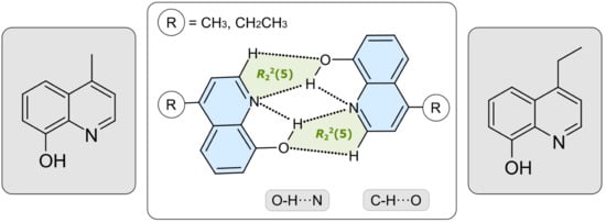
2.2. Structural Elucidation

The two alkyl-substituted 8-hydroxquinolines 1 and 2 crystallize from a mixture of diethyl ether and chloroform (v/v = 1:1) as colorless platelets and needles, respectively. Their molecular structures are shown in Figure 2. Both compounds crystallize in the triclinic space group P 1 ¯ , each with one molecule in the asymmetric unit of the cell. The distance between the hydroxy hydrogen atom and the nitrogen atom indicates the presence of intramolecular O-H∙∙∙N bonds [d(H∙∙∙N) 2.25(2), 2.27(2) Å].
Each of the crystal structures is composed of discrete molecular dimers held together by strong O-H∙∙∙N hydrogen bonds [d(H∙∙∙N) 2.17(2), 2.18(2) Å; ∠(O-H∙∙∙N) 135(2), 134(2)°] and weaker C-H∙∙∙O bonds [30,31,32,33,34,35] [d(H∙∙∙O) 2.45, 2.53 Å; ∠(C-H∙∙∙O) 117, 113°], the latter of which are formed with the participation of the arene hydrogen atom H(1). These hydrogen bonds create a pair of cyclic synthons of the structure R 2 2 ( 5 ) [36,37]. Taking into account the intramolecular hydrogen bond mentioned above, the nitrogen atom of the quinoline unit acts as a bifurcated binding site. The structure of the molecular dimer is shown exemplarily for compound 1 in Figure 3.
According to the planar geometry of these dimers, both crystal structures exhibit a layer-like packing arrangement. Since the outer region of these dimers is formed by the hydrophobic molecular units, only van der Waals forces exist between the molecular dimers within a given layer. A view of the crystal structures in the direction of their layer normal (Figure 4) shows that π∙∙∙π stacking forces [38,39] between the bicyclic units of the molecules contribute significantly to the stabilization of the crystal structures in the direction of the stacking axis of the molecular layers.
However, a comparison of the two crystal structures reveals differences, which are also reflected in the different cell dimensions. While the molecular layers present in the crystal of compound 1 extend parallel to the crystallographic 212 plane, those in the crystal structure of compound 2 run parallel to the 211 plane. A view at the sections of the molecular layers of 1 and 2 shown in Figure S1 also reveals differences. Apparently, the increased steric demand of the ethyl group in 2 induces a layer structure that differs from that of compound 1. In addition, the information in Table 1 on the π∙∙∙π stacking forces present in the crystal structures indicates a different degree of overlap between the quinoline groups of adjacent molecules.

3. Materials and Methods

3.1. Synthesis and Crystallization of 4-Alkyl-8-hydroxyquinolines 1 and 2

4-Methyl-8-hydroxyquinoline (1) and 4-ethyl-8-hydroxyquinoline (2) were synthesized according to the pathway shown in Scheme 1. The synthetic procedure, melting points and NMR spectroscopic data are given in the Supporting Information.
Recrystallization of the compounds from a saturated, hot 1:1-mixture of diethyl ether and chloroform yielded suitable crystals for X-ray structure analysis upon cooling. The crystallographic data are given below and in the Supporting Information (Tables S1 and S2).
Crystal Data for 1: C10H9NO (M = 159.18 g/mol): triclinic, space group P 1 ¯ , a = 7.396(5) Å, b = 7.622(5) Å, c = 8.321(5) Å, α = 63.82(5)°, β = 67.22(5)°, γ = 70.21(6)°, V = 380.1 Å3, Z = 2, T = 183, μ(MoKα) = 0.09 mm−1, Dcalc = 1.391 g/cm3, 6897 reflections measured (2.8° ≤ θ ≤ 25.7°), 1446 unique (Rint = 0.0295), which were used in all calculations. The final R was 0.0412 [I > 2σ(I)] and wR was 0.1160.
Crystal Data for 2: C11H11NO (M = 173.21 g/mol): triclinic, space group P 1 ¯ , a = 7.448(3) Å, b = 7.522(3) Å, c = 7.953(3) Å, α = 93.77(3)°, β = 91.95(3)°, γ = 106.82(3)°, V = 380.1 Å3, Z = 2, T = 183, μ(MoKα) = 0.09 mm−1, Dcalc = 1.354 g/cm3, 8463 reflections measured (2.6° ≤ θ ≤ 26.0°), 1663 unique (Rint = 0.0363), which were used in all calculations. The final R was 0.0488 [I > 2σ(I)] and wR was 0.1368.

3.2. X-Ray Structure Analysis

The data sets of the structures were recorded at a temperature of 183 K on a STOE diffractometer (MoKα radiation, λ = 0.71073 Å) equipped with an image plate system IPDS-2 (STOE & Cie. GmbH, Darmstadt, Germany). Indexing and integration of the reflections was performed using the IPDS software in the X-AREA program suite [40]. Data reduction and absorption correction were performed using the X-RED program [41]. Preliminary structure models were created using direct methods (SHELXT2018/2 [42] employing the program XSTEP-32 [43]) and then refined by full-matrix least-squares calculations based on F2 for all reflections using the program SHELXL [44]. All non-hydrogen atoms were refined anisotropically. With the exception of the O-H hydrogen atoms, all other H atoms are included in the structure models in calculated positions and were refined as constrained to bonding atoms. The graphical representation of the molecular structures was performed using the program ORTEP-III [45].

4. Conclusions

The studies reveal that hydrogen-bonded dimers constitute a characteristic structural motif in the crystal structures of 4-alkyl-substituted 8-hydroxyquinolines 1 and 2, as previously reported for unsubstituted 8-hydroxyquinoline [46]. The dimers are stabilized by strong O-H∙∙∙N hydrogen bonds and weaker C-H∙∙∙O interactions, forming a pair of cyclic synthons of the structure R 2 2 ( 5 ) . Considering the additional intramolecular O-H∙∙∙N hydrogen bond, the quinoline nitrogen atom acts as a bifurcated binding site.
While the primary hydrogen-bonding motifs remain consistent in 1 and 2, the crystal structures demonstrate that the supramolecular arrangement is sensitive to the nature of the alkyl substituent. Even minor alterations, such as replacing a methyl group with an ethyl group, can result in changes to the layer arrangement and π-overlap, demonstrating the sensitivity of the supramolecular architecture to subtle steric modifications.

Supplementary Materials

The following supporting information can be downloaded online. Table S1. Crystallographic and structure refinement data of the crystal structures examined. Table S2. Geometric parameters for non-covalent interactions in the crystal structures examined. Figure S1. A view of the molecular layer in the crystal structures of 1 (a) and 2 (b). Figure S2. 1H NMR (500 MHz) spectrum of 1 in acetone-d6. The hydroxy group of 1 is located at 8.72 ppm as a broad singlet. Figure S3. 13C NMR (125 MHz) spectrum of 1 in acetone-d6. Figure S4. 1H NMR (500 MHz) spectrum of 2 in CDCl3. The hydroxy group of 2 is located at 8.41ppm as a broad singlet. Figure S5. 13C NMR (125 MHz) spectrum of 2 in CDCl3. Figure S6. 1H,1H COSY (500 MHz) spectrum of 2 in CDCl3. Figure S7. 1H,13C HSQC spectrum of 2 in CDCl3.

Author Contributions

Conceptualization, M.M.; validation, S.B. and M.M.; software, S.B. and A.S.; formal analysis, S.B., A.S. and M.M.; investigation, S.B.; writing—original draft preparation, M.M. and S.B.; writing—review and editing, S.B. and M.M.; supervision, M.M. All authors have read and agreed to the published version of the manuscript.

Funding

This research received no external funding.

Data Availability Statement

CCDC 2541343 and 2541344 contain the supplementary crystallographic data for this paper. These data can be obtained free of charge via https://www.ccdc.cam.ac.uk/structures/? (or from the CCDC, 12 Union Road, Cambridge CB2 1EZ, UK; E-mail: deposit@ccdc.cam.ac.uk). Additional data supporting the findings of this study are available from the corresponding author upon reasonable request.

Conflicts of Interest

The authors declare no conflicts of interest.

References

  1. Rohini; Kamaldeep, P.; Luxami, V. 8-Hydroxyquinoline Fluorophore for Sensing of Metal Ions and Anions. Chem. Rec. 2020, 20, 1430–1473. [Google Scholar] [CrossRef]
  2. Phillips, J.P. The Reactions of 8-Quinolinol. Chem. Rev. 1956, 56, 271–297. [Google Scholar] [CrossRef]
  3. Zborowski, K.K.; Solá, M.; Poater, J.; Proniewicz, L.M. Aromatic properties of 8-hydroxyquinoline and its metal complexes. Cent. Eur. J. Chem. 2013, 11, 655–663. [Google Scholar] [CrossRef]
  4. Hollingshead, R.G.W. Oxine and Its Derivatives; Butterworths Scientific Publications: London, UK, 1956. [Google Scholar]
  5. Rao, T.P.; Gladis, J.M. Oxines as Preconcentration Agents in Inorganic Trace Analysis. Rev. Anal. Chem. 2001, 20, 145–159. [Google Scholar] [CrossRef]
  6. Chen, Y.; Zhang, T.; Lv, G.; Chao, X.; Yang, X. Extraction and Utilization of Valuable Elements from Bauxite and Bauxite Residue: A Review. Bull. Environ. Contam. Toxicol. 2022, 109, 228–237. [Google Scholar] [CrossRef] [PubMed]
  7. Melegari, M.; Neri, M.; Falco, A.; Tegoni, M.; Maffini, M.; Fornari, F.; Mucchino, C.; Artizzu, F.; Serpe, A.; Marchiò, L. Tailoring the Use of 8-Hydroxyquinolines for the Facile Separation of Iron, Dysprosium and Neodymium. ChemSusChem 2024, 17, e202400286. [Google Scholar] [CrossRef]
  8. Kekesi, T. Gallium extraction from synthetic Bayer liquors using Kelex 100-kerosene, the effect of loading and stripping conditions on selectivity. Hydrometallurgy 2007, 88, 170–179. [Google Scholar] [CrossRef]
  9. Berdimurodov, E.; Verma, C.; Berdimuradov, K.; Quraishi, M.A.; Kholikov, A.; Akbarov, K.; Umirov, N.; Borikhonov, B. 8–Hydroxyquinoline is key to the development of corrosion inhibitors: An advanced review. Inorg. Chem. Commun. 2022, 144, 109839. [Google Scholar] [CrossRef]
  10. Lewinska, G.; Sanetra, J.; Marszalek, K.W. Application of quinoline derivatives in third-generation photovoltaics. J. Mater. Sci. Mater. Electron. 2021, 32, 18451–18465. [Google Scholar] [CrossRef]
  11. Hamada, Y.; Sano, T.; Fujita, M.; Fujii, T.; Nishio, Y.; Shibata, K. Organic Electroluminescent Devices with 8-Hydroxyquinoline Derivative-Metal Complexes as an Emitter. Jpn. J. Appl. Phys. 1993, 32, L514. [Google Scholar] [CrossRef]
  12. Chen, C.H.; Shi, C. Metal chelates as emitting materials for organic electroluminescence. Coord. Chem. Rev. 1998, 171, 161–174. [Google Scholar] [CrossRef]
  13. Wu, S.; Li, S.; Li, C.-Z.; Shi, M.; Chen, H. Chemical modification of AlQ3 to a potential electron acceptor for solution-processed organic solar cells. Tetrahedron Lett. 2016, 57, 2797–2799. [Google Scholar] [CrossRef]
  14. Geffert, C.; Kuschel, M.; Mazik, M. Molecular Recognition of N-Acetylneuraminic Acid by Acyclic Pyridinium- and Quinolinium-Based Receptors in Aqueous Media: Recognition through Combination of Cationic and Neutral Recognition Sites. J. Org. Chem. 2013, 78, 292–300. [Google Scholar] [CrossRef] [PubMed]
  15. Mazik, M.; Geffert, C. 8-Hydroxyquinoline as a building block for artificial receptors: Binding preferences in the recognition of glycopyranosides. Org. Biomol. Chem. 2011, 9, 2319–2326. [Google Scholar] [CrossRef] [PubMed]
  16. Albrecht, M.; Fiege, M.; Osetska, O. 8-Hydroxyquinolines in metallosupramolecular chemistry. Coord. Chem. Rev. 2008, 252, 812–824. [Google Scholar] [CrossRef]
  17. Prachayasittikul, V.; Prachayasittikul, S.; Ruchirawar, S.; Prachayasittikul, V. 8-Hydroxyquinolines: A review of their metal chelating properties and medicinal applications. Drug Des. Dev. Ther. 2013, 7, 1157–1178. [Google Scholar] [CrossRef]
  18. Song, Y.; Xu, H.; Chen, W.; Zhan, P.; Liu, X. 8-Hydroxyquinoline: A privileged structure with a broad-ranging pharmacological potential. MedChemCommun 2015, 6, 61–74. [Google Scholar] [CrossRef]
  19. Gupta, R.; Luxami, V.; Paul, K. Insights of 8-hydroxyquinolines: A novel target in medicinal chemistry. Bioorg. Chem. 2021, 108, 104633. [Google Scholar] [CrossRef]
  20. Gasparin, C.B.; Pilger, D.A. 8-Hydroxyquinoline, Derivatives and Metal-Complexes: A Review of Antileukemia Activities. ChemistrySelect 2023, 8, e202204219. [Google Scholar] [CrossRef]
  21. Savić-Gajić, I.M.; Savić, I.M. Drug design strategies with metal-hydroxyquinoline complexes. Expert Opin. Drug Discov. 2019, 15, 383–390. [Google Scholar] [CrossRef]
  22. Schulze, M.; Löwe, R.; Pollex, R.; Mazik, M. Structure–extractability relationships for substituted 8-hydroxyquinolines: Solvent extraction of indium ions from acidic aqueous media. Monatsh. Chem. 2019, 150, 983–990. [Google Scholar] [CrossRef]
  23. Fuhrmann, B.; Meier, E.; Mazik, M. Crystal structure of bis(μ2-5-nonanoylquinolin-8-olato)bis[aquadichloridoindium(III)]. Acta Cryst. 2024, E80, 1020–1023. [Google Scholar] [CrossRef]
  24. Huang, D.-F.; Chow, T.J.; Wu, C.-Y.; Sun, S.-S.; Tsai, S.-H.; Wen, Y.-S.; Polosan, S.; Tsuboi, T. The Preparation of (8-Hydroxyquinolinato)Bis(2-Phenylpyridyl)Iridium Complexes and Their Photophysical Properties. J. Chin. Chem. Soc. 2013, 55, 439–448. [Google Scholar] [CrossRef]
  25. Nagata, Y.; Chujo, Y. Synthesis of Methyl-Substituted Main-Chain-Type Organoboron Quinolate Polymers and Their Emission Color Tuning. Macromolecules 2008, 41, 2809–2813. [Google Scholar] [CrossRef]
  26. Deaton, J.C.; Place, D.W.; Brown, C.T.; Rajeswaran, M.; Kondakova, M.E. The blue aluminum and gallium chelates for OLEDs. Inorg. Chim. Acta 2008, 361, 1020–1035. [Google Scholar] [CrossRef]
  27. El Safadi, M.; Wilson, K.A.; Strudwicke, I.J.; O’Mara, M.L.; Bhadbhade, M.; Rawling, T.; McDonagh, A.M. Amphetamine-like Deferiprone and Clioquinol Derivatives as Iron Chelating Agents. Molecules 2024, 29, 4213. [Google Scholar] [CrossRef]
  28. Phillips, J.P.; Elbinger, R.L.; Merritt, L.L. Preparation of Some Substituted 8-Hydroxy- and 8-Methoxyquinolines. J. Am. Chem. Soc. 1949, 71, 3986–3988. [Google Scholar] [CrossRef]
  29. Matsugi, M.; Tabusa, F.; Minamikawa, J. Doebner–Miller synthesis in a two-phase system: Practical preparation of quinolines. Tetrahedron Lett. 2000, 41, 8523–8525. [Google Scholar] [CrossRef]
  30. Steiner, T.; Desiraju, G.R. The Weak Hydrogen Bond in Chemistry and Structural Biology; Oxford University Press: New York, NY, USA, 1999. [Google Scholar]
  31. Desiraju, G.R. C–H⋯O and other weak hydrogen bonds. From crystal engineering to virtual screening. Chem. Commun. 2005, 2995–3001. [Google Scholar] [CrossRef]
  32. Mazik, M.; Bläser, D.; Boese, R. Intermolecular CH⋯N/CH⋯O hydrogen bonds in the crystal structures of α,β-unsaturated ketones carrying a terminal pyridine subunit. Tetrahedron 2001, 57, 5791–5797. [Google Scholar] [CrossRef]
  33. Mazik, M.; Hartmann, A.; Jones, P.G. Hydrogen and Halogen Bonding in the Crystal Structure of a 1,3,5-Substituted 2,4,6-Triethylbenzene Consisting of Three Phenanthroline Units. Eur. J. Org. Chem. 2010, 2010, 458–463. [Google Scholar] [CrossRef]
  34. Castellano, R.K. Progress Toward Understanding the Nature and Function of C-H⋯O Interactions. Curr. Org. Chem. 2004, 8, 845–865. [Google Scholar] [CrossRef]
  35. Steiner, T. Unrolling the hydrogen bond properties of C–H···O interactions. Chem. Commun. 1997, 727–734. [Google Scholar] [CrossRef]
  36. Etter, M.C.; MacDonald, J.C.; Bernstein, J. Graph set Analysis of Hydrogen Bond Patterns in Organic Crystals. Acta Crystallogr. Sect. B 1990, 46, 256–262. [Google Scholar] [CrossRef] [PubMed]
  37. Bernstein, J.; Davies, R.E.; Shimoni, L.; Chang, N.L. Patterns in Hydrogen Bonding: Functionality and Graph Set Analysis in Crystals. Angew. Chem. Int. Ed. 1995, 34, 1555–1573. [Google Scholar] [CrossRef]
  38. Dance, I. π-π Interactions: Theory and Scope. In Encyclopedia of Supramolecular Chemistry; Atwood, J.L., Steed, J.M., Eds.; Dekker: New York, NY, USA, 2004; pp. 1076–1092. [Google Scholar]
  39. James, S.L. π-π Stacking as a Crystal Engineering Tool. In Encyclopedia of Supramolecular Chemistry; Atwood, J.L., Steed, J.M., Eds.; Dekker: New York, NY, USA, 2004; pp. 1093–1099. [Google Scholar]
  40. X-AREA, version 2.7; Software Package for Data Collection and Data Evaluation; Stoe & Cie: Darmstadt, Germany, 2002.
  41. X-RED, version 2.3.3; Software Program for Data Reduction and Absorption Correction; Stoe & Cie: Darmstadt, Germany, 2002.
  42. Sheldrick, G.M. SHELXT—Integrated Space Group and Crystal Structure Determination. Acta Crystallogr. Sect. A 2015, 71, 3–8. [Google Scholar] [CrossRef]
  43. XSTEP-32, Revision 1.07e; Stoe & Cie: Darmstadt, Germany, 2000.
  44. Sheldrick, G.M. Crystal Structure Refinement with SHELXL. Acta Crystallogr. Sect. C 2015, 71, 3–8. [Google Scholar] [CrossRef]
  45. Farrugia, L.J. WinGX and ORTEP for Windows: An Update. J. Appl. Crystallogr. 2012, 45, 849–854. [Google Scholar] [CrossRef]
  46. Castaňeda, R.; Antal, S.A.; Draguta, S.; Timofeevaa, T.V.; Khrustalev, V.N. Crystal structure of 8-hydroxyquinoline: A new monoclinic polymorph. Acta Cryst. 2014, E70, o924–o925. [Google Scholar] [CrossRef]
Figure 1. Structures of 4-methyl-8-hydroxyquinoline (1) and 4-ethyl-8-hydroxyquinoline (2).
Molbank 2026 m2176 g001
Scheme 1. Synthesis of 4-methyl- (1) and 4-ethyl-8-hydroxyquinoline (2) according to the Doebner–Miller reaction. Reagents and conditions: 37 wt% HCl(aq)/toluene (ν/ν = 2:1), reflux, 6 h.
Molbank 2026 m2176 sch001
Figure 2. ORTEP plots of the molecular structures of 1 and 2. The displacement ellipsoids are drawn at a 50 % probability level.
Molbank 2026 m2176 g002
Figure 3. Structure of the molecular dimer in the crystal of 1. The five-membered supramolecular synthones [ R 2 2 ( 5 )] are marked by color highlighting.
Molbank 2026 m2176 g003
Figure 4. Excerpts of the crystal structures of compounds 1 (a) and 2 (b) viewed in the direction of the layer normal. The dashed lines correspond to hydrogen-bond interactions.
Molbank 2026 m2176 g004
Table 1. Non-covalent interactions in the crystal structures of 1 and 2.
AtomsDistance (Å)Angle (°)Slippage (Å)
D-H∙∙∙A
π∙∙∙π
D∙∙∙A
Cg∙∙∙Cg
H∙∙∙AD-H∙∙∙A
1
O(1)-H(10)···N(1)2.735(2)2.25(2)114(2)
O(1)-H(10)···N(1)2.869(3)2.17(2)135(2)
C(1)-H(1)···O(1)3.002(3)2.45117
Cg(A)···Cg(A) a3.556(3) 1.063
Cg(B)···Cg(B) a3.631(2) 1.234
Cg(A)···Cg(B) a3.719(3) 1.504
2
O(1)-H(10)···N(1)2.740(2)2.27(2)114(2)
O(1)-H(10)···N(1)2.859(2)2.18(2)134(2)
C(1)-H(1)···O(1)3.033(2)2.53113
Cg(A)···Cg(A) a3.662(2) 1.167
Cg(B)···Cg(B) a3.728(2) 1.345
Cg(A)···Cg(B) a3.753(2) 1.491
a Cg means the centroid (center of gravity) of the aromatic ring. 1, 2: Ring A: N(1),C(1)…C(4),C(9); ring B: C(4)…C(9).
Disclaimer/Publisher’s Note: The statements, opinions and data contained in all publications are solely those of the individual author(s) and contributor(s) and not of MDPI and/or the editor(s). MDPI and/or the editor(s) disclaim responsibility for any injury to people or property resulting from any ideas, methods, instructions or products referred to in the content.

