1. Introduction
Interest in materials with nonlinear optical (NLO) properties, particularly second harmonic generation (SHG), for use as biosensors has increased significantly over the years, as they offer several advantages compared to fluorescence-based methods [
1,
2,
3,
4,
5]. The enhanced sensitivity of SHG detectors allows for the exploration of a wide range of compounds with low SHG emission but high biocompatibility [
6,
7].
The most critical requirement for a crystalline material to exhibit SHG properties is the absence of a center of inversion in its structure. Low-cost carbohydrates, when incorporated into crystals with other substances, effectively induce this non-centrosymmetry [
8]. For many years, our research has focused on metal complexes with some monosaccharide ligands, which are biocompatible materials capable of significant SHG emission, making them good candidates for SHG bio-sensing.
We analyzed the SHG behavior of compounds featuring various alkaline earth metals (Ca
2+, Sr
2+), different sugars (
d-fructose,
d-ribose, 2-deoxy-
d-galactose) and various counterions (Cl
−, Br
−, I
−). We also had the opportunity to study isomorphous structures, Metal–Organic Frameworks (MOFs), and molecular crystals ([
9,
10] and references therein). More generally, carbohydrates have also been employed as building blocks in carbohydrate-containing Metal–Organic Frameworks, such as cyclodextrin-based MOFs, further demonstrating the versatility of saccharides in metal–organic architectures [
11,
12,
13]. Given that the crystal structure is the primary factor influencing SHG emission in this class of compounds, we observed that heavier anions generally have a significant influence on SHG intensity, while heavier cations contribute to a lesser extent. In this context, we decided to investigate an analog compound containing a very heavy metal, Lanthanum, to evaluate the effect of a heavier and more polarizable metal center on SHG efficiency.
In this work, a new β-d-fructose-lanthanum complex with the formula [La(C6H12O6)(H2O)5]Cl3 (LaFRUCl) was synthetized and characterized by single crystal X-ray diffraction (XRD). Its SHG response was measured with a Nd:YAG pulsed 1064 nm laser and compared with analog metal–sugar from our previous studies.
2. Results and Discussion
The synthesis of the LaFRUCl compound involves a simple, rapid, low-cost, and environmentally benign method in which the sugar and the metal salt are mixed in ethanol, and the product crystallizes directly from the reaction mixture during the slow evaporation of the solvent. This method exploits the natural capacity of the hydroxyl groups of the sugar to coordinate with the lanthanum ion. The product precipitates, forming high-quality crystals for X-ray diffraction.
Characterization by X-ray diffraction confirms the non-centrosymmetric structure (P212121), as expected from the intrinsically chiral fructose ligand, which results in an asymmetric coordination environment around the lanthanum(III) center.
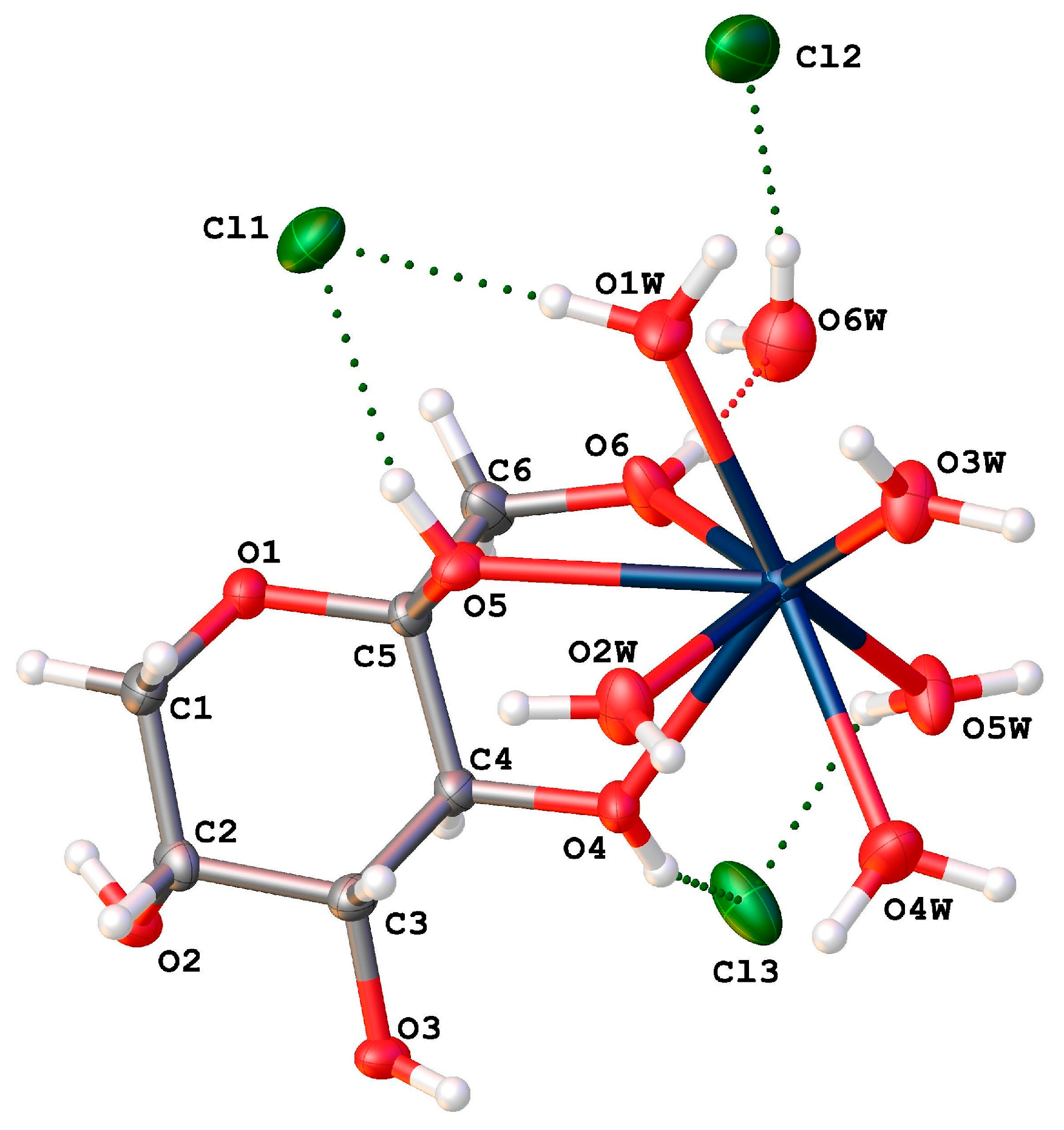
The asymmetric unit of LaFRUCl consists of one La
3+ ion, one fructose molecule, five water molecules coordinated to the metal center, one lattice water molecule, and three free chlorine anions (
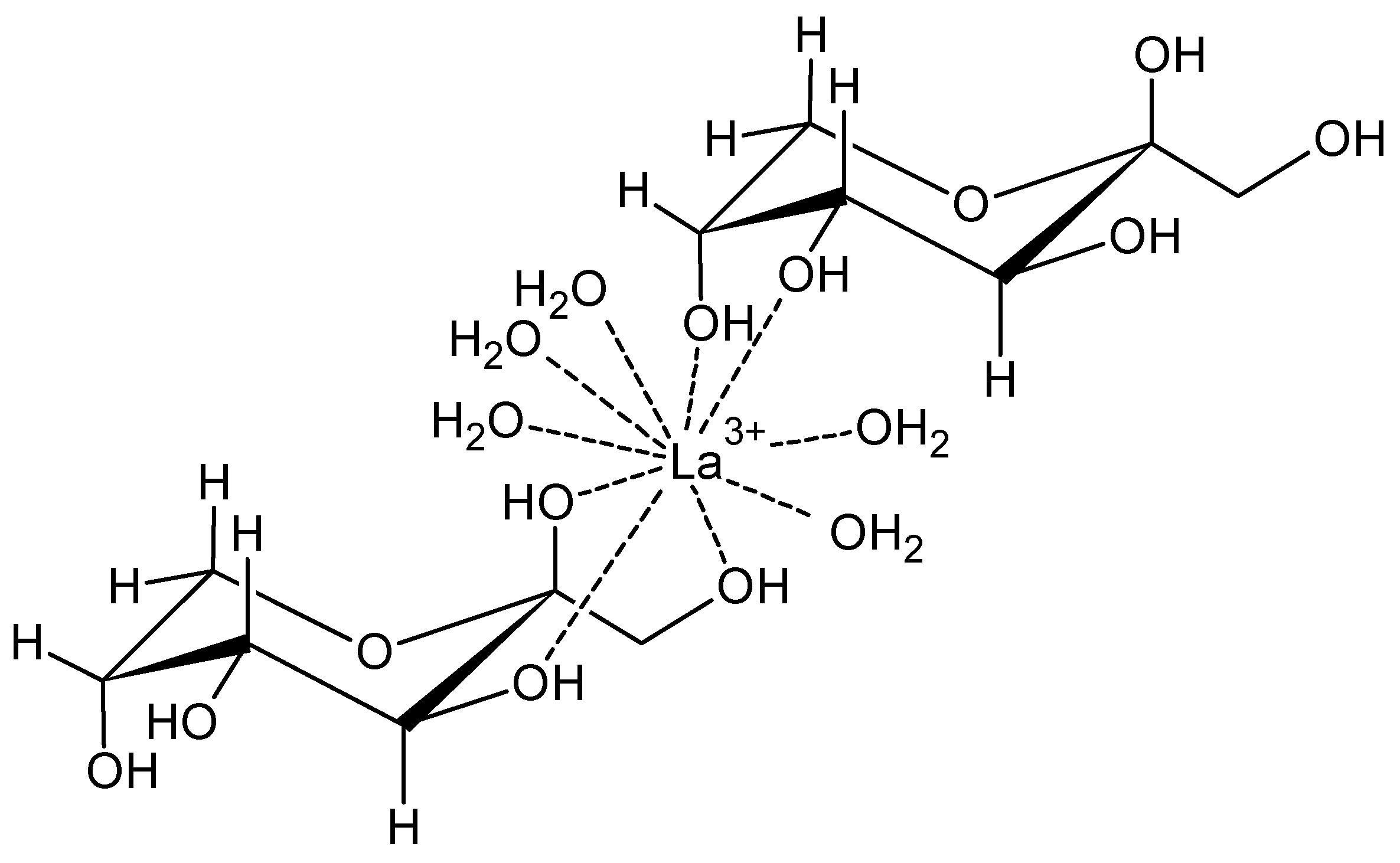
Figure 1). The fructose molecule exists in the pyranose form and adopts a chair conformation (
Scheme 1). To evaluate the deviation from the ideal chair conformation, we analyzed the endocyclic torsion angles of the β-
d-fructopyranose ring. The values range from 49.7(4)° to 57.2(4)°, in close agreement with the theoretical value of approximately 56° for an undistorted chair. The maximum deviation of ca. 6° (observed for the O1-C5-C4-C3 segment) indicates only a minor flattening of the ring. These data suggest that coordination to the La
3+ center occurs with minimal strain on the pyranose framework, largely preserving the ideal chair geometry. In fact, in the β form, the hydroxyl groups attached to the C4, C5, and C6 atoms are oriented on the same side of the chair plane. This facilitates optimal coordination of the La
3+ ion without significantly distorting the sugar’s chair configuration.
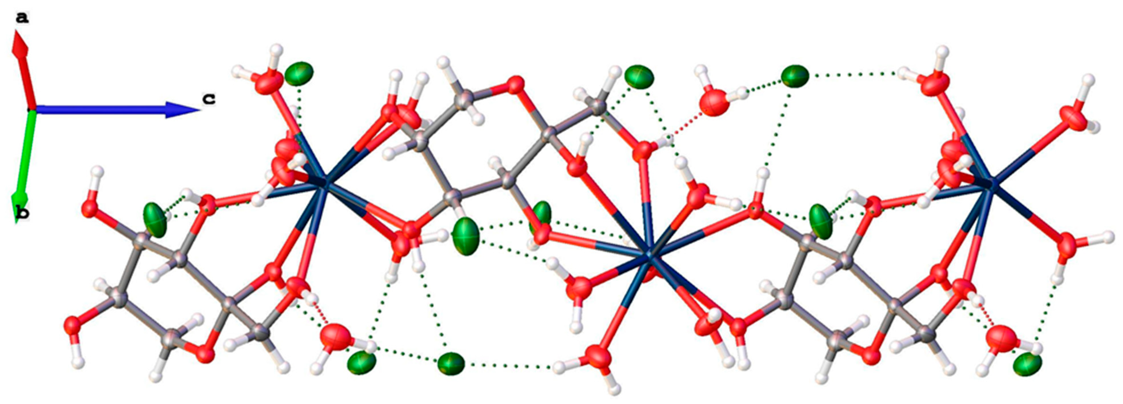
The fructose molecules bridge two metal cations using all their hydroxyl groups, forming infinite (La
3+-fructose)
n chains (
Figure 2) extending along the [001] direction. Consequently, the structure can be described as a 1D Metal–Organic Framework (MOF).
The chlorine atoms are positioned between these chains, stabilized by a network of strong hydrogen bonds involving the hydroxyl groups of the fructose and both the coordinated and lattice water molecules (see
Figure 2). A list of the most significant hydrogen bonds in the structure is provided in
Table S4 of the Supplementary Materials.
The SHG response of the compound was measured on a powder sample, which was hand-ground in an agate mortar. The powder was loaded in a capillary tube, and the SHG intensity was measured relative to a sucrose standard under the same experimental conditions. The I
2w/I
2wsucrose value recorded for LaFRUCl was 0.64. This value is consistent with the average magnitude observed for analogous compounds based on Ca, Sr, Cl, or Br combined with
d-fructose, 2-deoxy-
d-galactose, or
d-ribose [
10]. Consequently, the presence of the lanthanum ion did not result in the expected enhancement of SHG efficiency.
3. Materials and Methods
Lanthanum chloride heptahydrate and β-d-fructose were purchased from Sigma-Aldrich (Merck KGaA, Darmstadt, Germany) and used without additional purification.
3.1. Synthesis of [La(C6H12O6)(H2O)5]Cl3 (LaFRUCl)
Lanthanum chloride heptahydrate and β-d-fructose powders were mixed in a 1:1 stoichiometric ratio and ground in an agate mortar. During grinding, the reagents began to react, partially due to ambient humidity, as evidenced by the formation of a sticky material. XRPD patterns confirmed the amorphous nature of this intermediate. The resulting solid was partially dissolved in few drops of ethanol, and the solution was maintained in equilibrium with the solid for one week. Colorless crystals suitable for XRD analysis formed on the vial walls. These were briefly washed with ethanol and dried in an oven at 323 K.
3.2. Single Crystal X-Ray Diffraction (XRD) of LaFRUCl
X-ray diffraction data were collected at room temperature using a Xcalibur AtlasS2 Gemini R Ultra diffractometer Rigaku, Oxford Diffraction Ltd, Yarnton/Abingdon, Oxfordshire, United Kingdom) equipped with graphite monochromatized Mo-Kα radiation (0.71073 Å). The CrysAlisPro [
14] package was used for data collection and integration. The structure was solved using SHELXT [
15] and refined with SHELXL [
16], and Olex2 [
17] was used for graphics.
Crystal data: Orthorhombic, space group P212121, Z = 4, a = 10.0145(2), b = 12.3587(3) Å, c = 14.9211(3), and V = 1846.74(7) Å3. A total of 22876 reflections were collected, of which 5637 were unique (Rint = 0.0467). Fina refinement indices: R1 = 0.0288 (I > 2σ(I)); wR2 = 0.0607 (all data).
All non-hydrogen atoms were refined with anisotropic displacement parameters. Although hydrogen atom positions were visible in the difference Fourier maps, they were placed in calculated positions and refined using a riding model with the U
iso = 1.2 or 1.5 × U
eq of the parent atom. The interested reader can find further details on crystal data, data collection, least-squares refinements, and bond lengths, and angles in the
Supplementary Materials (Tables S1–S3) and CIF file (CCDC 2240232).
3.3. Second Harmonic Generation Measurements
SHG efficiency was determined by the Kurtz–Perry powder technique, using a nanosecond Nd:YAG pulsed (10 Hz) laser with a fundamental wavelength of 1064 nm. The SH signal generated by ground samples in capillary tubes was collected by an elliptical mirror, detected by a photomultiplier, and compared to the ground sucrose signal collected under the same conditions (sucrose displays an SHG equal to 0.7, that of KDP).
4. Conclusions
In this work, a new lanthanum/fructose-based MOF, [La(C6H12O6)(H2O)5]Cl3, was synthetized with a simple and low-cost method and structurally characterized by single-crystal X-ray diffraction. The compound crystallizes in the non-centrosymmetric orthorhombic space group P212121 and consists of infinite (La3+–fructose)ₙ chains extending along the [001] direction, which can be described as a 1D Metal–Organic Framework.
Second harmonic generation measurements performed using the Kurtz–Perry powder technique revealed an SHG efficiency comparable to that of previously reported alkaline earth metal–sugar analogs, indicating that the introduction of the lanthanum ion does not result in a significant enhancement of SHG intensity. Although the SHG response is significant enough for potential use as a biosensor, further studies on stability and potential metal ion release are required before considering biological applications.
Supplementary Materials
The following supporting information can be downloaded at: Table S1. Details on crystal data and structure refinement for LaFRUCl; Table S2. Bond Lengths for LaFRUCl; Table S3. Bond Angles for LaFRUCl; Table S4. List of stronger hydrogen bonds for LaFRUCl; CIF file.
Author Contributions
Conceptualization, P.B. and D.M.; methodology, D.M.; validation, P.B. and D.M.; formal analysis, P.B. and D.M.; investigation, P.B. and D.M.; data curation, D.M.; writing—original draft preparation, P.B. and D.M.; writing—review and editing, P.B. and D.M.; supervision, D.M.; project administration, D.M.; funding acquisition, D.M. All authors have read and agreed to the published version of the manuscript.
Funding
This research was funded by MUR (Ministero dell’Università e della Ricerca).
Data Availability Statement
The crystallographic data for this study have been deposited at the Cambridge Crystallographic Data Centre (CCDC 2240232). These data can be obtained free of charge via
www.ccdc.cam.ac.uk/structures/ (15 February 2026). Additional data supporting the findings of this study are available from the corresponding author upon reasonable request.
Acknowledgments
Financial support from MIUR (Ministero dell’Istruzione, dell’Università e della Ricerca) is acknowledged.
Conflicts of Interest
The authors declare no conflicts of interest.
References
- Liu, T.-M.; Conde, J.; Lipiński, T.; Bednarkiewicz, A.; Huang, C.-C. Smart NIR linear and nonlinear optical nanomaterials for cancer theranostics: Prospects in photomedicine. Prog. Mater. Sci. 2017, 88, 89–135. [Google Scholar] [CrossRef]
- Boyd, R.W. Nonlinear Optics; Academic Press: San Diego, CA, USA, 2003. [Google Scholar]
- Huang, X.; El-Sayed, I.H.; Qian, W.; El-Sayedet, M.A. Cancer Cell Imaging and Photothermal Therapy in the Near-Infrared Region by Using Gold Nanorods. J. Am. Chem. Soc. 2006, 128, 2115–2120. [Google Scholar] [CrossRef] [PubMed]
- Dempsey, W.P.; Fraser, S.E.; Pantazis, P. SHG nanoprobes: Advancing harmonic imaging in biology. BioEssays 2012, 34, 351–360. [Google Scholar] [CrossRef] [PubMed]
- Jin, Y. Engineering Plasmonic Gold Nanostructures and Metamaterials for Biosensing and Nanomedicine. Adv. Mater. 2012, 24, 5153–5165. [Google Scholar] [CrossRef] [PubMed]
- Shen, H.; Nguyen, N.; Gachet, D.; Maillard, V.; Toury, T.; Brasselet, S. Nanoscale optical properties of metal nanoparticles probed by Second Harmonic Generation microscopy. Opt. Express 2013, 21, 12318–12326. [Google Scholar] [CrossRef] [PubMed]
- Leonard, A.K.; Loughran, E.A.; Klymenko, Y.; Liu, Y.; Kim, O.; Asem, M.; McAbee, K.; Ravosa, M.; Stack, M.S. Chapter 4—Methods for the visualization and analysis of extracellular matrix protein structure and degradation. In Methods in Cell Biology; Academic Press: San Diego, CA, USA, 2018; Volume 143, pp. 79–95. [Google Scholar] [CrossRef]
- Clevers, S.; Burel, A.; Couvrat, N.; Dupray, V.; Coquerel, G. Relevance of Second Harmonic Generation Microscopy (SHG-M) in Material Sciences. In Proceedings of the BIWIC 2019 International Workshop on Industrial Crystallization, Rayong, Thailand, 25–27 August 2019; p. hal-02295938. [Google Scholar]
- Marabello, D.; Antoniotti, P.; Benzi, P.; Beccari, F.; Canepa, C.; Cariati, E.; Cioci, A.; Lo Presti, L. Crystal structure or chemical composition of salt–sugar-based metal–organic frameworks: What are the nonlinear optical properties due to? Acta Cryst. B 2021, 77, 506–514. [Google Scholar] [CrossRef]
- Marabello, D.; Antoniotti, P.; Benzi, P.; Canepa, C.; Cariati, E.; Cioci, A.; Lo Presti, L. Second Harmonic Generation behavior of two new d-ribose/d-fructose and metal halogenide-based coordination compounds and comparison to d-fructose and d-galactose analogues: An experimental and theoretical approach. Cryst. Growth Des. 2022, 22, 5923−5934. [Google Scholar] [CrossRef]
- Smaldone, R.A.; Forgan, R.S.; Furukawa, H.; Gassensmith, J.J.; Slawin, A.M.Z.; Yaghi, O.M.; Stoddart, J.F. Nanoporous Carbohydrate Metal–Organic Frameworks. J. Am. Chem. Soc. 2010, 132, 142–143. [Google Scholar] [CrossRef]
- Roy, I.; Stoddart, J.F. Cyclodextrin Metal–Organic Frameworks and Their Applications. Acc. Chem. Res. 2021, 54, 1440–1453. [Google Scholar] [CrossRef] [PubMed]
- Han, Y.; Liu, W.; Huang, J.; Qiu, S.; Zhong, H.; Liu, D.; Liu, J. Cyclodextrin-Based Metal–Organic Frameworks (CD-MOFs) in Pharmaceutics and Biomedicine. Pharmaceutics 2018, 10, 271. [Google Scholar] [CrossRef] [PubMed]
- CrysAlisPro, Version 1.171.37.31. Release 14-01-2014 CrysAlis171.NET. Agilent Technologies UK Ltd.: Oxford, UK, 2014.
- Sheldrick, G.M. SHELXT-Integrated Space-Group and Crystal-Structure Determination. Acta Cryst. A 2015, 71, 3–8. [Google Scholar] [CrossRef] [PubMed]
- Sheldrick, G.M. Crystal Structure Refinement with SHELXL. Acta Cryst. C 2015, 71, 3–8. [Google Scholar] [CrossRef] [PubMed]
- Dolomanov, O.V.; Bourhis, L.J.; Gildea, R.J.; Howard, J.A.K.; Puschmann, H. OLEX2: A Complete Structure Solution, Refinement and Analysis Program. J. Appl. Cryst. 2009, 42, 339–341. [Google Scholar] [CrossRef]
| Disclaimer/Publisher’s Note: The statements, opinions and data contained in all publications are solely those of the individual author(s) and contributor(s) and not of MDPI and/or the editor(s). MDPI and/or the editor(s) disclaim responsibility for any injury to people or property resulting from any ideas, methods, instructions or products referred to in the content. |







 Display in JSmol Viewer