
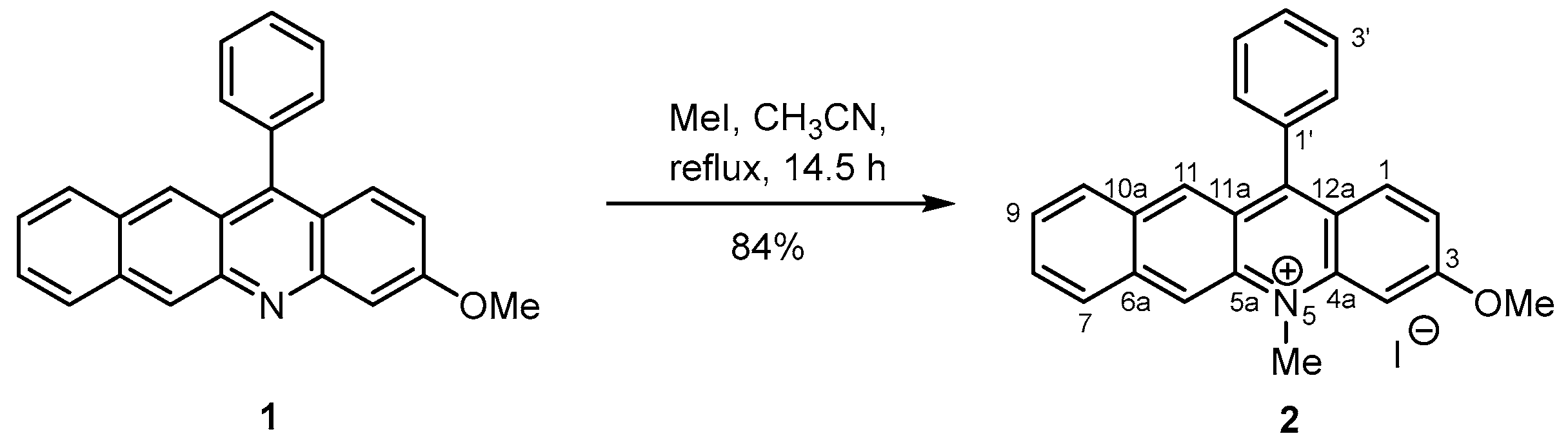
3-Methoxy-5-methyl-12-phenylbenzacridinium Iodide

 and
and 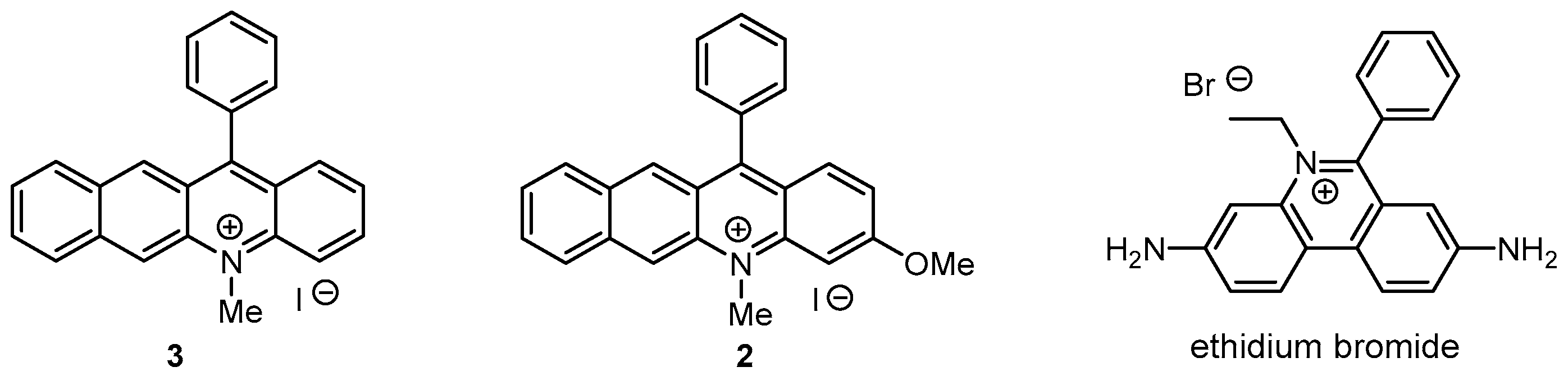
Abstract
:1. Introduction
2. Results and Discussion
3. Materials and Methods
3.1. Instrumentation
3.2. Synthesis of 3-Methoxy-5-methyl-12-phenylbenzacridinium Iodide (2)
4. Conclusions
Supplementary Materials
Author Contributions
Funding
Institutional Review Board Statement
Informed Consent Statement
Data Availability Statement
Acknowledgments
Conflicts of Interest
Sample Availability
References
- Fukuzumi, S.; Kotani, H.; Ohkubo, K.; Ogo, S.; Tkachenko, N.V.; Lemmetyinen, H. Electron-Transfer State of 9-Mesityl-10-methylacridinium Ion with a Much Longer Lifetime and Higher Energy than That of the Natural Photosynthetic Reaction Center. J. Am. Chem. Soc. 2004, 126, 1600–1601. [Google Scholar] [CrossRef] [PubMed]
- White, A.R.; Wang, L.; Nicewicz, D.A. Synthesis and Characterization of Acridinium Dyes for Photoredox Catalysis. Synlett 2019, 30, 827–832. [Google Scholar] [CrossRef]
- Koga, R.; Oishi, T.; Torikai, K. Tuned classical thermal aromatization furnishing an estrogenic benzoacridine. Synlett 2015, 26, 2801–2805. [Google Scholar]
- Torikai, K.; Koga, R.; Liu, X.; Umehara, K.; Kitano, T.; Watanabe, K.; Oishi, T.; Noguchi, H.; Shimohigashi, Y. Design and synthesis of benzoacridines as estrogenic and anti-estrogenic agents. Bioorg. Med. Chem. 2017, 25, 5216–5237. [Google Scholar] [CrossRef]
- Wungsintaweekul, B.; Abe, K.; Koga, R.; Katakura, Y.; Torikai, K. Antimicrobial and Anti-Oxidative Activities of 12-Arylbenzoacridines. Indones. J. Chem. 2020, 20, 1199–1205. [Google Scholar] [CrossRef]

Publisher’s Note: MDPI stays neutral with regard to jurisdictional claims in published maps and institutional affiliations. |
© 2022 by the authors. Licensee MDPI, Basel, Switzerland. This article is an open access article distributed under the terms and conditions of the Creative Commons Attribution (CC BY) license (https://creativecommons.org/licenses/by/4.0/).
Share and Cite
Uktamova, M.; Koga, R.; Mukhamedjonova, F.; Kholikov, T.; Ibragimov, B.; Torikai, K.; Khodjaniyazov, K. 3-Methoxy-5-methyl-12-phenylbenzacridinium Iodide. Molbank 2022, 2022, M1431. https://doi.org/10.3390/M1431
Uktamova M, Koga R, Mukhamedjonova F, Kholikov T, Ibragimov B, Torikai K, Khodjaniyazov K. 3-Methoxy-5-methyl-12-phenylbenzacridinium Iodide. Molbank. 2022; 2022(3):M1431. https://doi.org/10.3390/M1431
Chicago/Turabian StyleUktamova, Malokhat, Rintaro Koga, Fotima Mukhamedjonova, Tursunali Kholikov, Bakhtiyor Ibragimov, Kohei Torikai, and Khamid Khodjaniyazov. 2022. "3-Methoxy-5-methyl-12-phenylbenzacridinium Iodide" Molbank 2022, no. 3: M1431. https://doi.org/10.3390/M1431
APA StyleUktamova, M., Koga, R., Mukhamedjonova, F., Kholikov, T., Ibragimov, B., Torikai, K., & Khodjaniyazov, K. (2022). 3-Methoxy-5-methyl-12-phenylbenzacridinium Iodide. Molbank, 2022(3), M1431. https://doi.org/10.3390/M1431