Prenatal Whole-Genome Sequencing for Fetal Anomalies: Diagnostic Performance, Challenges, and Clinical Implications

 , , , , and
Abstract
1. Introduction
2. Scope and Method
3. Results
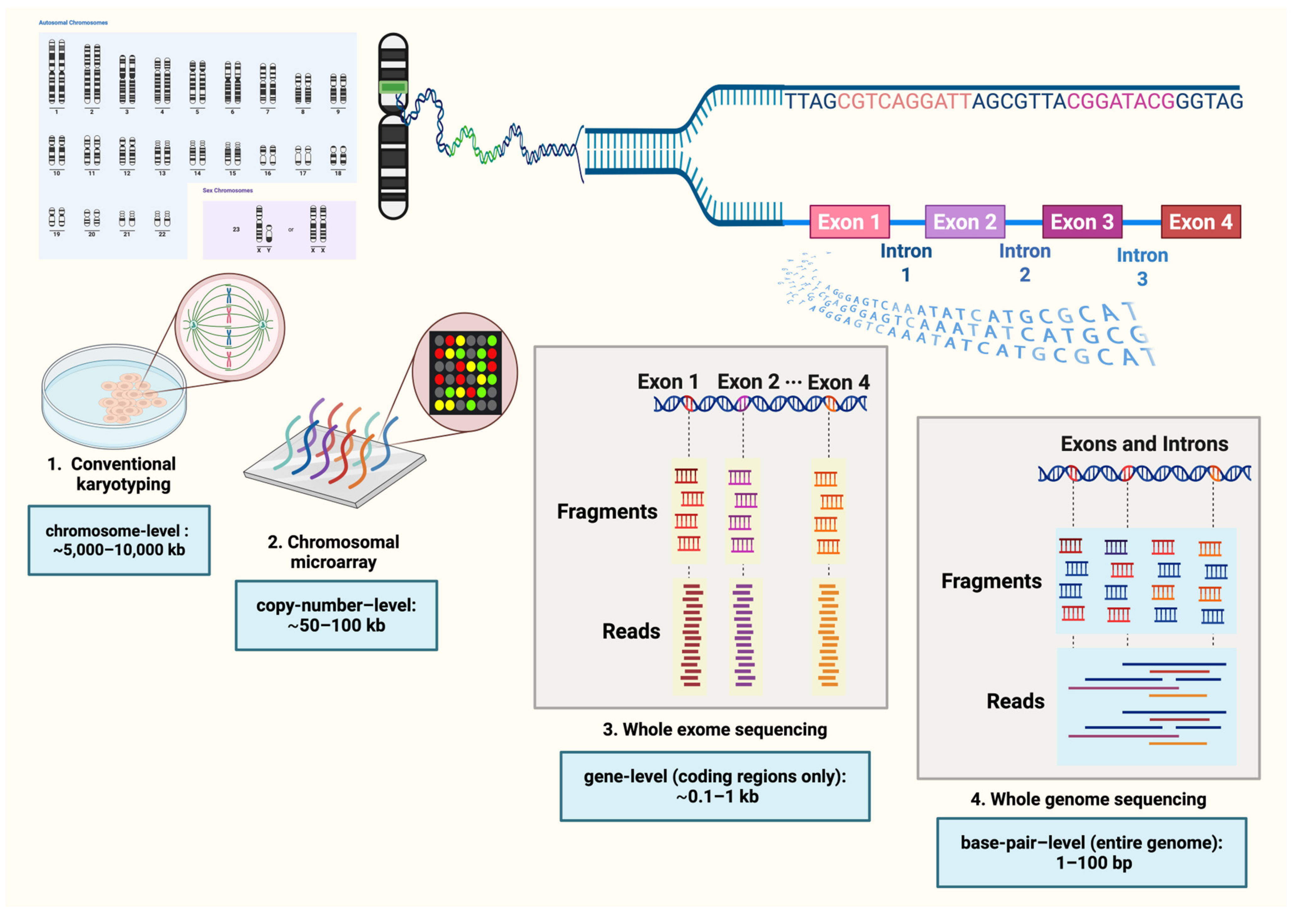
3.1. Prenatal WGS: Workflows
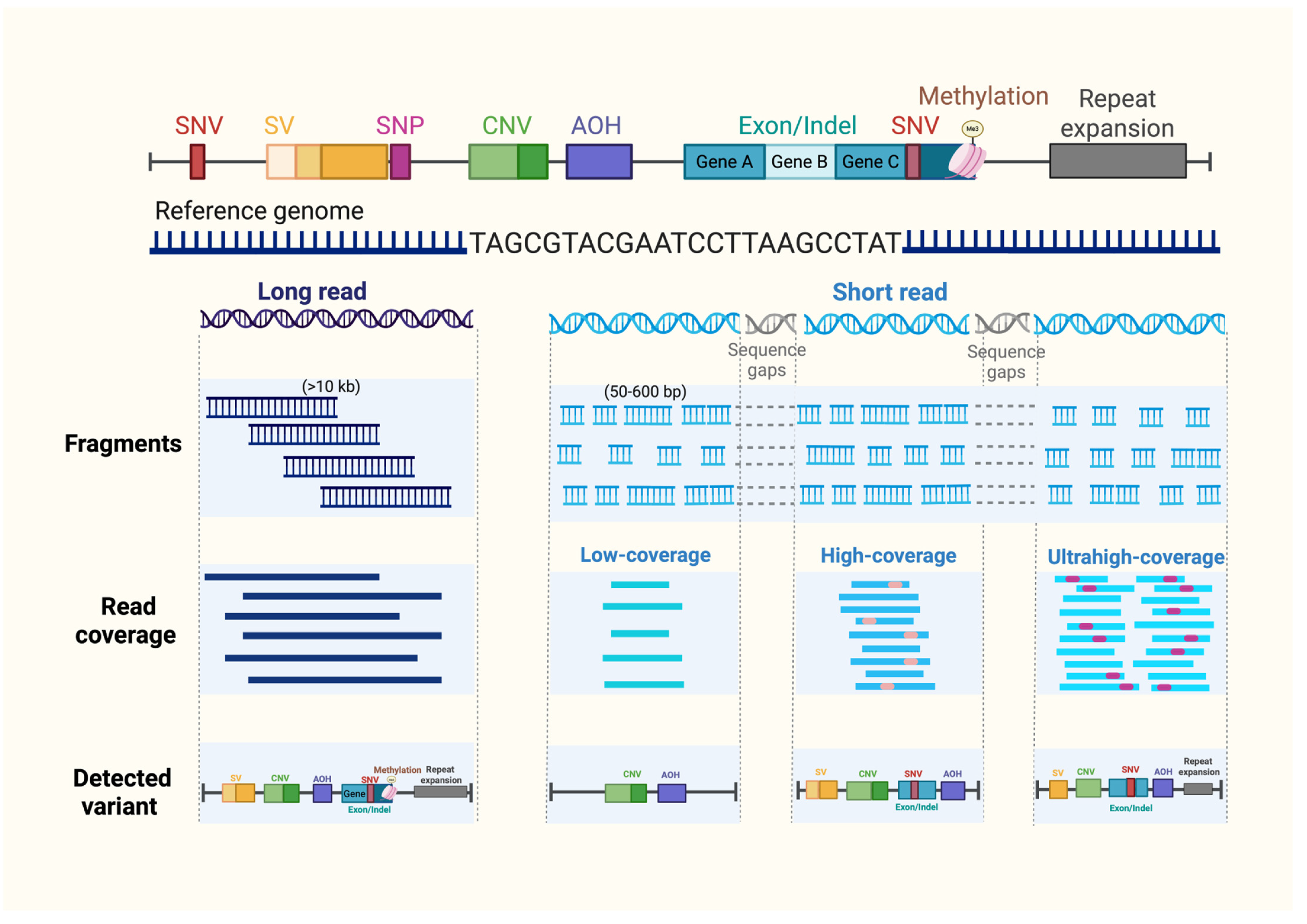
3.2. Prenatal WGS Strategies: Read Length, Sequencing Depth and Variant Detection
3.3. Variant Detection Performance Across Sequencing Coverage Depths in Prenatal WGS

| Author | Year | Population (Fetuses) | Type of Specimen | Methodology | Singleton (S)/Trio (T) | Coverage Depth | Sample Size (Cases) | Detection Rate (%) | Detected Variant | Rate of VUS (%) | Turnaround Time (Days) | Comparative Performance vs. Standard Methods |
|---|---|---|---|---|---|---|---|---|---|---|---|---|
| Walker L [44] | 2019 | Structural anomalies | AF, CVS | WGS vs. CMA | S | Low coverage | 40 | 7.5 | CNVs, mosaicism | 0 | NR | Comparable |
| Qi H [45] | 2018 | Miscarriage | POC | WGS vs. CMA and karyotype | S | Low coverage | 149 | 43.0 | Aneuploidy, CNVs, mosaicism | NR | NR | Comparable |
| Wang H [46] | 2020 | PND | AF, CB, CVS | WGS vs. CMA | S | ~0.25× | 1023 | 13.5 | CNVs, mosaicism | 5.2 | NR | Higher |
| Dong Z [41] | 2016 | Miscarriage, stillbirths | POC | WGS vs. CMA | S | ~0.25× | 384 | 41.4 | CNVs, mosaicism | NR | 10 | Comparable |
| Chau MHK [47] | 2020 | PND, miscarriage | AF, CB, CVS, POC | WGS vs. CMA | NR | ~0.25× | 429 | 27.3 | CNVs, AOH, mosaicism | 0.6 | 3 | Higher |
| Yang Y [48] | 2022 | CNS anomalies | AF, CVS | Two step WGS (0.5× → 40×) | S | ~0.5× | 162 | 38.3 | SNVs, small indels, CNVs, SVs, mosaicism | NR | 21 | Higher |
| Yin Y [49] | 2025 | PND | AF | WGS vs. CMA | S | 3× | 200 | 21.0 | CNVs, SVs, low-level mosaicism | 2.0 | NR | Higher |
| Pang J [53] | 2025 | Structural anomalies, PND | AF | WGS vs. CMA | S | ~5× | 42 | 61.9 | CNVs, AOH, mosaicism | NR | NR | Comparable |
| Chang J [50] | 2025 | Chromosomal mosaicism detected by CMA/SNP | AF, CB, CVS | WGS vs. CMA and karyotype/FISH | NR | ~5× | 34 | 100.0 * | Aneuploidy, CNVs, AOH, low-level mosaicism | 2.9 | NR | Comparable |
| Jiang Y [52] | 2025 | Structural anomalies, PND | AF | WGS vs. CMA and karyotype/FISH | S | ≥5× | 3973 | 24.7 | CNVs, AOH, low-level mosaicism | 15.7 | NR | Higher |
| Lü Y [51] | 2023 | Suspected chromosomal abnormalities, including AOH | AF, POC | WGS with AOH-specific bioinformatic algorithm | S | ≥5× | 24 | 33.3 | AOH, mosaicism | NR | NR | Enable to detect prenatal AOH |
| Hu P [58] | 2023 | Structural anomalies | AF | WGS vs. CMA | Both | ≥30× | 185 | 16.8 | SNVs, small indels, CNVs, STRs | NR | 21–28 | Higher |
| Wang Y [54] | 2022 | Structural anomalies | AF, CVS, fetal tissue | Single-step WGS vs. stepwise QF-PCR → CMA | S | >30× | 37 | 19.0 | SNVs, small indels, small CNVs | NR | NR | Higher |
| Westenius E [43] | 2024 | Structural anomalies | AF, CVS, fetal tissue | Uninformative QF-PCR and CMA → WGS | T | >30× | 50 | 26.0 | SNVs, small indels | NR | 14–21 | Higher |
| Gao Z [42] | 2026 | Structural anomalies | AF, CB, CVS | Single-step WGS vs. stepwise CNV-seq → WES | T | >30× | 96 | 34.4 | SNVs, small indels, CNVs, SVs, AOH, mosaicism | 6.2 | 21 | Higher |
| So PL [55] | 2022 | Structural anomalies | AF, CB, CVS, POC, fetal tissue | Uninformative QF-PCR and CMA → WGS | T | ≥30× | 14 | 35.7 | SNVs, small indels, CNVs, AOH | 35.7 | 19.5 | Higher |
| Fu F [59] | 2022 | Prenatally detected BCAs | AF | WGS vs. karyotype | S | ≥30× | 21 | 81.2 ** | SVs, non-coding variant | NR | NR | Enables identification of BCAs and gene disruptions |
| Qi Q [60] | 2024 | Structural anomalies | AF, CVS | Uninformative CMA and WES → WGS | T | >40× | 17 | 11.8 | SNVs, small CNVs | NR | 14 | Higher |
| Zhou J [24] | 2021 | Structural anomalies | AF, CB, CVS | WGS vs. CMA | T | ≥40× | 111 | 19.8 | SNVs, small indels, CNVs, SVs | NR | 12–24 | Higher |
| Miceikaite I [56] | 2023 | Structural anomalies or NT ≥ 5 mm | AF, CVS | WGS vs. CMA | T | 46.8 | 14 | 42.9 | Aneuploidy, CNVs, SNVs, low-level mosaicism | NR | 14 | Higher |
| Lioa Y [61] | 2022 | Abnormal Sylvian fissure | AF, CB | WGS | NR | Deep WGS | 28 | 57.1 | Aneuploidy, CNVs, SNVs | NR | NR | Higher |
3.4. Accuracy and Diagnostic Yield of WGS Across Anomaly Subgroups
| System | Author | Year | GA (Weeks) | Coverage Depth | Reported Diagnostic Yield (%) | Detected Variant |
|---|---|---|---|---|---|---|
| Cardiovascular (8–46%) | Li J [69] | 2025 | 20–25 | 5–8× | 8.5 | Aneuploidy, CNVs |
| Cao Y [70] | 2022 | 12–25 | ≥30× | 30.8 | SNVs, small indels, CNVs, SVs, low-level mosaicism | |
| Sweeney NM [71] | 2021 | Not reported | ≥40× | 45.8 | SNVs, small indels, CNVs | |
| Thick NT/hydrops fetalis (32–52%) | Choy KW [26] | 2019 | Not reported | ≥30× | 32.0 | SNVs, small indels, large and small CNVs, SVs, mosaicism |
| Westenius E [57] | 2022 | 13–37 | Not reported | 52.2 | SNVs, small indels, large and small CNVs, SVs, STRs | |
| Central nervous system (38–57%) | Yang Y [48] | 2022 | Not reported | ~0.5× | 38.3 | SNVs, small indels, CNVs, SVs, mosaicism |
| Liao Y [61] | 2022 | 21–30 | Deep WGS | 57.1 | Aneuploidy, CNVs, SNVs | |
| Skeletal dysplasia (70–90%) | Liu Y [72] | 2019 | 15–30 | 20–30× | 70 | SNVs, small indels, CNVs |
| Hammarsjö A [73] | 2021 | 18–33 | Not reported | 89.7 * | SNVs, small indels, CNVs |
4. Clinical Implications and Challenges of Prenatal WGS
4.1. Phenotype–Genotype Correlation and Bioinformatic Complexity
4.2. Unintentional Findings in Prenatal Whole-Genome Sequencing and Counseling Implications
- Variants of uncertain significance (VUS): Prenatal WGS is associated with a high rate of VUS, reflecting current limitations in genomic databases, incomplete genotype–phenotype correlations, and the evolving nature of variant interpretation. This challenge is particularly pronounced in the prenatal setting, where phenotyping information is often incomplete or dynamic, further complicating variant interpretation. In addition, higher sequencing depth and broader genomic interrogation may increase the likelihood of identifying variants with uncertain clinical significance. Previous studies have reported VUS rates ranging from approximately 5.2% at low coverage (~0.25×), increasing to around 15.7% at 5× coverage, and up to 35.7% at high coverage (≥30) [46,52,55]. Pre-test counseling should explicitly prepare parents for the possibility that WGS may identify variants that cannot be confidently classified as benign or pathogenic at the time of reporting and that such findings are not diagnostic. Trio-based sequencing, in which fetal and both biological parental samples are sequenced and analyzed concurrently, is strongly preferred for prenatal diagnostic sequencing, reduces the rate of uninterpretable variants, facilitates identification of de novo variants, enables determination of biallelic pathogenic variants (homozygous or compound heterozygous), and clarifies mode of inheritance, thereby improving diagnostic confidence. When proband-only (singleton) sequencing is performed, any potentially diagnostic fetal variant generally requires subsequent targeted testing of parental samples to confirm inheritance and clinical relevance [12,21].Several strategies may mitigate the burden of VUS in prenatal WGS, including phenotype-driven variant prioritization, restriction of analysis to clinically relevant gene sets, and the application of stringent filtering criteria. Incorporation of detailed prenatal phenotyping, together with the expansion of population-specific genomic databases, may further enhance variant interpretation. In addition, periodic reanalysis and multidisciplinary review are essential, as variant classification may evolve with accumulating genomic knowledge and the emergence of additional fetal or postnatal phenotypic data.Nevertheless, VUC cannot be entirely avoided, and parents should be counseled regarding the potential for future reclassification. This consideration is particularly important in the prenatal setting, where phenotypic features may evolve during gestation or become more apparent after birth, thereby refining genotype–phenotype correlations. Notably, approximately 21–27% of prenatally identified VUS are reclassified as likely pathogenic following the emergence of postnatal phenotypic features [78,79]. The identification may introduce uncertainty in clinical decision-making, especially when results inform pregnancy management. Therefore, a structured approach to VUS management, including multidisciplinary review, periodic reanalysis, and postnatal phenotypic follow-up, is essential to improve interpretation and clinical utility over time.
- Secondary findings (SFs) refer to pathogenic or likely pathogenic variants identified in genes unrelated to the fetal phenotype, but which are deliberately analyzed and reported according to recommendations from the American College of Medical Genetics and Genomics (ACMG) [35,82]. These genes are selected because they are associated with medically actionable conditions, for which early detection and clinical intervention may improve outcomes in otherwise asymptomatic individuals such as cancer predisposition syndromes (e.g., hereditary breast and ovarian cancer, Lynch syndrome) or cardiovascular disorders (e.g., cardiomyopathies, inherited arrhythmia syndromes). In the prenatal setting, secondary findings should not be used to guide reproductive decision-making, such as pregnancy continuation or termination. However, ACMG guidance emphasizes that the possibility of identifying secondary findings must be discussed during pre-test counseling, and parents must be offered the option to opt in or opt out of receiving such results as part of the informed consent process [12,36,81]. Reported incidence of SFs in prenatal WGS ranges from approximately 7.7–14.3% [55,70].
- Incidental findings include pathogenic or likely pathogenic variants that are unrelated to the primary testing indication, not included in the ACMG secondary findings gene list and identified unintentionally during genome-wide analysis. In the fetal context, incidental findings may involve genes associated with neurodevelopmental disorders, intellectual disability, or metabolic conditions that do not present with detectable prenatal ultrasound abnormalities. Importantly, highly penetrant pathogenic variants causing moderate-to-severe childhood onset disorders are generally recommended for reporting, as early diagnosis may have implications for postnatal management. Many of these conditions are not detectable by prenatal imaging. Incidental findings are considered ethically sensitive, particularly in pregnancy, due to their potential psychological impact and uncertain relevance to immediate prenatal care [12,81]. Reported rates of incidental findings in prenatal WGS range from approximately 1.6% to 30.8% [42,58].
4.3. Economic Feasibility
5. Future Perspectives
6. Conclusions
Author Contributions
Funding
Institutional Review Board Statement
Informed Consent Statement
Data Availability Statement
Conflicts of Interest
References
- Li, Y.; He, C.; Yu, H.; Wu, D.; Liu, L.; Zhang, X. Global, regional, and national epidemiology of congenital birth defects in children from 1990 to 2021: A cross-sectional study. BMC Pregnancy Childbirth 2025, 25, 484. [Google Scholar] [CrossRef]
- Duan, J.; Ding, R.; Yu, Y.; Li, M.; Ruan, Y.; Hu, Y.; He, Y.; Sun, Z. Global and regional burden of congenital birth defects, 1990-2021: Persistent healthcare disparities and emerging challenges from non-fatal health burden. BMJ Public Health 2025, 3, e001608. [Google Scholar] [CrossRef]
- Loane, M.; Dolk, H.; Garne, E.; Greenlees, R. Paper 3: EUROCAT data quality indicators for population-based registries of congenital anomalies. Birth Defects Res. A Clin. Mol. Teratol. 2011, 91, S23–S30. [Google Scholar] [CrossRef]
- Santoro, M.; Coi, A.; Masini, G.; Pasquini, L. Prenatal detection rate of congenital anomalies over a period of 30 years: A population-based registry study. Eur. J. Obstet. Gynecol. Reprod. Biol. 2025, 313, 114611. [Google Scholar] [CrossRef] [PubMed]
- Cleper, R.; Kapra, O.; Goldental, N.; Gross, R. The association between autism spectrum disorder and congenital malformations: A population-based nested case-control study. Mol. Psychiatry 2025, 31, 1697–1706. [Google Scholar] [CrossRef] [PubMed]
- Institute of Medicine Committee on Improving Birth Outcomes. Reducing Birth Defects: Meeting the Challenge in the Developing World; Bale, J.R., Stoll, B.J., Lucas, A.O., Eds.; National Academies Press (US): Washington, DC, USA, 2003. [Google Scholar]
- Rodan, L.H.; Stoler, J.; Chen, E.; Geleske, T. Genetic Evaluation of the Child with Intellectual Disability or Global Developmental Delay: Clinical Report. Pediatrics 2025, 156, e2025072219. [Google Scholar] [CrossRef] [PubMed]
- Wang, H.; Lin, X.; Lyu, G.; He, S.; Dong, B.; Yang, Y. Chromosomal abnormalities in fetuses with congenital heart disease: A meta-analysis. Arch. Gynecol. Obstet. 2023, 308, 797–811. [Google Scholar] [CrossRef]
- Best, S.; Wou, K.; Vora, N.; Van der Veyver, I.B.; Wapner, R.; Chitty, L.S. Promises, pitfalls and practicalities of prenatal whole exome sequencing. Prenat. Diagn. 2018, 38, 10–19. [Google Scholar] [CrossRef]
- Wojcik, M.H.; Agrawal, P.B. Deciphering congenital anomalies for the next generation. Cold Spring Harb. Mol. Case Stud. 2020, 6, a005504. [Google Scholar] [CrossRef]
- Dong, Z.; Wang, H.; Chen, H.; Jiang, H.; Yuan, J.; Yang, Z.; Wang, W.J.; Xu, F.; Guo, X.; Cao, Y.; et al. Identification of balanced chromosomal rearrangements previously unknown among participants in the 1000 Genomes Project: Implications for interpretation of structural variation in genomes and the future of clinical cytogenetics. Genet. Med. 2018, 20, 697–707. [Google Scholar] [CrossRef]
- Monaghan, K.G.; Leach, N.T.; Pekarek, D.; Prasad, P.; Rose, N.C. The use of fetal exome sequencing in prenatal diagnosis: A points to consider document of the American College of Medical Genetics and Genomics (ACMG). Genet. Med. 2020, 22, 675–680. [Google Scholar] [CrossRef]
- Steele, M.W.; Breg, W.R., Jr. Chromosome analysis of human amniotic-fluid cells. Lancet 1966, 287, 383–385. [Google Scholar] [CrossRef] [PubMed]
- Wapner, R.J.; Martin, C.L.; Levy, B.; Ballif, B.C.; Eng, C.M.; Zachary, J.M.; Savage, M.; Platt, L.D.; Saltzman, D.; Grobman, W.A.; et al. Chromosomal microarray versus karyotyping for prenatal diagnosis. N. Engl. J. Med. 2012, 367, 2175–2184. [Google Scholar] [CrossRef] [PubMed]
- Carss, K.J.; Hillman, S.C.; Parthiban, V.; McMullan, D.J.; Maher, E.R.; Kilby, M.D.; Hurles, M.E. Exome sequencing improves genetic diagnosis of structural fetal abnormalities revealed by ultrasound. Hum. Mol. Genet. 2014, 23, 3269–3277. [Google Scholar] [CrossRef] [PubMed]
- Wei, S.; Yuan, Y.; Tu, S.; Pang, C.; Chen, M.; Ren, M. Karyotyping with amniotic fluid in 6,572 pregnant women and pregnancy outcomes—A single-center retrospective study. PLoS ONE 2025, 20, e0324744. [Google Scholar] [CrossRef]
- Lord, J.; McMullan, D.J.; Eberhardt, R.Y.; Rinck, G.; Hamilton, S.J.; Quinlan-Jones, E.; Prigmore, E.; Keelagher, R.; Best, S.K.; Carey, G.K.; et al. Prenatal exome sequencing analysis in fetal structural anomalies detected by ultrasonography (PAGE): A cohort study. Lancet 2019, 393, 747–757. [Google Scholar] [CrossRef]
- Petrovski, S.; Aggarwal, V.; Giordano, J.L.; Stosic, M.; Wou, K.; Bier, L.; Spiegel, E.; Brennan, K.; Stong, N.; Jobanputra, V.; et al. Whole-exome sequencing in the evaluation of fetal structural anomalies: A prospective cohort study. Lancet 2019, 393, 758–767. [Google Scholar] [CrossRef]
- American College of Obstetricians and Gynecologists. Committee Opinion No. 682: Microarrays and Next-Generation Sequencing Technology: The Use of Advanced Genetic Diagnostic Tools in Obstetrics and Gynecology. Obstet. Gynecol. 2016, 128, e262–e268. [Google Scholar] [CrossRef]
- American College of Obstetricians and Gynecologists. ACOG Technology Assessment in Obstetrics and Gynecology No. 14: Modern Genetics in Obstetrics and Gynecology. Obstet. Gynecol. 2018, 132, e143–e168. [Google Scholar] [CrossRef]
- Jeanne, M.; Chung, W.K. Prenatal genomic sequencing: Navigating uncertainty. Semin. Perinatol. 2025, 49, 152058. [Google Scholar] [CrossRef]
- Stavropoulos, D.J.; Merico, D.; Jobling, R.; Bowdin, S.; Monfared, N.; Thiruvahindrapuram, B.; Nalpathamkalam, T.; Pellecchia, G.; Yuen, R.K.C.; Szego, M.J.; et al. Whole Genome Sequencing Expands Diagnostic Utility and Improves Clinical Management in Pediatric Medicine. NPJ Genom. Med. 2016, 1, 15012. [Google Scholar] [CrossRef]
- Raca, G.; Astbury, C.; Behlmann, A.; De Castro, M.J.; Hickey, S.E.; Karaca, E.; Lowther, C.; Riggs, E.R.; Seifert, B.A.; Thorland, E.C.; et al. Points to consider in the detection of germline structural variants using next-generation sequencing: A statement of the American College of Medical Genetics and Genomics (ACMG). Genet. Med. 2023, 25, 100316. [Google Scholar] [CrossRef] [PubMed]
- Zhou, J.; Yang, Z.; Sun, J.; Liu, L.; Zhou, X.; Liu, F.; Xing, Y.; Cui, S.; Xiong, S.; Liu, X.; et al. Whole Genome Sequencing in the Evaluation of Fetal Structural Anomalies: A Parallel Test with Chromosomal Microarray Plus Whole Exome Sequencing. Genes 2021, 12, 376. [Google Scholar] [CrossRef] [PubMed]
- Talkowski, M.E.; Ordulu, Z.; Pillalamarri, V.; Benson, C.B.; Blumenthal, I.; Connolly, S.; Hanscom, C.; Hussain, N.; Pereira, S.; Picker, J.; et al. Clinical diagnosis by whole-genome sequencing of a prenatal sample. N. Engl. J. Med. 2012, 367, 2226–2232. [Google Scholar] [CrossRef] [PubMed]
- Choy, K.W.; Wang, H.; Shi, M.; Chen, J.; Yang, Z.; Zhang, R.; Yan, H.; Wang, Y.; Chen, S.; Chau, M.H.K.; et al. Prenatal Diagnosis of Fetuses with Increased Nuchal Translucency by Genome Sequencing Analysis. Front. Genet. 2019, 10, 761. [Google Scholar] [CrossRef]
- Huang, J.; Liang, X.; Xuan, Y.; Geng, C.; Li, Y.; Lu, H.; Qu, S.; Mei, X.; Chen, H.; Yu, T.; et al. A reference human genome dataset of the BGISEQ-500 sequencer. Gigascience 2017, 6, gix024, Erratum in Gigascience 2018, 7, giy144. [Google Scholar] [CrossRef]
- Mao, Q.; Chin, R.; Xie, W.; Deng, Y.; Zhang, W.; Xu, H.; Zhang, R.Y.; Shi, Q.; Peters, E.E.; Gulbahce, N.; et al. Advanced Whole-Genome Sequencing and Analysis of Fetal Genomes from Amniotic Fluid. Clin. Chem. 2018, 64, 715–725. [Google Scholar] [CrossRef]
- Layer, R.M.; Chiang, C.; Quinlan, A.R.; Hall, I.M. LUMPY: A probabilistic framework for structural variant discovery. Genome Biol. 2014, 15, R84. [Google Scholar] [CrossRef]
- Pirooznia, M.; Goes, F.S.; Zandi, P.P. Whole-genome CNV analysis: Advances in computational approaches. Front. Genet. 2015, 6, 138. [Google Scholar] [CrossRef]
- Gabrielaite, M.; Torp, M.H.; Rasmussen, M.S.; Andreu-Sánchez, S.; Vieira, F.G.; Pedersen, C.B.; Kinalis, S.; Madsen, M.B.; Kodama, M.; Demircan, G.S.; et al. A Comparison of Tools for Copy-Number Variation Detection in Germline Whole Exome and Whole Genome Sequencing Data. Cancers 2021, 13, 6283. [Google Scholar] [CrossRef]
- Abou Tayoun, A.N.; Spinner, N.B.; Rehm, H.L.; Green, R.C.; Bianchi, D.W. Prenatal DNA Sequencing: Clinical, Counseling, and Diagnostic Laboratory Considerations. Prenat. Diagn. 2018, 38, 26–32. [Google Scholar] [CrossRef]
- Riggs, E.R.; Andersen, E.F.; Cherry, A.M.; Kantarci, S.; Kearney, H.; Patel, A.; Raca, G.; Ritter, D.I.; South, S.T.; Thorland, E.C.; et al. Technical standards for the interpretation and reporting of constitutional copy-number variants: A joint consensus recommendation of the American College of Medical Genetics and Genomics (ACMG) and the Clinical Genome Resource (ClinGen). Genet. Med. 2020, 22, 245–257, Erratum in Genet. Med. 2021, 23, 2230. https://doi.org/10.1038/s41436-021-01150-9. [Google Scholar] [CrossRef] [PubMed]
- Richards, S.; Aziz, N.; Bale, S.; Bick, D.; Das, S.; Gastier-Foster, J.; Grody, W.W.; Hegde, M.; Lyon, E.; Spector, E.; et al. Standards and guidelines for the interpretation of sequence variants: A joint consensus recommendation of the American College of Medical Genetics and Genomics and the Association for Molecular Pathology. Genet. Med. 2015, 17, 405–424. [Google Scholar] [CrossRef] [PubMed]
- Green, R.C.; Berg, J.S.; Grody, W.W.; Kalia, S.S.; Korf, B.R.; Martin, C.L.; McGuire, A.L.; Nussbaum, R.L.; O’Daniel, J.M.; Ormond, K.E.; et al. ACMG recommendations for reporting of incidental findings in clinical exome and genome sequencing. Genet. Med. 2013, 15, 565–574, Erratum in Genet. Med. 2017, 19, 606. [Google Scholar] [CrossRef] [PubMed]
- Miller, D.T.; Lee, K.; Gordon, A.S.; Amendola, L.M.; Adelman, K.; Bale, S.J.; Chung, W.K.; Gollob, M.H.; Harrison, S.M.; Herman, G.E.; et al. Recommendations for reporting of secondary findings in clinical exome and genome sequencing, 2021 update: A policy statement of the American College of Medical Genetics and Genomics (ACMG). Genet. Med. 2021, 23, 1391–1398. [Google Scholar] [CrossRef]
- Chau, M.H.K.; Choolani, M.; Dong, Z.; Cao, Y.; Choy, K.W. Genome sequencing in the prenatal diagnosis of structural malformations in the fetus. Best. Pract. Res. Clin. Obstet. Gynaecol. 2024, 97, 102539. [Google Scholar] [CrossRef]
- Karst, S.M.; Ziels, R.M.; Kirkegaard, R.H.; Sørensen, E.A.; McDonald, D.; Zhu, Q.; Knight, R.; Albertsen, M. High-accuracy long-read amplicon sequences using unique molecular identifiers with Nanopore or PacBio sequencing. Nat. Methods 2021, 18, 165–169. [Google Scholar] [CrossRef]
- Olivucci, G.; Iovino, E.; Innella, G.; Turchetti, D.; Pippucci, T.; Magini, P. Long read sequencing on its way to the routine diagnostics of genetic diseases. Front. Genet. 2024, 15, 1374860. [Google Scholar] [CrossRef]
- Basel-Salmon, L.; Brabbing-Goldstein, D. Fetal whole genome sequencing as a clinical diagnostic tool: Advantages, limitations and pitfalls. Best Pract. Res. Clin. Obstet. Gynaecol. 2024, 97, 102549. [Google Scholar] [CrossRef]
- Dong, Z.; Zhang, J.; Hu, P.; Chen, H.; Xu, J.; Tian, Q.; Meng, L.; Ye, Y.; Wang, J.; Zhang, M.; et al. Low-pass whole-genome sequencing in clinical cytogenetics: A validated approach. Genet. Med. 2016, 18, 940–948, Erratum in Genet. Med. 2017, 19, 129. [Google Scholar] [CrossRef]
- Gao, Z.; Liu, M.; Jiang, J.; Yang, X.; Li, Y.; Zhang, Y.; Wang, Y.; Hua, C.; Liu, N.; Zhu, X.; et al. Whole-genome sequencing for the prenatal evaluation of fetal structural anomalies: A prospective multicenter study. Am. J. Obstet. Gynecol. 2026, 234, 230–249. [Google Scholar] [CrossRef]
- Westenius, E.; Conner, P.; Pettersson, M.; Sahlin, E.; Papadogiannakis, N.; Lindstrand, A.; Iwarsson, E. Whole-genome sequencing in prenatally detected congenital malformations: Prospective cohort study in clinical setting. Ultrasound Obstet. Gynecol. 2024, 63, 658–663. [Google Scholar] [CrossRef] [PubMed]
- Walker, L.; Watson, C.M.; Hewitt, S.; Crinnion, L.A.; Bonthron, D.T.; Cohen, K.E. An alternative to array-based diagnostics: A prospectively recruited cohort, comparing arrayCGH to next-generation sequencing to evaluate foetal structural abnormalities. J. Obstet. Gynaecol. 2019, 39, 328–334. [Google Scholar] [CrossRef] [PubMed]
- Qi, H.; Xuan, Z.L.; Du, Y.; Cai, L.R.; Zhang, H.; Wen, X.H.; Kong, X.D.; Yang, K.; Mi, Y.; Fu, X.X.; et al. High resolution global chromosomal aberrations from spontaneous miscarriages revealed by low coverage whole genome sequencing. Eur. J. Obstet. Gynecol. Reprod. Biol. 2018, 224, 21–28. [Google Scholar] [CrossRef] [PubMed]
- Wang, H.; Dong, Z.; Zhang, R.; Chau, M.H.K.; Yang, Z.; Tsang, K.Y.C.; Wong, H.K.; Gui, B.; Meng, Z.; Xiao, K.; et al. Low-pass genome sequencing versus chromosomal microarray analysis: Implementation in prenatal diagnosis. Genet. Med. 2020, 22, 500–510. [Google Scholar] [CrossRef]
- Chau, M.H.K.; Wang, H.; Lai, Y.; Zhang, Y.; Xu, F.; Tang, Y.; Wang, Y.; Chen, Z.; Leung, T.Y.; Chung, J.P.W.; et al. Low-pass genome sequencing: A validated method in clinical cytogenetics. Hum. Genet. 2020, 139, 1403–1415. [Google Scholar] [CrossRef]
- Yang, Y.; Zhao, S.; Sun, G.; Chen, F.; Zhang, T.; Song, J.; Yang, W.; Wang, L.; Zhan, N.; Yang, X.; et al. Genomic architecture of fetal central nervous system anomalies using whole-genome sequencing. NPJ Genom. Med. 2022, 7, 31. [Google Scholar] [CrossRef]
- Yin, Y.; He, Y.; Chen, C.; He, Y.; Wang, J.; Qin, S.; Wang, H.; Ma, K.; Hu, D.; Xiao, R.; et al. A Prospective Evaluation of the Diagnostic Utility for Low-Coverage Genome Sequencing in Prenatal Samples: A Comparison With Chromosomal Microarray Analysis. Prenat. Diagn. 2025, 45, 1525–1538. [Google Scholar] [CrossRef]
- Chang, J.; Li, M.; Jiang, Y.; Zhou, X.; Hao, N.; Yu, Y.; Lü, Y.; Qi, Q. Prenatal diagnosis and pregnancy outcomes of mosaicism detected by CMA-seq. BMC Pregnancy Childbirth 2025, 25, 912. [Google Scholar] [CrossRef]
- Lü, Y.; Jiang, Y.; Zhou, X.; Hao, N.; Xu, C.; Guo, R.; Chang, J.; Li, M.; Zhang, H.; Zhou, J.; et al. Detection of Mosaic Absence of Heterozygosity (AOH) Using Low-Pass Whole Genome Sequencing in Prenatal Diagnosis: A Preliminary Report. Diagnostics 2023, 13, 2895. [Google Scholar] [CrossRef]
- Jiang, Y.; Liu, F.; Zhong, L.; Zhong, R.; Liang, M.; Ma, L.; Zhang, V.W.; Chen, B.; Zhang, Q.; Xu, L.; et al. Prenatal Diagnosis of Foetal Structural Anomalies Using Medium-Coverage Whole Genome Sequencing (CMA-Seq): A Large-Scale Comparative Study With CMA in 3973 Pregnancies. BJOG 2025, 132, 1489–1501. [Google Scholar] [CrossRef] [PubMed]
- Pang, J.; Zhou, L.; Hu, J.; Kuang, H.; Xi, H.; Ma, N.; Yang, S.; Yu, W.; Zhang, Y.; Zhang, Q.; et al. A Comparative Study of Medium-Coverage Genome Sequencing and SNP Array Technology in Identifying Chromosomal Abnormalities to Advance Prenatal and Postnatal Diagnosis. J. Mol. Diagn. 2025, 27, 736–746. [Google Scholar] [CrossRef] [PubMed]
- Wang, Y.; Greenfeld, E.; Watkins, N.; Belesiotis, P.; Zaidi, S.H.; Marshall, C.; Thiruvahindrapuram, B.; Shannon, P.; Roifman, M.; Chong, K.; et al. Diagnostic yield of genome sequencing for prenatal diagnosis of fetal structural anomalies. Prenat. Diagn. 2022, 42, 822–830. [Google Scholar] [CrossRef] [PubMed]
- So, P.L.; Hui, A.S.Y.; Ma, T.W.L.; Shu, W.; Hui, A.P.W.; Kong, C.W.; Lo, T.K.; Kan, A.N.C.; Kan, E.Y.L.; Chong, S.C.; et al. Implementation of Public Funded Genome Sequencing in Evaluation of Fetal Structural Anomalies. Genes 2022, 13, 2088. [Google Scholar] [CrossRef]
- Miceikaite, I.; Fagerberg, C.; Brasch-Andersen, C.; Torring, P.M.; Kristiansen, B.S.; Hao, Q.; Sperling, L.; Ibsen, M.H.; Löser, K.; Bendsen, E.A.; et al. Comprehensive prenatal diagnostics: Exome versus genome sequencing. Prenat. Diagn. 2023, 43, 1132–1141. [Google Scholar] [CrossRef]
- Westenius, E.; Sahlin, E.; Conner, P.; Lindstrand, A.; Iwarsson, E. Diagnostic yield using whole-genome sequencing and in-silico panel of 281 genes associated with non-immune hydrops fetalis in clinical setting. Ultrasound Obstet. Gynecol. 2022, 60, 487–493. [Google Scholar] [CrossRef]
- Hu, P.; Zhang, Q.; Cheng, Q.; Luo, C.; Zhang, C.; Zhou, R.; Meng, L.; Huang, M.; Wang, Y.; Wang, Y.; et al. Whole genome sequencing vs chromosomal microarray analysis in prenatal diagnosis. Am. J. Obstet. Gynecol. 2023, 229, 302.E1–302.E18. [Google Scholar] [CrossRef]
- Fu, F.; Li, R.; Dang, X.; Yu, Q.; Xu, K.; Gu, W.; Wang, D.; Yang, X.; Pan, M.; Zhen, L.; et al. Prenatal diagnosis of 21 fetuses with balanced chromosomal abnormalities (BCAs) using whole-genome sequencing. Front. Genet. 2022, 13, 951829. [Google Scholar] [CrossRef]
- Qi, Q.; Jiang, Y.; Zhou, X.; Lü, Y.; Xiao, R.; Bai, J.; Lou, H.; Sun, W.; Lian, Y.; Hao, N.; et al. Whole-genome sequencing analysis in fetal structural anomalies: Novel phenotype-genotype discoveries. Ultrasound Obstet. Gynecol. 2024, 63, 664–671. [Google Scholar] [CrossRef]
- Liao, Y.; Yang, Y.; Wen, H.; Wang, B.; Zhang, T.; Li, S. Abnormal Sylvian fissure at 20–30 weeks as indicator of malformations of cortical development: Role of prenatal whole-genome sequencing. Ultrasound Obstet. Gynecol. 2022, 59, 552–555. [Google Scholar] [CrossRef]
- Vavoulis, D.V.; Cutts, A.; Thota, N.; Brown, J.; Sugar, R.; Rueda, A.; Ardalan, A.; Howard, K.; Matos Santo, F.; Sannasiddappa, T.; et al. Multimodal cell-free DNA whole-genome TAPS is sensitive and reveals specific cancer signals. Nat. Commun. 2025, 16, 430. [Google Scholar] [CrossRef] [PubMed]
- Albuquerque, A.L.B.; Dos Santos, G.G.; Sadok, S.H.; Antonello, B.B.; de Jesus, L.M.; de Carvalho, M.E.A.; Mutarelli, A.; Ribeiro, P.V.Z. Diagnostic Yield of Genome Sequencing Versus Exome Sequencing in Pediatric Patients with Rare Phenotypes: A Systematic Review and Meta-Analysis. Am. J. Med. Genet. A 2025, 197, e64146. [Google Scholar] [CrossRef] [PubMed]
- Ganesamoorthy, D.; Robertson, A.J.; Chen, W.; Hall, M.B.; Cao, M.D.; Ferguson, K.; Lakhani, S.R.; Nones, K.; Simpson, P.T.; Coin, L.J.M. Whole genome deep sequencing analysis of cell-free DNA in samples with low tumour content. BMC Cancer 2022, 22, 85. [Google Scholar] [CrossRef] [PubMed]
- Tursky, M.L.; Artuz, C.M.; Rapadas, M.; Wittert, G.A.; Molloy, T.J.; Ma, D.D. Error-corrected ultradeep next-generation sequencing for detection of clonal haematopoiesis and haematological neoplasms—Sensitivity, specificity and accuracy. PLoS ONE 2025, 20, e0318300. [Google Scholar] [CrossRef]
- Rodin, R.E.; Dou, Y.; Kwon, M.; Sherman, M.A.; D’Gama, A.M.; Doan, R.N.; Rento, L.M.; Girskis, K.M.; Bohrson, C.L.; Kim, S.N.; et al. The landscape of somatic mutation in cerebral cortex of autistic and neurotypical individuals revealed by ultra-deep whole-genome sequencing. Nat. Neurosci. 2021, 24, 176–185, Erratum in Nat. Neurosci. 2021, 24, 611. Erratum in Nat. Neurosci. 2023, 26, 1833. [Google Scholar] [CrossRef]
- Shreeve, N.; Sproule, C.; Choy, K.W.; Dong, Z.; Gajewska-Knapik, K.; Kilby, M.D.; Mone, F. Incremental yield of whole-genome sequencing over chromosomal microarray analysis and exome sequencing for congenital anomalies in prenatal period and infancy: Systematic review and meta-analysis. Ultrasound Obstet. Gynecol. 2024, 63, 15–23. [Google Scholar] [CrossRef]
- Lim, K.M.X.; Gibbs, A.; Scotchman, E.; Smith, G.; Chitty, L.S.; Chandler, N.J. Diagnostic Yield of Sequencing for Prenatal Diagnosis of Fetal Structural Anomalies: An Updated Systematic Review. Prenat. Diagn. 2026. [Google Scholar] [CrossRef]
- Li, J.; Zhu, Y.; Tian, Y.; Li, Y.; Zhi, Y.; Ouyang, X.; Yang, X.; Xu, L.; Zhang, Q.; Zhang, W.V.; et al. Prenatal diagnostic value of medium-coverage WGS and clinical Exome sequencing in fetal congenital heart disease. Future Sci. OA 2025, 11, 2570035. [Google Scholar] [CrossRef]
- Cao, Y.; Chau, M.H.K.; Zheng, Y.; Zhao, Y.; Kwan, A.H.W.; Hui, S.Y.A.; Lam, Y.H.; Tan, T.Y.T.; Tse, W.T.; Wong, L.; et al. Exploring the diagnostic utility of genome sequencing for fetal congenital heart defects. Prenat. Diagn. 2022, 42, 862–872. [Google Scholar] [CrossRef]
- Sweeney, N.M.; Nahas, S.A.; Chowdhury, S.; Batalov, S.; Clark, M.; Caylor, S.; Cakici, J.; Nigro, J.J.; Ding, Y.; Veeraraghavan, N.; et al. Rapid whole genome sequencing impacts care and resource utilization in infants with congenital heart disease. NPJ Genom. Med. 2021, 6, 29, Erratum in NPJ Genom. Med. 2021, 6, 39. Erratum in NPJ Genom. Med. 2021, 6, 38. [Google Scholar] [CrossRef]
- Liu, Y.; Wang, L.; Yang, Y.K.; Liang, Y.; Zhang, T.J.; Liang, N.; Yang, L.M.; Li, S.J.; Shan, D.; Wu, Q.Q. Prenatal diagnosis of fetal skeletal dysplasia using targeted next-generation sequencing: An analysis of 30 cases. Diagn. Pathol. 2019, 14, 76. [Google Scholar] [CrossRef] [PubMed]
- Hammarsjö, A.; Pettersson, M.; Chitayat, D.; Handa, A.; Anderlid, B.M.; Bartocci, M.; Basel, D.; Batkovskyte, D.; Beleza-Meireles, A.; Conner, P.; et al. High diagnostic yield in skeletal ciliopathies using massively parallel genome sequencing, structural variant screening and RNA analyses. J. Hum. Genet. 2021, 66, 995–1008. [Google Scholar] [CrossRef] [PubMed]
- Zheng, Y.; Zhu, B.; Tan, J.; Guan, Y.; The Chinese Genomic Structural Variants; Morton, C.C.; Lu, G. Experience of Low-Pass Whole-Genome Sequencing-Based Copy Number Variant Analysis: A Survey of Chinese Tertiary Hospitals. Diagnostics 2022, 12, 1098. [Google Scholar] [CrossRef] [PubMed]
- Mazzonetto, P.C.; Villela, D.; Krepischi, A.C.V.; Pierry, P.M.; Bonaldi, A.; Almeida, L.G.D.; Paula, M.G.; Bürger, M.C.; de Oliveira, A.G.; Fonseca, G.G.G.; et al. Low-pass whole genome sequencing as a cost-effective alternative to chromosomal microarray analysis for low- and middle-income countries. Am. J. Med. Genet. A 2024, 194, e63802. [Google Scholar] [CrossRef]
- Nurchis, M.C.; Altamura, G.; Riccardi, M.T.; Radio, F.C.; Chillemi, G.; Bertini, E.S.; Garlasco, J.; Tartaglia, M.; Dallapiccola, B.; Damiani, G. Whole genome sequencing diagnostic yield for paediatric patients with suspected genetic disorders: Systematic review, meta-analysis, and GRADE assessment. Arch. Public Health 2023, 81, 93. [Google Scholar] [CrossRef]
- Kilby, M.D.; Morgan, S.; Mone, F.; Williams, D. Prenatal next-generation sequencing in the fetus with congenital malformations: How can we improve clinical utility? Am. J. Obstet. Gynecol. MFM 2023, 5, 100923. [Google Scholar] [CrossRef]
- Chandler, N.J.; Scotchman, E.; Mellis, R.; Ramachandran, V.; Roberts, R.; Chitty, L.S. Lessons learnt from prenatal exome sequencing. Prenat. Diagn. 2022, 42, 831–844. [Google Scholar] [CrossRef]
- Mone, F.; Abu Subieh, H.; Doyle, S.; Hamilton, S.; McMullan, D.J.; Allen, S.; Marton, T.; Williams, D.; Kilby, M.D. Evolving fetal phenotypes and clinical impact of progressive prenatal exome sequencing pathways: Cohort study. Ultrasound Obstet. Gynecol. 2022, 59, 723–730. [Google Scholar] [CrossRef]
- Liu, P.; Vossaert, L. Emerging technologies for prenatal diagnosis: The application of whole genome and RNA sequencing. Prenat. Diagn. 2022, 42, 686–696. [Google Scholar] [CrossRef]
- Van den Veyver, I.B.; Chandler, N.; Wilkins-Haug, L.E.; Wapner, R.J.; Chitty, L.S. International Society for Prenatal Diagnosis Updated Position Statement on the use of genome-wide sequencing for prenatal diagnosis. Prenat. Diagn. 2022, 42, 796–803. [Google Scholar] [CrossRef]
- Lee, K.; Abul-Husn, N.S.; Amendola, L.M.; Brothers, K.B.; Chung, W.K.; Gollob, M.H.; Gordon, A.S.; Harrison, S.M.; Hershberger, R.E.; Li, M.; et al. ACMG SF v3.3 list for reporting of secondary findings in clinical exome and genome sequencing: A policy statement of the American College of Medical Genetics and Genomics (ACMG). Genet. Med. 2025, 27, 101454. [Google Scholar] [CrossRef]
- Nurchis, M.C.; Radio, F.C.; Salmasi, L.; Heidar Alizadeh, A.; Raspolini, G.M.; Altamura, G.; Tartaglia, M.; Dallapiccola, B.; Pizzo, E.; Gianino, M.M.; et al. Cost-Effectiveness of Whole-Genome vs Whole-Exome Sequencing Among Children With Suspected Genetic Disorders. JAMA Netw. Open 2024, 7, e2353514. [Google Scholar] [CrossRef]
- Runheim, H.; Pettersson, M.; Hammarsjö, A.; Nordgren, A.; Henriksson, M.; Lindstrand, A.; Levin, L.; Soller, M.J. The cost-effectiveness of whole genome sequencing in neurodevelopmental disorders. Sci. Rep. 2023, 13, 6904. [Google Scholar] [CrossRef]
- Sanford Kobayashi, E.; Waldman, B.; Engorn, B.M.; Perofsky, K.; Allred, E.; Briggs, B.; Gatcliffe, C.; Ramchandar, N.; Gold, J.J.; Doshi, A.; et al. Cost Efficacy of Rapid Whole Genome Sequencing in the Pediatric Intensive Care Unit. Front. Pediatr. 2021, 9, 809536. [Google Scholar] [CrossRef]

| Variant Class/Category | Low Coverage (≤5×) | Moderate Coverage (20–40×) | High Coverage (60–100×) | Ultra-High Coverage (≥100×) | Long Read (30–50×) |
|---|---|---|---|---|---|
| Aneuploidy |  | | | | |
| CNVs | | | | | |
| AOH | | | | | |
| Exon deletion/duplication |  | | | | |
| Small indels (<50 bp) | | | | | |
| Low-level mosaicism |  | | | | |
| SVs | | | | | |
| SNVs | | | | | |
| STRs/repeat expansions | | | | | |
| None-coding/deep intron | | | | | |
| Mitochondrial variants | | | | | |
| Methylation | | | | | |
Not detectable | Limited detectable | Detectable | |||
Disclaimer/Publisher’s Note: The statements, opinions and data contained in all publications are solely those of the individual author(s) and contributor(s) and not of MDPI and/or the editor(s). MDPI and/or the editor(s) disclaim responsibility for any injury to people or property resulting from any ideas, methods, instructions or products referred to in the content. |
© 2026 by the authors. Licensee MDPI, Basel, Switzerland. This article is an open access article distributed under the terms and conditions of the Creative Commons Attribution (CC BY) license.
Share and Cite
Kamlungkuea, T.; Traisrisilp, K.; Luewan, S.; Klangjorhor, J.; Wattanasirichaigoon, D.; Tongprasert, F. Prenatal Whole-Genome Sequencing for Fetal Anomalies: Diagnostic Performance, Challenges, and Clinical Implications. Int. J. Mol. Sci. 2026, 27, 3568. https://doi.org/10.3390/ijms27083568
Kamlungkuea T, Traisrisilp K, Luewan S, Klangjorhor J, Wattanasirichaigoon D, Tongprasert F. Prenatal Whole-Genome Sequencing for Fetal Anomalies: Diagnostic Performance, Challenges, and Clinical Implications. International Journal of Molecular Sciences. 2026; 27(8):3568. https://doi.org/10.3390/ijms27083568
Chicago/Turabian StyleKamlungkuea, Threebhorn, Kuntharee Traisrisilp, Suchaya Luewan, Jeerawan Klangjorhor, Duangrurdee Wattanasirichaigoon, and Fuanglada Tongprasert. 2026. "Prenatal Whole-Genome Sequencing for Fetal Anomalies: Diagnostic Performance, Challenges, and Clinical Implications" International Journal of Molecular Sciences 27, no. 8: 3568. https://doi.org/10.3390/ijms27083568
APA StyleKamlungkuea, T., Traisrisilp, K., Luewan, S., Klangjorhor, J., Wattanasirichaigoon, D., & Tongprasert, F. (2026). Prenatal Whole-Genome Sequencing for Fetal Anomalies: Diagnostic Performance, Challenges, and Clinical Implications. International Journal of Molecular Sciences, 27(8), 3568. https://doi.org/10.3390/ijms27083568