Multiple Regulatory Mechanisms of Post-Translational Modifications and Therapeutic Potential of Mitotic Catastrophe

 ,
,
Abstract
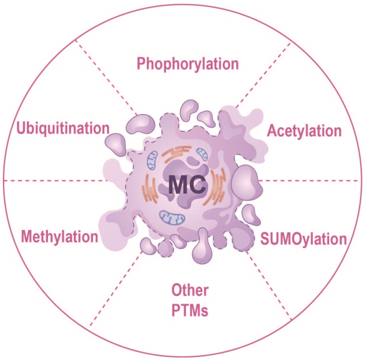
1. Introduction
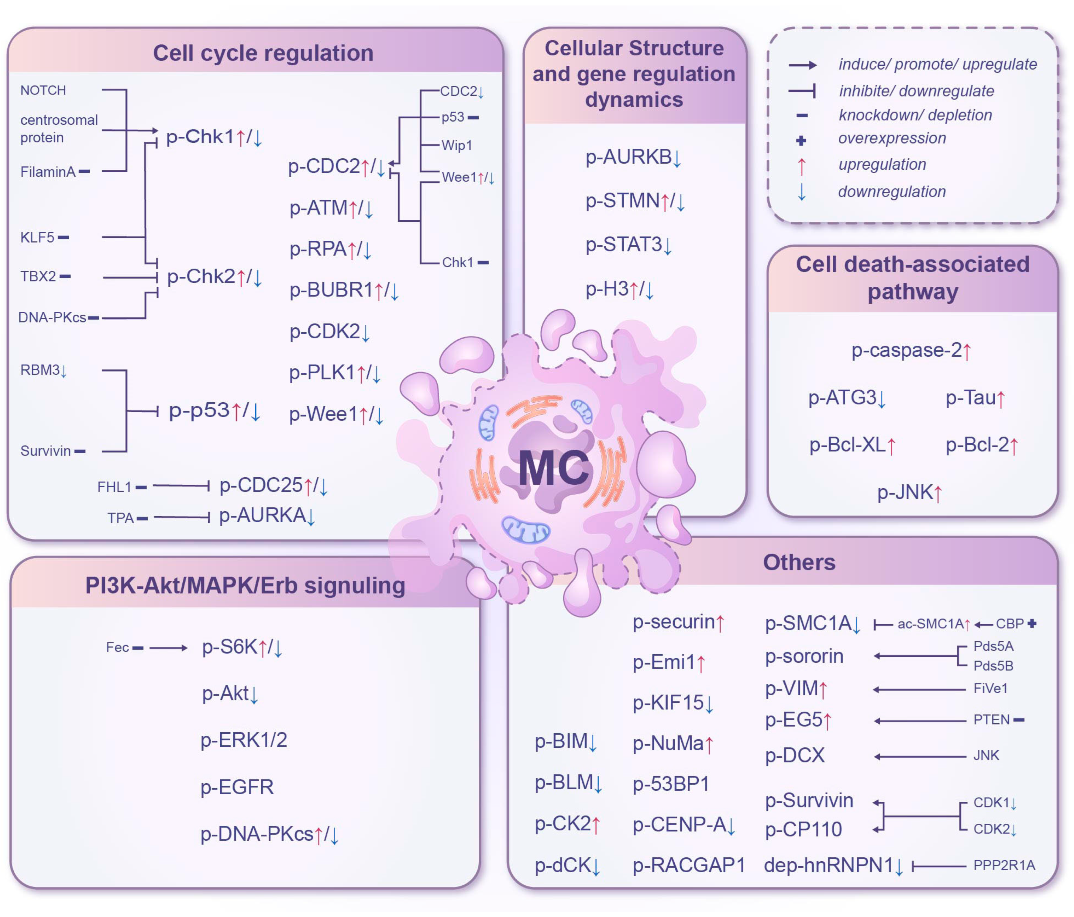
2. Mitotic Catastrophe and Phosphorylation
2.1. Cell Cycle Regulation
2.1.1. DNA Damage Response Pathway
2.1.2. G2-M Checkpoint Pathway
2.1.3. APC/C-Related Pathway
2.2. Cell Death-Associated Pathway
2.3. PI3K-Akt/MAPK/Erb Signaling
2.3.1. PI3K-Akt Signaling Pathway
2.3.2. MAPK/Erk Signaling Pathway
2.4. Cellular Structure and Gene Regulation Dynamics
2.4.1. Microtubule Dynamics
2.4.2. Epigenetics of Histone H3
2.5. Others
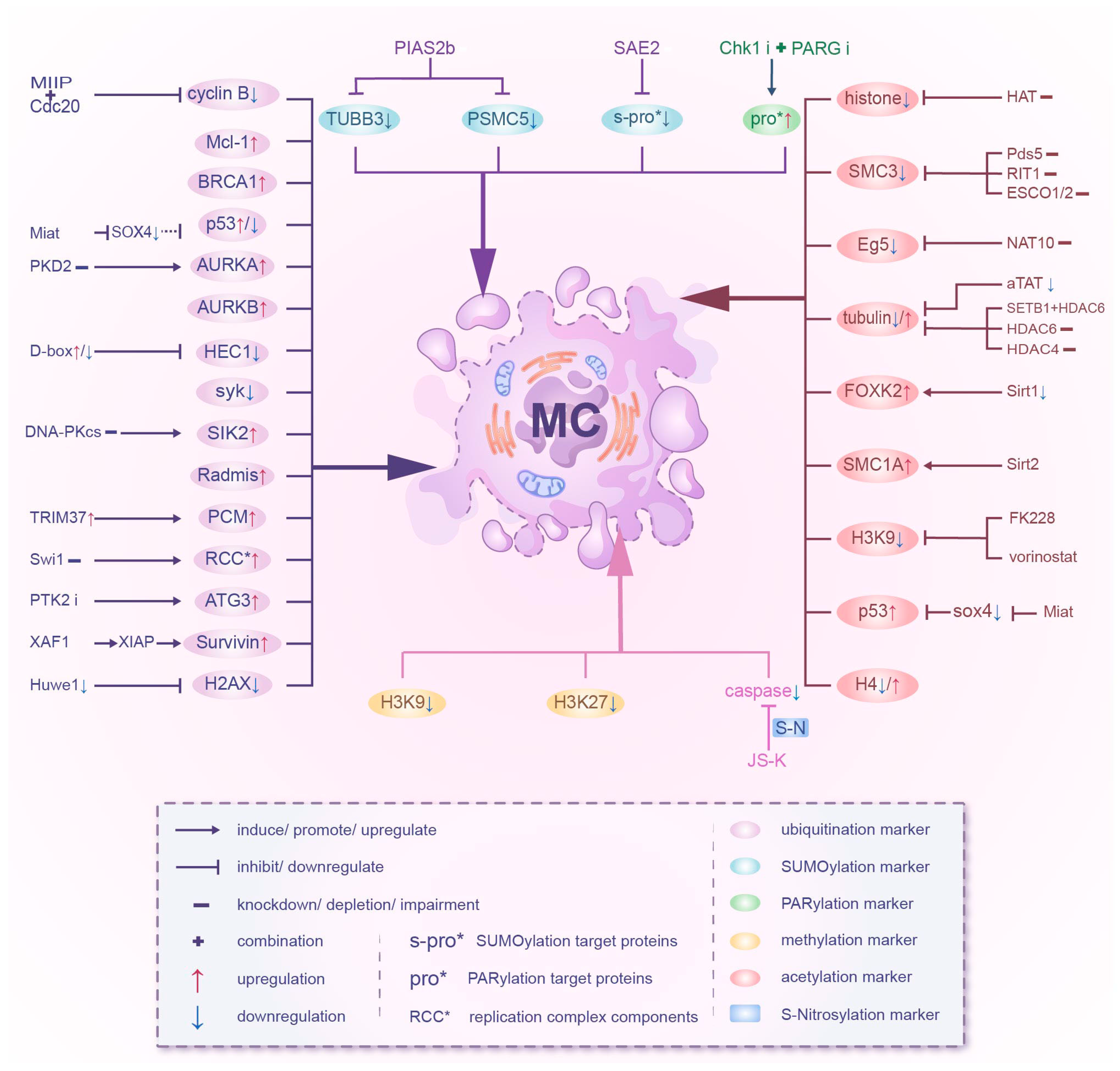
3. Mitotic Catastrophe and Ubiquitination
3.1. Cyclin B
3.2. Cell Cycle Checkpoints
3.3. Aurora Family
3.4. Others
4. Mitotic Catastrophe and Acetylation
4.1. KATs
4.2. KDACs
4.3. Others
5. Mitotic Catastrophe and Methylation
6. Mitotic Catastrophe and Other Post-Translational Modifications
7. Discussion and Future Perspectives
Author Contributions
Funding
Institutional Review Board Statement
Informed Consent Statement
Data Availability Statement
Conflicts of Interest
References
- Vitale, I.; Galluzzi, L.; Castedo, M.; Kroemer, G. Mitotic catastrophe: A mechanism for avoiding genomic instability. Nat. Rev. Mol. Cell Biol. 2011, 12, 385–392. [Google Scholar] [CrossRef]
- Pan, S.; Chen, R. Pathological implication of protein post-translational modifications in cancer. Mol. Aspects Med. 2022, 86, 101097. [Google Scholar] [CrossRef]
- Lee, J.M.; Hammarén, H.M.; Savitski, M.M.; Baek, S.H. Control of protein stability by post-translational modifications. Nat. Commun. 2023, 14, 201. [Google Scholar] [CrossRef]
- Venne, A.S.; Kollipara, L.; Zahedi, R.P. The next level of complexity: Crosstalk of posttranslational modifications. Proteomics 2014, 14, 513–524. [Google Scholar] [CrossRef]
- Ha, G.H.; Kim, H.S.; Lee, C.G.; Park, H.Y.; Kim, E.J.; Shin, H.J.; Lee, J.C.; Lee, K.W.; Lee, C.W. Mitotic catastrophe is the predominant response to histone acetyltransferase depletion. Cell Death Differ. 2009, 16, 483–497. [Google Scholar] [CrossRef]
- Rodrigues, J.S.; Chenlo, M.; Bravo, S.B.; Perez-Romero, S.; Suarez-Fariña, M.; Sobrino, T.; Sanz-Pamplona, R.; González-Prieto, R.; Blanco Freire, M.N.; Nogueiras, R.; et al. dsRNAi-Mediated Silencing of PIAS2beta Specifically Kills Anaplastic Carcinomas by Mitotic Catastrophe. Nat. Commun. 2024, 15, 3736, Correction in Nat. Commun. 2024, 15, 5835. [Google Scholar] [CrossRef]
- Sefton, B.M. Overview of protein phosphorylation. Curr. Protoc. Cell Biol. 2001, 14.1.1–14.1.3. [Google Scholar]
- Zhang, W.J.; Zhou, Y.; Zhang, Y.; Su, Y.H.; Xu, T. Protein phosphorylation: A molecular switch in plant signaling. Cell Rep. 2023, 42, 112729. [Google Scholar] [CrossRef] [PubMed]
- Jackson, S.P.; Bartek, J. The DNA-damage response in human biology and disease. Nature 2009, 461, 1071–1078. [Google Scholar] [CrossRef] [PubMed]
- Smith, J.; Tho, L.M.; Xu, N.; Gillespie, D.A. The ATM-Chk2 and ATR-Chk1 pathways in DNA damage signaling and cancer. Adv. Cancer Res. 2010, 108, 73–112. [Google Scholar] [PubMed]
- Niida, H.; Katsuno, Y.; Banerjee, B.; Hande, M.P.; Nakanishi, M. Specific role of Chk1 phosphorylations in cell survival and checkpoint activation. Mol. Cell. Biol. 2007, 27, 2572–2581. [Google Scholar] [CrossRef]
- Latif, C.; den Elzen, N.R.; O’Connell, M.J. DNA damage checkpoint maintenance through sustained Chk1 activity. J. Cell Sci. 2004, 117, 3489–3498. [Google Scholar] [CrossRef] [PubMed]
- Castedo, M.; Perfettini, J.L.; Roumier, T.; Yakushijin, K.; Horne, D.; Medema, R.; Kroemer, G. The cell cycle checkpoint kinase Chk2 is a negative regulator of mitotic catastrophe. Oncogene 2004, 23, 4353–4361. [Google Scholar] [CrossRef]
- King, C.; Diaz, H.B.; McNeely, S.; Barnard, D.; Dempsey, J.; Blosser, W.; Beckmann, R.; Barda, D.; Marshall, M.S. LY2606368 Causes Replication Catastrophe and Antitumor Effects through CHK1-Dependent Mechanisms. Mol. Cancer Ther. 2015, 14, 2004–2013. [Google Scholar] [CrossRef]
- Blackwood, E.; Epler, J.; Yen, I.; Flagella, M.; O’Brien, T.; Evangelista, M.; Schmidt, S.; Xiao, Y.; Choi, J.; Kowanetz, K.; et al. Combination drug scheduling defines a “window of opportunity” for chemopotentiation of gemcitabine by an orally bioavailable, selective ChK1 inhibitor, GNE-900. Mol. Cancer Ther. 2013, 12, 1968–1980. [Google Scholar] [CrossRef]
- Montano, R.; Chung, I.; Garner, K.M.; Parry, D.; Eastman, A. Preclinical development of the novel Chk1 inhibitor SCH900776 in combination with DNA-damaging agents and antimetabolites. Mol. Cancer Ther. 2012, 11, 427–438. [Google Scholar] [CrossRef]
- Oike, T.; Komachi, M.; Ogiwara, H.; Amornwichet, N.; Saitoh, Y.; Torikai, K.; Kubo, N.; Nakano, T.; Kohno, T. C646, a selective small molecule inhibitor of histone acetyltransferase p300, radiosensitizes lung cancer cells by enhancing mitotic catastrophe. Radiother. Oncol. 2014, 111, 222–227. [Google Scholar] [CrossRef]
- Riesterer, O.; Matsumoto, F.; Wang, L.; Pickett, J.; Molkentine, D.; Giri, U.; Milas, L.; Raju, U. A novel Chk inhibitor, XL-844, increases human cancer cell radiosensitivity through promotion of mitotic catastrophe. Investig. New Drugs 2011, 29, 514–522. [Google Scholar] [CrossRef]
- Lee, C.K.; Wang, S.; Huang, X.; Ryder, J.; Liu, B. HDAC inhibition synergistically enhances alkylator-induced DNA damage responses and apoptosis in multiple myeloma cells. Cancer Lett. 2010, 296, 233–240. [Google Scholar] [CrossRef] [PubMed]
- Zhou, X.; Kim, S.H.; Shen, L.; Lee, H.J.; Chen, C. Induction of mitotic catastrophe by PKC inhibition in Nf1-deficient cells. Cell Cycle 2014, 13, 2340–2348. [Google Scholar] [CrossRef] [PubMed]
- Wansleben, S.; Davis, E.; Peres, J.; Prince, S. A novel role for the anti-senescence factor TBX2 in DNA repair and cisplatin resistance. Cell Death Dis. 2013, 4, e846. [Google Scholar] [CrossRef]
- Shang, Z.F.; Huang, B.; Xu, Q.Z.; Zhang, S.M.; Fan, R.; Liu, X.D.; Wang, Y.; Zhou, P.K. Inactivation of DNA-dependent protein kinase leads to spindle disruption and mitotic catastrophe with attenuated checkpoint protein 2 Phosphorylation in response to DNA damage. Cancer Res. 2010, 70, 3657–3666. [Google Scholar] [CrossRef] [PubMed]
- Kimura, M.; Yoshioka, T.; Saio, M.; Banno, Y.; Nagaoka, H.; Okano, Y. Mitotic catastrophe and cell death induced by depletion of centrosomal proteins. Cell Death Dis. 2013, 4, e603. [Google Scholar] [CrossRef] [PubMed]
- Zhang, H.; Shao, F.; Guo, W.; Gao, Y.; He, J. Knockdown of KLF5 promotes cisplatin-induced cell apoptosis via regulating DNA damage checkpoint proteins in non-small cell lung cancer. Thorac. Cancer 2019, 10, 1069–1077. [Google Scholar] [CrossRef]
- Meng, X.; Yuan, Y.; Maestas, A.; Shen, Z. Recovery from DNA damage-induced G2 arrest requires actin-binding protein filamin-A/actin-binding protein 280. J. Biol. Chem. 2004, 279, 6098–6105. [Google Scholar] [CrossRef]
- Miao, K.; Lei, J.H.; Valecha, M.V.; Zhang, A.; Xu, J.; Wang, L.; Lyu, X.; Chen, S.; Miao, Z.; Zhang, X.; et al. NOTCH1 activation compensates BRCA1 deficiency and promotes triple-negative breast cancer formation. Nat. Commun. 2020, 11, 3256. [Google Scholar] [CrossRef]
- Sundararajan, V.; Tan, T.Z.; Lim, D.; Peng, Y.; Wengner, A.M.; Ngoi, N.Y.L.; Jeyasekharan, A.D.; Tan, D.S.P. Nuclear pCHK1 as a potential biomarker of increased sensitivity to ATR inhibition. J. Pathol. 2023, 259, 194–204. [Google Scholar] [CrossRef]
- Liu, Y.; Su, Z.; Tavana, O.; Gu, W. Understanding the complexity of p53 in a new era of tumor suppression. Cancer Cell 2024, 42, 946–967. [Google Scholar] [CrossRef] [PubMed]
- Sureban, S.M.; Ramalingam, S.; Natarajan, G.; May, R.; Subramaniam, D.; Bishnupuri, K.S.; Morrison, A.R.; Dieckgraefe, B.K.; Brackett, D.J.; Postier, R.G.; et al. Translation regulatory factor RBM3 is a proto-oncogene that prevents mitotic catastrophe. Oncogene 2008, 27, 4544–4556. [Google Scholar] [CrossRef]
- Rbah-Vidal, L.; Vidal, A.; Billaud, E.M.; Besse, S.; Ranchon-Cole, I.; Mishellany, F.; Perrot, Y.; Maigne, L.; Moins, N.; Guerquin-Kern, J.L.; et al. Theranostic Approach for Metastatic Pigmented Melanoma Using ICF15002, a Multimodal Radiotracer for Both PET Imaging and Targeted Radionuclide Therapy. Neoplasia 2017, 19, 17–27. [Google Scholar] [CrossRef]
- Martini, E.; Wittkopf, N.; Günther, C.; Leppkes, M.; Okada, H.; Watson, A.J.; Podstawa, E.; Backert, I.; Amann, K.; Neurath, M.F.; et al. Loss of Survivin in Intestinal Epithelial Progenitor Cells Leads to Mitotic Catastrophe and Breakdown of Gut Immune Homeostasis. Cell Rep. 2016, 14, 1062–1073. [Google Scholar] [CrossRef]
- Hung, J.Y.; Wen, C.W.; Hsu, Y.L.; Lin, E.S.; Huang, M.S.; Chen, C.Y.; Kuo, P.L. Subamolide a induces mitotic catastrophe accompanied by apoptosis in human lung cancer cells. Evid. Based Complement. Altern. Med. 2013, 2013, 828143, Erratum in Evid. Based Complement. Altern. Med. 2013, 2013, 687142. [Google Scholar] [CrossRef] [PubMed]
- Wakileh, G.A.; Bierholz, P.; Kotthoff, M.; Skowron, M.A.; Bremmer, F.; Stephan, A.; Anbuhl, S.M.; Heukers, R.; Smit, M.J.; Ströbel, P.; et al. Molecular characterization of the CXCR4/CXCR7 axis in germ cell tumors and its targetability using nanobody-drug-conjugates. Exp. Hematol. Oncol. 2023, 12, 96. [Google Scholar] [CrossRef]
- Yang, Y.C.; Chao, K.S.; Lin, C.P.; Chen, Y.Y.; Wang, K.L.; Chen, Y.J. Oxaliplatin regulates DNA repair responding to ionizing radiation and enhances radiosensitivity of human cervical cancer cells. Int. J. Gynecol. Cancer 2009, 19, 782–786. [Google Scholar] [CrossRef]
- Wang, J.; Zhang, Z.; Che, Y.; Yuan, Z.; Lu, Z.; Li, Y.; Wan, J.; Sun, H.; Chen, Z.; Pu, J.; et al. Rabdocoestin B exhibits antitumor activity by inducing G2/M phase arrest and apoptosis in esophageal squamous cell carcinoma. Cancer Chemother. Pharmacol. 2018, 81, 469–481. [Google Scholar] [CrossRef] [PubMed]
- Hu, L.; Li, B.; Chen, G.; Song, D.; Xu, Z.; Gao, L.; Xi, M.; Zhou, J.; Li, L.; Zhang, H.; et al. A novel M phase blocker, DCZ3301 enhances the sensitivity of bortezomib in resistant multiple myeloma through DNA damage and mitotic catastrophe. J. Exp. Clin. Cancer Res. 2020, 39, 105. [Google Scholar] [CrossRef] [PubMed]
- Ghelli Luserna di Rorà, A.; Cerchione, C.; Martinelli, G.; Simonetti, G. A WEE1 family business: Regulation of mitosis, cancer progression, and therapeutic target. J. Hematol. Oncol. 2020, 13, 126. [Google Scholar] [CrossRef]
- Göder, A.; Emmerich, C.; Nikolova, T.; Kiweler, N.; Schreiber, M.; Kühl, T.; Imhof, D.; Christmann, M.; Heinzel, T.; Schneider, G.; et al. HDAC1 and HDAC2 integrate checkpoint kinase phosphorylation and cell fate through the phosphatase-2A subunit PR130. Nat. Commun. 2018, 9, 764. [Google Scholar] [CrossRef]
- Filippi-Chiela, E.C.; Thomé, M.P.; Bueno e Silva, M.M.; Pelegrini, A.L.; Ledur, P.F.; Garicochea, B.; Zamin, L.L.; Lenz, G. Resveratrol abrogates the temozolomide-induced G2 arrest leading to mitotic catastrophe and reinforces the temozolomide-induced senescence in glioma cells. BMC Cancer 2013, 13, 147. [Google Scholar] [CrossRef]
- Roy, R.V.; Suman, S.; Das, T.P.; Luevano, J.E.; Damodaran, C. Withaferin A, a steroidal lactone from Withania somnifera, induces mitotic catastrophe and growth arrest in prostate cancer cells. J. Nat. Prod. 2013, 76, 1909–1915. [Google Scholar] [CrossRef]
- Caldwell, C.C.; Spies, M. Dynamic elements of replication protein A at the crossroads of DNA replication, recombination, and repair. Crit. Rev. Biochem. Mol. Biol. 2020, 55, 482–507. [Google Scholar] [CrossRef]
- Ramkumar, K.; Stewart, C.A.; Cargill, K.R.; Della Corte, C.M.; Wang, Q.; Shen, L.; Diao, L.; Cardnell, R.J.; Peng, D.H.; Rodriguez, B.L.; et al. AXL Inhibition Induces DNA Damage and Replication Stress in Non-Small Cell Lung Cancer Cells and Promotes Sensitivity to ATR Inhibitors. Mol. Cancer Res. 2021, 19, 485–497. [Google Scholar] [CrossRef]
- Menolfi, D.; Jiang, W.; Lee, B.J.; Moiseeva, T.; Shao, Z.; Estes, V.; Frattini, M.G.; Bakkenist, C.J.; Zha, S. Kinase-dead ATR differs from ATR loss by limiting the dynamic exchange of ATR and RPA. Nat. Commun. 2018, 9, 5351. [Google Scholar] [CrossRef]
- Ashley, A.K.; Shrivastav, M.; Nie, J.; Amerin, C.; Troksa, K.; Glanzer, J.G.; Liu, S.; Opiyo, S.O.; Dimitrova, D.D.; Le, P.; et al. DNA-PK phosphorylation of RPA32 Ser4/Ser8 regulates replication stress checkpoint activation, fork restart, homologous recombination and mitotic catastrophe. DNA Repair 2014, 21, 131–139. [Google Scholar] [CrossRef]
- Coleman, T.R.; Dunphy, W.G. Cdc2 regulatory factors. Curr. Opin. Cell Biol. 1994, 6, 877–882. [Google Scholar] [CrossRef]
- Berry, L.D.; Gould, K.L. Regulation of Cdc2 activity by phosphorylation at T14/Y15. Prog. Cell Cycle Res. 1996, 2, 99–105. [Google Scholar]
- Li, P.; Liu, X.; Zhao, T.; Li, F.; Wang, Q.; Zhang, P.; Hirayama, R.; Chen, W.; Jin, X.; Zheng, X.; et al. Comparable radiation sensitivity in p53 wild-type and p53 deficient tumor cells associated with different cell death modalities. Cell Death Discov. 2021, 7, 184. [Google Scholar] [CrossRef]
- Niida, H.; Tsuge, S.; Katsuno, Y.; Konishi, A.; Takeda, N.; Nakanishi, M. Depletion of Chk1 leads to premature activation of Cdc2-cyclin B and mitotic catastrophe. J. Biol. Chem. 2005, 280, 39246–39252. [Google Scholar] [CrossRef]
- Mak, J.P.; Man, W.Y.; Ma, H.T.; Poon, R.Y. Pharmacological targeting the ATR-CHK1-WEE1 axis involves balancing cell growth stimulation and apoptosis. Oncotarget 2014, 5, 10546–10557. [Google Scholar] [CrossRef] [PubMed]
- Mak, J.P.; Man, W.Y.; Chow, J.P.; Ma, H.T.; Poon, R.Y. Pharmacological inactivation of CHK1 and WEE1 induces mitotic catastrophe in nasopharyngeal carcinoma cells. Oncotarget 2015, 6, 21074–21084. [Google Scholar] [CrossRef]
- PosthumaDeBoer, J.; Würdinger, T.; Graat, H.C.; van Beusechem, V.W.; Helder, M.N.; van Royen, B.J.; Kaspers, G.J. WEE1 inhibition sensitizes osteosarcoma to radiotherapy. BMC Cancer 2011, 11, 156. [Google Scholar] [CrossRef]
- Lewis, C.W.; Bukhari, A.B.; Xiao, E.J.; Choi, W.S.; Smith, J.D.; Homola, E.; Mackey, J.R.; Campbell, S.D.; Gamper, A.M.; Chan, G.K. Upregulation of Myt1 Promotes Acquired Resistance of Cancer Cells to Wee1 Inhibition. Cancer Res. 2019, 79, 5971–5985. [Google Scholar] [CrossRef]
- Serpico, A.F.; D’Alterio, G.; Vetrei, C.; Della Monica, R.; Nardella, L.; Visconti, R.; Grieco, D. Wee1 Rather Than Plk1 Is Inhibited by AZD1775 at Therapeutically Relevant Concentrations. Cancers 2019, 11, 819. [Google Scholar] [CrossRef]
- Ma, H.; Takahashi, A.; Sejimo, Y.; Adachi, A.; Kubo, N.; Isono, M.; Yoshida, Y.; Kanai, T.; Ohno, T.; Nakano, T. Targeting of Carbon Ion-Induced G2 Checkpoint Activation in Lung Cancer Cells Using Wee-1 Inhibitor MK-1775. Radiat. Res. 2015, 184, 660–669, Erratum in Radiat. Res. 2016, 185, e52. [Google Scholar] [CrossRef]
- Martin, J.C.; Sims, J.R.; Gupta, A.; Bakin, A.V.; Ohm, J.E. WEE1 inhibition augments CDC7 (DDK) inhibitor-induced cell death in Ewing sarcoma by forcing premature mitotic entry and mitotic catastrophe. Cancer Res. Commun. 2022, 2, 471–482. [Google Scholar] [CrossRef] [PubMed]
- Lee, J.W.; Parameswaran, J.; Sandoval-Schaefer, T.; Eoh, K.J.; Yang, D.H.; Zhu, F.; Mehra, R.; Sharma, R.; Gaffney, S.G.; Perry, E.B.; et al. Combined Aurora Kinase A (AURKA) and WEE1 Inhibition Demonstrates Synergistic Antitumor Effect in Squamous Cell Carcinoma of the Head and Neck. Clin. Cancer Res. 2019, 25, 3430–3442. [Google Scholar] [CrossRef]
- Zhang, Z.; Zhang, G.; Kong, C. Targeted inhibition of Polo-like kinase 1 by a novel small-molecule inhibitor induces mitotic catastrophe and apoptosis in human bladder cancer cells. J. Cell. Mol. Med. 2017, 21, 758–767. [Google Scholar] [CrossRef] [PubMed]
- Wu, S.; Wang, S.; Gao, F.; Li, L.; Zheng, S.; Yung, W.K.A.; Koul, D. Activation of WEE1 confers resistance to PI3K inhibition in glioblastoma. Neuro Oncol. 2018, 20, 78–91. [Google Scholar] [CrossRef] [PubMed]
- Taylor, B.F.; McNeely, S.C.; Miller, H.L.; Lehmann, G.M.; McCabe, M.J., Jr.; States, J.C. p53 suppression of arsenite-induced mitotic catastrophe is mediated by p21CIP1/WAF1. J. Pharmacol. Exp. Ther. 2006, 318, 142–151. [Google Scholar] [CrossRef]
- Park, H.K.; Panneerselvam, J.; Dudimah, F.D.; Dong, G.; Sebastian, S.; Zhang, J.; Fei, P. Wip1 contributes to cell homeostasis maintained by the steady-state level of Wtp53. Cell Cycle 2011, 10, 2574–2582. [Google Scholar] [CrossRef]
- Karlsson-Rosenthal, C.; Millar, J.B. Cdc25: Mechanisms of checkpoint inhibition and recovery. Trends Cell Biol. 2006, 16, 285–292. [Google Scholar] [CrossRef]
- Goto, H.; Izawa, I.; Li, P.; Inagaki, M. Novel regulation of checkpoint kinase 1: Is checkpoint kinase 1 a good candidate for anti-cancer therapy? Cancer Sci. 2012, 103, 1195–1200. [Google Scholar] [CrossRef]
- Zhang, H.Y.; Gu, Y.Y.; Li, Z.G.; Jia, Y.H.; Yuan, L.; Li, S.Y.; An, G.S.; Ni, J.H.; Jia, H.T. Exposure of human lung cancer cells to 8-chloro-adenosine induces G2/M arrest and mitotic catastrophe. Neoplasia 2004, 6, 802–812. [Google Scholar] [CrossRef]
- Hirose, Y.; Berger, M.S.; Pieper, R.O. Abrogation of the Chk1-mediated G(2) checkpoint pathway potentiates temozolomide-induced toxicity in a p53-independent manner in human glioblastoma cells. Cancer Res. 2001, 61, 5843–5849. [Google Scholar]
- Xu, X.; Fan, Z.; Liang, C.; Li, L.; Wang, L.; Liang, Y.; Wu, J.; Chang, S.; Yan, Z.; Lv, Z.; et al. A signature motif in LIM proteins mediates binding to checkpoint proteins and increases tumour radiosensitivity. Nat. Commun. 2017, 8, 14059. [Google Scholar] [CrossRef]
- Yan, M.; Wang, C.; He, B.; Yang, M.; Tong, M.; Long, Z.; Liu, B.; Peng, F.; Xu, L.; Zhang, Y.; et al. Aurora-A Kinase: A Potent Oncogene and Target for Cancer Therapy. Med. Res. Rev. 2016, 36, 1036–1079. [Google Scholar] [CrossRef] [PubMed]
- Turaga, S.M.; Vishwakarma, V.; Hembruff, S.L.; Gibbs, B.K.; Sabu, P.; Puri, R.V.; Pathak, H.B.; Samuel, G.; Godwin, A.K. Inducing Mitotic Catastrophe as a Therapeutic Approach to Improve Outcomes in Ewing Sarcoma. Cancers 2023, 15, 4911. [Google Scholar] [CrossRef]
- Scaife, R.M. G2 cell cycle arrest, down-regulation of cyclin B, and induction of mitotic catastrophe by the flavoprotein inhibitor diphenyleneiodonium. Mol. Cancer Ther. 2004, 3, 1229–1237. [Google Scholar] [CrossRef]
- Danilov, A.V.; Hu, S.; Orr, B.; Godek, K.; Mustachio, L.M.; Sekula, D.; Liu, X.; Kawakami, M.; Johnson, F.M.; Compton, D.A.; et al. Dinaciclib Induces Anaphase Catastrophe in Lung Cancer Cells via Inhibition of Cyclin-Dependent Kinases 1 and 2. Mol. Cancer Ther. 2016, 15, 2758–2766. [Google Scholar] [CrossRef]
- Jennings, L.; Walters, H.A.; McCraw, T.J.; Turner, J.L.; Mason, J.M. FBH1 deficiency sensitizes cells to WEE1 inhibition by promoting mitotic catastrophe. DNA Repair 2024, 133, 103611. [Google Scholar] [CrossRef]
- Luo, M.L.; Li, J.; Shen, L.; Chu, J.; Guo, Q.; Liang, G.; Wu, W.; Chen, J.; Chen, R.; Song, E. The Role of APAL/ST8SIA6-AS1 lncRNA in PLK1 Activation and Mitotic Catastrophe of Tumor Cells. J. Natl. Cancer Inst. 2020, 112, 356–368, Correction in J. Natl. Cancer Inst. 2020, 112, 550. [Google Scholar] [CrossRef]
- Cheng, C.Y.; Liu, C.J.; Huang, Y.C.; Wu, S.H.; Fang, H.W.; Chen, Y.J. BI2536 induces mitotic catastrophe and radiosensitization in human oral cancer cells. Oncotarget 2018, 9, 21231–21243. [Google Scholar] [CrossRef]
- Ota, M.; Funakoshi, T.; Aki, T.; Unuma, K.; Uemura, K. Oxcarbazepine induces mitotic catastrophe and apoptosis in NRK-52E proximal tubular cells. Toxicol. Lett. 2021, 350, 240–248. [Google Scholar] [CrossRef]
- van de Weerdt, B.C.; van Vugt, M.A.; Lindon, C.; Kauw, J.J.; Rozendaal, M.J.; Klompmaker, R.; Wolthuis, R.M.; Medema, R.H. Uncoupling anaphase-promoting complex/cyclosome activity from spindle assembly checkpoint control by deregulating polo-like kinase 1. Mol. Cell. Biol. 2005, 25, 2031–2044. [Google Scholar] [CrossRef] [PubMed]
- Schrock, M.S.; Stromberg, B.R.; Scarberry, L.; Summers, M.K. APC/C ubiquitin ligase: Functions and mechanisms in tumorigenesis. Semin. Cancer Biol. 2020, 67, 80–91. [Google Scholar] [CrossRef]
- Topper, L.M.; Campbell, M.S.; Tugendreich, S.; Daum, J.R.; Burke, D.J.; Hieter, P.; Gorbsky, G.J. The dephosphorylated form of the anaphase-promoting complex protein Cdc27/Apc3 concentrates on kinetochores and chromosome arms in mitosis. Cell Cycle 2002, 1, 282–292. [Google Scholar] [CrossRef]
- Margottin-Goguet, F.; Hsu, J.Y.; Loktev, A.; Hsieh, H.M.; Reimann, J.D.; Jackson, P.K. Prophase destruction of Emi1 by the SCF(betaTrCP/Slimb) ubiquitin ligase activates the anaphase promoting complex to allow progression beyond prometaphase. Dev. Cell 2003, 4, 813–826. [Google Scholar] [CrossRef]
- Dowling, M.; Voong, K.R.; Kim, M.; Keutmann, M.K.; Harris, E.; Kao, G.D. Mitotic spindle checkpoint inactivation by trichostatin a defines a mechanism for increasing cancer cell killing by microtubule-disrupting agents. Cancer Biol. Ther. 2005, 4, 197–206. [Google Scholar] [CrossRef] [PubMed]
- Muenyi, C.S.; Trivedi, A.P.; Helm, C.W.; States, J.C. Cisplatin plus sodium arsenite and hyperthermia induces pseudo-G1 associated apoptotic cell death in ovarian cancer cells. Toxicol. Sci. 2014, 139, 74–82. [Google Scholar] [CrossRef] [PubMed]
- Park, S.E.; Chung, K.S.; Heo, S.W.; Kim, S.Y.; Lee, J.H.; Hassan, A.H.E.; Lee, Y.S.; Lee, J.Y.; Lee, K.T. Therapeutic role of 2-stearoxyphenethyl phosphocholine targeting microtubule dynamics and Wnt/β-catenin/EMT signaling in human colorectal cancer cells. Life Sci. 2023, 334, 122227. [Google Scholar] [CrossRef]
- Lim, Y.; De Bellis, D.; Sandow, J.J.; Capalbo, L.; D’Avino, P.P.; Murphy, J.M.; Webb, A.I.; Dorstyn, L.; Kumar, S. Phosphorylation by Aurora B kinase regulates caspase-2 activity and function. Cell Death Differ. 2021, 28, 349–366. [Google Scholar] [CrossRef]
- Choi, H.J.; Zhu, B.T. Upregulated cyclin B1/CDK1 mediates apoptosis following 2-methoxyestradiol-induced mitotic catastrophe: Role of Bcl-X(L) phosphorylation. Steroids 2019, 150, 108381. [Google Scholar] [CrossRef] [PubMed]
- Ma, K.; Fu, W.; Tang, M.; Zhang, C.; Hou, T.; Li, R.; Lu, X.; Wang, Y.; Zhou, J.; Li, X.; et al. PTK2-mediated degradation of ATG3 impedes cancer cells susceptible to DNA damage treatment. Autophagy 2017, 13, 579–591. [Google Scholar] [CrossRef]
- Bai, B. U1 snRNP Alteration and Neuronal Cell Cycle Reentry in Alzheimer Disease. Front. Aging Neurosci. 2018, 10, 75. [Google Scholar] [CrossRef]
- Chen, B.; Cheng, M.; Hong, D.J.; Sun, F.Y.; Zhu, C.Q. Okadaic acid induced cyclin B1 expression and mitotic catastrophe in rat cortex. Neurosci. Lett. 2006, 406, 178–182. [Google Scholar] [CrossRef]
- Glaviano, A.; Foo, A.S.C.; Lam, H.Y.; Yap, K.C.H.; Jacot, W.; Jones, R.H.; Eng, H.; Nair, M.G.; Makvandi, P.; Geoerger, B.; et al. PI3K/AKT/mTOR signaling transduction pathway and targeted therapies in cancer. Mol. Cancer 2023, 22, 138. [Google Scholar] [CrossRef]
- Lacombe, J.; Cretignier, T.; Meli, L.; Wijeratne, E.M.K.; Veuthey, J.L.; Cuendet, M.; Gunatilaka, A.A.L.; Zenhausern, F. Withanolide D Enhances Radiosensitivity of Human Cancer Cells by Inhibiting DNA Damage Non-homologous End Joining Repair Pathway. Front. Oncol. 2019, 9, 1468. [Google Scholar] [CrossRef]
- Zhou, B.; Yang, Y.; Pang, X.; Shi, J.; Jiang, T.; Zheng, X. Quercetin inhibits DNA damage responses to induce apoptosis via SIRT5/PI3K/AKT pathway in non-small cell lung cancer. Biomed. Pharmacother. 2023, 165, 115071. [Google Scholar] [CrossRef] [PubMed]
- Li, M.; Lang, Y.; Gu, M.M.; Shi, J.; Chen, B.P.C.; Yu, L.; Zhou, P.K.; Shang, Z.F. Vanillin derivative VND3207 activates DNA-PKcs conferring protection against radiation-induced intestinal epithelial cells injury in vitro and in vivo. Toxicol. Appl. Pharmacol. 2020, 387, 114855. [Google Scholar] [CrossRef] [PubMed]
- Manning, B.D.; Toker, A. AKT/PKB Signaling: Navigating the Network. Cell 2017, 169, 381–405. [Google Scholar] [CrossRef]
- Hagenbuchner, J.; Lungkofler, L.; Kiechl-Kohlendorfer, U.; Viola, G.; Ferlin, M.G.; Ausserlechner, M.J.; Obexer, P. The tubulin inhibitor MG-2477 induces autophagy-regulated cell death, ROS accumulation and activation of FOXO3 in neuroblastoma. Oncotarget 2017, 8, 32009–32026. [Google Scholar] [CrossRef] [PubMed]
- Kim, B.H.; Kim, Y.J.; Kim, M.H.; Na, Y.R.; Jung, D.; Seok, S.H.; Kim, J.; Kim, H.J. Identification of FES as a Novel Radiosensitizing Target in Human Cancers. Clin. Cancer Res. 2020, 26, 265–273. [Google Scholar] [CrossRef]
- Lima, K.; de Miranda, L.B.L.; Del Milagro Bernabe Garnique, A.; de Almeida, B.O.; do Nascimento, M.C.; Alcântara, G.A.S.; Machado-Santelli, G.M.; Rego, E.M.; Machado-Neto, J.A. The Multikinase Inhibitor AD80 Induces Mitotic Catastrophe and Autophagy in Pancreatic Cancer Cells. Cancers 2023, 15, 3866. [Google Scholar] [CrossRef]
- Titova, E.; Shagieva, G.; Dugina, V.; Kopnin, P. The Role of Aurora B Kinase in Normal and Cancer Cells. Biochemistry 2023, 88, 2054–2062. [Google Scholar]
- Dong, J.; Everitt, J.I.; Lau, S.S.; Monks, T.J. Induction of ERK1/2 and histone H3 phosphorylation within the outer stripe of the outer medulla of the Eker rat by 2,3,5-tris-(glutathion-S-yl)hydroquinone. Toxicol. Sci. 2004, 80, 350–357. [Google Scholar] [CrossRef]
- Chung, E.J.; Brown, A.P.; Asano, H.; Mandler, M.; Burgan, W.E.; Carter, D.; Camphausen, K.; Citrin, D. In vitro and in vivo radiosensitization with AZD6244 (ARRY-142886), an inhibitor of mitogen-activated protein kinase/extracellular signal-regulated kinase 1/2 kinase. Clin. Cancer Res. 2009, 15, 3050–3057. [Google Scholar] [CrossRef]
- Li, C.; Liao, J.; Wang, X.; Chen, F.X.; Guo, X.; Chen, X. Combined Aurora Kinase A and CHK1 Inhibition Enhances Radiosensitivity of Triple-Negative Breast Cancer Through Induction of Apoptosis and Mitotic Catastrophe Associated With Excessive DNA Damage. Int. J. Radiat. Oncol. Biol. Phys. 2023, 117, 1241–1254. [Google Scholar] [CrossRef] [PubMed]
- Huguet, F.; Fernet, M.; Giocanti, N.; Favaudon, V.; Larsen, A.K. Afatinib, an Irreversible EGFR Family Inhibitor, Shows Activity Toward Pancreatic Cancer Cells, Alone and in Combination with Radiotherapy, Independent of KRAS Status. Target. Oncol. 2016, 11, 371–381. [Google Scholar] [CrossRef]
- Chung, E.J.; Urick, M.E.; Kurshan, N.; Shield, W., 3rd; Asano, H.; Smith, P.D.; Scroggins, B.S.; Burkeen, J.; Citrin, D.E. MEK1/2 inhibition enhances the radiosensitivity of cancer cells by downregulating survival and growth signals mediated by EGFR ligands. Int. J. Oncol. 2013, 42, 2028–2036. [Google Scholar] [CrossRef]
- Su, C.C.; Chen, N.C.; Chyau, C.C.; Tseng, H.C.; Chou, F.P. Induction of Mitotic Catastrophe via Inhibition of Aurora B by Ionizing Radiation With Additive of Mulberry Water Extract in Human Bladder Cancer Cells. Integr. Cancer Ther. 2019, 18, 1534735418808586. [Google Scholar] [CrossRef] [PubMed]
- Mosca, L.; Pagano, C.; Tranchese, R.V.; Grillo, R.; Cadoni, F.; Navarra, G.; Coppola, L.; Pagano, M.; Mele, L.; Cacciapuoti, G.; et al. Antitumoral Activity of the Universal Methyl Donor S-Adenosylmethionine in Glioblastoma Cells. Molecules 2024, 29, 1708. [Google Scholar] [CrossRef]
- Holmfeldt, P.; Larsson, N.; Segerman, B.; Howell, B.; Morabito, J.; Cassimeris, L.; Gullberg, M. The catastrophe-promoting activity of ectopic Op18/stathmin is required for disruption of mitotic spindles but not interphase microtubules. Mol. Biol. Cell 2001, 12, 73–83. [Google Scholar] [CrossRef] [PubMed]
- Wegiel, B.; Wang, Y.; Li, M.; Jernigan, F.; Sun, L. Novel indolyl-chalcones target stathmin to induce cancer cell death. Cell Cycle 2016, 15, 1288–1294. [Google Scholar] [CrossRef]
- Urick, M.E.; Chung, E.J.; Shield, W.P., 3rd; Gerber, N.; White, A.; Sowers, A.; Thetford, A.; Camphausen, K.; Mitchell, J.; Citrin, D.E. Enhancement of 5-fluorouracil-induced in vitro and in vivo radiosensitization with MEK inhibition. Clin. Cancer Res. 2011, 17, 5038–5047. [Google Scholar] [CrossRef]
- Hans, F.; Dimitrov, S. Histone H3 phosphorylation and cell division. Oncogene 2001, 20, 3021–3027. [Google Scholar] [CrossRef]
- Huertas, D.; Soler, M.; Moreto, J.; Villanueva, A.; Martinez, A.; Vidal, A.; Charlton, M.; Moffat, D.; Patel, S.; McDermott, J.; et al. Antitumor activity of a small-molecule inhibitor of the histone kinase Haspin. Oncogene 2012, 31, 1408–1418. [Google Scholar] [CrossRef]
- Xu, C.; Gao, Q.; Wu, Z.; Lou, W.; Li, X.; Wang, M.; Wang, N.; Li, Q. Combined HASPIN and mTOR inhibition is synergistic against KRAS-driven carcinomas. Transl. Oncol. 2022, 26, 101540. [Google Scholar] [CrossRef]
- Libertini, S.; Abagnale, A.; Passaro, C.; Botta, G.; Barbato, S.; Chieffi, P.; Portella, G. AZD1152 negatively affects the growth of anaplastic thyroid carcinoma cells and enhances the effects of oncolytic virus dl922-947. Endocr. Relat. Cancer 2011, 18, 129–141. [Google Scholar] [CrossRef]
- Chen, M.; Guo, Y.; Zhao, R.; Wang, X.; Jiang, M.; Fu, H.; Zhang, X. Ophiopogonin B induces apoptosis, mitotic catastrophe and autophagy in A549 cells. Int. J. Oncol. 2016, 49, 316–324. [Google Scholar] [CrossRef] [PubMed]
- Qu, Z.; Zou, X.; Zhang, X.; Sheng, J.; Wang, Y.; Wang, J.; Wang, C.; Ji, Y. Chelidonine induces mitotic slippage and apoptotic-like death in SGC-7901 human gastric carcinoma cells. Mol. Med. Rep. 2016, 13, 1336–1344. [Google Scholar] [CrossRef] [PubMed]
- Chen, J.; Li, Z.; Jia, X.; Song, W.; Wu, H.; Zhu, H.; Xuan, Z.; Du, Y.; Zhu, X.; Song, G.; et al. Targeting anillin inhibits tumorigenesis and tumor growth in hepatocellular carcinoma via impairing cytokinesis fidelity. Oncogene 2022, 41, 3118–3130. [Google Scholar] [CrossRef]
- Zhong, R.; Xin, R.; Chen, Z.; Liang, N.; Liu, Y.; Ma, S.; Liu, X. The Role of Deoxycytidine Kinase (dCK) in Radiation-Induced Cell Death. Int. J. Mol. Sci. 2016, 17, 1939. [Google Scholar] [CrossRef]
- Zdioruk, M.; Want, A.; Mietelska-Porowska, A.; Laskowska-Kaszub, K.; Wojsiat, J.; Klejman, A.; Użarowska, E.; Koza, P.; Olejniczak, S.; Pikul, S.; et al. A New Inhibitor of Tubulin Polymerization Kills Multiple Cancer Cell Types and Reveals p21-Mediated Mechanism Determining Cell Death after Mitotic Catastrophe. Cancers 2020, 12, 2161. [Google Scholar] [CrossRef] [PubMed]
- Yi, F.; Zhang, Y.; Wang, Z.; Wang, Z.; Li, Z.; Zhou, T.; Xu, H.; Liu, J.; Jiang, B.; Li, X.; et al. The deacetylation-phosphorylation regulation of SIRT2-SMC1A axis as a mechanism of antimitotic catastrophe in early tumorigenesis. Sci. Adv. 2021, 7, eabe5518. [Google Scholar] [CrossRef] [PubMed]
- Tseng, H.H.; Chuah, Q.Y.; Yang, P.M.; Chen, C.T.; Chao, J.C.; Lin, M.D.; Chiu, S.J. Securin enhances the anti-cancer effects of 6-methoxy-3-(3′,4′,5′-trimethoxy-benzoyl)-1H-indole (BPR0L075) in human colorectal cancer cells. PLoS ONE 2012, 7, e36006. [Google Scholar] [CrossRef]
- Tanaka, K.; Yu, H.A.; Yang, S.; Han, S.; Selcuklu, S.D.; Kim, K.; Ramani, S.; Ganesan, Y.T.; Moyer, A.; Sinha, S.; et al. Targeting Aurora B kinase prevents and overcomes resistance to EGFR inhibitors in lung cancer by enhancing BIM- and PUMA-mediated apoptosis. Cancer Cell 2021, 39, 1245–1261.e6. [Google Scholar] [CrossRef]
- Al-Jomah, N.; Mukololo, L.; Anjum, A.; Al Madadha, M.; Patel, R. Pds5A and Pds5B Display Non-redundant Functions in Mitosis and Their Loss Triggers Chk1 Activation. Front. Cell Dev. Biol. 2020, 8, 531. [Google Scholar] [CrossRef]
- Zhang, X.; Zhang, Z.; Chen, G.; Zhao, M.; Wang, D.; Zhang, X.; Du, Z.; Xu, Y.; Yu, X. FK228 induces mitotic catastrophe in A549 cells by mistargeting chromosomal passenger complex localization through changing centromeric H3K9 hypoacetylation. Acta Biochim. Biophys. Sin. 2010, 42, 677–687. [Google Scholar] [PubMed]
- Bollong, M.J.; Pietilä, M.; Pearson, A.D.; Sarkar, T.R.; Ahmad, I.; Soundararajan, R.; Lyssiotis, C.A.; Mani, S.A.; Schultz, P.G.; Lairson, L.L. A vimentin binding small molecule leads to mitotic disruption in mesenchymal cancers. Proc. Natl. Acad. Sci. USA 2017, 114, E9903–E9912. [Google Scholar] [CrossRef]
- Yong, K.J.; Milenic, D.E.; Baidoo, K.E.; Brechbiel, M.W. 212Pb-radioimmunotherapy potentiates paclitaxel-induced cell killing efficacy by perturbing the mitotic spindle checkpoint. Br. J. Cancer 2013, 108, 2013–2020. [Google Scholar] [CrossRef]
- Sule, A.; Golding, S.E.; Ahmad, S.F.; Watson, J.; Ahmed, M.H.; Kellogg, G.E.; Bernas, T.; Koebley, S.; Reed, J.C.; Povirk, L.F.; et al. ATM phosphorylates PP2A subunit A resulting in nuclear export and spatiotemporal regulation of the DNA damage response. Cell. Mol. Life Sci. 2022, 79, 603. [Google Scholar] [CrossRef]
- St-Denis, N.A.; Derksen, D.R.; Litchfield, D.W. Evidence for Regulation of Mitotic Progression through Temporal Phosphorylation and Dephosphorylation of CK2α. Mol. Cell. Biol. 2009, 29, 2068–2081, Correction in Mol. Cell. Biol. 2021, 41, e00274-21. [Google Scholar] [CrossRef]
- He, J.; Zhang, Z.; Ouyang, M.; Yang, F.; Hao, H.; Lamb, K.L.; Yang, J.; Yin, Y.; Shen, W.H. PTEN regulates EG5 to control spindle architecture and chromosome congression during mitosis. Nat. Commun. 2016, 7, 12355. [Google Scholar] [CrossRef] [PubMed]
- Bayart, E.; Grigorieva, O.; Leibovitch, S.; Onclercq-Delic, R.; Amor-Guéret, M. A major role for mitotic CDC2 kinase inactivation in the establishment of the mitotic DNA damage checkpoint. Cancer Res. 2004, 64, 8954–8959. [Google Scholar] [CrossRef]
- Sui, J.D.; Tang, Z.; Chen, B.P.C.; Huang, P.; Yang, M.Q.; Wang, N.H.; Yang, H.N.; Tu, H.L.; Jiang, Q.M.; Zhang, J.; et al. Protein Phosphatase 2A-Dependent Mitotic hnRNPA1 Dephosphorylation and TERRA Formation Facilitate Telomere Capping. Mol. Cancer Res. 2022, 20, 583–595. [Google Scholar] [CrossRef] [PubMed]
- Santra, M.; Santra, S.; Roberts, C.; Zhang, R.L.; Chopp, M. Doublecortin induces mitotic microtubule catastrophe and inhibits glioma cell invasion. J. Neurochem. 2009, 108, 231–245. [Google Scholar] [CrossRef] [PubMed]
- Perfettini, J.L.; Nardacci, R.; Séror, C.; Raza, S.Q.; Sepe, S.; Saïdi, H.; Brottes, F.; Amendola, A.; Subra, F.; Del Nonno, F.; et al. 53BP1 represses mitotic catastrophe in syncytia elicited by the HIV-1 envelope. Cell Death Differ. 2010, 17, 811–820. [Google Scholar] [CrossRef]
- van Wijk, S.J.; Fulda, S.; Dikic, I.; Heilemann, M. Visualizing ubiquitination in mammalian cells. EMBO Rep. 2019, 20, e46520. [Google Scholar] [CrossRef]
- Popovic, D.; Vucic, D.; Dikic, I. Ubiquitination in disease pathogenesis and treatment. Nat. Med. 2014, 20, 1242–1253. [Google Scholar] [CrossRef]
- Hershko, A. Mechanisms and regulation of the degradation of cyclin B. Philos. Trans. R. Soc. Lond. B Biol. Sci. 1999, 354, 1571–1575; discussion 1575–1576. [Google Scholar] [CrossRef]
- Patel, D.; McCance, D.J. Compromised spindle assembly checkpoint due to altered expression of Ubch10 and Cdc20 in human papillomavirus type 16 E6- and E7-expressing keratinocytes. J. Virol. 2010, 84, 10956–10964. [Google Scholar] [CrossRef]
- O’Sullivan-Coyne, G.; O’Sullivan, G.C.; O’Donovan, T.R.; Piwocka, K.; McKenna, S.L. Curcumin induces apoptosis-independent death in oesophageal cancer cells. Br. J. Cancer 2009, 101, 1585–1595. [Google Scholar] [CrossRef]
- Ji, P.; Smith, S.M.; Wang, Y.; Jiang, R.; Song, S.W.; Li, B.; Sawaya, R.; Bruner, J.M.; Kuang, J.; Yu, H.; et al. Inhibition of gliomagenesis and attenuation of mitotic transition by MIIP. Oncogene 2010, 29, 3501–3508. [Google Scholar] [CrossRef]
- Huang, T.S.; Shu, C.H.; Chao, Y.; Chen, S.N.; Chen, L.L. Activation of MAD 2 checkprotein and persistence of cyclin B1/CDC 2 activity associate with paclitaxel-induced apoptosis in human nasopharyngeal carcinoma cells. Apoptosis 2000, 5, 235–241. [Google Scholar] [CrossRef]
- Chi, J.J.; Li, H.; Zhou, Z.; Izquierdo-Ferrer, J.; Xue, Y.; Wavelet, C.M.; Schiltz, G.E.; Zhang, B.; Cristofanilli, M.; Lu, X.; et al. A novel strategy to block mitotic progression for targeted therapy. eBioMedicine 2019, 49, 40–54. [Google Scholar] [CrossRef]
- Matthews, H.K.; Bertoli, C.; de Bruin, R.A.M. Cell cycle control in cancer. Nat. Rev. Mol. Cell Biol. 2022, 23, 74–88. [Google Scholar] [CrossRef] [PubMed]
- Li, L.; Zhou, Y.; Wang, G.F.; Liao, S.C.; Ke, Y.B.; Wu, W.; Li, X.H.; Zhang, R.L.; Fu, Y.C. Anaphase-promoting complex/cyclosome controls HEC1 stability. Cell Prolif. 2011, 44, 1–9. [Google Scholar] [CrossRef] [PubMed]
- Stecklein, S.R.; Kumaraswamy, E.; Behbod, F.; Wang, W.; Chaguturu, V.; Harlan-Williams, L.M.; Jensen, R.A. BRCA1 and HSP90 cooperate in homologous and non-homologous DNA double-strand-break repair and G2/M checkpoint activation. Proc. Natl. Acad. Sci. USA 2012, 109, 13650–13655. [Google Scholar] [CrossRef]
- Pérez-Fidalgo, J.A.; Gambardella, V.; Pineda, B.; Burgues, O.; Piñero, O.; Cervantes, A. Aurora kinases in ovarian cancer. ESMO Open 2020, 5, e000718. [Google Scholar] [CrossRef]
- Roy, A.; Veroli, M.V.; Prasad, S.; Wang, Q.J. Protein Kinase D2 Modulates Cell Cycle By Stabilizing Aurora A Kinase at Centrosomes. Mol. Cancer Res. 2018, 16, 1785–1797. [Google Scholar] [CrossRef] [PubMed]
- Gully, C.P.; Zhang, F.; Chen, J.; Yeung, J.A.; Velazquez-Torres, G.; Wang, E.; Yeung, S.C.; Lee, M.H. Antineoplastic effects of an Aurora B kinase inhibitor in breast cancer. Mol. Cancer 2010, 9, 42. [Google Scholar] [CrossRef]
- Zyss, D.; Montcourrier, P.; Vidal, B.; Anguille, C.; Mérezègue, F.; Sahuquet, A.; Mangeat, P.H.; Coopman, P.J. The Syk tyrosine kinase localizes to the centrosomes and negatively affects mitotic progression. Cancer Res. 2005, 65, 10872–10880. [Google Scholar] [CrossRef]
- Zhu, J.; Zhang, Y.; Yan, Z.; Wang, J.; Wang, P.; Liang, X.; Liu, Y.; Ao, X.; Zhu, M.; Zhou, P.; et al. Ubiquitination and degradation of SIK2 by DNA-PKcs deficiency promote radiation-induced mitotic catastrophe. Genes. Dis. 2023, 10, 313–316. [Google Scholar] [CrossRef]
- Yumoto, T.; Nakadate, K.; Nakamura, Y.; Sugitani, Y.; Sugitani-Yoshida, R.; Ueda, S.; Sakakibara, S. Radmis, a novel mitotic spindle protein that functions in cell division of neural progenitors. PLoS ONE 2013, 8, e79895. [Google Scholar] [CrossRef]
- Yeow, Z.Y.; Lambrus, B.G.; Marlow, R.; Zhan, K.H.; Durin, M.A.; Evans, L.T.; Scott, P.M.; Phan, T.; Park, E.; Ruiz, L.A.; et al. Targeting TRIM37-driven centrosome dysfunction in 17q23-amplified breast cancer. Nature 2020, 585, 447–452. [Google Scholar] [CrossRef]
- Wang, Z.; Chang, Y.; Liu, Y.; Liu, B.; Zhen, J.; Li, X.; Lin, J.; Yu, Q.; Lv, Z.; Wang, R. Inhibition of the lncRNA MIAT prevents podocyte injury and mitotic catastrophe in diabetic nephropathy. Mol. Ther. Nucleic Acids 2022, 28, 136–153. [Google Scholar] [CrossRef]
- Roseaulin, L.C.; Noguchi, C.; Martinez, E.; Ziegler, M.A.; Toda, T.; Noguchi, E. Coordinated degradation of replisome components ensures genome stability upon replication stress in the absence of the replication fork protection complex. PLoS Genet. 2013, 9, e1003213. [Google Scholar] [CrossRef]
- Fok, K.L.; Bose, R.; Sheng, K.; Chang, C.W.; Katz-Egorov, M.; Culty, M.; Su, S.; Yang, M.; Ruan, Y.C.; Chan, H.C.; et al. Huwe1 Regulates the Establishment and Maintenance of Spermatogonia by Suppressing DNA Damage Response. Endocrinology 2017, 158, 4000–4016. [Google Scholar] [CrossRef] [PubMed]
- Arora, V.; Cheung, H.H.; Plenchette, S.; Micali, O.C.; Liston, P.; Korneluk, R.G. Degradation of survivin by the X-linked inhibitor of apoptosis (XIAP)-XAF1 complex. J. Biol. Chem. 2007, 282, 26202–26209. [Google Scholar] [CrossRef] [PubMed]
- Allan, L.A.; Skowyra, A.; Rogers, K.I.; Zeller, D.; Clarke, P.R. Atypical APC/C-dependent degradation of Mcl-1 provides an apoptotic timer during mitotic arrest. EMBO J. 2018, 37, e96831. [Google Scholar] [CrossRef] [PubMed]
- Drazic, A.; Myklebust, L.M.; Ree, R.; Arnesen, T. The world of protein acetylation. Biochim. Biophys. Acta 2016, 1864, 1372–1401. [Google Scholar] [CrossRef]
- Shvedunova, M.; Akhtar, A. Modulation of cellular processes by histone and non-histone protein acetylation. Nat. Rev. Mol. Cell Biol. 2022, 23, 329–349. [Google Scholar] [CrossRef] [PubMed]
- Farria, A.; Li, W.; Dent, S.Y. KATs in cancer: Functions and therapies. Oncogene 2015, 34, 4901–4913. [Google Scholar] [CrossRef] [PubMed]
- Ogiwara, H.; Ui, A.; Shiotani, B.; Zou, L.; Yasui, A.; Kohno, T. Curcumin suppresses multiple DNA damage response pathways and has potency as a sensitizer to PARP inhibitor. Carcinogenesis 2013, 34, 2486–2497. [Google Scholar] [CrossRef] [PubMed]
- Zheng, J.; Tan, Y.; Liu, X.; Zhang, C.; Su, K.; Jiang, Y.; Luo, J.; Li, L.; Du, X. NAT10 regulates mitotic cell fate by acetylating Eg5 to control bipolar spindle assembly and chromosome segregation. Cell Death Differ. 2022, 29, 846–860. [Google Scholar] [CrossRef]
- Chien, J.Y.; Tsen, S.D.; Chien, C.C.; Liu, H.W.; Tung, C.Y.; Lin, C.H. αTAT1 downregulation induces mitotic catastrophe in HeLa and A549 cells. Cell Death Discov. 2016, 2, 16006. [Google Scholar] [CrossRef]
- Kawasumi, R.; Abe, T.; Arakawa, H.; Garre, M.; Hirota, K.; Branzei, D. ESCO1/2’s roles in chromosome structure and interphase chromatin organization. Genes. Dev. 2017, 31, 2136–2150. [Google Scholar] [CrossRef]
- Li, Y.; Seto, E. HDACs and HDAC Inhibitors in Cancer Development and Therapy. Cold Spring Harb. Perspect. Med. 2016, 6, a026831. [Google Scholar] [CrossRef]
- Wang, X.W.; Guo, Q.Q.; Yu, Y.; Zhou, T.T.; Zhang, S.Y.; Wang, Z.; Liu, J.W.; Tang, J.; Jiang, X.Y.; Wang, S.S.; et al. The deacetylation of Foxk2 by Sirt1 reduces chemosensitivity to cisplatin. J. Cell. Mol. Med. 2022, 26, 491–506. [Google Scholar] [CrossRef]
- Baschnagel, A.; Russo, A.; Burgan, W.E.; Carter, D.; Beam, K.; Palmieri, D.; Steeg, P.S.; Tofilon, P.; Camphausen, K. Vorinostat enhances the radiosensitivity of a breast cancer brain metastatic cell line grown in vitro and as intracranial xenografts. Mol. Cancer Ther. 2009, 8, 1589–1595. [Google Scholar] [CrossRef]
- Hernandez-Vicens, R.; Singh, J.; Pernicone, N.; Listovsky, T.; Gerlitz, G. SETDB1 regulates microtubule dynamics. Cell Prolif. 2022, 55, e13348. [Google Scholar] [CrossRef]
- Song, Y.W.; Lim, Y.; Cho, S.K. 2,4-Di-tert-butylphenol, a potential HDAC6 inhibitor, induces senescence and mitotic catastrophe in human gastric adenocarcinoma AGS cells. Biochim. Biophys. Acta Mol. Cell Res. 2018, 1865, 675–683. [Google Scholar] [CrossRef] [PubMed]
- Lee, S.J.; Krauthauser, C.; Maduskuie, V.; Fawcett, P.T.; Olson, J.M.; Rajasekaran, S.A. Curcumin-induced HDAC inhibition and attenuation of medulloblastoma growth in vitro and in vivo. BMC Cancer 2011, 11, 144. [Google Scholar] [CrossRef] [PubMed]
- Su, Y.; Lin, H.; Yu, J.; Mao, L.; Jin, W.; Liu, T.; Jiang, S.; Wu, Y.; Zhang, S.; Geng, Q.; et al. RIT1 regulates mitosis and promotes proliferation by interacting with SMC3 and PDS5 in hepatocellular carcinoma. J. Exp. Clin. Cancer Res. 2023, 42, 326. [Google Scholar] [CrossRef]
- Mouron, S.; Bueno, M.J.; Lluch, A.; Manso, L.; Calvo, I.; Cortes, J.; Garcia-Saenz, J.A.; Gil-Gil, M.; Martinez-Janez, N.; Apala, J.V.; et al. Phosphoproteomic analysis of neoadjuvant breast cancer suggests that increased sensitivity to paclitaxel is driven by CDK4 and filamin A. Nat. Commun. 2022, 13, 7529. [Google Scholar] [CrossRef] [PubMed]
- Wei, J.H.; Seemann, J. Nakiterpiosin targets tubulin and triggers mitotic catastrophe in human cancer cells. Mol. Cancer Ther. 2010, 9, 3375–3385. [Google Scholar] [CrossRef]
- Ashrafian, S.; Zarrineh, M.; Jensen, P.; Nawrocki, A.; Rezadoost, H.; Ansari, A.M.; Farahmand, L.; Ghassempour, A.; Larsen, M.R. Quantitative Phosphoproteomics and Acetylomics of Safranal Anticancer Effects in Triple-Negative Breast Cancer Cells. J. Proteome Res. 2022, 21, 2566–2585. [Google Scholar] [CrossRef]
- Bajbouj, K.; Mawrin, C.; Hartig, R.; Schulze-Luehrmann, J.; Wilisch-Neumann, A.; Roessner, A.; Schneider-Stock, R. P53-dependent antiproliferative and pro-apoptotic effects of trichostatin A (TSA) in glioblastoma cells. J. Neurooncol. 2012, 107, 503–516. [Google Scholar] [CrossRef]
- Gong, F.; Miller, K.M. Histone methylation and the DNA damage response. Mutat. Res. Rev. Mutat. Res. 2019, 780, 37–47. [Google Scholar] [CrossRef]
- Huang, J.; Berger, S.L. The emerging field of dynamic lysine methylation of non-histone proteins. Curr. Opin. Genet. Dev. 2008, 18, 152–158. [Google Scholar] [CrossRef]
- Mathison, A.; Salmonson, A.; Missfeldt, M.; Bintz, J.; Williams, M.; Kossak, S.; Nair, A.; de Assuncao, T.M.; Christensen, T.; Buttar, N.; et al. Combined AURKA and H3K9 Methyltransferase Targeting Inhibits Cell Growth By Inducing Mitotic Catastrophe. Mol. Cancer Res. 2017, 15, 984–997. [Google Scholar] [CrossRef]
- Karakashev, S.; Fukumoto, T.; Zhao, B.; Lin, J.; Wu, S.; Fatkhutdinov, N.; Park, P.H.; Semenova, G.; Jean, S.; Cadungog, M.G.; et al. EZH2 Inhibition Sensitizes CARM1-High, Homologous Recombination Proficient Ovarian Cancers to PARP Inhibition. Cancer Cell 2020, 37, 157–167.e6. [Google Scholar] [CrossRef]
- Yang, Y.; He, Y.; Wang, X.; Liang, Z.; He, G.; Zhang, P.; Zhu, H.; Xu, N.; Liang, S. Protein SUMOylation modification and its associations with disease. Open Biol. 2017, 7, 170167. [Google Scholar] [CrossRef]
- Kessler, J.D.; Kahle, K.T.; Sun, T.; Meerbrey, K.L.; Schlabach, M.R.; Schmitt, E.M.; Skinner, S.O.; Xu, Q.; Li, M.Z.; Hartman, Z.C.; et al. A SUMOylation-dependent transcriptional subprogram is required for Myc-driven tumorigenesis. Science 2012, 335, 348–353. [Google Scholar] [CrossRef]
- Xie, Y.W.; Wolin, M.S. Role of nitric oxide and its interaction with superoxide in the suppression of cardiac muscle mitochondrial respiration. Involvement in response to hypoxia/reoxygenation. Circulation 1996, 94, 2580–2586. [Google Scholar] [CrossRef] [PubMed]
- Günzle, J.; Osterberg, N.; Saavedra, J.E.; Weyerbrock, A. Nitric oxide released from JS-K induces cell death by mitotic catastrophe as part of necrosis in glioblastoma multiforme. Cell Death Dis. 2016, 7, e2349. [Google Scholar] [CrossRef]
- Liu, C.; Vyas, A.; Kassab, M.A.; Singh, A.K.; Yu, X. The role of poly ADP-ribosylation in the first wave of DNA damage response. Nucleic Acids Res. 2017, 45, 8129–8141. [Google Scholar] [CrossRef] [PubMed]
- Nie, L.; Wang, C.; Huang, M.; Liu, X.; Feng, X.; Tang, M.; Li, S.; Hang, Q.; Teng, H.; Shen, X.; et al. DePARylation is critical for S phase progression and cell survival. bioRxiv 2024. [Google Scholar] [CrossRef]
- Acharya, G.; Mani, C.; Sah, N.; Saamarthy, K.; Young, R.; Reedy, M.B.; Sobol, R.W.; Palle, K. CHK1 inhibitor induced PARylation by targeting PARG causes excessive replication and metabolic stress and overcomes chemoresistance in ovarian cancer. Cell Death Discov. 2024, 10, 278. [Google Scholar] [CrossRef]
- Yan, K.; Li, L.; Wang, X.; Hong, R.; Zhang, Y.; Yang, H.; Lin, M.; Zhang, S.; He, Q.; Zheng, D.; et al. The deubiquitinating enzyme complex BRISC is required for proper mitotic spindle assembly in mammalian cells. J. Cell Biol. 2015, 210, 209–224. [Google Scholar] [CrossRef]
- Ohata, H.; Miyazaki, M.; Otomo, R.; Matsushima-Hibiya, Y.; Otsubo, C.; Nagase, T.; Arakawa, H.; Yokota, J.; Nakagama, H.; Taya, Y.; et al. NuMA is required for the selective induction of p53 target genes. Mol. Cell. Biol. 2013, 33, 2447–2457. [Google Scholar] [CrossRef]
- Ota, J.; Yamashita, Y.; Okawa, K.; Kisanuki, H.; Fujiwara, S.; Ishikawa, M.; Lim Choi, Y.; Ueno, S.; Ohki, R.; Koinuma, K.; et al. Proteomic analysis of hematopoietic stem cell-like fractions in leukemic disorders. Oncogene 2003, 22, 5720–5728. [Google Scholar] [CrossRef]
- Cohen-Armon, M. Exclusive modifications of NuMA in malignant epithelial cells: A potential therapeutic mechanism. Drug Discov. Today 2022, 27, 1205–1209. [Google Scholar] [CrossRef]
- Chang, W.; Dynek, J.N.; Smith, S. NuMA is a major acceptor of poly(ADP-ribosyl)ation by tankyrase 1 in mitosis. Biochem. J. 2005, 391, 177–184. [Google Scholar] [CrossRef]
- Compton, D.A.; Luo, C. Mutation of the predicted p34cdc2 phosphorylation sites in NuMA impair the assembly of the mitotic spindle and block mitosis. J. Cell Sci. 1995, 108, 621–633. [Google Scholar] [CrossRef] [PubMed]
- Marotta, V.E.; Sabat-Pośpiech, D.; Fielding, A.B.; Ponsford, A.H.; Thomaz, A.; Querques, F.; Morgan, M.R.; Prior, I.A.; Coulson, J.M. OTUD6B regulates KIFC1-dependent centrosome clustering and breast cancer cell survival. EMBO Rep. 2025, 26, 1003–1035. [Google Scholar] [CrossRef] [PubMed]
- Yang, F.; Chen, Y.; Dai, W. Sumoylation of Kif18A plays a role in regulating mitotic progression. BMC Cancer 2015, 15, 197. [Google Scholar] [CrossRef] [PubMed]
- Zhou, Y.F.; Zhang, L.; Niu, Z.L.; Wang, X.; Storper, A.; Hunt, R.; Zhao, Y.; Sharifi, N.; Wang, Z.A. Chemical Proteomics Identifies Ketogenesis-Mediated Cysteine Modifications Regulating Redox Function. Angew. Chem. Int. Ed. Engl. 2026, 65, e19830. [Google Scholar] [CrossRef]
- Zhang, D.; Tang, Z.; Huang, H.; Zhou, G.; Cui, C.; Weng, Y.; Liu, W.; Kim, S.; Lee, S.; Perez-Neut, M.; et al. Metabolic regulation of gene expression by histone lactylation. Nature 2019, 574, 575–580. [Google Scholar] [CrossRef]
- Xie, Z.; Zhang, D.; Chung, D.; Tang, Z.; Huang, H.; Dai, L.; Qi, S.; Li, J.; Colak, G.; Chen, Y.; et al. Metabolic Regulation of Gene Expression by Histone Lysine β-Hydroxybutyrylation. Mol. Cell 2016, 62, 194–206. [Google Scholar] [CrossRef]



| Pharmacological Agents (Drugs/Inhibitors/Combos) | Phosphorylated Substrate | Effect on Substrate | Classic Function |
|---|---|---|---|
| LY2606308 | Chk1 | − | Chk1 inhibitor |
| GNE900 | Chk1 | − | Chk1 inhibitor |
| UCN-01 | Chk1 | − | Chk1 inhibitor |
| C646 | Chk1 | − | P300 inhibitor |
| SNDX-275 + melphalan | Chk1 | + | HDAC inhibitor |
| XL-844 | Chk | − | Chk inhibitor |
| ICF15002 | p53 | + | a melanin-targeted theranostic agent |
| CXCL12 | p53 | − | Stromal Cell-Derived Factor 1 |
| PI3K i | CDC2 | + | PI3K inhibitor |
| RO2380 | CDC2 | + | PLK inhibitor |
| AZD7762 | CDC2 | − | Chk inhibitor |
| AURKA i + DDK i | CDC2 | − | AURKA i + DDK i |
| Wee1 i | CDC2 | − | Wee1 inhibitor |
| metformin | CDC2 | − | Non-selective oral hypoglycemic drugs |
| PD0166285 | CDC2 | − | Wee1 and PKMYT1 inhibitor |
| AZD1775/MK-1775 | CDC2 | − | Wee1 inhibitor |
| DCZ3301 | CDC25 | + | M phase blocker |
| diphenyleneiodonium | AURKA | − | flavoprotein-specific inhibitor |
| PGE2 | CDC25 | − | Radioprotective |
| 8cl-Ado | CDC25 | − | chemotherapeutic agent |
| Chk i | CDC25 | − | Chk1 inhibitor |
| UCN-01 | CDC25 | − | Chk1 inhibitor |
| dinaciclib | Survivin | unknown | Multi-CDK inhibitor |
| dinaciclib | CP110 | unknown | Multi-CDK inhibitor |
| trichostatin A | BUBR1 | − | HDAC inhibitor |
| Cisplatin + sodium arsenite | BUBR1 | − | Cisplatin: Platinum-based chemotherapeutic Sodium arsenite: Antineoplastic agent |
| 2-stearoxyphenethyl phosphocholine | BUBR1 | + | Alkylphospholipid antineoplastic |
| resveratrol | Wee1 | − | Natural antitumor agent |
| MS275 | Wee1 | − | Class I HDAC inhibitor |
| withaferin A | Wee1 | + | Withanolide antitumor agent |
| BGB324 | RPA | + | Selective AXL inhibitor |
| DCZ3301 | ATM | + | M phase blocker |
| Oxaliplatin | ATM | − | Platinum-based chemotherapeutic |
| Rabdocoestin B | ATM | − | Antitumor precursor compound |
| 2-methoxyestradiol | JNK | + | Anti-tubulin anticancer agent |
| 2-methoxyestradiol | Bcl-2 | + | Anti-tubulin anticancer agent |
| 2-methoxyestradiol | Bcl-XL | + | Anti-tubulin anticancer agent |
| PF573228 | ATG3 | − | PTK2/FAK inhibitor |
| Okadaic acid | Tau | + | Phosphatase inhibitor |
| PTM Type | Substrate | Modification Site | Effect on Substrate |
|---|---|---|---|
| Ubiquitination | Cyclin B | Lysine residues | − |
| Ubiquitination | p53 | Lysine residues | + |
| Ubiquitination | BRCA1 | Lysine residues | + |
| Ubiquitination | HEC1 | D-box motif | − |
| Ubiquitination | Cdc20 | Lysine residues | − |
| Ubiquitination | MAD2 | Lysine residues | + |
| Ubiquitination | Aurora A | Lysine residues | − |
| Ubiquitination | Aurora B | Lysine residues | + |
| Ubiquitination | Syk | Lysine residues | + |
| Ubiquitination | SIK2 | Lysine residues | + |
| Ubiquitination | Radmis | Lysine residues | − |
| Ubiquitination | TRIM37 | Lysine residues | + |
| Ubiquitination | Survivin | Lysine residues | + |
| Ubiquitination | Mcl-1 | Lysine residues | + |
| S-Nitrosylation | Caspase-2 | Cysteine residues | + |
| PARylation | PARP substrates | ADP-ribose attachment sites | + |
| Acetylation | Histones | Lysine residues | − |
| Acetylation | p53 | Lysine residues | + |
| Acetylation | SMC1A | Lysine residues | − |
| Acetylation | Eg5 | Lysine 771 | − |
| Acetylation | Tubulin | Lysine residues | + |
| Acetylation | SMC3 | Lysine residues | − |
| Acetylation | H3K9 | Lysine 9 | − |
| Acetylation | H4 | Lysine residues | + |
| Acetylation | p21 | Lysine residues | + |
| Methylation | Histones | Lysine/arginine residues | − |
| Methylation | H3K9 | Lysine 9 | − |
| Methylation | H3K27 | Lysine 27 | + |
| SUMOylation | TUBB3 | Lysine residues | − |
| SUMOylation | PSMC5 | Lysine residues | − |
| SUMOylation | SMC3 | Lysine residues | − |
Disclaimer/Publisher’s Note: The statements, opinions and data contained in all publications are solely those of the individual author(s) and contributor(s) and not of MDPI and/or the editor(s). MDPI and/or the editor(s) disclaim responsibility for any injury to people or property resulting from any ideas, methods, instructions or products referred to in the content. |
© 2026 by the authors. Licensee MDPI, Basel, Switzerland. This article is an open access article distributed under the terms and conditions of the Creative Commons Attribution (CC BY) license.
Share and Cite
Zhang, Q.-Y.; Chen, X.; Li, S.-K.; Cao, L.-Z.; Wang, S.-Y.; He, Y.-J.; Zhang, X.-L.; Liu, J.-W.; Liu, X.-F. Multiple Regulatory Mechanisms of Post-Translational Modifications and Therapeutic Potential of Mitotic Catastrophe. Int. J. Mol. Sci. 2026, 27, 3370. https://doi.org/10.3390/ijms27083370
Zhang Q-Y, Chen X, Li S-K, Cao L-Z, Wang S-Y, He Y-J, Zhang X-L, Liu J-W, Liu X-F. Multiple Regulatory Mechanisms of Post-Translational Modifications and Therapeutic Potential of Mitotic Catastrophe. International Journal of Molecular Sciences. 2026; 27(8):3370. https://doi.org/10.3390/ijms27083370
Chicago/Turabian StyleZhang, Qing-Yue, Xia Chen, Shi-Kun Li, Liang-Zi Cao, Shi-Ying Wang, Ying-Jie He, Xiao-Lin Zhang, Jing-Wei Liu, and Xiao-Fang Liu. 2026. "Multiple Regulatory Mechanisms of Post-Translational Modifications and Therapeutic Potential of Mitotic Catastrophe" International Journal of Molecular Sciences 27, no. 8: 3370. https://doi.org/10.3390/ijms27083370
APA StyleZhang, Q.-Y., Chen, X., Li, S.-K., Cao, L.-Z., Wang, S.-Y., He, Y.-J., Zhang, X.-L., Liu, J.-W., & Liu, X.-F. (2026). Multiple Regulatory Mechanisms of Post-Translational Modifications and Therapeutic Potential of Mitotic Catastrophe. International Journal of Molecular Sciences, 27(8), 3370. https://doi.org/10.3390/ijms27083370