Review

Integrating Blood-Based Immune-Inflammation Biomarkers into Artificial Intelligence–Driven Prognostic Models in Oncology

by
Agata Dobrowolska-Szumowska
1,
Zbigniew Krzysztof Kamocki
1 and
Żaneta Anna Mierzejewska
2,*
1
Second Department of General and Gastroenterological Surgery, Medical University of Bialystok, M. Sklodowskiej-Curie 24A, 15-276 Bialystok, Poland
2
Institute of Biomedical Engineering, Faculty of Mechanical Department, Bialystok University of Technology, Wiejska 45C, 15-351 Bialystok, Poland
*
Author to whom correspondence should be addressed.
Int. J. Mol. Sci. 2026, 27(7), 3192; https://doi.org/10.3390/ijms27073192
Submission received: 12 March 2026 / Revised: 27 March 2026 / Accepted: 29 March 2026 / Published: 31 March 2026
(This article belongs to the Section Molecular Oncology)

Abstract

Stratification in oncology remains largely based on tumor-intrinsic characteristics, with limited integration of systemic host biology. The Systemic Immune-Inflammation Index (SII), derived from peripheral blood counts, reflects interactions between inflammation, thrombosis, and immune competence and has emerged as a simple, accessible prognostic biomarker. At the same time, artificial intelligence (AI)–based survival models enable integration of heterogeneous data and nonlinear relationships in outcome prediction. However, the structured incorporation of biologically interpretable host-derived markers into explainable AI frameworks remains insufficiently defined. A structured narrative review of PubMed/MEDLINE and Scopus was conducted to examine (i) the prognostic role of SII in solid tumors and (ii) AI-based survival modeling in oncology. Rather than quantitative synthesis, the review prioritized methodological quality, biological plausibility, validation strategies, and explainability relevant to clinical translation. Elevated SII is consistently associated with poorer survival across multiple solid malignancies and treatment contexts, although effect sizes vary and methodological heterogeneity remains considerable. AI-based survival models can incorporate continuous and multimodal variables, capture nonlinear associations, and model complex tumor–host interactions. We propose a translational framework integrating SII into explainable AI survival pipelines through continuous modeling, interaction-aware feature engineering, rigorous internal and external validation, and interpretable modeling approaches. Embedding SII as a biologically grounded continuous variable within multivariable prognostic architectures may enhance personalized risk stratification in precision oncology, pending prospective validation and standardized reporting.

1. Introduction

Accurate prognostic stratification remains a central challenge in contemporary oncology and a prerequisite for personalized treatment planning. Despite advances in molecular profiling, targeted therapies, and immunotherapy, clinical decision-making continues to rely largely on conventional systems such as tumor–node–metastasis (TNM) staging, histopathological grading, and selected molecular alterations. Although these parameters characterize tumor burden and biological subtype, they only partially explain interpatient heterogeneity [1,2,3].
Cancer progression and survival are influenced not only by tumor-intrinsic features but also by host-related biological processes, particularly systemic immune and inflammatory responses. Chronic inflammation promotes tumor progression through mechanisms such as genomic instability, angiogenesis, and immune evasion [4], whereas effective lymphocyte-mediated immunity is associated with improved outcomes across malignancies [5]. These observations have increased interest in biomarkers reflecting systemic immune status.
Peripheral blood–derived markers provide a practical approach to assessing systemic immune response. Ratios such as the neutrophil-to-lymphocyte ratio (NLR), platelet-to-lymphocyte ratio (PLR), and lymphocyte-to-monocyte ratio (LMR) have demonstrated prognostic associations across tumor types, but each captures only a limited aspect of the inflammatory response [6,7,8,9].
The Systemic Immune-Inflammation Index (SII), calculated from neutrophil, platelet, and lymphocyte counts, integrates inflammation, thrombosis, and immune competence into a single measure [7,10]. Elevated SII has been consistently associated with poorer survival across multiple solid tumors, although effect sizes vary and methodological heterogeneity remains substantial [11,12,13]. Parallel, artificial intelligence (AI)–based survival models enable integration of heterogeneous clinical, laboratory, and molecular data and capture nonlinear relationships. However, their performance is context-dependent, and improvements over conventional models are not consistent across studies [14,15,16,17].
Despite growing interest in both immune-inflammatory biomarkers and AI-based modeling, these domains are often studied separately [11,14,17,18]. Structured approaches for incorporating biologically interpretable host-derived markers into explainable and clinically validated AI models remain limited, particularly in light of concerns regarding methodological heterogeneity and risk of bias in prognostic modeling research [17,19,20].
This review adopts an integrative perspective and proposes a structured framework for incorporating SII into multivariable, explainable AI-based survival models. Rather than focusing on algorithm comparison, we emphasize biological rationale, methodological principles, and validation strategies relevant to clinical translation. This approach aims to align systemic immune biology with contemporary standards of prognostic modeling and improve the interpretability and applicability of AI-driven risk stratification in oncology.
The search strategy combined controlled vocabulary and free-text terms related to (i) “systemic immune-inflammation index” OR “SII” OR “immune-inflammatory biomarkers” AND (ii) “artificial intelligence” OR “machine learning” OR “deep learning” OR “survival analysis” OR “time-to-event modeling” AND (iii) “cancer” OR “oncology”. Searches were conducted in PubMed/MEDLINE and Scopus for studies published between January 2010 and January 2026.
Retrieved records were screened for relevance to oncologic survival outcomes and the methodological integration of immune-inflammatory biomarkers or AI-based modeling approaches. Priority was given to studies providing methodological detail, multivariable analyses, or validation procedures. The final body of literature included studies addressing the prognostic role of SII across multiple solid malignancies as well as methodological research on AI-based survival modeling.
The included SII literature covered hepatocellular carcinoma, pancreatic cancer, colorectal cancer, gastric cancer, lung cancer, urothelial carcinoma, breast cancer, and other solid tumors, with the largest representation observed in gastrointestinal and thoracic oncology cohorts.
Given the narrative and framework-oriented objective of this review, formal quantitative synthesis of effect estimates was not performed. Risk of bias was not assessed using structured instruments such as PROBAST, as the aim was conceptual integration rather than systematic evidence grading. Consequently, the findings should be interpreted as a structured conceptual synthesis rather than a formal systematic evaluation of evidence.

2. Prognostic Biomarkers in Oncology: From Tumor-Centered to Host-Related Factors

2.1. Tumor-Centered Prognostic Paradigms and Their Limitations

Prognostic assessment in oncology has traditionally relied on tumor-focused parameters, including tumor size, nodal status, metastatic spread, histopathological subtype, grading, and selected molecular alterations. These variables underpin staging systems and therapeutic algorithms across malignancies and remain essential for estimating tumor burden and intrinsic aggressiveness [21,22].
However, tumor-centered approaches incompletely account for the heterogeneity observed among patients with similar clinicopathological profiles. Individuals with comparable stage and molecular characteristics may exhibit markedly different survival outcomes and treatment responses [23]. This suggests that tumor-intrinsic features alone do not fully determine disease course.
In addition, conventional staging systems are largely static and may not adequately reflect biological processes that evolve during disease progression or under therapeutic pressure [24]. For example, they do not capture temporal changes in systemic immune response, treatment-induced alterations in tumor biology, or dynamic tumor–host interactions that may influence clinical outcomes. As oncology advances toward adaptive and individualized care, prognostic strategies increasingly require integration of tumor characteristics with systemic biological context.

2.2. Host-Related Immune-Inflammatory Biomarkers

Recognition of the host immune system as an active determinant of cancer progression has expanded interest in systemic immune-inflammatory biomarkers. Chronic inflammation contributes to multiple hallmarks of cancer, including genomic instability, angiogenesis, immune evasion, and metastatic dissemination [25,26]. Conversely, effective antitumor immune surveillance, particularly lymphocyte-mediated adaptive responses, is associated with improved outcomes across malignancies [5].
Peripheral blood–derived markers provide a practical means of assessing systemic immune status. Complete blood counts are routinely available and reflect circulating inflammatory and immune cell populations [27]. Ratios such as the neutrophil-to-lymphocyte ratio, platelet-to-lymphocyte ratio, and lymphocyte-to-monocyte ratio have shown prognostic associations in multiple tumor types and treatment settings [28,29,30].
These indices, however, capture only pairwise cellular relationships and may not represent the broader immune-inflammatory milieu [7,30]. Their performance varies across disease stages and therapeutic contexts, highlighting the need for more integrative measures.
Composite indices incorporating multiple hematologic compartments have, therefore, been proposed to provide a more comprehensive representation of systemic immune-inflammatory status. Among these, the Systemic Immune-Inflammation Index (SII) has been extensively studied and demonstrates consistent prognostic associations across solid malignancies [31,32,33]. For clarity, the most commonly used peripheral blood–based immune-inflammatory biomarkers and their key biological interpretations are summarized in Table 1.

2.3. Emerging Prognostic Biomarkers and Multimodal Risk Stratification

Recent advances in oncology have expanded the prognostic biomarker landscape beyond conventional clinicopathological and hematological parameters toward molecular and computational approaches. Circulating tumor DNA enables detection of minimal residual disease, early relapse prediction, and longitudinal monitoring of treatment response [34]. Similarly, immune profiling, including tumor-infiltrating lymphocytes, peripheral immune cell subsets, and cytokine signatures, provides insight into antitumor immunity and therapeutic susceptibility, particularly in the context of immunotherapy [35].
Radiomics and radiogenomics extract quantitative features from routine imaging and link them to tumor heterogeneity and clinical outcomes. Multi-omics approaches integrating genomic, transcriptomic, proteomic, and metabolomic data further enhance characterization of disease biology [36,37,38].
Despite their depth, many of these approaches are limited by cost, technical complexity, and restricted availability in routine practice [39,40]. In addition, they primarily capture tumor-intrinsic properties and may only indirectly reflect systemic immune status. In this context, peripheral blood–based indices such as the Systemic Immune-Inflammation Index (SII) provide accessible information on host-related biological processes and may complement tumor-derived data [11,41,42].
From a modeling perspective, AI-based prognostic systems must be aligned with both the intended clinical application and the characteristics of available data [43,44]. Variables that are widely available, reproducible, and biologically interpretable are particularly valuable in this context [17]. Immune-inflammatory indices such as SII meet these criteria and may function as stabilizing features within multivariable models. Unlike high-dimensional molecular data, these biomarkers are routinely measured, relatively standardized, and less sensitive to technical variability. They, therefore, provide consistent signals that can enhance model robustness and generalizability across datasets. Their interpretability also aligns with the increasing emphasis on explainable artificial intelligence, facilitating clinical trust and potential implementation [11].

3. The Systemic Immune-Inflammation Index (SII)

3.1. Definition and Conceptual Framework

The Systemic Immune-Inflammation Index (SII) is a composite biomarker derived from routinely available peripheral blood parameters [45] and is calculated as follows:
SII = (Platelet count × Neutrophil count)/Lymphocyte count
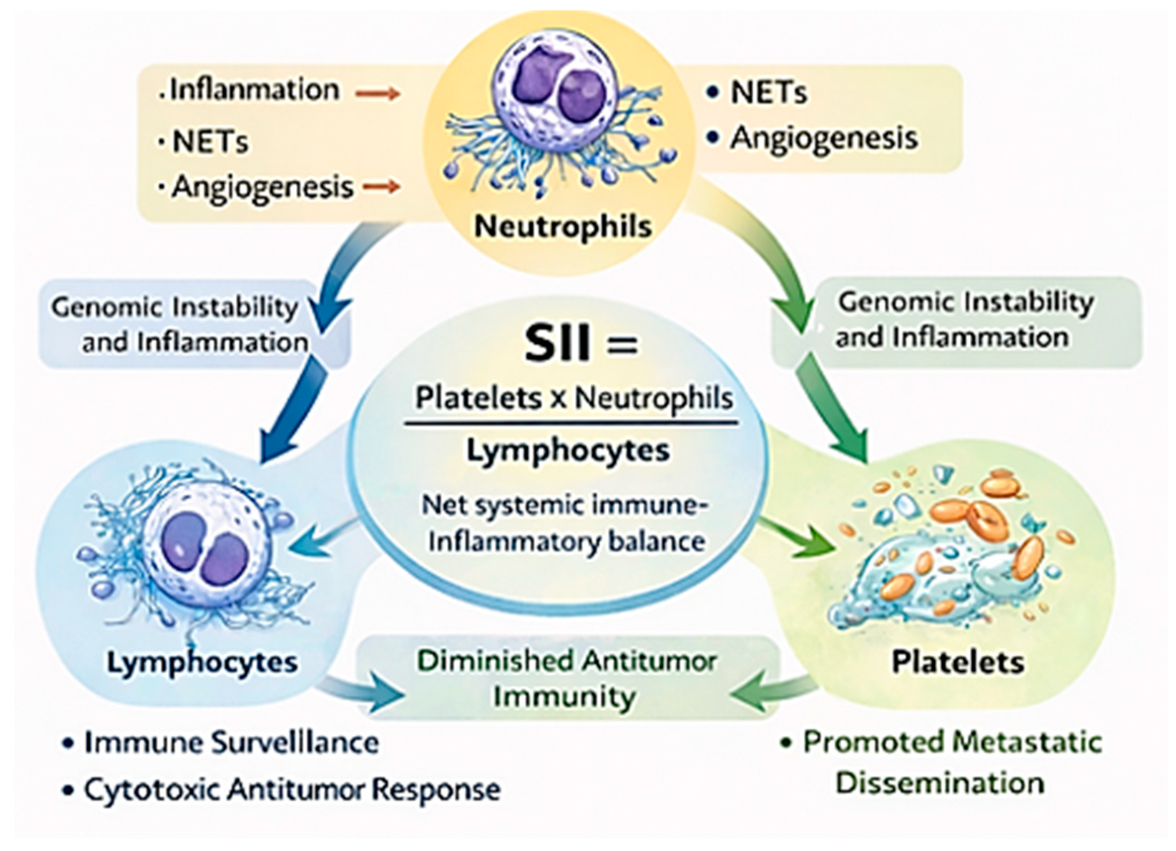
SII integrates three key hematologic compartments involved in cancer-related processes: neutrophils (inflammation), platelets (thrombosis and tumor promotion), and lymphocytes (adaptive immunity) [46]. It reflects the balance between pro-inflammatory and immunosuppressive mechanisms and antitumor immune surveillance [7,11].
Importantly, SII does not provide pathway-specific mechanistic information. Rather, it serves as a summary indicator of systemic immune-inflammatory status associated with cancer progression and survival [47]. It should, therefore, be interpreted within a multivariable clinical context rather than as a mechanistically specific marker.
Compared with other systemic inflammatory markers, such as C-reactive protein (CRP), the modified Glasgow Prognostic Score (mGPS), or the Prognostic Nutritional Index (PNI), SII integrates multiple hematologic components into a single measure [48,49]. However, as an indirect surrogate of inflammation, it may capture overlapping biological processes, and comparative evidence remains limited [50].

3.2. Biological Rationale: Linking Inflammation, Thrombosis, and Immune Suppression

The biological plausibility of SII as a prognostic biomarker is supported by extensive evidence implicating neutrophils, platelets, and lymphocytes in cancer-related processes. These cell populations contribute to inflammation, tumor progression, and immune regulation. Neutrophils are key mediators of systemic inflammation and promote tumor progression through the release of cytokines, proteases, and reactive oxygen species [51]. They also form neutrophil extracellular traps, which facilitate tumor cell adhesion, immune evasion, and metastatic dissemination [52].
Platelets contribute to tumor progression by protecting circulating tumor cells from immune clearance, enhancing endothelial adhesion, and releasing pro-angiogenic factors [53,54,55]. Increased platelet activity has been associated with adverse outcomes across multiple malignancies.
In contrast, lymphocytes, particularly cytotoxic T cells, mediate antitumor immunity. Reduced lymphocyte counts may reflect impaired immune surveillance and are associated with poorer clinical outcomes. Elevated SII values, reflecting higher neutrophil and platelet counts relative to lymphocytes, therefore, correspond to a systemic state characterized by inflammation and immune suppression, which is associated with adverse prognosis [31,56,57] (Figure 1).

3.3. Clinical and Prognostic Evidence Across Cancer Types and Treatment Contexts

The prognostic relevance of SII has been evaluated across multiple solid malignancies and treatment settings [11,31]. Elevated pre-treatment SII is consistently associated with shorter overall survival and progression-free survival in hepatocellular, pancreatic, colorectal, gastric, and lung cancers [45,47,58,59,60,61].
Most studies report independent associations after multivariable adjustment. However, formal evaluation of incremental predictive value remains limited. In particular, direct comparisons between base clinicopathological models and models extended with SII, using discrimination, calibration, or decision-analytic metrics, are rarely performed. As a result, statistical significance does not necessarily translate into improved risk prediction. As summarized in Table 2, meta-analyses report pooled hazard ratios typically ranging from approximately 1.5 to 2.5, although moderate to substantial heterogeneity is common (I2 often > 50%). This variability likely reflects differences in study design, patient populations, disease stage distribution, treatment modalities, and analytical approaches.
Most available studies are retrospective and frequently single-center, which may introduce selection bias and limit external validity [17]. In addition, effect sizes appear to vary by disease stage and availability of molecular stratification markers, suggesting that the incremental contribution of SII is context-dependent.
Across treatment settings, including surgery, chemotherapy, radiotherapy, targeted therapy, and immunotherapy, elevated SII has generally been associated with poorer outcomes [62,63,64]. Although several studies have explored its potential relevance in patients receiving immune checkpoint inhibitors [58,65,66], formal interaction analyses demonstrating treatment-specific effects remain limited. Current evidence, therefore, supports a primarily prognostic rather than predictive role [67,68].
From a methodological perspective, it is important to distinguish between prognostic and predictive biomarkers. Prognostic markers provide information on outcome independent of treatment, whereas predictive biomarkers identify differential treatment benefit. Without formal interaction testing, interpretation of SII as a predictive marker remains premature.
Overall, SII demonstrates consistent prognostic associations across cancer types. However, robust evidence of incremental predictive value beyond established models remains limited, and further validation in well-designed prospective studies is required.

3.4. Methodological, Analytical, and Biological Limitations

Despite consistent associations with clinical outcomes, several factors limit the clinical application of SII. A major challenge is the lack of standardized cut-off values for risk stratification [69]. Reported thresholds vary across studies due to differences in patient populations, laboratory practices, and statistical methods, which complicates comparison and clinical implementation [32,33].
Most available evidence is derived from retrospective cohorts, introducing potential selection bias and confounding [11,17]. In addition, SII is influenced by non-malignant conditions such as infection, chronic inflammatory disorders, and concomitant medications, limiting its specificity for cancer-related processes. Single baseline measurements may also fail to reflect temporal changes during treatment or disease progression [46,69].
From a statistical perspective, dichotomization remains common but results in information loss and may inflate effect estimates [70,71]. Data-driven threshold selection can introduce optimism bias and reduce transportability. Modeling SII as a continuous variable may mitigate these limitations.
Meta-analyses frequently report moderate to high heterogeneity and potential publication bias [72,73,74]. External validation remains limited, further constraining generalizability. Beyond baseline assessment, longitudinal evaluation of SII using serial measurements may provide additional insight into dynamic tumor–host interactions. Time-dependent modeling approaches could capture treatment-related changes and evolving systemic immune status, potentially improving risk stratification over the disease course. However, the clinical utility of such approaches requires prospective validation.
Overall, SII should not be used as an isolated risk classifier. Its clinical value is better assessed within multivariable, externally validated prediction models that account for continuous effects and context-dependent variability [75,76,77,78].

4. Artificial Intelligence in Prognostic Modeling of Cancer Outcomes

4.1. From Traditional Statistical Models to Data-Driven Prognostication

Prognostic modeling in oncology has traditionally relied on statistical approaches, most notably the Cox proportional hazards model. These models are widely used due to their interpretability and well-established theoretical foundation. However, they rely on assumptions such as proportional hazards and predefined relationships between variables, which may not fully capture the complexity of cancer progression [79].
Cancer outcomes reflect interactions among tumor characteristics, host-related factors, treatment variables, and temporal dynamics [80,81,82]. Modeling these relationships within predefined regression structures may require simplification and can limit the representation of nonlinear or higher-order effects [83].
Machine learning and deep learning approaches provide greater flexibility by accommodating nonlinear relationships and interactions without explicit prespecification [81,84]. These methods can integrate heterogeneous data sources and identify patterns that may not be captured by conventional models [85,86].
However, empirical evidence indicates that machine learning does not consistently outperform well-specified regression models in structured clinical datasets [17,82]. In many cases, Cox-based models achieve comparable discrimination. Therefore, the choice of method should be guided by data characteristics, predictor complexity, and clinical context rather than assumptions of superiority.
Artificial intelligence should be viewed as an extension of traditional modeling rather than a replacement. Its value depends on appropriate application, validation, and interpretability within clinically relevant settings.

4.2. Machine Learning Approaches for Time-to-Event Outcomes

Most clinically relevant prognostic questions in oncology involve time-to-event outcomes, such as overall survival, progression-free survival, recurrence, or treatment failure. This has led to the development of machine learning methods adapted to censored survival data [87,88].
Common approaches include random survival forests, gradient boosting methods, and neural network–based survival models [89]. Random survival forests extend tree-based models to survival analysis without relying on proportional hazards assumptions [90]. Boosting methods iteratively optimize predictive performance and can model complex relationships in structured data [91,92,93]. Neural network–based models, such as DeepSurv and DeepHit, allow flexible estimation of risk functions and time-dependent effects [94,95].
These approaches can incorporate continuous predictors and capture nonlinear associations without explicit model specification, which may be relevant for biomarkers such as SII [91,96]. However, increased model flexibility is associated with a higher risk of overfitting, particularly in datasets with limited sample size or event numbers. Robust internal validation and independent external validation are, therefore, essential to ensure generalizability [17].
The choice of modeling approach should be guided by dataset characteristics, predictor dimensionality, and clinical objectives. In structured clinical datasets, simpler and well-calibrated models may perform comparably to more complex methods.

4.3. Multimodal Data Integration and Prognostic Performance

A key advantage of AI-based prognostic systems is the ability to integrate multimodal data. In oncology, patient outcomes are influenced by tumor characteristics, systemic factors, treatment variables, and temporal dynamics. Combining these data sources within a single model may improve representation of disease complexity [97,98,99,100].
AI-based approaches can incorporate clinical variables, laboratory parameters, imaging features, and molecular data within a unified analytical framework. This may enable identification of patterns that are not captured by predefined regression structures.
However, reported improvements in predictive performance are generally modest and vary across studies, as illustrated in Table 3 [17,82]. In structured clinical datasets with limited predictor dimensionality, well-specified Cox models often achieve comparable discrimination [68,71,101]. Therefore, the value of multimodal integration depends on data quality, sample size, and clinical context rather than model complexity alone.
Model evaluation should extend beyond discrimination metrics. The concordance index reflects ranking ability but does not assess agreement between predicted and observed outcomes. Calibration is essential for individual risk estimation, yet it remains underreported in many studies [101]. In addition, improvements in statistical performance do not necessarily translate into clinical benefit [102]. Decision curve analysis and related approaches can help determine whether differences in model performance lead to meaningful changes in clinical decision-making.
Incorporating host-related biomarkers alongside tumor-centered variables may improve predictive stability in selected contexts [102]. However, the contribution of individual variables should be evaluated within fully specified multivariable models and validated in independent datasets.
Table 3. Comparative performance of Cox and AI-based survival models in oncology.
AuthorCancer TypeNModel ComparisonC-Index (Cox)C-Index (AI)External Validation
Katzman et al. [103]Breast cancer (METABRIC)~1980Cox PH vs. DeepSurv0.630.65Internal validation
Li et al.
[104]
Colorectal cancer416Cox variants vs. DeepSurv0.690.77Internal test set
Li et al.
[105]
Early-stage young breast cancer7850Cox PH vs. Random Survival Forest0.700.70Yes (external cohort)
Ma et al.
[106]
Pan-cancer (TCGA, 20 types)Multiple cohortsLasso-Cox vs. XGBoost~0.64–0.66 *0.69Cross-validation across datasets
Huang et al. [107]Ampullary adenocarcinoma2935Cox PH vs. DeepSurv0.690.73Internal test split
* Range across datasets or approximated from reported results.

4.4. Interpretability, Explainability, and Clinical Trust

In light of the cited studies, interpretability remains a central barrier to the clinical implementation of machine learning models in oncology. Evidence from studies such as Chalkidis et al. (2023) indicates that even well-performing and externally validated prediction models may have limited clinical utility if their decision-making processes are not transparent to clinicians [108]. Similarly, Collins et al. (2025) emphasize that “black-box” models can undermine clinical trust and hinder adoption, particularly in high-stakes decision-making contexts [109]. In this regard, explainable artificial intelligence (XAI) methods are increasingly recognized as essential for bridging the gap between predictive performance and clinical usability. As highlighted in the scoping review by Santos and Amorim-Lopes (2025), techniques such as feature importance measures and SHAP values can enhance transparency by clarifying how individual variables contribute to model outputs [110]. Moreover, Biziaev et al. (2023) demonstrate that incorporating clinically interpretable predictors within modeling frameworks can further support their practical applicability [111].
However, it is important to note that improved interpretability does not equate to causal understanding. XAI methods describe patterns and associations within the data rather than underlying biological mechanisms. Therefore, as consistently emphasized across these studies, model interpretation must be contextualized within clinical expertise and supported by rigorous external validation to ensure reliability and avoid misinterpretation [108,109,110,111,112].
Despite their predictive potential, AI-based models are often criticized for limited interpretability. Opaque “black-box” outputs may hinder clinical adoption, particularly in high-stakes decision-making. Interpretability is, therefore, a key requirement for implementation in oncology [113,114].
Explainable artificial intelligence (XAI) methods aim to improve transparency by quantifying how individual variables contribute to model predictions. Common approaches include feature importance measures, partial dependence plots, accumulated local effects, and Shapley additive explanations (SHAP), which provide both global and individual-level interpretations [115,116,117,118,119,120].
In this context, biologically interpretable variables such as immune-inflammatory biomarkers may enhance the credibility of model outputs. Markers such as SII are routinely measured and clinically familiar, which facilitates interpretation within multivariable models [121,122].
However, variable importance does not imply causality. XAI methods describe associations within a given dataset but do not establish mechanistic relationships. Interpretation of model outputs, therefore, requires clinical context and independent validation to avoid overinterpretation or bias [113,116,121].

4.5. Challenges and Limitations of AI-Based Prognostic Models

Despite ongoing advances, several challenges limit the clinical implementation of AI-based prognostic models. A major concern is overfitting, particularly in datasets with limited sample size, low event rates, or high predictor dimensionality [123,124]. Model performance may, therefore, be overestimated in internal validation and fail to generalize to independent cohorts.
Data quality and heterogeneity represent additional challenges. Variability in data acquisition, feature definitions, and preprocessing can affect model reproducibility and stability across studies [123,124]. Differences in laboratory standards, imaging protocols, and patient populations further limit transportability between institutions [76,125].
External validation remains essential but is often insufficiently performed. Models developed in single-center or highly selected cohorts may not maintain performance in broader clinical settings [17,82].
Implementation in clinical practice also requires appropriate infrastructure, integration with existing workflows, and clinician engagement. Ethical and regulatory considerations, including data privacy, algorithmic bias, and accountability, must be addressed to ensure responsible use [126,127].
AI-based prognostic models should, therefore, be regarded as decision-support tools that complement, rather than replace, clinical judgment by providing probabilistic risk estimates [128,129,130]. This perspective is consistent with broader ethical and regulatory considerations, which emphasize that AI systems in healthcare must support—not substitute–physician responsibility and accountability in decision-making [131].
From a multidisciplinary standpoint, Amann et al. [132] argue that explainability and human oversight are fundamental to ensuring that AI systems are used appropriately in clinical contexts, reinforcing the role of clinicians as final decision-makers [132]. Furthermore, evidence from comparative studies, such as the meta-analysis by Liu et al. [133], shows that while AI models can achieve performance comparable to healthcare professionals in specific diagnostic tasks, their optimal use lies in augmenting clinical expertise rather than replacing it [133].
Together, these findings support the view that AI-driven prognostic tools should be integrated into clinical workflows as supportive instruments, enhancing risk stratification and decision-making while preserving the central role of clinical judgment and responsibility [131,132,133].

5. Integrating the Systemic Immune-Inflammation Index into AI-Based Prognostic Models

Integrating the Systemic Immune-Inflammation Index (SII) into AI-based prognostic systems combines a routinely available host-derived biomarker with data-driven analytical methods. SII reflects systemic immune-inflammatory status, while artificial intelligence enables joint analysis of heterogeneous variables relevant to cancer outcomes [134,135]. Owing to its low cost and widespread availability, SII can be incorporated across tumor types and clinical settings, facilitating large-scale data aggregation and external validation [136]. As a clinically familiar laboratory index, it may also support interpretation of individualized risk estimates [137].
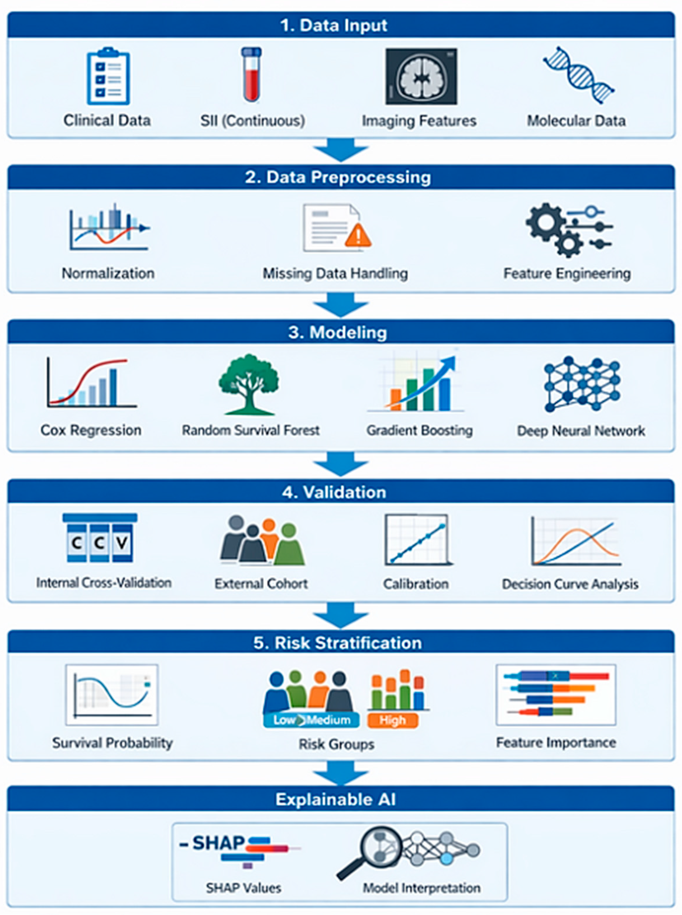
In survival analysis, SII is most appropriately modeled as a continuous variable, preserving information lost through dichotomization and enabling flexible estimation of associations [70,71]. When included in multivariable models, potential redundancy should be considered. Simultaneous inclusion of SII and its individual components or related inflammatory indices may introduce multicollinearity and affect model stability [138,139]. Appropriate variable selection and sensitivity analyses are, therefore, required.
Beyond baseline assessment, SII may be evaluated longitudinally using serial measurements to capture temporal variation during treatment or disease progression. Although conceptually attractive, such approaches remain insufficiently validated and require prospective evaluation [140,141]. The prognostic impact of SII may also vary across disease stage, treatment modality, and molecular subtype [142].
A structured modeling pipeline for incorporating SII into prognostic systems is illustrated in Figure 2. This framework includes data harmonization, feature selection, multicollinearity assessment, internal validation, and independent external validation. These steps are essential to ensure robustness and transportability across clinical settings.
Within multivariable models, SII provides information on systemic immune status that is not directly captured by tumor-derived features [143,144,145]. Its contribution should, therefore, be evaluated empirically rather than assumed.
Beyond statistical significance, the incremental predictive value of SII should be formally assessed. Demonstrating independent association does not necessarily imply meaningful improvement in risk prediction. Evaluation should, therefore, include changes in discrimination, calibration, and clinical utility when SII is added to established prognostic models [146,147].
Discrimination can be assessed using concordance indices, although improvements should be interpreted cautiously, particularly in structured datasets where conventional models may perform similarly [14,82]. Calibration is essential to evaluate agreement between predicted and observed outcomes and should be examined before and after inclusion of SII [82,109]. Clinical usefulness should be assessed using decision-analytic approaches such as decision curve analysis, which determine whether model extensions translate into meaningful changes in clinical decision-making [68,71].
As illustrated in Table 2, independent multivariable association does not necessarily correspond to improved predictive performance. Current evidence supports the prognostic relevance of SII; however, robust demonstration of incremental benefit beyond established models remains limited. Explainability methods may identify SII as an influential predictor in specific datasets [146,147,148]. Such findings reflect statistical contribution within a model but do not establish causality [149]. Interpretation, therefore, requires clinical context and biological plausibility.
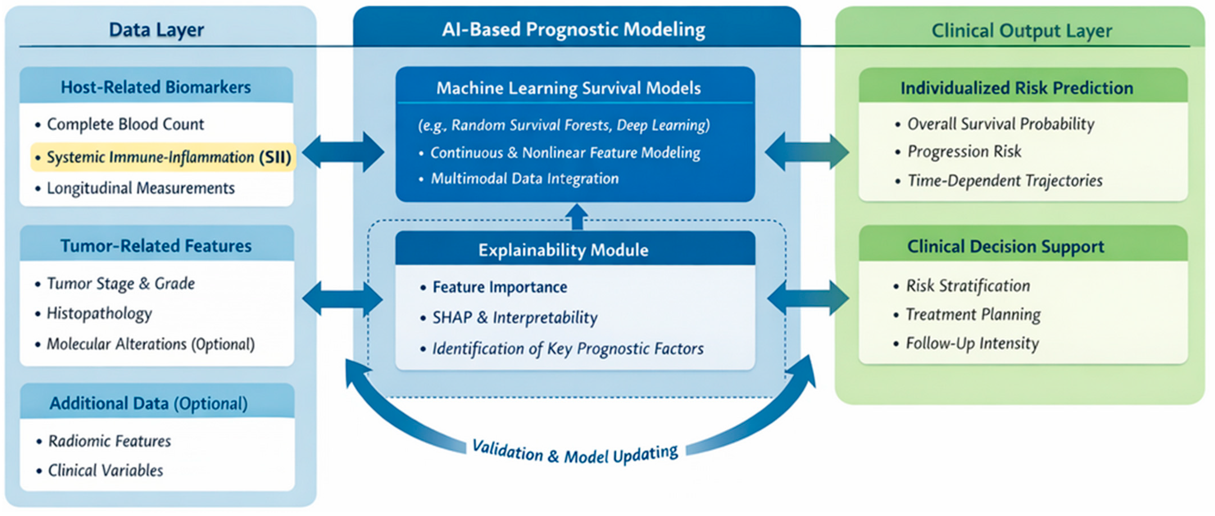
A structured implementation pathway includes model development in representative cohorts, internal validation, independent external validation, and evaluation of both discrimination and calibration prior to clinical use [113,122,150,151]. Ongoing performance monitoring and model updating are required as clinical practices evolve (Figure 3).
Although the layers are based on similar underlying data types, they differ in their functional roles. The Data Layer represents raw clinical and biological inputs generated in routine practice. The AI-based Layer transforms these inputs using algorithmic approaches to produce predictive structures and risk estimates. In contrast, the Clinical Output Layer contextualizes these results within clinical decision-making. While overlap exists due to shared data sources, the layers differ in terms of processing, abstraction, and application.
Clinically, incorporating systemic immune-inflammatory status alongside tumor characteristics may support pre-treatment risk stratification and individualized therapeutic planning [137,138,146]. Longitudinal incorporation of SII may enable dynamic risk assessment during treatment, although prospective validation remains essential [140,141,142]. In research settings, such models may support trial stratification and exploratory analyses of treatment response [143,144,145,149].
SII should, therefore, be considered as one component of a multivariable, externally validated prognostic system rather than as an independent determinant of outcome. When appropriately integrated, it may contribute clinically interpretable information to AI-based risk estimation in oncology.

6. Clinical Implications and Translational Potential

The Systemic Immune-Inflammation Index offers practical advantages, including routine availability, low cost, and reproducibility across healthcare settings [11,45,59]. Its translational value is maximized when incorporated into validated multivariable prediction systems rather than applied in isolation [14,44]. Importantly, such integration should be supported by demonstrable incremental predictive performance beyond established clinicopathological models, rather than by statistical association alone.
In pre-treatment assessment, models integrating SII with tumor characteristics may support more refined risk stratification and individualized therapeutic planning [11,14,45]. These approaches may help identify patients who require intensified monitoring or alternative strategies, while also informing de-escalation in selected low-risk contexts [44,67,109]. Importantly, such models should provide probabilistic estimates that support shared decision-making rather than replace clinical judgment [97,134,149].
In longitudinal settings, serial assessment of SII may contribute to dynamic risk evaluation during treatment. Changes in systemic immune-inflammatory status may reflect therapeutic response or disease progression [64,87,141]. Time-updated models could enable earlier identification of patients at increased risk, although prospective validation remains necessary [2,34,103,142].
In clinical research, incorporating SII into multivariable models may improve trial stratification, reduce outcome heterogeneity, and enhance endpoint prediction [44,85,134]. These approaches may also support exploratory analyses of immune-inflammatory correlates of treatment response and inform biomarker-driven study design [24,51,149].
Routine implementation requires standardized data acquisition, appropriate digital infrastructure, and clinician engagement [109,123]. In the context of AI-based prognostic models, ethical and regulatory considerations acquire additional importance. Issues such as algorithmic transparency, potential bias in training data, and accountability for model-informed decisions are particularly relevant when risk estimates may influence clinical management. Ensuring interpretability, external validation, and clear delineation of clinical responsibility is, therefore, essential for safe implementation [102,126,130]. Ongoing evaluation and model updating are required as clinical practice evolves.
From a clinical perspective, integrating simple and routinely available biomarkers such as SII into advanced modeling frameworks represents a pragmatic step toward more applicable precision oncology. Rather than replacing clinical judgment, such models may support more structured and transparent decision-making.

7. Limitations and Challenges

Several biological, methodological, and implementation-related limitations should be acknowledged [7,46].
Biologically, SII is a non-specific marker of systemic immune-inflammatory status. Neutrophil, platelet, and lymphocyte counts are influenced by infections, chronic inflammatory conditions, comorbidities, and medications, limiting specificity for cancer-related processes [4,51,53,54].
Beyond non-malignant inflammation, SII may also reflect broader systemic vulnerability rather than tumor-specific biology. Frailty, sarcopenia, cachexia, and age-related immunosenescence are associated with alterations in circulating immune cell profiles and may confound associations between SII and survival outcomes [57]. Elevated SII may, therefore, act as a surrogate of impaired physiological reserve rather than a direct mediator of tumor progression.
Reverse causality represents an additional concern. Advanced disease and occult metastases may induce systemic inflammation, leading to elevated SII values. As a result, SII may partly reflect underlying disease burden rather than independently influencing prognosis.
Treatment-related factors further complicate interpretation. Chemotherapy, corticosteroids, granulocyte colony-stimulating factors, immunotherapy, and radiation can alter peripheral blood counts. Both baseline and on-treatment SII may, therefore, reflect a combination of host biology and therapy-related effects. Failure to account for sampling timing may introduce bias.
These considerations indicate that SII should be interpreted as a composite systemic marker influenced by tumor biology, host vulnerability, and treatment-related factors rather than as a tumor-specific biomarker.
Methodologically, most evidence derives from retrospective studies with heterogeneous populations, tumor types, and analytical strategies. Variability in laboratory standards, sampling timing, and statistical approaches limits comparability. Study-specific dichotomization thresholds further reduce reproducibility and may inflate effect estimates [70,74,83].
AI-based prognostic models introduce additional challenges related to overfitting, dataset quality, and transportability. Performance estimates from internal validation may not generalize to independent cohorts, underscoring the need for rigorous external validation and transparent reporting [17,82,138]. Model outputs also require careful interpretation, as feature importance does not establish causality [113,115,132].
Implementation requires appropriate digital infrastructure, standardized data pipelines, clinician engagement, and clear governance structures. Ethical and regulatory considerations, including data privacy, bias mitigation, and accountability, must be addressed prior to routine deployment [44,83,126].
Finally, the conceptual nature of this review and the absence of quantitative synthesis limit direct comparison of modeling strategies. The proposed framework should, therefore, be considered hypothesis-generating pending prospective validation [70,74].

8. Future Directions

Future research should prioritize prospective, multicenter validation with standardized data collection and reporting [68,76,109]. Continuous modeling of SII within multivariable systems may improve reproducibility and transportability across clinical settings [79,90].
Longitudinal incorporation of serial SII measurements represents a promising approach to dynamic risk prediction, although prospective confirmation of clinical benefit remains necessary [134,141].
Integration with emerging biomarker modalities—including circulating tumor DNA, immune profiling, radiomics, and multi-omics platforms—may further refine risk stratification by combining systemic and tumor-derived information [36,145].
Advances in explainable AI and adherence to reporting standards such as TRIPOD-AI and CONSORT-AI will be essential to improve transparency, reproducibility, and comparability of prognostic models [113,130,132,150].
Progress in this field will depend on interdisciplinary collaboration among clinicians, data scientists, and translational researchers to ensure that analytical advances translate into clinically meaningful outcomes.

9. Conclusions

The Systemic Immune-Inflammation Index represents a readily available indicator of systemic immune-inflammatory status with reproducible prognostic associations across solid malignancies. However, its clinical utility is maximized when incorporated into rigorously developed and externally validated multivariable prediction models rather than applied as a dichotomized standalone marker.
Embedding biologically interpretable host-derived biomarkers within explainable AI-based survival models enables more context-sensitive risk estimation. Such systems should be regarded as decision-support tools that complement clinical expertise by providing probabilistic risk estimates derived from integrated data sources. Realizing their potential requires prospective validation, standardized reporting, and careful implementation.
Structured integration of immune-inflammatory biomarkers into AI-based survival models may contribute to more precise and clinically relevant risk stratification in oncology. Incorporating both baseline and longitudinal representations of systemic immune-inflammatory status may further enhance their clinical relevance, although prospective validation remains essential.

Author Contributions

Conceptualization, A.D.-S. and Ż.A.M.; methodology, A.D.-S. and Ż.A.M.; validation, A.D.-S., Z.K.K. and Ż.A.M.; formal analysis, A.D.-S. and Ż.A.M.; investigation, Ż.A.M.; resources, Ż.A.M.; data curation, A.D.-S. and Ż.A.M.; writing—original draft preparation, A.D.-S. and Ż.A.M.; writing—review and editing, A.D.-S., Z.K.K. and Ż.A.M.; visualization, A.D.-S. and Ż.A.M.; supervision, Z.K.K.; project administration, A.D.-S. and Ż.A.M.; funding acquisition, A.D.-S. and Z.K.K. All authors have read and agreed to the published version of the manuscript.

Funding

This research was funded under the Medical University of Bialystok (UMB) project with the funding number: B.SUB.26.446.

Institutional Review Board Statement

Not applicable.

Informed Consent Statement

Not applicable.

Data Availability Statement

No new data were created or analyzed in this study.

Acknowledgments

The language of this article was refined with the support of artificial intelligence (AI) tools.

Conflicts of Interest

The authors declare no conflicts of interest.

Abbreviations

The following abbreviations are used in this manuscript:
AIArtificial Intelligence
MLMachine Learning
XAIExplainable Artificial Intelligence
SIISystemic Immune-Inflammation Index
NLRNeutrophil-to-Lymphocyte Ratio
PLRPlatelet-to-Lymphocyte Ratio
LMRLymphocyte-to-Monocyte Ratio
CRPC-Reactive Protein
MgpsModified Glasgow Prognostic Score
PNIPrognostic Nutritional Index
TNMTumor–Node–Metastasis staging system
CtDNACirculating Tumor DNA
PRISMAPreferred Reporting Items for Systematic Reviews and Meta-Analyses
SHAPShapley Additive Explanations
NSCLCNon-Small Cell Lung Cancer
PD-1Programmed Cell Death Protein 1
XELOXCapecitabine + Oxaliplatin chemotherapy regimen
SEERSurveillance, Epidemiology, and End Results database
TRIPOD-AITransparent Reporting of a Multivariable Prediction Model for Individual Prognosis or Diagnosis–Artificial Intelligence extension
PROBASTPrediction Model Risk of Bias Assessment Tool
CONSORT-AIConsolidated Standards of Reporting Trials–Artificial Intelligence extension
SPIRIT-AIStandard Protocol Items: Recommendations for Interventional Trials–Artificial Intelligence extension

References

  1. Yang, H.-T.; Shah, R.H.; Tegay, D.; Onel, K. Precision oncology: Lessons learned and challenges for the future. Cancer Manag. Res. 2019, 11, 7525–7536. [Google Scholar] [CrossRef]
  2. Tsimberidou, A.M.; Fountzilas, E.; Nikanjam, M.; Kurzrock, R. Review of precision cancer medicine: Evolution of the treatment paradigm. Cancer Treat. Rev. 2020, 86, 102019. [Google Scholar] [CrossRef]
  3. Ramraj, A.; Saravanan, H. A comparative analysis of cancer stage classification systems for registries. ecancermedicalscience 2025, 19, 1920. [Google Scholar] [CrossRef] [PubMed]
  4. Zhao, H.; Wu, L.; Yan, G.; Chen, Y.; Zhou, M.; Wu, Y.; Li, Y. Inflammation and tumor progression: Signaling pathways and targeted intervention. Signal Transduct. Target. Ther. 2021, 6, 263. [Google Scholar] [CrossRef] [PubMed]
  5. Yan, S.; Gong, X.; Liu, R.; Jia, X. Prognostic significance of systemic pan-immune-inflammation value in locally advanced cervical cancer. Front. Oncol. 2024, 14, 1492251. [Google Scholar] [CrossRef]
  6. Portale, G.; Bartolotta, P.; Azzolina, D.; Gregori, D.; Fiscon, V. Prognostic role of platelet-to-lymphocyte ratio, neutrophil-to-lymphocyte, and lymphocyte-to-monocyte ratio in operated rectal cancer patients: Systematic review and meta-analysis. Langenbeck’s Arch. Surg. 2023, 408, 85. [Google Scholar] [CrossRef]
  7. Islam, M.M.; Satici, M.O.; Eroglu, S.E. Unraveling the clinical significance and prognostic value of the neutrophil-to-lymphocyte ratio, platelet-to-lymphocyte ratio, systemic immune-inflammation index, systemic inflammation response index, and delta neutrophil index. Turk. J. Emerg. Med. 2024, 24, 8–19. [Google Scholar] [CrossRef]
  8. Tan, S.; Zheng, Q.; Zhang, W.; Zhou, M.; Xia, C.; Feng, W. Prognostic value of inflammatory markers NLR, PLR, and LMR in gastric cancer patients treated with immune checkpoint inhibitors: A meta-analysis and systematic review. Front. Immunol. 2024, 15, 1408700. [Google Scholar] [CrossRef]
  9. Yu, L.; Yuan, J.; Lou, S.; Huang, A.; Cai, X.; Ji, J.; Dai, X.; Mao, X.; Mao, Y.; Sun, L. Factors affecting survival in patients with colorectal cancer: An umbrella review. J. Transl. Med. 2025, 23, 1185. [Google Scholar] [CrossRef]
  10. Nan, H.; Yang, P.; Wang, L.; Li, W.; Liu, Y.; Huang, F.; Xu, F.; Shi, M.; Bai, Y. Prognostic Significance of Systemic Immune-Inflammation Index in Patients with de Novo non-M3 Acute Myeloid Leukemia. J. Inflamm. Res. 2025, 18, 16331–16346. [Google Scholar] [CrossRef]
  11. Zhong, J.-H.; Huang, D.-H.; Chen, Z.-Y. Prognostic role of systemic immune-inflammation index in solid tumors: A systematic review and meta-analysis. Oncotarget 2017, 8, 75381–75388. [Google Scholar] [CrossRef]
  12. Huang, H.; Liu, Q.; Zhu, L.; Zhang, Y.; Lu, X.; Wu, Y.; Liu, L. Prognostic Value of Preoperative Systemic Immune-Inflammation Index in Patients with Cervical Cancer. Sci. Rep. 2019, 9, 3284. [Google Scholar] [CrossRef]
  13. Meng, L.; Yang, Y.; Hu, X.; Zhang, R.; Li, X. Prognostic value of the pretreatment systemic immune-inflammation index in patients with prostate cancer: A systematic review and meta-analysis. J. Transl. Med. 2023, 21, 79. [Google Scholar] [CrossRef] [PubMed]
  14. Huang, Y.; Bazzazzadehgan, S.; Li, J.; Arabshomali, A.; Li, M.; Bhattacharya, K.; Bentley, J.P. Comparison of machine learning methods versus traditional Cox regression for survival prediction in cancer using real-world data: A systematic literature review and meta-analysis. BMC Med. Res. Methodol. 2025, 25, 243. [Google Scholar] [CrossRef]
  15. Zhang, B.; Shi, H.; Wang, H. Machine Learning and AI in Cancer Prognosis, Prediction, and Treatment Selection: A Critical Approach. J. Multidiscip. Healthc. 2023, 16, 1779–1791. [Google Scholar] [CrossRef]
  16. AlOsaimi, H.M.; Alshilash, A.M.; Al-Saif, L.K.; Bosbait, J.M.; Albeladi, R.S.; Almutairi, D.R.; Alhazzaa, A.A.; Alluqmani, T.A.; Al Qahtani, S.M.; Almohammadi, S.A.; et al. AI models for the identification of prognostic and predictive biomarkers in lung cancer: A systematic review and meta-analysis. Front. Oncol. 2025, 15, 1424647. [Google Scholar] [CrossRef] [PubMed]
  17. Dhiman, P.; Ma, J.; Navarro, C.L.A.; Speich, B.; Bullock, G.; Damen, J.A.A.; Hooft, L.; Kirtley, S.; Riley, R.D.; Van Calster, B.; et al. Risk of bias of prognostic models developed using machine learning: A systematic review in oncology. Diagn. Progn. Res. 2022, 6, 13. [Google Scholar] [CrossRef]
  18. Ligero, M.; El Nahhas, O.S.; Aldea, M.; Kather, J.N. Artificial intelligence-based biomarkers for treatment decisions in oncology. Trends Cancer 2025, 11, 232–244. [Google Scholar] [CrossRef]
  19. Zhao, H.; Zhang, S.; Wang, Y.; Zhang, H.; Du, P.; Cao, A. Immuno-inflammatory signature for predicting therapeutic response and survival after stereotactic radiosurgery in NSCLC patients with brain metastases: A retrospective cohort study. Front. Immunol. 2025, 16, 1739812. [Google Scholar] [CrossRef]
  20. Cheng, X.; Meng, F.; Wang, R.; Liu, S.; Li, Q.; Chen, B.; Xi, M. Prognostic value of immuno-inflammatory biomarkers in esophageal squamous cell carcinoma patients receiving immunotherapy combined with chemoradiotherapy and its association with immuno-genomic landscape. BMC Cancer 2024, 24, 1518. [Google Scholar] [CrossRef]
  21. Xue, Q.; Wang, Y.; Zheng, Q.; Chen, L.; Lin, Y.; Jin, Y.; Shen, X.; Li, Y. Prognostic value of tumor immune microenvironment factors in patients with stage I lung adenocarcinoma. Am. J. Cancer Res. 2023, 13, 950–963. [Google Scholar]
  22. Segal, S.; An, J.; Berkovsky, M.; Jung, G.; Stone, A.; Yau, V.; Lin, J.; Smith, R.V.; Yin, S. The Role of Tumor Immune Microenvironment and Clinical Factors in Head and Neck Cancer Prognosis Among African American Men and Women. Cancers 2026, 18, 481. [Google Scholar] [CrossRef] [PubMed]
  23. Chen, S.; Liu, Z.; Wang, J.; Zhang, Z.; Dekker, A.; Wee, L.; Kalendralis, P.; Sun, Y.; Tong, T. Automatically quantifying spatial heterogeneity of immune and tumor hypoxia environment and predicting disease-free survival for patients with rectal cancer. Cancer Immunol. Immunother. 2025, 74, 380. [Google Scholar] [CrossRef] [PubMed]
  24. Pagès, F.; Mlecnik, B.; Marliot, F.; Bindea, G.; Ou, F.-S.; Bifulco, C.; Lugli, A.; Zlobec, I.; Rau, T.T.; Berger, M.D.; et al. International validation of the consensus Immunoscore for the classification of colon cancer: A prognostic and accuracy study. Lancet 2018, 391, 2128–2139. [Google Scholar] [CrossRef] [PubMed]
  25. Mao, H.; Zhao, X.; Sun, S.C. NF-κB in inflammation and cancer. Cell. Mol. Immunol. 2025, 22, 811–839. [Google Scholar] [CrossRef]
  26. Tripathi, S.; Sharma, Y.; Kumar, D. Unveiling the link between chronic inflammation and cancer. Metab. Open 2025, 25, 100347. [Google Scholar] [CrossRef]
  27. An, H.J.; Chon, H.J.; Kim, C. Peripheral Blood-Based Biomarkers for Immune Checkpoint Inhibitors. Int. J. Mol. Sci. 2021, 22, 9414. [Google Scholar] [CrossRef]
  28. Liu, N.; Mao, J.; Tao, P.; Chi, H.; Jia, W.; Dong, C. The relationship between NLR/PLR/LMR levels and survival prognosis in patients with non-small cell lung carcinoma treated with immune checkpoint inhibitors. Medicine 2022, 101, e28617, Erratum in Medicine 2022, 101, e31359. https://doi.org/10.1097/MD.0000000000031359. [Google Scholar] [CrossRef]
  29. Allahyari, A.; Fallah, F.; Taqanaki, P.B.; Tafti, S.P.; Vakilzadeh, M.M.; Nodeh, M.M.; Kamandi, M.; Noferesti, A. Evaluating the neutrophil-to-lymphocyte ratio (NLR) and platelet-to-lymphocyte ratio (PLR) as prognostic and treatment response biomarkers in stage IV colorectal cancer patients. Oncol. Clin. Pract. 2024, 21, 200–205. [Google Scholar] [CrossRef]
  30. Mireștean, C.C.; Stan, M.C.; Iancu, R.I.; Iancu, D.P.T.; Bădulescu, F. The Prognostic Value of Platelet–Lymphocyte Ratio, Neutrophil–Lymphocyte Ratio, and Monocyte–Lymphocyte Ratio in Head and Neck Squamous Cell Carcinoma (HNSCC)—A Retrospective Single Center Study and a Literature Review. Diagnostics 2023, 13, 3396. [Google Scholar] [CrossRef]
  31. Kou, J.; Huang, J.; Li, J.; Wu, Z.; Ni, L. Systemic immune-inflammation index predicts prognosis and responsiveness to immunotherapy in cancer patients: A systematic review and meta-analysis. Clin. Exp. Med. 2023, 23, 3895–3905. [Google Scholar] [CrossRef] [PubMed]
  32. Wang, Y.; Hao, X.; Li, G. Prognostic and clinical pathological significance of the systemic immune-inflammation index in urothelial carcinoma: A systematic review and meta-analysis. Front. Oncol. 2024, 14, 1322897. [Google Scholar] [CrossRef] [PubMed]
  33. Cao, W.; Shao, Y.; Zou, S.; Wang, N.; Wang, J. Prognostic significance of systemic immune-inflammation index in patients with bladder cancer: A systematic review and meta-analysis. Medicine 2022, 101, e30380. [Google Scholar] [CrossRef] [PubMed]
  34. Reinert, T.; Schøler, L.V.; Thomsen, R.; Tobiasen, H.; Vang, S.; Nordentoft, I.; Lamy, P.; Kannerup, A.-S.; Mortensen, F.V.; Stribolt, K.; et al. Analysis of circulating tumour DNA to monitor disease burden following colorectal cancer surgery. Gut 2015, 65, 625–634. [Google Scholar] [CrossRef]
  35. Denkert, C.; Von Minckwitz, G.; Darb-Esfahani, S.; Lederer, B.; Heppner, B.I.; Weber, K.E.; Budczies, J.; Huober, J.; Klauschen, F.; Furlanetto, J.; et al. Tumour-infiltrating lymphocytes and prognosis in different subtypes of breast cancer: A pooled analysis of 3771 patients treated with neoadjuvant therapy. Lancet Oncol. 2018, 19, 40–50. [Google Scholar] [CrossRef]
  36. Lin, P.; Zheng, J.-M.; Liu, C.-W.; Tang, Q.-Q.; Pang, J.-S.; Qin, Q.; Lin, Z.-H.; Yang, H. Radiogenomic insights suggest that multiscale tumor heterogeneity is associated with interpretable radiomic features and outcomes in cancer patients. Comput. Med Imaging Graph. 2025, 124, 102586. [Google Scholar] [CrossRef]
  37. Wu, X.; Dai, W. Beyond Imaging: Integrating Radiomics, Genomics, and Multi-Omics for Precision Breast Cancer Management. Cancers 2025, 17, 3408. [Google Scholar] [CrossRef]
  38. Huang, Y.; Peng, W.; Yang, H.; Liu, Z.; Zhang, T.; Jin, L.; He, W.; Du, M.; Chen, Z. Radiomics and radiogenomics in ovarian cancer: A review with a focus on ultrasound applications. Cancer Imaging 2025, 25, 1–14. [Google Scholar] [CrossRef]
  39. Aiello, M. Is Radiomics Growing Towards Clinical Practice? J. Pers. Med. 2022, 12, 1373. [Google Scholar] [CrossRef]
  40. Avanzo, M.; Soda, P.; Bertolini, M.; Bettinelli, A.; Rancati, T.; Stancanello, J.; Rampado, O.; Pirrone, G.; Drigo, A. Robust radiomics: A review of guidelines for radiomics in medical imaging. Front. Radiol. 2026, 5, 1701110. [Google Scholar] [CrossRef]
  41. Chan, H.T.; Nagayama, S.; Otaki, M.; Chin, Y.M.; Fukunaga, Y.; Ueno, M.; Nakamura, Y.; Low, S.-K. Tumor-informed or tumor-agnostic circulating tumor DNA as a biomarker for risk of recurrence in resected colorectal cancer patients. Front. Oncol. 2023, 12, 1055968. [Google Scholar] [CrossRef]
  42. Loft, M.; To, Y.H.; Gibbs, P.; Tie, J. Clinical application of circulating tumour DNA in colorectal cancer. Lancet Gastroenterol. Hepatol. 2023, 8, 837–852. [Google Scholar] [CrossRef] [PubMed]
  43. Fountzilas, E.; Pearce, T.; Baysal, M.A.; Chakraborty, A.; Tsimberidou, A.M. Convergence of evolving artificial intelligence and machine learning techniques in precision oncology. NPJ Digit. Med. 2025, 8, 75. [Google Scholar] [CrossRef]
  44. El Naqa, I.; Karolak, A.; Luo, Y.; Folio, L.; Tarhini, A.A.; Rollison, D.; Parodi, K. Translation of AI into oncology clinical practice. Oncogene 2023, 42, 3089–3097. [Google Scholar] [CrossRef] [PubMed]
  45. Hu, B.; Yang, X.-R.; Xu, Y.; Sun, Y.-F.; Sun, C.; Guo, W.; Zhang, X.; Wang, W.-M.; Qiu, S.-J.; Zhou, J.; et al. Systemic Immune-Inflammation Index Predicts Prognosis of Patients after Curative Resection for Hepatocellular Carcinoma. Clin. Cancer Res. 2014, 20, 6212–6222. [Google Scholar] [CrossRef] [PubMed]
  46. Fest, J.; Ruiter, R.; Mulder, M.; Koerkamp, B.G.; Ikram, M.A.; Stricker, B.H.; van Eijck, C.H. The systemic immune-inflammation index is associated with an increased risk of incident cancer—A population-based cohort study. Int. J. Cancer 2019, 146, 692–698. [Google Scholar] [CrossRef]
  47. Aziz, M.H.; Sideras, K.; Aziz, N.A.; Mauff, K.; Haen, R.; Roos, D.; van Eijck, C.H. The systemic-immune-inflammation index independently predicts survival and recurrence in resectable pancreatic cancer and its prognostic value depends on bilirubin levels: A retrospective multicenter cohort study. Ann. Surg. 2019, 270, 139–146. [Google Scholar] [CrossRef]
  48. Bai, X.; Feng, L. Correlation between Prognostic Nutritional Index, Glasgow Prognostic Score, Systemic Inflammatory Response, and TNM Staging in Colorectal Cancer Patients. Nutr. Cancer 2019, 72, 1170–1177. [Google Scholar] [CrossRef]
  49. Yamamoto, S.; Adachi, S.; Wada, T.; Narui, K.; Kimura, A.; Oshi, M.; Yamada, A.; Misumi, T.; Endo, I. The Modified Glasgow Prognostic Score and Prognostic Nutritional Index as Prognostic Markers in Patients With Metastatic Breast Cancer Treated With Eribulin. Vivo 2022, 36, 1854–1859. [Google Scholar] [CrossRef]
  50. Menzel, A.; Samouda, H.; Dohet, F.; Loap, S.; Ellulu, M.S.; Bohn, T. Common and Novel Markers for Measuring Inflammation and Oxidative Stress Ex Vivo in Research and Clinical Practice—Which to Use Regarding Disease Outcomes? Antioxidants 2021, 10, 414. [Google Scholar] [CrossRef]
  51. Shaul, M.E.; Fridlender, Z.G. Tumour-associated neutrophils in patients with cancer. Nat. Rev. Clin. Oncol. 2019, 16, 601–620. [Google Scholar] [CrossRef]
  52. Yang, L.; Liu, Q.; Zhang, X.; Liu, X.; Zhou, B.; Chen, J.; Huang, D.; Li, J.; Li, H.; Chen, F.; et al. DNA of neutrophil extracellular traps promotes cancer metastasis via CCDC25. Nature 2020, 583, 133–138. [Google Scholar] [CrossRef]
  53. Braun, A.; Anders, H.-J.; Gudermann, T.; Mammadova-Bach, E. Platelet-Cancer Interplay: Molecular Mechanisms and New Therapeutic Avenues. Front. Oncol. 2021, 11, 665534. [Google Scholar] [CrossRef]
  54. Xu, X.R.; Yousef, G.M.; Ni, H. Cancer and platelet crosstalk: Opportunities and challenges for aspirin and other antiplatelet agents. Blood 2018, 131, 1777–1789. [Google Scholar] [CrossRef]
  55. Bekendam, R.H.; Ravid, K. Mechanisms of platelet activation in cancer-associated thrombosis: A focus on myeloproliferative neoplasms. Front. Cell Dev. Biol. 2023, 11, 1207395. [Google Scholar] [CrossRef] [PubMed]
  56. Galon, J.; Bruni, D. Approaches to treat immune hot, altered and cold tumours with combination immunotherapies. Nat. Rev. Drug Discov. 2019, 18, 197–218. [Google Scholar] [CrossRef] [PubMed]
  57. Rodriguez, J.E.; Naigeon, M.; Goldschmidt, V.; Dugage, M.R.; Seknazi, L.; Danlos, F.X.; Champiat, S.; Marabelle, A.; Michot, J.-M.; Massard, C.; et al. Immunosenescence, inflammaging, and cancer immunotherapy efficacy. Expert Rev. Anticancer Ther. 2022, 22, 915–926. [Google Scholar] [CrossRef] [PubMed]
  58. Chen, G.; Bao, B.; Ye, Y.; Hu, A.; Sun, J.; Liu, W. Prognostic value of the systemic immune-inflammation index in non-small cell lung cancer patients treated with immune checkpoint inhibitors: A systematic review and meta-analysis. Front. Oncol. 2025, 15, 1532343. [Google Scholar] [CrossRef]
  59. Yang, R.; Chang, Q.; Meng, X.; Gao, N.; Wang, W. Prognostic value of Systemic immune-inflammation index in cancer: A meta-analysis. J. Cancer 2018, 9, 3295–3302. [Google Scholar] [CrossRef]
  60. Yang, X.; Wu, C. Systemic immune inflammation index and gastric cancer prognosis: A systematic review and meta-analysis. Exp. Ther. Med. 2024, 27, 122. [Google Scholar] [CrossRef]
  61. Shui, Y.; Li, M.; Su, J.; Chen, M.; Gu, X.; Guo, W. Prognostic and clinicopathological significance of systemic immune-inflammation index in pancreatic cancer: A meta-analysis of 2365 patients. Aging 2021, 13, 20585–20597. [Google Scholar] [CrossRef] [PubMed]
  62. He, K.; Si, L.; Pan, X.; Sun, L.; Wang, Y.; Lu, J.; Wang, X. Preoperative Systemic Immune–Inflammation Index (SII) as a Superior Predictor of Long-Term Survival Outcome in Patients With Stage I–II Gastric Cancer After Radical Surgery. Front. Oncol. 2022, 12, 829689. [Google Scholar] [CrossRef] [PubMed]
  63. Ding, P.; Guo, H.; Sun, C.; Yang, P.; Kim, N.H.; Tian, Y.; Liu, Y.; Liu, P.; Li, Y.; Zhao, Q. Combined systemic immune-inflammatory index (SII) and prognostic nutritional index (PNI) predicts chemotherapy response and prognosis in locally advanced gastric cancer patients receiving neoadjuvant chemotherapy with PD-1 antibody sintilimab and XELOX: A prospective study. BMC Gastroenterol. 2022, 22, 121. [Google Scholar] [CrossRef] [PubMed]
  64. Tan, Y.; Hu, B.; Li, Q.; Cao, W. Prognostic value and clinicopathological significance of pre-and post-treatment systemic immune-inflammation index in colorectal cancer patients: A meta-analysis. World J. Surg. Oncol. 2025, 23, 11. [Google Scholar] [CrossRef]
  65. Mezquita, L.; Auclin, E.; Ferrara, R.; Charrier, M.; Remon, J.; Planchard, D.; Ponce, S.; Ares, L.P.; Leroy, L.; Audigier-Valette, C.; et al. Association of the Lung Immune Prognostic Index With Immune Checkpoint Inhibitor Outcomes in Patients With Advanced Non–Small Cell Lung Cancer. JAMA Oncol. 2018, 4, 351–357. [Google Scholar] [CrossRef]
  66. Sun, T.; Guo, Y.; Sun, B.; Chen, L.; Ren, Y.; Zhu, L.; Zhang, L.; Liu, Y.; Zheng, C. Association of the pretreatment lung immune prognostic index with immune checkpoint inhibitor outcomes in patients with advanced hepatocellular carcinoma. Eur. J. Med. Res. 2023, 28, 225. [Google Scholar] [CrossRef]
  67. Kappen, T.H.; van Klei, W.A.; van Wolfswinkel, L.; Kalkman, C.J.; Vergouwe, Y.; Moons, K.G.M. Evaluating the impact of prediction models: Lessons learned, challenges, and recommendations. Diagn. Progn. Res. 2018, 2, 11. [Google Scholar] [CrossRef]
  68. Van Calster, B.; Valentin, L.; Froyman, W.; Landolfo, C.; Ceusters, J.; Testa, A.C.; Wynants, L.; Sladkevicius, P.; Van Holsbeke, C.; Domali, E.; et al. Validation of models to diagnose ovarian cancer in patients managed surgically or conservatively: Multicentre cohort study. BMJ 2020, 370, m2614. [Google Scholar] [CrossRef]
  69. Kinoshita, T.; Goto, T. Links between Inflammation and Postoperative Cancer Recurrence. J. Clin. Med. 2021, 10, 228. [Google Scholar] [CrossRef]
  70. Riley, R.D.; Moons, K.G.M.; Snell, K.I.E.; Ensor, J.; Hooft, L.; Altman, D.G.; Hayden, J.; Collins, G.S.; Debray, T.P.A. A guide to systematic review and meta-analysis of prognostic factor studies. BMJ 2019, 364, k4597. [Google Scholar] [CrossRef]
  71. Wynants, L.; Van Calster, B.; Collins, G.S.; Riley, R.D.; Heinze, G.; Schuit, E.; Bonten, M.M.J.; Dahly, D.L.; Damen, J.A.; Debray, T.P.A.; et al. Prediction models for diagnosis and prognosis of COVID-19: Systematic review and critical appraisal. BMJ 2020, 369, m1328, Erratum in BMJ 2020, 369, m2204. https://doi.org/10.1136/bmj.m2204. [Google Scholar] [CrossRef] [PubMed]
  72. Tian, B.-W.; Yang, Y.-F.; Yang, C.-C.; Yan, L.-J.; Ding, Z.-N.; Liu, H.; Xue, J.-S.; Dong, Z.-R.; Chen, Z.-Q.; Hong, J.-G.; et al. Systemic Immune–Inflammation Index Predicts Prognosis of Cancer Immunotherapy: Systemic Review and Meta-Analysis. Immunotherapy 2022, 14, 1481–1496. [Google Scholar] [CrossRef] [PubMed]
  73. Li, X.; Lin, H.; Ouyang, R.; Yang, Y.; Peng, J. Prognostic significance of the systemic immune-inflammation index in pancreatic carcinoma patients: A meta-analysis. Biosci. Rep. 2021, 41, BSR20204401. [Google Scholar] [CrossRef] [PubMed]
  74. Page, M.J.; Sterne, J.A.C.; Higgins, J.P.T.; Egger, M. Investigating and dealing with publication bias and other reporting biases in meta-analyses of health research: A review. Res. Synth. Methods 2020, 12, 248–259. [Google Scholar] [CrossRef]
  75. Riley, R.D.; Snell, K.I.; Ensor, J.; Burke, D.L.; Harrell, F.E.; Moons, K.G.; Collins, G.S. Minimum sample size for developing a multivariable prediction model: Part I—Continuous outcomes. Stat. Med. 2018, 38, 1262–1275. [Google Scholar] [CrossRef]
  76. Collins, G.S.; Dhiman, P.; Navarro, C.L.A.; Ma, J.; Hooft, L.; Reitsma, J.B.; Logullo, P.; Beam, A.L.; Peng, L.; Van Calster, B.; et al. Protocol for development of a reporting guideline (TRIPOD-AI) and risk of bias tool (PROBAST-AI) for diagnostic and prognostic prediction model studies based on artificial intelligence. BMJ Open 2021, 11, e048008. [Google Scholar] [CrossRef]
  77. van der Laak, J.; Litjens, G.; Ciompi, F. Deep learning in histopathology: The path to the clinic. Nat. Med. 2021, 27, 775–784. [Google Scholar] [CrossRef]
  78. Wang, Y.; Ni, Q. Prognostic and clinicopathological significance of Systemic Immune-Inflammation Index in cancer patients receiving immune checkpoint inhibitors: A meta-analysis. Ann. Med. 2023, 55, 808–819. [Google Scholar] [CrossRef]
  79. Bellavia, A.; Melloni, G.E.M.; Park, J.-G.; Discacciati, A.; Murphy, S.A. Estimating and presenting hazard ratios and absolute risks from a Cox model with complex nonlinear interactions. Am. J. Epidemiol. 2024, 193, 1155–1160. [Google Scholar] [CrossRef]
  80. Topol, E.J. High-performance medicine: The convergence of human and artificial intelligence. Nat. Med. 2019, 25, 44–56. [Google Scholar] [CrossRef]
  81. Bi, W.L.; Hosny, A.; Schabath, M.B.; Giger, M.L.; Birkbak, N.J.; Mehrtash, A.; Allison, T.; Arnaout, O.; Abbosh, C.; Dunn, I.F.; et al. Artificial intelligence in cancer imaging: Clinical challenges and applications. CA Cancer J. Clin. 2019, 69, 127–157. [Google Scholar] [CrossRef]
  82. Christodoulou, E.; Ma, J.; Collins, G.S.; Steyerberg, E.W.; Verbakel, J.Y.; Van Calster, B. A systematic review shows no performance benefit of machine learning over logistic regression for clinical prediction models. J. Clin. Epidemiol. 2019, 110, 12–22. [Google Scholar] [CrossRef] [PubMed]
  83. Van Calster, B.; McLernon, D.J.; Van Smeden, M.; Wynants, L.; Steyerberg, E.W. Calibration: The Achilles heel of predictive analytics. BMC Med. 2019, 17, 230. [Google Scholar] [CrossRef] [PubMed]
  84. Kelly, C.J.; Karthikesalingam, A.; Suleyman, M.; Corrado, G.; King, D. Key challenges for delivering clinical impact with artificial intelligence. BMC Med. 2019, 17, 195. [Google Scholar] [CrossRef] [PubMed]
  85. Ibrahim, H.; Liu, X.; Rivera, S.C.; Moher, D.; Chan, A.-W.; Sydes, M.R.; Calvert, M.J.; Denniston, A.K. Reporting guidelines for clinical trials of artificial intelligence interventions: The SPIRIT-AI and CONSORT-AI guidelines. Trials 2021, 22, 11. [Google Scholar] [CrossRef]
  86. Luchini, C.; Pea, A.; Scarpa, A. Artificial intelligence in oncology: Current applications and future perspectives. Br. J. Cancer 2021, 126, 4–9. [Google Scholar] [CrossRef]
  87. Wiegrebe, S.; Kopper, P.; Sonabend, R.; Bischl, B.; Bender, A. Deep learning for survival analysis: A review. Artif. Intell. Rev. 2024, 57, 65. [Google Scholar] [CrossRef]
  88. Garcia-Lopez, A.; Jiménez-Gómez, M.; Gomez-Montero, A.; Gonzalez-Sierra, J.C.; Cabas, S.; Giron-Luque, F. Survival analysis using machine learning in transplantation: A practical introduction. BMC Med. Inform. Decis. Mak. 2025, 25, 141. [Google Scholar] [CrossRef]
  89. Spooner, A.; Chen, E.; Sowmya, A.; Sachdev, P.; Kochan, N.A.; Trollor, J.; Brodaty, H. A comparison of machine learning methods for survival analysis of high-dimensional clinical data for dementia prediction. Sci. Rep. 2020, 10, 20410. [Google Scholar] [CrossRef]
  90. Nagpal, C.; Li, X.; Dubrawski, A. Deep Survival Machines: Fully Parametric Survival Regression and Representation Learning for Censored Data With Competing Risks. IEEE J. Biomed. Health Inform. 2021, 25, 3163–3175. [Google Scholar] [CrossRef]
  91. Pölsterl, S. scikit-survival: A library for time-to-event analysis built on top of scikit-learn. J. Mach. Learn. Res. 2020, 21, 1–6. [Google Scholar]
  92. Peng, Z.-H.; Huang, Z.-X.; Tian, J.-H.; Chong, T.; Li, Z.-L. The application of time-to-event analysis in machine learning prognostic models. J. Transl. Med. 2024, 22, 146. [Google Scholar] [CrossRef] [PubMed]
  93. Eley, A.; Hlaing, T.T.; Breininger, D.; Helforoush, Z.; Kachouie, N.N. Monte Carlo Gradient Boosted Trees for Cancer Staging: A Machine Learning Approach. Cancers 2025, 17, 2452. [Google Scholar] [CrossRef] [PubMed]
  94. Poursaeed, R.; Mohammadzadeh, M.; Safaei, A.A. Survival prediction of glioblastoma patients using machine learning and deep learning: A systematic review. BMC Cancer 2024, 24, 1581. [Google Scholar] [CrossRef]
  95. Baidoo, T.G.; Rodrigo, H. Data-driven survival modeling for breast cancer prognostics: A comparative study with machine learning and traditional survival modeling methods. PLoS ONE 2025, 20, e0318167. [Google Scholar] [CrossRef]
  96. Kar, I.; Kocaman, G.; İbrahimov, F.; Enön, S.; Coşgun, E.; Elhan, A.H. Comparison of deep learning-based recurrence-free survival with random survival forest and Cox proportional hazard models in Stage-I NSCLC patients. Cancer Med. 2023, 12, 19272–19278. [Google Scholar] [CrossRef]
  97. Acosta, J.N.; Falcone, G.J.; Rajpurkar, P.; Topol, E.J. Multimodal biomedical AI. Nat. Med. 2022, 28, 1773–1784. [Google Scholar] [CrossRef]
  98. Xiang, J.; Wang, X.; Zhang, X.; Xi, Y.; Eweje, F.; Chen, Y.; Li, Y.; Bergstrom, C.; Gopaulchan, M.; Kim, T.; et al. A vision–language foundation model for precision oncology. Nature 2025, 638, 769–778. [Google Scholar] [CrossRef]
  99. Williamson, S.M.; Prybutok, V. Balancing Privacy and Progress: A Review of Privacy Challenges, Systemic Oversight, and Patient Perceptions in AI-Driven Healthcare. Appl. Sci. 2024, 14, 675. [Google Scholar] [CrossRef]
  100. Hunter, B.; Hindocha, S.; Lee, R.W. The Role of Artificial Intelligence in Early Cancer Diagnosis. Cancers 2022, 14, 1524. [Google Scholar] [CrossRef]
  101. Zhao, J.; Chen, X.; Zhang, Z.; Zhao, D.; Cao, X.; Mao, F.; Sun, Q.; Peng, L. Prognostic value of the systemic immune-inflammation index in recurrent/metastatic triple-negative breast cancer: A retrospective cohort study. Gland. Surg. 2025, 14, 1858–1872. [Google Scholar] [CrossRef]
  102. Van Calster, B.; Wynants, L.; Riley, R.D.; van Smeden, M.; Collins, G.S. Methodology over metrics: Current scientific standards are a disservice to patients and society. J. Clin. Epidemiol. 2021, 138, 219–226. [Google Scholar] [CrossRef] [PubMed]
  103. Katzman, J.L.; Shaham, U.; Cloninger, A.; Bates, J.; Jiang, T.; Kluger, Y. DeepSurv: Personalized treatment recommender system using a Cox proportional hazards deep neural network. BMC Med. Res. Methodol. 2018, 18, 24. [Google Scholar] [CrossRef] [PubMed]
  104. Li, W.; Lin, S.; He, Y.; Wang, J.; Pan, Y. Deep learning survival model for colorectal cancer patients (DeepCRC) with Asian clinical data compared with different theories. Arch. Med. Sci. 2023, 19, 264–269. [Google Scholar] [CrossRef] [PubMed]
  105. Li, L.-W.; Liu, X.; Shen, M.-L.; Zhao, M.-J.; Liu, H. Development and validation of a random survival forest model for predicting long-term survival of early-stage young breast cancer patients based on the SEER database and an external validation cohort. Am. J. Cancer Res. 2024, 14, 1609–1621. [Google Scholar] [CrossRef]
  106. Ma, B.; Yan, G.; Chai, B.; Hou, X. XGBLC: An improved survival prediction model based on XGBoost. Bioinformatics 2021, 38, 410–418. [Google Scholar] [CrossRef]
  107. Huang, T.; Huang, L.; Yang, R.; Li, S.; He, N.; Feng, A.; Li, L.; Lyu, J. Machine learning models for predicting survival in patients with ampullary adenocarcinoma. Asia-Pac. J. Oncol. Nurs. 2022, 9, 100141. [Google Scholar] [CrossRef]
  108. Chalkidis, G.; McPherson, J.P.; Beck, A.; Newman, M.G.; Guo, J.-W.; Sloss, E.A.; Staes, C.J. External Validation of a Machine Learning Model to Predict 6-Month Mortality for Patients With Advanced Solid Tumors. JAMA Netw. Open 2023, 6, e2327193. [Google Scholar] [CrossRef]
  109. Collins, G.S.; Chester-Jones, M.; Gerry, S.; Ma, J.; Matos, J.; Sehjal, J.; Tsegaye, B.; Dhiman, P. Clinical prediction models using machine learning in oncology: Challenges and recommendations. BMJ Oncol. 2025, 4, e000914. [Google Scholar] [CrossRef]
  110. Santos, C.S.; Amorim-Lopes, M. Externally validated and clinically useful machine learning algorithms to support patient-related decision-making in oncology: A scoping review. BMC Med. Res. Methodol. 2025, 25, 45. [Google Scholar] [CrossRef]
  111. Biziaev, T.; Aktary, M.L.; Wang, Q.; Chekouo, T.; Bhatti, P.; Shack, L.; Robson, P.J.; Kopciuk, K.A. Development and External Validation of Partial Proportional Odds Risk Prediction Models for Cancer Stage at Diagnosis among Males and Females in Canada. Cancers 2023, 15, 3545. [Google Scholar] [CrossRef] [PubMed]
  112. Clift, A.K.; Dodwell, D.; Lord, S.; Petrou, S.; Brady, M.; Collins, G.S.; Hippisley-Cox, J. Development and internal-external validation of statistical and machine learning models for breast cancer prognostication: Cohort study. BMJ 2023, 381, e073800. [Google Scholar] [CrossRef] [PubMed]
  113. Lu, S.-C.; Swisher, C.L.; Chung, C.; Jaffray, D.; Sidey-Gibbons, C. On the importance of interpretable machine learning predictions to inform clinical decision making in oncology. Front. Oncol. 2023, 13, 1129380. [Google Scholar] [CrossRef] [PubMed]
  114. Hrinivich, W.T.; Wang, T.; Wang, C. Editorial: Interpretable and explainable machine learning models in oncology. Front. Oncol. 2023, 13, 1184428. [Google Scholar] [CrossRef]
  115. Sun, C.; Liu, Z.-P. Discovering explainable biomarkers for breast cancer anti-PD1 response via network Shapley value analysis. Comput. Methods Programs Biomed. 2024, 257, 108481. [Google Scholar] [CrossRef]
  116. Tideman, L.E.; Migas, L.G.; Djambazova, K.V.; Patterson, N.H.; Caprioli, R.M.; Spraggins, J.M.; Van de Plas, R. Automated biomarker candidate discovery in imaging mass spectrometry data through spatially localized Shapley additive explanations. Anal. Chim. Acta 2021, 1177, 338522. [Google Scholar] [CrossRef]
  117. Huang, W.; Suominen, H.; Liu, T.; Rice, G.; Salomon, C.; Barnard, A.S. Explainable discovery of disease biomarkers: The case of ovarian cancer to illustrate the best practice in machine learning and Shapley analysis. J. Biomed. Inform. 2023, 141, 104365. [Google Scholar] [CrossRef]
  118. Miao, K.; Hounye, A.H.; Su, L.; Pan, Q.; Wang, J.; Hou, M.; Xiong, L. Exploring explainable machine learning and Shapley additive exPlanations (SHAP) technique to uncover key factors of HNSC cancer: An analysis of the best practices. Biomed. Signal Process. Control 2023, 89, 105752. [Google Scholar] [CrossRef]
  119. Ganie, S.M.; Pramanik, P.K.D.; Zhao, Z. Enhanced and Interpretable Prediction of Multiple Cancer Types Using a Stacking Ensemble Approach with SHAP Analysis. Bioengineering 2025, 12, 472. [Google Scholar] [CrossRef]
  120. Iqbal, A.B.; Masoodi, T.A.; Bhat, A.A.; Macha, M.A.; Assad, A.; Shah, S.Z.A. Explainable AI-driven prediction of APE1 inhibitors: Enhancing cancer therapy with machine learning models and feature importance analysis. Mol. Divers. 2025, 29, 3371–3390. [Google Scholar] [CrossRef]
  121. Al-Ewaidat, O.A.; Naffaa, M.M. Emerging AI- and Biomarker-Driven Precision Medicine in Autoimmune Rheumatic Diseases: From Diagnostics to Therapeutic Decision-Making. Rheumato 2025, 5, 17. [Google Scholar] [CrossRef]
  122. Cai, H.; Shao, Y.; Liu, X.-Y.; Li, C.-Y.; Ran, H.-Y.; Shi, H.-M.; Zhang, C.; Wu, Q.-C. Interpretable prognostic modeling for long-term survival of Type A aortic dissection patients using support vector machine algorithm. Eur. J. Med. Res. 2025, 30, 277. [Google Scholar] [CrossRef] [PubMed]
  123. Cohen, J.P.; Luck, M.; Honari, S. Distribution Matching Losses Can Hallucinate Features in Medical Image Translation. In Proceedings of the International Conference on Medical Image Computing and Computer-Assisted Intervention, Granada, Spain, 16–20 September 2018; pp. 529–536. [Google Scholar]
  124. Loftus, T.J.; Shickel, B.; Ozrazgat-Baslanti, T.; Ren, Y.; Glicksberg, B.S.; Cao, J.; Singh, K.; Chan, L.; Nadkarni, G.N.; Bihorac, A. Artificial intelligence-enabled decision support in nephrology. Nat. Rev. Nephrol. 2022, 18, 452–465. [Google Scholar] [CrossRef] [PubMed]
  125. Elhassan, A.B.E. Socio-technical issues and implementation barriers in integrating artificial intelligence into clinical risk. Transnatl. Med. Health Sci. J. 2025, 1, 36–44. [Google Scholar]
  126. Morley, J.; Machado, C.C.V.; Burr, C.; Cowls, J.; Joshi, I.; Taddeo, M.; Floridi, L. The ethics of AI in health care: A mapping review. Soc. Sci. Med. 2020, 260, 113172. [Google Scholar] [CrossRef]
  127. Rajkomar, A.; Dean, J.; Kohane, I. Machine learning in medicine. N. Engl. J. Med. 2019, 380, 1347–1358. [Google Scholar] [CrossRef]
  128. Kang, C.-Y.; Yoon, J.H. Current challenges in adopting machine learning to critical care and emergency medicine. Clin. Exp. Emerg. Med. 2023, 10, 132–137. [Google Scholar] [CrossRef]
  129. Shah, W.S.; Elkhwesky, Z.; Jasim, K.M.; Elkhwesky, E.F.Y.; Elkhwesky, F.F.Y. Artificial intelligence in healthcare services: Past, present and future research directions. Rev. Manag. Sci. 2023, 18, 941–963. [Google Scholar] [CrossRef]
  130. Ardic, N.; Dinc, R. Artificial Intelligence in Healthcare: Current Regulatory Landscape and Future Directions. Br. J. Hosp. Med. 2025, 86, 8. [Google Scholar] [CrossRef]
  131. El Arab, R.A.; Al Moosa, O.A.; Sagbakken, M. Economic, ethical, and regulatory dimensions of artificial intelligence in healthcare: An integrative review. Front. Public Health 2025, 13, 1617138. [Google Scholar] [CrossRef]
  132. Amann, J.; Blasimme, A.; Vayena, E.; Frey, D.; Madai, V.I. Explainability for artificial intelligence in healthcare: A multidisciplinary perspective. BMC Med. Inform. Decis. Mak. 2020, 20, 310. [Google Scholar] [CrossRef] [PubMed]
  133. Liu, X.; Faes, L.; Kale, A.U.; Wagner, S.K.; Fu, D.J.; Bruynseels, A.; Mahendiran, T.; Moraes, G.; Shamdas, M.; Kern, C.; et al. A comparison of deep learning performance against health-care professionals in detecting diseases from medical imaging: A systematic review and meta-analysis. Lancet Digit. Health 2019, 1, e271–e297, Correction in Lancet Digit. Health 2019, 1, e334. https://doi.org/10.1016/S2589-7500(19)30160-8. [Google Scholar] [CrossRef] [PubMed]
  134. Shimizu, H.; Nakayama, K.I. Artificial intelligence in oncology. Cancer Sci. 2020, 111, 1452–1460. [Google Scholar] [CrossRef] [PubMed]
  135. Vyas, A.; Kumar, K.; Sharma, A.; Verma, D.; Bhatia, D.; Wahi, N.; Yadav, A.K. Advancing the frontier of artificial intelligence on emerging technologies to redefine cancer diagnosis and care. Comput. Biol. Med. 2025, 191, 110178. [Google Scholar] [CrossRef]
  136. Esteva, A.; Robicquet, A.; Ramsundar, B.; Kuleshov, V.; DePristo, M.; Chou, K.; Cui, C.; Corrado, G.; Thrun, S.; Dean, J. A guide to deep learning in healthcare. Nat. Med. 2019, 25, 24–29. [Google Scholar] [CrossRef]
  137. Jiang, F.; Jiang, Y.; Zhi, H.; Dong, Y.; Li, H.; Ma, S.; Wang, Y.; Dong, Q.; Shen, H.; Wang, Y. Artificial intelligence in healthcare: Past, present and future. Stroke Vasc. Neurol. 2017, 2, 230–243. [Google Scholar] [CrossRef]
  138. Riley, R.D.; Snell, K.I.; Ensor, J.; Burke, D.L.; Harrell, F.E., Jr.; Moons, K.G.; Collins, G.S. Minimum sample size for developing a multivariable prediction model: PART II—Binary and time-to-event outcomes. Stat. Med. 2019, 38, 1276–1296, Correction in Stat. Med. 2019, 38, 5672. https://doi.org/10.1002/sim.8409. [Google Scholar] [CrossRef]
  139. Huang, E.P.; O’cOnnor, J.P.B.; McShane, L.M.; Giger, M.L.; Lambin, P.; Kinahan, P.E.; Siegel, E.L.; Shankar, L.K. Criteria for the translation of radiomics into clinically useful tests. Nat. Rev. Clin. Oncol. 2022, 20, 69–82. [Google Scholar] [CrossRef]
  140. Wang, P.; Li, Y.; Reddy, C.K. Machine learning for survival analysis: A survey. ACM Comput. Surv. 2019, 51, 110. [Google Scholar] [CrossRef]
  141. Lee, C.; Yoon, J.; van der Schaar, M. Dynamic-DeepHit: A Deep Learning Approach for Dynamic Survival Analysis With Competing Risks Based on Longitudinal Data. IEEE Trans. Biomed. Eng. 2019, 67, 122–133. [Google Scholar] [CrossRef]
  142. Chen, S.; Wang, D.; Che, C.; Wei, Z.; Zhong, Z. A heterogeneous data fusion mortality prediction model based on time-aware self-attention mechanism. Int. J. Mach. Learn. Cybern. 2025, 16, 6381–6397. [Google Scholar] [CrossRef]
  143. Parvin, N.; Joo, S.W.; Jung, J.H.; Mandal, T.K. Multimodal AI in biomedicine: Pioneering the future of biomaterials, diagnostics, and personalized healthcare. Nanomaterials 2025, 15, 895. [Google Scholar] [CrossRef]
  144. Doan, L.M.T.; Shahhosseini, K.; Verma, S.; Marefat, A.; Locicero, G.; Verma, S.; Angione, C.; Occhipinti, A. Bridging modalities with AI: A review of AI advances in multimodal biomedical imaging. Commun. Eng. 2026, 5, 30. [Google Scholar] [CrossRef]
  145. Liu, J.; Zhang, Y.-D.; Cai, H. Editorial: Multi-modal learning and its application for biomedical data. Front. Med. 2024, 10, 1342374. [Google Scholar] [CrossRef] [PubMed]
  146. Huang, S.; Yang, J.; Fong, S.; Zhao, Q. Artificial intelligence in cancer diagnosis and prognosis: Opportunities and challenges. Cancer Lett. 2020, 471, 61–71. [Google Scholar] [CrossRef] [PubMed]
  147. Varnosfaderani, S.M.; Forouzanfar, M. The Role of AI in Hospitals and Clinics: Transforming Healthcare in the 21st Century. Bioengineering 2024, 11, 337. [Google Scholar] [CrossRef] [PubMed]
  148. Arrambide, G.; Comabella, M.; Tur, C. Big data and artificial intelligence applied to blood and CSF fluid biomarkers in multiple sclerosis. Front. Immunol. 2024, 15, 1459502. [Google Scholar] [CrossRef]
  149. Chen, R.J.; Lu, M.Y.; Williamson, D.F.; Chen, T.Y.; Lipkova, J.; Noor, Z.; Shaban, M.; Shady, M.; Williams, M.; Joo, B.; et al. Pan-cancer integrative histology-genomic analysis via multimodal deep learning. Cancer Cell 2022, 40, 865–878.e6. [Google Scholar] [CrossRef]
  150. González-Alday, R.; García-Cuesta, E.; Kulikowski, C.A.; Maojo, V. A Scoping Review on the Progress, Applicability, and Future of Explainable Artificial Intelligence in Medicine. Appl. Sci. 2023, 13, 10778. [Google Scholar] [CrossRef]
  151. Hossain, I.; Zamzmi, G.; Mouton, P.R.; Salekin, S.; Sun, Y.; Goldgof, D. Explainable AI for Medical Data: Current Methods, Limitations, and Future Directions. ACM Comput. Surv. 2025, 57, 1–46. [Google Scholar] [CrossRef]
Figure 1. Biological rationale of the Systemic Immune-Inflammation Index. This figure was created in BioRender. Mierzejewska, A. (2026) (Available online: https://BioRender.com/3pzt40f; accessed date: 13 March 2026).
Ijms 27 03192 g001
Figure 2. Technical workflow of an AI-based survival modeling pipeline integrating the Systemic Immune-Inflammation Index (SII). This figure was created in BioRender. Mierzejewska, A. (2026) (Available online: https://BioRender.com/v4a9dux; accessed date: 13 March 2026).
Ijms 27 03192 g002
Figure 3. Multilayer conceptual framework for integrating the Systemic Immune-Inflammation Index into AI-based prognostic models in oncology. This figure was created in BioRender. Mierzejewska, A. (2026) (Available online: https://BioRender.com/seupuxr; accessed date: 13 March 2026).
Ijms 27 03192 g003
Table 1. Comparison of selected peripheral blood–based immune-inflammatory biomarkers used in oncology.
BiomarkerComponentsBiological InterpretationCommon Application ContextMain Limitations
NLRNeutrophils/
Lymphocytes
Balance between systemic inflammation and adaptive immune competenceBaseline prognostic stratification; treatment response estimation across solid tumorsIgnores platelet involvement; limited reflection of thrombocytic processes
PLRPlatelets/
Lymphocytes
Thrombosis-related tumor promotion versus immune surveillancePrognostic assessment in advanced disease and perioperative settingsLimited inflammatory context; variability across tumor types
LMRLymphocytes/
Monocytes
Immune surveillance vs. macrophage precursors activityPrognosis in selected hematological and solid malignanciesLess robust and consistent across cancer types
SIIPlatelets × Neutrophils/LymphocytesIntegrated inflammation, thrombosis, and immune suppressionGlobal host-related prognostic stratification; emerging use in multimodal and AI-based prognostic modelsLack of standardized cut-offs; influenced by non-malignant inflammatory conditions
Table 2. Representative primary studies and meta-analyses evaluating the prognostic value of SII in solid tumors.
AuthorStudy TypeCancer TypeNTreatment ContextSII Cut-OffOutcomeHR (95% CI)Multivariable AdjustmentIncremental Model ComparisonΔDiscrimination/Calibration Reported
Hu et al.
[45]
Primary cohortHepatocellular carcinoma133Surgical
resection
330OS2.21 (1.35–3.61)YesNo formal base vs. extended model comparisonNot reported
Aziz et al. [47]Pancreatic cancer321440 *1.88 (1.20–2.95)No
Zhong et al. [11]Meta-analysisMultiple solid tumors22 studies (n ≈ 7657)MixedStudy-specific1.69 (1.42–2.01) **Adjusted HR pooledNot applicable
Yang et al. [59]Multiple cancers100 studies (n > 19,000)1.85 (1.63–2.10) **Not applicable
Shui et al. [61]Pancreatic cancer10 studies (n = 2365)1.87 (1.49–2.35) **No
* cut-off example; varies across studies, ** pooled hazard ratio. Abbreviations: HR, hazard ratio; N, number of patients.
Disclaimer/Publisher’s Note: The statements, opinions and data contained in all publications are solely those of the individual author(s) and contributor(s) and not of MDPI and/or the editor(s). MDPI and/or the editor(s) disclaim responsibility for any injury to people or property resulting from any ideas, methods, instructions or products referred to in the content.