Association Between Myocardial Dysfunction and Septic Shock

 ,
, 
 , , , , and
Abstract
1. Introduction
2. Pathophysiological and Clinical Mechanisms of Sepsis-Induced Myocardial Dysfunction

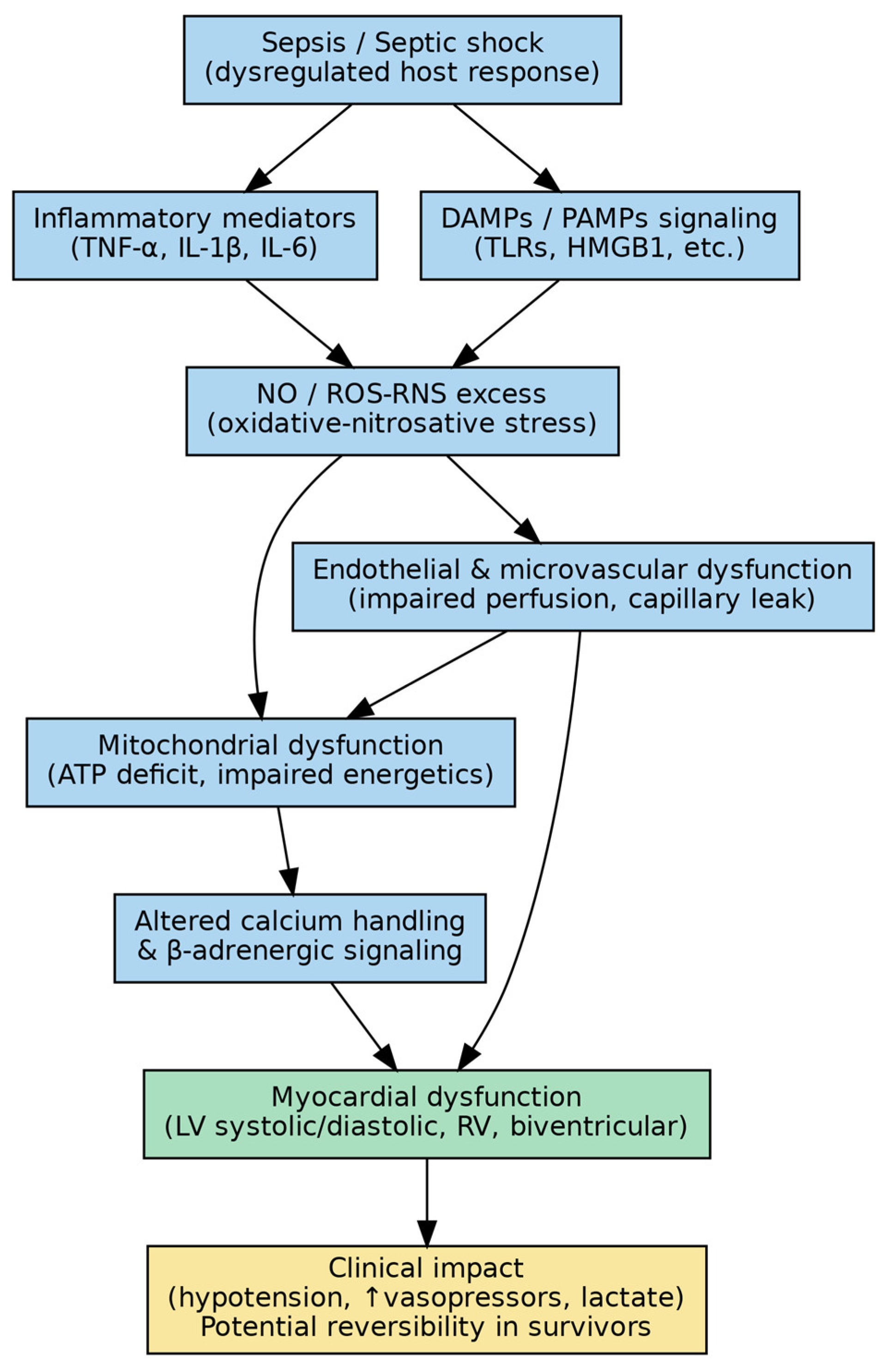
2.1. Clinical Signs of Myocardial Dysfunction Brought on by Sepsis
2.2. Risk Factors for Cardiac Dysfunction Brought on by Sepsis
- Sepsis severity: Myocardial dysfunction is more likely in cases of severe sepsis, such as septic shock. Widespread inflammation and severe infections can have a major effect on heart function.
- Preexisting cardiovascular disease: Patients with pre-existing cardiovascular conditions—such as hypertension, coronary artery disease, or heart failure—are more susceptible to sepsis-induced myocardial dysfunction, as these comorbidities may limit the heart’s ability to tolerate the additional hemodynamic and inflammatory stress imposed by sepsis.
- Associated comorbidities: Because they affect general health and the inflammatory response, illnesses like diabetes mellitus or chronic obstructive pulmonary disease (COPD) can increase the risk.
- High levels of inflammatory markers: Elevated levels of inflammatory markers, particularly pro-inflammatory cytokines, may exacerbate cardiac dysfunction and are frequently observed in severe sepsis.
- High acute illness severity: Myocardial dysfunction is more likely to occur in patients who have severe organ dysfunction and high Sequential Organ Failure Assessment (SOFA) scores.
- Nutritional status: Inadequate nutrition can weaken the immune system and make a person more vulnerable to severe sepsis and cardiac abnormalities.
- Inadequate or delayed treatment: More serious systemic effects, such as heart malfunction, might result from inadequate management of sepsis or from delays in treating it.
- High lactic acid: SIMD is independently associated with elevated serum lactate levels (>4.0 mmol/L) at ICU admission [22,23,24]. Accordingly, early identification of at-risk patients, prompt initiation of appropriate therapy, and optimized supportive care are essential to mitigate the impact of sepsis-induced myocardial dysfunction.
2.3. Pathophysiology of Cardiac Dysfunction Caused by Sepsis
2.3.1. Myocardial Circulation
2.3.2. Dysfunction of Mitochondrial Metabolism
Oxidative Stress and Damage to Mitochondria
Abnormal Calcium Transport in the Mitochondria
DNA in Mitochondria
Dynamics of Mitochondria
The Biogenesis of Mitochondria
The Process of Mitophagy
2.3.3. Myocardial Depression Directly
The Cytokines
Nitric Oxide
Endothelin-1
Intracellular Adhesion Molecules
Prostanoids
High Mobility Group Box 1 (HMGB1) and Histones
Complement System
Additional Mediators
2.3.4. Epigenetic Mechanisms
3. Discussion
3.1. Monitoring Strategies
- Hemodynamic monitoring: Regular evaluation of vital signs, such as blood pressure, heart rate, and central venous pressure, aids in determining the general state of the cardiovascular system. Advanced hemodynamic monitoring tools, like pulmonary artery catheters and the transpulmonary thermodilution method, offer comprehensive data on cardiac output, LV ejection fraction, preload, and afterload. They also assist in assessing the efficacy of fluid resuscitation and the sufficiency of perfusion. In cases of sepsis-induced myocardial dysfunction, measuring cardiac output (CO) and other hemodynamic parameters is essential. Although the pulmonary artery catheter (PAC) was once often used to monitor hemodynamics in critically ill patients, its use has decreased because there is no proof that it reduces patient mortality. Another technique for measuring CO and cardiac performance indicators such as the global ejection fraction (GEF) and cardiac function index (CFI) is the transpulmonary thermodilution method. According to reports, cardiac dysfunction in septic patients can be detected by low CFI and GEF readings from transpulmonary thermodilution. Furthermore, it has been determined that pulse contour analysis is a reliable method for continuous CO assessment in sepsis. Nevertheless, more research is needed to confirm these techniques’ effectiveness in identifying SIMD [80,81,82]. There is evidence that non-invasive hemodynamic techniques, such as end-expiratory occlusion, passive straight leg raise, inferior vena cava collapse, pulse contour analysis, pulse pressure variation, and stroke volume fluctuation, are effective in determining fluid responsiveness in septic shock patients. These methods do have certain drawbacks, though. Following the first stage of resuscitation, cautious fluid control is crucial. Dynamic measurements of fluid responsiveness ought to serve as guidance for this.
- Echocardiography: This imaging modality provides real-time assessment of cardiac anatomy and function, enabling the evaluation of ventricular size, ejection fraction, and regional wall motion abnormalities—key features of SIMD. In addition, continuous electrocardiographic monitoring is essential for the detection of ischemic changes or arrhythmias that may arise secondary to sepsis-related cardiac dysfunction.
- Cardiac Blood Biomarkers: Cardiac troponins and B-type natriuretic peptide (BNP) are widely used biomarkers for assessing myocardial injury and wall stress. Elevated concentrations of these markers are associated with cardiac dysfunction and may aid in risk stratification and therapeutic decision-making.
- Lactate levels: Serial monitoring of serum lactate levels assists in the assessment of tissue perfusion and oxygenation. Elevated lactate concentrations may indicate inadequate cardiac output and can inform timely adjustments in therapeutic management.
- Clinical assessment: Frequent examination of clinical indicators, such as edema, jugular venous distension, and altered mental status, can reveal information concerning declining fluid balance and cardiac function. Integrating these data points is necessary for effective monitoring in order to give a thorough picture of heart function and direct suitable therapeutic techniques for sepsis patients.
3.2. Differential Diagnosis of Myocardial Dysfunction in Septic Patients
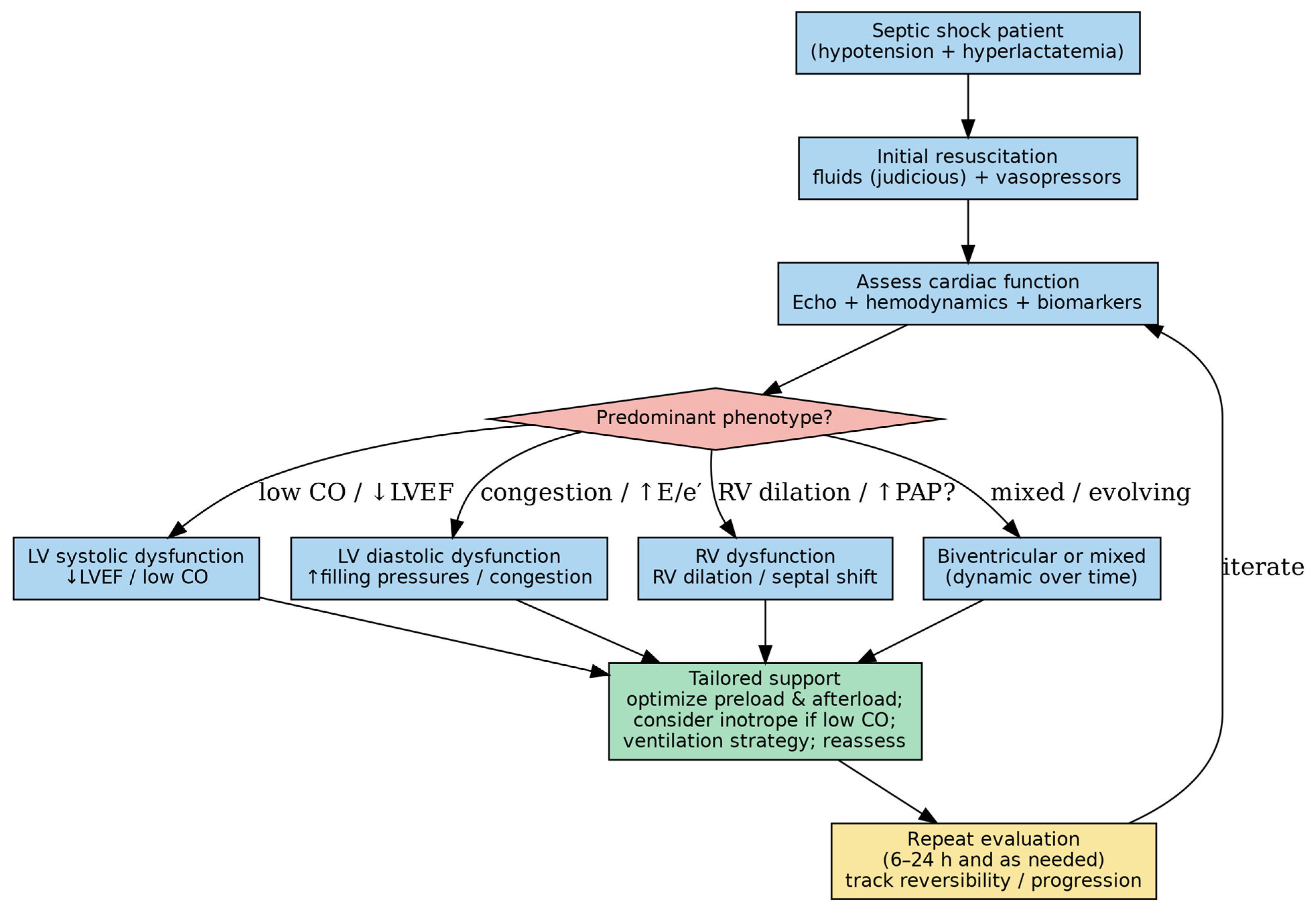
3.3. Handling Myocardial Dysfunction
3.4. Emerging and Targeted Therapeutic Approaches
4. Materials and Methods
5. Conclusions
Author Contributions
Funding
Institutional Review Board Statement
Informed Consent Statement
Data Availability Statement
Acknowledgments
Conflicts of Interest
References
- Singer, M.; Deutschman, C.S.; Seymour, C.W.; Shankar-Hari, M.; Annane, D.; Bauer, M.; Bellomo, R.; Bernard, G.R.; Chiche, J.-D.; Coopersmith, C.M.; et al. The Third International Consensus Definitions for sepsis and septic shock (Sepsis-3). JAMA 2016, 315, 801–810. [Google Scholar] [CrossRef] [Scilit]
- Laura, E.; Andrew, R.; Waleed, A.; Massimo, A.; Craig, M.C.; Craig, F.; Flávia, R.M.; Lauralyn, M.; Marlies, O.; Hallie, C.P.; et al. Surviving Sepsis Campaign: International guidelines for management of sepsis and septic shock 2021. Intensive Care Med. 2021, 47, 1181–1247. [Google Scholar] [CrossRef] [Scilit]
- Vieillard-Baron, A. Septic cardiomyopathy. Ann. Intensive Care 2011, 1, 6. [Google Scholar] [CrossRef] [Scilit] [PubMed]
- Hotchkiss, R.S.; Karl, I.E. Pathophysiology and treatment of sepsis. N. Engl. J. Med. 2003, 348, 138–150. [Google Scholar] [CrossRef] [Scilit]
- Geri, G.; Vignon, P.; Aubry, A.; Fedou, A.L.; Charron, C.; Silva, S.; Repessé, X.; Vieillard-Baron, A. Cardiovascular clusters in septic shock combining clinical and echocardiographic parameters: A post hoc analysis. Intensive Care Med. 2019, 45, 657–667. [Google Scholar] [CrossRef] [Scilit]
- Wasyluk, W.; Nowicka-Stążka, P.; Zwolak, A. Heart metabolism in sepsis induced cardiomyopathy—Unusual metabolic dysfunction of the heart. Int. J. Environ. Res. Public Health 2021, 18, 7598. [Google Scholar] [CrossRef] [Scilit]
- Habimana, R.; Choi, I.; Cho, H.J.; Kim, D.; Lee, K.; Jeong, I. Sepsis-induced cardiac dysfunction: A review of pathophysiology. Acute Crit. Care 2020, 35, 57–66. [Google Scholar] [CrossRef] [Scilit]
- Fan, D.; Wu, R. Mechanisms of the septic heart: From inflammatory response to myocardial edema. J. Mol. Cell Cardiol. 2024, 195, 73–82. [Google Scholar] [CrossRef] [Scilit]
- Kim, J.S.; Kim, Y.J.; Kim, M.; Ryoo, S.M.; Kim, W.Y. Association between right ventricle dysfunction and poor outcome in patients with septic shock. Heart 2020, 106, 1665–1671. [Google Scholar] [CrossRef] [Scilit]
- Lin, Y.; Xu, Y.; Zhang, Z. Sepsis-induced myocardial dysfunction (SIMD): The pathophysiological mechanisms and therapeutic strategies targeting mitochondria. Inflammation 2020, 43, 1184–1200. [Google Scholar] [CrossRef] [Scilit] [PubMed]
- Thupakula, S.; Nimmala, S.S.; Ravula, H.; Chekuri, S.; Padiya, R. Emerging biomarkers for the detection of cardiovascular diseases. Egypt. Heart J. 2022, 74, 77. [Google Scholar] [CrossRef] [Scilit]
- Ehrman, R.R.; Sullivan, A.N.; Favot, M.J.; Sherwin, R.L.; Reynolds, C.A.; Abidov, A.; Levy, P.D. Pathophysiology, echocardiographic evaluation, biomarker findings, and prognostic implications of septic cardiomyopathy: A review of the literature. Crit. Care 2018, 22, 112. [Google Scholar] [CrossRef] [Scilit] [PubMed]
- Fernandes, C.J., Jr.; de Assuncao, M.S. Myocardial dysfunction in sepsis: A large, unsolved puzzle. Crit. Care Res. Pract. 2012, 2012, 896430. [Google Scholar] [CrossRef] [Scilit]
- L’Heureux, M.; Sternberg, M.; Brath, L.; Turlington, J.; Kashiouris, M.G. Sepsis-induced cardiomyopathy: A comprehensive review. Curr. Cardiol. Rep. 2020, 22, 35. [Google Scholar] [CrossRef] [Scilit]
- Pulido, J.N.; Afessa, B.; Masaki, M.; Yuasa, T.; Do, S.G.; Herasevich, V.; Brown, D.R.; Oh, J.K. Clinical spectrum, frequency, and significance of myocardial dysfunction in severe sepsis and septic shock. Mayo Clin. Proc. 2012, 87, 620–628. [Google Scholar] [CrossRef] [Scilit]
- Muller-Werdan, U.; Buerke, M.; Ebelt, H.; Heinroth, K.M.; Herklotz, A.; Loppnow, H.; Ruß, M.; Schlegel, F.; Schlitt, A.; Schmidt, H.B.; et al. Septic cardiomyopathy: A not yet discovered cardiomyopathy? Exp. Clin. Cardiol. 2006, 11, 226–236. [Google Scholar]
- Hoesel, L.M.; Niederbichler, A.D.; Ward, P.A. Complement-related molecular events in sepsis leading to heart failure. Mol. Immunol. 2007, 44, 95–102. [Google Scholar] [CrossRef] [Scilit]
- Parker, M.M.; Suffredini, A.F.; Natanson, C.; Ognibene, F.P.; Shelhamer, J.H.; Parrillo, J.E. Responses of left ventricular function in survivors and nonsurvivors of septic shock. J. Crit. Care 1989, 4, 19–25. [Google Scholar] [CrossRef] [Scilit]
- Poelaert, J.; Declerck, C.; Vogelaers, D.; Colardyn, F.; Visser, C.A. Left ventricular systolic and diastolic function in septic shock. Intensive Care Med. 1997, 23, 553–560. [Google Scholar] [CrossRef] [Scilit] [PubMed]
- Parker, M.M.; Shelhamer, J.H.; Bacharach, S.L.; Green, M.V.; Natanson, C.; Frederick, T.M.; Damske, B.A.; Parrillo, J.E. Profound but reversible myocardial depression in patients with septic shock. Ann. Intern. Med. 1984, 100, 483–490. [Google Scholar] [CrossRef] [Scilit] [PubMed]
- Parker, M.M.; McCarthy, K.E.; Ognibene, F.P.; Parrillo, J.E. Right ventricular dysfunction and dilatation, similar to left ventricular changes, characterize the cardiac depression of septic shock in humans. Chest 1990, 97, 126–131. [Google Scholar] [CrossRef] [Scilit]
- Li, Y.; Li, H. Analysis of incidence and risk factors of septic cardiomyopathy. Zhonghua Jizhen Yixue Zazhi 2019, 28, 836–840. [Google Scholar]
- Sato, R.; Kuriyama, A.; Takada, T.; Nasu, M.; Luthe, S.K. Prevalence, and risk factors of sepsis-induced cardiomyopathy: A retrospective cohort study. Medicine 2016, 95, e5031. [Google Scholar] [CrossRef] [Scilit]
- Jeong, H.S.; Lee, T.H.; Bang, C.H.; Kim, J.-H.H.S.J. Risk factors and outcomes of sepsis-induced myocardial dysfunction and stress induced cardiomyopathy in sepsis or septic shock: A comparative retrospective study. Medicine 2018, 97, e0263. [Google Scholar] [CrossRef] [Scilit] [PubMed]
- Akira, S.; Uematsu, S.; Takeuchi, O. Pathogen recognition and innate immunity. Cell 2006, 124, 783–801. [Google Scholar] [CrossRef] [Scilit] [PubMed]
- Raeburn, C.D.; Sheppard, F.; Barsness, K.A.; Arya, J.; Harken, A.H. Cytokines for surgeons. Am. J. Surg. 2002, 183, 268–273. [Google Scholar] [CrossRef] [Scilit]
- Levy, R.J.; Piel, D.A.; Acton, P.D.; Zhou, R.; Ferrari, V.A.; Karp, J.S.; Deutschman, C.S. Evidence of myocardial hibernation in the septic heart. Crit. Care Med. 2005, 33, 2752–2756. [Google Scholar] [CrossRef] [Scilit]
- Antonucci, E.; Fiaccadori, E.; Donadello, K.; Taccone, F.S.; Franchi, F.; Scolletta, S. Myocardial depression in sepsis: From pathogenesis to clinical manifestations and treatment. J. Crit. Care 2014, 29, 500–511. [Google Scholar] [CrossRef] [Scilit] [PubMed]
- Jang, D.H.; Greenwood, J.C.; Spyres, M.B.; Eckmann, D.M. Measurement of mitochondrial respiration and motility in acute care: Sepsis, trauma, and poisoning. J. Intensive Care Med. 2017, 32, 86–94. [Google Scholar] [CrossRef] [Scilit]
- Carré, J.E.; Singer, M. Cellular energetic metabolism in sepsis: The need for a systems approach. Biochim. Biophys. Acta 2008, 1777, 763–771. [Google Scholar] [CrossRef] [Scilit]
- Brealey, D.; Brand, M.; Hargreaves, I.; Heales, S.; Land, J.; Smolenski, R.; Davies, N.A.; Cooper, C.E.; Singer, M. Association between mitochondrial dysfunction and severity and outcome of septic shock. Lancet 2002, 360, 219–223. [Google Scholar] [CrossRef] [Scilit]
- Potz, B.A.; Sellke, F.W.; Abid, M.R. Endothelial ROS and impaired myocardial oxygen consumption in sepsis-induced cardiac dysfunction. J. Intensive Crit. Care 2016, 2, 20. [Google Scholar] [CrossRef] [Scilit]
- Larsen, F.J.; Schiffer, T.A.; Weitzberg, E.; Lundberg, J.O. Regulation of mitochondrial function and energetics by reactive nitrogen oxides. Free Radic. Biol. Med. 2012, 53, 1919–1928. [Google Scholar] [CrossRef] [Scilit]
- Crouser, E.D. Mitochondrial dysfunction in septic shock and multiple organ dysfunction syndrome. Mitochondrion 2004, 4, 729–741. [Google Scholar] [CrossRef] [Scilit]
- Lowes, D.A.; Webster, N.R.; Murphy, M.P.; Galley, H.F. Antioxidants that protect mitochondria reduce interleukin-6 and oxidative stress, improve mitochondrial function, and reduce biochemical markers of organ dysfunction in a rat model of acute sepsis. Br. J. Anaesth. 2013, 110, 472–480. [Google Scholar] [CrossRef] [Scilit] [PubMed]
- Kayali, R.; Aydin, S.; Cakatay, U. Effect of gender on main clinical chemistry parameters in aged rats. Curr. Aging Sci. 2009, 2, 71. [Google Scholar] [CrossRef]
- Galley, H.F. Oxidative stress and mitochondrial dysfunction in sepsis. Br. J. Anaesth. 2011, 107, 57–64. [Google Scholar] [CrossRef] [Scilit]
- Bernardi, P.; Di Lisa, F. The mitochondrial permeability transition pore: Molecular nature and role as a target in cardioprotection. J. Mol. Cell. Cardiol. 2015, 78, 100–106. [Google Scholar] [CrossRef] [Scilit] [PubMed]
- Duncan, D.J.; Yang, Z.; Hopkins, P.M.; Steele, D.S.; Harrison, S.M. TNF alpha and IL-1beta increase Ca2+ leak from the sarcoplasmic reticulum and susceptibility to arrhythmia in rat ventricular myocytes. Cell Calcium 2010, 47, 378–386. [Google Scholar] [CrossRef] [Scilit] [PubMed]
- Hassoun, S.M.; Marechal, X.; Montaigne, D.; Bouazza, Y.; Decoster, B.; Lancel, S.; Neviere, R. Prevention of endotoxin induced sarcoplasmic reticulum calcium leak improves mitochondrial and myocardial dysfunction. Crit. Care Med. 2008, 36, 2590–2596. [Google Scholar] [CrossRef] [Scilit]
- Shanmuganathan, S.; Hausenloy, D.J.; Duchen, M.R.; Yellon, D.M. Mitochondrial permeability transition pore as a target for cardioprotection in the human heart. Am. J. Physiol. Heart Circ. Physiol. 2005, 289, H237–H242. [Google Scholar] [CrossRef] [Scilit] [PubMed]
- Smeding, L.; Plötz, F.B.; Groeneveld, A.B.; Kneyber, M.C. Structural changes of the heart during severe sepsis or septic shock. Shock 2012, 37, 449–456. [Google Scholar] [CrossRef] [Scilit]
- Finkel, M.S.; Oddis, C.V.; Jacob, T.D.; Watkins, S.C.; Hattler, B.G.; Simmons, R.L. Negative inotropic effects of cytokines on the heart mediated by nitric oxide. Science 1992, 257, 387–389. [Google Scholar] [CrossRef] [Scilit]
- Wu, L.L.; Ji, Y.; Dong, L.W.; Liu, M.S. Calcium uptake by sarcoplasmic reticulum is impaired during the hypodynamic phase of sepsis in the rat heart. Shock 2001, 15, 49–55. [Google Scholar] [CrossRef] [Scilit]
- Salami, O.M.; Habimana, O.; Peng, J.F.; Yi, G.H. Therapeutic strategies targeting mitochondrial dysfunction in sepsis induced cardiomyopathy. Cardiovasc. Drugs Ther. 2024, 38, 163–180. [Google Scholar] [CrossRef] [Scilit] [PubMed]
- Nunnari, J.; Suomalainen, A. Mitochondria: In sickness and in health. Cell 2012, 148, 1145–1159. [Google Scholar] [CrossRef] [Scilit] [PubMed]
- Nakahira, K.; Kyung, S.-Y.; Rogers, A.J.; Gazourian, L.; Youn, S.; Massaro, A.F.; Quintana, C.; Osorio, J.C.; Wang, Z.; Zhao, Y.; et al. Circulating mitochondrial DNA in patients in the ICU as a marker of mortality: Derivation and validation. PLoS Med. 2013, 10, e1001577. [Google Scholar] [CrossRef] [Scilit]
- Sánchez-Villamil, J.P.; D’aNnunzio, V.; Finocchietto, P.; Holod, S.; Rebagliati, I.; Pérez, H.; Peralta, J.G.; Gelpi, R.J.; Poderoso, J.J.; Carreras, M.C. Cardiac-specific overexpression of thioredoxin 1 attenuates mitochondrial and myocardial dysfunction in septic mice. Int. J. Biochem. Cell Biol. 2016, 81, 323–334. [Google Scholar] [CrossRef] [Scilit] [PubMed]
- Zhan, M.; Brooks, C.; Liu, F.; Sun, L.; Dong, Z. Mitochondrial dynamics: Regulatory mechanisms and emerging role in renal pathophysiology. Kidney Int. 2013, 83, 568–581. [Google Scholar] [CrossRef] [Scilit]
- Liesa, M.; Palacín, M.; Zorzano, A. Mitochondrial dynamics in mammalian health and disease. Physiol. Rev. 2009, 89, 799–845. [Google Scholar] [CrossRef] [Scilit]
- Wu, Y.; Yao, Y.M.; Lu, Z.Q. Mitochondrial quality control mechanisms as potential therapeutic targets in sepsis-induced multiple organ failure. J. Mol. Med. 2019, 97, 451–462. [Google Scholar] [CrossRef] [Scilit] [PubMed]
- Singer, M. The role of mitochondrial dysfunction in sepsis-induced multi organ failure. Virulence 2014, 5, 66–72. [Google Scholar] [CrossRef] [Scilit]
- Gunst, J.; Derese, I.; Aertgeerts, A.; Ververs, E.-J.; Wauters, A.; Berghe, G.V.D.; Vanhorebeek, I. Insufficient autophagy contributes to mitochondrial dysfunction, organ failure, and adverse outcome in an animal model of critical illness. Crit. Care Med. 2013, 41, 182–194. [Google Scholar] [CrossRef] [Scilit]
- Rocha, M.; Herance, R.; Rovira, S.; Hernández-Mijares, A.; Victor, V.M. Mitochondrial dysfunction and antioxidant therapy in sepsis. Infect. Disord. Drug Targets 2012, 12, 161–178. [Google Scholar] [CrossRef] [Scilit]
- Kakihana, Y.; Ito, T.; Nakahara, M.; Yamaguchi, K.; Yasuda, T. Sepsis-induced myocardial dysfunction: Pathophysiology and management. J. Intensive Care 2016, 4, 22. [Google Scholar] [CrossRef] [Scilit] [PubMed]
- Kumar, A.; Krieger, A.; Symeoneides, S.; Kumar, A.; Parrillo, J.E. Myocardial dysfunction in septic shock. Part II. Role Cytokines Nitric oxide. J. Cardiothorac. Vasc. Anesth. 2001, 15, 485–511. [Google Scholar] [CrossRef] [Scilit] [PubMed]
- Pathan, N.; A Hemingway, C.; A Alizadeh, A.; Stephens, A.C.; Boldrick, J.C.; E Oragui, E.; McCabe, C.; Welch, S.B.; Whitney, A.; O′Gara, P.; et al. Role of interleukin 6 in myocardial dysfunction of meningococcal septic shock. Lancet 2004, 363, 203–209. [Google Scholar] [CrossRef] [Scilit]
- Loppnow, H.; Werdan, K.; Reuter, G.; Flad, H.D. The interleukin-1 and interleukin-1 converting enzyme families in the cardiovascular system. Eur. Cytokine Netw. 1998, 9, 675–680. [Google Scholar] [PubMed]
- Kelly, R.A.; Balligand, J.L.; Smith, T.W. Nitric oxide and cardiac function. Circ. Res. 1996, 79, 363–380. [Google Scholar] [CrossRef] [Scilit]
- Francis, S.E.; Holden, H.; Holt, C.M.; Duff, G.W. Interleukin-1 in myocardium and coronary arteries of patients with dilated cardiomyopathy. J. Mol. Cell. Cardiol. 1998, 30, 215–223. [Google Scholar] [CrossRef] [Scilit]
- Opal, S.M.; Fisher, C.J., Jr.; Dhainaut, J.F.; Vincent, J.L.; Brase, R.; Lowry, S.F.; Sadoff, J.C.; Slotman, G.J.; Levy, H.; Balk, R.A. Confirmatory interleukin-1 receptor antagonist trial in severe sepsis: A phase III, randomized, double-blind, placebo-controlled, multicenter trial. The interleukin-1 receptor antagonist sepsis investigator group. Crit. Care Med. 1997, 25, 1115–1124. [Google Scholar] [CrossRef] [Scilit] [PubMed]
- Müller-Werdan, U.; Werdan, K. Immune modul. by catecholamines: A potential mech. cytokine release heart fail? Herz 2000, 25, 271–273. [Google Scholar] [CrossRef] [Scilit]
- Geppert, A.; Steiner, A.; Zorn, G.; Delle-Karth, G.; Koreny, M.; Haumer, M.; Siostrzonek, P.; Huber, K.; Heinz, G. Multiple organ failure in patients with cardiogenic shock is associated with high plasma levels of interleukin-6. Crit. Care Med. 2002, 30, 1987–1994. [Google Scholar] [CrossRef] [Scilit] [PubMed]
- Rudiger, A.; Singer, M. Mechanisms of sepsis-induced cardiac dysfunction. Crit. Care Med. 2007, 35, 1599–1608. [Google Scholar] [CrossRef] [Scilit]
- Zanotti-Cavazzoni, S.L.; Hollenberg, S.M. Cardiac dysfunction in severe sepsis and septic shock. Curr. Opin. Crit. Care 2009, 15, 392–397. [Google Scholar] [CrossRef] [Scilit] [PubMed]
- Crouser, E.D.; Julian, M.W.; Huff, J.E.B.; Joshi, M.S.; Bauer, J.A.; Gadd, M.E.; Wewers, M.D.; Pfeiffer, D.R. Abnormal permeability of inner and outer mitochondrial membranes contributes independently to mitochondrial dysfunction in the liver during acute endotoxemia. Crit. Care Med. 2004, 32, 478–488. [Google Scholar] [CrossRef] [Scilit]
- Konrad, D.; Oldner, A.; Rossi, P.; Wanecek, M.; Rudehill, A.; Weitzberg, E. Differentiated and dose-related cardiovascular effects of a dual endothelin receptor antagonist in endotoxin shock. Crit. Care Med. 2004, 32, 1192–1199. [Google Scholar] [CrossRef] [Scilit]
- Raeburn, C.D.; Calkins, C.M.; Zimmerman, M.A.; Song, Y.; Ao, L.; Banerjee, A.; Harken, A.H.; Meng, X. ICAM-1 and VCAM-1 mediate endotoxemic myocardial dysfunction independent of neutrophil accumulation. Am. J. Physiol. Regul. Integr. Comp. Physiol. 2002, 283, R47786. [Google Scholar] [CrossRef] [Scilit]
- Raeburn, C.D.; Calkins, C.M.; Zimmerman, M.A.; Song, Y.; Ao, L.; Banerjee, A.; Meng, X.; Harken, A.H. Vascular cell adhesion molecule: 1 expression is obligatory for endotoxin-induced myocardial neutrophil accumulation and contractile dysfunction. Surgery 2001, 130, 319–325. [Google Scholar] [CrossRef] [Scilit]
- Memiş, D.; Karamanlioğlu, B.; Turan, A.; Koyuncu, O.; Pamukçu, Z. Effects of lornoxicam on the physiology of severe sepsis. Crit. Care 2004, 8, R474–R482. [Google Scholar] [CrossRef] [Scilit]
- Alhamdi, Y.; Abrams, S.T.; Cheng, Z.; Jing, S.; Su, D.; Liu, Z.; Lane, S.; Welters, I.; Wang, G.; Toh, C.-H. Circulating histones are major mediators of cardiac injury in patients with sepsis. Crit. Care Med. 2015, 43, 2094–2103. [Google Scholar] [CrossRef] [Scilit]
- Zhang, C.; Mo, M.; Ding, W.; Liu, W.; Yan, D.; Deng, J.; Luo, X.; Liu, J. High-mobility group box 1 (HMGB1) impaired cardiac excitation-contraction coupling by enhancing the sarcoplasmic reticulum (SR) Ca (2+) leak through TLR4-ROS signaling in cardiomyocytes. J. Mol. Cell. Cardiol. 2014, 74, 260–273. [Google Scholar] [CrossRef] [Scilit]
- Pei, X.B.; Liu, B. Research progress on the mechanism and management of septic cardiomyopathy: A comprehensive review. Emerg. Med. Int. 2023, 2023, 8107336. [Google Scholar] [CrossRef] [Scilit]
- Niederbichler, A.D.; Hoesel, L.M.; Westfall, M.V.; Gao, H.; Ipaktchi, K.R.; Sun, L.; Zetoune, F.S.; Su, G.L.; Arbabi, S.; Sarma, J.V.; et al. An essential role for complement C5a in the pathogenesis of septic cardiac dysfunction. J. Exp. Med. 2006, 203, 53–61. [Google Scholar] [CrossRef] [Scilit]
- Court, O.; Kumar, A.; Parrillo, J.E.; Kumar, A. Clinical review: Myocardial depression in sepsis and septic shock. Crit. Care 2002, 6, 500–508. [Google Scholar] [CrossRef] [Scilit]
- Witthaut, R.; Busch, C.; Fraunberger, P.; Walli, A.; Seidel, D.; Pilz, G.; Stuttmann, R.; Speichermann, N.; Verner, L.; Werdan, K. Plasma atrial natriuretic peptide and brain natriuretic peptide are increased in septic shock: Impact of interleukin-6 and sepsis-associated left ventricular dysfunction. Intensive Care Med. 2003, 29, 1696–1702. [Google Scholar] [CrossRef] [Scilit] [PubMed]
- Grandel, U.; Hopf, M.; Buerke, M.; Hattar, K.; Heep, M.; Fink, L.; Bohle, R.M.; Morath, S.; Hartung, T.; Pullamsetti, S.; et al. Mechanisms of cardiac depression caused by lipoteichoic acids from Staphylococcus aureus in isolated rat hearts. Circulation 2005, 112, 691–698. [Google Scholar] [CrossRef] [Scilit] [PubMed]
- Zhang, G.; Dong, D.; Wan, X.; Zhang, Y. Cardiomyocyte death in sepsis: Mechanisms and regulation (Review). Mol. Med. Rep. 2022, 26, 257. [Google Scholar] [CrossRef] [Scilit] [PubMed]
- Liu, Z.; Li, F.; Li, N.; Chen, Y.; Chen, Z. MicroRNAs as regulators of cardiac dysfunction in sepsis: Pathogenesis and diagnostic potential. Front. Cardiovasc. Med. 2025, 12, 1517323. [Google Scholar] [CrossRef] [Scilit]
- Wiener, R.S.; Welch, H.G. Trends in the use of the pulmonary artery catheter in the United States, 1993–2004. J. Am. Med. Assoc. 2007, 298, 423–429. [Google Scholar] [CrossRef] [Scilit]
- Ritter, S.; Rudiger, A.; Maggiorini, M. Transpulmonary thermodilution-derived cardiac function index identifies cardiac dysfunction in acute heart failure and septic patients: An observational study. Crit. Care 2009, 13, R133. [Google Scholar] [CrossRef] [Scilit]
- Schuerholz, T.; Meyer, M.C.; Friedrich, L.; Przemeck, M.; Sümpelmann, R.; Marx, G. Reliability of continuous cardiac output determination by pulse-contour analysis in porcine septic shock. Acta Anaesthesiol. Scand. 2006, 50, 407–413. [Google Scholar] [CrossRef] [Scilit]
- Rhodes, A.; Evans, L.E.; Alhazzani, W.; Levy, M.M.; Antonelli, M.; Ferrer, R.; Kumar, A.; Sevransky, J.E.; Sprung, C.L.; Nunnally, M.E.; et al. Surviving sepsis campaign: International guidelines for management of sepsis and septic shock: 2016. Crit. Care Med. 2017, 45, 486–552. [Google Scholar] [CrossRef] [Scilit]
- Brown, R.M.; Semler, M.W. Fluid management in sepsis. J. Intensive Care Med. 2019, 34, 364–373. [Google Scholar] [CrossRef] [Scilit] [PubMed]
- Marik, P.E.; Monnet, X.; Teboul, J.L. Hemodynamic parameters to guide fluid therapy. Ann. Intensive Care 2011, 1, 1. [Google Scholar] [CrossRef] [Scilit] [PubMed]
- Morelli, A.; De Castro, S.; Teboul, J.-L.; Singer, M.; Rocco, M.; Conti, G.; De Luca, L.; Di Angelantonio, E.; Orecchioni, A.; Pandian, N.G.; et al. Effects of levosimendan on systemic and regional hemodynamics in septic myocardial depression. Intensive Care Med. 2005, 31, 638–644. [Google Scholar] [CrossRef] [Scilit]
- Liu, D.-H.; Ning, Y.-L.; Lei, Y.-Y.; Chen, J.; Liu, Y.-Y.; Lin, X.-F.; Yang, Z.-Q.; Xian, S.-X.; Chen, W.-T. Levosimendan versus dobutamine for sepsis-induced cardiac dysfunction: A systematic review and meta-analysis. Sci. Rep. 2021, 11, 20333. [Google Scholar] [CrossRef] [Scilit] [PubMed]
- Whitehouse, T.; Hossain, A.; Perkins, G.D.; Gordon, A.C.; Bion, J.; Young, D.; McAuley, D.; Singer, M.; Lord, J.; Gates, S.; et al. Landiolol and organ failure in patients with septic shock: The STRESS-L randomized clinical trial. J. Am. Med. Assoc. 2023, 330, 1641–1652. [Google Scholar] [CrossRef] [Scilit]
- Wada, K.; Bunya, N.; Kakizaki, R.; Kasai, T.; Uemura, S.; Harada, K.; Narimatsu, E. Successful use of venoarterial extracorporeal membrane oxygenation for septic cardiomyopathy in a patient with preexisting chronic heart failure. Acute Med. Surg. 2019, 6, 301–304. [Google Scholar] [CrossRef] [Scilit]
Disclaimer/Publisher’s Note: The statements, opinions and data contained in all publications are solely those of the individual author(s) and contributor(s) and not of MDPI and/or the editor(s). MDPI and/or the editor(s) disclaim responsibility for any injury to people or property resulting from any ideas, methods, instructions or products referred to in the content. |
© 2026 by the authors. Licensee MDPI, Basel, Switzerland. This article is an open access article distributed under the terms and conditions of the Creative Commons Attribution (CC BY) license.
Share and Cite
Pădureanu, V.; Caragea, D.C.; Pîrșcoveanu, D.F.V.; Dop, D.; Munteanu, A.C.; Rădulescu, D.; Popa, D.G.; Forțofoiu, D.; Drăgoescu, A.N.; Pădureanu, R. Association Between Myocardial Dysfunction and Septic Shock. Int. J. Mol. Sci. 2026, 27, 2552. https://doi.org/10.3390/ijms27062552
Pădureanu V, Caragea DC, Pîrșcoveanu DFV, Dop D, Munteanu AC, Rădulescu D, Popa DG, Forțofoiu D, Drăgoescu AN, Pădureanu R. Association Between Myocardial Dysfunction and Septic Shock. International Journal of Molecular Sciences. 2026; 27(6):2552. https://doi.org/10.3390/ijms27062552
Chicago/Turabian StylePădureanu, Vlad, Daniel Cosmin Caragea, Denisa Floriana Vasilica Pîrșcoveanu, Dalia Dop, Alexandru Claudiu Munteanu, Dumitru Rădulescu, Dragoș George Popa, Dragoș Forțofoiu, Alice Nicoleta Drăgoescu, and Rodica Pădureanu. 2026. "Association Between Myocardial Dysfunction and Septic Shock" International Journal of Molecular Sciences 27, no. 6: 2552. https://doi.org/10.3390/ijms27062552
APA StylePădureanu, V., Caragea, D. C., Pîrșcoveanu, D. F. V., Dop, D., Munteanu, A. C., Rădulescu, D., Popa, D. G., Forțofoiu, D., Drăgoescu, A. N., & Pădureanu, R. (2026). Association Between Myocardial Dysfunction and Septic Shock. International Journal of Molecular Sciences, 27(6), 2552. https://doi.org/10.3390/ijms27062552