Margin of Error: The Emerging Role of Field Cancerization in Predicting Recurrence Risk of Ductal Carcinoma In Situ

 ,
,
Abstract
1. Introduction
2. Breaking New Ground: Ductal Carcinoma In Situ to Invasive Ductal Carcinoma
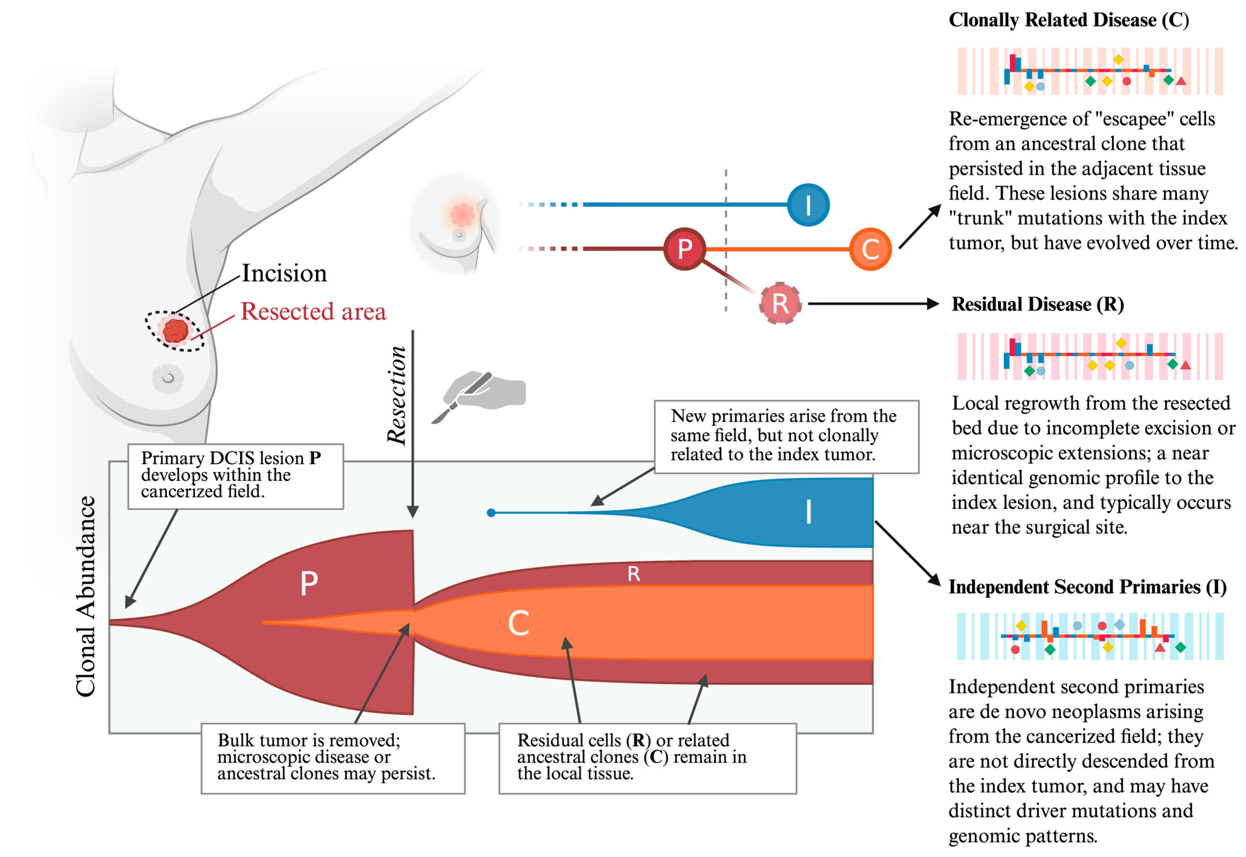
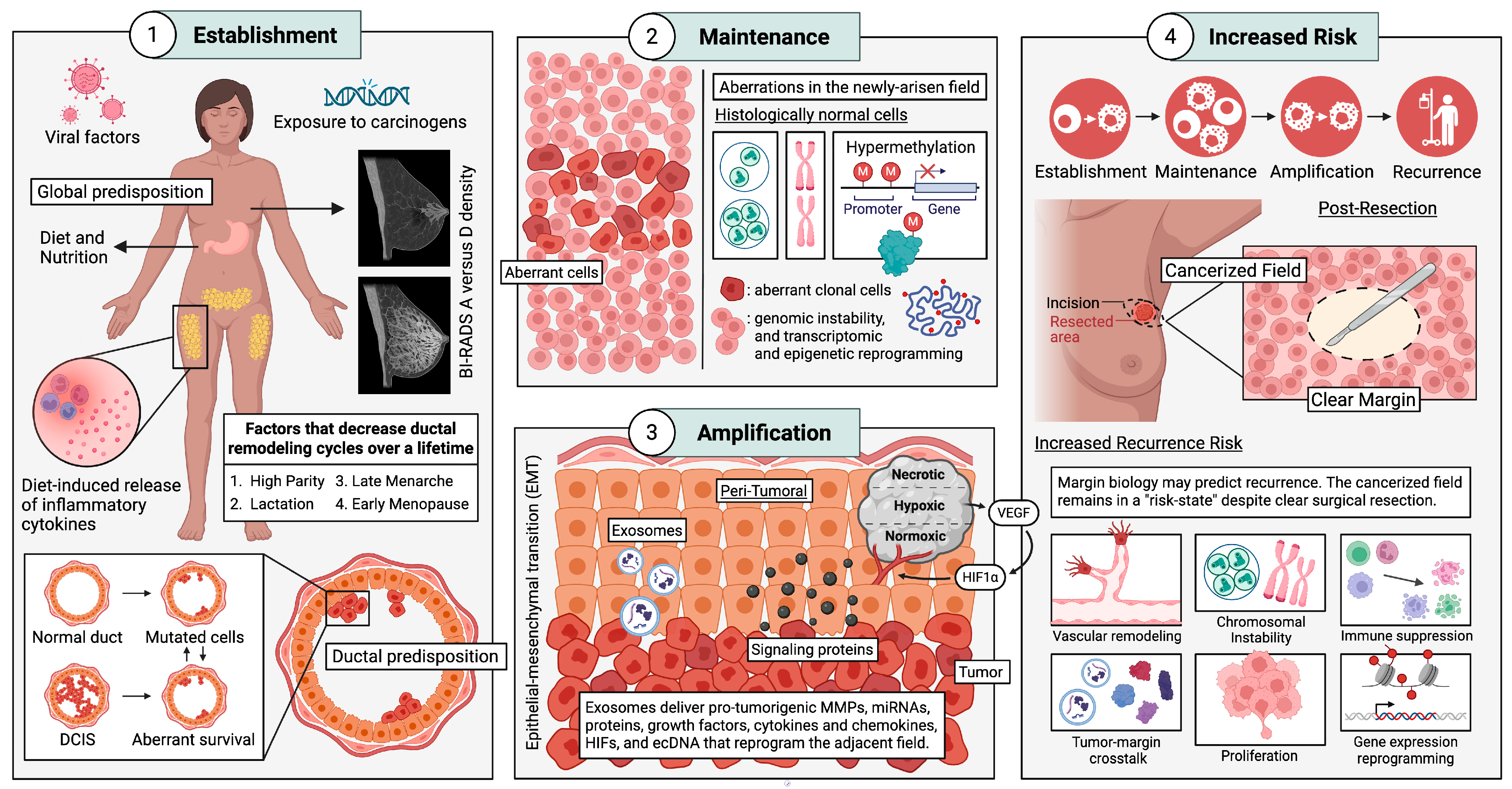
3. Ring of Fire: Field Cancerization
4. Pre-Tumorigenesis: Drivers of Field Cancerization
4.1. Non-Biological Drivers
4.1.1. Endocrine Disruptors
4.1.2. Diet, Alcohol, and Supplementation
4.2. Biological Drivers
4.2.1. Inflammatory Cytokines and Mammographic Density
4.2.2. Menstrual and Reproductive Factors
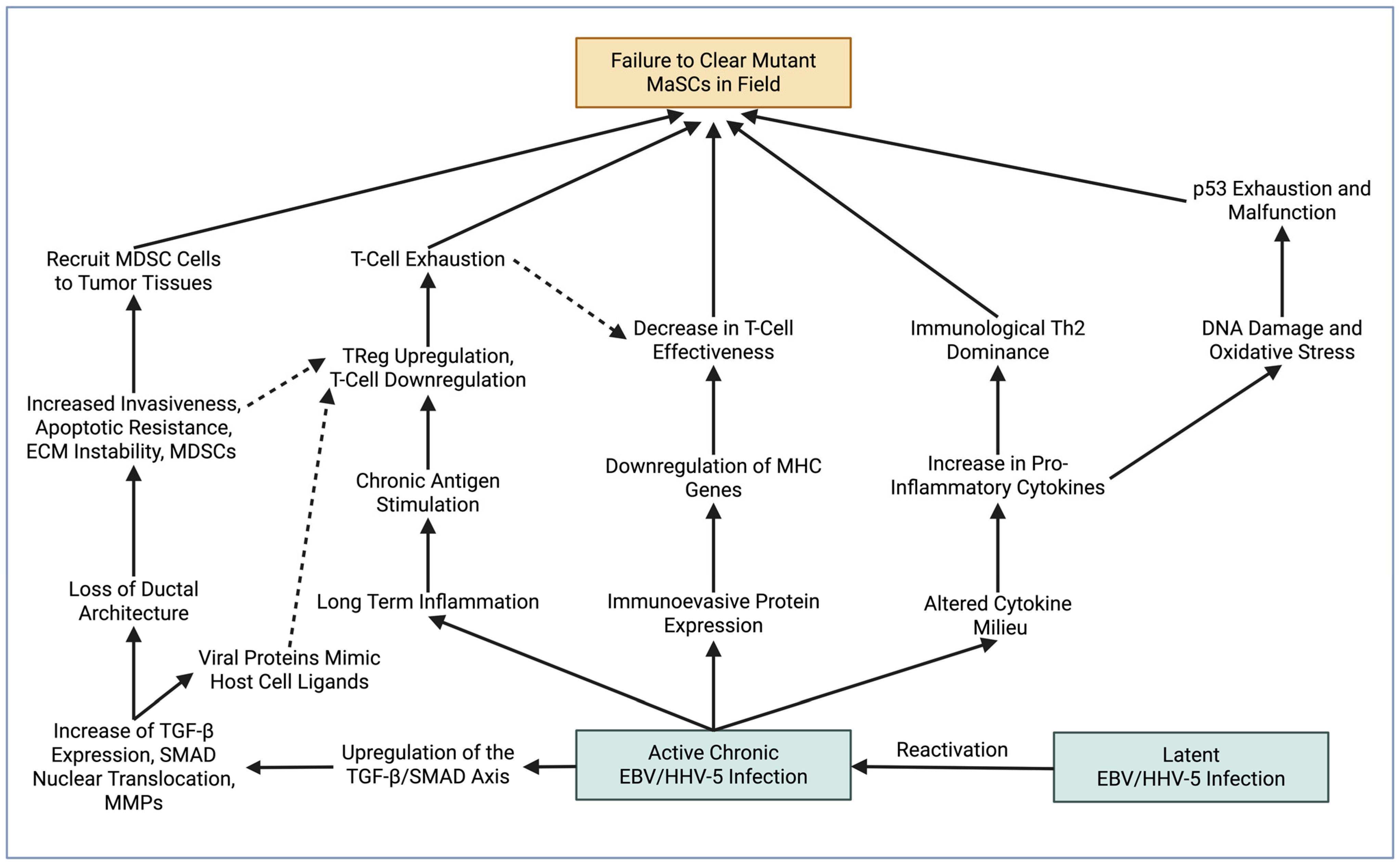
4.2.3. Going Viral: Viral Mechanisms Implicated in Field Cancerization
4.2.4. Inflammation, Immune Evasion, and Immune Suppression
5. Post-Tumorigenesis: Secondary Drivers of Field Cancerization
6. The Crux of the Matter: Genomic Instability Is Widespread in Peritumoral Tissues
7. Top-Down Effect: Epigenetic and Transcriptomic Changes in Tumor Margin Cells
7.1. Shifting Gears: Telomere Erosion and Telomerase Expression in Tumor-Adjacent Tissues
7.2. The Bystander Effect: Other Changes Observed in Peritumoral Tissues
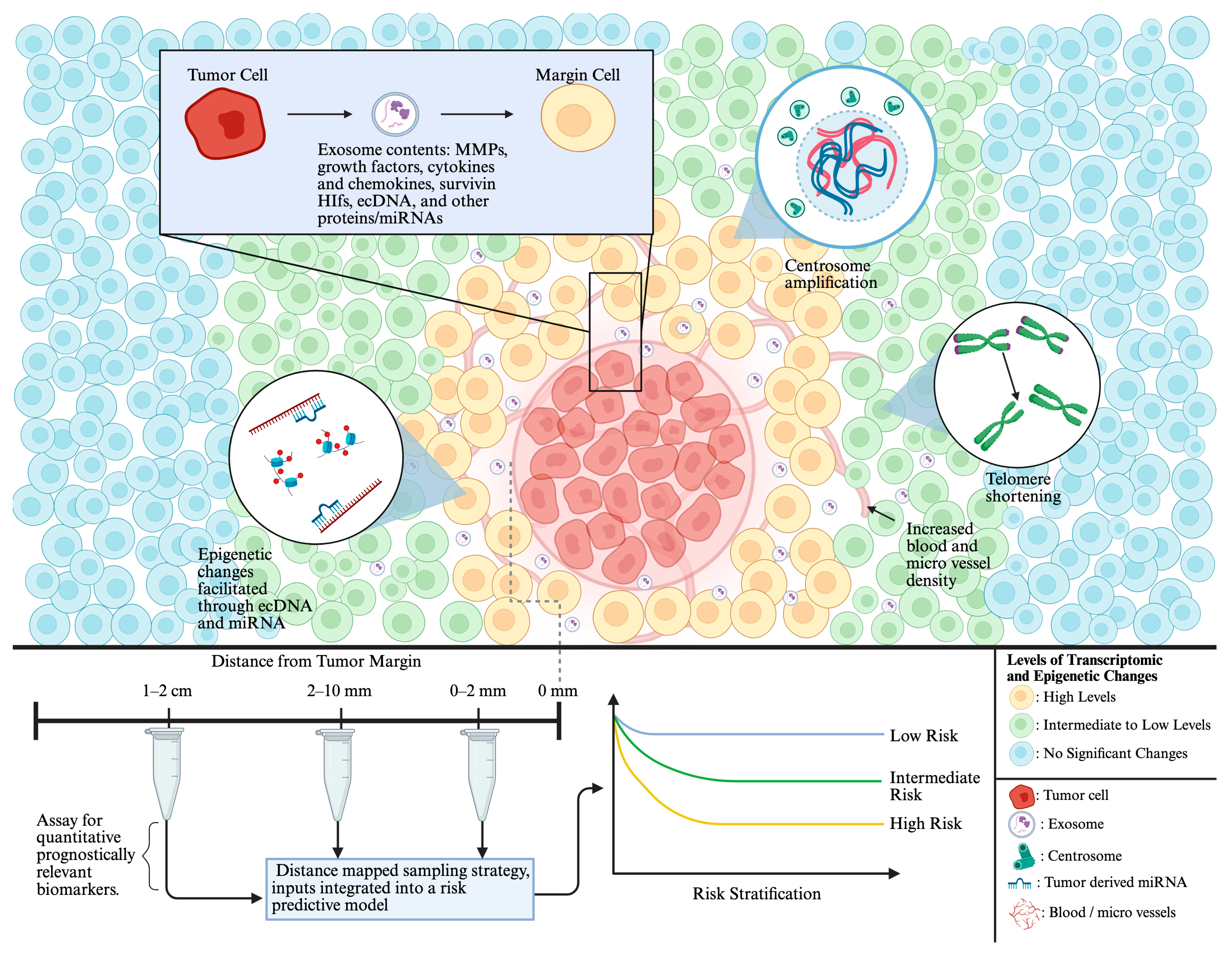
7.3. Home Invasion: Roles of Exosomes in Field Cancerization
8. Exosomal miRNAs
8.1. Exosomal miRNA Within Stromal Cells
8.2. Exosomal Cytokines, MMPs and Hypoxia-Inducible Factor, and Other Proteins
8.3. Exosomal Growth Factors
8.4. Exosomal Survivin
8.5. ecDNA as a Driver of Breast Tumor Metastasis
8.6. Immune Modulation by Tumor Cells
9. Connecting the Dots: Shifting Emphasis from “Clear Margins” to “Field-Informed” Care
Author Contributions
Funding
Data Availability Statement
Acknowledgments
Conflicts of Interest
References
- Kalwaniya, D.S.; Gairola, M.; Gupta, S.; Pawan, G.; Gurivelli, P. Ductal Carcinoma in Situ: A Detailed Review of Current Practices. Cureus 2023, 15, e37932. [Google Scholar] [CrossRef] [Scilit]
- Chang, Y.-W.; Ryu, J.K.; An, J.K.; Choi, N.; Park, Y.M.; Ko, K.H.; Han, K. Artificial intelligence for breast cancer screening in mammography (AI-STREAM): Preliminary analysis of a prospective multicenter cohort study. Nat. Commun. 2025, 16, 2248. [Google Scholar] [CrossRef] [Scilit]
- Siegel, R.L.; Miller, K.D.; Fuchs, H.E.; Jemal, A. Cancer statistics. CA Cancer J. Clin. 2022, 72, 7–33. [Google Scholar] [CrossRef] [Scilit]
- Siegel, R.L.; Giaquinto, A.N.; Jemal, A. Cancer statistics, 2024. CA A Cancer J. Clin. 2024, 74, 12–49, Erratum in CA Cancer J. Clin. 2024, 74, 203. https://doi.org/10.3322/caac.21830. [Google Scholar] [CrossRef] [Scilit] [PubMed]
- Co, M.; Cheng, K.; Yeung, Y.; Lau, K.; Qian, Z.; Wong, C.; Wong, B.; Sin, E.; Wong, H.; Ma, C. Clinical Outcomes of Conservative Treatment for Low-Risk Ductal Carcinoma in Situ: A Systematic Review and Pooled Analysis. Clin. Oncol. 2023, 35, 255–261. [Google Scholar] [CrossRef] [Scilit] [PubMed]
- Williams, L.J.; Kunkler, I.H.; Taylor, K.J.; Dunlop, J.; Piper, T.; Caldwell, J.; Jack, W.; Loane, J.F.; Elder, K.; Bartlett, J.M.S.; et al. Postoperative radiotherapy in women with early operable breast cancer (Scottish Breast Conservation Trial): 30-year update of a randomised, controlled, phase 3 trial. Lancet Oncol. 2024, 25, 1213–1221. [Google Scholar] [CrossRef] [Scilit] [PubMed]
- Withrow, D.R.; Morton, L.M.; Curtis, R.E.; Schonfeld, S.J.; de González, A.B. Radiotherapy for ductal carcinoma in situ and risk of second non-breast cancers. Breast Cancer Res. Treat. 2017, 166, 299–306. [Google Scholar] [CrossRef] [Scilit]
- Wadsten, C.; Wennstig, A.-K.; Garmo, H.; Nilsson, G.; Blomqvist, C.; Holmberg, L.; Fredriksson, I.; Wärnberg, F.; Sund, M. Risk of ischemic heart disease after radiotherapy for ductal carcinoma in situ. Breast Cancer Res. Treat. 2018, 171, 95–101. [Google Scholar] [CrossRef] [Scilit]
- Solin, L.J.; Gray, R.; Hughes, L.L.; Wood, W.C.; Lowen, M.A.; Badve, S.S.; Baehner, F.L.; Ingle, J.N.; Perez, E.A.; Recht, A.; et al. Surgical Excision Without Radiation for Ductal Carcinoma in Situ of the Breast: 12-Year Results From the ECOG-ACRIN E5194 Study. J. Clin. Oncol. 2015, 33, 3938–3944. [Google Scholar] [CrossRef] [Scilit] [PubMed]
- Rudloff, U.; Jacks, L.M.; Goldberg, J.I.; Wynveen, C.A.; Brogi, E.; Patil, S.; Van Zee, K.J. Nomogram for Predicting the Risk of Local Recurrence After Breast-Conserving Surgery for Ductal Carcinoma In Situ. J. Clin. Oncol. 2010, 28, 3762–3769. [Google Scholar] [CrossRef] [Scilit]
- Milonas, D.; Venclovas, Z.; Muilwijk, T.; Jievaltas, M.; Joniau, S. External validation of Memorial Sloan Kettering Cancer Center nomogram and prediction of optimal candidate for lymph node dissection in clinically localized prostate cancer. Central Eur. J. Urol. 2020, 73, 19–25. [Google Scholar] [CrossRef] [Scilit] [PubMed]
- Choi, J.D.W.; Hughes, T.M.D.; Marx, G.; Boyages, J.; Rutovitz, J.; Hasovits, C.; Parasyn, A.; Edirimanne, S.; Ngui, N.K. The Utility of the Oncotype DX Test for Breast Cancer Patients in an Australian Multidisciplinary Setting. Breast J. 2022, 2022, 1199245. [Google Scholar] [CrossRef] [Scilit]
- Shah, C.; Bremer, T.; Cox, C.; Whitworth, P.; Patel, R.; Patel, A.; Brown, E.; Gold, L.; Rock, D.; Riley, L.; et al. The Clinical Utility of DCISionRT® on Radiation Therapy Decision Making in Patients with Ductal Carcinoma In Situ Following Breast-Conserving Surgery. Ann. Surg. Oncol. 2021, 28, 5974–5984, Erratum in Ann Surg Oncol. 2021, 28, 878. https://doi.org/10.1245/s10434-021-10138-3. [Google Scholar] [CrossRef] [Scilit] [PubMed] [PubMed Central]
- Pawloski, K.R.; Tadros, A.B.; Sevilimedu, V.; Newman, A.; Gentile, L.; Zabor, E.C.; Morrow, M.; Van Zee, K.J.; Kirstein, L.J. Patterns of invasive recurrence among patients originally treated for ductal carcinoma in situ by breast-conserving surgery versus mastectomy. Breast Cancer Res. Treat. 2021, 186, 617–624. [Google Scholar] [CrossRef] [Scilit]
- Tamburelli, F.; Maggiorotto, F.; Marchiò, C.; Balmativola, D.; Magistris, A.; Kubatzki, F.; Sgandurra, P.; Di Virgilio, M.R.; Regge, D.; Montemurro, F.; et al. Reoperation rate after breast conserving surgery as quality indicator in breast cancer treatment: A reappraisal. Breast 2020, 53, 181–188. [Google Scholar] [CrossRef] [Scilit]
- Co, M.; Fung, M.W.Y.; Kwong, A. Surgical margin and local recurrence of ductal carcinoma in situ. Cancer Treat. Res. Commun. 2024, 39, 100793. [Google Scholar] [CrossRef] [Scilit]
- Schmitz, R.S.J.M.; Belt-Dusebout, A.W.v.D.; Clements, K.; Ren, Y.; Cresta, C.; Timbres, J.; Liu, Y.-H.; Byng, D.; Lynch, T.; A Menegaz, B.; et al. Association of DCIS size and margin status with risk of developing breast cancer post-treatment: Multinational, pooled cohort study. BMJ 2023, 383, e076022. [Google Scholar] [CrossRef] [Scilit]
- Chien, J.-C.; Huang, W.-T.; Shih, L.-C.; Liu, W.-C.; Chen, Y.-C.; Chou, K.-J.; Shiue, Y.-L.; Lin, P.-C. Local treatment options for young women with ductal carcinoma in situ: A systematic review and meta-analysis comparing breast conserving surgery with or without adjuvant radiotherapy, and mastectomy. Breast 2022, 63, 29–36. [Google Scholar] [CrossRef] [Scilit]
- Gadaleta, E.; Thorn, G.J.; Ross-Adams, H.; Jones, L.J.; Chelala, C. Field cancerization in breast cancer. J. Pathol. 2022, 257, 561–574. [Google Scholar] [CrossRef] [Scilit]
- Wilson, G.M.; Dinh, P.; Pathmanathan, N.; Graham, J.D. Ductal Carcinoma in Situ: Molecular Changes Accompanying Disease Progression. J. Mammary Gland. Biol. Neoplasia 2022, 27, 101–131. [Google Scholar] [CrossRef] [Scilit] [PubMed]
- Moumen, M.; Chiche, A.; Cagnet, S.; Petit, V.; Raymond, K.; Faraldo, M.M.; Deugnier, M.-A.; Glukhova, M.A. The mammary myoepithelial cell. Int. J. Dev. Biol. 2011, 55, 763–771. [Google Scholar] [CrossRef] [Scilit] [PubMed]
- Siegel, R.L.; Miller, K.D.; Jemal, A. Cancer statistics, 2020. CA Cancer J. Clin. 2020, 70, 7–30. [Google Scholar] [CrossRef] [Scilit] [PubMed]
- Farabegoli, F.; Champeme, M.; Bieche, I.; Santini, D.; Ceccarelli, C.; Derenzini, M.; Lidereau, R. Genetic pathways in the evolution of breast ductal carcinoma in situ. J. Pathol. 2002, 196, 280–286. [Google Scholar] [CrossRef] [Scilit]
- Bièche, I.; Lidereau, R. Genetic alterations in breast cancer. Genes, Chromosom. Cancer 1995, 14, 227–251. [Google Scholar] [CrossRef] [Scilit]
- Russell, T.D.; Jindal, S.; Agunbiade, S.; Gao, D.; Troxell, M.; Borges, V.F.; Schedin, P. Myoepithelial Cell Differentiation Markers in Ductal Carcinoma in Situ Progression. Am. J. Pathol. 2015, 185, 3076–3089. [Google Scholar] [CrossRef] [Scilit]
- Mardekian, S.K.; Bombonati, A.; Palazzo, J.P. Ductal carcinoma in situ of the breast: The importance of morphologic and molecular interactions. Hum. Pathol. 2016, 49, 114–123. [Google Scholar] [CrossRef] [Scilit]
- Rohilla, M.; Bal, A.; Singh, G.; Joshi, K. Phenotypic and Functional Characterization of Ductal Carcinoma In Situ–Associated Myoepithelial Cells. Clin. Breast Cancer 2015, 15, 335–342. [Google Scholar] [CrossRef] [Scilit] [PubMed]
- Allen, M.D.; Thomas, G.J.; Clark, S.; Dawoud, M.M.; Vallath, S.; Payne, S.J.; Gomm, J.J.; Dreger, S.A.; Dickinson, S.; Edwards, D.R.; et al. Altered Microenvironment Promotes Progression of Preinvasive Breast Cancer: Myoepithelial Expression of αvβ6 Integrin in DCIS Identifies High-risk Patients and Predicts Recurrence. Clin. Cancer Res. 2014, 20, 344–357. [Google Scholar] [CrossRef] [Scilit]
- Toss, M.; Miligy, I.; Gorringe, K.; Mittal, K.; Aneja, R.; Ellis, I.; Green, A.; Rakha, E. Prognostic significance of cathepsin V (CTSV/CTSL2) in breast ductal carcinoma in situ. J. Clin. Pathol. 2019, 73, 76–82. [Google Scholar] [CrossRef] [Scilit]
- Toss, M.S.; Miligy, I.M.; Gorringe, K.L.; AlKawaz, A.; Khout, H.; Ellis, I.O.; Green, A.R.; Rakha, E.A. Prolyl-4-hydroxylase A subunit 2 (P4HA2) expression is a predictor of poor outcome in breast ductal carcinoma in situ (DCIS). Br. J. Cancer 2018, 119, 1518–1526. [Google Scholar] [CrossRef] [Scilit]
- van Seijen, M.; Lips, E.H.; Thompson, A.M.; Nik-Zainal, S.; Futreal, A.; Hwang, E.S.; Verschuur, E.; Lane, J.; Jonkers, J.; Rea, D.W.; et al. Ductal carcinoma in situ: To treat or not to treat, that is the question. Br. J. Cancer 2019, 121, 285–292. [Google Scholar] [CrossRef] [Scilit]
- Yates, L.R.; Gerstung, M.; Knappskog, S.; Desmedt, C.; Gundem, G.; Van Loo, P.; Aas, T.; Alexandrov, L.B.; Larsimont, D.; Davies, H.; et al. Subclonal diversification of primary breast cancer revealed by multiregion sequencing. Nat. Med. 2015, 21, 751–759. [Google Scholar] [CrossRef] [Scilit]
- Casasent, A.K.; Edgerton, M.; Navin, N.E. Genome evolution in ductal carcinoma in situ: Invasion of the clones. J. Pathol. 2016, 241, 208–218. [Google Scholar] [CrossRef] [Scilit] [PubMed]
- Wang, J.; Li, B.; Luo, M.; Huang, J.; Zhang, K.; Zheng, S.; Zhang, S.; Zhou, J. Progression from ductal carcinoma in situ to invasive breast cancer: Molecular features and clinical significance. Signal Transduct. Target. Ther. 2024, 9, 83. [Google Scholar] [CrossRef] [Scilit] [PubMed]
- Bray, F.; McCarron, P.; Parkin, D.M. The changing global patterns of female breast cancer incidence and mortality. Breast Cancer Res. 2004, 6, 229–239. [Google Scholar] [CrossRef] [Scilit] [PubMed]
- Diamanti-Kandarakis, E.; Bourguignon, J.-P.; Giudice, L.C.; Hauser, R.; Prins, G.S.; Soto, A.M.; Zoeller, R.T.; Gore, A.C. Endocrine-Disrupting Chemicals: An Endocrine Society Scientific Statement. Endocr. Rev. 2009, 30, 293–342. [Google Scholar] [CrossRef] [Scilit]
- Segovia-Mendoza, M.; Palacios-Arreola, M.I.; Monroy-Escamilla, L.M.; Soto-Piña, A.E.; Nava-Castro, K.E.; Becerril-Alarcón, Y.; Camacho-Beiza, R.; Aguirre-Quezada, D.E.; Cardoso-Peña, E.; Amador-Muñoz, O.; et al. Association of Serum Levels of Plasticizers Compounds, Phthalates and Bisphenols, in Patients and Survivors of Breast Cancer: A Real Connection? Int. J. Environ. Res. Public Heal. 2022, 19, 8040. [Google Scholar] [CrossRef] [Scilit]
- Ahern, T.P.; Broe, A.; Lash, T.L.; Cronin-Fenton, D.P.; Ulrichsen, S.P.; Christiansen, P.M.; Cole, B.F.; Tamimi, R.M.; Sørensen, H.T.; Damkier, P. Phthalate Exposure and Breast Cancer Incidence: A Danish Nationwide Cohort Study. J. Clin. Oncol. 2019, 37, 1800–1809. [Google Scholar] [CrossRef] [Scilit]
- Yang, P.-J.; Hou, M.-F.; Ou-Yang, F.; Hsieh, T.-H.; Lee, Y.-J.; Tsai, E.-M.; Wang, T.-N. Association between recurrent breast cancer and phthalate exposure modified by hormone receptors and body mass index. Sci. Rep. 2022, 12, 2858. [Google Scholar] [CrossRef] [Scilit]
- Stillwater, B.J.; Bull, A.C.; Romagnolo, D.F.; Neumayer, L.A.; Donovan, M.G.; Selmin, O.I. Bisphenols and Risk of Breast Cancer: A Narrative Review of the Impact of Diet and Bioactive Food Components. Front. Nutr. 2020, 7, 581388. [Google Scholar] [CrossRef] [Scilit]
- Scoccianti, C.; Lauby-Secretan, B.; Bello, P.-Y.; Chajes, V.; Romieu, I. Female Breast Cancer and Alcohol Consumption. Am. J. Prev. Med. 2014, 46, S16–S25. [Google Scholar] [CrossRef] [Scilit]
- Simapivapan, P.; Boltong, A.; Hodge, A. To what extent is alcohol consumption associated with breast cancer recurrence and second primary breast cancer?: A systematic review. Cancer Treat. Rev. 2016, 50, 155–167. [Google Scholar] [CrossRef] [Scilit] [PubMed]
- Allen, N.E.; Beral, V.; Casabonne, D.; Kan, S.W.; Reeves, G.K.; Brown, A.; Green, J.; on behalf of the Million Women Study Collaborators. Moderate Alcohol Intake and Cancer Incidence in Women. JNCI J. Natl. Cancer Inst. 2009, 101, 296–305. [Google Scholar] [CrossRef] [Scilit]
- Kwan, M.L.; Kushi, L.H.; Weltzien, E.; Tam, E.K.; Castillo, A.; Sweeney, C.; Caan, B.J. Alcohol Consumption and Breast Cancer Recurrence and Survival Among Women with Early-Stage Breast Cancer: The Life After Cancer Epidemiology Study. J. Clin. Oncol. 2010, 28, 4410–4416. [Google Scholar] [CrossRef] [Scilit]
- Kabat, G.C.; Kim, M.; Shikany, J.M.; Rodgers, A.K.; Wactawski-Wende, J.; Lane, D.; Powell, L.; Stefanick, M.L.; Freiberg, M.S.; Kazlauskaite, R.; et al. Alcohol Consumption and Risk of Ductal Carcinoma In situ of the Breast in a Cohort of Postmenopausal Women. Cancer Epidemiol. Biomark. Prev. 2010, 19, 2066–2072. [Google Scholar] [CrossRef] [Scilit] [PubMed]
- Messina, M.; Hilakivi-Clarke, L. Early Intake Appears to Be the Key to the Proposed Protective Effects of Soy Intake Against Breast Cancer. Nutr. Cancer 2009, 61, 792–798. [Google Scholar] [CrossRef] [Scilit]
- Shu, X.O.; Zheng, Y.; Cai, H.; Gu, K.; Chen, Z.; Zheng, W.; Lu, W. Soy Food Intake and Breast Cancer Survival. JAMA 2009, 302, 2437–2443. [Google Scholar] [CrossRef] [Scilit]
- Nechuta, S.J.; Caan, B.J.; Chen, W.Y.; Lu, W.; Chen, Z.; Kwan, M.L.; Flatt, S.W.; Zheng, Y.; Zheng, W.; Pierce, J.P.; et al. Soy food intake after diagnosis of breast cancer and survival: An in-depth analysis of combined evidence from cohort studies of US and Chinese women. Am. J. Clin. Nutr. 2012, 96, 123–132. [Google Scholar] [CrossRef] [Scilit]
- Ismail, A.; El Awady, R.; Abdelsalam, G.; Hussein, M.; Ramadan, S. Prognostic Significance of Serum Vitamin D Levels in Egyptian Females with Breast Cancer. Asian Pac. J. Cancer Prev. 2018, 19, 571–576. [Google Scholar] [CrossRef] [Scilit]
- Goodwin, P.J.; Ennis, M.; Pritchard, K.I.; Koo, J.; Hood, N. Prognostic Effects of 25-Hydroxyvitamin D Levels in Early Breast Cancer. J. Clin. Oncol. 2009, 27, 3757–3763. [Google Scholar] [CrossRef] [Scilit] [PubMed]
- de Sousa Almeida-Filho, B.; De Luca Vespoli, H.; Pessoa, E.C.; Machado, M.; Nahas-Neto, J.; Nahas, E.A.P. Vitamin D deficiency is associated with poor breast cancer prognostic features in postmenopausal women. J. Steroid Biochem. Mol. Biol. 2017, 174, 284–289. [Google Scholar] [CrossRef] [Scilit]
- Jacobs, E.T.; A Thomson, C.; Flatt, S.W.; Al-Delaimy, W.K.; A Hibler, E.; A Jones, L.; LeRoy, E.C.; A Newman, V.; A Parker, B.; Rock, C.L.; et al. Vitamin D and breast cancer recurrence in the Women’s Healthy Eating and Living (WHEL) Study. Am. J. Clin. Nutr. 2011, 93, 108–117. [Google Scholar] [CrossRef] [Scilit]
- Augustin, L.S.; Libra, M.; Crispo, A.; Grimaldi, M.; De Laurentiis, M.; Rinaldo, M.; D’aiuto, M.; Catalano, F.; Banna, G.; Ferrau’, F.; et al. Low glycemic index diet, exercise and vitamin D to reduce breast cancer recurrence (DEDiCa): Design of a clinical trial. BMC Cancer 2017, 17, 69. [Google Scholar] [CrossRef] [Scilit]
- Redwan, E.M.; Linjawi, M.H.; Uversky, V.N. Looking at the carcinogenicity of human insulin analogues via the intrinsic disorder prism. Sci. Rep. 2016, 6, 23320. [Google Scholar] [CrossRef] [Scilit]
- A Habel, L.; Daling, J.R.; A Newcomb, P.; Self, S.G.; Porter, P.L.; Stanford, J.L.; Seidel, K.; Weiss, N.S. Risk of recurrence after ductal carcinoma in situ of the breast. Cancer Epidemiol. Biomark. Prev. 1998, 7, 689–696. [Google Scholar]
- Lee, H.-M.; Lee, H.-J.; Chang, J.-E. Inflammatory Cytokine: An Attractive Target for Cancer Treatment. Biomedicines 2022, 10, 2116. [Google Scholar] [CrossRef] [Scilit]
- Takeuchi, Y.; Gotoh, N. Inflammatory cytokine-enriched microenvironment plays key roles in the development of breast cancers. Cancer Sci. 2023, 114, 1792–1799. [Google Scholar] [CrossRef] [Scilit] [PubMed]
- Sparano, J.A.; O’nEill, A.; Graham, N.; Northfelt, D.W.; Dang, C.T.; Wolff, A.C.; Sledge, G.W.; Miller, K.D. Inflammatory cytokines and distant recurrence in HER2-negative early breast cancer. npj Breast Cancer 2022, 8, 16. [Google Scholar] [CrossRef] [Scilit]
- Cuzick, J.; Otto, F.; Baron, J.A.; Brown, P.H.; Burn, J.; Greenwald, P.; Jankowski, J.; La Vecchia, C.; Meyskens, F.; Senn, H.J.; et al. Aspirin and non-steroidal anti-inflammatory drugs for cancer prevention: An international consensus statement. Lancet Oncol. 2009, 10, 501–507. [Google Scholar] [CrossRef] [Scilit] [PubMed]
- de Pedro, M.; Baeza, S.; Escudero, M.-T.; Dierssen-Sotos, T.; Gómez-Acebo, I.; Pollán, M.; Llorca, J. Effect of COX-2 inhibitors and other non-steroidal inflammatory drugs on breast cancer risk: A meta-analysis. Breast Cancer Res. Treat. 2015, 149, 525–536. [Google Scholar] [CrossRef] [Scilit] [PubMed]
- Groenvold, M.; Petersen, M.A.; Idler, E.; Bjorner, J.B.; Fayers, P.M.; Mouridsen, H.T. Psychological distress and fatigue predicted recurrence and survival in primary breast cancer patients. Breast Cancer Res. Treat. 2007, 105, 209–219. [Google Scholar] [CrossRef] [Scilit]
- Yan, J.; Chen, Y.; Luo, M.; Hu, X.; Li, H.; Liu, Q.; Zou, Z. Chronic stress in solid tumor development: From mechanisms to interventions. J. Biomed. Sci. 2023, 30, 8. [Google Scholar] [CrossRef] [Scilit] [PubMed]
- Martin, L.J.; Boyd, N.F. Mammographic density. Potential mechanisms of breast cancer risk associated with mammographic density: Hypotheses based on epidemiological evidence. Breast Cancer Res. 2008, 10, 201. [Google Scholar] [CrossRef] [Scilit] [PubMed]
- Li, T.; Sun, L.; Miller, N.; Nicklee, T.; Woo, J.; Hulse-Smith, L.; Tsao, M.-S.; Khokha, R.; Martin, L.; Boyd, N. The Association of Measured Breast Tissue Characteristics with Mammographic Density and Other Risk Factors for Breast Cancer. Cancer Epidemiol. Biomark. Prev. 2005, 14, 343–349. [Google Scholar] [CrossRef] [Scilit]
- McConnell, J.C.; O’cOnnell, O.V.; Brennan, K.; Weiping, L.; Howe, M.; Joseph, L.; Knight, D.; O’cUalain, R.; Lim, Y.; Leek, A.; et al. Increased peri-ductal collagen micro-organization may contribute to raised mammographic density. Breast Cancer Res. 2016, 18, 5. [Google Scholar] [CrossRef] [Scilit] [PubMed]
- Vinnicombe, S. Breast density: Why all the fuss? Clin. Radiol. 2018, 73, 334–357. [Google Scholar] [CrossRef] [Scilit]
- Boyd, N.F.; Rommens, J.M.; Vogt, K.; Lee, V.; Hopper, J.L.; Yaffe, M.J.; Paterson, A.D. Mammographic breast density as an intermediate phenotype for breast cancer. Lancet Oncol. 2005, 6, 798–808, Erratum in Lancet Oncol. 2005, 6, 826. [Google Scholar] [CrossRef] [Scilit] [PubMed]
- Zdanowski, A.; Sartor, H.; Feldt, M.; Skarping, I. Mammographic density in relation to breast cancer recurrence and survival in women receiving neoadjuvant chemotherapy. Front. Oncol. 2023, 13, 1177310. [Google Scholar] [CrossRef] [Scilit]
- Nazari, S.S.; Mukherjee, P. An overview of mammographic density and its association with breast cancer. Breast Cancer 2018, 25, 259–267. [Google Scholar] [CrossRef] [Scilit]
- Fletcher, S.W.; Elmore, J.G. Mammographic Screening for Breast Cancer. N. Engl. J. Med. 2003, 348, 1672–1680. [Google Scholar] [CrossRef] [Scilit]
- Woolcott, C.G.; Koga, K.; Conroy, S.M.; Byrne, C.; Nagata, C.; Ursin, G.; Vachon, C.M.; Yaffe, M.J.; Pagano, I.; Maskarinec, G. Mammographic density, parity and age at first birth, and risk of breast cancer: An analysis of four case–control studies. Breast Cancer Res. Treat. 2012, 132, 1163–1171. [Google Scholar] [CrossRef] [Scilit]
- Kerlikowske, K.; Cook, A.J.; Buist, D.S.; Cummings, S.R.; Vachon, C.; Vacek, P.; Miglioretti, D.L. Breast Cancer Risk by Breast Density, Menopause, and Postmenopausal Hormone Therapy Use. J. Clin. Oncol. 2010, 28, 3830–3837. [Google Scholar] [CrossRef] [Scilit]
- Faguy, K. Fibrocystic Breast Changes. Radiol Technol. 2022, 93, 303M–315M. [Google Scholar] [PubMed]
- Collaborative Group on Hormonal Factors in Breast Cancer. Menarche, menopause, and breast cancer risk: Individual participant meta-analysis, including 118 964 women with breast cancer from 117 epidemiological studies. Lancet Oncol. 2012, 13, 1141–1151. [Google Scholar] [CrossRef] [Scilit]
- Shamliyan, T.; Wang, S.-Y.; Virnig, B.A.; Tuttle, T.M.; Kane, R.L. Association Between Patient and Tumor Characteristics with Clinical Outcomes in Women with Ductal Carcinoma In Situ. JNCI Monogr. 2010, 2010, 121–129. [Google Scholar] [CrossRef] [Scilit]
- Miller, K. Estrogen and DNA Damage: The Silent Source of Breast Cancer? JNCI J. Natl. Cancer Inst. 2003, 95, 100–102. [Google Scholar] [CrossRef] [Scilit]
- Barclay, J.; Ernster, V.; Kerlikowske, K.; Grady, D.; Sickles, E.A. Comparison of Risk Factors for Ductal Carcinoma In Situ and Invasive Breast Cancer. JNCI J. Natl. Cancer Inst. 1997, 89, 76–82. [Google Scholar] [CrossRef] [Scilit] [PubMed]
- Ma, H.; Henderson, K.D.; Sullivan-Halley, J.; Duan, L.; Marshall, S.F.; Ursin, G.; Horn-Ross, P.L.; Largent, J.; Deapen, D.M.; Lacey, J.V.; et al. Pregnancy-related factors and the risk of breast carcinoma in situand invasive breast cancer among postmenopausal women in the California Teachers Study cohort. Breast Cancer Res. 2010, 12, R35. [Google Scholar] [CrossRef] [Scilit] [PubMed]
- Merrill, R.M.; A Folsom, J. Female breast cancer incidence and survival in Utah according to religious preference, 1985–1999. BMC Cancer 2005, 5, 49. [Google Scholar] [CrossRef] [Scilit] [PubMed]
- McDaniel, S.M.; Rumer, K.K.; Biroc, S.L.; Metz, R.P.; Singh, M.; Porter, W.; Schedin, P. Remodeling of the Mammary Microenvironment after Lactation Promotes Breast Tumor Cell Metastasis. Am. J. Pathol. 2006, 168, 608–620. [Google Scholar] [CrossRef] [Scilit]
- Reunanen, N.; Kähäri, V.M. Matrix Metalloproteinases in Cancer Cell Invasion. In Madame Curie Bioscience Database [Internet]; Landes Bioscience: Austin, TX, USA, 2000; pp. 2000–2013. [Google Scholar]
- Gwak, H.; Woo, S.S.; Lee, E.-S.; Park, M.H.; Lee, S.; Youn, H.J.; Park, S.; Suh, I.S.; Kim, S.H. Survival of women with pregnancy-associated breast cancer according to clinical characteristics: A propensity score matching study. Medicine 2022, 101, e30831. [Google Scholar] [CrossRef] [Scilit]
- Chia, S.B.; Johnson, B.J.; Hu, J.; Valença-Pereira, F.; Chadeau-Hyam, M.; Guntoro, F.; Montgomery, H.; Boorgula, M.P.; Sreekanth, V.; Goodspeed, A.; et al. Respiratory viral infections awaken metastatic breast cancer cells in lungs. Nature 2025, 645, 496–506. [Google Scholar] [CrossRef] [Scilit] [PubMed]
- Yang, S.; Zhao, M.; Jia, S. Macrophage: Key player in the pathogenesis of autoimmune diseases. Front. Immunol. 2023, 14, 1080310. [Google Scholar] [CrossRef] [Scilit]
- McLaughlin-Drubin, M.E.; Munger, K. Viruses associated with human cancer. Biochim. Biophys. Acta (BBA)—Mol. Basis Dis. 2008, 1782, 127–150. [Google Scholar] [CrossRef] [Scilit] [PubMed]
- Jiang, Y.; Li, Y.; Zhu, B. T-cell exhaustion in the tumor microenvironment. Cell Death Dis. 2015, 6, e1792. [Google Scholar] [CrossRef] [Scilit] [PubMed]
- Quinn, L.L.; Williams, L.R.; White, C.; Forrest, C.; Zuo, J.; Rowe, M. The Missing Link in Epstein-Barr Virus Immune Evasion: The BDLF3 Gene Induces Ubiquitination and Downregulation of Major Histocompatibility Complex Class I (MHC-I) and MHC-II. J. Virol. 2016, 90, 356–367. [Google Scholar] [CrossRef] [Scilit]
- Gabor, F.; Jahn, G.; Sedmak, D.D.; Sinzger, C. In vivo Downregulation of MHC Class I Molecules by HCMV Occurs During All Phases of Viral Replication but Is Not Always Complete. Front. Cell. Infect. Microbiol. 2020, 10, 283. [Google Scholar] [CrossRef] [Scilit]
- Cornel, A.M.; Mimpen, I.L.; Nierkens, S. MHC Class I Downregulation in Cancer: Underlying Mechanisms and Potential Targets for Cancer Immunotherapy. Cancers 2020, 12, 1760. [Google Scholar] [CrossRef] [Scilit]
- Aloni-Grinstein, R.; Charni-Natan, M.; Solomon, H.; Rotter, V. p53 and the Viral Connection: Back into the Future ‡. Cancers 2018, 10, 178. [Google Scholar] [CrossRef] [Scilit]
- Keynan, Y.; Card, C.M.; McLaren, P.J.; Dawood, M.R.; Kasper, K.; Fowke, K.R. The Role of Regulatory T Cells in Chronic and Acute Viral Infections. Clin. Infect. Dis. 2008, 46, 1046–1052. [Google Scholar] [CrossRef] [Scilit]
- Buchbinder, E.I.; Desai, A. CTLA-4 and PD-1 Pathways: Similarities, Differences, and Implications of Their Inhibition. Am. J. Clin. Oncol. 2016, 39, 98–106. [Google Scholar] [CrossRef] [Scilit]
- Deng, Z.; Fan, T.; Xiao, C.; Tian, H.; Zheng, Y.; Li, C.; He, J. TGF-β signaling in health, disease and therapeutics. Signal Transduct. Target. Ther. 2024, 9, 61. [Google Scholar] [CrossRef] [Scilit]
- Lebrun, J.-J. The Dual Role of TGFβ in Human Cancer: From Tumor Suppression to Cancer Metastasis. ISRN Mol. Biol. 2012, 2012, 381428, Erratum in Int. Sch. Res. Not. 2018, 1, 1497218. https://doi.org/10.1155/2018/1497218. [Google Scholar] [CrossRef] [Scilit]
- Yeung, K.T.; Yang, J. Epithelial–mesenchymal transition in tumor metastasis. Mol. Oncol. 2016, 11, 28–39. [Google Scholar] [CrossRef] [Scilit] [PubMed]
- Javelaud, D.; Alexaki, V.I.; Dennler, S.; Mohammad, K.S.; Guise, T.A.; Mauviel, A. TGF-β/SMAD/GLI2 Signaling Axis in Cancer Progression and Metastasis. Cancer Res. 2011, 71, 5606–5610. [Google Scholar] [CrossRef] [Scilit]
- Tzavlaki, K.; Moustakas, A. TGF-β Signaling. Biomolecules 2020, 10, 487. [Google Scholar] [CrossRef] [Scilit] [PubMed]
- Shi, Y.; Massagué, J. Mechanisms of TGF-β Signaling from Cell Membrane to the Nucleus. Cell 2003, 113, 685–700. [Google Scholar] [CrossRef] [Scilit] [PubMed]
- Hill, C.S. Transcriptional Control by the SMADs. Cold Spring Harb. Perspect. Biol. 2016, 8, a022079. [Google Scholar] [CrossRef] [Scilit]
- Shi, X.; Yang, J.; Deng, S.; Xu, H.; Wu, D.; Zeng, Q.; Wang, S.; Hu, T.; Wu, F.; Zhou, H. TGF-β signaling in the tumor metabolic microenvironment and targeted therapies. J. Hematol. Oncol. 2022, 15, 135. [Google Scholar] [CrossRef] [Scilit]
- Pham, A.H.; Mitchell, J.; Botto, S.; Pryke, K.M.; DeFilippis, V.R.; Hancock, M.H. Human cytomegalovirus blocks canonical TGFβ signaling during lytic infection to limit induction of type I interferons. PLoS Pathog. 2021, 17, e1009380. [Google Scholar] [CrossRef] [Scilit]
- Silva, J.d.M.; Alves, C.E.d.C.; Pontes, G.S. Epstein-Barr virus: The mastermind of immune chaos. Front. Immunol. 2024, 15, 1297994. [Google Scholar] [CrossRef] [Scilit]
- Mahmud, J.; Geiler, B.W.; Biswas, J.; Miller, M.J.; Myers, J.E.; Matthews, S.M.; Wass, A.B.; O’Connor, C.M.; Chan, G.C. Virion-associated US28 rapidly modulates Akt activity to suppress HCMV lytic replication in monocytes. bioRxiv 2023. Update in Sci. Signal. 2024, 17, eadn8727. https://doi.org/10.1126/scisignal.adn8727. [Google Scholar] [CrossRef] [Scilit] [PubMed] [PubMed Central]
- Jasek-Gajda, E.; Jurkowska, H.; Jasińska, M.; Lis, G.J. Targeting the MAPK/ERK and PI3K/AKT Signaling Pathways Affects NRF2, Trx and GSH Antioxidant Systems in Leukemia Cells. Antioxidants 2020, 9, 633. [Google Scholar] [CrossRef] [Scilit]
- Paulus, C.; Nevels, M. The Human Cytomegalovirus Major Immediate-Early Proteins as Antagonists of Intrinsic and Innate Antiviral Host Responses. Viruses 2009, 1, 760–779. [Google Scholar] [CrossRef] [Scilit]
- Dunn, E.F.; Connor, J.H. HijAkt: The PI3K/Akt pathway in virus replication and pathogenesis. Prog. Mol. Biol. Transl. Sci. 2012, 106, 223–250. [Google Scholar] [CrossRef] [Scilit] [PubMed] [PubMed Central]
- Roy, S.; Arav-Boger, R. New Cell-Signaling Pathways for Controlling Cytomegalovirus Replication. Am. J. Transplant. 2014, 14, 1249–1258. [Google Scholar] [CrossRef] [Scilit]
- Tang, Y.-L.; Wang, S.-S.; Jiang, J.; Liang, X.-H. Links between cancer stem cells and epithelial– mesenchymal transition. OncoTargets Ther. 2015, 8, 2973–2980. [Google Scholar] [CrossRef] [Scilit] [PubMed]
- Furler, R.L.; Nixon, D.F.; Brantner, C.A.; Popratiloff, A.; Uittenbogaart, C.H. TGF-β Sustains Tumor Progression through Biochemical and Mechanical Signal Transduction. Cancers 2018, 10, 199. [Google Scholar] [CrossRef] [Scilit] [PubMed]
- Krstic, J.; Santibanez, J.F. Transforming Growth Factor-Beta and Matrix Metalloproteinases: Functional Interactions in Tumor Stroma-Infiltrating Myeloid Cells. Sci. World J. 2014, 2014, 521754. [Google Scholar] [CrossRef] [Scilit] [PubMed]
- Biernacka, A.; Dobaczewski, M.; Frangogiannis, N.G. TGF-β signaling in fibrosis. Growth Factors 2011, 29, 196–202. [Google Scholar] [CrossRef] [Scilit]
- Yuan, S.; Zhu, W.; Wang, Y.; Yu, L. The Function of the TGFβ Signaling Pathway in Connective Tissue Diseases: From Biology to Clinical Application. J. Inflamm. Res. 2025, ume 18, 14679–14696. [Google Scholar] [CrossRef] [Scilit]
- Kim, Y.; Stolarska, M.A.; Othmer, H.G. The role of the microenvironment in tumor growth and invasion. Prog. Biophys. Mol. Biol. 2011, 106, 353–379. [Google Scholar] [CrossRef] [Scilit] [PubMed]
- Lieberman, P.M. Epigenetics and Genetics of Viral Latency. Cell Host Microbe 2016, 19, 619–628. [Google Scholar] [CrossRef] [Scilit]
- Murata, T.; Sugimoto, A.; Inagaki, T.; Yanagi, Y.; Watanabe, T.; Sato, Y.; Kimura, H. Molecular Basis of Epstein–Barr Virus Latency Establishment and Lytic Reactivation. Viruses 2021, 13, 2344. [Google Scholar] [CrossRef] [Scilit] [PubMed]
- Baasch, S.; Ruzsics, Z.; Henneke, P. Cytomegaloviruses and Macrophages—Friends and Foes From Early on? Front. Immunol. 2020, 11, 793. [Google Scholar] [CrossRef] [Scilit]
- Chen, L.; Deng, H.; Cui, H.; Fang, J.; Zuo, Z.; Deng, J.; Li, Y.; Wang, X.; Zhao, L. Inflammatory responses and inflammation-associated diseases in organs. Oncotarget 2017, 9, 7204–7218. [Google Scholar] [CrossRef] [Scilit] [PubMed]
- Lindau, D.; Gielen, P.; Kroesen, M.; Wesseling, P.; Adema, G.J. The immunosuppressive tumour network: Myeloid-derived suppressor cells, regulatory T cells and natural killer T cells. Immunology 2012, 138, 105–115. [Google Scholar] [CrossRef] [Scilit]
- Varani, S.; Landini, M.P. Cytomegalovirus-induced immunopathology and its clinical consequences. Herpesviridae 2011, 2, 6. [Google Scholar] [CrossRef] [Scilit]
- Alsaadawe, M.; Radman, B.A.; Hu, L.; Long, J.; Luo, Q.; Tan, C.; Amirat, H.S.; Alsaadawi, M.; Lyu, X. From Viral Infection to Malignancy: The Dual Threat of EBV and COVID-19 in Cancer Development. Viruses 2025, 17, 1195. [Google Scholar] [CrossRef] [Scilit]
- Ortmann, B.M. Hypoxia-inducible factor in cancer: From pathway regulation to therapeutic opportunity. BMJ Oncol. 2024, 3, e000154. [Google Scholar] [CrossRef] [Scilit] [PubMed] [PubMed Central]
- Scott, R.S. Epstein–Barr virus: A master epigenetic manipulator. Curr. Opin. Virol. 2017, 26, 74–80. [Google Scholar] [CrossRef] [Scilit] [PubMed]
- Wei, H.; Zhou, M.-M. Viral-encoded enzymes that target host chromatin functions. Biochim. Biophys. Acta (BBA)—Gene Regul. Mech. 2009, 1799, 296–301. [Google Scholar] [CrossRef] [Scilit]
- Sadanandam, A.; Lal, A.; Benz, S.C.; Eppenberger-Castori, S.; Scott, G.; Gray, J.W.; Spellman, P.; Waldman, F.; Benz, C.C. Genomic aberrations in normal tissue adjacent to HER2-amplified breast cancers: Field cancerization or contaminating tumor cells? Breast Cancer Res. Treat. 2012, 136, 693–703. [Google Scholar] [CrossRef] [Scilit]
- Heaphy, C.M.; Bisoffi, M.; Fordyce, C.A.; Haaland, C.M.; Hines, W.C.; Joste, N.E.; Griffith, J.K. Telomere DNA content and allelic imbalance demonstrate field cancerization in histologically normal tissue adjacent to breast tumors. Int. J. Cancer 2006, 119, 108–116. [Google Scholar] [CrossRef] [Scilit]
- Aran, D.; Camarda, R.; Odegaard, J.; Paik, H.; Oskotsky, B.; Krings, G.; Goga, A.; Sirota, M.; Butte, A.J. Comprehensive analysis of normal adjacent to tumor transcriptomes. Nat. Commun. 2017, 8, 1077. [Google Scholar] [CrossRef] [Scilit]
- Huang, X.; Stern, D.F.; Zhao, H. Transcriptional Profiles from Paired Normal Samples Offer Complementary Information on Cancer Patient Survival—Evidence from TCGA Pan-Cancer Data. Sci. Rep. 2016, 6, 20567. [Google Scholar] [CrossRef] [Scilit]
- Trujillo, K.A.; Hines, W.C.; Vargas, K.M.; Jones, A.C.; Joste, N.E.; Bisoffi, M.; Griffith, J.K. Breast Field Cancerization: Isolation and Comparison of Telomerase-Expressing Cells in Tumor and Tumor Adjacent, Histologically Normal Breast Tissue. Mol. Cancer Res. 2011, 9, 1209–1221. [Google Scholar] [CrossRef] [Scilit]
- Lewis, C.M.; Cler, L.R.; Bu, D.-W.; ZöcHbauer-MülLer, S.; Milchgrub, S.; Naftalis, E.Z.; Leitch, A.M.; Minna, J.D.; Euhus, D.M. Promoter Hypermethylation in Benign Breast Epithelium in Relation to Predicted Breast Cancer Risk. Clin. Cancer Res. 2005, 11, 166–172. [Google Scholar] [CrossRef] [Scilit] [PubMed]
- Lehmann, U.; Länger, F.; Feist, H.; Glöckner, S.; Hasemeier, B.; Kreipe, H. Quantitative Assessment of Promoter Hypermethylation during Breast Cancer Development. Am. J. Pathol. 2002, 160, 605–612. [Google Scholar] [CrossRef] [Scilit]
- Forsberg, L.A.; Rasi, C.; Pekar, G.; Davies, H.; Piotrowski, A.; Absher, D.; Razzaghian, H.R.; Ambicka, A.; Halaszka, K.; Przewoźnik, M.; et al. Signatures of post-zygotic structural genetic aberrations in the cells of histologically normal breast tissue that can predispose to sporadic breast cancer. Genome Res. 2015, 25, 1521–1535. [Google Scholar] [CrossRef] [Scilit] [PubMed]
- Försti, A.; Louhelainen, J.; Söderberg, M.; Wijkström, H.; Hemminki, K. Loss of heterozygosity in tumour-adjacent normal tissue of breast and bladder cancer. Eur. J. Cancer 2001, 37, 1372–1380. [Google Scholar] [CrossRef] [Scilit]
- Gadaleta, E.; Fourgoux, P.; Pirró, S.; Thorn, G.J.; Nelan, R.; Ironside, A.; Rajeeve, V.; Cutillas, P.R.; Lobley, A.E.; Wang, J.; et al. Characterization of four subtypes in morphologically normal tissue excised proximal and distal to breast cancer. npj Breast Cancer 2020, 6, 38. [Google Scholar] [CrossRef] [Scilit]
- Graham, K.; Morenas, A.d.L.; Tripathi, A.; King, C.; Kavanah, M.; Mendez, J.; Stone, M.; Slama, J.; Miller, M.; Antoine, G.; et al. Gene expression in histologically normal epithelium from breast cancer patients and from cancer-free prophylactic mastectomy patients shares a similar profile. Br. J. Cancer 2010, 102, 1284–1293. [Google Scholar] [CrossRef] [Scilit]
- Larson, P.; Morenas, A.d.L.; Cerda, S.; Bennett, S.; Cupples, L.; Rosenberg, C. Quantitative analysis of allele imbalance supports atypical ductal hyperplasia lesions as direct breast cancer precursors. J. Pathol. 2006, 209, 307–316. [Google Scholar] [CrossRef] [Scilit] [PubMed]
- Román-Pérez, E.; Casbas-Hernández, P.; Pirone, J.R.; Rein, J.; Carey, L.A.; Lubet, R.A.; Mani, S.A.; Amos, K.D.; Troester, M.A. Gene expression in extratumoral microenvironment predicts clinical outcome in breast cancer patients. Breast Cancer Res. 2012, 14, R51. [Google Scholar] [CrossRef] [Scilit]
- A Troester, M.; A Hoadley, K.; D’aRcy, M.; Cherniack, A.D.; Stewart, C.; Koboldt, D.C.; Robertson, A.G.; Mahurkar, S.; Shen, H.; Wilkerson, M.D.; et al. DNA defects, epigenetics, and gene expression in cancer-adjacent breast: A study from The Cancer Genome Atlas. npj Breast Cancer 2016, 2, 16007. [Google Scholar] [CrossRef] [Scilit]
- Trujillo, K.A.; Heaphy, C.M.; Mai, M.; Vargas, K.M.; Jones, A.C.; Vo, P.; Butler, K.S.; Joste, N.E.; Bisoffi, M.; Griffith, J.K. Markers of fibrosis and epithelial to mesenchymal transition demonstrate field cancerization in histologically normal tissue adjacent to breast tumors. Int. J. Cancer 2010, 129, 1310–1321. [Google Scholar] [CrossRef] [Scilit] [PubMed]
- Klimov, S.; Miligy, I.M.; Gertych, A.; Jiang, Y.; Toss, M.S.; Rida, P.; Ellis, I.O.; Green, A.; Krishnamurti, U.; Rakha, E.A.; et al. A whole slide image-based machine learning approach to predict ductal carcinoma in situ (DCIS) recurrence risk. Breast Cancer Res. 2019, 21, 83. [Google Scholar] [CrossRef] [Scilit]
- Lengauer, C.; Kinzler, K.W.; Vogelstein, B. Genetic instabilities in human cancers. Nature 1998, 396, 643–649. [Google Scholar] [CrossRef] [Scilit] [PubMed]
- Ellsworth, D.L.; Ellsworth, R.E.; Love, B.; Deyarmin, B.; Lubert, S.M.; Mittal, V.; Shriver, C.D. Genomic patterns of allelic imbalance in disease free tissue adjacent to primary breast carcinomas. Breast Cancer Res. Treat. 2004, 88, 131–139. [Google Scholar] [CrossRef] [Scilit]
- Larson, P.S.; Morenas, A.D.L.; Cupples, L.A.; Huang, K.; Rosenberg, C.L. Genetically abnormal clones in histologically normal breast tissue. Am. J. Pathol. 1998, 152, 1591–1598. [Google Scholar]
- Larson, P.S.; Morenas, A.d.L.; Bennett, S.R.; Cupples, L.A.; Rosenberg, C.L. Loss of Heterozygosity or Allele Imbalance in Histologically Normal Breast Epithelium Is Distinct from Loss of Heterozygosity or Allele Imbalance in Co-Existing Carcinomas. Am. J. Pathol. 2002, 161, 283–290. [Google Scholar] [CrossRef] [Scilit] [PubMed]
- Li, Z.; Moore, D.H.; Meng, Z.H.; Ljung, B.-M.; Gray, J.W.; Dairkee, S.H. Increased risk of local recurrence is associated with allelic loss in normal lobules of breast cancer patients. Cancer Res. 2002, 62, 1000–1003. [Google Scholar]
- Kreiger, N.; Sloan, M.; Cotterchio, M.; Kirsh, V. The risk of breast cancer following reproductive surgery. Eur. J. Cancer 1999, 35, 97–101. [Google Scholar] [CrossRef] [Scilit]
- Askenase, P.W. Exosomes provide unappreciated carrier effects that assist transfers of their miRNAs to targeted cells; I. They are ‘The Elephant in the Room’. RNA Biol. 2021, 18, 2038–2053. [Google Scholar] [CrossRef] [Scilit]
- Yue, W.; Santen, R.; Wang, J.-P.; Li, Y.; Verderame, M.; Bocchinfuso, W.; Korach, K.; Devanesan, P.; Todorovic, R.; Rogan, E.; et al. Genotoxic metabolites of estradiol in breast: Potential mechanism of estradiol induced carcinogenesis. J. Steroid Biochem. Mol. Biol. 2003, 86, 477–486. [Google Scholar] [CrossRef] [Scilit] [PubMed]
- Ellsworth, D.L.; Ellsworth, R.E.; Liebman, M.N.; Hooke, J.A.; Shriver, C.D. Genomic instability in histologically normal breast tissues: Implications for carcinogenesis. Lancet Oncol. 2004, 5, 753–758. [Google Scholar] [CrossRef] [Scilit] [PubMed]
- Teo, N.B.; Shoker, B.; Jarvis, C.; Martin, L.; Sloane, J.; Holcombe, C. Angiogenesis and invasive recurrence in ductal carcinoma in situ of the breast. Eur. J. Cancer 2003, 39, 38–44. [Google Scholar] [CrossRef] [Scilit] [PubMed]
- Mittal, K.; Toss, M.S.; Wei, G.; Kaur, J.; Choi, D.H.; Melton, B.D.; Osan, R.M.; Miligy, I.M.; Green, A.R.; Janssen, E.A.; et al. A Quantitative Centrosomal Amplification Score Predicts Local Recurrence of Ductal Carcinoma In Situ. Clin. Cancer Res. 2020, 26, 2898–2907. [Google Scholar] [CrossRef] [Scilit]
- Denu, R.A.; Zasadil, L.M.; Kanugh, C.; Laffin, J.; Weaver, B.A.; Burkard, M.E. Centrosome amplification induces high grade features and is prognostic of worse outcomes in breast cancer. BMC Cancer 2016, 16, 47. [Google Scholar] [CrossRef] [Scilit]
- Nojima, H. G1 and S-phase checkpoints, chromosome instability, and cancer. Methods Mol. Biol. 2004, 280, 3–49. [Google Scholar] [CrossRef] [Scilit] [PubMed]
- Sun, X.; Ma, X.; Wang, J.; Zhao, Y.; Wang, Y.; Bihl, J.C.; Chen, Y.; Jiang, C. Glioma stem cells-derived exosomes promote the angiogenic ability of endothelial cells through miR-21/VEGF signal. Oncotarget 2017, 8, 36137–36148. [Google Scholar] [CrossRef] [Scilit]
- King, H.W.; Michael, M.Z.; Gleadle, J.M. Hypoxic enhancement of exosome release by breast cancer cells. BMC Cancer 2012, 12, 421. [Google Scholar] [CrossRef] [Scilit]
- Lucero, R.; Zappulli, V.; Sammarco, A.; Murillo, O.D.; Cheah, P.S.; Srinivasan, S.; Tai, E.; Ting, D.T.; Wei, Z.; Roth, M.E.; et al. Glioma-Derived miRNA-Containing Extracellular Vesicles Induce Angiogenesis by Reprogramming Brain Endothelial Cells. Cell Rep. 2020, 30, 2065–2074.e4. [Google Scholar] [CrossRef] [Scilit]
- Yang, S.; Kuo, C.; Bisi, J.E.; Kim, M.K. PML-dependent apoptosis after DNA damage is regulated by the checkpoint kinase hCds1/Chk2. Nat. Cell Biol. 2002, 4, 865–870. [Google Scholar] [CrossRef] [Scilit]
- Muse, M.E.; Titus, A.J.; Salas, L.A.; Wilkins, O.M.; Mullen, C.; Gregory, K.J.; Schneider, S.S.; Crisi, G.M.; Jawale, R.M.; Otis, C.N.; et al. Enrichment of CpG island shore region hypermethylation in epigenetic breast field cancerization. Epigenetics 2020, 15, 1093–1106. [Google Scholar] [CrossRef] [Scilit] [PubMed]
- Yan, P.S.; Venkataramu, C.; Ibrahim, A.; Liu, J.C.; Shen, R.Z.; Diaz, N.M.; Centeno, B.; Weber, F.; Leu, Y.-W.; Shapiro, C.L.; et al. Mapping Geographic Zones of Cancer Risk with Epigenetic Biomarkers in Normal Breast Tissue. Clin. Cancer Res. 2006, 12, 6626–6636. [Google Scholar] [CrossRef] [Scilit] [PubMed]
- Gao, Y.; Widschwendter, M.; Teschendorff, A.E. DNA Methylation Patterns in Normal Tissue Correlate more Strongly with Breast Cancer Status than Copy-Number Variants. eBioMedicine 2018, 31, 243–252. [Google Scholar] [CrossRef] [Scilit]
- Mani, S.A.; Guo, W.; Liao, M.-J.; Eaton, E.N.; Ayyanan, A.; Zhou, A.Y.; Brooks, M.; Reinhard, F.; Zhang, C.C.; Shipitsin, M.; et al. The Epithelial-Mesenchymal Transition Generates Cells with Properties of Stem Cells. Cell 2008, 133, 704–715. [Google Scholar] [CrossRef] [Scilit]
- Lebya, K.; Garcia-Smith, R.; Swaminathan, R.; Jones, A.; Russell, J.; Joste, N.; Bisoffi, M.; Trujillo, K. Towards a personalized surgical margin for breast conserving surgery—Implications of field cancerization in local recurrence. J. Surg. Oncol. 2017, 115, 109–115. [Google Scholar] [CrossRef] [Scilit] [PubMed]
- Saliminejad, K.; Khorram Khorshid, H.R.; Soleymani Fard, S.; Ghaffari, S.H. An overview of microRNAs: Biology, functions, therapeutics, and analysis methods. J. Cell. Physiol. 2019, 234, 5451–5465. [Google Scholar] [CrossRef] [Scilit]
- Shen, S.; Sun, Q.; Liang, Z.; Cui, X.; Ren, X.; Chen, H.; Zhang, X.; Zhou, Y. A Prognostic Model of Triple-Negative Breast Cancer Based on miR-27b-3p and Node Status. PLoS ONE 2014, 9, e100664. [Google Scholar] [CrossRef] [Scilit]
- Mathe, A.; Scott, R.J.; Avery-Kiejda, K.A. miRNAs and Other Epigenetic Changes as Biomarkers in Triple Negative Breast Cancer. Int. J. Mol. Sci. 2015, 16, 28347–28376. [Google Scholar] [CrossRef] [Scilit]
- McKee, A.M.; Kirkup, B.M.; Madgwick, M.; Fowler, W.J.; Price, C.A.; Dreger, S.A.; Ansorge, R.; Makin, K.A.; Caim, S.; Le Gall, G.; et al. Antibiotic-induced disturbances of the gut microbiota result in accelerated breast tumor growth. iScience 2021, 24, 103012. [Google Scholar] [CrossRef] [Scilit] [PubMed]
- Ingman, W.V. The Gut Microbiome: A New Player in Breast Cancer Metastasis. Cancer Res. 2019, 79, 3539–3541. [Google Scholar] [CrossRef] [Scilit] [PubMed]
- Urbaniak, C.; Gloor, G.B.; Brackstone, M.; Scott, L.; Tangney, M.; Reid, G. The Microbiota of Breast Tissue and Its Association with Breast Cancer. Appl. Environ. Microbiol. 2016, 82, 5039–5048. [Google Scholar] [CrossRef] [Scilit]
- Xuan, C.; Shamonki, J.M.; Chung, A.; DiNome, M.L.; Chung, M.; Sieling, P.A.; Lee, D.J. Microbial Dysbiosis Is Associated with Human Breast Cancer. PLoS ONE 2014, 9, e83744. [Google Scholar] [CrossRef] [Scilit] [PubMed]
- Banerjee, S.; Tian, T.; Wei, Z.; Shih, N.; Feldman, M.D.; Peck, K.N.; DeMichele, A.M.; Alwine, J.C.; Robertson, E.S. Distinct Microbial Signatures Associated with Different Breast Cancer Types. Front. Microbiol. 2018, 9, 951. [Google Scholar] [CrossRef] [Scilit]
- Becker, A.; Thakur, B.K.; Weiss, J.M.; Kim, H.S.; Peinado, H.; Lyden, D. Extracellular Vesicles in Cancer: Cell-to-Cell Mediators of Metastasis. Cancer Cell 2016, 30, 836–848. [Google Scholar] [CrossRef] [Scilit]
- Li, C.; Zhou, T.; Chen, J.; Li, R.; Chen, H.; Luo, S.; Chen, D.; Cai, C.; Li, W. The role of Exosomal miRNAs in cancer. J. Transl. Med. 2022, 20, 6. [Google Scholar] [CrossRef] [Scilit]
- Guay, C.; Regazzi, R. Exosomes as new players in metabolic organ cross-talk. Diabetes, Obes. Metab. 2017, 19, 137–146. [Google Scholar] [CrossRef] [Scilit] [PubMed]
- Whiteside, T.L. Exosome and mesenchymal stem cell cross-talk in the tumor microenvironment. Semin. Immunol. 2018, 35, 69–79. [Google Scholar] [CrossRef] [Scilit]
- Fang, T.; Lv, H.; Lv, G.; Li, T.; Wang, C.; Han, Q.; Yu, L.; Su, B.; Guo, L.; Huang, S.; et al. Tumor-derived exosomal miR-1247-3p induces cancer-associated fibroblast activation to foster lung metastasis of liver cancer. Nat. Commun. 2018, 9, 191. [Google Scholar] [CrossRef] [Scilit]
- Tang, X.; Hou, Y.; Yang, G.; Wang, X.; Tang, S.; Du, Y.-E.; Yang, L.; Yu, T.; Zhang, H.; Zhou, M.; et al. Stromal miR-200s contribute to breast cancer cell invasion through CAF activation and ECM remodeling. Cell Death Differ. 2015, 23, 132–145. [Google Scholar] [CrossRef] [Scilit]
- Liu, Y.; Li, X.; Zhang, Y.; Wang, H.; Rong, X.; Peng, J.; He, L.; Peng, Y. An miR-340-5p-macrophage feedback loop modulates the progression and tumor microenvironment of glioblastoma multiforme. Oncogene 2019, 38, 7399–7415. [Google Scholar] [CrossRef] [Scilit]
- De Silva, N.; Samblas, M.; Martínez, J.A.; I Milagro, F. Effects of exosomes from LPS-activated macrophages on adipocyte gene expression, differentiation, and insulin-dependent glucose uptake. J. Physiol. Biochem. 2018, 74, 559–568. [Google Scholar] [CrossRef] [Scilit]
- Rodríguez-Martínez, A.; de Miguel-Pérez, D.; Ortega, F.G.; García-Puche, J.L.; Robles-Fernández, I.; Exposito, J.; Martorell-Marugan, J.; Carmona-Sáez, P.; Garrido-Navas, M.d.C.; Rolfo, C.; et al. Exosomal miRNA profile as complementary tool in the diagnostic and prediction of treatment response in localized breast cancer under neoadjuvant chemotherapy. Breast Cancer Res. 2019, 21, 1–9. [Google Scholar] [CrossRef] [Scilit] [PubMed]
- Tai, Y.-L.; Chen, K.-C.; Hsieh, J.-T.; Shen, T.-L. Exosomes in cancer development and clinical applications. Cancer Sci. 2018, 109, 2364–2374. [Google Scholar] [CrossRef] [Scilit]
- Zhao, Z.; Yang, S.; Zhou, A.; Li, X.; Fang, R.; Zhang, S.; Zhao, G.; Li, P. Small Extracellular Vesicles in the Development, Diagnosis, and Possible Therapeutic Application of Esophageal Squamous Cell Carcinoma. Front. Oncol. 2021, 11, 732702. [Google Scholar] [CrossRef] [Scilit] [PubMed]
- Yan, W.; Wu, X.; Zhou, W.; Fong, M.Y.; Cao, M.; Liu, J.; Liu, X.; Chen, C.-H.; Fadare, O.; Pizzo, D.P.; et al. Cancer-cell-secreted exosomal miR-105 promotes tumour growth through the MYC-dependent metabolic reprogramming of stromal cells. Nat. Cell Biol. 2018, 20, 597–609. [Google Scholar] [CrossRef] [Scilit]
- Petrova, V.; Annicchiarico-Petruzzelli, M.; Melino, G.; Amelio, I. The hypoxic tumour microenvironment. Oncogenesis 2018, 7, 10. [Google Scholar] [CrossRef] [Scilit]
- Pontecorvi, G.; Bellenghi, M.; Puglisi, R.; Carè, A.; Mattia, G. Tumor-derived extracellular vesicles and microRNAs: Functional roles, diagnostic, prognostic and therapeutic options. Cytokine Growth Factor Rev. 2020, 51, 75–83. [Google Scholar] [CrossRef] [Scilit]
- Zhou, Y.; Ren, H.; Dai, B.; Li, J.; Shang, L.; Huang, J.; Shi, X. Hepatocellular carcinoma-derived exosomal miRNA-21 contributes to tumor progression by converting hepatocyte stellate cells to cancer-associated fibroblasts. J. Exp. Clin. Cancer Res. 2018, 37, 324, Erratum in J. Exp. Clin. Cancer Res. 2022, 41, 359. https://doi.org/10.1186/s13046-022-02575-z. [Google Scholar] [CrossRef] [Scilit] [PubMed] [PubMed Central]
- Tan, S.; Xia, L.; Yi, P.; Han, Y.; Tang, L.; Pan, Q.; Tian, Y.; Rao, S.; Oyang, L.; Liang, J.; et al. Exosomal miRNAs in tumor microenvironment. J. Exp. Clin. Cancer Res. 2020, 39, 67. [Google Scholar] [CrossRef] [Scilit]
- Mei, W.; Tabrizi, S.F.; Godina, C.; Lovisa, A.F.; Isaksson, K.; Jernström, H.; Tavazoie, S.F. A commonly inherited human PCSK9 germline variant drives breast cancer metastasis via LRP1 receptor. Cell 2024, 188, 371–389.e28. [Google Scholar] [CrossRef] [Scilit]
- Webber, J.; Steadman, R.; Mason, M.D.; Tabi, Z.; Clayton, A. Cancer Exosomes Trigger Fibroblast to Myofibroblast Differentiation. Cancer Res. 2010, 70, 9621–9630. [Google Scholar] [CrossRef] [Scilit] [PubMed]
- Fattore, L.; Ruggiero, C.F.; Pisanu, M.E.; Liguoro, D.; Cerri, A.; Costantini, S.; Capone, F.; Acunzo, M.; Romano, G.; Nigita, G.; et al. Reprogramming miRNAs global expression orchestrates development of drug resistance in BRAF mutated melanoma. Cell Death Differ. 2019, 26, 1267–1282. [Google Scholar] [CrossRef] [Scilit] [PubMed]
- Fernando, M.R.; Jiang, C.; Krzyzanowski, G.D.; Ryan, W.L. New evidence that a large proportion of human blood plasma cell-free DNA is localized in exosomes. PLoS ONE 2017, 12, e0183915. [Google Scholar] [CrossRef] [Scilit]
- Ruksha, T.; Palkina, N. Role of exosomes in transforming growth factor-β-mediated cancer cell plasticity and drug resistance. Explor. Target. Antitumor Ther. 2025, 6, 1002322. [Google Scholar] [CrossRef] [Scilit] [PubMed]
- Khan, S.; Jutzy, J.M.S.; Aspe, J.R.; McGregor, D.W.; Neidigh, J.W.; Wall, N.R. Survivin is released from cancer cells via exosomes. Apoptosis 2010, 16, 1–12. [Google Scholar] [CrossRef] [Scilit]
- Khan, S.; Bennit, H.F.; Wall, N.R. The emerging role of exosomes in survivin secretion. Histol. Histopathol. 2014, 30, 43–50. [Google Scholar] [CrossRef] [Scilit]
- Wu, S.; Turner, K.M.; Nguyen, N.; Raviram, R.; Erb, M.; Santini, J.; Luebeck, J.; Rajkumar, U.; Diao, Y.; Li, B.; et al. Circular ecDNA promotes accessible chromatin and high oncogene expression. Nature 2019, 575, 699–703. [Google Scholar] [CrossRef] [Scilit] [PubMed]
- Turner, K.M.; Deshpande, V.; Beyter, D.; Koga, T.; Rusert, J.; Lee, C.; Li, B.; Arden, K.; Ren, B.; Nathanson, D.A.; et al. Extrachromosomal oncogene amplification drives tumour evolution and genetic heterogeneity. Nature 2017, 543, 122–125. [Google Scholar] [CrossRef] [Scilit]
- Pecorino, L.T.; Verhaak, R.G.; Henssen, A.; Mischel, P.S. Extrachromosomal DNA (ecDNA): An origin of tumor heterogeneity, genomic remodeling, and drug resistance. Biochem. Soc. Trans. 2022, 50, 1911–1920. [Google Scholar] [CrossRef] [Scilit]
- Elzanowska, J.; Semira, C.; Costa-Silva, B. DNA in extracellular vesicles: Biological and clinical aspects. Mol. Oncol. 2020, 15, 1701–1714. [Google Scholar] [CrossRef] [Scilit]
- Moroishi, T.; Hayashi, T.; Pan, W.-W.; Fujita, Y.; Holt, M.V.; Qin, J.; Carson, D.A.; Guan, K.-L. The Hippo Pathway Kinases LATS1/2 Suppress Cancer Immunity. Cell 2016, 167, 1525–1539.e17. [Google Scholar] [CrossRef] [Scilit] [PubMed]
- Muhsin-Sharafaldine, M.-R.; Saunderson, S.C.; Dunn, A.C.; Faed, J.M.; Kleffmann, T.; McLellan, A.D. Procoagulant and immunogenic properties of melanoma exosomes, microvesicles and apoptotic vesicles. Oncotarget 2016, 7, 56279–56294. [Google Scholar] [CrossRef] [Scilit] [PubMed]
- Yang, Y.; Li, C.-W.; Chan, L.-C.; Wei, Y.; Hsu, J.-M.; Xia, W.; Cha, J.-H.; Hou, J.; Hsu, J.L.; Sun, L.; et al. Exosomal PD-L1 harbors active defense function to suppress T cell killing of breast cancer cells and promote tumor growth. Cell Res. 2018, 28, 862–864. [Google Scholar] [CrossRef] [Scilit]
- Zhou, M.; Chen, J.; Zhou, L.; Chen, W.; Ding, G.; Cao, L. Pancreatic cancer derived exosomes regulate the expression of TLR4 in dendritic cells via miR-203. Cell. Immunol. 2014, 292, 65–69. [Google Scholar] [CrossRef] [Scilit]



| Potential Drivers of Recurrence (Examples) | Mechanistic Link to Field Cancerization | Field Alterations/Measurable Biomarkers (Examples) | How It Could Inform Clinical Decisions (Examples) |
|---|---|---|---|
| Endocrine disruptors (phthalates, bisphenols) | Estrogen-pathway perturbation promotes proliferative signaling and inflammatory conditioning | Estrogen receptor and progesterone receptor signaling/activation signatures; inflammatory cytokine programs; epigenetic shifts; proliferation markers | Identify hormone-driven field activity → consider endocrine prevention/optimization; tighter surveillance; emphasize margin profiling even if histology is negative |
| Alcohol/acetaldehyde exposure | DNA damage. Oxidative stress may subsequently expand into a field with damaged DNA (especially with chronic exposure) | DNA damage response (DDR) signatures; oxidative stress markers; genomic instability signatures | High DDR/instability in margins → may support escalated local control (radiotherapy benefit) and closer follow-up |
| Dietary protective factors (soy/isoflavones) | Differentiation-promoting and potentially anti-inflammatory effects may limit field expansion | Differentiation-associated gene programs; reduced proliferative/inflammatory signatures | Low-risk field biology → may support de-escalation strategies when lesion pathology is borderline |
| High glycemic index/hyperinsulinemia | Growth-factor–like signaling fosters tumor-favorable microenvironment and may strengthen or expand a cancerized field | Insulin/IGF signaling signatures; proliferation programs; stromal activation | Pro-growth field biology → may support more aggressive local control (radiotherapy) and monitoring for recurrence |
| Inflammatory cytokine milieu (obesity, distress, chronic inflammation) | Cytokine-rich environment supports survival and immune evasion | IL-6/IL-8/TGF-β programs; immune suppression/exhaustion signatures; CAF activation; ECM remodeling | Inflamed + immunosuppressed margins → consider radiotherapy benefit; avoid de-escalation; increase surveillance intensity |
| High mammographic density | Collagen deposition/stiffness + stromal architecture creates a permissive, cancerization-prone microenvironment | Collagen alignment, stiffness-associated programs; CAF signatures; increased risk | Dense breasts + high-risk field signatures → could potentially justify supplemental imaging, more intensive surveillance, potentially lower threshold for RT |
| Ductal remodeling (menarche/menopause, parity, involution) | Cyclical turnover may inadvertently expand surviving mutant clones | Clonal expansion markers; mutation burden/AI/LOH in normal-appearing epithelium; proliferative programs | Younger/premenopausal + high-risk field biomarkers → avoid de-escalation; tailor surveillance and adjuvant decisions |
| Viral persistence (EBV, HHV-5/HCMV)—speculative for DCIS recurrence | Immune dysregulation + chronic inflammation + pathway perturbation may amplify field survival and remodeling | Immune exhaustion signatures; inflammatory cytokines; TGF-β/MMP activation; viral burden/latency markers | If validated: viral/immune-high-risk field state could justify closer surveillance and inform immunologic risk models |
| Post-tumorigenesis alterations in adjacent tissue | Peritumoral histological normal tissue has been shown to acquire genetic/epigenetic/telomeric abnormalities in a distance-dependent gradient; microenvironment within the margin gains increased vascular density, and immune-rich stroma | AI/LOH; CNV burden; ERBB2 copy gain; methylation changes; transcriptomic shift; telomere shortening/telomerase expression; Micro vessel/periductal vascular density; immune infiltration patterns | Potentially high-risk margin biology despite clear histology → may support wider re-excision, RT benefit, and higher-intensity surveillance |
| Centrosome amplification (CA/CAS) | Early driver of chromosomal instability; present in tumor-adjacent histological normal cells; prognostic for recurrence | Numerical/structural CA; CIN programs; recurrence-associated CAS | High CA/CIN field state → higher recurrence risk → may support RT, careful margin management; avoid de-escalation |
| Exosomes/miRNAs/cargo (HIFs, MMPs, cytokines, survivin, ecDNA) | Tumor-to-field communication reprograms adjacent tissue; supports EMT, immune suppression, angiogenesis, survival | Exosomal miRNA signatures; cytokine/MMP programs; survivin presence; ecDNA-related oncogene dosage effects | If validated: non-invasive field activity tracking → informs surveillance intensity and recurrence-risk updates over time |
| (a) | ||||||
| Recurrence Category | Field Biology Driver | Expected Molecular Pattern in Margin/Field | Clonality Signature | Time Horizon | Projected Risk Stratification | Investigational Implications |
| Residual disease | Incomplete excision of clonally identical tumor cells | Identical driver mutations; concordant AI/LOH; ERBB2 copy concordance; high centrosome amplification localized within 0–2 mm margin | Genomically identical to index lesion | Early (short interval) | High | Consider re-excision; strong radiotherapy recommendation; intensified early surveillance |
| Clonally related recurrence | Pre-existing cancerized ductal field with evolutionary divergence | Shared ancestral mutations with subclonal divergence; distance-dependent AI gradient; telomere shortening; CA in histologically normal epithelium; epigenetic field signatures | Partially shared ancestry with genomic divergence | Intermediate | Intermediate–High | Margin molecular profiling; radiotherapy consideration; avoid de-escalation in high-field-burden cases |
| Independent second primary tumor | Diffuse genomic instability or systemic predisposition (e.g., BRCA/HRD background) | Distinct mutation profile from index lesion; global AI/CNV burden; HRD signature; widespread epigenetic dysregulation | Genomically distinct from primary tumor | Late/Long-term | Variable but sustained recurrence risk | Genetic counseling/testing; systemic risk-reduction strategies; bilateral imaging surveillance; preventive interventions |
| (b) | ||||||
| Field Amplifier | Mechanistic Role | Effect on Recurrence Categories | Translational Considerations | |||
| Inflammatory/cytokine-enriched stroma | Has been demonstrated to promote survival, immune evasion, and clonal expansion within field | Increases likelihood of residual disease persistence and clonal expansion | Evaluate radiotherapy; increase surveillance intensity | |||
| Centrosome amplification | Established driver of chromosomal instability and subclonal diversification | Promotes progression of residual disease, and can additionally drive clonally related and independent events | Test integration of CA/CIN metrics into recurrence nomograms; avoid de-escalation if elevated | |||
| Exosome-mediated signaling | Potential horizontal transfer of oncogenic miRNAs, proteins, survivin, ecDNA | Expands field permissiveness across all categories | Future liquid biopsy monitoring; closer follow-up in high exosomal burden states | |||
| High mammographic density/stromal stiffness | Collagen deposition and mechanical signaling have been shown to promote field permissiveness | Amplifies local recurrence risk independent of margin width | Assess need for supplemental imaging; risk-adapted radiotherapy consideration | |||
| Field Cancerization Stage | Core Biological Processes (Integrated Model) | Example Assays/Readouts | How It Translates to Recurrence Risk and Aids in Risk Management |
|---|---|---|---|
| (1) Establishment [pre-tumorigenesis] | Selection and expansion of aberrant epithelial stem/progenitor clones during cyclic remodeling and cumulative exposures. Predisposition widens susceptible tissue compartment. |
| May help identify individuals with a biologically expanded at-risk field. Such information could support closer surveillance or risk counseling, although prospective validation is required. |
| (2) Maintenance [persistence and spatial spread] | Ongoing genomic instability, and transcriptional and epigenetic reprogramming in histologically normal peri-tumoral cells, enabling spatial persistence of altered clones. |
| May highlight margins with biologic alterations, supporting further investigation of risk beyond margin width alone in selected contexts. |
| (3) Amplification [post-tumorigenic tumor-margin crosstalk] | Tumor-derived exosomes, cytokines, and hypoxia signaling remodel adjacent tissue through delivery of miRNAs, ecDNA, and proteins. Immune and stromal reprogramming. EMT/stemness. |
| Provides a conceptual framework for considering tumor-field interactions in adjuvant strategy development, although clinical utility remains to be prospectively validated. |
| (4) Increased Recurrence Risk [clinical conversion: risk state → recurrence event] | Quantitative thresholds of instability and a permissive microenvironment yield recurrence (residual, clonally related, or new primary along a continuum). |
| May contribute to the development and refinement of multi-factorial risk models incorporating margin-derived biomarkers. If validated, such models could inform risk-adapted decisions regarding radiotherapy, re-excision, or surveillance intensity while identifying patients appropriate for de-escalation. |
| Limitation | Why It Matters | Practical Mitigation (Study Design/Assay) |
|---|---|---|
| Biological heterogeneity of DCIS Some examples: Heterogeneity in grade, architecture, extent (radial span, number of ducts involved, unifocal vs. multifocal vs. multicentric, and segmental vs. diffuse growth), molecular subtype | Tumor-derived “field” signals may be diverse, and pooling across heterogeneous DCIS samples can blur associations or confound biomarker discovery |
|
| Sampling bias (where sample collected may not represent the true biology of the tumor or the margin area), inconsistencies in samples collected, and limited spatial resolution of molecular assays. Some examples:
|
|
|
| Difficulty distinguishing field effects from occult residual disease or microscopic multifocality |
|
|
|
|
|
| Confounding effects of treatment and clinical management (radiation therapy/endocrine therapy; re-excision) | Treatment effects can confound associations between field markers and recurrence, limiting causal interpretation | Model treatments as covariates and test interactions |
| Technical variation in fixation/processing/batch effects |
|
|
Disclaimer/Publisher’s Note: The statements, opinions and data contained in all publications are solely those of the individual author(s) and contributor(s) and not of MDPI and/or the editor(s). MDPI and/or the editor(s) disclaim responsibility for any injury to people or property resulting from any ideas, methods, instructions or products referred to in the content. |
© 2026 by the authors. Licensee MDPI, Basel, Switzerland. This article is an open access article distributed under the terms and conditions of the Creative Commons Attribution (CC BY) license.
Share and Cite
Hu-Lieskovan, S.; Banks, O.; Davidson, R.; Franklin, D.; Rida, P.; Jinna, N. Margin of Error: The Emerging Role of Field Cancerization in Predicting Recurrence Risk of Ductal Carcinoma In Situ. Int. J. Mol. Sci. 2026, 27, 2523. https://doi.org/10.3390/ijms27062523
Hu-Lieskovan S, Banks O, Davidson R, Franklin D, Rida P, Jinna N. Margin of Error: The Emerging Role of Field Cancerization in Predicting Recurrence Risk of Ductal Carcinoma In Situ. International Journal of Molecular Sciences. 2026; 27(6):2523. https://doi.org/10.3390/ijms27062523
Chicago/Turabian StyleHu-Lieskovan, Sophia, Olivia Banks, Rose Davidson, Dana Franklin, Padmashree Rida, and Nikita Jinna. 2026. "Margin of Error: The Emerging Role of Field Cancerization in Predicting Recurrence Risk of Ductal Carcinoma In Situ" International Journal of Molecular Sciences 27, no. 6: 2523. https://doi.org/10.3390/ijms27062523
APA StyleHu-Lieskovan, S., Banks, O., Davidson, R., Franklin, D., Rida, P., & Jinna, N. (2026). Margin of Error: The Emerging Role of Field Cancerization in Predicting Recurrence Risk of Ductal Carcinoma In Situ. International Journal of Molecular Sciences, 27(6), 2523. https://doi.org/10.3390/ijms27062523