Review

Exercise-Induced Irisin: A Novel Strategy for Neuroinflammation Alleviation and Neurorepair in Diabetic Retinopathy

1
Department of Ophthalmology, The First Clinical College, School of Medicine, Wuhan University of Science and Technology, Wuhan 430064, China
2
Department of Ophthalmology, Tongji Hospital, Tongji Medical College, Huazhong University of Science and Technology, Wuhan 430030, China
*
Authors to whom correspondence should be addressed.
Int. J. Mol. Sci. 2026, 27(4), 1849; https://doi.org/10.3390/ijms27041849
Submission received: 14 January 2026 / Revised: 7 February 2026 / Accepted: 12 February 2026 / Published: 14 February 2026
(This article belongs to the Section Molecular Endocrinology and Metabolism)

Abstract

Diabetic retinopathy (DR) stands as a classic microvascular complication of diabetes mellitus. DR is characterized by multidimensional pathological changes in retinal neurons, microvasculature and supportive cells, leading to an intricate damage network. It is predominantly marked by neuropathy, encompassing retinal neuronal dysfunction, aberrant activation of glial cells, and degeneration of synaptic structures. In severe instances, it can result in visual impairment and, in the worst-case scenario, blindness. As diabetes progresses, retinal nerve tissue frequently sustains damage owing to oxidative stress, inflammatory responses, and compromised mitochondrial function. Although the precise neuroprotective mechanisms remain elusive, exercise has the ability to bolster mitochondrial function in retinal cells, diminish oxidative stress, and curb inflammatory reactions, thereby safeguarding the neurophysiological function of the retina. Irisin is a myokine primarily secreted by skeletal muscles in response to exercise stimulation. Moreover, being produced in trace amounts across a variety of tissues, it has the capacity to regulate the physiological processes of multiple organs. Recent studies have indicated that irisin can exert powerful neuroprotective effects by enhancing cellular glucose uptake, improving mitochondrial function, inhibiting the expression of pro-inflammatory factors, and resisting ferroptosis. In this review, we systematically collated and synthesized existing evidence on irisin-related signaling pathways and comprehensively assessed its regulatory potential in alleviating neuroinflammation and promoting neural repair in diabetic retinopathy and offer insights into future research directions in this field.

1. Introduction

Diabetic retinopathy (DR), the most common microvascular complication of diabetes, is characterized by retinal microvascular damage and neovascularization. It represents a leading cause of visual impairment in individuals with diabetes. According to World Health Organization (WHO) research, the number of people with diabetes is projected to rise to 578 million by 2030 and 700 million by 2045. Globally, the prevalence of DR among diabetic patients is 22.27% [1,2,3].
DR exhibits multidimensional pathological changes, and understanding this complexity requires recognizing the retina’s highly ordered, sophisticated architecture. The neuroretina, a transparent neural tissue in the innermost layer of the eyeball, adheres closely to the retinal pigment epithelium (RPE); it comprises nine layers from the vitreal to choroidal side, together forming the widely cited ten retinal layers with the RPE. The internal limiting membrane (ILM)—a basement membrane formed by fused Müller glial cell end-feet—acts as a physical barrier, provides structural support, and separates the neural retina from the vitreous body. The nerve fiber layer (NFL) consists of unmyelinated ganglion cell axons that converge at the optic disc to form the optic nerve, and its extreme thinness in the macula minimizes obstruction to photoreception. The ganglion cell layer (GCL) contains ganglion cell somata, the retina’s output neurons, while the inner plexiform layer (IPL), a pure synaptic layer, mediates signal integration. The inner nuclear layer (INL) houses somata of various interneurons and Müller cell nuclei, serving as the core for initial visual signal processing. The outer plexiform layer (OPL), another synaptic layer, facilitates signal transmission between photoreceptors and interneurons and contains most capillaries of the outer retina. The outer nuclear layer (ONL) is a dense cluster of cone and rod nuclei, sustaining the metabolic homeostasis of photoreceptors, whereas the external limiting membrane (ELM)—composed of adherens junctions—provides structural support and separates nuclear layers from photoreceptor layers. The cone-rod (photoreceptor) layer has outer segments rich in visual pigments: cones, concentrated in the macular fovea, govern photopic and color vision, while rods, dominant in the periphery, mediate scotopic vision and dim light perception. As a non-neural component, the RPE is a melanin-containing cuboidal epithelial monolayer that phagocytoses shed photoreceptor membranous discs, transports nutrients and waste, absorbs scattered light, and constitutes the critical outer blood–retinal barrier (BRB) [4].
Under diabetic conditions, the neural retina is highly vulnerable to chronic hyperglycemia. Metabolic dysregulation induced by diabetes, including oxidative stress, the accumulation of advanced glycation end products (AGEs), and persistent inflammation, directly impairs retinal cellular components and disrupts their dynamic homeostasis. Critically, early neuroretinal alterations precede and drive classical microvascular pathology [5]. According to recent research, degenerative alterations such as retinal neuron death, glial cell activation, and neuroinflammatory reactions have already occurred in DR patients before visible vascular abnormalities manifest [6]. These early neural alterations not only constitute a critical initial stage in the pathological progression of DR but also directly influence the prognosis of a patient’s visual function. This challenges the conventional view that focuses solely on vascular pathology. Specifically, retinal ganglion cells (RGCs) and amacrine cells in the inner retina undergo apoptosis, resulting in thinning of the nerve fiber and ganglion cell layers [7,8,9]; photoreceptors are subjected to oxidative stress and subsequent apoptotic loss, further compromising visual function; synaptic connections within the IPL and OPL are disrupted [10,11], impairing visual signal transmission. Additionally, Müller cells undergo reactive gliosis, upregulating the expression of inflammatory cytokines and vascular endothelial growth factor (VEGF), thereby exacerbating neuronal stress and vascular leakage. Microglia become aberrantly activated, shifting toward a pro-inflammatory phenotype that amplifies neuroinflammation and promotes pathological neovascularization. These processes lead to functional deficits such as reduced contrast sensitivity and abnormal electroretinogram responses [12].
Although clinical management of DR has become relatively standardized and includes treatments such as intravitreal glucocorticoid injections, pars plana vitrectomy, retinal laser photocoagulation, and anti-VEGF therapy, these approaches remain predominantly focused on mid- to late-stage vascular complications. By contrast, early neuroprotective interventions still lack demonstrated efficacy, and no effective strategies currently exist to counteract neuroinflammation and neuronal loss in the initial phases of the disease. Consequently, by the time DR is diagnosed, most patients have already progressed to a stage where retinal damage is largely irreversible [13,14,15].
Lifestyle interventions, particularly regular exercise, have demonstrated a clear protective effect against diabetes and its complications. Irisin, a key myokine released in response to exercise, is considered a central mediator of this protection. As type 2 diabetes mellitus (T2DM) represents the primary population affected by DR [16], modifiable risk factors such as obesity are of major clinical relevance. Regular exercise improves glycemic control, blood pressure regulation, weight management, and overall complication risk [17]. Both resistance training and aerobic exercise have proven beneficial in T2DM management [18,19]. Irisin is secreted by skeletal muscle during physical activity. Initially identified by Boström et al. [20] as a factor that promotes browning of white adipose tissue and modulates energy metabolism, irisin has since been recognized for its antidiabetic properties. Subsequent studies indicate that it enhances glucose homeostasis and reduces insulin resistance [21]. Notably, serum irisin levels are decreased in T2DM patients and inversely correlated with the incidence of new-onset T2DM [22,23,24,25]. Beyond its metabolic role, growing evidence suggests that irisin exerts multiple therapeutic effects, including improved cognitive function [22,26,27,28], antifibrotic activity [29,30], inhibition of pathological neovascularization [31], neuroprotection [28,32,33], and prevention of ferroptosis [34]. Therefore, we hypothesize that irisin, by mediating anti-inflammatory, antioxidant, and anti-ferroptotic mechanisms, may serve as a novel therapeutic agent for early intervention in DR.

2. Biological Properties of Irisin

2.1. Source, Molecular Characteristics and Receptors of Irisin

Irisin was first identified in 2012 as a myokine produced by the proteolytic cleavage of fibronectin type III domain-containing protein 5 (FNDC5) [20,35]. Its production is initiated when physical exercise activates skeletal muscle, leading to the upregulation of peroxisome proliferator-activated receptor gamma coactivator 1-alpha (PGC-1)—a master transcriptional coactivator that drives the expression of FNDC5. Following translation, FNDC5 undergoes post-translational modifications and cleavage of its C-terminal transmembrane segment, releasing a soluble 112–amino acid N-terminal fragment known as irisin.
Structurally, irisin is a protein hormone with a molecular mass of approximately 12,000–12,587 Da. Its amino acid sequence is highly conserved among mammals; human and mouse irisin, for example, share 95.83% identity, underscoring its evolutionary preservation of essential biological roles. The active peptide is derived from the N-terminal fibronectin type III (FNIII) domain of FNDC5 and can form a dimer in vivo. This dimerization enhances receptor binding by optimizing the ligand–receptor interface, thereby amplifying downstream signaling and biological activity [36].
The biological activity of irisin and its precursor FNDC5 is precisely regulated by post-translational modifications, which collectively determine the generation, intensity, and functional specificity of irisin signaling. Phosphorylation acts as the core mechanism underlying irisin signal transduction; as a primary upstream activator, irisin induces the phosphorylation of key downstream kinases such as p38 MAPK [37], JNK [37], AMPK [38], ERK [39], and STAT4 [40], thereby activating multiple signaling pathways and playing a central role in metabolic regulation [39], cell differentiation [37], and cytoprotection, and it can also modulate the phosphorylation status of target proteins (such as the glucocorticoid receptor) to exert direct biological effects [41]. Glycosylation mainly regulates the bioavailability of irisin and FNDC5: N-glycosylation of the FNDC5 protein at Asn36 and Asn81 is critical for its stability, proper processing, and the secretion of active irisin [42,43], which constitutes a prerequisite for its effective biosynthesis, while to date, there is no evidence that FNDC5 or irisin itself undergoes O-GlcNAcylation. Notably, O-GlcNAcylation is often hyperactivated under metabolic stress, and its potential association with the irisin system remains an uncharted research area. In summary, phosphorylation precisely controls the “switch” and pathway selection of irisin signaling, while glycosylation ensures its stable “output,” and together, these two post-translational modifications synergistically shape the overall biological activity of irisin.
The tissue distribution of irisin is far more extensive than initially recognized. Although first identified in mouse skeletal muscle [20], subsequent research has confirmed its presence in a wide variety of tissues and biological fluids [44]. In humans, FNDC5 mRNA is expressed not only in skeletal muscle but also in numerous other organs and structures, including the brain, heart, liver, kidneys, retina, spinal cord, thyroid, adrenal glands, pituitary gland, ovaries, testes, and various components of the digestive, circulatory, and reproductive systems. Irisin or its precursor has also been detected in plasma, cerebrospinal fluid, saliva, and breast milk [45,46,47]. Similarly, in rodents, both FNDC5 mRNA and irisin protein have been found in adipose tissue, cardiomyocytes, the hippocampus, and other non-muscular sites [48,49]. This broad expression pattern strongly suggests that irisin’s physiological functions extend well beyond the regulation of energy metabolism.
The exertion of irisin’s physiological functions relies on its binding to specific receptors. Extensive existing research has demonstrated that integrin αVβ5 is the key functional receptor mediating irisin’s effects in various tissue cells. This receptor is widely expressed across multiple tissues, including the intestines, nerves, pancreas, heart, adipose tissue, muscles and eyes [33,49,50,51,52,53]. Specifically, in the retina, its expression has been verified in vascular endothelial cells [54], RPE cells [55], retinal microglia [56], and astrocytes [57]. These findings suggest that irisin may help maintain retinal homeostasis through neuroprotective, metabolic, and vasoprotective mechanisms. However, the precise signaling pathways, upstream regulators, and clinical potential of irisin in the retina require further systematic investigation.

2.2. Regulation of Irisin Secretion by Exercise

Different modes of exercise differentially regulate irisin secretion, yet they all converge on the core PGC-1/FNDC5 signaling axis. Exercise activates the AMPK/PGC-1α pathway, which upregulates FNDC5 expression and enhances its proteolytic cleavage to release irisin. Among various exercise types, endurance exercise, acute exercise, and resistance exercise have different effects on irisin secretion.
Endurance exercise, characterized by sustained rhythmic physical activity, has been shown in numerous studies to regulate irisin secretion, with significant heterogeneity in the intensity of this regulatory effect (Table 1). The core influencing factors include exercise intensity, exercise duration, and individual characteristics of participants such as age [58]. There is a consensus in clinical and basic research that moderate-intensity aerobic exercise is a reliable means of inducing irisin production: animal experiments have demonstrated that 4 weeks of swimming training significantly increases plasma irisin concentrations in both rats and mice [59]; studies in both humans and animals have confirmed that sustained moderate-intensity aerobic exercise can upregulate irisin expression, simultaneously increasing its circulating concentration and local synthesis in skeletal muscle [60,61,62]; population studies have also found that the blood irisin concentration in long-term endurance exercisers is twice that in sedentary individuals [20]. Discrepancies in existing research mainly focus on the regulatory roles of exercise intensity and age, which are also the core variables leading to result heterogeneity. Studies on healthy, untrained middle-aged men have shown that high-intensity endurance training does not significantly alter the expression levels of PGC-1α and FNDC5 in skeletal muscle or serum irisin concentrations [63]. This negative result is associated with three factors: reduced physiological responsiveness of middle-aged men to high-intensity exercise, methodological differences in training protocols including variations in training frequency and recovery periods, and technical limitations in experimental procedures including differences in detection sensitivity and deviations in blood sampling timing. Additionally, rigorous controlled studies have confirmed that age is a key regulatory factor in irisin response—8 weeks of endurance training significantly increases serum irisin levels in healthy middle-aged and elderly participants, while no statistically significant effect is observed in younger participants [64]. These findings offer a targeted framework for irisin’s clinical and experimental use, advising moderate-intensity aerobic exercise for older adults and individualized high-intensity training parameters for younger populations; standardizing experimental and training protocols is also critical to boost research reproducibility, translational value and support the clinical and experimental advancement of irisin interventions.
Acute exercise elicits variable effects on serum irisin levels (Table 2). Some studies have shown that there is no significant difference in serum irisin levels between the exercise group and the control group after high-intensity interval training (HIIT) and resistance training [65]; however, other studies have reported that acute exercise can significantly increase circulating irisin levels [66,67]. The mode of exercise appears critical: for instance, high-intensity exhaustive running stimulates irisin release during and after exertion, whereas an equivalent cycling protocol does not produce a similar response [68]. Rope skipping—a form of HIIT—has been found to significantly enhance irisin secretion, particularly in obese adolescents. In one intervention, combining rope skipping with dark chocolate supplementation not only elevated serum irisin but also improved body composition and reduced inflammatory markers [69]. In addition, serum irisin levels in football players are positively correlated with whole-body bone mineral density (TB-BMD), and football matches can significantly increase irisin levels at different time periods (morning, afternoon, and evening), and this effect is consistent in all subjects [70,71]. Similarly, a single 20-s all-out bicycle sprint led to significant increases in irisin at 15, 30, and 60 min into recovery [72]. At the molecular level, Continuous HIIT has been shown to upregulate FNDC5 mRNA expression in skeletal muscle [73]; however, this transcriptional activation does not always translate into detectable changes in plasma irisin levels [74]. Collectively, these findings indicate that not only is acute exercise-induced irisin release largely independent of training status, but it may also be modulated by the specific mode of training.
Resistance exercise can significantly stimulate irisin secretion, although research findings on its effects remain inconsistent (Table 3). Clinically, resistance training may induce a more pronounced irisin response than endurance exercise alone, with combined training yielding the greatest effect [75]. However, the duration and design of resistance interventions appear critical. For instance, an 8-week resistance training program did not significantly alter serum irisin levels in healthy individuals [76], whereas 12 consecutive weeks of resistance training effectively elevated circulating irisin in both mice and humans [77,78]. Acute responses to resistance exercise are similarly variable. One study reported a transient rise in blood irisin one hour after a single high-intensity strength session (e.g., weightlifting), with levels returning to baseline thereafter [79]. In contrast, another study found no significant irisin change despite clear increases in lactate and heart rate following similar resistance exercise [80]. Moreover, Specific forms of resistance training also influence irisin dynamics. High-intensity static exercises targeting the lower limbs and core, such as leg raises, elevate both serum irisin and local FNDC5 expression in muscle [77]. Similarly, compound bodyweight movements like squats and push-ups can stimulate irisin secretion [81], highlighting the ability of varied resistance modalities to modulate this myokine.
In summary, while irisin levels are modulated by exercise type, intensity, duration, and individual factors such as age and health status, future research should prioritize two key objectives: first, elucidating the mechanisms behind these interindividual differences, and second, translating this understanding into precise, personalized exercise regimens capable of effectively leveraging irisin for the prevention and treatment of metabolism-related diseases.

3. Early Neurodegeneration in Diabetic Retinopathy: The Key Therapeutic Target of Irisin

Irisin is an exercise-induced endogenous peptide capable of crossing the BRB. It plays a critical role in modulating neuroinflammation, oxidative stress, ferroptosis, and the homeostasis of the neurovascular unit (NVU) [26,82,83], thereby emerging as a promising therapeutic target for early neurodegeneration in DR.

3.1. Neuroinflammation: Abnormal Activation of Microglia

Chronic inflammation is a key contributor to the development of diabetes and its complications [84] and serves as a central driver in the pathogenesis of DR. DR is now recognized as a chronic, low-grade inflammatory state perpetuated by sustained hyperglycemia and metabolic dysregulation. In this pathological setting, it is the high-glucose microenvironment that aberrantly activates retinal microglia (the resident immune cells), leading them to initiate and amplify neuroinflammation.
Hyperglycemia drives microglial activation through interconnected pathways: it induces oxidative and endoplasmic reticulum stress [85], leading to activation of NF-κB and other pro-inflammatory transcription factors; simultaneously, it promotes the accumulation of AGEs, which engage RAGE and further stimulate inflammatory signaling [86,87]. Guided by these signals, microglia shift from a surveillance to an activated phenotype, releasing inflammatory cytokines such as TNF-α and IL-1β, thereby driving a self-perpetuating inflammatory cascade. Notably, irisin has been shown to reprogram microglial activation via the integrin αVβ5 receptor [56]. This interaction suggests a novel therapeutic strategy for mitigating neuroinflammation and BRB dysfunction in DR, whereby irisin may disrupt the pathogenic cascade at the cellular level through the αVβ5/AMPK signaling axis.
In the inflammatory milieu of DR, vascular endothelial cells exposed to a high-glucose environment upregulate adhesion molecules and recruit macrophages, neutrophils, and other inflammatory cells into the retinal tissue. These infiltrating cells coalesce with activated microglia to form localized inflammatory foci. Activated immune cells release pro-inflammatory cytokines, such as TNF-α and IL-1β, which act directly on retinal neurons [88], disrupting their energy metabolism and synaptic transmission, ultimately leading to neuronal dysfunction and apoptosis, which is also an important pathological basis of early neurodegeneration in DR. In parallel, inflammatory factors, together with VEGF and matrix metalloproteinases (MMPs), synergistically disrupt BRB integrity by degrading tight junction proteins, increasing vascular permeability [89,90,91], and promoting retinal edema [92]. Elevated vitreous TNF-α levels in diabetic patients and diabetic rats correlate with DR severity and BRB leakage [93,94]. Moreover, chronic inflammation driven by hyperglycemia induces VEGF overexpression, which stimulates pathological neovascularization [95,96,97] and accelerates the progression of DR from the non-proliferative phase to the proliferative phase.

3.2. Oxidative Stress and Ferroptosis

Oxidative stress, a pathological state caused by excessive production of reactive oxygen species (ROS) and impaired antioxidant defenses, plays a central role in the pathogenesis of DR. In the hyperglycemic retinal milieu, mitochondrial dysfunction, particularly in photoreceptors, disrupts the electron transport chain and drives overproduction of superoxide anions, representing a key initiating event in DR [98,99]. Sustained oxidative stress triggers mitochondrial damage, apoptosis, neurodegeneration, lipid peroxidation, microvascular leakage, and inflammatory activation, which jointly promote the progression of DR [100,101]. It is worth noting that oxidative stress does not act in isolation but engages in a self-amplifying loop with major hyperglycemia-induced pathways—including the polyol and hexosamine pathways, AGEs formation, and protein kinase C (PKC) activation—thereby mutually amplifying damage and accelerating the progression of DR [102].
In DR, hyperglycemia activates the polyol pathway of glucose metabolism. Through the utilization of NADPH, glucose is reduced to sorbitol by aldose reductase (AR) in this pathway, a process that increases intracellular osmotic pressure and leads to capillary damage and cell death. Sorbitol is subsequently oxidized by sorbitol dehydrogenase to generate fructose, accompanied by the reduction of NAD+ to NADH [103,104,105]. The resulting NADH surplus serves as a substrate for NADH oxidase, promoting further ROS generation in retinal cells and aggravating oxidative stress [106]. Elevated ROS levels inhibit glyceraldehyde-3-phosphate dehydrogenase (GAPDH) activity. Concurrently, increased influx of phosphorylated glucose activates the hexosamine pathway. Glucosamine produced via this pathway enhances hydrogen peroxide (H2O2) generation, which in turn promotes oxidative stress, endothelial dysfunction, increased vascular permeability, and angiogenesis [107].
High glucose–induced oxidative stress inhibits the activity of GPX4, a key enzyme responsible for neutralizing lipid peroxides, thereby increasing the susceptibility of retinal cells, including Müller cells, retinal pigment epithelial cells, and microglia, to ferroptosis [108,109,110,111]. This mechanism provides a novel explanation for hyperglycemia-mediated retinal injury. Not only does ferroptosis exacerbate oxidative damage in retinal tissue, but it also intensifies inflammatory responses to drive the progression of DR.

3.3. Neurovascular Unit (NVU) Injury and Blood–Retinal Barrier Disruption

The retinal NVU is essential for maintaining retinal homeostasis, integrating vascular perfusion with metabolic demand and preserving BRB integrity. Disruption of NVU function and breakdown of the BRB are early and fundamental events in the pathogenesis of DR [112,113], with impairment of the inner BRB (iBRB) serving as a key marker of disease onset [114]. The BRB consists of an iBRB and an outer barrier (oBRB) [115], which together regulate the exchange of substances between the blood and retinal tissue, prevent harmful agents from entering the retina [116], and maintain a stable microenvironment for retinal nerve cells. Chronic hyperglycemia disrupts retinal vascular homeostasis by activating pathways such as the polyol and PKC pathways, thereby establishing the foundation for BRB damage [117] and promoting the progression of DR.
The core of the iBRB is formed by tight junctions (TJs) between retinal capillary endothelial cells [117] and the pericytes that ensheathe them. Early iBRB damage is a hallmark of DR [118]. In a hyperglycemic environment, endothelial TJs are compromised through two major mechanisms. First, activation of the AGEs-RAGE and PKC pathways induces the release of inflammatory factors such as TNF-α, IL-6, and ROS, which degrade TJ proteins or suppress their gene expression [119,120]. Second, high glucose-induced retinal ischemia and hypoxia stimulate Müller cells and astrocytes to secrete VEGF. VEGF binding to endothelial VEGFR2 accelerates TJ degradation and widens intercellular gaps, thereby increasing vascular permeability, promoting extravasation of blood macromolecules, and contributing to vasogenic edema [121,122]. Early pericyte loss compromises iBRB structural and functional integrity. Hyperglycemia induces pericyte apoptosis via the polyol pathway and AGE accumulation, or activates AGEs–RAGE signaling in pericytes to inhibit glucose uptake and trigger cell death [123,124], thereby reducing vascular wall elasticity and increasing permeability. Additionally, chronic hyperglycemia thickens and stiffens the retinal capillary basement membrane [125,126], which impairs nutrient exchange, mechanically stresses endothelial junctions, and indirectly disrupts TJ function, ultimately establishing a self-perpetuating cycle of ischemia and barrier breakdown.
The oBRB consists of the choriocapillaris, Bruch’s membrane, and RPE cells [127,128]. Located between photoreceptor outer segments and the choriocapillaris, it mediates nutrient supply and waste clearance for the outer retina [129,130]. Although oBRB impairment is often subtle in early DR, its dysfunction becomes more pronounced as the disease progresses. RPE cell tight junctions are mainly composed of claudin-19 and ZO-1 [131,132], which is the key to preventing choroidal blood macromolecules from entering the outer layer of the retina. Once the structure of its TJs is destroyed, choroidal substances will abnormally penetrate into the retinal tissue. Hyperglycemia disrupts RPE function [133,134] in several ways. It impairs the phagocytosis of shed photoreceptor outer segments [135], blocking the transport of essential nutrients for photoreceptor cells such as glucose, retinol, vitamin A, and fatty acids, leading to photoreceptor cell degeneration and apoptosis [136,137]. Additionally, hyperglycemia reduces the secretion of neurotrophic factors like pigment epithelium–derived factor (PEDF) [138], diminishing neuroprotection and potentially exacerbating barrier dysfunction through disrupted vascular homeostasis, thereby accelerating DR progression.

3.4. Relative Contributions of Retinal Neurons, Microvasculature and Supportive Cells to DR Pathogenesis

DR pathogenesis is best characterized as a dynamic, multicompartmental disorder. The dysfunction of retinal neurons, the microvasculature, and support cells—primarily Müller glia and microglia—evolves temporally in an interwoven manner, creating a synergistic and self-amplifying network of damage. The relative contributions and chronological sequence of these three compartments progressively shift as the disease advances.
In the early stages, neuronal dysfunction serves as the initiating core, with the neural and NVU affected first, typically in a clinically silent manner [139]. Retinal neurons and photoreceptors undergo subtle metabolic stress and oxidative damage before detectable structural changes appear, representing the initial trigger of injury [140]. Concurrently, mild BRB leakage occurs in the microvasculature, and support cells (Müller glia and microglia) become activated, initiating an immune response and collectively fostering a pro-inflammatory microenvironment. At this stage, neuronal injury is the primary driver, whereas microvascular leakage and glial activation represent secondary responses. Functional impairments mainly manifest as reduced contrast sensitivity and abnormal electroretinogram responses [141,142], whereas classical microvascular signs such as microaneurysms and dot hemorrhages emerge later [143]. As DR progresses, a pathological shift occurs. Microvascular pathology becomes the dominant driver, with widespread capillary occlusion and progressive ischemia replacing neuronal injury as the central pathogenic force. This shift propels the disease into the proliferative stage, characterized by pathological neovascularization and ultimately leading to severe vision loss.
This progression involves all three compartments in a coordinated manner. Chronic hyperglycemia acts as the initiating factor, directly inducing metabolic stress and oxidative damage in neurons and photoreceptors while simultaneously injuring microvascular endothelial cells and pericytes, resulting in increased vascular permeability and luminal occlusion. The ensuing ischemia, together with signals released from damaged neurons, potently activates Müller glia and microglia, driving their transition to a pro-inflammatory phenotype. These activated cells release various cytokines, chemokines, and VEGF, which further exacerbate BRB disruption, promote pathological neovascularization [144], and accelerate neuronal apoptosis [145]. Worsening vascular leakage and ischemia, in turn, aggravate neuronal damage and abnormal glial activation, establishing a vicious cycle of “neuronal injury–microvascular occlusion–glial activation” in which the three components mutually reinforce and amplify damage.
In summary, DR is a classic “multicomponent synergistic injury disorder.” Injury to retinal neurons, the microvasculature, and support cells does not occur in isolation but exists within a tightly interconnected, mutually amplifying causal network: neuronal injury is central in the early phase, with the microvasculature and support cells cooperating in damage initiation; in the advanced phase, microvascular ischemia and occlusion become dominant, reciprocally exacerbating neuronal and glial dysfunction. Therefore, pleiotropic intervention strategies capable of simultaneously modulating the function of all three compartments hold greater therapeutic potential. Exercise-induced irisin, with its demonstrated anti-inflammatory, antioxidant, and neuroprotective properties, represents one such multi-target strategy. It promises to act on several key nodes within this pathogenic network, offering simultaneous protection to neurons, stabilization of the microvascular barrier, and regulation of support cell activation.

4. Neuroprotective Mechanism of Irisin in Diabetic Retinopathy

4.1. Anti-Inflammatory Effect: Regulating Glial Cell Homeostasis

Chronic low-grade inflammation plays a central role in the progression of DR. Hyperglycemia activates multiple inflammatory pathways and promotes the activation of transcription factors such as NF-κB, leading to the release of inflammatory mediators including TNF-α, IL-1β, IL-6, and VEGF. This cascade triggers leukocyte adhesion, vascular leakage, neuroinflammation, and pathological angiogenesis. Irisin exerts potent anti-inflammatory effects and represents a key molecular mechanism underlying the beneficial health effects of exercise. It plays an important role in mediating the protective effects against DR. Specifically, irisin regulates glial cell homeostasis through two primary mechanisms.
Irisin can effectively inhibit core inflammatory pathways such as NF-κB. S Studies confirm that irisin reduces β-cell insulin resistance and inflammation by suppressing the TLR4/NF-κB pathway [146]. This mechanism is also relevant in DR, where irisin downregulates NF-κB activity, thereby reducing the expression of critical pro-inflammatory factors such as TNF-α, IL-1β, IL-6 and VEGF in retinal tissue by down-regulating NF-κ B activity, thereby inhibiting leukocyte adhesion and vascular leakage, modulating the retinal immune microenvironment, and mitigating inflammatory damage to both blood vessels and neurons. Irisin also activates the nuclear factor erythroid 2–related factor 2/heme oxygenase-1 (Nrf2/HO-1) antioxidant pathway, which indirectly enhances its anti-inflammatory actions [147]. Notably, IL-17A has been identified as an independent risk factor for non-proliferative diabetic retinopathy (NPDR) (OR = 1.22). Intriguingly, when irisin was included in the statistical model, the association between IL-17A and NPDR disappeared. Partial correlation analysis further revealed a significant negative correlation between irisin and IL-17A levels (r = −0.252), suggesting that irisin may help prevent DR through a potential anti-IL-17A mechanism [24].
Irisin modulates microglia and macrophage polarization, promoting a shift toward the anti-inflammatory M2 phenotype. Glial cell activation is a major driver of neuroinflammation in DR, and hyperglycemic environment can significantly activate retinal microglia, leading to inflammation [148,149]. Irisin mitigates neuroinflammation by reprogramming activated microglia and macrophages, shifting their polarization from the M1 toward the M2 phenotype [150]. This polarization reduces the release of pro-inflammatory cytokines while enhancing the secretion of anti-inflammatory factors, thereby helping to restore retinal glial homeostasis and attenuate neuroinflammatory damage in DR.

4.2. Anti-Ferroptosis and Anti-Oxidative Stress of Irisin

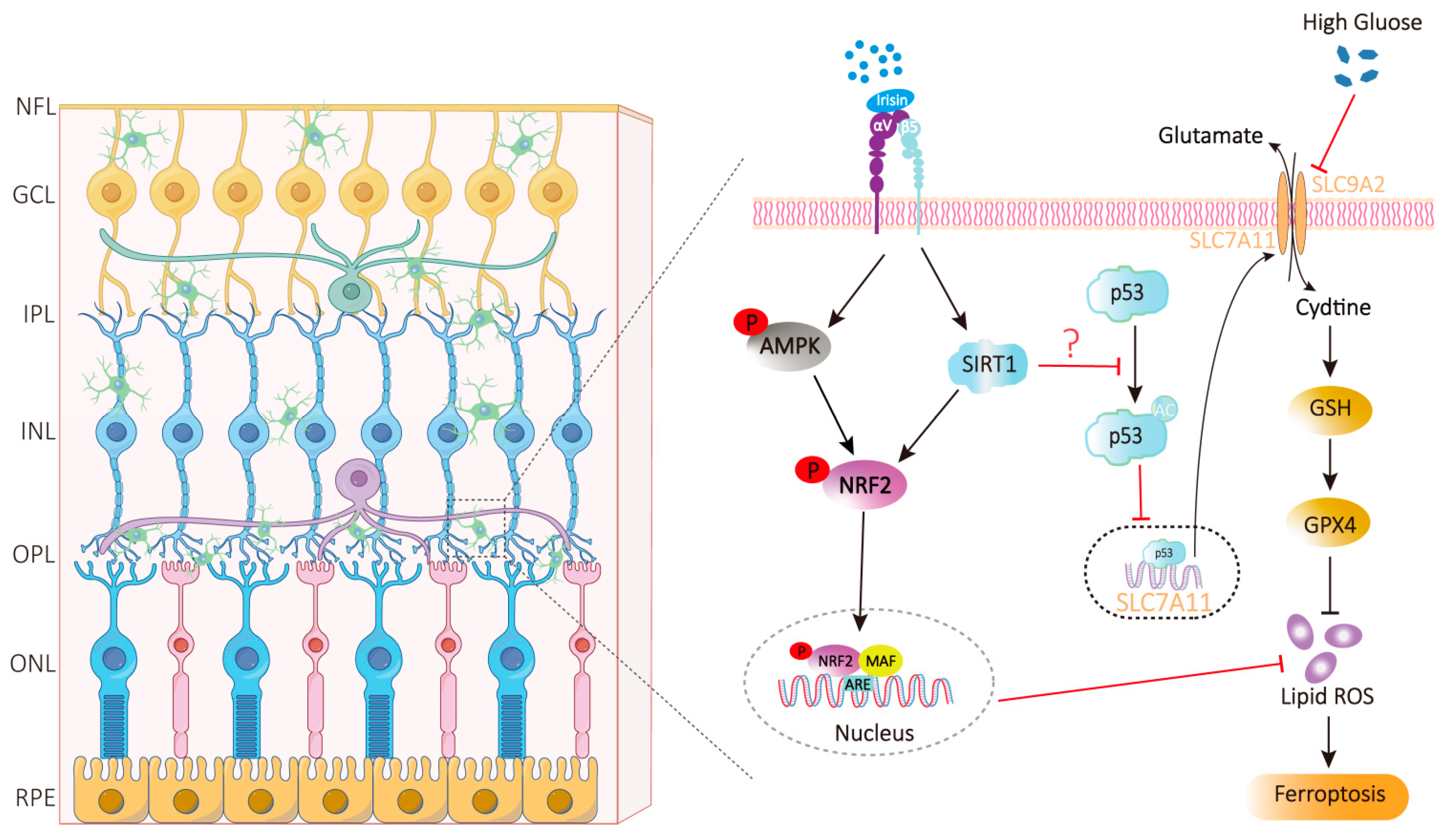
Characterized by the pathological accumulation of lipid peroxides, ferroptosis serves as a critical mechanism underlying both cell loss and vascular damage in DR. Hyperglycemia disrupts iron homeostasis in RPE cells and capillary pericytes, leading to excessive ROS generation and lipid peroxidation. In retinal endothelial cells, hyperglycemia significantly elevates levels of free iron, lipid peroxides (LPOs), and ROS, while concurrently suppressing the activity of GPX4 [109]. Irisin mainly exerts anti-ferroptosis and anti-oxidative stress effects through two core pathways (Figure 1).
Irisin inhibits lipid peroxidation by activating the AMPK/Nrf2/GPX4 signaling axis. Irisin can activate AMPK signaling molecules, thereby initiating the activation of nuclear factor E2-related factor 2 (Nrf2) [151]. Once in the nucleus, Nrf2 transcriptionally upregulates antioxidant genes such as GPX4 (a key inhibitor of ferroptosis, which can specifically reduce lipid peroxides), heme oxygenase-1 (HO-1), thereby enhancing cellular antioxidant capacity [152]. By effectively blocking lipid peroxidation, irisin acts through two complementary mechanisms: preserving reduced glutathione (GSH) homeostasis to support GPX4 activity, and modulating iron metabolism to reduce labile iron pools. This protective mechanism closely resembles that of astaxanthin in DR models, where astaxanthin attenuates retinal tissue damage by activating the Nrf2/GPX4 pathway, thereby reducing malondialdehyde (MDA) levels and enhancing GPX4 activity [153]. In models of renal and cardiac ischemia–reperfusion injury, irisin exerts protective effects that are closely associated with a marked reduction in lipid peroxide levels and upregulated expression of GPX4 and superoxide dismutase (SOD) [154,155], further supporting the robustness of this pathway in mediating irisin’s antioxidant actions.
Irisin exerts anti-ferroptotic effects primarily via the SIRT1/Nrf2 or SIRT1-p53-SLC7A11 pathway. Ferroptosis is hallmarked by aberrant expression of SLC7A11 and GPX4, concomitant with elevated MDA levels and reduced GSH content [83,156]. At present, studies have shown that irisin exerts anti-ferroptotic effects by activating the SIRT1/Nrf2 pathway [157]. The upregulation of SLC7A11 expression by irisin is mediated through two key events: the promotion of SIRT1-dependent deacetylation and the downregulation of p53 [83,158]. Through this SIRT1–p53–SLC7A11 axis, irisin upregulates SLC7A11 and GPX4, enhances antioxidant capacity, and reduces MDA levels, counteracting ferroptosis. Although this pathway has not been directly confirmed in the retina, irisin was shown in a type 1 diabetic cardiomyopathy model to reverse ferroptosis-induced increases in MDA and decreases in GSH, SLC7A11, and GPX4 [83]. Given the similar ferroptotic features in DR, this pathway likely contributes to irisin’s retinal protective effects.

4.3. Maintain BRB Integrity

Hyperglycemia can destroy BRB integrity through multiple mechanisms such as inflammatory response and ferroptosis. By concurrently targeting both anti-inflammatory and anti-ferroptotic pathways, irisin exerts protective effects on retinal pigment epithelial cells and retinal endothelial cells. This protection contributes to maintaining the tight junctions of the BRB and thereby safeguards the neurovascular units.
Irisin’s protection of the BRB against hyperglycemia-induced damage involves a coordinated network of anti-inflammatory and antioxidant mechanisms. It effectively suppresses the aberrant activation of key pro-inflammatory signaling cascades, specifically the TLR4/MyD88 [159] and NF-κB/MAPK [160] pathways, leading to a marked reduction in the secretion of inflammatory mediators such as TNF-α, IL-6, and IL-17A. In addition, given the structural and functional similarities between the BBB and the BRB, and considering that irisin has been shown to upregulate the expression of tight junction proteins, such as Claudin and Occludin [161], it is plausible that irisin similarly reinforces BRB integrity by modulating these critical junctional proteins. Irisin may also activate the Notch signaling pathway via Notch1 receptor, thereby promoting the repair of high glucose–induced damage in vascular endothelial cells [162].
Irisin may effectively inhibit high glucose-induced ferroptosis in retinal cells, thereby safeguarding both the oBRB and the iBRB. Regarding the oBRB, irisin demonstrates potent anti-ferroptotic and neuroprotective properties in neuronal HT22 cells under high-glucose conditions [163,164]. Given that RPE cells and Müller cells, akin to HT22 cells, originate from the ectoderm and exhibit similar susceptibility to oxidative stress, it is reasonable to hypothesize that irisin could mitigate high glucose or hypoxia-induced ferroptosis in RPE and Müller cells through its well-established anti-ferroptotic mechanisms. This, in turn, would preserve their vital functions, including phagocytosis, trophic support, and ion homeostasis, all of which are crucial for maintaining oBRB stability. Endothelial cells and pericytes of the retinal microvasculature are core structural components of the iBRB and are particularly vulnerable to injury under diabetic conditions. Irisin has been proven to suppress ferroptosis and enhance mitochondrial function in cardiomyocytes and hepatocytes [34,155], indicating its potential to also shield retinal vascular endothelial cells and pericytes from ferroptosis.
Clinical evidence further supports the physiological relevance of these actions. A significant decrease in serum irisin levels, which are negatively correlated with VEGF levels [165], is observed in patients with DR. These findings link low irisin levels to BRB impairment, confirming its key role in preserving barrier function and suggesting its potential involvement in the clinical pathology.

4.4. Clinical Correlation Between Irisin Levels and DR Severity

A retrospective study of 3031 adults demonstrated that individuals who maintain high levels of physical activity over the long term have a significantly lower risk of developing DR [166]; Conversely, a sedentary lifestyle markedly increases the likelihood of DR in patients with diabetes [167,168], underscoring the protective role of exercise against DR onset.
Irisin enhances glucose uptake ability in skeletal muscle and adipocytes by activating AMPK and PI3K/Akt signaling pathways, thereby improving insulin sensitivity and reducing blood glucose levels [169,170,171,172]. Studies have confirmed that circulating irisin levels in patients with T2DM are significantly lower than those in healthy controls [173]. Simple regression analysis indicates that in T2DM patients, serum irisin levels are inversely correlated with age, fasting blood glucose, insulin resistance homeostasis model assessment, blood urea nitrogen, and creatinine, and positively correlated with creatinine clearance and ACEI/ARB treatment [23]. Moreover, irisin levels decline further in T2DM patients who develop complications such as diabetic nephropathy or DR [25]. Specifically, in DR-related studies, plasma irisin levels were higher in patients without diabetic retinopathy (No DR) than in those with DR, and higher in patients with NPDR than in those with proliferative diabetic retinopathy (PDR) [174]. Consistently, irisin concentrations in both serum and vitreous humor of PDR patients were significantly lower than those in healthy individuals and in T2DM patients with no DR [23].

4.5. The Function of Irisin in Diabetic Complications

Irisin exerts a complex role and holds broad therapeutic promise in T2DM and its associated complications. Clinical studies have confirmed that circulating irisin levels in T2DM patients are lower than those in healthy individuals [23,174,175], while such levels decrease more markedly in patients with concurrent chronic complications [25].
Irisin exerts distinct protective effects against multiple diabetic complications. In diabetic cardiomyopathy, myocardial FNDC5/irisin expression and plasma irisin levels are downregulated in T2DM mouse models; exogenous irisin supplementation or FNDC5 overexpression ameliorates diastolic dysfunction, alleviates myocardial apoptosis, fibrosis, and hypertrophy, largely via activation of the integrin αV/β5-AKT pathway and attenuation of oxidative/nitrosative stress [176]. Serum irisin levels in patients with diabetic nephropathy exhibit a notable decline during the early disease stages [177]. Correspondingly, in db/db diabetic mouse models, genetically mediated upregulation or exogenous administration of irisin can significantly alleviate glomerular injury, renal tubular injury, and renal interstitial fibrosis, ameliorate renal function, and reduce proteinuria [29,177]. A hyperglycemic environment exerts prominent neurotoxic effects on the nervous system; in neuronal cells, hyperglycemia can induce ferroptosis, which is characterized by upregulated ACSL4 expression and elevated levels of iron ions and MDA, while irisin treatment effectively mitigates such neurotoxicity and ferroptosis and enhances cellular antioxidant capacity [163]. Meanwhile, at the cognitive level, hyperglycemia impairs hippocampal synaptic plasticity, whereas irisin confers protection against hyperglycemia-induced cognitive impairment by modulating hippocampal synaptic plasticity via the FNDC5/BDNF-Trk axis [178]. In the context of diabetic osteopathy, irisin treatment alleviates osteoblast ferroptosis by inhibiting the endoplasmic reticulum stress-mediated eIF2α/ATF4/CHOP signaling pathway, thereby ameliorating bone loss in type 1 diabetes mellitus (T1DM) mouse models [179]. In contrast, in T2DM mouse models, exercise intervention or exogenous irisin supplementation downregulates oxidative stress-dependent miR-150 (a microRNA that suppresses FNDC5 expression), which in turn inhibits NLRP3 inflammasome-associated pyroptosis, ultimately promoting osteogenesis and improving bone microstructure [61]. Regarding diabetic retinopathy, the negative correlation between irisin expression and disease severity has been elaborated in the preceding text. Irisin may exert a retinal protective effect to counteract the occurrence and progression of DR through anti-inflammatory, anti-angiogenic, and other pathways [24,31].
In summary, as a key molecular bridge linking exercise and health benefits, irisin plays a complex role in the pathological processes of diabetes mellitus and its complications. Existing evidence supports its multi-organ protective properties, including the amelioration of diabetic cardiomyopathy, diabetic nephropathy, diabetic osteopathy, diabetic neuropathy, and diabetic retinopathy.

4.6. Irisin in the Retina: Expression, Receptor Distribution, and Multi-Cellular Targeting Potential

The therapeutic promise of irisin in ameliorating DR pathology is closely associated with its intraretinal distribution and the functional expression of receptors on relevant target cells (Table 4). Irisin has been detected in the neural retina and the RPE [31,180], indicating that its local production could complement endocrine signaling from exercise-stimulated peripheral tissues such as skeletal muscle. Additionally, FNDC5 mRNA, which encodes the irisin precursor, is reported in aqueous humor, serum, and retinal tissue [31,181,182], supporting its probable expression within discrete retinal cell types including photoreceptors, RGCs, and Müller glial cells. This broad expression profile further substantiates the hypothesis of local irisin synthesis and establishes a foundation for exploring irisin-mediated intercompartmental signaling within the retina.
The biological functions of irisin are primarily mediated by integrin receptors, with integrin αVβ5 being the most characterized in the retina. Expression of integrin αVβ5 has been confirmed in vascular endothelial cells [54], RPE cells [55], retinal microglia [56], and astrocytes [57], providing direct evidence for irisin signaling in these cell types and suggesting its potential to modulate diverse retinal cell populations. Based on this receptor distribution, we propose that irisin exerts distinct yet overlapping regulatory effects across different retinal compartments, and that its synergistic actions may mitigate multiple pathological features of DR (Table 2). These compartment-specific effects are likely influenced by localized irisin availability and receptor expression patterns, as well as the unique functional role of each cell type, though further validation of their precise contributions in DR is warranted.
Irisin demonstrates multi-targeted neuroprotective potential in the retina, with mechanisms and contributions that are cell type-specific. At the neuronal level, irisin has been shown in central neuronal models like the hippocampus to demonstrate neuroprotective properties, promote neurogenesis, and regulate synaptic plasticity [27], supporting the likelihood of similar functions in retinal neurons. Within the vascular compartment, irisin contributes to vascular homeostasis by inhibiting endothelial inflammation and reducing oxidative stress [183], effects that help maintain vascular integrity, protect blood-brain barrier function [186], and restrain pathological neovascularization [31]. Based on these systemic actions, we hypothesize that irisin likely operates through analogous pathways in the retinal vasculature. In the glial network, irisin exerts key immunomodulatory actions, promoting a shift in microglia from the M1 to the M2 phenotype [150] and possibly modulating Müller cell activation, which collectively attenuates neuroinflammation and the release of factors such as VEGF. Together, through coordinated regulation of neuronal survival, vascular stability, and glial function, irisin establishes an integrated, multi-layered protective network in the retina, highlighting its promise as a therapeutic candidate and warranting further investigation.

4.7. Relative Contribution and Synergy of Local and Distal Irisin in DR Protection

The protective effects of irisin against DR depend on the synergistic interplay between its distally and locally produced forms. Among these, endogenously derived irisin from distal sites acts as an endocrine factor and serves as the cornerstone of systemic retinal protection, with skeletal muscle being its primary source. Exercise activates PGC-1α, which in turn upregulates FNDC5 expression and promotes irisin cleavage and subsequent release into the circulation [20]. A significant inverse correlation between serum irisin levels and DR severity [23,174] validates the clinical significance of systemic irisin. Circulating irisin can cross the BRB; it exerts anti-inflammatory and antioxidant effects to maintain BRB integrity, ameliorate the systemic pro-diabetic microenvironment, and establish a baseline protective threshold for the retina. However, reduced irisin synthesis in skeletal muscle under diabetic conditions [23,174,187] lowers this protective threshold, thereby increasing the risk of retinal damage.
Locally produced irisin acts in a paracrine/autocrine manner and functions as a precise modulator of retinal protection. Both irisin and its precursor FNDC5 are endogenously expressed in retinal neurons, vascular endothelial cells, and RPE cells [31,181,182], with their synthesis likely regulated by retinal cellular stress and metabolic status. By operating via paracrine/autocrine signaling pathways, this local irisin elicits rapid responses to local perturbations such as hyperglycemia and oxidative stress. It not only maintains retinal microenvironmental homeostasis and regulates the functions of adjacent cells, but also initiates prompt protective responses through αVβ5 integrin receptors expressed on retinal cells [54,55,56,57], thus compensating for the relatively slow, sustained signaling of circulating irisin.
These two sources of irisin complement each other and collectively mediate the protective effects against DR. Specifically, myokine-derived irisin acts as a core mediator linking exercise to retinal protection, laying a systemic foundation for defending against metabolic disorders and inflammatory injuries. In contrast, retina-derived irisin undertakes the precise regulation of the local microenvironment and maintains retinal homeostasis through regional defense and repair mechanisms.

5. Summary and Outlook

This article offers a systematic overview of the neuroprotective effects of irisin in DR and the underlying molecular mechanisms. Accumulating evidence indicates that irisin ameliorates glucose metabolism disorders by activating the AMPK/PI3K/Akt signaling pathway. Moreover, irisin may act by suppressing NF-κB activation and modulating microglia/macrophage polarization, thereby alleviating neuroinflammation and reducing vascular leakage in DR. Notably, irisin demonstrates significant anti-ferroptotic potential, likely mediated via the AMPK/Nrf2/GPX4 axis or the SIRT1p53 pathway. These mechanisms enhance cellular antioxidant defenses and restore iron metabolism homeostasis, thereby inhibiting high-glucose-induced lipid peroxidation and ferroptotic death of retinal cells, ultimately preserving BRB integrity. Clinically, both circulating and intraocular irisin levels are markedly reduced in patients with DR and exhibit a negative correlation with disease severity. This suggests that irisin may function as an endogenous protective factor involved in the pathophysiological progression of DR. However, it is important to note that clinical diagnosis of DR and the efficacy of treatment are exclusively based on the vasculature. Nevertheless, research on irisin in the context of DR remains in its early stages. A key challenge in this field lies in elucidating the differential effects of various exercise modalities on irisin secretion and defining the optimal parameters for its clinical application.
Current clinical therapies for diabetic retinopathy, including anti-VEGF agents and laser photocoagulation, primarily target late-stage vascular lesions, offer insufficient intervention against early neurodegeneration and inflammation, and are often associated with adverse effects. Consequently, their therapeutic efficacy falls short of meeting comprehensive clinical demands. Current evidence regarding irisin’s function in DR is derived primarily from animal studies and in vitro experiments. Its precise protective effects and underlying mechanisms in human diabetic retinopathy have not yet been fully elucidated. To achieve the dual goals of neuroprotection and vascular stabilization, it is essential to further investigate the multi-target effects of irisin. Such research will not only help clarify how exercise provides ocular protection but also offer critical evidence for developing future exercise-based therapeutic approaches. Therefore, it is recommended that individuals with T2DM engage in at least 150 min of moderate-intensity aerobic exercise weekly to aid in preventing or delaying the onset of DR.
To further harness the therapeutic potential of irisin in DR, future research should prioritize the following directions: First, it is essential to systematically analyze the interaction between exercise and individual baseline health status, including factors such as age, glycemic control level, the extent of basic fundus lesions, and the presence of metabolic syndrome. This analysis should clarify the differences in the responses of individuals at high risk of DR and early-stage patients with varying baseline characteristics to irisin exercise, particularly regarding potential “exercise thresholds” or differences in “response sensitivity.” Second, it is crucial to accurately define the optimal exercise parameters that promote retinal neuroprotection. This includes quantitatively grading exercise intensity, such as relative intensity based on maximum oxygen uptake (VO2max), as well as determining the duration of exercise, encompassing both single exercise sessions and the total intervention period, and identifying the differential effects of various exercise types, including endurance training, HIIT, and resistance training. It is precisely by addressing these research gaps that we can gain deeper insight into the mechanisms by which irisin regulates retinal homeostasis and establish an evidence-based clinical foundation for developing personalized exercise intervention protocols for DR. This will contribute to the creation of integrated management strategies that combine exercise with pharmacological therapy, thereby more effectively preventing and mitigating the onset and progression of DR.

Author Contributions

The conception and design of this paper were jointly completed by C.D., C.W., H.S. and W.H. H.S. was responsible for the retrieval, review, and analysis of literature and also undertook the manuscript drafting. Y.J. and S.Z. handled data collection. In addition, all authors participated in the revision and review of the paper and agreed on the final published version. All authors have read and agreed to the published version of the manuscript.

Funding

This work was supported by the National Natural Science Foundation of China (82101125) and the Health Commission Foundation of Hubei Province (WJ2021M211).

Institutional Review Board Statement

Not applicable.

Informed Consent Statement

Not applicable.

Data Availability Statement

No new data were created or analyzed in this study. Data sharing is not applicable to this article.

Conflicts of Interest

There are no competing interests among all authors.

References

  1. Cheung, N.; Mitchell, P.; Wong, T.Y. Diabetic retinopathy. Lancet 2010, 376, 124–136. [Google Scholar] [CrossRef]
  2. Sun, H.; Saeedi, P.; Karuranga, S.; Pinkepank, M.; Ogurtsova, K.; Duncan, B.B.; Stein, C.; Basit, A.; Chan, J.C.N.; Mbanya, J.C.; et al. IDF Diabetes Atlas: Global, regional and country-level diabetes prevalence estimates for 2021 and projections for 2045. Diabetes Res. Clin. Pract. 2022, 183, 109119, Erratum in Diabetes Res. Clin. Pract. 2023, 204, 110945. [Google Scholar] [CrossRef] [PubMed]
  3. Teo, Z.L.; Tham, Y.C.; Yu, M.; Chee, M.L.; Rim, T.H.; Cheung, N.; Bikbov, M.M.; Wang, Y.X.; Tang, Y.; Lu, Y.; et al. Global Prevalence of Diabetic Retinopathy and Projection of Burden through 2045: Systematic Review and Meta-analysis. Ophthalmology 2021, 128, 1580–1591. [Google Scholar] [CrossRef] [PubMed]
  4. Kiser, P.D.; Palczewski, K. Membrane-binding and enzymatic properties of RPE65. Prog. Retin. Eye Res. 2010, 29, 428–442. [Google Scholar] [CrossRef] [PubMed]
  5. Hammer, S.S.; Vieira, C.P.; McFarland, D.; Sandler, M.; Levitsky, Y.; Dorweiler, T.F.; Lydic, T.A.; Asare-Bediako, B.; Adu-Agyeiwaah, Y.; Sielski, M.S.; et al. Fasting and fasting-mimicking treatment activate SIRT1/LXRα and alleviate diabetes-induced systemic and microvascular dysfunction. Diabetologia 2021, 64, 1674–1689. [Google Scholar] [CrossRef]
  6. Barber, A.J. Diabetic retinopathy: Recent advances towards understanding neurodegeneration and vision loss. Sci. China Life Sci. 2015, 58, 541–549. [Google Scholar] [CrossRef]
  7. Barber, A.J.; Lieth, E.; Khin, S.A.; Antonetti, D.A.; Buchanan, A.G.; Gardner, T.W. Neural apoptosis in the retina during experimental and human diabetes. Early onset and effect of insulin. J. Clin. Investig. 1998, 102, 783–791. [Google Scholar] [CrossRef]
  8. Simó, R.; Stitt, A.W.; Gardner, T.W. Neurodegeneration in diabetic retinopathy: Does it really matter? Diabetologia 2018, 61, 1902–1912. [Google Scholar] [CrossRef]
  9. Zhang, C.; Xu, Y.; Tan, H.Y.; Li, S.; Wang, N.; Zhang, Y.; Feng, Y. Neuroprotective effect of He-Ying-Qing-Re formula on retinal ganglion cell in diabetic retinopathy. J. Ethnopharmacol. 2018, 214, 179–189. [Google Scholar] [CrossRef]
  10. Umino, Y.; Cuenca, N.; Everhart, D.; Fernandez-Sanchez, L.; Barlow, R.B.; Solessio, E. Partial rescue of retinal function in chronically hypoglycemic mice. Investig. Ophthalmol. Vis. Sci. 2012, 53, 915–923. [Google Scholar] [CrossRef]
  11. Jung, S.H.; Kim, Y.S.; Lee, Y.R.; Kim, J.S. High glucose-induced changes in hyaloid-retinal vessels during early ocular development of zebrafish: A short-term animal model of diabetic retinopathy. Br. J. Pharmacol. 2016, 173, 15–26. [Google Scholar] [CrossRef]
  12. Saadane, A.; Veenstra, A.A.; Minns, M.S.; Tang, J.; Du, Y.; Abubakr Elghazali, F.; Lessieur, E.M.; Pearlman, E.; Kern, T.S. CCR2-positive monocytes contribute to the pathogenesis of early diabetic retinopathy in mice. Diabetologia 2023, 66, 590–602. [Google Scholar] [CrossRef]
  13. Wong, T.Y.; Cheung, C.M.; Larsen, M.; Sharma, S.; Simó, R. Diabetic retinopathy. Nat. Rev. Dis. Primers 2016, 2, 16012. [Google Scholar] [CrossRef]
  14. Levine, S.R.; Sapieha, P.; Dutta, S.; Sun, J.K.; Gardner, T.W. It is time for a moonshot to find “Cures” for diabetic retinal disease. Prog. Retin. Eye Res. 2022, 90, 101051. [Google Scholar] [CrossRef]
  15. Li, Y.; Liu, Y.; Liu, S.; Gao, M.; Wang, W.; Chen, K.; Huang, L.; Liu, Y. Diabetic vascular diseases: Molecular mechanisms and therapeutic strategies. Signal Transduct. Target. Ther. 2023, 8, 152. [Google Scholar] [CrossRef] [PubMed]
  16. Radikova, Z.; Mosna, L.; Eckerstorfer, C.; Bajer, B.; Havranova, A.; Imrich, R.; Vlcek, M.; Penesova, A. Plasma irisin and the brain-derived neurotrophic factor levels in sedentary subjects: Effect of 8-weeks lifestyle intervention. Endocr. Regul. 2024, 58, 115–128. [Google Scholar] [CrossRef]
  17. Yano, N.; Zhao, Y.T.; Zhao, T.C. The Physiological Role of Irisin in the Regulation of Muscle Glucose Homeostasis. Endocrines 2021, 2, 266–283. [Google Scholar] [CrossRef] [PubMed]
  18. Fernández-Rodríguez, R.; Monedero-Carrasco, S.; Bizzozero-Peroni, B.; Garrido-Miguel, M.; Mesas, A.E.; Martínez-Vizcaíno, V. Effectiveness of Resistance Exercise on Inflammatory Biomarkers in Patients with Type 2 Diabetes Mellitus: A Systematic Review with Meta-Analysis. Diabetes Metab. J. 2023, 47, 118–134. [Google Scholar] [CrossRef] [PubMed]
  19. Cai, Y.; Wang, M.; Zong, Y.; Li, C.; Fu, S.; Xie, K. Demethylation of miR-299-5p by aerobic exercise relieves insulin resistance in the vascular endothelium by repressing resistin. Diabetes Res. Clin. Pract. 2023, 195, 110176. [Google Scholar] [CrossRef]
  20. Boström, P.; Wu, J.; Jedrychowski, M.P.; Korde, A.; Ye, L.; Lo, J.C.; Rasbach, K.A.; Boström, E.A.; Choi, J.H.; Long, J.Z.; et al. A PGC1-α-dependent myokine that drives brown-fat-like development of white fat and thermogenesis. Nature 2012, 481, 463–468. [Google Scholar] [CrossRef]
  21. Novelle, M.G.; Contreras, C.; Romero-Picó, A.; López, M.; Diéguez, C. Irisin, two years later. Int. J. Endocrinol. 2013, 2013, 746281. [Google Scholar] [CrossRef]
  22. Li, R.; Zheng, F.; Xu, P.; Lv, L.; Mu, Y.; Zhuang, X.; Chen, S. Correlation of mild cognitive impairment with the thickness of retinal nerve fiber layer and serum indicators in type 2 diabetic patients. Front. Endocrinol. 2023, 14, 1299206, Corrigendum in Front. Endocrinol. 2024, 15, 1380375. [Google Scholar]
  23. Hu, W.; Wang, R.; Li, J.; Zhang, J.; Wang, W. Association of irisin concentrations with the presence of diabetic nephropathy and retinopathy. Ann. Clin. Biochem. 2016, 53, 67–74. [Google Scholar] [CrossRef]
  24. Wang, C.; Wang, L.; Liu, J.; Song, J.; Sun, Y.; Lin, P.; Liang, K.; Liu, F.; He, T.; Sun, Z.; et al. Irisin modulates the association of interleukin-17A with the presence of non-proliferative diabetic retinopathy in patients with type 2 diabetes. Endocrine 2016, 53, 459–464. [Google Scholar] [CrossRef]
  25. Hou, Q.; Song, R.; Zhao, X.; Yang, C.; Feng, Y. Lower circulating irisin levels in type 2 diabetes mellitus patients with chronic complications: A meta-analysis. Heliyon 2023, 9, e21859. [Google Scholar] [CrossRef]
  26. Zhang, H.; Liang, J.; Huang, J.; Wang, M.; Wu, L.; Wu, T.; Chen, N. Exerkine irisin mitigates cognitive impairment by suppressing gut-brain axis-mediated inflammation. J. Adv. Res. 2025, 75, 843–862. [Google Scholar] [CrossRef]
  27. Lourenco, M.V.; Frozza, R.L.; de Freitas, G.B.; Zhang, H.; Kincheski, G.C.; Ribeiro, F.C.; Gonçalves, R.A.; Clarke, J.R.; Beckman, D.; Staniszewski, A.; et al. Exercise-linked FNDC5/irisin rescues synaptic plasticity and memory defects in Alzheimer’s models. Nat. Med. 2019, 25, 165–175. [Google Scholar] [CrossRef]
  28. Zhao, R.; Tian, X.; Xu, H.; Wang, Y.; Lin, J.; Wang, B. Aerobic Exercise Restores Hippocampal Neurogenesis and Cognitive Function by Decreasing Microglia Inflammasome Formation Through Irisin/NLRP3 Pathway. Aging Cell 2025, 24, e70061. [Google Scholar] [CrossRef] [PubMed]
  29. Kim, J.H.; Lee, S.; Jang, H.; Jung, S.; Jung, M.H.; Yun, J.W.; Jeon, H.; Kim, H.J.; Chang, S.H.; Lee, E.J.; et al. Transcriptional Intermediary Factor 1γ-Induced Irisin in Skeletal Muscle Attenuates Renal Fibrosis in Diabetic Nephropathy. J. Cachexia Sarcopenia Muscle 2025, 16, e13810. [Google Scholar] [CrossRef] [PubMed]
  30. Ren, Y.; Zhang, J.; Wang, M.; Bi, J.; Wang, T.; Qiu, M.; Lv, Y.; Wu, Z.; Wu, R. Identification of irisin as a therapeutic agent that inhibits oxidative stress and fibrosis in a murine model of chronic pancreatitis. Biomed. Pharmacother. 2020, 126, 110101. [Google Scholar] [CrossRef] [PubMed]
  31. Zhang, J.; Liu, Z.; Wu, H.; Chen, X.; Hu, Q.; Li, X.; Luo, L.; Ye, S.; Ye, J. Irisin Attenuates Pathological Neovascularization in Oxygen-Induced Retinopathy Mice. Investig. Ophthalmol. Vis. Sci. 2022, 63, 21. [Google Scholar] [CrossRef] [PubMed]
  32. Lourenco, M.V. Irisin limits amyloid-β buildup in Alzheimer’s disease. Trends Endocrinol. Metab. 2024, 35, 94–96. [Google Scholar] [CrossRef]
  33. Wang, Y.; Tian, M.; Tan, J.; Pei, X.; Lu, C.; Xin, Y.; Deng, S.; Zhao, F.; Gao, Y.; Gong, Y. Irisin ameliorates neuroinflammation and neuronal apoptosis through integrin αVβ5/AMPK signaling pathway after intracerebral hemorrhage in mice. J. Neuroinflamm. 2022, 19, 82. [Google Scholar] [CrossRef] [PubMed]
  34. Wei, S.; Bi, J.; Yang, L.; Zhang, J.; Wan, Y.; Chen, X.; Wang, Y.; Wu, Z.; Lv, Y.; Wu, R. Serum irisin levels are decreased in patients with sepsis, and exogenous irisin suppresses ferroptosis in the liver of septic mice. Clin. Transl. Med. 2020, 10, e173. [Google Scholar] [CrossRef] [PubMed]
  35. Kelly, D.P. Medicine. Irisin, light my fire. Science 2012, 336, 42–43. [Google Scholar] [CrossRef]
  36. Schumacher, M.A.; Chinnam, N.; Ohashi, T.; Shah, R.S.; Erickson, H.P. The structure of irisin reveals a novel intersubunit β-sheet fibronectin type III (FNIII) dimer: Implications for receptor activation. J. Biol. Chem. 2013, 288, 33738–33744. [Google Scholar] [CrossRef]
  37. Mao, M.Z.; Zheng, M.H.; Guo, B.; Ling, Y.L.; Lin, X.; Li, F.X.; Shan, S.K.; Dai, D.X.; Qiu, L.; Cai, X.Y.; et al. FNDC5/irisin-enriched sEVs conjugated with bone-targeting aptamer alleviate osteoporosis: A potential alternative to exercise. J. Nanobiotechnol. 2025, 23, 504. [Google Scholar] [CrossRef]
  38. Zhao, L.; Zhang, T.; Zhao, Z.; Ren, X.; Huang, X.; Hao, Y.; Zhou, X. Irisin improves acute kidney injury induced by ischemia-reperfusion through targeting energy metabolism reprogramming. Int. J. Biol. Macromol. 2025, 323, 147073. [Google Scholar] [CrossRef]
  39. Xiong, X.Q.; Chen, D.; Sun, H.J.; Ding, L.; Wang, J.J.; Chen, Q.; Li, Y.H.; Zhou, Y.B.; Han, Y.; Zhang, F.; et al. FNDC5 overexpression and irisin ameliorate glucose/lipid metabolic derangements and enhance lipolysis in obesity. Biochim. Biophys. Acta-Mol. Basis Dis. 2015, 1852, 1867–1875. [Google Scholar] [CrossRef]
  40. Zhao, Y.; Li, J.; Ma, A.; Wang, Z.; Ni, Y.; Wu, D.; Zhou, Y.; Zhang, N.; Zhang, L.; Chang, Y.; et al. Irisin alleviates hepatic steatosis by activating the autophagic SIRT3 pathway. Chin. Med. J. 2025, 139, 443–456. [Google Scholar] [CrossRef]
  41. Yang, J.; Zhang, P.; An, Y.; Tan, S.; Sun, S.; Liu, Y.; Zhu, L.; Wang, H.; Gao, T.; Dong, Y. Irisin regulates the phosphorylation of glucocorticoid receptor Ser212 and Ser234 and mediates glucocorticoid-induced muscle atrophy in mice. Commun. Biol. 2025, 8, 1746. [Google Scholar] [CrossRef] [PubMed]
  42. Nie, Y.; Liu, D. N-Glycosylation is required for FDNC5 stabilization and irisin secretion. Biochem. J. 2017, 474, 3167–3177. [Google Scholar] [CrossRef]
  43. Grzeszczuk, M.; Mrozowska, M.; Kmiecik, A.; Rusak, A.; Jabłońska, K.; Ciesielska, U.; Dzięgiel, P.; Nowińska, K. The Effect of Hypoxia on Irisin Expression in HL-1 Cardiomyocytes. In Vivo 2024, 38, 2126–2133. [Google Scholar] [CrossRef]
  44. Peng, Q.; Wang, X.; Wu, K.; Liu, K.; Wang, S.; Chen, X. Irisin attenuates H(2)O(2)-induced apoptosis in cardiomyocytes via microRNA-19b/AKT/mTOR signaling pathway. Int. J. Clin. Exp. Pathol. 2017, 10, 7707–7717. [Google Scholar]
  45. Perakakis, N.; Triantafyllou, G.A.; Fernández-Real, J.M.; Huh, J.Y.; Park, K.H.; Seufert, J.; Mantzoros, C.S. Physiology and role of irisin in glucose homeostasis. Nat. Rev. Endocrinol. 2017, 13, 324–337. [Google Scholar] [CrossRef]
  46. Zhang, Y.; Zhao, L.; Gao, H.; Zhai, J.; Song, Y. Potential role of irisin in digestive system diseases. Biomed. Pharmacother. 2023, 166, 115347. [Google Scholar] [CrossRef]
  47. Parr, E.B.; Camera, D.M.; Burke, L.M.; Phillips, S.M.; Coffey, V.G.; Hawley, J.A. Circulating MicroRNA Responses between ‘High’ and ‘Low’ Responders to a 16-Wk Diet and Exercise Weight Loss Intervention. PLoS ONE 2016, 11, e0152545. [Google Scholar] [CrossRef] [PubMed]
  48. Song, D.; Chen, X.; Zhou, N.; Yuan, Y.; Geng, S.; Zhang, C.; Zhao, Z.; Wang, X.; Bao, X.; Lan, X.; et al. Low-intensity pulsed ultrasound triggers a beneficial neuromodulation in dementia mice with chronic cerebral hypoperfusion via activation of hippocampal Fndc5/irisin signaling. J. Transl. Med. 2023, 21, 139. [Google Scholar] [CrossRef]
  49. Zhou, X.; Xu, M.; Bryant, J.L.; Ma, J.; Xu, X. Exercise-induced myokine FNDC5/irisin functions in cardiovascular protection and intracerebral retrieval of synaptic plasticity. Cell Biosci. 2019, 9, 32. [Google Scholar] [CrossRef]
  50. Bi, J.; Zhang, J.; Ren, Y.; Du, Z.; Li, T.; Wang, T.; Zhang, L.; Wang, M.; Wu, Z.; Lv, Y.; et al. Irisin reverses intestinal epithelial barrier dysfunction during intestinal injury via binding to the integrin αVβ5 receptor. J. Cell. Mol. Med. 2020, 24, 996–1009. [Google Scholar] [CrossRef] [PubMed]
  51. Tan, A.; Li, T.; Yang, J.; Li, X.; Li, W.; Yu, J. Irisin regulates integrin αvβ5/FAK/ERK to inhibit neutrophil extracellular traps formation and reduce pancreatic beta-cells pyroptosis in type 2 diabetes mellitus. Diabetol. Metab. Syndr. 2025, 17, 279. [Google Scholar] [CrossRef] [PubMed]
  52. Kumar, M.; Sengar, A.S.; Lye, A.; Kumar, P.; Mukherjee, S.; Kumar, D.; Das, P.; Chatterjee, S.; Stewart, A.; Maity, B. FNDC5/irisin mitigates the cardiotoxic impacts of cancer chemotherapeutics by modulating ROS-dependent and -independent mechanisms. Redox Biol. 2025, 80, 103527, Corrigendum in Redox Biol. 2025, 85, 103734. [Google Scholar] [CrossRef]
  53. Mu, A.; Wales, T.E.; Zhou, H.; Draga-Coletă, S.V.; Gorgulla, C.; Blackmore, K.A.; Mittenbühler, M.J.; Kim, C.R.; Bogoslavski, D.; Zhang, Q.; et al. Irisin acts through its integrin receptor in a two-step process involving extracellular Hsp90α. Mol. Cell 2023, 83, 1903–1920.e1912. [Google Scholar] [CrossRef]
  54. Bi, J.; Zhang, J.; Ren, Y.; Du, Z.; Zhang, Y.; Liu, C.; Wang, Y.; Zhang, L.; Shi, Z.; Wu, Z.; et al. Exercise hormone irisin mitigates endothelial barrier dysfunction and microvascular leakage-related diseases. JCI Insight 2020, 5, e136277. [Google Scholar] [CrossRef]
  55. Nandrot, E.F.; Kim, Y.; Brodie, S.E.; Huang, X.; Sheppard, D.; Finnemann, S.C. Loss of synchronized retinal phagocytosis and age-related blindness in mice lacking alphavbeta5 integrin. J. Exp. Med. 2004, 200, 1539–1545. [Google Scholar] [CrossRef] [PubMed]
  56. Zhang, Q.; Xiang, S.; Chen, X.; Rong, Y.; Huang, L.; Chen, Z.; Yao, K.; Chen, W.; Deng, C.; Wang, J. Irisin attenuates acute glaucoma-induced neuroinflammation by activating microglia-integrin αVβ5/AMPK and promoting autophagy. Int. Immunopharmacol. 2024, 138, 112545. [Google Scholar] [CrossRef]
  57. Yun, J.H.; Park, S.W.; Kim, J.H.; Park, Y.J.; Cho, C.H.; Kim, J.H. Angiopoietin 2 induces astrocyte apoptosis via αvβ5-integrin signaling in diabetic retinopathy. Cell Death Dis. 2016, 7, e2101. [Google Scholar] [CrossRef]
  58. Dae-Eun, K.; Il-Young, P.; Jae-Hoon, R.; Tae-Hyung, L.; Kyung-Su, C. The Changes in Plasma Irisin Levels During Recovery and Immediately After Triathlon According to Course Types. J. Sport Leis. Stud. 2019, 78, 453–459. [Google Scholar] [CrossRef]
  59. Zhou, W.; Shi, Y.; Wang, H.; Chen, L.; Yu, C.; Zhang, X.; Yang, L.; Zhang, X.; Wu, A. Exercise-induced FNDC5/irisin protects nucleus pulposus cells against senescence and apoptosis by activating autophagy. Exp. Mol. Med. 2022, 54, 1038–1048. [Google Scholar] [CrossRef] [PubMed]
  60. Wu, F.; Li, Z.; Cai, M.; Xi, Y.; Xu, Z.; Zhang, Z.; Li, H.; Zhu, W.; Tian, Z. Aerobic exercise alleviates oxidative stress-induced apoptosis in kidneys of myocardial infarction mice by inhibiting ALCAT1 and activating FNDC5/Irisin signaling pathway. Free Radic. Biol. Med. 2020, 158, 171–180. [Google Scholar] [CrossRef]
  61. Behera, J.; Ison, J.; Voor, M.J.; Tyagi, N. Exercise-Linked Skeletal Irisin Ameliorates Diabetes-Associated Osteoporosis by Inhibiting the Oxidative Damage-Dependent miR-150-FNDC5/Pyroptosis Axis. Diabetes 2022, 71, 2777–2792. [Google Scholar] [CrossRef] [PubMed]
  62. Arabzadeh, E.; Shirvani, H.; Ebadi Zahmatkesh, M.; Riyahi Malayeri, S.; Meftahi, G.H.; Rostamkhani, F. Irisin/FNDC5 influences myogenic markers on skeletal muscle following high and moderate-intensity exercise training in STZ-diabetic rats. 3 Biotech 2022, 12, 193. [Google Scholar] [CrossRef] [PubMed]
  63. Pekkala, S.; Wiklund, P.K.; Hulmi, J.J.; Ahtiainen, J.P.; Horttanainen, M.; Pöllänen, E.; Mäkelä, K.A.; Kainulainen, H.; Häkkinen, K.; Nyman, K.; et al. Are skeletal muscle FNDC5 gene expression and irisin release regulated by exercise and related to health? J. Physiol. 2013, 591, 5393–5400. [Google Scholar] [CrossRef]
  64. Miyamoto-Mikami, E.; Sato, K.; Kurihara, T.; Hasegawa, N.; Fujie, S.; Fujita, S.; Sanada, K.; Hamaoka, T.; Tabata, I.; Iemitsu, M. Endurance training-induced increase in circulating irisin levels is associated with reduction of abdominal visceral fat in middle-aged and older adults. PLoS ONE 2015, 10, e0120354. [Google Scholar] [CrossRef] [PubMed]
  65. He, Z.; Tian, Y.; Valenzuela, P.L.; Huang, C.; Zhao, J.; Hong, P.; He, Z.; Yin, S.; Lucia, A. Myokine Response to High-Intensity Interval vs. Resistance Exercise: An Individual Approach. Front. Physiol. 2018, 9, 1735. [Google Scholar] [CrossRef]
  66. Huh, J.Y.; Panagiotou, G.; Mougios, V.; Brinkoetter, M.; Vamvini, M.T.; Schneider, B.E.; Mantzoros, C.S. FNDC5 and irisin in humans: I. Predictors of circulating concentrations in serum and plasma and II. mRNA expression and circulating concentrations in response to weight loss and exercise. Metab. Clin. Exp. 2012, 61, 1725–1738. [Google Scholar] [CrossRef]
  67. Brenmoehl, J.; Albrecht, E.; Komolka, K.; Schering, L.; Langhammer, M.; Hoeflich, A.; Maak, S. Irisin is elevated in skeletal muscle and serum of mice immediately after acute exercise. Int. J. Biol. Sci. 2014, 10, 338–349. [Google Scholar] [CrossRef]
  68. Qiu, S.; Bosnyák, E.; Treff, G.; Steinacker, J.M.; Nieß, A.M.; Krüger, K.; Mooren, F.C.; Zügel, M.; Schumann, U. Acute exercise-induced irisin release in healthy adults: Associations with training status and exercise mode. Eur. J. Sport Sci. 2018, 18, 1226–1233. [Google Scholar] [CrossRef]
  69. Eskandari, M.; Hooshmand Moghadam, B.; Bagheri, R.; Ashtary-Larky, D.; Eskandari, E.; Nordvall, M.; Dutheil, F.; Wong, A. Effects of Interval Jump Rope Exercise Combined with Dark Chocolate Supplementation on Inflammatory Adipokine, Cytokine Concentrations, and Body Composition in Obese Adolescent Boys. Nutrients 2020, 12, 3011. [Google Scholar] [CrossRef]
  70. Colaianni, G.; Notarnicola, A.; Sanesi, L.; Brunetti, G.; Lippo, L.; Celi, M.; Moretti, L.; Pesce, V.; Vicenti, G.; Moretti, B.; et al. Irisin levels correlate with bone mineral density in soccer players. J. Biol. Regul. Homeost. Agents 2017, 31, 21–28. [Google Scholar]
  71. Ozcelik, O.; Algul, S.; Yilmaz, B. Nesfatin-1 and irisin levels in response to the soccer matches performed in morning, afternoon and at night in young trained male subjects. Cell. Mol. Biol. 2018, 64, 130–133. [Google Scholar] [CrossRef]
  72. Wiecek, M.; Szymura, J.; Maciejczyk, M.; Kantorowicz, M.; Szygula, Z. Acute Anaerobic Exercise Affects the Secretion of Asprosin, Irisin, and Other Cytokines—A Comparison Between Sexes. Front. Physiol. 2018, 9, 1782. [Google Scholar] [CrossRef]
  73. Eaton, M.; Granata, C.; Barry, J.; Safdar, A.; Bishop, D.; Little, J.P. Impact of a single bout of high-intensity interval exercise and short-term interval training on interleukin-6, FNDC5, and METRNL mRNA expression in human skeletal muscle. J. Sport Health Sci. 2018, 7, 191–196. [Google Scholar] [CrossRef]
  74. Archundia-Herrera, C.; Macias-Cervantes, M.; Ruiz-Muñoz, B.; Vargas-Ortiz, K.; Kornhauser, C.; Perez-Vazquez, V. Muscle irisin response to aerobic vs. HIIT in overweight female adolescents. Diabetol. Metab. Syndr. 2017, 9, 101. [Google Scholar] [CrossRef]
  75. Tsuchiya, Y.; Ando, D.; Takamatsu, K.; Goto, K. Resistance exercise induces a greater irisin response than endurance exercise. Metab. Clin. Exp. 2015, 64, 1042–1050. [Google Scholar] [CrossRef]
  76. Bang, H.S.; Seo, D.Y.; Chung, Y.M.; Oh, K.M.; Park, J.J.; Arturo, F.; Jeong, S.H.; Kim, N.; Han, J. Ursolic Acid-induced elevation of serum irisin augments muscle strength during resistance training in men. Korean J. Physiol. Pharmacol. 2014, 18, 441–446, Corrigendum in Korean J. Physiol. Pharmacol. 2014, 18, 531. [Google Scholar] [CrossRef]
  77. Zhao, J.; Su, Z.; Qu, C.; Dong, Y. Effects of 12 Weeks Resistance Training on Serum Irisin in Older Male Adults. Front. Physiol. 2017, 8, 171. [Google Scholar] [CrossRef]
  78. Kim, H.J.; So, B.; Choi, M.; Kang, D.; Song, W. Resistance exercise training increases the expression of irisin concomitant with improvement of muscle function in aging mice and humans. Exp. Gerontol. 2015, 70, 11–17. [Google Scholar] [CrossRef]
  79. Nygaard, H.; Slettaløkken, G.; Vegge, G.; Hollan, I.; Whist, J.E.; Strand, T.; Rønnestad, B.R.; Ellefsen, S. Irisin in blood increases transiently after single sessions of intense endurance exercise and heavy strength training. PLoS ONE 2015, 10, e0121367. [Google Scholar] [CrossRef] [PubMed]
  80. Larissa, F.; Marcos Antônio, B.; Gabriela, N.; Lucélia Scarabeli Silva, B.; Heliana, B.F.; Antonio Felipe, S.-G.; Aline Silva, M.; Albená, N.-S. Effects of a single strength training session on the peripheral concentrations of irisin in trained men. Res. Sq. 2023; preprint. [Google Scholar]
  81. Junsoo, L.; Hee-Cheol, K.; Sun-Young, J.; Dae-Young, K. Effects of high-intensity circuit training on muscle strength, irisin, FGF-21, and adiponectin in obese female college students. In Proceedings of the Korean Society of Exercise Rehabilitation Academic Conference, Busan, Republic of Korea, 17 May 2019. [Google Scholar]
  82. Mazur-Bialy, A.I.; Pocheć, E. The Time-Course of Antioxidant Irisin Activity: Role of the Nrf2/HO-1/HMGB1 Axis. Antioxidants 2021, 10, 88. [Google Scholar] [CrossRef]
  83. Tang, Y.J.; Zhang, Z.; Yan, T.; Chen, K.; Xu, G.F.; Xiong, S.Q.; Wu, D.Q.; Chen, J.; Jose, P.A.; Zeng, C.Y.; et al. Irisin attenuates type 1 diabetic cardiomyopathy by anti-ferroptosis via SIRT1-mediated deacetylation of p53. Cardiovasc. Diabetol. 2024, 23, 116. [Google Scholar] [CrossRef]
  84. Li, M.; Trapika, I.; Tang, S.Y.S.; Cho, J.L.; Qi, Y.; Li, C.G.; Li, Y.; Yao, M.; Yang, D.; Liu, B.; et al. Mechanisms and Active Compounds Polysaccharides and Bibenzyls of Medicinal Dendrobiums for Diabetes Management. Front. Nutr. 2021, 8, 811870. [Google Scholar] [CrossRef] [PubMed]
  85. Yang, X.; Yao, W.; Liu, H.; Gao, Y.; Liu, R.; Xu, L. Tangluoning, a traditional Chinese medicine, attenuates in vivo and in vitro diabetic peripheral neuropathy through modulation of PERK/Nrf2 pathway. Sci. Rep. 2017, 7, 1014. [Google Scholar] [CrossRef]
  86. Wang, Y.H.; Liu, Y.H.; He, G.R.; Lv, Y.; Du, G.H. Esculin improves dyslipidemia, inflammation and renal damage in streptozotocin-induced diabetic rats. BMC Complement. Altern. Med. 2015, 15, 402. [Google Scholar] [CrossRef] [PubMed]
  87. Rui, W.; Xingchen, W.; Jinmei, O. Network pharmacology-based approach reveals that Fructus mume exerts therapeutic effects against ulcerative colitis via the AGE/RAGE signaling pathway. Arab. J. Chem. 2023, 17, 105534. [Google Scholar]
  88. Tang, Y.; Hu, H.; Xie, Q.; Shen, J. GAS6/AXL signaling promotes M2 microglia efferocytosis to alleviate neuroinflammation in sepsis-associated encephalopathy. Cell Death Discov. 2025, 11, 268, Correction in Cell Death Discov. 2025, 11, 531. [Google Scholar] [CrossRef] [PubMed]
  89. Kim, J.H.; Kim, J.H.; Kim, D.H.; Park, W.Y.; Kim, K.W.; Yu, Y.S. Intravenously administered anti-recoverin antibody alone does not pass through the blood-retinal barrier. Korean J. Ophthalmol. 2011, 25, 189–195. [Google Scholar] [CrossRef]
  90. Rohowetz, L.J.; Kraus, J.G.; Koulen, P. Reactive Oxygen Species-Mediated Damage of Retinal Neurons: Drug Development Targets for Therapies of Chronic Neurodegeneration of the Retina. Int. J. Mol. Sci. 2018, 19, 3362. [Google Scholar] [CrossRef]
  91. Ye, E.A.; Liu, L.; Steinle, J.J. miR-15a/16 inhibits TGF-beta3/VEGF signaling and increases retinal endothelial cell barrier proteins. Vis. Res. 2017, 139, 23–29. [Google Scholar] [CrossRef]
  92. Yin, R.; Zhang, N.; Zhang, D.; Zhao, W.; Ke, J.; Zhao, D. Higher levels of circulating ANGPTL2 are associated with macular edema in patients with type 2 diabetes. Medicine 2021, 100, e24638. [Google Scholar] [CrossRef]
  93. Koya, D.; King, G.L. Protein kinase C activation and the development of diabetic complications. Diabetes 1998, 47, 859–866. [Google Scholar] [CrossRef]
  94. Bolinger, M.T.; Antonetti, D.A. Moving Past Anti-VEGF: Novel Therapies for Treating Diabetic Retinopathy. Int. J. Mol. Sci. 2016, 17, 3362. [Google Scholar] [CrossRef]
  95. Dammak, A.; Huete-Toral, F.; Carpena-Torres, C.; Martin-Gil, A.; Pastrana, C.; Carracedo, G. From Oxidative Stress to Inflammation in the Posterior Ocular Diseases: Diagnosis and Treatment. Pharmaceutics 2021, 13, 1376. [Google Scholar] [CrossRef] [PubMed]
  96. Madhusudhan, S.; Gupta, N.V.; Rahamathulla, M.; Chidambaram, S.B.; Osmani, R.A.M.; Ghazwani, M.; Ahmed, M.M.; Farhana, S.A.; Sarhan, M.Y.; Tousif, A.H. Subconjunctival Delivery of Sorafenib-Tosylate-Loaded Cubosomes for Facilitated Diabetic Retinopathy Treatment: Formulation Development, Evaluation, Pharmacokinetic and Pharmacodynamic (PKPD) Studies. Pharmaceutics 2023, 15, 2419. [Google Scholar] [CrossRef] [PubMed]
  97. Yuan, Z.; Yi, Y.; Jiawen, Z.; Lei, L.; Danyang, L.; Jiawei, H.; Yiming, G.; Wei, L.; Ning, L.; Liting, J. Characterisation of macrophage infiltration and polarisation based on integrated transcriptomic and histological analyses in Primary Sjögren’s syndrome. Front. Immunol. 2023, 14, 1292146. [Google Scholar] [CrossRef]
  98. Eshaq, R.S.; Wright, W.S.; Harris, N.R. Oxygen delivery, consumption, and conversion to reactive oxygen species in experimental models of diabetic retinopathy. Redox Biol. 2014, 2, 661–666. [Google Scholar] [CrossRef]
  99. Du, Y.; Veenstra, A.; Palczewski, K.; Kern, T.S. Photoreceptor cells are major contributors to diabetes-induced oxidative stress and local inflammation in the retina. Proc. Natl. Acad. Sci. USA 2013, 110, 16586–16591. [Google Scholar] [CrossRef] [PubMed]
  100. Kang, Q.; Yang, C. Oxidative stress and diabetic retinopathy: Molecular mechanisms, pathogenetic role and therapeutic implications. Redox Biol. 2020, 37, 101799. [Google Scholar] [CrossRef]
  101. Wu, M.Y.; Yiang, G.T.; Lai, T.T.; Li, C.J. The Oxidative Stress and Mitochondrial Dysfunction during the Pathogenesis of Diabetic Retinopathy. Oxidative Med. Cell. Longev. 2018, 2018, 3420187. [Google Scholar] [CrossRef]
  102. Mahajan, N.; Arora, P.; Sandhir, R. Perturbed Biochemical Pathways and Associated Oxidative Stress Lead to Vascular Dysfunctions in Diabetic Retinopathy. Oxidative Med. Cell. Longev. 2019, 2019, 8458472. [Google Scholar] [CrossRef]
  103. Kang, Y.L.; Kim, J.; Kwak, S.B.; Kim, Y.S.; Huh, J.; Park, J.W. The polyol pathway and nuclear ketohexokinase A signaling drive hyperglycemia-induced metastasis of gastric cancer. Exp. Mol. Med. 2024, 56, 220–234. [Google Scholar] [CrossRef]
  104. Schwab, A.; Siddiqui, M.A.; Ramesh, V.; Gollavilli, P.N.; Turtos, A.M.; Møller, S.S.; Pinna, L.; Havelund, J.F.; Rømer, A.M.A.; Ersan, P.G.; et al. Polyol pathway-generated fructose is indispensable for growth and survival of non-small cell lung cancer. Cell Death Differ. 2025, 32, 587–597. [Google Scholar] [CrossRef]
  105. Yuan, T.; Yang, T.; Chen, H.; Fu, D.; Hu, Y.; Wang, J.; Yuan, Q.; Yu, H.; Xu, W.; Xie, X. New insights into oxidative stress and inflammation during diabetes mellitus-accelerated atherosclerosis. Redox Biol. 2019, 20, 247–260. [Google Scholar] [CrossRef]
  106. Mrugacz, M.; Pony-Uram, M.; Bryl, A.; Zorena, K. Current Approach to the Pathogenesis of Diabetic Cataracts. Int. J. Mol. Sci. 2023, 24, 6317. [Google Scholar] [CrossRef] [PubMed]
  107. Yang, J.; Liu, Z. Mechanistic Pathogenesis of Endothelial Dysfunction in Diabetic Nephropathy and Retinopathy. Front. Endocrinol. 2022, 13, 816400. [Google Scholar] [CrossRef]
  108. Chen, Z.; Liu, B.; Zhou, D.; Lei, M.; Yang, J.; Hu, Z.; Duan, W. AQP4 regulates ferroptosis and oxidative stress of Muller cells in diabetic retinopathy by regulating TRPV4. Exp. Cell Res. 2024, 439, 114087. [Google Scholar] [CrossRef] [PubMed]
  109. Malaviya, P.; Kumar, J.; Kowluru, R.A. Role of ferroptosis in mitochondrial damage in diabetic retinopathy. Free Radic. Biol. Med. 2024, 225, 821–832. [Google Scholar] [CrossRef]
  110. Fan, X.; Xu, M.; Wang, Z.; Sun, X.; Fan, Y.; Chen, J.; Hao, J.; Wang, R.; Jia, W. Arctiin suppress Th17 cells response and ameliorates experimental autoimmune uveitis through JAK/STAT signaling. Cell. Immunol. 2025, 409–410, 104927. [Google Scholar] [CrossRef] [PubMed]
  111. Jiang, Y.; Cao, Y.; Li, Y.; Bi, L.; Wang, L.; Chen, Q.; Lin, Y.; Jin, H.; Xu, X.; Peng, R.; et al. SNP alleviates mitochondrial homeostasis dysregulation-mediated developmental toxicity in diabetic zebrafish larvae. Biomed. Pharmacother. 2024, 177, 117117. [Google Scholar] [CrossRef]
  112. Ramos, H.; Bogdanov, P.; Simó, R.; Deàs-Just, A.; Hernández, C. Transcriptomic Analysis Reveals That Retinal Neuromodulation Is a Relevant Mechanism in the Neuroprotective Effect of Sitagliptin in an Experimental Model of Diabetic Retinopathy. Int. J. Mol. Sci. 2022, 24, 571. [Google Scholar] [CrossRef] [PubMed]
  113. Rudraraju, M.; Narayanan, S.P.; Somanath, P.R. Regulation of blood-retinal barrier cell-junctions in diabetic retinopathy. Pharmacol. Res. 2020, 161, 105115. [Google Scholar] [CrossRef]
  114. Jadeja, R.N.; Jones, M.A.; Abdelrahman, A.A.; Powell, F.L.; Thounaojam, M.C.; Gutsaeva, D.; Bartoli, M.; Martin, P.M. Inhibiting microRNA-144 potentiates Nrf2-dependent antioxidant signaling in RPE and protects against oxidative stress-induced outer retinal degeneration. Redox Biol. 2020, 28, 101336. [Google Scholar] [CrossRef]
  115. Hosoya, K.; Tachikawa, M. The inner blood-retinal barrier: Molecular structure and transport biology. Adv. Exp. Med. Biol. 2012, 763, 85–104. [Google Scholar]
  116. Liu, L.; Liu, X. Roles of Drug Transporters in Blood-Retinal Barrier. Adv. Exp. Med. Biol. 2019, 1141, 467–504. [Google Scholar]
  117. Bădulescu, O.; Bădescu, C.; Ciocoiu, M.; Bădescu, M. Interleukin-1-beta and dyslipidemic syndrome as major risk factors for thrombotic complications in type 2 diabetes mellitus. Mediat. Inflamm. 2013, 2013, 169420. [Google Scholar] [CrossRef]
  118. Seo, H.; Chung, W.G.; Kwon, Y.W.; Kim, S.; Hong, Y.M.; Park, W.; Kim, E.; Lee, J.; Lee, S.; Kim, M.; et al. Smart Contact Lenses as Wearable Ophthalmic Devices for Disease Monitoring and Health Management. Chem. Rev. 2023, 123, 11488–11558. [Google Scholar] [CrossRef]
  119. Wang, W.; Lo, A.C.Y. Diabetic Retinopathy: Pathophysiology and Treatments. Int. J. Mol. Sci. 2018, 19, 1816. [Google Scholar] [CrossRef]
  120. Giebel, S.J.; Menicucci, G.; McGuire, P.G.; Das, A. Matrix metalloproteinases in early diabetic retinopathy and their role in alteration of the blood-retinal barrier. Lab. Investig. A J. Tech. Methods Pathol. 2005, 85, 597–607. [Google Scholar] [CrossRef] [PubMed]
  121. Janine, Y.; David, G.; Lucia, S. Interleukin-6 and Macular Edema: A Review of Outcomes with Inhibition. Int. J. Mol. Sci. 2023, 24, 4676. [Google Scholar] [CrossRef] [PubMed]
  122. Xiang, X.H.; Wei, J.; Wang, X.F.; Xu, Q.; Yu, C.L.; He, C.L.; Long, T.; Guo, M.S.; Chen, X.; Zhou, X.G.; et al. Lychee seed polyphenol ameliorates DR via inhibiting inflammasome/apoptosis and angiogenesis in hRECs and db/db mice. Biomed. Pharmacother. 2023, 167, 115478. [Google Scholar] [CrossRef] [PubMed]
  123. Li, X.M.; Zhao, Z.Y.; Yu, X.; Xia, Q.D.; Zhou, P.; Wang, S.G.; Wu, H.L.; Hu, J. Exploiting E3 ubiquitin ligases to reeducate the tumor microenvironment for cancer therapy. Exp. Hematol. Oncol. 2023, 12, 34. [Google Scholar] [CrossRef] [PubMed]
  124. Bronowicka-Szydełko, A.; Gostomska-Pampuch, K.; Kuzan, A.; Pietkiewicz, J.; Krzystek-Korpacka, M.; Gamian, A. Effect of advanced glycation end-products in a wide range of medical problems including COVID-19. Adv. Med. Sci. 2024, 69, 36–50. [Google Scholar] [CrossRef]
  125. Takamura, Y.; Yamada, Y.; Inatani, M. Role of Microaneurysms in the Pathogenesis and Therapy of Diabetic Macular Edema: A Descriptive Review. Medicina 2023, 59, 435. [Google Scholar] [CrossRef] [PubMed]
  126. Fanaro, G.B.; Marques, M.R.; Calaza, K.D.C.; Brito, R.; Pessoni, A.M.; Mendonça, H.R.; Lemos, D.E.A.; de Brito Alves, J.L.; de Souza, E.L.; Cavalcanti Neto, M.P. New Insights on Dietary Polyphenols for the Management of Oxidative Stress and Neuroinflammation in Diabetic Retinopathy. Antioxidants 2023, 12, 1237. [Google Scholar] [CrossRef]
  127. D’Amico, A.G.; Maugeri, G.; Magrì, B.; Bucolo, C.; D’Agata, V. Targeting the PINK1/Parkin pathway: A new perspective in the prevention and therapy of diabetic retinopathy. Exp. Eye Res. 2024, 247, 110024. [Google Scholar] [CrossRef]
  128. Fields, M.A.; Del Priore, L.V.; Adelman, R.A.; Rizzolo, L.J. Interactions of the choroid, Bruch’s membrane, retinal pigment epithelium, and neurosensory retina collaborate to form the outer blood-retinal-barrier. Prog. Retin. Eye Res. 2020, 76, 100803. [Google Scholar] [CrossRef]
  129. Song, M.J.; Quinn, R.; Nguyen, E.; Hampton, C.; Sharma, R.; Park, T.S.; Koster, C.; Voss, T.; Tristan, C.; Weber, C.; et al. Bioprinted 3D outer retina barrier uncovers RPE-dependent choroidal phenotype in advanced macular degeneration. Nat. Methods 2023, 20, 149–161. [Google Scholar] [CrossRef]
  130. Houssier, M.; Raoul, W.; Lavalette, S.; Keller, N.; Guillonneau, X.; Baragatti, B.; Jonet, L.; Jeanny, J.C.; Behar-Cohen, F.; Coceani, F.; et al. CD36 deficiency leads to choroidal involution via COX2 down-regulation in rodents. PLoS Med. 2008, 5, e39. [Google Scholar] [CrossRef]
  131. Liu, F.; Xu, T.; Peng, S.; Adelman, R.A.; Rizzolo, L.J. Claudins regulate gene and protein expression of the retinal pigment epithelium independent of their association with tight junctions. Exp. Eye Res. 2020, 198, 108157. [Google Scholar] [CrossRef]
  132. Obert, E.; Strauss, R.; Brandon, C.; Grek, C.; Ghatnekar, G.; Gourdie, R.; Rohrer, B. Targeting the tight junction protein, zonula occludens-1, with the connexin43 mimetic peptide, αCT1, reduces VEGF-dependent RPE pathophysiology. J. Mol. Med. 2017, 95, 535–552. [Google Scholar] [CrossRef]
  133. Wang, D.; Chen, Y.; Li, J.; Wu, E.; Tang, T.; Singla, R.K.; Shen, B.; Zhang, M. Natural products for the treatment of age-related macular degeneration. Phytomedicine Int. J. Phytother. Phytopharm. 2024, 130, 155522. [Google Scholar] [CrossRef]
  134. Kang, M.K.; Lee, E.J.; Kim, Y.H.; Kim, D.Y.; Oh, H.; Kim, S.I.; Kang, Y.H. Chrysin Ameliorates Malfunction of Retinoid Visual Cycle through Blocking Activation of AGE-RAGE-ER Stress in Glucose-Stimulated Retinal Pigment Epithelial Cells and Diabetic Eyes. Nutrients 2018, 10, 1046. [Google Scholar] [CrossRef]
  135. Wittig, D.; Wang, X.; Walter, C.; Gerdes, H.H.; Funk, R.H.; Roehlecke, C. Multi-level communication of human retinal pigment epithelial cells via tunneling nanotubes. PLoS ONE 2012, 7, e33195. [Google Scholar] [CrossRef] [PubMed]
  136. Chucair-Elliott, A.J.; Ocañas, S.R.; Pham, K.; Machalinski, A.; Plafker, S.; Stout, M.B.; Elliott, M.H.; Freeman, W.M. Age- and sex- divergent translatomic responses of the mouse retinal pigmented epithelium. Neurobiol. Aging 2024, 140, 41–59. [Google Scholar] [CrossRef] [PubMed]
  137. Leuenberger, P.M.; Novikoff, A.B. Studies on microperoxisomes. VII. Pigment epithelial cells and other cell types in the retina of rodents. J. Cell Biol. 1975, 65, 324–334. [Google Scholar] [CrossRef]
  138. Kota, S.K.; Meher, L.K.; Jammula, S.; Kota, S.K.; Krishna, S.V.; Modi, K.D. Aberrant angiogenesis: The gateway to diabetic complications. Indian J. Endocrinol. Metab. 2012, 16, 918–930. [Google Scholar] [CrossRef]
  139. Lechner, J.; Medina, R.J.; Lois, N.; Stitt, A.W. Advances in cell therapies using stem cells/progenitors as a novel approach for neurovascular repair of the diabetic retina. Stem Cell Res. Ther. 2022, 13, 388. [Google Scholar] [CrossRef]
  140. Yu, H.; Wark, L.; Ji, H.; Willard, L.; Jaing, Y.; Han, J.; He, H.; Ortiz, E.; Zhang, Y.; Medeiros, D.M.; et al. Dietary wolfberry upregulates carotenoid metabolic genes and enhances mitochondrial biogenesis in the retina of db/db diabetic mice. Mol. Nutr. Food Res. 2013, 57, 1158–1169. [Google Scholar] [CrossRef]
  141. Chong, R.S.; Martin, K.R. Glial cell interactions and glaucoma. Curr. Opin. Ophthalmol. 2015, 26, 73–77, Erratum in Curr. Opin. Ophthalmol. 2015, 26, 553. https://doi.org/10.1097/ICU.0000000000000222. [Google Scholar] [CrossRef]
  142. Reichenbach, A.; Bringmann, A. New functions of Müller cells. Glia 2013, 61, 651–678. [Google Scholar] [CrossRef] [PubMed]
  143. Whitehead, M.; Wickremasinghe, S.; Osborne, A.; Van Wijngaarden, P.; Martin, K.R. Diabetic retinopathy: A complex pathophysiology requiring novel therapeutic strategies. Expert Opin. Biol. Ther. 2018, 18, 1257–1270. [Google Scholar] [CrossRef]
  144. Xu, Y.; Peng, Y.; Wu, X.; Ni, F.; Sun, D.; Zhang, P.; Yang, Y.; Yan, M.; Mi, J.; Tian, G. VEGF-B prevents chronic hyperglycemia-induced retinal vascular leakage by regulating the CDC42-ZO1/VE-cadherin pathway. FASEB J. 2024, 38, e70019. [Google Scholar] [CrossRef]
  145. Song, P.; Fogerty, J.; Cianciolo, L.T.; Stupay, R.; Perkins, B.D. Cone Photoreceptor Degeneration and Neuroinflammation in the Zebrafish Bardet-Biedl Syndrome 2 (bbs2) Mutant Does Not Lead to Retinal Regeneration. Front. Cell Dev. Biol. 2020, 8, 578528. [Google Scholar] [CrossRef]
  146. Zheng, S.; Chen, N.; Kang, X.; Hu, Y.; Shi, S. Irisin alleviates FFA induced β-cell insulin resistance and inflammatory response through activating PI3K/AKT/FOXO1 signaling pathway. Endocrine 2022, 75, 740–751. [Google Scholar] [CrossRef]
  147. Mazur-Bialy, A.I.; Kozlowska, K.; Pochec, E.; Bilski, J.; Brzozowski, T. Myokine irisin-induced protection against oxidative stress in vitro. Involvement of heme oxygenase-1 and antioxidazing enzymes superoxide dismutase-2 and glutathione peroxidase. J. Physiol. Pharmacol. Off. J. Pol. Physiol. Soc. 2018, 69, 117–125. [Google Scholar]
  148. Zhu, S.H.; Liu, B.Q.; Hao, M.J.; Fan, Y.X.; Qian, C.; Teng, P.; Zhou, X.W.; Hu, L.; Liu, W.T.; Yuan, Z.L.; et al. Paeoniflorin Suppressed High Glucose-Induced Retinal Microglia MMP-9 Expression and Inflammatory Response via Inhibition of TLR4/NF-κB Pathway Through Upregulation of SOCS3 in Diabetic Retinopathy. Inflammation 2017, 40, 1475–1486. [Google Scholar] [CrossRef]
  149. Liang, D.; Qi, Y.; Liu, L.; Chen, Z.; Tang, S.; Tang, J.; Chen, N. Jin-Gui-Shen-Qi Wan ameliorates diabetic retinopathy by inhibiting apoptosis of retinal ganglion cells through the Akt/HIF-1α pathway. Chin. Med. 2023, 18, 130. [Google Scholar] [CrossRef] [PubMed]
  150. Wang, M.; Huang, X.; Shu, J.; Li, H.; Yang, T.; Li, N.; Yang, P. Irisin Alleviates Autoimmune Uveitis Through Promoting Retinal Microglial M1 to M2 Phenotypic Polarization Mediated by HIF-1α Pathway. Inflammation 2025, 48, 1716–1727. [Google Scholar] [CrossRef] [PubMed]
  151. Zhuo, C.; Xin, J.; Huang, W.; Zhang, D.; Yan, X.; Li, R.; Li, H.; Lan, J.; Lin, L.; Li, L.; et al. Irisin protects against doxorubicin-induced cardiotoxicity by improving AMPK-Nrf2 dependent mitochondrial fusion and strengthening endogenous anti-oxidant defense mechanisms. Toxicology 2023, 494, 153597. [Google Scholar] [CrossRef]
  152. Gui, S.; Zhu, C.; Lu, Y. Fibronectin type III domain containing protein 5/irisin alleviated sepsis-induced acute kidney injury by abating ferroptosis through the adenosine 5′-monophosphate-activated protein kinase/nuclear factor erythroid-2-related factor 2 signaling pathway. CytoJournal 2024, 21, 54. [Google Scholar] [CrossRef] [PubMed]
  153. Fang, J.; Bai, W.; Yang, L. Astaxanthin inhibits oxidative stress and apoptosis in diabetic retinopathy. Acta Histochem. 2023, 125, 152069. [Google Scholar] [CrossRef] [PubMed]
  154. Zhang, J.; Bi, J.; Ren, Y.; Du, Z.; Li, T.; Wang, T.; Zhang, L.; Wang, M.; Wei, S.; Lv, Y.; et al. Involvement of GPX4 in irisin’s protection against ischemia reperfusion-induced acute kidney injury. J. Cell. Physiol. 2021, 236, 931–945. [Google Scholar] [CrossRef]
  155. Cao, G.; Yang, C.; Jin, Z.; Wei, H.; Xin, C.; Zheng, C.; Xu, J.; Huang, Q.; Zhang, Z.; Hu, T. FNDC5/irisin reduces ferroptosis and improves mitochondrial dysfunction in hypoxic cardiomyocytes by Nrf2/HO-1 axis. Cell Biol. Int. 2022, 46, 723–736. [Google Scholar] [CrossRef] [PubMed]
  156. Yan, S.; Yin, L.; Dong, R. Inhibition of IEC-6 Cell Proliferation and the Mechanism of Ulcerative Colitis in C57BL/6 Mice by Dandelion Root Polysaccharides. Foods 2023, 12, 3800. [Google Scholar] [CrossRef]
  157. Qiongyue, Z.; Xin, Y.; Meng, P.; Sulin, M.; Yanlin, W.; Xinyi, L.; Xuemin, S. Post-treatment With Irisin Attenuates Acute Kidney Injury in Sepsis Mice Through Anti-Ferroptosis via the SIRT1/Nrf2 Pathway. Front. Pharmacol. 2022, 13, 857067. [Google Scholar] [CrossRef]
  158. Zhou, L.; Han, S.; Guo, J.; Qiu, T.; Zhou, J.; Shen, L. Ferroptosis-A New Dawn in the Treatment of Organ Ischemia-Reperfusion Injury. Cells 2022, 11, 3653. [Google Scholar] [CrossRef]
  159. Mazur-Bialy, A.I.; Pocheć, E.; Zarawski, M. Anti-Inflammatory Properties of Irisin, Mediator of Physical Activity, Are Connected with TLR4/MyD88 Signaling Pathway Activation. Int. J. Mol. Sci. 2017, 18, 701. [Google Scholar] [CrossRef]
  160. Jiang, X.; Shen, Z.; Chen, J.; Wang, C.; Gao, Z.; Yu, S.; Yu, X.; Chen, L.; Xu, L.; Chen, Z.; et al. Irisin Protects Against Motor Dysfunction of Rats with Spinal Cord Injury via Adenosine 5′-Monophosphate (AMP)-Activated Protein Kinase-Nuclear Factor Kappa-B Pathway. Front. Pharmacol. 2020, 11, 582484. [Google Scholar] [CrossRef]
  161. Guo, P.; Jin, Z.; Wang, J.; Sang, A.; Wu, H. Irisin Rescues Blood-Brain Barrier Permeability following Traumatic Brain Injury and Contributes to the Neuroprotection of Exercise in Traumatic Brain Injury. Oxidative Med. Cell. Longev. 2021, 2021, 1118981. [Google Scholar] [CrossRef]
  162. Wang, H.; Pei, S.; Fang, S.; Jin, S.; Deng, S.; Zhao, Y.; Feng, Y. Irisin restores high glucose-induced cell injury in vascular endothelial cells by activating Notch pathway via Notch receptor 1. Biosci. Biotechnol. Biochem. 2021, 85, 2093–2102. [Google Scholar] [CrossRef]
  163. Yang, L.; Zhou, X.; Heng, T.; Zhu, Y.; Gong, L.; Liu, N.; Yao, X.; Luo, Y. FNDC5/Irisin mitigates high glucose-induced neurotoxicity in HT22 cell via ferroptosis. Biosci. Trends 2024, 18, 465–475. [Google Scholar] [CrossRef]
  164. Fang, C.; Huang, L.; Gu, J.; Song, T. Exosomal irisin from FNDC5-engineered BMSCs improves ischemic stroke via inhibiting YAP/EGR1/ACSL4-mediated ferroptosis. Exp. Neurol. 2025, 387, 115172. [Google Scholar] [CrossRef] [PubMed]
  165. Abu-Yaghi, N.E.; Abu Tarboush, N.M.; Abojaradeh, A.M.; Al-Akily, A.S.; Abdo, E.M.; Emoush, L.O. Relationship between Serum Vascular Endothelial Growth Factor Levels and Stages of Diabetic Retinopathy and Other Biomarkers. J. Ophthalmol. 2020, 2020, 8480193. [Google Scholar] [CrossRef]
  166. Wang, Y.X.; Wei, W.B.; Xu, L.; Jonas, J.B. Physical activity and eye diseases. The Beijing Eye Study. Acta Ophthalmol. 2019, 97, 325–331. [Google Scholar] [CrossRef]
  167. Loprinzi, P.D. Association of Accelerometer-Assessed Sedentary Behavior With Diabetic Retinopathy in the United States. JAMA Ophthalmol. 2016, 134, 1197–1198. [Google Scholar] [CrossRef]
  168. Ren, C.; Liu, W.; Li, J.; Cao, Y.; Xu, J.; Lu, P. Physical activity and risk of diabetic retinopathy: A systematic review and meta-analysis. Acta Diabetol. 2019, 56, 823–837. [Google Scholar] [CrossRef]
  169. Lee, H.J.; Lee, J.O.; Kim, N.; Kim, J.K.; Kim, H.I.; Lee, Y.W.; Kim, S.J.; Choi, J.I.; Oh, Y.; Kim, J.H.; et al. Irisin, a Novel Myokine, Regulates Glucose Uptake in Skeletal Muscle Cells via AMPK. Mol. Endocrinol. 2015, 29, 873–881. [Google Scholar] [CrossRef] [PubMed]
  170. Zhao, J.; Qiao, L.; Dong, J.; Wu, R. Antioxidant Effects of Irisin in Liver Diseases: Mechanistic Insights. Oxidative Med. Cell. Longev. 2022, 2022, 3563518. [Google Scholar] [CrossRef]
  171. Junjie, L.; Xu, L.; Yalan, Z.; Baishu, Z.; Yuanxin, W.; Wei, C.; Yan, P.; Bin, W.; Zhao, C.; Renqing, Z. Molecular Basis of Irisin Regulating the Effects of Exercise on Insulin Resistance. Appl. Sci. 2022, 12, 5837. [Google Scholar] [CrossRef]
  172. Shaoyang, Z.; Liping, Y.; Guokun, Y.; Chaobin, Q.; Xiao, Y.; Mingming, N.; Wenlei, Z.; Mingyu, L.; Mengjuan, Z.; Guoxing, N. Irisin Regulates Hepatic Glucose Metabolism via AMPK and PI3K/Akt Activation. Aquac. Nutr. 2022, 2022, 1946960. [Google Scholar]
  173. Song, R.; Zhao, X.; Zhang, D.Q.; Wang, R.; Feng, Y. Lower levels of irisin in patients with type 2 diabetes mellitus: A meta-analysis. Diabetes Res. Clin. Pract. 2021, 175, 108788. [Google Scholar] [CrossRef]
  174. Tarboush, N.A.; Abu-Yaghi, N.E.; Al Ejeilat, L.H.; Wahed, R.K.A.; Jeris, I.N. Association of Irisin Circulating Level with Diabetic Retinopathy: A Case-Control Study. Exp. Clin. Endocrinol. Diabetes Off. J. Ger. Soc. Endocrinol. Ger. Diabetes Assoc. 2021, 129, 36–42. [Google Scholar] [CrossRef]
  175. Shelbaya, S.; Abu Shady, M.M.; Nasr, M.S.; Bekhet, M.M.; Mageed, Y.A.; Abbas, M. Study of Irisin Hormone Level in Type 2 Diabetic Patients and Patients with Diabetic Nephropathy. Curr. Diabetes Rev. 2018, 14, 481–486. [Google Scholar] [CrossRef]
  176. Lin, C.; Guo, Y.; Xia, Y.; Li, C.; Xu, X.; Qi, T.; Zhang, F.; Fan, M.; Hu, G.; Zhao, H.; et al. FNDC5/Irisin attenuates diabetic cardiomyopathy in a type 2 diabetes mouse model by activation of integrin αV/β5-AKT signaling and reduction of oxidative/nitrosative stress. J. Mol. Cell. Cardiol. 2021, 160, 27–41. [Google Scholar] [CrossRef]
  177. Lai, W.; Luo, D.; Li, Y.; Li, Y.; Wang, Q.; Hu, Z.; Ye, Z.; Peng, H. Irisin ameliorates diabetic kidney disease by restoring autophagy in podocytes. FASEB J. 2023, 37, e23175. [Google Scholar] [CrossRef] [PubMed]
  178. Xiang, Q.; Tao, J.S.; Fu, C.J.; Liao, L.X.; Liu, L.N.; Deng, J.; Li, X.H. The integrated analysis and underlying mechanisms of FNDC5 on diabetic induced cognitive deficits. Int. J. Geriatr. Psychiatry 2024, 39, e6047. [Google Scholar] [CrossRef] [PubMed]
  179. Dong, Q.; Han, Z.; Gao, M.; Tian, L. FNDC5/irisin ameliorates bone loss of type 1 diabetes by suppressing endoplasmic reticulum stress-mediated ferroptosis. J. Orthop. Surg. Res. 2024, 19, 205. [Google Scholar] [CrossRef]
  180. Gençer Tarakçı, B.; Girgin, A.; Timurkaan, S.; Yalçın, M.H.; Gür, F.M.; Karan, M. Immunohistochemical localization of irisin in skin, eye, and thyroid and pineal glands of the crested porcupine (Hystrix cristata). Biotech. Histochem. Off. Publ. Biol. Stain Comm. 2016, 91, 423–427. [Google Scholar] [CrossRef]
  181. Sherwin, A.A.R.; Jayashree, K.; Senthilkumar, G.P.; Thomas, S.E.; Babu, K.R. An Assessment of Serum Irisin and Intercellular Adhesion Molecule-1 as Potential Indicators of Retinopathy in Type 2 Diabetes Mellitus. Niger. Postgrad. Med. J. 2025, 32, 240–246. [Google Scholar] [CrossRef]
  182. Li, X.; Cao, X.; Zhao, M.; Bao, Y. The Changes of Irisin and Inflammatory Cytokines in the Age-Related Macular Degeneration and Retinal Vein Occlusion. Front. Endocrinol. 2022, 13, 861757. [Google Scholar] [CrossRef]
  183. Remuzgo-Martínez, S.; Rueda-Gotor, J.; Pulito-Cueto, V.; López-Mejías, R.; Corrales, A.; Lera-Gómez, L.; Pérez-Fernández, R.; Portilla, V.; González-Mazón, Í.; Blanco, R.; et al. Irisin as a Novel Biomarker of Subclinical Atherosclerosis, Cardiovascular Risk and Severe Disease in Axial Spondyloarthritis. Front. Immunol. 2022, 13, 894171. [Google Scholar] [CrossRef] [PubMed]
  184. Kim, E.; Kim, H.; Jedrychowski, M.P.; Bakiasi, G.; Park, J.; Kruskop, J.; Choi, Y.; Kwak, S.S.; Quinti, L.; Kim, D.Y.; et al. Irisin reduces amyloid-β by inducing the release of neprilysin from astrocytes following downregulation of ERK-STAT3 signaling. Neuron 2023, 111, 3619–3633.e3618. [Google Scholar] [CrossRef]
  185. Wang, K.; Li, H.; Wang, H.; Wang, J.H.; Song, F.; Sun, Y. Irisin Exerts Neuroprotective Effects on Cultured Neurons by Regulating Astrocytes. Mediat. Inflamm. 2018, 2018, 9070341. [Google Scholar] [CrossRef]
  186. Ge, Y.; Wu, X.; Cai, Y.; Hu, Q.; Wang, J.; Zhang, S.; Zhao, B.; Cui, W.; Wu, Y.; Wang, Q.; et al. FNDC5 prevents oxidative stress and neuronal apoptosis after traumatic brain injury through SIRT3-dependent regulation of mitochondrial quality control. Cell Death Dis. 2024, 15, 364, Correction in Cell Death Dis. 2024, 15, 527. [Google Scholar] [CrossRef] [PubMed]
  187. Zhu, D.; Zhang, X.; Wang, F.; Ye, Q.; Yang, C.; Liu, D. Irisin rescues diabetic cardiac microvascular injury via ERK1/2/Nrf2/HO-1 mediated inhibition of oxidative stress. Diabetes Res. Clin. Pract. 2022, 183, 109170. [Google Scholar] [CrossRef] [PubMed]
Figure 1. In diabetic retinopathy lies the mechanism through which irisin exerts its inhibitory effect on microglial ferroptosis. (These pathways are largely inferred from research on non-retinal cells; their specific roles in DR have not been conclusively validated.) Under high glucose stimulation, irisin binds to the αV/β3 receptor on the surface of microglia to activate a signal cascade. This cascade regulates glutathione (GSH) synthesis and lipid reactive oxygen species (Lipid ROS) levels through key molecules such as AMPK, NRF2, p53, and SIRT1, ultimately affecting the ferroptosis process. Nerve Fiber Layer (NFL), Ganglion Cell Layer (GCL), Inner Plexiform Layer (IPL), Inner Nuclear Layer (INL), Outer Plexiform Layer (OPL), Outer Nuclear Layer (ONL), and Retinal Pigment Epithelium (RPE). The figure was created with Adobe Illustrator 2026 (v30.1).
Ijms 27 01849 g001
Table 1. Endurance Exercise Effects on Irisin.
Exercise TypeStudy SubjectsGender; AgeExercise ProtocolsStudy OutcomesReferences
Endurance exerciseHumanmales; aged 30 to 49 yearsHigh-intensity aerobic exercise (running, swimming, cycling)Significantly increased irisin secretion levels[58]
Mice, ratsunspecified; six-month-old mice and twelve-month-old rats4 weeks of swimming trainingSignificantly increased plasma irisin levels[59]
HumanUnspecifiedRegular endurance trainingBlood irisin concentration twice that of non-exercise[20]
Healthy untrained middle-aged menmales; aged 35 to 59 yearsHigh-intensity endurance trainingNo significant changes in skeletal muscle PGC-1a, FNDC5, or serum irisin[63]
Healthy middle-aged and elderlymen and women; 67 ± 8 years8 weeks of endurance trainingSerum irisin levels significantly increased[64]
Healthy young adultsmen and women; 21 ± 1 years8 weeks of endurance trainingNo significant effect on serum irisin levels[64]
Table 2. Acute Exercise Effects on Irisin.
Exercise TypeStudy SubjectsGender; AgeExercise ProtocolsStudy OutcomesReferences
Acute exerciseHumanmales; 23 ± 2 yearsHigh-intensity interval training (HIIT), resistance trainingNo significant difference in serum irisin levels between exercise and control groups[65]
Humanmen and women; 27.4 ± 3.8 yearsExhaustive running (high-intensity exercise)Induced irisin release increases both during exercise and recovery[68]
Humanmen and women; 27.4 ± 3.8 yearsExhaustive cycling (high-intensity exercise)Weak promoting effect on irisin release[68]
Obese adolescentsmales; 15.4 ± 1.1 yearsSkipping rope (HIIT form), combined with dark chocolate supplementationSignificantly increased irisin secretion, improved body composition and inflammatory markers[69]
Humanmales; 18.4 ± 0.1 yearsSoccer match (morning, afternoon, evening)Serum irisin levels significantly increased[70,71]
Humanmales; 21.64 ± 1.22 years, women; 22.64 ± 1.49 years20-s all-out cycling sprint, observed at 15/30/60 min recoveryIrisin concentration significantly increased at all recovery time points[72]
Humanmen; 20.5 ± 1.5 yearsConsecutive HIITIncreased FNDC5 mRNA expression in skeletal muscle[73]
Humanfemale; aged 14 to 18 yearsConsecutive HIITNo significant difference in plasma irisin expression[74]
Humanmales; 24 + 2 yearsEndurance exercise alone, resistance exercise alone, combined resistance + endurance exerciseResistance exercise-induced irisin response better than endurance alone, combined intervention optimal[75]
Table 3. Resistance Exercise Effects on Irisin.
Exercise TypeStudy SubjectsGender, AgeExercise ProtocolsStudy OutcomesReferences
Resistance exerciseHumanmales; 29.37 ± 5.14 years8 weeks of resistance trainingNo significant effect on serum irisin levels[76]
Humanmales; 62 years12 weeks of lower limb and core high-intensity intermittent static training (e.g., leg raises) resistance trainingIncreased serum irisin levels[77]
C57BL/6 micemales; fourteen months12 weeks of resistance trainingIncreased serum irisin levels[78]
Humanmen and women; unspecifiedSingle session high-intensity strength training (e.g., weightlifting), observed 1 h post-trainingBlood irisin concentration transiently increased, peaked at 1 h post-training, then returned to baseline[79]
Humanmales; unspecifiedSingle session weight trainingNo significant change in irisin levels[80]
Humanwomen; 21.00 ± 1.33 yearsCompound resistance exercises like squats, push-upsInduced irisin secretion[81]
Table 4. Irisin/FNDC5 and Integrin Receptor Expression and Function in Retinal Cells.
Retinal Cell TypeExpression of Irisin/FNDC5Expression of Irisin Receptors (Integrins)Primary FunctionReferences
Ganglion CellsExpressionUndefinedAnti-apoptosis, anti-oxidative stress.[31,56]
Vascular Endothelial CellsExpressionαVβ5Anti-inflammatory, anti-oxidative stress, anti-apoptosis, inhibit pathological neovascularization.[31,54,183]
Astrocytes UndefinedαVβ5Protection against apoptosis, anti-inflammatory, inhibit pathological neovascularization. (inferred from studies on non-retinal astrocytes)[31,57,184,185]
Microglia UndefinedαVβ5Polarization from M1 to M2 phenotype, reduced neuroinflammation.[56,150]
RPE CellsExpressionαVβ5Antioxidative stress, maintenance of phagocytic and trophic functions.[31,55]
Disclaimer/Publisher’s Note: The statements, opinions and data contained in all publications are solely those of the individual author(s) and contributor(s) and not of MDPI and/or the editor(s). MDPI and/or the editor(s) disclaim responsibility for any injury to people or property resulting from any ideas, methods, instructions or products referred to in the content.