Deciphering the Genetic Basis of Congenital Vertebral Malformations Through a Stepwise Diagnostic Approach
 , ,
, ,  , ,
, ,
Abstract
1. Introduction
2. Results
2.1. Cohort Constitution
2.2. Genetic Results
3. Discussion
4. Methods
4.1. Patient Recruitment and Clinical Evaluation
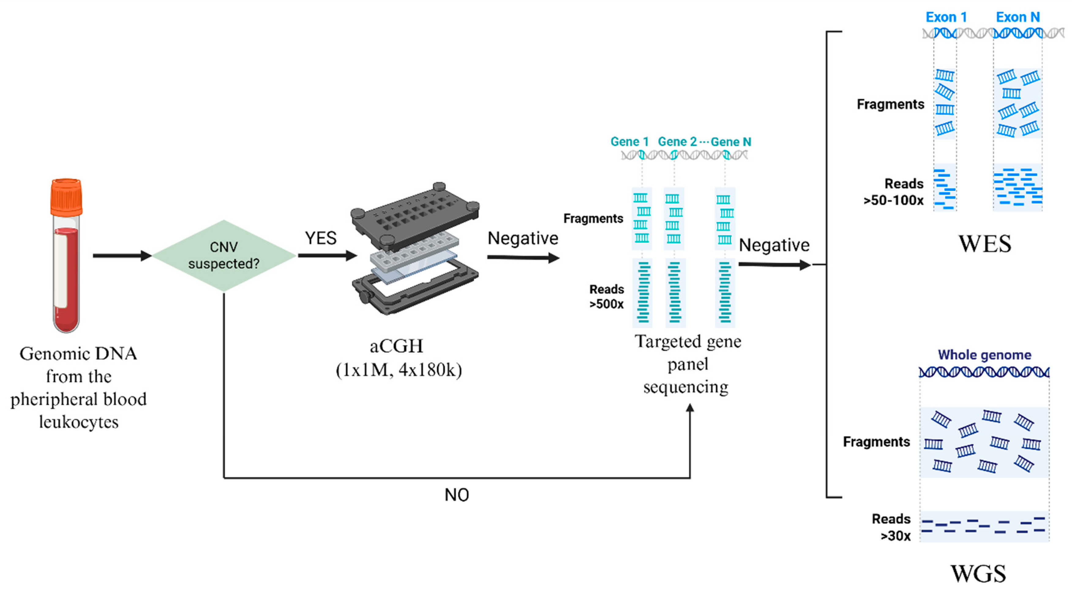
4.2. Genetic Analyses
4.2.1. aCGH
4.2.2. Targeted NGS Panel
4.2.3. WES and WGS
4.2.4. Sanger Sequencing
Supplementary Materials
Author Contributions
Funding
Institutional Review Board Statement
Informed Consent Statement
Data Availability Statement
Acknowledgments
Conflicts of Interest
References
- Chen, Y.; Liu, Z.; Chen, J.; Zuo, Y.; Liu, S.; Chen, W.; Liu, G.; Qiu, G.; Giampietro, P.F.; Wu, N.; et al. The genetic landscape and clinical implications of vertebral anomalies in VACTERL association. J. Med. Genet. 2016, 53, 431–437. [Google Scholar] [CrossRef]
- Erol, B.; Tracy, M.R.; Dormans, J.P.; Zackai, E.H.; Maisenbacher, M.K.; O’Brien, M.L.; Turnpenny, P.D.; Kusumi, K. Congenital scoliosis and vertebral malformations: Characterization of segmental defects for genetic analysis. J. Pediatr. Orthop. 2004, 24, 674–682. [Google Scholar] [CrossRef]
- Giampietro, P.F.; Dunwoodie, S.L.; Kusumi, K.; Pourquié, O.; Tassy, O.; Offiah, A.C.; Cornier, A.S.; Alman, B.A.; Blank, R.D.; Raggio, C.L.; et al. Progress in the understanding of the genetic etiology of vertebral segmentation disorders in humans. Ann. N. Y. Acad. Sci. 2009, 1151, 38–67. [Google Scholar] [CrossRef]
- Szoszkiewicz, A.; Bukowska-Olech, E.; Jamsheer, A. Molecular landscape of congenital vertebral malformations: Recent discoveries and future directions. Orphanet J. Rare Dis. 2024, 19, 32. [Google Scholar] [CrossRef] [PubMed]
- Turnpenny, P.D.; Alman, B.; Cornier, A.S.; Giampietro, P.F.; Offiah, A.; Tassy, O.; Pourquié, O.; Kusumi, K.; Dunwoodie, S. Abnormal vertebral segmentation and the notch signaling pathway in man. Dev. Dyn. 2007, 236, 1456–1474. [Google Scholar] [CrossRef] [PubMed]
- Liu, J.; Wu, N.; Yang, N.; Takeda, K.; Chen, W.; Li, W.; Du, R.; Liu, S.; Zhou, Y.; Zhang, L.; et al. TBX6-associated congenital scoliosis (TACS) as a clinically distinguishable subtype of congenital scoliosis: Further evidence supporting the compound inheritance and TBX6 gene dosage model. Genet. Med. 2019, 21, 1548–1558. [Google Scholar] [CrossRef] [PubMed]
- Wu, N.; Ming, X.; Xiao, J.; Wu, Z.; Chen, X.; Shinawi, M.; Shen, Y.; Yu, G.; Liu, J.; Xie, H.; et al. TBX6 null variants and a common hypomorphic allele in congenital scoliosis. N. Engl. J. Med. 2015, 372, 341–350. [Google Scholar] [CrossRef]
- Zhao, S.; Zhang, Y.; Chen, W.; Li, W.; Wang, S.; Wang, L.; Zhao, Y.; Lin, M.; Ye, Y.; Lin, J.; et al. Diagnostic yield and clinical impact of exome sequencing in early-onset scoliosis (EOS). J. Med. Genet. 2021, 58, 41–47. [Google Scholar] [CrossRef]
- Li, Z.; Zhao, S.; Cai, S.; Zhang, Y.; Wang, L.; Niu, Y.; Li, X.; Hu, J.; Chen, J.; Wang, S.; et al. The mutational burden and oligogenic inheritance in Klippel-Feil syndrome. BMC Musculoskelet. Disord. 2020, 21, 220. [Google Scholar] [CrossRef]
- Saker, E.; Loukas, M.; Oskouian, R.J.; Tubbs, R.S. The intriguing history of vertebral fusion anomalies: The Klippel-Feil syndrome. ChNS 2016, 32, 1599–1602. [Google Scholar] [CrossRef]
- Alazami, A.M.; Kentab, A.Y.; Faqeih, E.; Mohamed, J.Y.; Alkhalidi, H.; Hijazi, H.; Alkuraya, F.S. A novel syndrome of Klippel-Feil anomaly, myopathy, and characteristic facies is linked to a null mutation in MYO18B. J. Med. Genet. 2015, 52, 400–404. [Google Scholar] [CrossRef]
- Karaca, E.; Yuregir, O.O.; Bozdogan, S.T.; Aslan, H.; Pehlivan, D.; Jhangiani, S.N.; Akdemir, Z.C.; Gambin, T.; Bayram, Y.; Atik, M.M.; et al. Rare variants in the notch signaling pathway describe a novel type of autosomal recessive Klippel-Feil syndrome. Am. J. Med. Genet. A 2015, 167A, 2795–2799. [Google Scholar] [CrossRef] [PubMed]
- Li, Z.Q.; Geng, M.Z.; Zhao, S.; Wu, Z.H.; Zhang, J.G.; Wu, N.; Wang, Y.P. Clinical Characteristics and Genetic Analysis of Klippel-Feil Syndrome. Acta Acad. Med. Sin. 2021, 43, 25–31. [Google Scholar]
- Szoszkiewicz, A.; Bukowska-Olech, E.; Kurzawa, P.; Sowińska-Seidler, A.; Niedziela, M.; Kolesińska, Z.; Jamsheer, A. Upstream SOX9 deletion in a 46,XY girl with acampomelic campomelic dysplasia and absent minipuberty. Orphanet J. Rare Dis. 2025, 21, 41. [Google Scholar] [CrossRef] [PubMed]
- Beauregard-Lacroix, E.; Tardif, J.; Camurri, M.V.; Lemyre, E.; Barchi, S.; Parent, S.; Campeau, P.M. Retrospective Analysis of Congenital Scoliosis: Associated Anomalies and Genetic Diagnoses. Spine 2017, 42, E841–E847. [Google Scholar] [CrossRef]
- Zhao, S.; Zhao, H.; Zhao, L.; Cheng, X.; Zheng, Z.; Wu, M.; Wen, W.; Wang, S.; Zhou, Z.; Xie, H.; et al. Unraveling the genetic architecture of congenital vertebral malformation with reference to the developing spine. Nat. Commun. 2024, 15, 1125. [Google Scholar] [CrossRef]
- Mitter, D.; Krakow, D.; Farrington-Rock, C.; Meinecke, P. Expanded clinical spectrum of spondylocarpotarsal synostosis syndrome and possible manifestation in a heterozygous father. Am. J. Med. Genet. A 2008, 146, 779–783. [Google Scholar] [CrossRef]
- Coêlho, K.E.; Ramos, E.S.; Felix, T.M.; Martelli, L.; de Pina-Neto, J.M.; Niikawa, N. Three new cases of spondylocarpotarsal synostosis syndrome: Clinical and radiographic studies. Am. J. Med. Genet. 1998, 77, 12–15. [Google Scholar] [CrossRef]
- Isidor, B.; Cormier-Daire, V.; Le Merrer, M.; Lefrancois, T.; Hamel, A.; Le Caignec, C.; David, A.; Jacquemont, S. Autosomal dominant spondylocarpotarsal synostosis syndrome: Phenotypic homogeneity and genetic heterogeneity. Am. J. Med. Genet. A 2008, 146A, 1593–1597. [Google Scholar] [CrossRef]
- Yang, C.-F.; Wang, C.-H.; Siong H’ng, W.; Chang, C.-P.; Lin, W.-D.; Chen, Y.-T.; Wu, J.-Y.; Tsai, F.-J. Filamin B Loss-of-Function Mutation in Dimerization Domain Causes Autosomal-Recessive Spondylocarpotarsal Synostosis Syndrome with Rib Anomalies. Hum. Mutat. 2017, 38, 540–547. [Google Scholar] [CrossRef]
- Farrington-Rock, C.; Kirilova, V.; Dillard-Telm, L.; Borowsky, A.D.; Chalk, S.; Rock, M.J.; Cohn, D.H.; Krakow, D. Disruption of the Flnb gene in mice phenocopies the human disease spondylocarpotarsal synostosis syndrome. Hum. Mol. Genet. 2008, 17, 631–641. [Google Scholar] [CrossRef] [PubMed]
- Krakow, D.; Robertson, S.P.; King, L.M.; Morgan, T.; Sebald, E.T.; Bertolotto, C.; Wachsmann-Hogiu, S.; Acuna, D.; Shapiro, S.S.; Takafuta, T.; et al. Mutations in the gene encoding filamin B disrupt vertebral segmentation, joint formation and skeletogenesis. Nat. Genet. 2004, 36, 405–410. [Google Scholar] [CrossRef] [PubMed]
- Ramos-Mejía, R.; Del Pino, M.; Aza-Carmona, M.; Abbate, S.; Obregon, M.G.; Heath, K.E.; Fano, V. Novel FLNB Variants in Seven Argentinian Cases with Spondylocarpotarsal Synostosis Syndrome. J. Pediatr. Genet. 2024, 13, 167–174. [Google Scholar] [CrossRef] [PubMed]
- Fukushima, K.; Parthasarathy, P.; Wade, E.M.; Morgan, T.; Gowrishankar, K.; Markie, D.M.; Robertson, S.P. Intragenic Deletions in FLNB Are Part of the Mutational Spectrum Causing Spondylocarpotarsal Synostosis Syndrome. Genes 2021, 12, 528. [Google Scholar] [CrossRef]
- Yasin, S.; Makitie, O.; Naz, S. Spondylocarpotarsal synostosis syndrome due to a novel loss of function FLNB variant: A case report. BMC Musculoskelet. Disord. 2021, 22, 31. [Google Scholar] [CrossRef]
- Lu, J.; Lian, G.; Lenkinski, R.; De Grand, A.; Vaid, R.R.; Bryce, T.; Stasenko, M.; Boskey, A.; Walsh, C.; Sheen, V. Filamin B mutations cause chondrocyte defects in skeletal development. Hum. Mol. Genet. 2007, 16, 1661–1675. [Google Scholar] [CrossRef]
- Zhou, X.; Tian, F.; Sandzén, J.; Cao, R.; Flaberg, E.; Szekely, L.; Cao, Y.; Ohlsson, C.; Bergo, M.O.; Borén, J.; et al. Filamin B deficiency in mice results in skeletal malformations and impaired microvascular development. Proc. Natl. Acad. Sci. USA 2007, 104, 3919–3924. [Google Scholar] [CrossRef]
- Niikawa, N.; Matsuura, N.; Fukushima, Y.; Ohsawa, T.; Kajii, T. Kabuki make-up syndrome: A syndrome of mental retardation, unusual facies, large and protruding ears, and postnatal growth deficiency. J. Pediatr. 1981, 99, 565–569. [Google Scholar] [CrossRef]
- Fan, H.; Wang, Y.; Wu, Y.; Jia, L.; Wang, L.; Shen, Y. Clinical and genetic characteristics of four children with Kabuki syndrome due to de novo variants of KMT2D gene. Chin. J. Med. Genet. 2024, 41, 546–550. [Google Scholar]
- Di Candia, F.; Fontana, P.; Paglia, P.; Falco, M.; Rosano, C.; Piscopo, C.; Cappuccio, G.; Siano, M.A.; De Brasi, D.; Mandato, C.; et al. Clinical heterogeneity of Kabuki syndrome in a cohort of Italian patients and review of the literature. Eur. J. Pediatr. 2022, 181, 171–187. [Google Scholar] [CrossRef]
- White, S.M.; Thompson, E.M.; Kidd, A.; Savarirayan, R.; Turner, A.; Amor, D.; Delatycki, M.B.; Fahey, M.; Baxendale, A.; White, S.; et al. Growth, behavior, and clinical findings in 27 patients with Kabuki (Niikawa-Kuroki) syndrome. Am. J. Med. Genet. A 2004, 127A, 118–127. [Google Scholar] [CrossRef] [PubMed]
- Gao, C.W.; Lin, W.; Riddle, R.C.; Chopra, S.; Kim, J.; Boukas, L.; Hansen, K.D.; Björnsson, H.T.; Fahrner, J.A. Growth deficiency in a mouse model of Kabuki syndrome 2 bears mechanistic similarities to Kabuki syndrome 1. PLoS Genet. 2024, 20, e1011310. [Google Scholar] [CrossRef] [PubMed]
- Shpargel, K.B.; Mangini, C.L.; Xie, G.; Ge, K.; Magnuson, T. The KMT2D Kabuki syndrome histone methylase controls neural crest cell differentiation and facial morphology. Development 2020, 147, dev187997. [Google Scholar] [CrossRef] [PubMed]
- Zanoni, P.; Steindl, K.; Sengupta, D.; Joset, P.; Bahr, A.; Sticht, H.; Lang-Muritano, M.; van Ravenswaaij-Arts, C.M.A.; Shinawi, M.; Andrews, M.; et al. Loss-of-function and missense variants in NSD2 cause decreased methylation activity and are associated with a distinct developmental phenotype. Genet. Med. 2021, 23, 1474–1483. [Google Scholar] [CrossRef]
- Nishi, E.; Yanagi, K.; Kaname, T.; Okamoto, N. Clinical details of individuals with Rauch-Steindl syndrome due to NSD2 truncating variants. Mol. Genet. Genom. Med. 2024, 12, e2396. [Google Scholar] [CrossRef]
- Chen, S.; Lei, Y.; Yang, Y.; Liu, C.; Kuang, L.; Jin, L.; Finnell, R.H.; Yang, X.; Wang, H. A mutation in TBXT causes congenital vertebral malformations in humans and mice. J. Genet. Genom. 2024, 51, 433–442. [Google Scholar] [CrossRef]
- Postma, A.V.; Alders, M.; Sylva, M.; Bilardo, C.M.; Pajkrt, E.; van Rijn, R.R.; Schulte-Merker, S.; Bulk, S.; Stefanovic, S.; Ilgun, A.; et al. Mutations in the T (brachyury) gene cause a novel syndrome consisting of sacral agenesis, abnormal ossification of the vertebral bodies and a persistent notochordal canal. J. Med. Genet. 2014, 51, 90–97. [Google Scholar] [CrossRef]
- Ghebranious, N.; Blank, R.D.; Raggio, C.L.; Staubli, J.; McPherson, E.; Ivacic, L.; Rasmussen, K.; Jacobsen, F.S.; Faciszewski, T.; Burmester, J.K.; et al. A missense T (Brachyury) mutation contributes to vertebral malformations. J. Bone Miner. Res. 2008, 23, 1576–1583. [Google Scholar] [CrossRef]
- Yang, N.; Wu, N.; Zhang, L.; Zhao, Y.; Liu, J.; Liang, X.; Ren, X.; Li, W.; Chen, W.; Dong, S.; et al. TBX6 compound inheritance leads to congenital vertebral malformations in humans and mice. Hum. Mol. Genet. 2019, 28, 539–547. [Google Scholar] [CrossRef]
- Bukowska-Olech, E.; Popiel, D.; Koczyk, G.; Sowińska-Seidler, A.; Socha, M.; Wojciechowicz, B.; Dawidziuk, A.; Larysz, D.; Jamsheer, A. Adapting SureSelect enrichment protocol to the Ion Torrent S5 platform in molecular diagnostics of craniosynostosis. Sci. Rep. 2020, 10, 4159. [Google Scholar] [CrossRef]



| Characteristics | Cohort with CVMs (n, %) |
|---|---|
| Sex | |
| Female | 21 (62%) |
| Male | 13 (38%) |
| CVMs | |
| Failure of formation | 10 (29%) |
| Failure of segmentation | 12 (35%) |
| Mixed | 13 (38%) |
| Forms of CVMs | |
| Syndromes | |
| Klippel–Feil syndrome | 21 (62%) |
| Jarcho–Levin syndrome | 16 (47%) |
| Campomelic dysplasia | 1 (3%) |
| Sacral agenesis with vertebral anomalies | 1 (3%) |
| Kabuki syndrome | 1 (3%) |
| Spondylocarpotarsal synostosis syndrome | 1 (3%) |
| Isolated CVMs | 13 (38%) |
| Location of CVMs | |
| Cervical | 27 (79%) |
| Thoracic | 24 (71%) |
| Lumbar | 9 (26%) |
| Sacral | 4 (12%) |
| Coccygeal | 1 (3%) |
| Associated anomalies | |
| Cardiovascular disorders | 8 (24%) |
| Facial dysmorphism | 12 (35%) |
| Gastrointestinal anomalies | 5 (14%) |
| Hearing loss | 4 (12%) |
| Limbs | 6 (18%) |
| Neurological anomalies | 7 (21%) |
| Renal abnormalities | 9 (26%) |
| Rib malformations | 15 (44%) |
| Vision impairment | 6 (18%) |
| Patient | Gene | Transcript | Variant | Zygosity | Inheritance | gnomAD Exomes (v4) | ACMG Variant Classification | Criteria | Molecular Method |
|---|---|---|---|---|---|---|---|---|---|
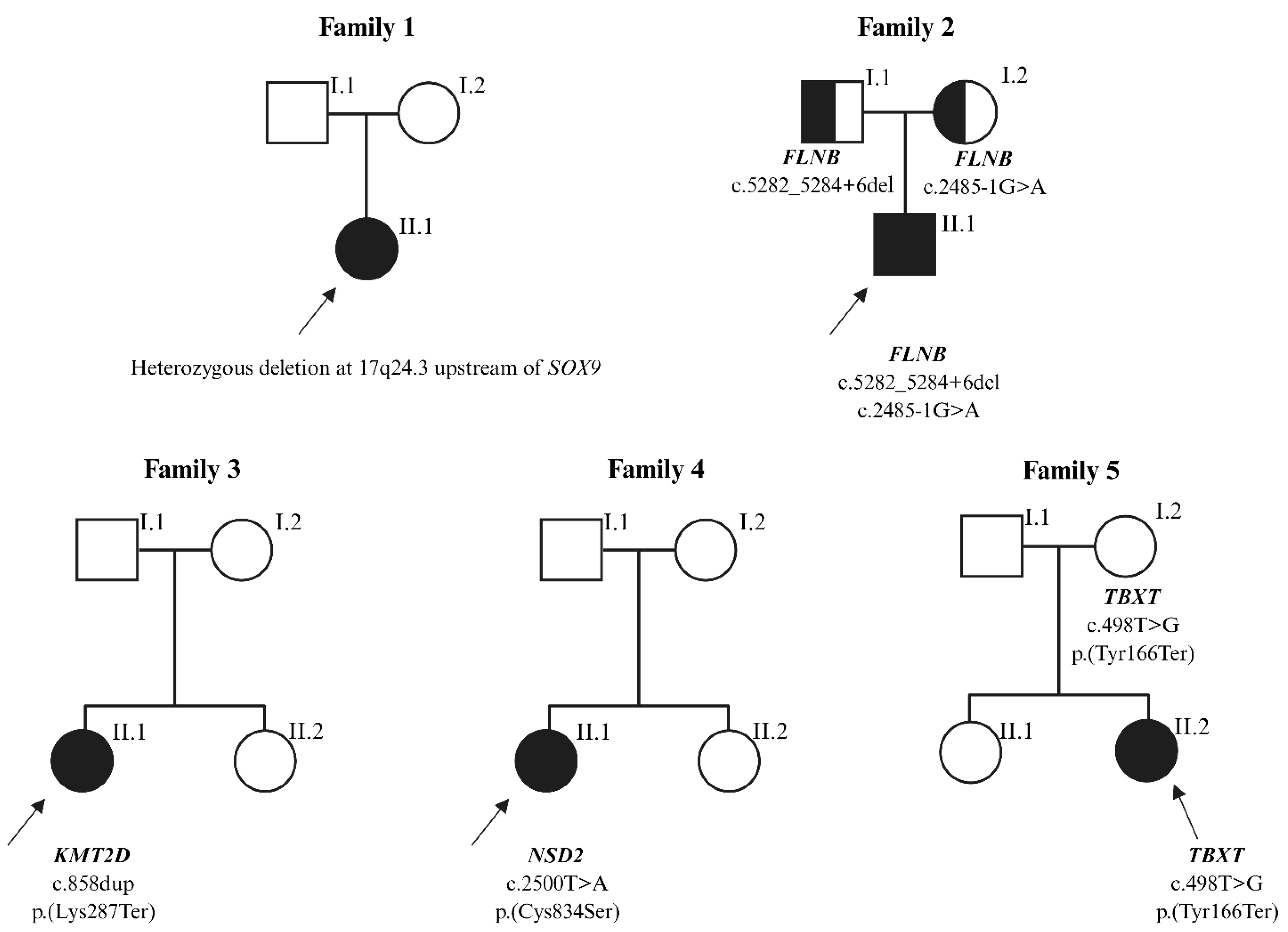
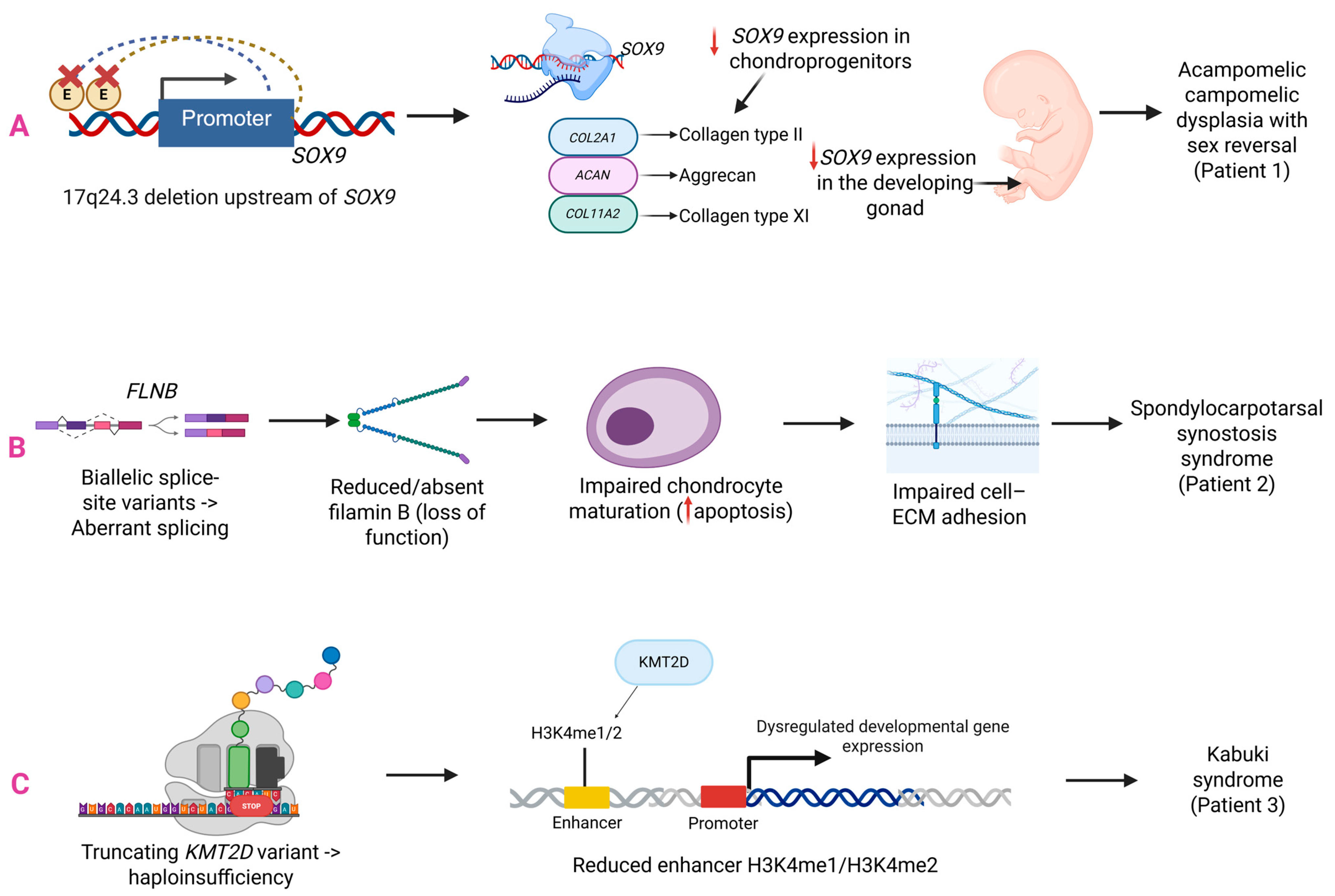
| P1 | SOX9 regulatory region | N/A | NC_000017.11:g. 70259128_71930429del | Het | de novo | N/A | Pathogenic | ND | aCGH +breakpoint mapping |
| P2 | FLNB | NM_001457.4 | c.2485-1G>A a c.5282_5284+6del b | Het Het | Maternal Paternal | 0 0.000000684 | Likely pathogenic Likely pathogenic | PVS1 Very Strong PM2 Supporting PVS1 Very Strong PM2 Supporting | WES |
| P3 | KMT2D | NM_003482.4 | c.858dup p.(Lys287Ter) | Het | de novo | 0 | Pathogenic | PS4 Moderate PVS1 Very Strong PM2 Moderate PS2 Strong | WES |
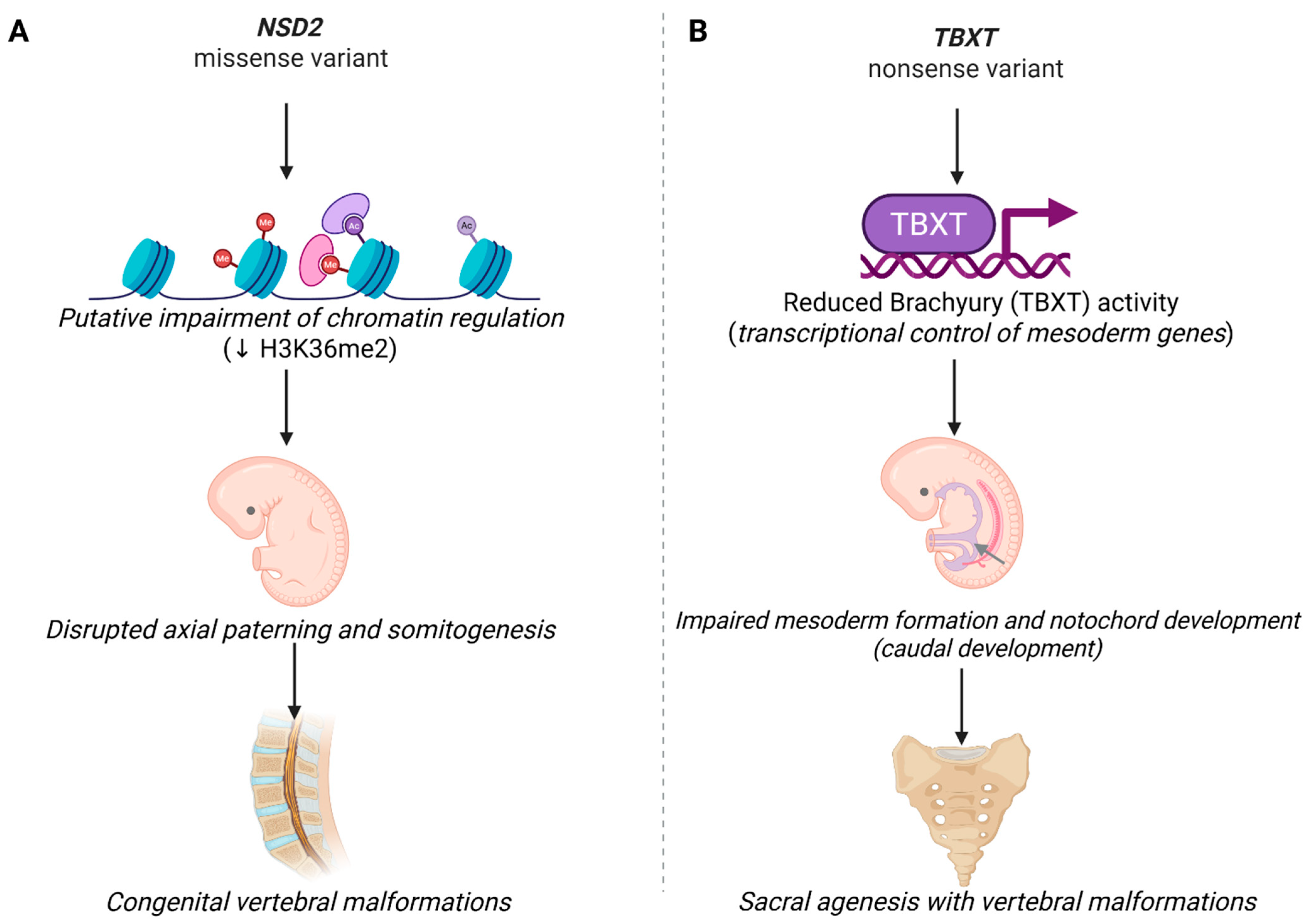
| P4 | NSD2 | NM_001042424.3 | c.2500T>A p.(Cys834Ser) | Het | de novo | 0 | VUS | PP3 Strong PP2 Supporting PM2 Supporting PS2 Strong BP1 Supporting | WES |
| P5 | TBXT | NM_001366285.2 | c.498T>G p.(Tyr166Ter) | Het | Maternal | 0 | Likely pathogenic | PVS1 Very Strong PM2 Moderate | WES |
| Patient | Sex | Age at Admission | Syndrome | Skeletal Anomalies | Other Malformations |
|---|---|---|---|---|---|
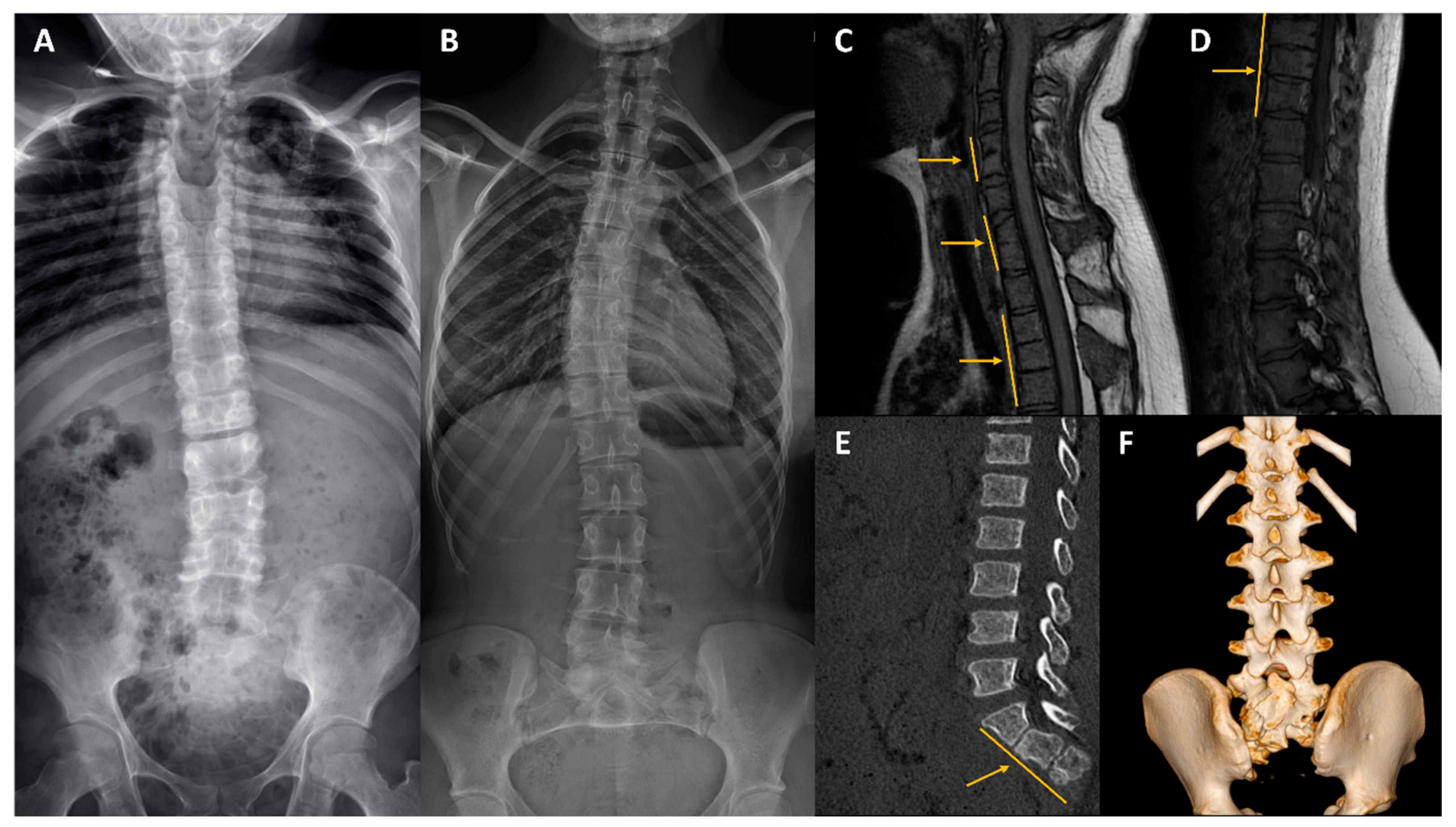
| P1 | F | 2 | Acampomelic campomelic dysplasia | Severe thoracic kyphoscoliosis, reduced vertebral height, rotation, canal stenosis, bell-shaped thorax, hypoplastic scapulae, narrow iliac wings, short neck * | Facial dysmorphism, cleft palate, widely spaced nipples, a sandal gap, 46, XY complete gonadal dysgenesis * |
| P2 | M | 2 | Spondylocarpotarsal synostosis syndrome | Right-sided torticollis, bony block of the vertebral arches from Th5-Th12 and L2-S1, the thoracic and lumbar lordosis, lumbar kyphosis, short neck, fifth finger clinodactyly | Short stature, psychomotor developmental delay, facial dysmorphia (dolichocephaly, frontal prominence, micrognathia, epicanthal folds, facial asymmetry), limited tongue protrusion, dilated renal pelvis, cerebellar hypoplasia |
| P3 | F | 2 | Kabuki syndrome | Butterfly vertebrae at Th6 and Th12, genu valgum, left hip dysplasia | Facial dysmorphia (hypertelorism, broad nasal bridge, high forehead, retrognathia), psychomotor developmental delay, speech delay, duplex kidney, horseshoe kidney, joint laxity |
| P4 | F | 13 | Rauch-Steindl syndrome | Scoliosis with vertebral rotation, cervical and lumbar lordosis with thoracic kyphosis, bony blocks at C4-C5, C7-Th1, Th3-Th5, Th8-Th10, bilateral cervical ribs, fifth finger clinodactyly | Short stature, postnatal growth retardation, failure to thrive, small head circumference, microcephaly, facial dysmorphism (short philtrum, prominent glabella, hypertelorism, low-set ears, posteriorly rotated ears, arched eyebrows, wide nasal bridge, thin lips, thin downturned corners of the mouth), astigmatism, craniofacial asymmetry, atrial septal defect type II, dental abnormalities, developmental delay, delayed walking, hypotonia, impaired intellectual development |
| P5 | F | 2 | Sacral agenesis with vertebral anomalies | Hypoplastic sacrum with only segments S1–S3 present (S1–S2 hypoplastic, S3 partially formed), agenesis of the coccyx, congenital hip dysplasia, bilateral clubfoot | Spinal cord malformations (absent conus medullaris), right ectopic pelvic kidney with hypoplasia, dysmorphic features (abnormal gluteal crease, abnormal cutaneous sinus tract) |
Disclaimer/Publisher’s Note: The statements, opinions and data contained in all publications are solely those of the individual author(s) and contributor(s) and not of MDPI and/or the editor(s). MDPI and/or the editor(s) disclaim responsibility for any injury to people or property resulting from any ideas, methods, instructions or products referred to in the content. |
© 2026 by the authors. Licensee MDPI, Basel, Switzerland. This article is an open access article distributed under the terms and conditions of the Creative Commons Attribution (CC BY) license.
Share and Cite
Szoszkiewicz, A.; Sowińska-Seidler, A.; Wnuk-Kłosińska, A.; Bukowska-Olech, E.; Biel, K.; Matuszewska, K.; Biel, M.; Badura-Stronka, M.; Glazar, R.; Jakubiuk-Tomaszuk, A.; et al. Deciphering the Genetic Basis of Congenital Vertebral Malformations Through a Stepwise Diagnostic Approach. Int. J. Mol. Sci. 2026, 27, 1752. https://doi.org/10.3390/ijms27041752
Szoszkiewicz A, Sowińska-Seidler A, Wnuk-Kłosińska A, Bukowska-Olech E, Biel K, Matuszewska K, Biel M, Badura-Stronka M, Glazar R, Jakubiuk-Tomaszuk A, et al. Deciphering the Genetic Basis of Congenital Vertebral Malformations Through a Stepwise Diagnostic Approach. International Journal of Molecular Sciences. 2026; 27(4):1752. https://doi.org/10.3390/ijms27041752
Chicago/Turabian StyleSzoszkiewicz, Anna, Anna Sowińska-Seidler, Aleksandra Wnuk-Kłosińska, Ewelina Bukowska-Olech, Karolina Biel, Karolina Matuszewska, Marcin Biel, Magdalena Badura-Stronka, Renata Glazar, Anna Jakubiuk-Tomaszuk, and et al. 2026. "Deciphering the Genetic Basis of Congenital Vertebral Malformations Through a Stepwise Diagnostic Approach" International Journal of Molecular Sciences 27, no. 4: 1752. https://doi.org/10.3390/ijms27041752
APA StyleSzoszkiewicz, A., Sowińska-Seidler, A., Wnuk-Kłosińska, A., Bukowska-Olech, E., Biel, K., Matuszewska, K., Biel, M., Badura-Stronka, M., Glazar, R., Jakubiuk-Tomaszuk, A., Krawczyński, M., Szczałuba, K., Śledzińska, K., Wiśniewska, M., & Jamsheer, A. (2026). Deciphering the Genetic Basis of Congenital Vertebral Malformations Through a Stepwise Diagnostic Approach. International Journal of Molecular Sciences, 27(4), 1752. https://doi.org/10.3390/ijms27041752