Review

Therapeutic Modulation of Nitric Oxide Pathways to Address Insulin Resistance in Cardiovascular–Kidney–Metabolic Syndrome

by
Ligia-Maria Ceteraș
1,2,
Vlad Dumitru Brata
3,*,
Ioana Dobrotă
1,2,
Rahela Borbei
1,2,
Mihai Clim
1,2,
Teodora-Gabriela Alexescu
1,2,
Mircea-Vasile Milaciu
1,2,
Mirela-Georgiana Perne
1,2,
Cezara-Andreea Gerdanovics
1,2,
Angela Cozma
1,2 and
Olga-Hilda Orășan
1,2
1
Department of Internal Medicine, 4th Medical Discipline, “Iuliu Hațieganu” University of Medicine and Pharmacy, 400015 Cluj-Napoca, Romania
2
Faculty of Medicine, “Iuliu Hațieganu” University of Medicine and Pharmacy, 400347 Cluj-Napoca, Romania
3
Department of Gastroenterology, Regional Institute of Gastroenterology and Hepatology “Prof. Dr. Octavian Fodor”, 400394 Cluj-Napoca, Romania
*
Author to whom correspondence should be addressed.
Int. J. Mol. Sci. 2026, 27(17), 7701; https://doi.org/10.3390/ijms27177701 (registering DOI)
Submission received: 13 July 2026 / Revised: 15 August 2026 / Accepted: 19 August 2026 / Published: 28 August 2026
(This article belongs to the Special Issue New Insights into the Treatment of Metabolic Syndrome and Diabetes)

Abstract

Cardiovascular–kidney–metabolic (CKM) syndrome encompasses the convergent pathophysiology of obesity, insulin resistance, type 2 diabetes, chronic kidney disease, and cardiovascular disease, conditions whose interactions account for a substantial proportion of cardiovascular morbidity and mortality despite contemporary guideline-directed therapy. Nitric oxide (NO) pathway dysfunction constitutes a unifying mechanism across this continuum, linking endothelial dysfunction, impaired insulin signaling, and multiorgan injury through endothelial NO synthase (eNOS) uncoupling, increased arginase activity, asymmetric dimethylarginine (ADMA) accumulation, and paradoxical inducible NO synthase (iNOS)-driven nitrosative stress. Established cardiometabolic therapies—sodium-glucose cotransporter-2 (SGLT2) inhibitors, glucagon-like peptide-1 receptor agonists (GLP-1 RAs), renin–angiotensin–aldosterone system (RAAS) inhibitors, statins, and metformin—improve NO signaling indirectly through reductions in oxidative stress and inflammation yet fail to fully restore NO bioavailability and leave substantial residual cardiovascular and renal risk unaddressed. Direct NO-restoring strategies, including soluble guanylate cyclase (sGC) modulators, arginase inhibition, ADMA-lowering approaches, and microbiome-targeted interventions, demonstrate mechanistic promise in preclinical and early translational studies but currently lack outcome-level evidence. Biomarkers of NO pathway dysfunction—ADMA, flow-mediated dilation (FMD), the tetrahydrobiopterin–dihydrobiopterin (BH4/BH2) ratio, cyclic guanosine monophosphate (cGMP), and endothelial microparticles (EMPs)—offer a foundation for patient phenotyping but remain insufficiently standardized for clinical use. A NO-centered framework provides a biologically coherent model for understanding residual cardiometabolic risk; its translation into personalized therapy will require validated biomarker panels and biomarker-guided outcome trials.

1. Introduction

Cardiovascular–kidney–metabolic (CKM) syndrome reflects the convergence of obesity, insulin resistance, type 2 diabetes mellitus, chronic kidney disease (CKD), and cardiovascular disease (CVD). The global prevalence of obesity has nearly tripled over the past five decades; the number of adults living with diabetes has exceeded 500 million, and CKD affects approximately 10–15% of the adult population. Individuals with coexisting metabolic, renal, and cardiovascular abnormalities have a substantially greater risk of heart failure, atherosclerotic cardiovascular disease, kidney failure, and all-cause mortality than those with a single disease entity [1,2,3].
Recognizing the need for an integrated framework, the American Heart Association introduced CKM syndrome as a systemic disorder characterized by pathological interactions among metabolic risk factors, kidney dysfunction, and cardiovascular disease. Its staging system describes a continuum from stage 0, denoting the absence of CKM risk factors, to stage 4, denoting established cardiovascular disease in the presence of metabolic and/or kidney dysfunction. This framework supports earlier identification of high-risk individuals and emphasizes the shared contributions of obesity, insulin resistance, inflammation, endothelial dysfunction, and neurohormonal activation to progressive cardiovascular and renal injury [3].
Nitric oxide (NO) signaling is one of the molecular pathways connecting metabolic dysfunction with cardiovascular and renal injury. NO regulates vascular homeostasis through endothelium-dependent vasodilation, inhibition of platelet aggregation, modulation of inflammation, maintenance of microvascular perfusion, and regulation of mitochondrial function. It also contributes to metabolic regulation by facilitating insulin-mediated glucose uptake, skeletal muscle perfusion, and insulin signaling. Experimental and clinical evidence links reduced NO bioavailability to endothelial dysfunction, impaired glucose disposal, hypertension, vascular remodeling, glomerular injury, and atherosclerosis. However, the timing and relative contribution of NO dysfunction across the CKM continuum remain incompletely established [4,5,6,7].
Several additional knowledge gaps limit clinical translation. Reliable biomarkers of NO bioavailability are lacking, and the mechanisms of impaired NO signaling vary among CKM populations. Direct NO-restoring interventions have also been evaluated predominantly in mechanistic or early-phase studies rather than in large outcome trials. Consequently, the most appropriate targets within the NO–soluble guanylate cyclase–cyclic guanosine monophosphate (NO–sGC–cGMP) pathway, the patients most likely to benefit, and the long-term clinical effects of NO-centered interventions remain uncertain [2,5,8,9].
Previous reviews have examined NO biology in obesity and insulin resistance, cardiorenal physiology, kidney regulation, endothelial dysfunction, or nitrate–nitrite pathways, generally within individual diseases or specific components of NO signaling [6,7,10,11,12,13]. The present review instead applies an integrated CKM framework to connect impaired NO production and bioavailability with endothelial and microvascular insulin resistance, organ-specific injury, and residual cardiovascular and renal risk. It also distinguishes established cardiometabolic therapies with indirect effects on NO signaling from more direct or experimental NO-targeted strategies according to their level of clinical evidence. Finally, it critically evaluates candidate biomarkers for biological phenotyping and therapeutic stratification while emphasizing their current limitations and need for prospective validation. Table 1 positions the scope and contribution of the present review relative to the previous literature.

2. Materials and Methods

This article was designed as a narrative review and was not intended to provide a systematic or exhaustive synthesis of all available evidence. No review protocol was registered. PubMed/MEDLINE and Embase were searched for publications from January 1990 to June 2026. Searches combined terms related to NO signaling and insulin resistance, including “nitric oxide,” “insulin resistance,” “cardiovascular-kidney-metabolic syndrome,” “endothelial dysfunction,” “endothelial nitric oxide synthase,” “eNOS,” “cyclic guanosine monophosphate,” “cGMP,” “asymmetric dimethylarginine,” “ADMA,” “arginase,” and related therapeutic terms. Search terms and combinations were adapted to the syntax of each database. Reference lists of relevant publications were also screened manually.
Studies were considered eligible when they addressed NO signaling in relation to insulin resistance, CKM-related pathophysiology, organ-specific injury, biomarkers, or NO-targeted therapeutic strategies. Human, animal, and mechanistic studies were included. Priority was given to randomized controlled trials, systematic reviews, meta-analyses, and clinically relevant observational and mechanistic studies. Publications that did not address the objectives of the review or provided information already adequately represented by higher-level or more directly relevant evidence were excluded.
No new quantitative synthesis was performed, and findings were reported as presented in the cited publications. No formal risk-of-bias or evidence-certainty instrument was applied. Evidence was interpreted according to study design, relevance to the CKM continuum, directness of NO-pathway assessment, and degree of clinical validation. These methodological features should be considered when interpreting the conclusions of this narrative review.

3. NO Biology Relevant to Insulin Sensitivity

Nitric oxide contributes to insulin sensitivity through vasodilation, enhanced microvascular perfusion, and direct modulation of insulin receptor signaling. In endothelial cells, insulin activates the phosphoinositide 3-kinase/protein kinase B (PI3K/Akt) pathway, which stimulates endothelial nitric oxide synthase (eNOS) and increases NO production. The resulting improvement in microvascular perfusion facilitates the delivery of insulin and glucose to skeletal muscle, while NO also supports insulin signaling within insulin-responsive cells [17,18,19].
Reduced NO bioavailability may therefore be both a consequence of insulin resistance and a mechanism that contributes to its progression. The underlying defects are heterogeneous and may include reduced eNOS activity, increased oxidative NO scavenging, and eNOS uncoupling. This distinction has therapeutic implications because strategies that increase NO production may be less effective when accelerated NO degradation or impaired downstream signaling is the predominant abnormality [20,21,22,23].

3.1. eNOS-NO-cGMP Signaling

The eNOS–NO–cGMP pathway contributes to insulin sensitivity, particularly through its actions in the vasculature and skeletal muscle [24,25,26,27].
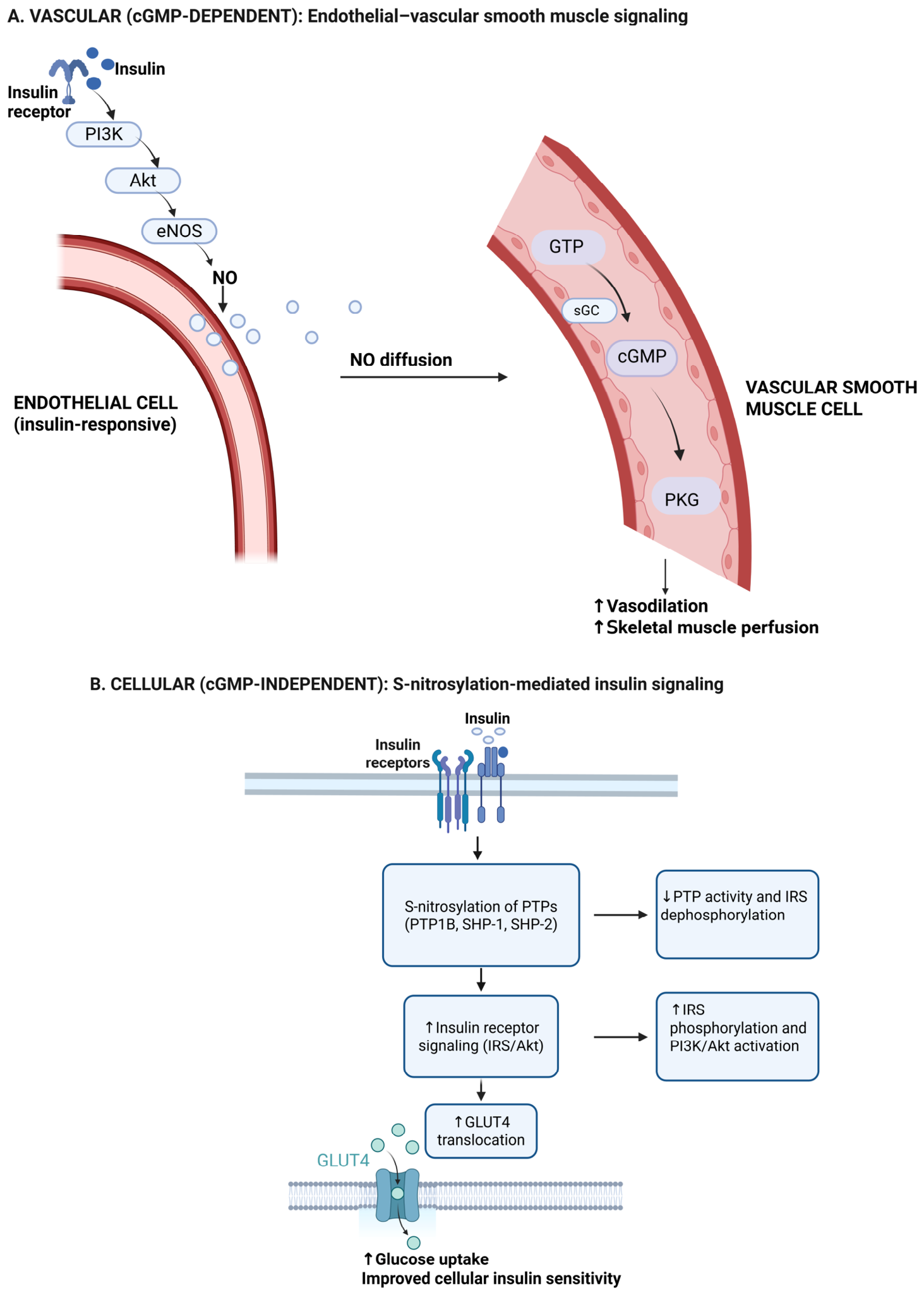
Following insulin stimulation, the PI3K/Akt pathway activates eNOS in endothelial cells and increases NO production. NO then diffuses into adjacent vascular smooth muscle cells, where it activates sGC, which catalyzes the conversion of guanosine triphosphate (GTP) to cGMP. Through cGMP-dependent signaling, NO induces vasodilation and improves skeletal muscle perfusion, thereby facilitating the delivery of insulin and glucose to target tissues [28,29].
NO also modulates insulin signaling through cGMP-independent protein S-nitrosylation [18,30,31]. Relevant targets include protein tyrosine phosphatase 1B (PTP1B) and Src homology region 2 domain-containing phosphatases 1 and 2 (SHP-1 and SHP-2), which negatively regulate insulin receptor signaling. Their S-nitrosylation reduces phosphatase activity and can thereby enhance insulin receptor activation, Akt phosphorylation, and glucose transporter type 4 (GLUT4) translocation in endothelial cells and insulin-responsive tissues, including skeletal muscle, liver, and adipose tissue [17,18,19]. These effects support insulin responsiveness [5], although their relative contribution in human CKM syndrome remains incompletely established.
Thus, NO supports insulin sensitivity through two complementary mechanisms: cGMP-dependent vasodilation and microvascular recruitment, and cGMP-independent modulation of insulin receptor signaling (Figure 1). Dysfunction in either mechanism may impair glucose disposal.

3.2. Mechanisms Reducing NO Production and NO Bioavailability in Insulin Resistance

NO bioavailability refers to the amount of biologically active NO available to exert physiological effects and is determined by the balance between its production and inactivation through reactions with superoxide, hemoglobin, and other scavengers [32,33,34]. Unlike NO production, which describes its synthesis by nitric oxide synthases, bioavailability also depends on protection from oxidative degradation and effective signaling in target tissues. It is therefore more informative than production alone when evaluating the relationship between NO and insulin sensitivity [30,31,34].
The mechanisms underlying impaired NO signaling in insulin resistance and CKM syndrome can be broadly divided into those that reduce NO production and those that accelerate its inactivation or disrupt its biological activity.

3.2.1. Mechanisms Reducing NO Production

Under conditions of oxidative stress, cofactor deficiency, or substrate limitation, eNOS can become uncoupled and generate superoxide rather than NO. The relevant determinant is the ratio of reduced tetrahydrobiopterin (BH4) to its oxidized form, dihydrobiopterin (BH2), rather than the absolute BH4 concentration [35,36,37]. Because eNOS binds BH2 and BH4 with comparable affinity, oxidation of BH4 favors the formation of BH2-bound eNOS and superoxide production [36]. Superoxide subsequently reacts with NO to form peroxynitrite, which further oxidizes BH4 and sustains eNOS uncoupling, NO scavenging, and oxidative vascular injury [33,38].
Arginase competes with nitric oxide synthase (NOS) for their common substrate, L-arginine. Arginase 1 (Arg1) is cytosolic and predominantly hepatic, whereas arginase 2 (Arg2) is mitochondrial and is expressed in endothelial cells, vascular smooth muscle, kidney, and skeletal muscle [32]. Increased arginase activity in obesity, insulin resistance, and type 2 diabetes reduces L-arginine availability for NO synthesis [34,36,37]. In insulin-resistant patients with morbid obesity, increased Arg2 expression was associated with impaired endothelium-dependent vasodilation, which improved following L-arginine supplementation or arginase inhibition [39]. Erythrocyte-derived Arg1 may further contribute to systemic L-arginine depletion in type 2 diabetes and heart failure [40].
Asymmetric dimethylarginine (ADMA) accumulates in insulin-resistant states when the activity of dimethylarginine dimethylaminohydrolase (DDAH) is reduced [39,41,42]. Higher ADMA concentrations correlate inversely with insulin sensitivity and endothelium-dependent vasodilation [39,43,44]. ADMA competitively inhibits NOS and may also reduce eNOS phosphorylation through activation of the mitogen-activated protein kinase (MAPK) pathway [45]. DDAH1 accounts for most systemic ADMA clearance, whereas DDAH2 regulates local ADMA concentrations near sites of eNOS activity [46]. By contrast, symmetric dimethylarginine (SDMA) does not directly inhibit NOS and primarily reflects renal clearance rather than NO-pathway dysfunction [47].

3.2.2. Mechanisms Reducing NO Bioavailability

Oxidative stress reduces NO bioavailability through increased production of reactive oxygen species (ROS). Superoxide (O2) rapidly reacts with NO to form peroxynitrite (ONOO) [35,37,38]. Major sources of ROS include nicotinamide adenine dinucleotide phosphate (NADPH) oxidase isoforms 1–5 (NOX1–NOX5), mitochondria during substrate overload, and xanthine oxidase in hyperuricemic states [48,49]. Peroxynitrite further oxidizes BH4, nitrates tyrosine residues, inactivates sGC, and promotes S-glutathionylation of eNOS [5,33,38]. These reactions couple accelerated NO inactivation with progressive eNOS uncoupling and endothelial dysfunction.
Inducible nitric oxide synthase (iNOS; NOS2) represents a distinct mechanism in which NO signaling becomes dysregulated despite increased NO production. It is transcriptionally induced through nuclear factor kappa B (NF-κB)- and Janus kinase/signal transducer and activator of transcription (JAK–STAT)-dependent signaling in response to tumor necrosis factor alpha (TNF-α), interleukin-1 beta (IL-1β), interferon gamma (IFN-γ), and lipopolysaccharide. Unlike eNOS, it generates sustained, high-output NO [50]. In the presence of superoxide, this NO promotes peroxynitrite formation and may induce pathological S-nitrosylation of insulin receptor substrate 1 (IRS-1), Akt inactivation, and kinase nitration, thereby impairing insulin signaling [51]. Nevertheless, iNOS activity is not uniformly pathogenic; in acute settings, iNOS-derived NO contributes to antimicrobial defense and ischemic preconditioning [52].
These mechanisms should not be interpreted as producing a uniform deficiency of NO across all tissues. At the endothelial–vascular interface, eNOS-derived NO may be reduced, whereas inflammatory activation can increase iNOS-derived NO in specific cellular compartments. The biological consequences therefore depend on the cellular source, concentration, spatial distribution, redox environment, and downstream signaling capacity of NO. This distinction may explain why increasing total NO production does not necessarily reproduce the metabolic effects of restoring physiological eNOS-derived signaling.

4. NO and Insulin Resistance: Core Mechanisms

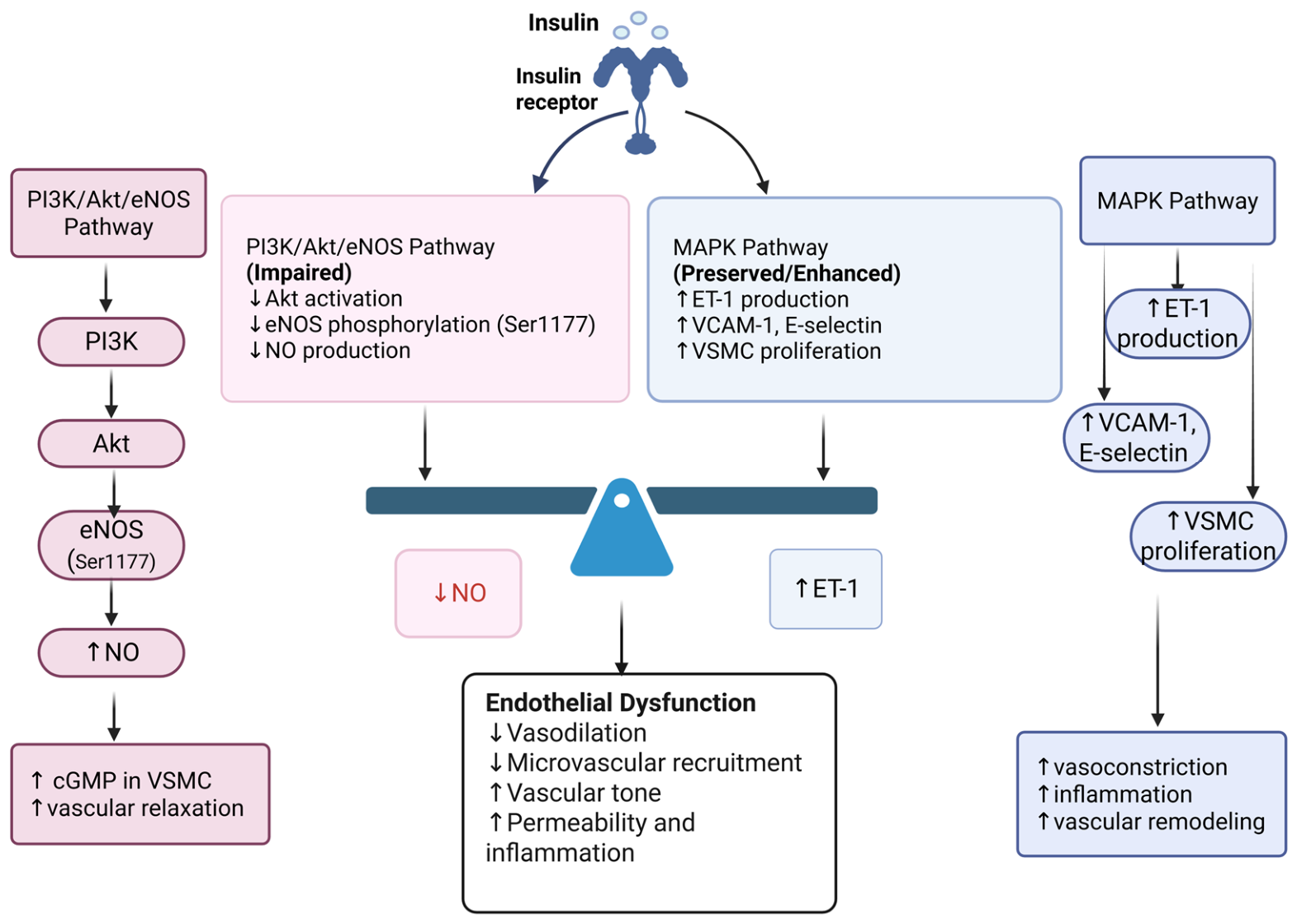
In insulin-resistant states, glucotoxicity, lipotoxicity, and chronic low-grade inflammation selectively impair PI3K/Akt/eNOS signaling, which normally couples endothelial insulin action to NO production. The resulting reduction in NO bioavailability becomes functionally important at the endothelial–microvascular interface, where impaired perfusion and substrate delivery contribute to systemic insulin resistance [14,20,29,53,54,55].

Endothelial and Microvascular Insulin Resistance

Endothelial insulin resistance refers to impaired insulin signaling within vascular endothelial cells, with reduced NO production and endothelial dysfunction as its principal manifestations. Microvascular insulin resistance describes the parallel failure of insulin to recruit capillaries and increase microvascular perfusion in target tissues, particularly skeletal muscle. These processes are closely linked through the PI3K/Akt/eNOS pathway and together influence peripheral glucose disposal [14,15,56].
Insulin activates two endothelial signaling branches with different vascular effects. The PI3K/Akt/eNOS branch promotes vasodilation, capillary recruitment, and substrate delivery to skeletal muscle, whereas the MAPK branch promotes endothelin-1 (ET-1) production, vascular smooth muscle proliferation, and expression of adhesion molecules such as vascular cell adhesion molecule 1 (VCAM-1) and E-selectin [29,57]. In insulin resistance, PI3K/Akt/eNOS signaling is selectively suppressed, whereas MAPK signaling remains intact or becomes hyperactive during compensatory hyperinsulinemia [14,29,57]. This imbalance shifts the endothelium from a vasodilatory, anti-inflammatory phenotype toward a vasoconstrictive, pro-inflammatory one, characterized by reduced NO availability, increased ET-1 signaling, elevated vascular tone, and impaired microvascular perfusion (Figure 2).
Physiological concentrations of insulin recruit skeletal muscle microvasculature within 5–10 min, preceding both myocyte signaling and increased glucose disposal. This response is NOS-dependent and can be abolished by the NOS inhibitor NG-monomethyl-L-arginine (L-NMMA) [25]. The hemodynamic actions of insulin include relaxation of resistance arterioles, which increases total blood flow; relaxation of terminal arterioles, which recruits previously unperfused capillaries; and expansion of the microvascular surface available for insulin and substrate exchange.
Insulin resistance adds a structural limitation through capillary rarefaction, defined as a sustained reduction in capillary density. This further restricts the surface available for insulin and glucose exchange [58,59]. In mice, muscle-specific deletion of vascular endothelial growth factor (VEGF) reduced skeletal muscle capillary density by approximately 60% and produced insulin resistance independently of primary myocyte signaling defects [58]. These findings indicate that loss of microvascular architecture can contribute to impaired insulin sensitivity.
Endothelial dysfunction and insulin resistance may therefore reinforce each other. Insulin resistance impairs PI3K/Akt/eNOS signaling and reduces NO production, whereas endothelial dysfunction limits microvascular recruitment and substrate delivery, further aggravating peripheral insulin resistance [14]. This bidirectional model suggests that impaired insulin action should not be viewed exclusively as an intracellular defect in insulin-responsive tissues; part of the impairment may arise upstream, at the level of insulin delivery and microvascular recruitment. The endothelial compartment therefore occupies an intermediate position between circulating insulin and its metabolic targets, although it remains unclear how much insulin resistance in humans is attributable to vascular rather than cellular defects.

5. Organ-Specific Pathophysiology in CKM Syndrome

The consequences of impaired NO signaling vary across the organs affected by CKM syndrome but share a common pathophysiological pattern: reduced eNOS-derived NO at the endothelial interface, amplification by oxidative and inflammatory stress, and tissue-dependent induction of iNOS in immune or parenchymal cells. The relative contribution of these mechanisms depends on cellular composition, vascular architecture, and metabolic demand, producing distinct patterns of organ injury.
In the vasculature, metabolic risk factors promote oxidative stress, inflammation, eNOS uncoupling, and reduced NO production [10,11,12]. The resulting loss of endothelium-dependent vasodilation increases vascular tone and arterial stiffness and promotes atherogenesis [11,12,13]. With sustained metabolic stress, this functional disturbance progresses toward vascular inflammation, remodeling, and structural atherosclerotic disease [10,11,13].
In the heart, reduced NO bioavailability impairs NO–sGC–cGMP–PKG signaling, promoting cardiomyocyte hypertrophy, myocardial stiffness, diastolic dysfunction, and adverse remodeling [60,61]. Metabolic oxidative and nitrosative stress further promotes inflammation and fibrosis, contributing to the development of heart failure with preserved ejection fraction [60,61,62]. Concurrent impairment of coronary microvascular NO signaling limits the matching of myocardial perfusion to metabolic demand [60,62]. Vascular dysfunction and cardiomyocyte remodeling may consequently reinforce one another within the metabolic HFpEF phenotype.
Renal NO signaling regulates glomerular hemodynamics, tubular transport, and renal blood flow [11,62]. Reduced NO bioavailability favors arteriolar vasoconstriction, impaired natriuresis, sodium retention, and altered glomerular filtration [16,62,63]. Chronic kidney disease further increases ADMA concentrations through impaired renal clearance and altered DDAH activity, thereby aggravating NOS inhibition and endothelial dysfunction [13,16,45,63]. Oxidative stress within the renal vasculature additionally suppresses eNOS activity and shifts the balance toward vasoconstrictor signaling [11,16,63]. These interacting hemodynamic and molecular abnormalities promote glomerular and vascular injury and establish a feedback loop between renal dysfunction and systemic endothelial impairment.
In the liver, inflammatory induction of iNOS generates sustained, high-output NO that promotes S-nitrosylation and tyrosine nitration of insulin-signaling proteins, thereby impairing hepatic insulin action [23,64,65,66]. Excessive iNOS-derived NO can also disrupt autophagy and lysosomal function, contributing to lipid accumulation and metabolic dysfunction [23,66]. By contrast, physiological NO generated by constitutive NOS isoforms supports hepatic glucose and lipid metabolism [23]. Liver-specific iNOS expression is sufficient to induce hepatic insulin resistance in experimental models, supporting a causal role for nitrosative stress rather than a purely associative relationship [64]. Altered S-nitrosoglutathione metabolism may further connect abnormal NO signaling with defective cellular quality control and hepatic steatosis [66].
In skeletal muscle, NO contributes to insulin-stimulated glucose uptake and GLUT4 translocation [67,68,69,70]. Reduced endothelial NO availability impairs microvascular recruitment and capillary perfusion, limiting the delivery of insulin and glucose to myocytes [11,23]. Within muscle cells, iNOS-derived NO and nitrosative stress can directly inhibit insulin signaling [23,68]. Skeletal muscle insulin resistance therefore includes both a vascular defect in substrate delivery and a cellular defect in NO-dependent insulin signaling [67,68,69,70]. Impaired microvascular perfusion may precede and amplify intracellular abnormalities, increasing their contribution to systemic insulin resistance.
In adipose tissue, chronic inflammation and iNOS induction promote nitrosative stress, impaired insulin signaling, lipolysis, free fatty acid release, and systemic inflammation [23,71]. Reduced vascular NO–cGMP signaling may itself promote adipose inflammation, indicating that endothelial dysfunction can actively contribute to adipose tissue pathology [72]. In visceral adipose tissue from individuals with obesity, vascular TNF-α production has been associated with reduced NO availability in small arteries [73]. This interaction is particularly relevant in perivascular adipose tissue surrounding coronary and renal vessels. During obesity, perivascular adipose tissue loses its vasoprotective phenotype and releases pro-inflammatory mediators that impair NO signaling in the adjacent vascular wall, thereby linking adipose dysfunction with coronary and renal microvascular injury [74,75,76].
The principal NO-related mechanisms and their organ-specific consequences across the CKM continuum are summarized in Table 2.

6. Clinical Manifestations of Insulin Resistance in CKM Syndrome

The clinical phenotype of CKM syndrome reflects the convergence of insulin resistance and impaired NO signaling across multiple organs. Its manifestations combine metabolic abnormalities, including hyperinsulinemia, dyslipidemia, ectopic adiposity, and hyperglycemia, with vascular abnormalities such as endothelial dysfunction, reduced NO bioavailability, increased vascular tone, and capillary rarefaction. The interaction between these processes contributes to the elevated cardiovascular and renal risk associated with CKM syndrome [14,71].
Atherogenic dyslipidemia is characterized by elevated triglycerides, reduced high-density lipoprotein cholesterol, and increased concentrations of small dense low-density lipoprotein particles. Oxidized low-density lipoprotein further impairs endothelial function by increasing oxidative stress, promoting eNOS uncoupling, and reducing NO bioavailability, thereby accelerating vascular inflammation and atherogenesis [33,38,77,78].
Visceral and ectopic adiposity contribute to insulin resistance through the release of free fatty acids and pro-inflammatory adipokines, including TNF-α, IL-6, and leptin. These mediators increase oxidative stress, impair eNOS activity, and reduce vascular NO bioavailability [65,72,73,79,80,81,82,83]. In the liver, chronic lipotoxicity and inflammation contribute to metabolic dysfunction-associated steatotic liver disease (MASLD) and steatohepatitis (MASH). Increased iNOS activity promotes nitrosative modification of insulin-signaling proteins, whereas reduced sinusoidal NO availability impairs hepatic microvascular function and increases intrahepatic vascular resistance [23,64,65,66,79].
Hypertension in CKM syndrome reflects the interaction of hyperinsulinemia, sympathetic nervous system activation, renin–angiotensin–aldosterone system activity, renal sodium retention, and endothelial dysfunction. Reduced NO bioavailability shifts vascular regulation toward vasoconstrictor signaling mediated by angiotensin II and ET-1, increasing vascular resistance and blood pressure [10,13,14,15,71].
Heart failure with preserved ejection fraction is a prominent cardiac phenotype within the CKM continuum. Systemic and coronary microvascular endothelial dysfunction reduces NO bioavailability and impairs NO–sGC–cGMP–PKG signaling in cardiomyocytes. The resulting titin hypophosphorylation, myocardial stiffness, inflammation, and fibrosis contribute to diastolic dysfunction and adverse cardiac remodeling [60,61,62,84].
NO inhibits platelet activation, adhesion, and aggregation through cGMP-dependent signaling. Impaired NO bioavailability may therefore favor a prothrombotic vascular state in insulin resistance and CKM syndrome [85].
Renal involvement may develop before a measurable decline in glomerular filtration rate. Early abnormalities can include glomerular hyperfiltration, altered tubuloglomerular feedback, activation of the renin–angiotensin system, and progressive endothelial dysfunction. Because the contribution of NO can vary with disease stage and renal compartment, hyperfiltration should not be attributed exclusively to reduced NO availability [10,11,13,16,63]. Albuminuria, quantified using the urinary albumin-to-creatinine ratio (UACR), reflects glomerular and systemic vascular injury and is associated with both cardiovascular and renal risk [86]. UACR is therefore a clinically accessible risk marker that complements, but does not directly measure, NO-pathway dysfunction.
Chronic low-grade inflammation and oxidative stress provide a systemic link among these clinical phenotypes. Elevated C-reactive protein, TNF-α, and IL-6 concentrations and increased NF-κB activity can suppress eNOS expression, promote eNOS uncoupling, and accelerate NO inactivation. Reduced NO bioavailability can, in turn, facilitate endothelial activation, adhesion-molecule expression, and further inflammatory signaling [31,33,34,35,36,37,38,61,82,87,88,89]. This relationship is context-dependent: acute inflammatory responses may be adaptive, whereas persistent metabolic inflammation shifts NO signaling toward oxidative and nitrosative stress. Consequently, suppression of inflammatory signaling alone cannot be assumed to restore endothelial NO bioavailability.

7. Therapeutic Strategies Affecting the NO Pathway

Therapeutic interventions relevant to NO signaling range from established cardiometabolic treatments with demonstrated cardiovascular or renal benefits to experimental strategies that directly target NO production, bioavailability, or downstream signaling. The categories below are descriptive and distinguish therapies according to their clinical evidence base and proximity to the NO pathway. They should not be interpreted as a formal evidence-grading system or as proof that NO modulation mediates the observed clinical benefits.

7.1. Established Therapies with Cardiovascular or Renal Outcome Evidence

Sodium–glucose cotransporter 2 (SGLT2) inhibitors have demonstrated cardiovascular and renal benefits in trials including EMPA-REG OUTCOME, DAPA-HF, EMPEROR-Preserved, DAPA-CKD, and EMPA-KIDNEY, across populations with type 2 diabetes, heart failure, and chronic kidney disease. Proposed NO-related mechanisms include reduced NADPH oxidase activity, mitochondrial dysfunction, oxidative stress, and inflammatory signaling [90,91].
Glucagon-like peptide-1 receptor agonists (GLP-1 RAs) have demonstrated cardiovascular, metabolic, or renal benefits in LEADER, SUSTAIN-6, REWIND, and FLOW. Mechanistic studies suggest that reduced oxidative stress and inflammation, enhanced eNOS activity, and improved mitochondrial function may contribute to their vascular effects [90,91].
Statins have well-established cardiovascular benefits through lowering of atherogenic lipoproteins and may additionally improve endothelial NO signaling by increasing eNOS expression and activity, limiting eNOS uncoupling, and reducing NADPH oxidase-dependent oxidative stress [92,93,94].
Inhibitors of the renin–angiotensin–aldosterone system reduce cardiovascular and renal risk in appropriately selected patients. Angiotensin-converting enzyme inhibitors, angiotensin receptor blockers, and mineralocorticoid receptor antagonists may preserve NO bioavailability by reducing angiotensin II-mediated oxidative stress, supporting eNOS function, and limiting vascular inflammation [8,95]. Finerenone reduced kidney disease progression and cardiovascular events in patients with chronic kidney disease and type 2 diabetes in FIDELIO-DKD and FIGARO-DKD [96,97].
EMPA-REG OUTCOME, DAPA-HF, EMPEROR-Preserved, DAPA-CKD, EMPA-KIDNEY, LEADER, SUSTAIN-6, REWIND, FLOW, FIDELIO-DKD, and FIGARO-DKD were designed to evaluate clinical outcomes rather than NO-pathway activity. Evidence for NO modulation therefore derives mainly from experimental studies and surrogate measures of endothelial function, oxidative stress, or NO-related signaling [84,90,91,92,93,94,95,96,97]. Direct measurements of NO production or tissue-specific bioavailability were rarely prespecified, so the clinical benefits observed in these trials cannot be attributed specifically to restoration of NO signaling.
Future mechanistic studies should prespecify biochemical or functional measures—such as ADMA, nitrate and nitrite, cGMP, or flow-mediated dilation—and determine whether treatment-induced changes are associated with clinical outcomes.

7.2. Clinically Available or Evaluated Interventions with Limited CKM-Specific NO Evidence

sGC acts downstream of NO synthesis and increases cGMP generation through direct stimulation of sGC. Riociguat is indicated for pulmonary arterial hypertension and selected forms of chronic thromboembolic pulmonary hypertension. Vericiguat demonstrated clinical benefit in VICTORIA and is indicated for symptomatic chronic heart failure with an ejection fraction below 45% following a recent worsening event. VICTORIA did not establish that this benefit was mediated by restoration of systemic or tissue-specific NO bioavailability. Neither agent has demonstrated efficacy for insulin resistance or CKM syndrome as an integrated condition, while sGC activators remain investigational [84].
Pioglitazone improves insulin sensitivity and may influence endothelial function through effects on inflammation, adipokine signaling, and eNOS expression. In IRIS, it reduced recurrent stroke or myocardial infarction among patients with insulin resistance after ischemic stroke or transient ischemic attack [98]. This population should not be equated with the broader CKM population, and fluid retention, weight gain, and heart failure risk constrain its clinical use.
Metformin remains widely used for glycemic management in type 2 diabetes. The experimental and mechanistic literature has linked it to AMP-activated protein kinase signaling, enhanced eNOS phosphorylation, reduced mitochondrial oxidative stress, and attenuation of inflammatory pathways [12,85]. However, direct human evidence that restoration of NO signaling mediates its clinical effects is limited, and NO-related biomarkers have not generally been incorporated as prespecified endpoints in metformin trials.
Strategies based on tetrahydrobiopterin include BH4 supplementation, sepiapterin administration, and folate-mediated support of BH4-dependent eNOS coupling. Although these approaches can improve endothelial function or vascular reactivity under selected experimental conditions, clinical evidence remains largely restricted to surrogate outcomes [36,99,100,101]. Their efficacy is also likely to depend on whether BH4 oxidation and eNOS uncoupling are dominant abnormalities in the treated population.
L-arginine and L-citrulline supplementation seek to increase substrate availability for NOS. Some pharmacokinetic studies suggest that L-citrulline can produce more sustained increases in circulating L-arginine because it undergoes less presystemic metabolism [102,103]. Nevertheless, clinical benefits have generally been modest and have focused on blood pressure or endothelial-function endpoints rather than cardiovascular or renal events [13,102,103].
Organic nitrates remain established treatments for symptomatic ischemic heart disease but are not therapies for CKM syndrome itself. Their long-term use may be limited by tolerance, oxidative stress, and nitrate-associated endothelial dysfunction, and they have not demonstrated sustained cardiovascular or renal protection across CKM populations [104,105].
Phosphodiesterase type 5 (PDE5) inhibitors prevent cGMP degradation and thereby amplify signaling downstream of NO. Their effectiveness depends on sufficiently preserved upstream NO–sGC activity, which may be compromised in advanced oxidative vascular disease. Although these agents have established clinical uses outside CKM syndrome, evidence supporting their use for insulin resistance or multiorgan CKM outcomes remains insufficient [106].

7.3. Experimental NO-Restoring Strategies

The inorganic nitrate–nitrite–NO pathway provides a NOS-independent route for NO generation through enterosalivary nitrate reduction. Small mechanistic studies have reported improvements in blood pressure, endothelial function, or arterial stiffness, but metabolic effects have been inconsistent and clinical outcome evidence is lacking [10,12,107,108,109]. Chronic nitrate supplementation also failed to improve metabolic health and worsened aspects of disease progression in a mouse model of diet-induced obesity [110]. These findings indicate that the effects of nitrate supplementation depend on biological context, including disease stage, redox environment, diet, and microbiome activity.
Antioxidant interventions such as resveratrol, N-acetylcysteine, and vitamin C have been investigated as indirect means of preserving NO bioavailability and limiting eNOS uncoupling. Although experimental findings are often favorable, clinical studies have produced inconsistent results, possibly because nonspecific suppression of oxidative signaling does not correct every cause of NO-pathway dysfunction [38,111].
Modulation of endogenous S-nitrosothiol metabolism represents another experimental approach. Altered S-nitrosoglutathione reductase activity has been linked to defective autophagy and hepatic insulin resistance in obesity [66]. This finding supports the biological relevance of S-nitrosothiol homeostasis but does not establish the therapeutic efficacy of administering S-nitrosothiols or modifying their metabolism in CKM syndrome.

7.4. Biomarkers and Current Limitations of NO Pathway Assessment

Clinical translation of NO-targeted strategies is limited by the difficulty of measuring NO bioavailability in vivo, the heterogeneity of pathway dysfunction across patients, and the absence of validated thresholds for treatment selection or monitoring [84,112]. Because NO is highly reactive and has a short biological half-life, direct measurement is largely restricted to experimental settings. Clinical and translational studies therefore rely on biochemical surrogates and functional vascular measurements, each capturing a different aspect of NO biology [88,113,114].
ADMA reflects endogenous NOS inhibition and has been associated with endothelial dysfunction, insulin resistance, cardiovascular events, and CKD progression [39,41,42,44,45,47]. SDMA does not directly inhibit NOS and primarily provides information about renal clearance and methylarginine metabolism [47]. Plasma nitrate and nitrite, commonly reported together as NOx, provide indirect estimates of systemic NO metabolism but are affected by dietary intake, oral microbiome activity, renal clearance, medication use, and oxidative conditions [10,11,12,88,114]. The BH4/BH2 ratio may reflect eNOS coupling more accurately than either metabolite alone, although its measurement remains largely research-based [35,36,37,38]. cGMP provides information about downstream signaling but is not specific to NO because it can also be generated through natriuretic peptide pathways [60,84,115].
Functional measurements provide complementary information. Flow-mediated dilation assesses endothelium-dependent vasodilatory capacity but is not exclusively determined by NO and is sensitive to vascular remodeling, sympathetic activity, metabolic conditions, operator technique, and pre-test preparation [6,16,116,117]. Pulse wave velocity reflects arterial stiffness and cumulative vascular injury but is not specific to NO dysfunction [16,62]. Circulating endothelial microparticles may indicate endothelial injury or activation, although differences in isolation and quantification methods limit comparability among studies [118,119,120].
Several practical barriers currently prevent routine implementation. Most biochemical markers lack universally accepted reference ranges, standardized sampling conditions, and clinically validated thresholds for diagnosis, risk stratification, or treatment selection. ADMA and SDMA are influenced by kidney function, whereas nitrate and nitrite measurements require control of diet, medication exposure, oral microbiome activity, and renal clearance [10,11,12,39,41,42,45,47,88,114]. Assessment of the BH4/BH2 ratio and endothelial microparticles requires specialized analytical methods and remains insufficiently standardized across laboratories [35,36,37,118,119,120]. Flow-mediated dilation requires strict pre-test conditions, dedicated equipment, technical expertise, and operator training, while its reproducibility is affected by methodological and physiological variability [6,16,116,117]. Cost, assay availability, turnaround time, and the need for repeated standardized measurements create additional obstacles to routine clinical use.
Candidate biomarkers, their potential applications, and their principal limitations are summarized in Table 3.

8. Emerging and Future Therapeutic Targets

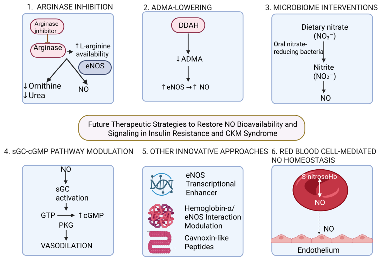
Emerging NO-directed strategies target substrate competition, endogenous NOS inhibition, microbiome-dependent nitrate reduction, downstream cGMP signaling, and erythrocyte–endothelial interactions. Their translational maturity varies substantially, and most remain supported primarily by preclinical or early mechanistic evidence. The principal emerging strategies are summarized in Figure 3.
Arginase inhibition seeks to preserve L-arginine availability for eNOS. In rodent models of metabolic syndrome and obesity, pharmacological arginase inhibition improved vascular function, reduced blood pressure, and enhanced insulin sensitivity [121,122]. Whether these effects translate into cardiovascular or renal benefits in patients with CKM syndrome remains unknown.
Enhancement of DDAH activity represents a complementary strategy for lowering ADMA. A long-acting recombinant DDAH preparation reduced ADMA, improved endothelial function, lowered blood pressure, and limited ischemia–reperfusion injury in preclinical models [123]. These findings demonstrate target engagement but have not yet been validated clinically.
Pharmacological eNOS transcriptional enhancers, including AVE3085 and AVE9488, increase eNOS expression and may limit eNOS uncoupling in experimental models [78]. Cavnoxin-like peptides are designed to disrupt the inhibitory interaction between caveolin-1 and eNOS, thereby increasing eNOS-derived NO, but evidence remains preclinical [115]. Modulation of the hemoglobin-α/eNOS interaction at myoendothelial junctions offers another potential means of increasing local NO bioavailability, although this approach remains mechanistic [85].
Microbiome-targeted strategies are based on the essential contribution of oral and gut bacteria to bioactivation of dietary nitrate through the nitrate–nitrite–NO pathway [124,125]. Human observational data have associated the nitrite-generating and nitrite-depleting capacity of the oral microbiome with cardiometabolic risk profiles [126]. However, these associations do not establish that probiotics, prebiotics, or other microbiome interventions can restore NO signaling or improve clinical outcomes.
The reduced, heme-containing and oxidized, heme-deficient forms of sGC represent pharmacologically distinct targets for sGC stimulators and activators, respectively [127]. Next-generation agents, including the sGC stimulator praliciguat and the sGC activator ataciguat, are being investigated for broader cardiovascular and metabolic applications, but their efficacy in insulin resistance or CKM syndrome has not been established [115].
Erythrocyte-mediated regulation of vascular NO provides another emerging target. Erythrocytes from patients with type 2 diabetes have been shown to induce endothelial dysfunction through increased arginase I activity in experimental vascular assays [128]. These findings implicate erythrocyte arginase and NO metabolism in vascular dysfunction but do not yet establish a clinically actionable therapeutic strategy [85,128].

9. Clinical Implications and Key Messages

NO-pathway dysfunction provides an integrative mechanistic framework for understanding the vascular, metabolic, cardiac, and renal manifestations of CKM syndrome. However, it should not be interpreted as a uniform deficiency state or as a clinically validated basis for treatment selection. Reduced eNOS-derived NO, eNOS uncoupling, ADMA accumulation, increased arginase activity, oxidative NO inactivation, impaired sGC–cGMP signaling, and iNOS-mediated nitrosative stress represent biologically distinct abnormalities that may require different interventions [33,38,39,42,45,64,65,66,84].
Established CKM therapies, including SGLT2 inhibitors, GLP-1 receptor agonists, renin–angiotensin–aldosterone system inhibitors, finerenone, and statins, provide cardiovascular or renal benefits in appropriately selected patients. Experimental and surrogate-endpoint studies suggest that several of these treatments may also improve NO-related signaling, but the major outcome trials were not designed to establish NO modulation as the mediator of clinical benefit [90,91,92,93,94,95,96,97]. Treatment decisions should therefore remain based on demonstrated clinical indications rather than presumed effects on the NO pathway.
Mechanism-based selection of NO-targeted therapy remains a research objective. Strategies that increase NO production may be ineffective when oxidative inactivation or impaired downstream responsiveness predominates, whereas cGMP-directed interventions may bypass some upstream defects. Conversely, excessive iNOS-derived NO requires control of nitrosative stress rather than indiscriminate enhancement of NO production. These distinctions support biological phenotyping but do not yet justify a clinical treatment algorithm.
Available biomarkers also remain insufficient for routine therapeutic selection. ADMA, nitrate and nitrite, the BH4/BH2 ratio, cGMP, flow-mediated dilation, and pulse wave velocity assess different aspects of NO biology, but none directly quantifies tissue-specific NO bioavailability or has validated thresholds for selecting or monitoring therapy [10,11,12,39,45,47,113,114,116,117]. Multimarker panels should therefore be regarded as investigational until their analytical validity, reproducibility, prognostic value, and association with treatment response have been established prospectively.
Future evaluation of NO-based combination therapy should follow a stepwise design. Initial randomized phase II studies should test an NO-targeted intervention as add-on therapy to optimized guideline-directed CKM treatment, using candidate biomarkers for enrichment or stratification while treating them as investigational. Primary objectives should include safety and prespecified target engagement, supported by functional vascular measurements; UACR and other organ-level measures may serve as secondary outcomes but should not be interpreted as direct measures of NO activity. Combination regimens should proceed only after their individual components demonstrate target engagement and acceptable safety and should preferably address complementary defects, such as upstream eNOS dysfunction and impaired downstream cGMP signaling. If early studies demonstrate biological activity, adequately powered trials should evaluate cardiovascular death, heart-failure events, and kidney disease progression. Until such evidence becomes available, NO-targeted and biomarker-guided strategies remain investigational.

10. Conclusions

NO-pathway dysfunction provides an integrative framework linking insulin resistance with vascular and multiorgan injury across the CKM continuum. Reduced eNOS-derived NO, eNOS uncoupling, increased arginase activity, ADMA-mediated NOS inhibition, oxidative NO inactivation, iNOS-derived nitrosative stress, and impaired sGC–cGMP signaling may each contribute. Their relative importance varies among organs, disease stages, and metabolic phenotypes, and current human evidence does not establish NO dysfunction as the sole or dominant cause of residual cardiovascular and renal risk.
Contemporary CKM therapies improve clinical outcomes, but these benefits cannot be attributed specifically to restoration of NO signaling. Direct NO-targeted interventions remain predominantly preclinical or at an early translational stage. Further progress requires mechanism-appropriate interventions, standardized and prospectively validated measures of pathway activity, and trials that distinguish molecular target engagement from clinical benefit. NO homeostasis should therefore be viewed as a useful framework for integrating vascular, metabolic, cardiac, and renal pathophysiology and generating testable therapeutic strategies, rather than as an established biomarker-defined treatment paradigm.

Author Contributions

Conceptualization, L.-M.C.; Data curation, O.-H.O., V.D.B., T.-G.A., I.D., A.C. and M.-V.M.; Formal analysis, M.-V.M., M.-G.P., T.-G.A., V.D.B. and O.-H.O.; Investigation, L.-M.C., O.-H.O., V.D.B. and I.D.; Methodology, O.-H.O., V.D.B. and I.D.; Supervision, O.-H.O., V.D.B., T.-G.A., M.-V.M., M.-G.P., I.D., A.C. and C.-A.G.; Writing—original draft, L.-M.C.; Writing—review and editing, L.-M.C., V.D.B., O.-H.O., R.B., C.-A.G. and M.C. All authors have read and agreed to the published version of the manuscript.

Funding

This research received no external funding.

Institutional Review Board Statement

Not applicable.

Informed Consent Statement

Not applicable.

Data Availability Statement

No new data were created or analyzed in this study. Data sharing is not applicable to this article.

Acknowledgments

During the preparation of this manuscript, the authors used ChatGPT (GPT-5, OpenAI) between February and June 2026 to assist with language editing and the reformulation of text drafted by the authors, including revisions to phrasing and structure for clarity and precision. This tool was not used to determine study eligibility, extract or interpret data, assess evidence certainty, perform statistical analyses, or formulate scientific conclusions. No citation was included without retrieval and verification of the original publication by the authors. All AI-assisted revisions were reviewed and approved by the authors, who take full responsibility for the content, accuracy, and integrity of the manuscript.

Conflicts of Interest

The authors declare no conflict of interest.

Abbreviations

The following abbreviations are used in this manuscript:
ADMAasymmetric dimethylarginine
Aktprotein kinase B
Arg1arginase 1
Arg2arginase 2
BH2dihydrobiopterin
BH4tetrahydrobiopterin
cGMPcyclic guanosine monophosphate
CKDchronic kidney disease
CKMcardiovascular–kidney–metabolic
CVDcardiovascular disease
DDAHdimethylarginine dimethylaminohydrolase
DDAH1dimethylarginine dimethylaminohydrolase 1
DDAH2dimethylarginine dimethylaminohydrolase 2
EMPsendothelial microparticles
eNOSendothelial nitric oxide synthase
ET-1endothelin-1
FMDflow-mediated dilation
GLP-1 RAglucagon-like peptide-1 receptor agonist
GLUT4glucose transporter type 4
GTPguanosine triphosphate
HFpEFheart failure with preserved ejection fraction
IFN-γinterferon gamma
IL-1βinterleukin-1 beta
IL-6interleukin-6
iNOSinducible nitric oxide synthase
IRSinsulin receptor substrate
IRS-1insulin receptor substrate 1
JAK/STATJanus kinase/signal transducer and activator of transcription
L-NMMANG-monomethyl-L-arginine
MAPKmitogen-activated protein kinase
MASHmetabolic dysfunction-associated steatohepatitis
MASLDmetabolic dysfunction-associated steatotic liver disease
NADPHnicotinamide adenine dinucleotide phosphate
NF-κBnuclear factor kappa B
NOnitric oxide
NO2nitrite
NO3nitrate
NOSnitric oxide synthase
NOS2nitric oxide synthase 2, the gene encoding iNOS
NOxnitrate and nitrite metabolites
NOX1–NOX5NADPH oxidase isoforms 1–5
O2superoxide anion
ONOOperoxynitrite
PDE5phosphodiesterase type 5
PI3Kphosphoinositide 3-kinase
PKGprotein kinase G
PTPprotein tyrosine phosphatase
PTP1Bprotein tyrosine phosphatase 1B
PWVpulse wave velocity
ROSreactive oxygen species
SDMAsymmetric dimethylarginine
Ser1177serine residue at position 1177
sGCsoluble guanylate cyclase
SGLT2sodium–glucose cotransporter 2
SHP-1Src homology region 2 domain-containing phosphatase 1
SHP-2Src homology region 2 domain-containing phosphatase 2
SNO-HbS-nitrosohemoglobin
TNF-αtumor necrosis factor alpha
UACRurinary albumin-to-creatinine ratio
VCAM-1vascular cell adhesion molecule 1
VEGFvascular endothelial growth factor
VSMCvascular smooth muscle cell

References

  1. Jager, K.J.; Kovesdy, C.; Langham, R.; Rosenberg, M.; Jha, V.; Zoccali, C. A single number for advocacy and communication—Worldwide more than 850 million individuals have kidney diseases. Kidney Int. 2019, 96, 1048–1050. [Google Scholar] [CrossRef] [Scilit] [PubMed]
  2. Powell-Wiley, T.M.; Poirier, P.; Burke, L.E.; Després, J.P.; Gordon-Larsen, P.; Lavie, C.J.; Lear, S.A.; Ndumele, C.E.; Neeland, I.J.; Sanders, P.; et al. Obesity and Cardiovascular Disease: A Scientific Statement From the American Heart Association. Circulation 2021, 143, e984–e1010. [Google Scholar] [CrossRef] [Scilit] [PubMed]
  3. Ndumele, C.E.; Rangaswami, J.; Chow, S.L.; Neeland, I.J.; Tuttle, K.R.; Khan, S.S.; Coresh, J.; Mathew, R.O.; Baker-Smith, C.M.; Carnethon, M.R.; et al. Cardiovascular-Kidney-Metabolic Health: A Presidential Advisory From the American Heart Association. Circulation 2023, 148, 1606–1635, Correction in Circulation 2024, 149, 13. https://doi.org/10.1161/CIR.0000000000001241. [Google Scholar] [CrossRef] [Scilit] [PubMed]
  4. Tessari, P.; Cecchet, D.; Cosma, A.; Vettore, M.; Coracina, A.; Millioni, R.; Iori, E.; Puricelli, L.; Avogaro, A.; Vedovato, M. Nitric oxide synthesis is reduced in subjects with type 2 diabetes and nephropathy. Diabetes 2010, 59, 2152–2159. [Google Scholar] [CrossRef] [Scilit] [PubMed]
  5. Förstermann, U.; Sessa, W.C. Nitric oxide synthases: Regulation and function. Eur. Heart J. 2012, 33, 829–837. [Google Scholar] [CrossRef] [Scilit] [PubMed]
  6. Tousoulis, D.; Kampoli, A.M.; Tentolouris, C.; Papageorgiou, N.; Stefanadis, C. The role of nitric oxide on endothelial function. Curr. Vasc. Pharmacol. 2012, 10, 4–18. [Google Scholar] [CrossRef] [Scilit] [PubMed]
  7. Sansbury, B.E.; Hill, B.G. Regulation of obesity and insulin resistance by nitric oxide. Free Radic. Biol. Med. 2014, 73, 383–399. [Google Scholar] [CrossRef] [Scilit] [PubMed]
  8. Förstermann, U.; Münzel, T. Endothelial nitric oxide synthase in vascular disease: From marvel to menace. Circulation 2006, 113, 1708–1714. [Google Scholar] [CrossRef] [Scilit] [PubMed]
  9. Khan, S.S.; Ning, H.; Wilkins, J.T.; Allen, N.; Carnethon, M.; Berry, J.D.; Sweis, R.N.; Lloyd-Jones, D.M. Association of Body Mass Index With Lifetime Risk of Cardiovascular Disease and Compression of Morbidity. JAMA Cardiol. 2018, 3, 280–287. [Google Scholar] [CrossRef] [Scilit] [PubMed]
  10. Carlström, M.; Montenegro, M.F. Therapeutic value of stimulating the nitrate-nitrite-nitric oxide pathway to attenuate oxidative stress and restore nitric oxide bioavailability in cardiorenal disease. J. Intern. Med. 2019, 285, 2–18. [Google Scholar] [CrossRef] [Scilit] [PubMed]
  11. Carlström, M. Nitric oxide signalling in kidney regulation and cardiometabolic health. Nat. Rev. Nephrol. 2021, 17, 575–590. [Google Scholar] [CrossRef] [Scilit] [PubMed]
  12. Liao, G.Z.; He, C.H.; Zhang, Y.H.; Zhang, J. Addressing the “Nitric Oxide Crisis” in Cardiovascular-Kidney-Metabolic Syndrome: Therapeutic Potential of the Inorganic Nitrate-Nitrite-NO Pathway. Obes. Rev. 2026, 27, e70021. [Google Scholar] [CrossRef] [Scilit] [PubMed]
  13. Rajapakse, N.W.; Nanayakkara, S.; Kaye, D.M. Pathogenesis and treatment of the cardiorenal syndrome: Implications of L-arginine-nitric oxide pathway impairment. Pharmacol. Ther. 2015, 154, 1–12. [Google Scholar] [CrossRef] [Scilit] [PubMed]
  14. Kim, J.A.; Montagnani, M.; Koh, K.K.; Quon, M.J. Reciprocal relationships between insulin resistance and endothelial dysfunction: Molecular and pathophysiological mechanisms. Circulation 2006, 113, 1888–1904. [Google Scholar] [CrossRef] [Scilit] [PubMed]
  15. Muniyappa, R.; Montagnani, M.; Koh, K.K.; Quon, M.J. Cardiovascular actions of insulin. Endocr. Rev. 2007, 28, 463–491. [Google Scholar] [CrossRef] [Scilit] [PubMed]
  16. Roumeliotis, S.; Mallamaci, F.; Zoccali, C. Endothelial Dysfunction in Chronic Kidney Disease, from Biology to Clinical Outcomes: A 2020 Update. J. Clin. Med. 2020, 9, 2359. [Google Scholar] [CrossRef] [Scilit] [PubMed]
  17. Hsu, M.F.; Meng, T.C. Enhancement of insulin responsiveness by nitric oxide-mediated inactivation of protein-tyrosine phosphatases. J. Biol. Chem. 2010, 285, 7919–7928. [Google Scholar] [CrossRef] [Scilit] [PubMed]
  18. Wang, H.; Wang, A.X.; Aylor, K.; Barrett, E.J. Nitric oxide directly promotes vascular endothelial insulin transport. Diabetes 2013, 62, 4030–4042. [Google Scholar] [CrossRef] [Scilit] [PubMed]
  19. Hsu, M.F.; Pan, K.T.; Chang, F.Y.; Khoo, K.H.; Urlaub, H.; Cheng, C.F.; Chang, G.-D.; Haj, F.G.; Meng, T.-C. S-nitrosylation of endogenous protein tyrosine phosphatases in endothelial insulin signaling. Free Radic. Biol. Med. 2016, 99, 199–213. [Google Scholar] [CrossRef] [Scilit] [PubMed]
  20. Kashyap, S.R.; Roman, L.J.; Lamont, J.; Masters, B.S.S.; Bajaj, M.; Suraamornkul, S.; Belfort, R.; Berria, R.; Kellogg, D.L.; Liu, Y.; et al. Insulin resistance is associated with impaired nitric oxide synthase activity in skeletal muscle of type 2 diabetic subjects. J. Clin. Endocrinol. Metab. 2005, 90, 1100–1105. [Google Scholar] [CrossRef] [Scilit] [PubMed]
  21. Wu, G.; Meininger, C.J. Nitric oxide and vascular insulin resistance. Biofactors 2009, 35, 21–27. [Google Scholar] [CrossRef] [Scilit] [PubMed]
  22. Molina, M.N.; Ferder, L.; Manucha, W. Emerging Role of Nitric Oxide and Heat Shock Proteins in Insulin Resistance. Curr. Hypertens. Rep. 2016, 18, 1. [Google Scholar] [CrossRef] [Scilit] [PubMed]
  23. Bahadoran, Z.; Mirmiran, P.; Ghasemi, A. Role of Nitric Oxide in Insulin Secretion and Glucose Metabolism. Trends Endocrinol. Metab. 2020, 31, 118–130. [Google Scholar] [CrossRef] [Scilit] [PubMed]
  24. Vincent, M.A.; Barrett, E.J.; Lindner, J.R.; Clark, M.G.; Rattigan, S. Inhibiting NOS blocks microvascular recruitment and blunts muscle glucose uptake in response to insulin. Am. J. Physiol. Endocrinol. Metab. 2003, 285, E123–E129. [Google Scholar] [CrossRef] [Scilit] [PubMed]
  25. Vincent, M.A.; Clerk, L.H.; Lindner, J.R.; Klibanov, A.L.; Clark, M.G.; Rattigan, S.; Barrett, E.J. Microvascular recruitment is an early insulin effect that regulates skeletal muscle glucose uptake in vivo. Diabetes 2004, 53, 1418–1423. [Google Scholar] [CrossRef] [Scilit] [PubMed]
  26. Barrett, E.J.; Eggleston, E.M.; Inyard, A.C.; Wang, H.; Li, G.; Chai, W.; Liu, Z. The vascular actions of insulin control its delivery to muscle and regulate the rate-limiting step in skeletal muscle insulin action. Diabetologia 2009, 52, 752–764. [Google Scholar] [CrossRef] [Scilit] [PubMed]
  27. Barrett, E.J.; Wang, H.; Upchurch, C.T.; Liu, Z. Insulin regulates its own delivery to skeletal muscle by feed-forward actions on the vasculature. Am. J. Physiol. Endocrinol. Metab. 2011, 301, E252–E263. [Google Scholar] [CrossRef] [Scilit] [PubMed]
  28. Vincent, M.A.; Montagnani, M.; Quon, M.J. Molecular and physiologic actions of insulin related to production of nitric oxide in vascular endothelium. Curr. Diab Rep. 2003, 3, 279–288. [Google Scholar] [CrossRef] [Scilit] [PubMed]
  29. Muniyappa, R.; Chen, H.; Montagnani, M.; Sherman, A.; Quon, M.J. Endothelial dysfunction due to selective insulin resistance in vascular endothelium: Insights from mechanistic modeling. Am. J. Physiol. Endocrinol. Metab. 2020, 319, E629–46. [Google Scholar] [CrossRef] [Scilit] [PubMed]
  30. Ceriello, A. New insights on oxidative stress and diabetic complications may lead to a “causal” antioxidant therapy. Diabetes Care 2003, 26, 1589–1596. [Google Scholar] [CrossRef] [Scilit] [PubMed]
  31. Rains, J.L.; Jain, S.K. Oxidative stress, insulin signaling, and diabetes. Free Radic. Biol. Med. 2011, 50, 567–575. [Google Scholar] [CrossRef] [Scilit] [PubMed]
  32. Pernow, J.; Jung, C. Arginase as a potential target in the treatment of cardiovascular disease: Reversal of arginine steal? Cardiovasc. Res. 2013, 98, 334–343. [Google Scholar] [CrossRef] [Scilit] [PubMed]
  33. Münzel, T.; Camici, G.G.; Maack, C.; Bonetti, N.R.; Fuster, V.; Kovacic, J.C. Impact of Oxidative Stress on the Heart and Vasculature: Part 2 of a 3-Part Series. J. Am. Coll. Cardiol. 2017, 70, 212–229. [Google Scholar] [CrossRef] [Scilit] [PubMed]
  34. Meza, C.A.; La Favor, J.D.; Kim, D.H.; Hickner, R.C. Endothelial Dysfunction: Is There a Hyperglycemia-Induced Imbalance of NOX and NOS? Int. J. Mol. Sci. 2019, 20, 3775. [Google Scholar] [CrossRef] [Scilit] [PubMed]
  35. Shinozaki, K.; Kashiwagi, A.; Nishio, Y.; Okamura, T.; Yoshida, Y.; Masada, M.; Toda, N.; Kikkawa, R. Abnormal biopterin metabolism is a major cause of impaired endothelium-dependent relaxation through nitric oxide/O2- imbalance in insulin-resistant rat aorta. Diabetes 1999, 48, 2437–2445. [Google Scholar] [CrossRef] [Scilit] [PubMed]
  36. Crabtree, M.J.; Smith, C.L.; Lam, G.; Goligorsky, M.S.; Gross, S.S. Ratio of 5,6,7,8-tetrahydrobiopterin to 7,8-dihydrobiopterin in endothelial cells determines glucose-elicited changes in NO vs. superoxide production by eNOS. Am. J. Physiol. Heart Circ. Physiol. 2008, 294, H1530–H1540. [Google Scholar] [CrossRef] [Scilit] [PubMed]
  37. Kietadisorn, R.; Juni, R.P.; Moens, A.L. Tackling endothelial dysfunction by modulating NOS uncoupling: New insights into its pathogenesis and therapeutic possibilities. Am. J. Physiol. Endocrinol. Metab. 2012, 302, E481–E495. [Google Scholar] [CrossRef] [Scilit] [PubMed]
  38. Li, H.; Förstermann, U. Uncoupling of endothelial NO synthase in atherosclerosis and vascular disease. Curr. Opin. Pharmacol. 2013, 13, 161–167. [Google Scholar] [CrossRef] [Scilit] [PubMed]
  39. El Assar, M.; Angulo, J.; Santos-Ruiz, M.; Ruiz de Adana, J.C.; Pindado, M.L.; Sánchez-Ferrer, A.; Hernández, A.; Rodríguez-Mañas, L. Asymmetric dimethylarginine [ADMA] elevation and arginase up-regulation contribute to endothelial dysfunction related to insulin resistance in rats and morbidly obese humans. J. Physiol. 2016, 594, 3045–3060. [Google Scholar] [CrossRef] [Scilit] [PubMed]
  40. Song, M.; Dai, H.; Zhou, Q.; Meng, X. The immunology of diabetic cardiomyopathy. Front. Endocrinol. 2025, 16, 1542208. [Google Scholar] [CrossRef] [Scilit] [PubMed]
  41. Böger, R.H. Asymmetric dimethylarginine, an endogenous inhibitor of nitric oxide synthase, explains the “L-arginine paradox” and acts as a novel cardiovascular risk factor. J. Nutr. 2004, 134, 2842S–2847S. [Google Scholar] [CrossRef] [Scilit] [PubMed]
  42. Leiper, J.; Nandi, M. The therapeutic potential of targeting endogenous inhibitors of nitric oxide synthesis. Nat. Rev. Drug Discov. 2011, 10, 277–291. [Google Scholar] [CrossRef] [Scilit] [PubMed]
  43. Cardounel, A.J.; Cui, H.; Samouilov, A.; Johnson, W.; Kearns, P.; Tsai, A.L.; Berka, V.; Zweier, J.L. Evidence for the pathophysiological role of endogenous methylarginines in regulation of endothelial NO production and vascular function. J. Biol. Chem. 2007, 282, 879–887. [Google Scholar] [CrossRef] [Scilit] [PubMed]
  44. Siervo, M.; Bluck, L.J.C. In vivo nitric oxide synthesis, insulin sensitivity, and asymmetric dimethylarginine in obese subjects without and with metabolic syndrome. Metabolism 2012, 61, 680–688. [Google Scholar] [CrossRef] [Scilit] [PubMed]
  45. Dobrian, A.D. ADMA and NOS regulation in chronic renal disease: Beyond the old rivalry for l-arginine. Kidney Int. 2012, 81, 722–724. [Google Scholar] [CrossRef] [Scilit] [PubMed]
  46. Lange, C.; Mowat, F.; Sayed, H.; Mehad, M.; Duluc, L.; Piper, S.; Luhmann, U.; Nandi, M.; Kelly, P.; Smith, A.; et al. Dimethylarginine dimethylaminohydrolase-2 deficiency promotes vascular regeneration and attenuates pathological angiogenesis. Exp. Eye Res. 2016, 147, 148–155. [Google Scholar] [CrossRef] [Scilit] [PubMed]
  47. Schwedhelm, E.; Böger, R.H. The role of asymmetric and symmetric dimethylarginines in renal disease. Nat. Rev. Nephrol. 2011, 7, 275–285. [Google Scholar] [CrossRef] [Scilit] [PubMed]
  48. Anilkumar, N.; Sirker, A.; Shah, A.M. Redox sensitive signaling pathways in cardiac remodeling, hypertrophy and failure. Front. Biosci. 2009, 14, 3168–3187. [Google Scholar] [CrossRef] [Scilit] [PubMed]
  49. Battelli, M.G.; Bortolotti, M.; Polito, L.; Bolognesi, A. Metabolic syndrome and cancer risk: The role of xanthine oxidoreductase. Redox Biol. 2019, 21, 101070. [Google Scholar] [CrossRef] [Scilit] [PubMed]
  50. Aktan, F. INOS-mediated nitric oxide production and its regulation. Life Sci. 2004, 75, 639–653. [Google Scholar] [CrossRef] [Scilit] [PubMed]
  51. Carvalho-Filho, M.A.; Ueno, M.; Hirabara, S.M.; Seabra, A.B.; Carvalheira, J.B.C.; de Oliveira, M.G.; Velloso, L.A.; Curi, R.; Saad, M.J. S-nitrosation of the insulin receptor, insulin receptor substrate 1, and protein kinase B/Akt: A novel mechanism of insulin resistance. Diabetes 2005, 54, 959–967. [Google Scholar] [CrossRef] [Scilit] [PubMed]
  52. Bolli, R.; Marbán, E. Molecular and cellular mechanisms of myocardial stunning. Physiol. Rev. 1999, 79, 609–634. [Google Scholar] [CrossRef] [Scilit] [PubMed]
  53. Muniyappa, R.; Quon, M.J. Insulin action and insulin resistance in vascular endothelium. Curr. Opin. Clin. Nutr. Metab. Care 2007, 10, 523–530. [Google Scholar] [CrossRef] [Scilit] [PubMed]
  54. Kubota, T.; Kubota, N.; Kumagai, H.; Yamaguchi, S.; Kozono, H.; Takahashi, T.; Inoue, M.; Itoh, S.; Takamoto, I.; Sasako, T.; et al. Impaired insulin signaling in endothelial cells reduces insulin-induced glucose uptake by skeletal muscle. Cell Metab. 2011, 13, 294–307. [Google Scholar] [CrossRef] [Scilit] [PubMed]
  55. Parry, S.A.; Turner, M.C.; Woods, R.M.; James, L.J.; Ferguson, R.A.; Cocks, M.; Whytock, K.L.; A Strauss, J.; O Shepherd, S.; Wagenmakers, A.J.M.; et al. High-Fat Overfeeding Impairs Peripheral Glucose Metabolism and Muscle Microvascular eNOS Ser1177 Phosphorylation. J. Clin. Endocrinol. Metab. 2020, 105, dgz018. [Google Scholar] [CrossRef] [Scilit] [PubMed]
  56. Kubota, T.; Kubota, N.; Kadowaki, T. The role of endothelial insulin signaling in the regulation of glucose metabolism. Rev. Endocr. Metab. Disord. 2013, 14, 207–216. [Google Scholar] [CrossRef] [Scilit] [PubMed]
  57. Mechanick, J.I.; Farkouh, M.E.; Newman, J.D.; Garvey, W.T. Cardiometabolic-Based Chronic Disease, Adiposity and Dysglycemia Drivers: JACC State-of-the-Art Review. J. Am. Coll. Cardiol. 2020, 75, 525–538. [Google Scholar] [CrossRef] [Scilit] [PubMed]
  58. Bonner, J.S.; Lantier, L.; Hasenour, C.M.; James, F.D.; Bracy, D.P.; Wasserman, D.H. Muscle-specific vascular endothelial growth factor deletion induces muscle capillary rarefaction creating muscle insulin resistance. Diabetes 2013, 62, 572–580. [Google Scholar] [CrossRef] [Scilit] [PubMed]
  59. Williams, I.M.; Wasserman, D.H. Capillary Endothelial Insulin Transport: The Rate-limiting Step for Insulin-stimulated Glucose Uptake. Endocrinology 2022, 163, bqab252. [Google Scholar] [CrossRef] [Scilit] [PubMed]
  60. Pechánová, O.; Varga, Z.V.; Cebová, M.; Giricz, Z.; Pacher, P.; Ferdinandy, P. Cardiac NO signalling in the metabolic syndrome. Br. J. Pharmacol. 2015, 172, 1415–1433. [Google Scholar] [CrossRef] [Scilit] [PubMed]
  61. Murphy, S.P.; Kakkar, R.; McCarthy, C.P.; Januzzi, J.L. Inflammation in Heart Failure: JACC State-of-the-Art Review. J. Am. Coll. Cardiol. 2020, 75, 1324–1340. [Google Scholar] [CrossRef] [Scilit] [PubMed]
  62. Marti, C.N.; Gheorghiade, M.; Kalogeropoulos, A.P.; Georgiopoulou, V.V.; Quyyumi, A.A.; Butler, J. Endothelial dysfunction, arterial stiffness, and heart failure. J. Am. Coll. Cardiol. 2012, 60, 1455–1469. [Google Scholar] [CrossRef] [Scilit] [PubMed]
  63. Amador-Martínez, I.; Pérez-Villalva, R.; Uribe, N.; Cortés-González, C.; Bobadilla, N.A.; Barrera-Chimal, J. Reduced endothelial nitric oxide synthase activation contributes to cardiovascular injury during chronic kidney disease progression. Am. J. Physiol. Ren. Physiol. 2019, 317, F275–F285. [Google Scholar] [CrossRef] [Scilit] [PubMed]
  64. Shinozaki, S.; Choi, C.S.; Shimizu, N.; Yamada, M.; Kim, M.; Zhang, T.; Dong, H.H.; Kim, Y.-B.; Kaneki, M. Liver-specific inducible nitric-oxide synthase expression is sufficient to cause hepatic insulin resistance and mild hyperglycemia in mice. J. Biol. Chem. 2011, 286, 34959–34975. [Google Scholar] [CrossRef] [Scilit] [PubMed]
  65. Kumar, D.; Shankar, K.; Patel, S.; Gupta, A.; Varshney, S.; Gupta, S.; Rajan, S.; Srivastava, A.; Vishwakarma, A.L.; Gaikwad, A.N. Chronic hyperinsulinemia promotes meta-inflammation and extracellular matrix deposition in adipose tissue: Implications of nitric oxide. Mol. Cell Endocrinol. 2018, 477, 15–28. [Google Scholar] [CrossRef] [Scilit] [PubMed]
  66. Qian, Q.; Zhang, Z.; Orwig, A.; Chen, S.; Ding, W.X.; Xu, Y.; Kunz, R.C.; Lind, N.R.; Stamler, J.S.; Yang, L. S-Nitrosoglutathione Reductase Dysfunction Contributes to Obesity-Associated Hepatic Insulin Resistance via Regulating Autophagy. Diabetes 2018, 67, 193–207. [Google Scholar] [CrossRef] [Scilit] [PubMed]
  67. Etgen, G.J.; Fryburg, D.A.; Gibbs, E.M. Nitric oxide stimulates skeletal muscle glucose transport through a calcium/contraction—And phosphatidylinositol-3-kinase-independent pathway. Diabetes 1997, 46, 1915–1919. [Google Scholar] [CrossRef] [Scilit] [PubMed]
  68. Kapur, S.; Bédard, S.; Marcotte, B.; Côté, C.H.; Marette, A. Expression of nitric oxide synthase in skeletal muscle: A novel role for nitric oxide as a modulator of insulin action. Diabetes 1997, 46, 1691–1700. [Google Scholar] [CrossRef] [Scilit] [PubMed]
  69. Kellogg, D.L.; McCammon, K.M.; Hinchee-Rodriguez, K.S.; Adamo, M.L.; Roman, L.J. Neuronal nitric oxide synthase mediates insulin- and oxidative stress-induced glucose uptake in skeletal muscle myotubes. Free Radic. Biol. Med. 2017, 110, 261–269. [Google Scholar] [CrossRef] [Scilit] [PubMed]
  70. Zhang, X.; Hiam, D.; Hong, Y.H.; Zulli, A.; Hayes, A.; Rattigan, S.; McConell, G.K. Nitric oxide is required for the insulin sensitizing effects of contraction in mouse skeletal muscle. J. Physiol. 2017, 595, 7427–7439. [Google Scholar] [CrossRef] [Scilit] [PubMed]
  71. Ndumele, C.E.; Neeland, I.J.; Tuttle, K.R.; Chow, S.L.; Mathew, R.O.; Khan, S.S.; Coresh, J.; Baker-Smith, C.M.; Carnethon, M.R.; Després, J.-P.; et al. A Synopsis of the Evidence for the Science and Clinical Management of Cardiovascular-Kidney-Metabolic [CKM] Syndrome: A Scientific Statement From the American Heart Association. Circulation 2023, 148, 1636–1664. [Google Scholar] [CrossRef] [Scilit] [PubMed]
  72. Handa, P.; Tateya, S.; Rizzo, N.O.; Cheng, A.M.; Morgan-Stevenson, V.; Han, C.Y.; Clowes, A.W.; Daum, G.; O’BRien, K.D.; Schwartz, M.W.; et al. Reduced vascular nitric oxide-cGMP signaling contributes to adipose tissue inflammation during high-fat feeding. Arter. Thromb. Vasc. Biol. 2011, 31, 2827–2835. [Google Scholar] [CrossRef] [Scilit] [PubMed]
  73. Virdis, A.; Santini, F.; Colucci, R.; Duranti, E.; Salvetti, G.; Rugani, I.; Segnani, C.; Anselmino, M.; Bernardini, N.; Blandizzi, C.; et al. Vascular generation of tumor necrosis factor-α reduces nitric oxide availability in small arteries from visceral fat of obese patients. J. Am. Coll. Cardiol. 2011, 58, 238–247. [Google Scholar] [CrossRef] [Scilit] [PubMed]
  74. Costa, R.M.; Neves, K.B.; Tostes, R.C.; Lobato, N.S. Perivascular Adipose Tissue as a Relevant Fat Depot for Cardiovascular Risk in Obesity. Front. Physiol. 2018, 9, 253. [Google Scholar] [CrossRef] [Scilit] [PubMed]
  75. Kim, H.W.; Belin de Chantemèle, E.J.; Weintraub, N.L. Perivascular Adipocytes in Vascular Disease. Arterioscler. Thromb. Vasc. Biol. 2019, 39, 2220–2227. [Google Scholar] [CrossRef] [Scilit] [PubMed]
  76. Kim, H.W.; Shi, H.; Winkler, M.A.; Lee, R.; Weintraub, N.L. Perivascular Adipose Tissue and Vascular Perturbation/Atherosclerosis. Arterioscler. Thromb. Vasc. Biol. 2020, 40, 2569–2576. [Google Scholar] [CrossRef] [Scilit] [PubMed]
  77. Li, H.; Förstermann, U. Prevention of atherosclerosis by interference with the vascular nitric oxide system. Curr. Pharm. Des. 2009, 15, 3133–3145. [Google Scholar] [CrossRef] [Scilit] [PubMed]
  78. Förstermann, U.; Li, H. Therapeutic effect of enhancing endothelial nitric oxide synthase [eNOS] expression and preventing eNOS uncoupling. Br. J. Pharmacol. 2011, 164, 213–223. [Google Scholar] [CrossRef] [Scilit] [PubMed]
  79. Kaneki, M.; Shimizu, N.; Yamada, D.; Chang, K. Nitrosative stress and pathogenesis of insulin resistance. Antioxid. Redox Signal. 2007, 9, 319–329. [Google Scholar] [CrossRef] [Scilit] [PubMed]
  80. Prieto, D.; Contreras, C.; Sánchez, A. Endothelial dysfunction, obesity and insulin resistance. Curr. Vasc. Pharmacol. 2014, 12, 412–426. [Google Scholar] [CrossRef] [Scilit] [PubMed]
  81. Engin, A. Endothelial Dysfunction in Obesity. Adv. Exp. Med. Biol. 2017, 960, 345–379. [Google Scholar] [CrossRef] [Scilit] [PubMed]
  82. Atawia, R.T.; Bunch, K.L.; Toque, H.A.; Caldwell, R.B.; Caldwell, R.W. Mechanisms of obesity-induced metabolic and vascular dysfunctions. Front. Biosci. 2019, 24, 890–934. [Google Scholar] [CrossRef] [Scilit] [PubMed]
  83. Bahadoran, Z.; Mirmiran, P.; Ghasemi, A. Adipose organ dysfunction and type 2 diabetes: Role of nitric oxide. Biochem. Pharmacol. 2024, 221, 116043. [Google Scholar] [CrossRef] [Scilit] [PubMed]
  84. Triposkiadis, F.; Xanthopoulos, A.; Skoularigis, J.; Starling, R.C. Therapeutic augmentation of NO-sGC-cGMP signalling: Lessons learned from pulmonary arterial hypertension and heart failure. Heart Fail. Rev. 2022, 27, 1991–2003. [Google Scholar] [CrossRef] [Scilit] [PubMed]
  85. Carlström, M.; Weitzberg, E.; Lundberg, J.O. Nitric Oxide Signaling and Regulation in the Cardiovascular System: Recent Advances. Pharmacol. Rev. 2024, 76, 1038–1062. [Google Scholar] [CrossRef] [Scilit] [PubMed]
  86. Claudel, S.E.; Verma, A. Albuminuria in cardiovascular, kidney, and metabolic disorders: A state-of-the-art review. Circulation 2025, 151, 716–732. [Google Scholar] [CrossRef] [Scilit] [PubMed]
  87. Marchesi, C.; Ebrahimian, T.; Angulo, O.; Paradis, P.; Schiffrin, E.L. Endothelial nitric oxide synthase uncoupling and perivascular adipose oxidative stress and inflammation contribute to vascular dysfunction in a rodent model of metabolic syndrome. Hypertension 2009, 54, 1384–1392. [Google Scholar] [CrossRef] [Scilit] [PubMed]
  88. Griendling, K.K.; Touyz, R.M.; Zweier, J.L.; Dikalov, S.; Chilian, W.; Chen, Y.R.; Harrison, D.G.; Bhatnagar, A. Measurement of Reactive Oxygen Species, Reactive Nitrogen Species, and Redox-Dependent Signaling in the Cardiovascular System: A Scientific Statement From the American Heart Association. Circ. Res. 2016, 119, e39–e75. [Google Scholar] [CrossRef] [Scilit] [PubMed]
  89. Lee, H.; Jose, P.A. Coordinated Contribution of NADPH Oxidase- and Mitochondria-Derived Reactive Oxygen Species in Metabolic Syndrome and Its Implication in Renal Dysfunction. Front. Pharmacol. 2021, 12, 670076. [Google Scholar] [CrossRef] [Scilit] [PubMed]
  90. Correale, M.; Lamacchia, O.; Ciccarelli, M.; Dattilo, G.; Tricarico, L.; Brunetti, N.D. Vascular and metabolic effects of SGLT2i and GLP-1 in heart failure patients. Heart Fail. Rev. 2023, 28, 733–744. [Google Scholar] [CrossRef] [Scilit] [PubMed]
  91. Luna-Marco, C.; Iannantuoni, F.; Hermo-Argibay, A.; Devos, D.; Salazar, J.D.; Víctor, V.M.; Rovira-Llopis, S. Cardiovascular benefits of SGLT2 inhibitors and GLP-1 receptor agonists through effects on mitochondrial function and oxidative stress. Free Radic. Biol. Med. 2024, 213, 19–35. [Google Scholar] [CrossRef] [Scilit] [PubMed]
  92. Margaritis, M.; Channon, K.M.; Antoniades, C. Statins as regulators of redox state in the vascular endothelium: Beyond lipid lowering. Antioxid. Redox Signal 2014, 20, 1198–1215. [Google Scholar] [CrossRef] [Scilit] [PubMed]
  93. Tousoulis, D.; Psarros, C.; Demosthenous, M.; Patel, R.; Antoniades, C.; Stefanadis, C. Innate and adaptive inflammation as a therapeutic target in vascular disease: The emerging role of statins. J. Am. Coll. Cardiol. 2014, 63, 2491–2502. [Google Scholar] [CrossRef] [Scilit] [PubMed]
  94. Chen, W.H.; Chen, C.H.; Hsu, M.C.; Chang, R.W.; Wang, C.H.; Lee, T.S. Advances in the molecular mechanisms of statins in regulating endothelial nitric oxide bioavailability: Interlocking biology between eNOS activity and L-arginine metabolism. BioMed. Pharmacother. 2024, 171, 116192. [Google Scholar] [CrossRef] [Scilit] [PubMed]
  95. Virdis, A.; Duranti, E.; Taddei, S. Oxidative Stress and Vascular Damage in Hypertension: Role of Angiotensin II. Int. J. Hypertens. 2011, 2011, 916310. [Google Scholar] [CrossRef] [Scilit] [PubMed]
  96. Bakris, G.L.; Agarwal, R.; Anker, S.D.; Pitt, B.; Ruilope, L.M.; Rossing, P.; Kolkhof, P.; Nowack, C.; Schloemer, P.; Joseph, A.; et al. Effect of Finerenone on Chronic Kidney Disease Outcomes in Type 2 Diabetes. N. Engl. J. Med. 2020, 383, 2219–2229. [Google Scholar] [CrossRef] [Scilit] [PubMed]
  97. Pitt, B.; Filippatos, G.; Agarwal, R.; Anker, S.D.; Bakris, G.L.; Rossing, P.; Joseph, A.; Kolkhof, P.; Nowack, C.; Schloemer, P.; et al. Cardiovascular Events with Finerenone in Kidney Disease and Type 2 Diabetes. N. Engl. J. Med. 2021, 385, 2252–2263. [Google Scholar] [CrossRef] [Scilit] [PubMed]
  98. Kernan, W.N.; Viscoli, C.M.; Furie, K.L.; Young, L.H.; Inzucchi, S.E.; Gorman, M.; Guarino, P.D.; Lovejoy, A.M.; Peduzzi, P.N.; Conwit, R.; et al. Pioglitazone after ischemic stroke or transient ischemic attack. N. Engl. J. Med. 2016, 374, 1321–1331. [Google Scholar] [CrossRef] [Scilit] [PubMed]
  99. Kuzkaya, N.; Weissmann, N.; Harrison, D.G.; Dikalov, S. Interactions of peroxynitrite, tetrahydrobiopterin, ascorbic acid, and thiols: Implications for uncoupling endothelial nitric-oxide synthase. J. Biol. Chem. 2003, 278, 22546–22554. [Google Scholar] [CrossRef] [Scilit] [PubMed]
  100. Antoniades, C.; Bakogiannis, C.; Leeson, P.; Guzik, T.J.; Zhang, M.H.; Tousoulis, D.; Antonopoulos, A.S.; Demosthenous, M.; Marinou, K.; Hale, A.; et al. Rapid, direct effects of statin treatment on arterial redox state and nitric oxide bioavailability in human atherosclerosis via tetrahydrobiopterin-mediated endothelial nitric oxide synthase coupling. Circulation 2011, 124, 335–345. [Google Scholar] [CrossRef] [Scilit] [PubMed]
  101. Werner, E.R.; Blau, N.; Thöny, B. Tetrahydrobiopterin: Biochemistry and pathophysiology. Biochem. J. 2011, 438, 397–414. [Google Scholar] [CrossRef] [Scilit] [PubMed]
  102. Morita, M.; Hayashi, T.; Ochiai, M.; Maeda, M.; Yamaguchi, T.; Ina, K.; Kuzuya, M. Oral supplementation with a combination of L-citrulline and L-arginine rapidly increases plasma L-arginine concentration and enhances NO bioavailability. Biochem. Biophys. Res. Commun. 2014, 454, 53–57. [Google Scholar] [CrossRef] [Scilit] [PubMed]
  103. Tsuboi, T.; Maeda, M.; Hayashi, T. Administration of L-arginine plus L-citrulline or L-citrulline alone successfully retarded endothelial senescence. PLoS ONE 2018, 13, e0192252. [Google Scholar] [CrossRef] [Scilit] [PubMed]
  104. Münzel, T.; Steven, S.; Daiber, A. Organic nitrates: Update on mechanisms underlying vasodilation, tolerance and endothelial dysfunction. Vasc. Pharmacol. 2014, 63, 105–113. [Google Scholar] [CrossRef] [Scilit] [PubMed]
  105. Daiber, A.; Münzel, T. Organic Nitrate Therapy, Nitrate Tolerance, and Nitrate-Induced Endothelial Dysfunction: Emphasis on Redox Biology and Oxidative Stress. Antioxid. Redox Signal 2015, 23, 899–942. [Google Scholar] [CrossRef] [Scilit] [PubMed]
  106. Benza, R.L.; Grünig, E.; Sandner, P.; Stasch, J.P.; Simonneau, G. The nitric oxide-soluble guanylate cyclase-cGMP pathway in pulmonary hypertension: From PDE5 to soluble guanylate cyclase. Eur. Respir. Rev. 2024, 33, 230183. [Google Scholar] [CrossRef] [Scilit] [PubMed]
  107. Omar, S.A.; Webb, A.J.; Lundberg, J.O.; Weitzberg, E. Therapeutic effects of inorganic nitrate and nitrite in cardiovascular and metabolic diseases. J. Intern. Med. 2016, 279, 315–336. [Google Scholar] [CrossRef] [Scilit] [PubMed]
  108. Lundberg, J.O.; Carlström, M.; Weitzberg, E. Metabolic Effects of Dietary Nitrate in Health and Disease. Cell Metab. 2018, 28, 9–22. [Google Scholar] [CrossRef] [Scilit] [PubMed]
  109. Jones, A.M.; Vanhatalo, A.; Seals, D.R.; Rossman, M.J.; Piknova, B.; Jonvik, K.L. Dietary Nitrate and Nitric Oxide Metabolism: Mouth, Circulation, Skeletal Muscle, and Exercise Performance. Med. Sci. Sports Exerc. 2021, 53, 280–294. [Google Scholar] [CrossRef] [Scilit] [PubMed]
  110. Sowton, A.P.; Holzner, L.M.W.; Krause, F.N.; Baxter, R.; Mocciaro, G.; Krzyzanska, D.K.; Minnion, M.; O’brien, K.A.; Harrop, M.C.; Darwin, P.M.; et al. Chronic inorganic nitrate supplementation does not improve metabolic health and worsens disease progression in mice with diet-induced obesity. Am. J. Physiol. Endocrinol. Metab. 2025, 328, E69–E91. [Google Scholar] [CrossRef] [Scilit] [PubMed]
  111. Pinheiro, L.C.; Tanus-Santos, J.E.; Castro, M.M. The potential of stimulating nitric oxide formation in the treatment of hypertension. Expert Opin. Ther. Targets 2017, 21, 543–556. [Google Scholar] [CrossRef] [Scilit] [PubMed]
  112. Humbert, M.; Kovacs, G.; Hoeper, M.M.; Badagliacca, R.; Berger, R.M.F.; Brida, M.; Carlsen, J.; Coats, A.J.; Escribano-Subias, P.; Ferrari, P.; et al. 2022 ESC/ERS Guidelines for the diagnosis and treatment of pulmonary hypertension. Eur. Respir. J. 2023, 61, 2200879. [Google Scholar] [CrossRef] [Scilit] [PubMed]
  113. Ibrahimi, K.; De Graaf, Y.; Draijer, R.; Jan Danser, A.H.; Maassen VanDenBrink, A.; van den Meiracker, A.H. Reproducibility and agreement of different non-invasive methods of endothelial function assessment. Microvasc. Res. 2018, 117, 50–56. [Google Scholar] [CrossRef] [Scilit] [PubMed]
  114. Piknova, B.; Park, J.W.; Cassel, K.S.; Gilliard, C.N.; Schechter, A.N. Measuring Nitrite and Nitrate, Metabolites in the Nitric Oxide Pathway, in Biological Materials using the Chemiluminescence Method. J. Vis. Exp. 2016, 118, e54879. [Google Scholar] [CrossRef] [Scilit] [PubMed]
  115. Divakaran, S.; Loscalzo, J. The Role of Nitroglycerin and Other Nitrogen Oxides in Cardiovascular Therapeutics. J. Am. Coll. Cardiol. 2017, 70, 2393–2410. [Google Scholar] [CrossRef] [Scilit] [PubMed]
  116. Ghiadoni, L.; Versari, D.; Giannarelli, C.; Faita, F.; Taddei, S. Non-invasive diagnostic tools for investigating endothelial dysfunction. Curr. Pharm. Des. 2008, 14, 3715–3722. [Google Scholar] [CrossRef] [Scilit] [PubMed]
  117. Wei, C.; Vanhatalo, A.; Black, M.I.; Blackwell, J.R.; Rajaram, R.; Kadach, S.; Jones, A.M. Relationships between nitric oxide biomarkers and physiological outcomes following dietary nitrate supplementation. Nitric Oxide 2024, 148, 23–33. [Google Scholar] [CrossRef] [Scilit] [PubMed]
  118. Amabile, N.; Guérin, A.P.; Leroyer, A.; Mallat, Z.; Nguyen, C.; Boddaert, J.; London, G.M.; Tedgui, A.; Boulanger, C.M. Circulating endothelial microparticles are associated with vascular dysfunction in end-stage renal failure. J. Am. Soc. Nephrol. 2005, 16, 3381–3388. [Google Scholar] [CrossRef] [Scilit] [PubMed]
  119. Jansen, F.; Nickenig, G.; Werner, N. Extracellular vesicles in cardiovascular disease: Potential applications in diagnosis, prognosis, and epidemiology. Cardiovasc. Res. 2017, 120, 1649–1657. [Google Scholar] [CrossRef] [Scilit] [PubMed]
  120. Burger, D.; Touyz, R.M. Cellular biomarkers of endothelial health: Microparticles, extracellular vesicles and endothelial progenitor cells. J. Am. Soc. Hypertens. 2012, 6, 85–99. [Google Scholar] [CrossRef] [Scilit] [PubMed]
  121. El-Bassossy, H.M.; El-Fawal, R.; Fahmy, A.; Watson, M.L. Arginase inhibition alleviates hypertension in the metabolic syndrome. Br. J. Pharmacol. 2013, 169, 693–703. [Google Scholar] [CrossRef] [Scilit] [PubMed]
  122. Peyton, K.J.; Liu, X.M.; Shebib, A.R.; Johnson, F.K.; Johnson, R.A.; Durante, W. Arginase inhibition prevents the development of hypertension and improves insulin resistance in obese rats. Amino Acids 2018, 50, 747–754. [Google Scholar] [CrossRef] [Scilit] [PubMed]
  123. Lee, Y.; Mehrotra, P.; Basile, D.; Ullah, M.; Singh, A.; Skill, N.; Younes, S.T.; Sasser, J.; Shekhar, A.; Singh, J. Specific Lowering of Asymmetric Dimethylarginine by Pharmacological Dimethylarginine Dimethylaminohydrolase Improves Endothelial Function, Reduces Blood Pressure and Ischemia-Reperfusion Injury. J. Pharmacol. Exp. Ther. 2021, 376, 181–189, Erratum in J. Pharmacol. Exp. Ther. 2021, 376, 463. [Google Scholar] [CrossRef] [Scilit] [PubMed]
  124. Moretti, C.; Zhuge, Z.; Zhang, G.; Haworth, S.M.; Paulo, L.L.; Guimarães, D.D.; Cruz, J.C.; Montenegro, M.F.; Cordero-Herrera, I.; Braga, V.A.; et al. The obligatory role of host microbiota in bioactivation of dietary nitrate. Free Radic. Biol. Med. 2019, 145, 342–348. [Google Scholar] [CrossRef] [Scilit] [PubMed]
  125. Koch, C.D.; Gladwin, M.T.; Freeman, B.A.; Lundberg, J.O.; Weitzberg, E.; Morris, A. Enterosalivary nitrate metabolism and the microbiome: Intersection of microbial metabolism, nitric oxide and diet in cardiac and pulmonary vascular health. Free Radic. Biol. Med. 2017, 105, 48–67. [Google Scholar] [CrossRef] [Scilit] [PubMed]
  126. Goh, C.E.; Bohn, B.; Marotz, C.; Molinsky, R.; Roy, S.; Paster, B.J.; Chen, C.; Rosenbaum, M.; Yuzefpolskaya, M.; Colombo, P.C.; et al. Nitrite Generating and Depleting Capacity of the Oral Microbiome and Cardiometabolic Risk: Results from ORIGINS. J. Am. Heart Assoc. 2022, 11, e023038. [Google Scholar] [CrossRef] [Scilit] [PubMed]
  127. Derbyshire, E.R.; Marletta, M.A. Structure and regulation of soluble guanylate cyclase. Annu. Rev. Biochem. 2012, 81, 533–559. [Google Scholar] [CrossRef] [Scilit] [PubMed]
  128. Zhou, Z.; Mahdi, A.; Tratsiakovich, Y.; Zahorán, S.; Kövamees, O.; Nordin, F.; Uribe Gonzalez, A.E.; Alvarsson, M.; Östenson, C.-G.; Andersson, D.C.; et al. Erythrocytes From Patients With Type 2 Diabetes Induce Endothelial Dysfunction Via Arginase I. J. Am. Coll. Cardiol. 2018, 72, 769–780. [Google Scholar] [CrossRef] [Scilit] [PubMed]
Figure 1. Complementary vascular and cellular mechanisms linking nitric oxide signaling to insulin action. (A) Vascular cGMP-dependent mechanism. Activation of endothelial insulin receptors stimulates the PI3K/Akt/eNOS pathway and NO production. NO diffuses into vascular smooth muscle cells, activates sGC, promotes the conversion of GTP to cGMP, and activates PKG, thereby increasing vasodilation and skeletal muscle perfusion. (B) Cellular cGMP-independent mechanism. NO-mediated S-nitrosylation of PTP1B, SHP-1, and SHP-2 inhibits protein tyrosine phosphatase activity, limits IRS dephosphorylation, and supports IRS/PI3K/Akt signaling. This promotes GLUT4 translocation, glucose uptake, and cellular insulin sensitivity. Abbreviations: Akt, protein kinase B; cGMP, cyclic guanosine monophosphate; eNOS, endothelial nitric oxide synthase; GLUT4, glucose transporter type 4; GTP, guanosine triphosphate; IRS, insulin receptor substrate; NO, nitric oxide; PI3K, phosphoinositide 3-kinase; PKG, protein kinase G; PTP, protein tyrosine phosphatase; PTP1B, protein tyrosine phosphatase 1B; sGC, soluble guanylate cyclase; SHP-1 and SHP-2, Src homology 2 domain-containing protein tyrosine phosphatases 1 and 2. Created in BioRender. Ceteras, L. (2026) https://BioRender.com/475tk5e.
Ijms 27 07701 g001
Figure 2. Selective endothelial insulin resistance and imbalance between PI3K/Akt/eNOS and MAPK signaling. Under physiological conditions, insulin activates the endothelial PI3K/Akt/eNOS pathway, promoting eNOS phosphorylation at Ser1177, NO production, cGMP generation in vascular smooth muscle cells, and vascular relaxation. In insulin-resistant states, this pathway is selectively impaired, whereas MAPK signaling remains preserved or becomes enhanced during compensatory hyperinsulinemia. The resulting increases in ET-1 production, VCAM-1 and E-selectin expression, and vascular smooth muscle cell proliferation shift the endothelium toward a vasoconstrictive and pro-inflammatory phenotype. Reduced NO availability combined with increased ET-1 signaling impairs vasodilation and microvascular recruitment while increasing vascular tone, permeability, inflammation, and remodeling. Abbreviations: Akt, protein kinase B; cGMP, cyclic guanosine monophosphate; eNOS, endothelial nitric oxide synthase; ET-1, endothelin-1; MAPK, mitogen-activated protein kinase; NO, nitric oxide; PI3K, phosphoinositide 3-kinase; Ser1177, serine 1177; VCAM-1, vascular cell adhesion molecule 1; VSMC, vascular smooth muscle cell. Created in BioRender. Ceteras, L. (2026) https://BioRender.com/5mbxa3q.
Ijms 27 07701 g002
Figure 3. Emerging therapeutic strategies targeting nitric oxide bioavailability and signaling in insulin resistance and cardiovascular–kidney–metabolic syndrome. The proposed approaches include: (1) arginase inhibition to preserve L-arginine availability for eNOS-dependent NO production; (2) enhancement of DDAH-mediated ADMA degradation to relieve endogenous NOS inhibition; (3) microbiome-dependent bioactivation of dietary nitrate through the nitrate–nitrite–NO pathway; (4) pharmacological modulation of sGC–cGMP–PKG signaling to promote vasodilation; (5) enhancement of eNOS expression or activity through transcriptional enhancers, modulation of the hemoglobin-α/eNOS interaction, and Cavnoxin-like peptides; and (6) modulation of erythrocyte-mediated NO homeostasis, including the proposed contribution of S-nitrosohemoglobin to NO bioactivity at the endothelium. These strategies remain predominantly preclinical or at an early translational stage. Abbreviations: ADMA, asymmetric dimethylarginine; cGMP, cyclic guanosine monophosphate; CKM, cardiovascular–kidney–metabolic; DDAH, dimethylarginine dimethylaminohydrolase; eNOS, endothelial nitric oxide synthase; GTP, guanosine triphosphate; Hbα, hemoglobin α; NO, nitric oxide; NO2, nitrite; NO3, nitrate; PKG, protein kinase G; sGC, soluble guanylate cyclase; SNO-Hb, S-nitrosohemoglobin. Created in BioRender. Ceteras, L. (2026) https://BioRender.com/250lphv.
Ijms 27 07701 g003
Table 1. Positioning of the current review relative to the previous literature.
Previous ReviewMain FocusKey LimitationsWhat the Current Review AddsReferences
Sansbury and Hill, 2014Role of NO in obesity and insulin resistanceFocused primarily on metabolic regulation; predates the CKM syndrome concept and contemporary cardiometabolic therapiesIntegrates insulin resistance into the broader CKM continuum and links NO dysfunction to cardiovascular and renal outcomes[7]
Rajapakse et al., 2015L-arginine–NO pathway in cardiorenal syndromeRestricted to heart–kidney interactions; does not incorporate obesity, diabetes, or CKM stagingExpands the framework to include the full cardiovascular–kidney–metabolic spectrum and residual cardiorenal risk[13]
Carlström, 2021NO signaling in kidney regulation and cardiometabolic healthBroad review of NO biology with emphasis on renal physiology; not centered on insulin resistance or CKM syndromeProvides a disease-oriented model linking NO deficiency, insulin resistance, endothelial dysfunction, and CKM progression[11]
Reviews of nitrate–nitrite–NO biology and NO restoration strategiesFocus on alternative NO-generating pathways and NO replacement approachesTypically examine isolated NO pathways rather than integrated CKM pathophysiologyPlaces emerging NO-restoring therapies within a clinically relevant CKM framework and stratifies them according to level of clinical evidence[10,12]
Reviews of endothelial dysfunction in diabetes and cardiovascular diseaseFocus on vascular biology and endothelial impairmentUsually address individual diseases rather than multisystem interactionsLinks endothelial dysfunction with insulin resistance, CKD progression, HFpEF, and residual cardiorenal risk[6,14,15,16]
Current ReviewNO pathway dysfunction across CKM syndromeNarrative synthesis; the proposed biomarker-guided framework requires prospective validationIntegrates molecular mechanisms, organ-specific effects, biomarkers, and NO-targeted therapies according to the available level of evidence
Abbreviations: CKM, cardiovascular–kidney–metabolic; NO, nitric oxide.
Table 2. Organ-specific mechanisms and consequences of impaired nitric oxide signaling in cardiovascular–kidney–metabolic syndrome.
Organ or TissuePrincipal NO-Related MechanismsMain Pathophysiological ConsequencesReferences
VasculatureReduced eNOS-derived NO, eNOS uncoupling, and oxidative stressImpaired endothelium-dependent vasodilation, increased vascular tone and arterial stiffness, vascular remodeling, and atherogenesis[10,11,12,13]
HeartImpaired NO–sGC–cGMP–PKG signaling and increased oxidative and nitrosative stressCardiomyocyte hypertrophy, myocardial stiffness, fibrosis, diastolic dysfunction, and HFpEF[60,61,62]
KidneyReduced NO bioavailability, ADMA accumulation, altered DDAH activity, and impaired renal hemodynamicsArteriolar vasoconstriction, sodium retention, glomerular and endothelial injury, and CKD progression[11,13,16,45,62,63]
LiveriNOS-derived nitrosative stress, S-nitrosylation and nitration of insulin-signaling proteins, and impaired autophagyHepatic insulin resistance, defective cellular quality control, and steatosis[23,64,65,66]
Skeletal muscleImpaired microvascular NO signaling and iNOS-mediated disruption of myocyte insulin signalingReduced microvascular recruitment, impaired glucose uptake, and peripheral insulin resistance[11,23,67,68,69,70]
Adipose tissueiNOS-mediated nitrosative stress, reduced vascular NO–cGMP signaling, and pro-inflammatory signaling from perivascular adipose tissueImpaired adipocyte insulin signaling, increased lipolysis and inflammation, and coronary and renal microvascular dysfunction[23,71,72,73,74,75,76]
Abbreviations: ADMA, asymmetric dimethylarginine; CKD, chronic kidney disease; cGMP, cyclic guanosine monophosphate; DDAH, dimethylarginine dimethylaminohydrolase; eNOS, endothelial nitric oxide synthase; HFpEF, heart failure with preserved ejection fraction; iNOS, inducible nitric oxide synthase; NO, nitric oxide; PKG, protein kinase G; sGC, soluble guanylate cyclase.
Table 3. Candidate biochemical and functional markers of nitric oxide-pathway dysfunction in cardiovascular–kidney–metabolic syndrome.
BiomarkerPathway AssessedPotential ApplicationMain LimitationsReferences
ADMAEndogenous NOS inhibitionCandidate marker of endothelial dysfunction and cardiovascular and renal riskInfluenced by kidney function and analytical method; no validated threshold for treatment selection[39,41,42,44,45,47]
SDMAMethylarginine metabolism and renal clearanceMay support interpretation of methylarginine concentrations in CKDDoes not directly inhibit NOS; strongly influenced by kidney function[47]
NOxSystemic NO metabolismPharmacodynamic assessment in mechanistic and interventional studiesInfluenced by diet, oral microbiome activity, kidney function, medication exposure, and pre-analytical conditions; does not distinguish NO sources[10,11,12,88,114]
BH4/BH2 ratioRedox balance and eNOS couplingMechanism-specific assessment of eNOS uncouplingRequires specialized assays and careful sample handling; lacks standardized clinical thresholds[35,36,37,38]
cGMPDownstream cyclic-nucleotide signalingPotential pharmacodynamic marker of sGC-targeted interventionsNot specific to NO because natriuretic peptides also stimulate cGMP production; does not identify tissue-specific signaling[60,84,115]
EMPsEndothelial injury and activationComplementary indicator of endothelial dysfunctionIsolation, phenotyping, and quantification methods are insufficiently standardized; not specific to NO dysfunction[118,119,120]
FMDEndothelium-dependent vasodilatory capacityFunctional assessment of vascular phenotype and treatment-associated changesNot determined exclusively by NO; requires strict pre-test conditions, technical expertise, and operator-dependent measurement[6,16,116,117]
PWVArterial stiffness and cumulative vascular injuryFunctional assessment of vascular damage and cardiovascular riskNot specific to NO dysfunction; influenced by age, blood pressure, and structural vascular remodeling[16,62]
Abbreviations: ADMA, asymmetric dimethylarginine; BH2, 7,8-dihydrobiopterin; BH4, tetrahydrobiopterin; cGMP, cyclic guanosine monophosphate; CKD, chronic kidney disease; EMPs, endothelial microparticles; eNOS, endothelial nitric oxide synthase; FMD, flow-mediated dilation; NO, nitric oxide; NOS, nitric oxide synthase; NOx, nitrate/nitrite metabolites; PWV, pulse wave velocity; SDMA, symmetric dimethylarginine; sGC, soluble guanylate cyclase.
Disclaimer/Publisher’s Note: The statements, opinions and data contained in all publications are solely those of the individual author(s) and contributor(s) and not of MDPI and/or the editor(s). MDPI and/or the editor(s) disclaim responsibility for any injury to people or property resulting from any ideas, methods, instructions or products referred to in the content.