A Perfect Storm of Pollutants: Environmental Mixtures in the Pathogenesis of Atherosclerosis

 ,
,  ,
,  ,
,  , and
, and 
Abstract
1. Introduction
2. Literature Search Strategy
3. Biological Mechanisms Underlying the Atherogenic Effects of Environmental Mixtures
3.1. Inflammation
3.2. Oxidative Stress
3.3. Lipid Dysregulation
3.4. Endothelial Dysfunction
3.5. Epigenetic Modifications
4. Evidence Integration and Mechanistic Insights
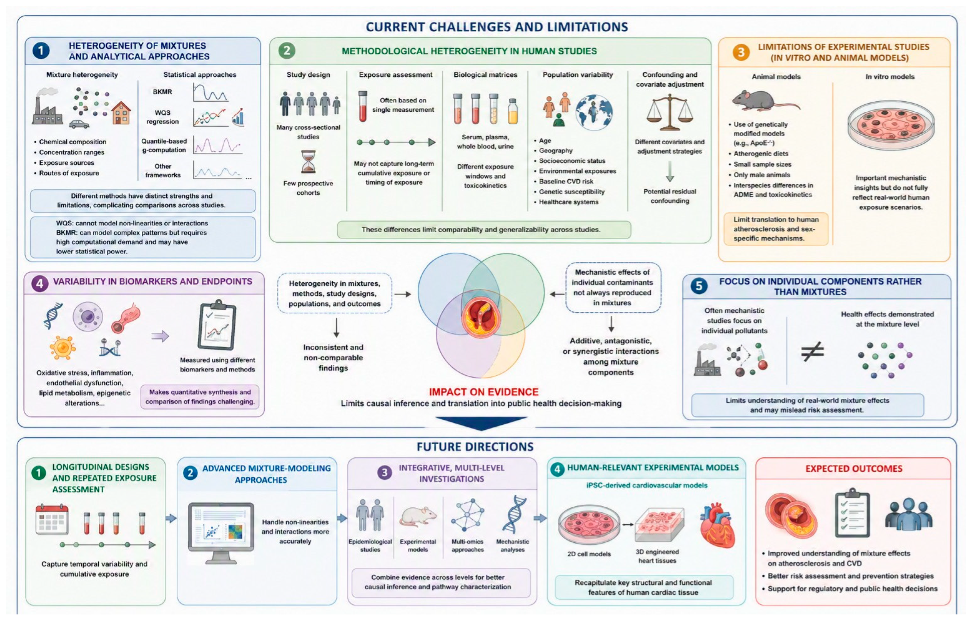
5. Challenges and Knowledge Gaps
6. Conclusions
Supplementary Materials
Author Contributions
Funding
Institutional Review Board Statement
Informed Consent Statement
Data Availability Statement
Acknowledgments
Conflicts of Interest
Abbreviations
| 8-OHdG | 8-hydroxy-2′-deoxyguanosine |
| AhR | Aryl hydrocarbon receptor |
| ASCVD | Atherosclerotic cardiovascular disease |
| BKMR | Bayesian kernel machine regression |
| BPA | Bisphenol A |
| CAPs | Concentrated ambient particulate matter |
| CIMT | Carotid intima-media thickness |
| CVD | Cardiovascular disease |
| EDC | Endocrine disrupting chemical |
| HDL | High-density lipoprotein |
| IHD | Ischemic heart disease |
| IL | Interleukin |
| LDL | Low-density lipoprotein |
| MCP-1 | Macrophage/monocyte chemoattractant protein-1 |
| MI | Myocardial infarction |
| NF-κB | Nuclear factor kappa-light-chain-enhancer of activated B cells |
| NO | Nitric oxide |
| Nrf2 | Nuclear factor erythroid 2-related factor 2 |
| OBS | Oxidative Balance Score |
| OxPL | Oxidized phospholipid |
| PAH | Polycyclic aromatic hydrocarbon |
| PCB | Polychlorinated biphenyl |
| PFAS | Per- and polyfluoroalkyl substances |
| PFOS | Perfluorooctane sulfonic acid |
| POP | Persistent organic pollutant |
| PM | Particulate matter |
| RAS | Renin–angiotensin system |
| ROS | Reactive oxygen species |
| THM | Trihalomethane |
| TNF-α | Tumor necrosis factor alpha |
| TyG | Triglyceride–glucose index |
| VOC | Volatile organic compound |
| WQS | Weighted quantile sum |
References
- Fenercioğlu, A.K.; Ünal, D.Ö. The Role of Endocrine Disrupting Chemicals in the Development of Atherosclerosis. Cardiovasc. Toxicol. 2025, 25, 1706–1717. [Google Scholar] [CrossRef] [Scilit]
- Johri, A.M.; Hill, B.; Grubic, N.; Sirwani, B.; Fraser, M.; Hétu, M.-F.; Douglas, P.S.; Fuster, V.; Ibanez, B.; Bundgaard, H.; et al. The prevalence of carotid subclinical atherosclerosis according to age: A systematic review and meta-analysis of the young to middle-age. J. Clin. Lipidol. 2026, 20, 250–261. [Google Scholar] [CrossRef] [Scilit]
- Xu, S.; Liu, Y.; Zhu, M.; Chen, K.; Xu, F.; Liu, Y. Global Burden of Atherosclerotic Cardiovascular Disease Attributed to Lifestyle and Metabolic Risks. Sci. China Life Sci. 2025, 68, 2739–2754. [Google Scholar] [CrossRef] [Scilit]
- Chong, B.; Jayabaskaran, J.; Jauhari, S.M.; Chan, S.P.; Goh, R.; Kueh, M.T.W.; Li, H.; Chin, Y.H.; Kong, G.; Anand, V.V.; et al. Global Burden of Cardiovascular Diseases: Projections from 2025 to 2050. Eur. J. Prev. Cardiol. 2025, 32, 1001–1015. [Google Scholar] [CrossRef] [Scilit] [PubMed]
- Li, Z.; Yang, Y.; Wang, X.; Yang, N.; He, L.; Wang, J.; Ping, F.; Xu, L.; Zhang, H.; Li, W.; et al. Comparative Analysis of Atheroscle-rotic Cardiovascular Disease Burden between Ages 20–54 and over 55 Years: Insights from the Global Burden of Disease Study 2019. BMC Med. 2024, 22, 303. [Google Scholar] [CrossRef] [Scilit]
- Gómez-Delgado, F.; Raya-Cruz, M.; Romero-Cabrera, J.L.; Pérez-Martínez, P. Environmental Pollution and Cardiovascular Health: Challenges and New Perspectives. Clin. Investig. Arterioscler. 2026, 38, 500802. [Google Scholar] [CrossRef] [Scilit]
- World Heart Federation. World Heart Report 2024. Clearing the Air to Address Pollution’s Cardiovascular Health Effects. Available online: https://world-heart-federation.org/report2024/ (accessed on 18 June 2026).
- Goleij, P.; Tabari, M.A.K.; Sanaye, P.M.; Moradi, A.; Karimi, H.; Rezaee, A.; Kumar, A.P.; Khan, H. Epigenetic Alterations Induced by Air Pollution: A Key Driver in Atherosclerosis Development. Cardiovasc. Toxicol. 2025, 25, 1288–1303. [Google Scholar] [CrossRef] [Scilit]
- Münzel, T.; Sørensen, M.; Hahad, O.; Nieuwenhuijsen, M.; Daiber, A. The Contribution of the Exposome to the Burden of Cardiovascular Disease. Nat. Rev. Cardiol. 2023, 20, 651–669. [Google Scholar] [CrossRef] [Scilit]
- Bhatnagar, A. Cardiovascular Effects of Particulate Air Pollution. Annu. Rev. Med. 2022, 73, 393–406. [Google Scholar] [CrossRef] [Scilit]
- Farzan, S.F.; Eunus, H.M.; Haque, S.E.; Sarwar, G.; Hasan, A.R.; Wu, F.; Islam, T.; Ahmed, A.; Shahriar, M.; Jasmine, F.; et al. Arsenic Exposure from Drinking Water and Endothelial Dysfunction in Bangladeshi Adolescents. Environ. Res. 2022, 208, 112697. [Google Scholar] [CrossRef] [Scilit]
- Grau-Pérez, M.; Caballero-Mateos, M.J.; Domingo-Relloso, A.; Navas-Acién, A.; Gómez-Ariza, J.L.; García-Barrera, T.; León-Latre, M.; Soriano-Gil, Z.; Jarauta, E.; Cenarro, A.; et al. Toxic Metals and Subclinical Atherosclerosis in Carotid, Femoral, and Coronary Vascular Territories: The Aragon Workers Health Study. Arter. Thromb. Vasc. Biol. 2022, 42, 87–99. [Google Scholar] [CrossRef] [Scilit]
- Harari, F.; Barregard, L.; Östling, G.; Sallsten, G.; Hedblad, B.; Forsgard, N.; Borné, Y.; Fagerberg, B.; Engström, G. Blood Lead Levels and Risk of Atherosclerosis in the Carotid Artery: Results from a Swedish Cohort. Environ. Health Perspect. 2019, 127, 127002. [Google Scholar] [CrossRef] [Scilit]
- Wu, X.; Wei, D.; Zhou, Y.; Cao, Q.; Han, G.; Han, E.; Chen, Z.; Guo, Y.; Huo, W.; Wang, C.; et al. Pesticide Exposures and 10-Year Atherosclerotic Cardiovascular Disease Risk: Integrated Epidemiological and Bioinformatics Analysis. J. Hazard. Mater. 2025, 485, 136835. [Google Scholar] [CrossRef] [Scilit]
- Mallah, M.A.; Mallah, M.A.; Liu, Y.; Xi, H.; Wang, W.; Feng, F.; Zhang, Q. Relationship Between Polycyclic Aromatic Hydrocarbons and Cardiovascular Diseases: A Systematic Review. Front. Public Health 2021, 9, 763706. [Google Scholar] [CrossRef] [Scilit]
- Gorini, F.; Tonacci, A.; Palazzo, M.; Borghini, A. From Exposure to Atherosclerosis: Mechanistic Insights into Phthalate-Driven Ischemic Heart Disease and Prevention Strategies. Life 2026, 16, 327. [Google Scholar] [CrossRef] [Scilit]
- Gorini, F.; Tonacci, A.; Palazzo, M.; Bustaffa, E.; Minichilli, F.; Borghini, A. Per- and Polyfluoroalkyl Substances Exposure and Ischemic Heart Disease: Emerging Evidence from the Literature. Antioxidants 2026, 15, 718. [Google Scholar] [CrossRef] [Scilit]
- Aimo, A.; Panichella, G.; Tommasi, E.; Revuelta-López, E.; Berastegui, E.; Bayés-Genís, A. The Effects of Microplastics and Nanoplastics on Cardiovascular Disease: Mechanisms and Perspectives. Nat. Rev. Cardiol. 2026. [Google Scholar] [CrossRef] [Scilit]
- Borghini, A.; Palazzo, M.; Tonacci, A.; Minichilli, F.; Wu, H.; Gorini, F. Environmental Exposure to Micro- and Nanoplastics: Linking Cardiovascular Disease and Cancer Through Shared Biological Pathways—A Critical Review. Antioxidants 2026, 15, 786. [Google Scholar] [CrossRef] [Scilit]
- Münzel, T.; Sørensen, M.; Lelieveld, J.; Landrigan, P.J.; Kuntic, M.; Nieuwenhuijsen, M.; Miller, M.R.; Schneider, A.; Daiber, A. A Comprehensive Review/Expert Statement on Environmental Risk Factors of Cardiovascular Disease. Cardiovasc. Res. 2025, 121, 1653–1678. [Google Scholar] [CrossRef] [Scilit]
- Libby, P.; Soehnlein, O. Inflammation in Atherosclerosis: Lessons and Therapeutic Implications. Immunity 2025, 58, 2383–2401. [Google Scholar] [CrossRef] [Scilit]
- Madaudo, C.; Coppola, G.; Parlati, A.L.M.; Corrado, E. Discovering Inflammation in Atherosclerosis: Insights from Pathogenic Pathways to Clinical Practice. Int. J. Mol. Sci. 2024, 25, 6016. [Google Scholar] [CrossRef] [Scilit]
- Montano, L.; Baldini, G.M.; Piscopo, M.; Liguori, G.; Lombardi, R.; Ricciardi, M.; Esposito, G.; Pinto, G.; Fontanarosa, C.; Spinelli, M.; et al. Polycyclic Aromatic Hydrocarbons (PAHs) in the Environment: Occupational Exposure, Health Risks and Fertility Implications. Toxics 2025, 13, 151. [Google Scholar] [CrossRef] [Scilit]
- Venkatraman, G.; Giribabu, N.; Mohan, P.S.; Muttiah, B.; Govindarajan, V.K.; Alagiri, M.; Abdul Rahman, P.S.; Karsani, S.A. Environmental impact and human health effects of polycyclic aromatic hydrocarbons and remedial strategies: A detailed review. Chemosphere 2024, 351, 141227. [Google Scholar] [CrossRef] [Scilit]
- Mallah, M.A.; Changxing, L.; Mallah, M.A.; Naveed, M.; Liu, Y.; Noreen, S.; Xi, H.; Wang, W.; Feng, F.; Zhang, Q.; et al. Association of Urinary Polycyclic Aromatic Hydrocarbon Metabolites and Cardiovascular Disease among US Population: A Cross-Sectional Study. Environ. Res. 2022, 209, 112775. [Google Scholar] [CrossRef] [Scilit]
- Rojas, G.A.; Saavedra, N.; Saavedra, K.; Hevia, M.; Morales, C.; Lanas, F.; Salazar, L.A. Polycyclic Aromatic Hydrocarbons (PAHs) Exposure Triggers Inflammation and Endothelial Dysfunction in BALB/c Mice: A Pilot Study. Toxics 2022, 10, 497. [Google Scholar] [CrossRef] [Scilit]
- Grebenciucova, E.; VanHaerents, S. Interleukin 6: At the Interface of Human Health and Disease. Front. Immunol. 2023, 14, 1255533. [Google Scholar] [CrossRef] [Scilit]
- Kaur, K. Role of Interferon-Gamma (IFN-γ) in Pathophysiology and Management of Deep Vein Thrombosis. Immuno 2025, 5, 46. [Google Scholar] [CrossRef] [Scilit]
- Lin, Y.; Jamison, S.; Lin, W. Interferon-γ Activates Nuclear Factor-κB in Oligodendrocytes through a Process Mediated by the Unfolded Protein Response. PLoS ONE 2012, 7, e36408. [Google Scholar] [CrossRef] [Scilit]
- He, J.; Pang, Q.; Huang, C.; Xie, J.; Hu, J.; Wang, L.; Wang, C.; Meng, L.; Fan, R. Environmental Dose of 16 Priority-Controlled PAHs Mixture Induce Damages of Vascular Endothelial Cells Involved in Oxidative Stress and Inflammation. Toxicol. In Vitro 2022, 79, 105296. [Google Scholar] [CrossRef] [Scilit]
- Du, T.; Shen, X.; Zhan, R. Association of Mixed Polycyclic Aromatic Hydrocarbons Exposure with Cardiovascular Disease and the Mediating Role of Inflammatory Indices in US Adults. Environ. Health Prev. Med. 2024, 29, 70. [Google Scholar] [CrossRef] [Scilit]
- Borghini, A.; Mercuri, A.; Andreassi, M.G. Neutrophil-to-Lymphocyte, Platelet-to-Lymphocyte Ratios, and Systemic Immune-Inflammation Index as Predictors of Mortality in Coronary Artery Disease. J. Cardiovasc. Transl. Res. 2023, 16, 473–475. [Google Scholar] [CrossRef] [Scilit]
- Zhang, P.; Cui, D.; Zhang, P.; Wang, H.; Hao, Y.; Ma, J.; Li, Q.; Zhang, A.; Li, D.; Li, X. Correlation between Blood Inflammatory Indices and Carotid Intima-Media Thickness in the Middle-Aged and Elderly Adults. J. Stroke Cerebrovasc. Dis. 2024, 33, 107715. [Google Scholar] [CrossRef] [Scilit]
- Zhao, S.; Liu, H.; Zhong, H.; Qin, Y.; Duan, J.; Liu, A. Systemic Inflammation Mediates the Association Between Blood Trihalomethane Concentrations and Cardiovascular Disease in U.S. Individuals Over 45: Insights from NHANES 2005–2012. Cardiovasc. Toxicol. 2025, 25, 1055–1066. [Google Scholar] [CrossRef] [Scilit]
- de Castro Medeiros, L.; de Alencar, F.L.S.; Navoni, J.A.; de Araujo, A.L.C.; do Amaral, V.S. Toxicological Aspects of Trihalomethanes: A Systematic Review. Environ. Sci. Pollut. Res. 2019, 26, 5316–5332. [Google Scholar] [CrossRef] [Scilit]
- Li, X.F.; Mitch, W.A. Drinking Water Disinfection Byproducts (DBPs) and Human Health Effects: Multidisciplinary Challenges and Opportunities. Environ. Sci. Technol. 2018, 52, 1681–1689. [Google Scholar] [CrossRef] [Scilit]
- Chen, T.; He, H.; Tang, W.; Liu, Z.; Zhang, H. Association of Blood Trihalomethane Concentrations with Diabetes Mellitus in Older Adults in the US: A Cross-Sectional Study of NHANES 2013–2018. Front. Endocrinol. 2024, 15, 1401131. [Google Scholar] [CrossRef] [Scilit]
- Makris, K.C.; Andrianou, X.D.; Charisiadis, P.; Burch, J.B.; Seth, R.K.; Ioannou, A.; Picolos, M.; Christophi, C.A.; Chatterjee, S. Association between Exposures to Brominated Trihalomethanes, Hepatic Injury and Type II Diabetes Mellitus. Environ. Int. 2016, 92–93, 486–493. [Google Scholar] [CrossRef] [Scilit]
- Ma, L.; Yang, W.; Gao, W.; Liu, X.; Dong, M.; An, G.; Meng, X. IL-17 as a Therapeutic Target in Cardiovascular Diseases: Mechanistic Insights and Translational Opportunities. Pharmacol. Res. 2025, 219, 107879. [Google Scholar] [CrossRef] [Scilit]
- Shekhar, C.; Khosya, R.; Thakur, K.; Mahajan, D.; Kumar, R.; Kumar, S.; Sharma, A.K. A Systematic Review of Pesticide Exposure, Associated Risks, and Long-Term Human Health Impacts. Toxicol. Rep. 2024, 13, 101840. [Google Scholar] [CrossRef] [Scilit]
- Xu, W.; Dong, Y.; Liu, S.; Hu, F.; Cai, Y. Association between organophosphorus pesticides and obesity among American adults. Environ. Health 2024, 23, 65. [Google Scholar] [CrossRef] [Scilit]
- Dong, Y.; Xu, W.; Liu, S.; Xu, Z.; Qiao, S.; Cai, Y. Serum albumin and liver dysfunction mediate the associations between organophosphorus pesticide exposure and hypertension among US adults. Sci. Total Environ. 2024, 948, 174748. [Google Scholar] [CrossRef] [Scilit]
- Park, J.; Park, S.K.; Choi, Y.H. Environmental pyrethroid exposure and diabetes in U.S. adults. Environ. Res. 2019, 172, 399–407. [Google Scholar] [CrossRef] [Scilit]
- Linton, M.F.; Fazio, S. Cyclooxygenase-2 and atherosclerosis. Curr. Opin. Lipidol. 2002, 13, 497–504. [Google Scholar] [CrossRef] [Scilit]
- Mansuri, A.; Trivedi, C.; Chokshi, S.; Jantrania, K.; Kumar, A. Phthalate Exposure: Prevalence, Health Effects, Regulatory Frameworks, and Remediation. Chem. Res. Toxicol. 2025, 38, 1291–1308. [Google Scholar] [CrossRef] [Scilit]
- Gong, W.; Zhu, H.; Sun, X.; Zhang, J.; Lin, M.; Sun, P. The association between exposure to phthalates and cardiovascular disease: A comprehensive study utilizing NHANES data from 2005 to 2018 and network toxicology. Chem. Biol. Interact. 2025, 420, 111651. [Google Scholar] [CrossRef] [Scilit]
- Hu, X.; Li, J.; Fu, M.; Zhao, X.; Wang, W. The JAK/STAT signaling pathway: From bench to clinic. Signal Transduct. Target Ther. 2021, 6, 402. [Google Scholar] [CrossRef] [Scilit]
- Sun, L.; Liu, C.; Xu, X.; Ying, Z.; Maiseyeu, A.; Wang, A.; Allen, K.; Lewandowski, R.P.; Bramble, L.A.; Morishita, M.; et al. Ambient fine particulate matter and ozone exposures induce inflammation in epicardial and perirenal adipose tissues in rats fed a high fructose diet. Part. Fibre Toxicol. 2013, 10, 43. [Google Scholar] [CrossRef] [Scilit]
- Dai, J.; Sun, C.; Yao, Z.; Chen, W.; Yu, L.; Long, M. Exposure to concentrated ambient fine particulate matter disrupts vascular endothelial cell barrier function via the IL-6/HIF-1α signaling pathway. FEBS Open Bio 2016, 6, 720–728. [Google Scholar] [CrossRef] [Scilit]
- Zhang, Z.; Wang, C.; Lin, C.; Wu, Y.; Wei, J.; Lu, J.; Chen, B.; Wu, C.; Zhang, X.; Yang, Y.; et al. Association of long-term exposure to ozone with cardiovascular mortality and its metabolic mediators: Evidence from a nationwide, population-based, prospective cohort study. Lancet Reg. Health West. Pac. 2024, 52, 101222. [Google Scholar] [CrossRef] [Scilit]
- Phipps, B.L.; Suwannasual, U.; Lucero, J.; Mitchell, N.A.; Lund, A.K. Vehicle emissions-exposure alters expression of systemic and tissue-specific components of the renin-angiotensin system and promotes outcomes associated with cardiovascular disease and obesity in wild-type C57BL/6 male mice. Toxicol. Rep. 2021, 8, 846–862. [Google Scholar] [CrossRef] [Scilit]
- Jantzen, K.; Jensen, A.; Kermanizadeh, A.; Elholm, G.; Sigsgaard, T.; Møller, P.; Roursgaard, M.; Loft, S. Inhalation of House Dust and Ozone Alters Systemic Levels of Endothelial Progenitor Cells, Oxidative Stress, and Inflammation in Elderly Subjects. Toxicol. Sci. 2018, 163, 353–363. [Google Scholar] [CrossRef] [Scilit]
- Cavusoglu, E.; Marmur, J.D.; Yanamadala, S.; Chopra, V.; Hegde, S.; Nazli, A.; Singh, K.P.; Zhang, M.; Eng, C. Elevated baseline plasma IL-8 levels are an independent predictor of long-term all-cause mortality in patients with acute coronary syndrome. Atherosclerosis 2015, 242, 589–594. [Google Scholar] [CrossRef] [Scilit]
- Aragon, M.J.; Chrobak, I.; Brower, J.; Roldan, L.; Fredenburgh, L.E.; McDonald, J.D.; Campen, M.J. Inflammatory and Vasoactive Effects of Serum Following Inhalation of Varied Complex Mixtures. Cardiovasc. Toxicol. 2016, 16, 163–171. [Google Scholar] [CrossRef] [Scilit]
- Shan, Q.; Wang, J.; Huang, F.; Lv, X.; Ma, M.; Du, Y. Augmented atherogenesis in ApoE-null mice co-exposed to polychlorinated biphenyls and 2,3,7,8-tetrachlorodibenzo-p-dioxin. Toxicol. Appl. Pharmacol. 2014, 276, 136–146. [Google Scholar] [CrossRef] [Scilit]
- Othman, N.; Ismail, Z.; Selamat, M.I.; Sheikh Abdul Kadir, S.H.; Shibraumalisi, N.A. A Review of Polychlorinated Biphenyls (PCBs) Pollution in the Air: Where and How Much Are We Exposed To? Int. J. Environ. Res. Public Health 2022, 19, 13923. [Google Scholar] [CrossRef] [Scilit]
- Blackowicz, M.J.; Persky, V.W.; Sargis, R.M.; Freels, S.; Anderson, H.A.; Turyk, M.E. Polychlorinated biphenyls and incident coronary heart disease-related outcomes in Great Lakes fish consumers. Environ. Res. 2024, 255, 119071. [Google Scholar] [CrossRef] [Scilit]
- Akinyemi, B.; Obeng-Gyasi, E. Combined Effects of Metals, PCBs, Dioxins, and Furans on Cardiovascular Dysfunction. J. Xenobiot. 2025, 15, 94. [Google Scholar] [CrossRef] [Scilit]
- Moghadasian, M.H.; McManus, B.M.; Nguyen, L.B.; Shefer, S.; Nadji, M.; Godin, D.V.; Green, T.J.; Hill, J.; Yang, Y.; Scudamore, C.H.; et al. Pathophysiology of apolipoprotein E deficiency in mice: Relevance to apo E-related disorders in humans. FASEB J. 2001, 15, 2623–2630. [Google Scholar] [CrossRef] [Scilit]
- Wang, F.; Xia, W.; Liu, F.; Li, J.; Wang, G.; Gu, J. Interferon regulator factor 1/retinoic inducible gene I (IRF1/RIG-I) axis mediates 25-hydroxycholesterol-induced interleukin-8 production in atherosclerosis. Cardiovasc. Res. 2012, 93, 190–199. [Google Scholar] [CrossRef] [Scilit]
- Roth, K.; Yang, Z.; Agarwal, M.; Gurdziel, K.; Petriello, M.C. Exposure to a PFAS mixture alters cholesterol lipoprotein subfractions and induces a foam cell-like aortic macrophage expression profile in hyperlipidemic LDLr−/− mice. Toxicol. Appl. Pharmacol. 2026, 507, 117683. [Google Scholar] [CrossRef] [Scilit]
- Yang, X.; Li, X.; Li, X.; Zhang, H.; Wang, C.; Chen, X. Per- and polyfluoroalkyl substances and cardiovascular disease: A mechanistic and epidemiological synthesis. Ecotoxicol. Environ. Saf. 2026, 313, 119901. [Google Scholar] [CrossRef] [Scilit]
- Pan, Y.; Du, Z.; Ma, Y.; Chen, C.; He, S.; Zhang, M.; Baral, K.; Xu, L.; Xu, M.; Zhao, M. Suppression effect of folate on poly- and perfluoroalkyl substance-induced alterations in lipids and the atherogenic index of plasma in adolescents. Lipids Health Dis. 2025, 24, 213. [Google Scholar] [CrossRef] [Scilit]
- Deng, P.; Wang, C.; Wahlang, B.; Sexton, T.; Morris, A.J.; Hennig, B. Co-exposure to PCB126 and PFOS increases biomarkers associated with cardiovascular disease risk and liver injury in mice. Toxicol. Appl. Pharmacol. 2020, 409, 115301. [Google Scholar] [CrossRef] [Scilit]
- Durham, J.; Tessmann, J.W.; Deng, P.; Hennig, B.; Zaytseva, Y.Y. The role of perfluorooctane sulfonic acid (PFOS) exposure in inflammation of intestinal tissues and intestinal carcinogenesis. Front. Toxicol. 2023, 5, 1244457. [Google Scholar] [CrossRef] [Scilit]
- Sun, Z.; Xu, Y.; Liu, Y.; Tao, X.; Zhou, P.; Feng, H.; Weng, Y.; Lu, X.; Wu, J.; Wei, Y.; et al. Associations of Exposure to 56 Serum Trace Elements with the Prevalence and Severity of Acute Myocardial Infarction: Omics, Mixture, and Mediation Analysis. Biol. Trace Elem. Res. 2025, 203, 4466–4478. [Google Scholar] [CrossRef] [Scilit]
- Fang, H.; Fang, G.H.; Chen, D.J.; Chang, H.J.; Yang, M.H.; Zhang, H.; Jiang, Y.J.; Cai, Y.F.; Hu, Z.J.; Peng, X.E. The association of environmental toxicants exposure with cardiovascular disease risk: A comprehensive analysis from population to molecular mechanism. Ecotoxicol. Environ. Saf. 2026, 309, 119562. [Google Scholar] [CrossRef] [Scilit]
- Ma, Y.; Niu, R.; Sun, Z.; Wang, J.; Luo, G.; Zhang, J.; Wang, J. Inflammatory responses induced by fluoride and arsenic at toxic concentration in rabbit aorta. Arch. Toxicol. 2012, 86, 849–856. [Google Scholar] [CrossRef] [Scilit]
- Baltoo, R.; Thakur, R.; Rana, S. Cardiovascular Health and Fluoride Toxicity: A Qualitative Systematic Review. Cardiovasc. Toxicol. 2026, 26, 40. [Google Scholar] [CrossRef] [Scilit]
- Al-Forkan, M.; Wali, F.B.; Khaleda, L.; Alam, M.J.; Chowdhury, R.H.; Datta, A.; Rahman, M.Z.; Hosain, N.; Maruf, M.F.; Chowdhury, M.A.Q.; et al. Association of arsenic-induced cardiovascular disease susceptibility with genetic polymorphisms. Sci. Rep. 2021, 11, 6263. [Google Scholar] [CrossRef] [Scilit]
- Subramaniam, N.K.; Gagnon, N.; Makhani, K.; Kukolj, N.; Mouradian, M.H.; Giles, B.H.; Srikannan, H.; Fruh, V.; Meliker, J.; Wellenius, G.A.; et al. In vitro and in vivo approaches to assess atherosclerosis following exposure to low-dose mixtures of arsenic and cadmium. Toxicol. Appl. Pharmacol. 2023, 481, 116763. [Google Scholar] [CrossRef] [Scilit]
- Oliveira, T.F.; Batista, P.R.; Leal, M.A.; Campagnaro, B.P.; Nogueira, B.V.; Vassallo, D.V.; Meyrelles, S.S.; Padilha, A.S. Chronic Cadmium Exposure Accelerates the Development of Atherosclerosis and Induces Vascular Dysfunction in the Aorta of ApoE−/− Mice. Biol. Trace Elem. Res. 2019, 187, 163–171. [Google Scholar] [CrossRef] [Scilit]
- Kattoor, A.J.; Pothineni, N.V.K.; Palagiri, D.; Mehta, J.L. Oxidative Stress in Atherosclerosis. Curr. Atheroscler. Rep. 2017, 19, 42. [Google Scholar] [CrossRef] [Scilit]
- Vomund, S.; Schäfer, A.; Parnham, M.J.; Brüne, B.; von Knethen, A. Nrf2, the Master Regulator of Anti-Oxidative Responses. Int. J. Mol. Sci. 2017, 18, 2772. [Google Scholar] [CrossRef] [Scilit]
- Zhang, D.; Liu, X.; Xiao, Q.; Han, L.; Yang, J.; Li, X.; Xu, J.; Zheng, Q.; Ma, J.; Chen, J.; et al. Co-Exposure to Bisphenols, Parabens, and Antimicrobials and Association with Coronary Heart Disease: Oxidative Stress as a Potential Mediating Factor? Environ. Sci. Technol. 2023, 57, 531–538. [Google Scholar] [CrossRef] [Scilit]
- Fonseca, M.I.; Lorigo, M.; Cairrao, E. Endocrine-Disrupting Effects of Bisphenol A on the Cardiovascular System: A Review. J. Xenobiot. 2022, 12, 181–213. [Google Scholar] [CrossRef] [Scilit]
- Yin, T.; Zhu, X.; Cheang, I.; Zhou, Y.; Liao, S.; Lu, X.; Zhou, Y.; Yao, W.; Li, X.; Zhang, H. Urinary phenols and parabens metabolites associated with cardiovascular disease among adults in the United States. Environ. Sci. Pollut. Res. Int. 2023, 30, 25093–25102. [Google Scholar]
- Marques, A.C.; Mariana, M.; Cairrao, E. Triclosan and Its Consequences on the Reproductive, Cardiovascular and Thyroid Levels. Int. J. Mol. Sci. 2022, 23, 11427. [Google Scholar] [CrossRef] [Scilit]
- Yang, N.; Chen, J.; Zhu, Y.; Shan, W.; Cao, Z.; Fu, Y.; Cao, H.; Li, Y.; Xiang, Y.; Ding, S.; et al. Human cardiac organoid model reveals antibacterial triclocarban promotes myocardial hypertrophy by interfering with endothelial cell metabolism. Sci. Bull. 2025, 70, 342–346. [Google Scholar] [CrossRef] [Scilit]
- Senderovic, A.; Galijasevic, S. The Role of Inducible Nitric Oxide Synthase in Assessing the Functional Level of Coronary Artery Lesions in Chronic Coronary Syndrome. Cardiol. Res. 2024, 15, 330–339. [Google Scholar] [CrossRef] [Scilit]
- Zhang, Y.; Ji, X.; Ku, T.; Li, G.; Sang, N. Heavy metals bound to fine particulate matter from northern China induce season-dependent health risks: A study based on myocardial toxicity. Environ. Pollut. 2016, 216, 380–390. [Google Scholar] [CrossRef] [Scilit]
- Guo, L.C.; Lv, Z.; Ma, W.; Xiao, J.; Lin, H.; He, G.; Li, X.; Zeng, W.; Hu, J.; Zhou, Y.; et al. Contribution of heavy metals in PM2.5 to cardiovascular disease mortality risk, a case study in Guangzhou, China. Chemosphere 2022, 297, 134102. [Google Scholar] [CrossRef] [Scilit]
- Tenório, M.C.D.S.; Graciliano, N.G.; Moura, F.A.; Oliveira, A.C.M.; Goulart, M.O.F. N-Acetylcysteine (NAC): Impacts on Human Health. Antioxidants 2021, 10, 967. [Google Scholar] [CrossRef] [Scilit]
- Saul, D.; Lischer, C.; Bruns, H.; Ziegler, N.; Kannt, A.; Michel, M.; Mougiakakos, D. OGG1 activation improves T cell resilience to oxidative stress after allo-SCT and T cell engager exposure. Leukemia 2025, 39, 3037–3041. [Google Scholar] [CrossRef] [Scilit]
- Zhang, J.; Liang, R.; Ding, X.; Chen, B.; Tan, Q.; Wang, M.; Hu, Y.; Liu, Q.; Chen, W.; Zhou, M. Dinitroaniline herbicide exposure, mitochondrial DNA copy number, and 10-year risk of atherosclerotic cardiovascular disease: A community-based cohort study. Environ. Pollut. 2025, 373, 126113. [Google Scholar] [CrossRef] [Scilit]
- Scardino, B.; Xing, D.; Agrawal, A.; Bhuiyan, M.S.; Conrad, S.A.; Vanchiere, J.A.; Bhuiyan, M.M.R.; Orr, A.W.; Kevil, C.G.; Bhuiyan, M.A.N. Combined effects of heavy metals and volatile organic compounds on oxidative stress and cardiovascular risk. Am. Heart J. Plus 2026, 66, 100801. [Google Scholar] [CrossRef] [Scilit]
- Meng, L.; Wang, Y.; Wang, X.; Mu, M.; Zheng, H. Association between exposure to volatile organic compounds and atherogenic index of plasma in NHANES 2011-2018. Sci. Rep. 2025, 15, 9024. [Google Scholar] [CrossRef] [Scilit]
- Pan, Z.; Gong, T.; Liang, P. Heavy Metal Exposure and Cardiovascular Disease. Circ. Res. 2024, 134, 1160–1178. [Google Scholar] [CrossRef] [Scilit]
- Ross, D.; Siegel, D. NQO1 in protection against oxidative stress. Curr. Opin. Toxicol. 2018, 7, 67–72. [Google Scholar] [CrossRef] [Scilit]
- Ma, Y.; Ma, Z.; Yin, S.; Yan, X.; Wang, J. Arsenic and fluoride induce apoptosis, inflammation and oxidative stress in cultured human umbilical vein endothelial cells. Chemosphere 2017, 167, 454–461. [Google Scholar] [CrossRef] [Scilit]
- Brandes, R.P.; Weissmann, N.; Schröder, K. NADPH oxidases in cardiovascular disease. Free Radic. Biol. Med. 2010, 49, 687–706. [Google Scholar] [CrossRef] [Scilit]
- Ortega-Romero, M.; Rojas Lima, E.; Rubio-Gutiérrez, J.C.; Narváez Morales, J.; Bravo Carvajal, I.Y.; Esparza García, M.; Mejia, M.Á.; Tamayo Y Orozco, J.A.; Mendez-Hernández, P.; Barbier, O.C.; et al. Oxidative stress and cardiovascular risk related to urinary metal(loid) levels in a pediatric population. Environ. Geochem. Health 2026, 48, 311. [Google Scholar] [CrossRef] [Scilit]
- Cvetkovic, T.; Saric, S.; Stefanovic, N.; Stojiljkovic, V.; Djordjevic, B.; Stojanovic, D.; Cvetkovic, M.; Deljanin Ilic, M. Plasma advanced oxidation products as an additional tool in assessment of post-infarction heart failure. J. Int. Med. Res. 2022, 50, 3000605221139711. [Google Scholar] [CrossRef] [Scilit]
- Schalkwijk, C.G.; Stehouwer, C.D.A. Methylglyoxal, a Highly Reactive Dicarbonyl Compound, in Diabetes, Its Vascular Complications, and Other Age-Related Diseases. Physiol. Rev. 2020, 100, 407–461. [Google Scholar] [CrossRef] [Scilit] [PubMed]
- Aratani, Y. Myeloperoxidase: Its role for host defense, inflammation, and neutrophil function. Arch. Biochem. Biophys. 2018, 40, 47–52. [Google Scholar] [CrossRef] [Scilit]
- Li, Z.; Wang, L.; Ren, Y.; Huang, Y.; Liu, W.; Lv, Z.; Qian, L.; Yu, Y.; Xiong, Y. Arginase: Shedding light on the mechanisms and opportunities in cardiovascular diseases. Cell Death Discov. 2022, 8, 413. [Google Scholar] [CrossRef] [Scilit]
- Caga-Anan, M.V.; Dasanayaka, N.N.; Seneviratne, A.N. Environmental Factors and Lipid Metabolism in Atherosclerosis Development. Lipidology 2026, 3, 7. [Google Scholar] [CrossRef] [Scilit]
- Matar, D.B.; Elahi, M.A.; Sukkarieh, H.; Nassar, W.K.; Aljada, A. Unlocking the secrets: Adipose tissue dysfunction and atherosclerosis-mechanisms and innovative therapeutic approaches. Atherosclerosis 2025, 408, 120424. [Google Scholar] [CrossRef] [Scilit]
- Chen, J.; Zhou, Q.; Wang, Y.; Wu, Y.; Li, M.; Wang, H.; Zheng, G.; Hao, Y.; Cao, X.; Yang, W.; et al. Association of PM2.5-bound multiple metals co-exposure with early cardiovascular damage: A panel study in young adults combining metabolomics. Environ. Pollut. 2025, 371, 125964. [Google Scholar] [CrossRef] [Scilit]
- Yang, X.F.; Shang, D.J. The role of peroxisome proliferator-activated receptor γ in lipid metabolism and inflammation in atherosclerosis. Cell Biol. Int. 2023, 47, 1469–1487. [Google Scholar] [CrossRef] [Scilit]
- Cai, D.; Fang, Z.Q.; Cui, N.H.; Wang, B.; Gao, M.J.; Wang, X.B. Per- and Polyfluoroalkyl Substances, Serum Lipidome, and Clinical Outcomes after Percutaneous Coronary Intervention in Type 2 Diabetic Patients: A Prospective Nested Case-control Study. J. Atheroscler. Thromb. 2025, 32, 1486–1522. [Google Scholar] [CrossRef] [Scilit]
- Hu, Y.; Kong, Y.; Tian, X.; Zhang, X.; Zuo, Y. Association between Heavy metals and triglyceride-glucose-related index: A mediation analysis of inflammation indicators. Lipids Health Dis. 2025, 24, 46. [Google Scholar] [CrossRef] [Scilit]
- Zhang, D.; Liu, X.; Tu, J.; Xiao, Q.; Han, L.; Fu, J.; Bian, J.; Zhang, R.; Chen, J.; Shao, Y.; et al. Mediating Role of Glucose-Lipid Metabolism in the Association between the Increased Risk of Coronary Heart Disease and Exposure to Organophosphate Esters, Phthalates, and Polycyclic Aromatic Hydrocarbons. Environ. Health 2024, 2, 170–179. [Google Scholar] [CrossRef] [Scilit]
- Yang, L.; Li, X.; Ni, L.; Lin, Y. Treatment of endothelial cell dysfunction in atherosclerosis: A new perspective integrating traditional and modern approaches. Front. Physiol. 2025, 16, 1555118. [Google Scholar] [CrossRef] [Scilit]
- Quan, C.; Sun, Q.; Lippmann, M.; Chen, L.C. Comparative effects of inhaled diesel exhaust and ambient fine particles on inflammation, atherosclerosis, and vascular dysfunction. Inhal. Toxicol. 2010, 22, 738–753. [Google Scholar] [CrossRef] [Scilit]
- Gurevitz, C.; Zadok, O.I.B.; Leshem-Lev, D.; Hodeda, L.; Rotholz, A.; Kornowski, R.; Eisen, A. Circulating Endothelial Progenitor Cells in Patients with Established Cardiovascular Disease Treated with PCSK9 Monoclonal Antibodies. Am. J. Prev. Cardiol. 2024, 20, 100896. [Google Scholar] [CrossRef] [Scilit]
- McGraw, K.E.; Riggs, D.W.; Rai, S.; Navas-Acien, A.; Xie, Z.; Lorkiewicz, P.; Lynch, J.; Zafar, N.; Krishnasamy, S.; Taylor, K.C.; et al. Exposure to volatile organic compounds—Acrolein, 1,3-butadiene, and crotonaldehyde—Is associated with vascular dysfunction. Environ. Res. 2021, 196, 110903. [Google Scholar] [CrossRef] [Scilit]
- Moreno-Gómez-Toledano, R.; Delgado-Marín, M.; Sánchez-Esteban, S.; Cook-Calvete, A.; Ortiz, S.; Bosch, R.J.; Saura, M. Combination of Bisphenol A and Its Emergent Substitute Molecules Is Related to Heart Disease and Exerts a Differential Effect on Vascular Endothelium. Int. J. Mol. Sci. 2023, 24, 12188. [Google Scholar] [CrossRef] [Scilit]
- Lakshmanan, I.; Batra, S.K. Protocol for Apoptosis Assay by Flow Cytometry Using Annexin V Staining Method. Bio Protoc. 2013, 3, e374. [Google Scholar] [CrossRef] [Scilit]
- Dho, S.H.; Cho, M.; Woo, W.; Jeong, S.; Kim, L.K. Caspases as master regulators of programmed cell death: Apoptosis, pyroptosis and beyond. Exp. Mol. Med. 2025, 57, 1121–1132. [Google Scholar] [CrossRef] [Scilit]
- Morgan, M.J.; Kim, Y.S. Roles of RIPK3 in necroptosis, cell signaling, and disease. Exp. Mol. Med. 2022, 54, 1695–1704. [Google Scholar] [CrossRef] [Scilit]
- Knapp, M.; Gil-Mika, M.; Sawicki, R.; Lisowska, A.; Kaminski, M.; Sobkowicz, B.; Ptaszynska, K. Pentraxin 3 as a marker of development and severity of stable coronary artery disease. Adv. Med. Sci. 2024, 69, 391–397. [Google Scholar] [CrossRef] [Scilit]
- Zheng, Y.Y.; Hua, C.; Lin, X.X. PI3K/AKT/mTOR axis in vascular malformations: From molecular insights to targeted clinical trials. Orphanet J. Rare Dis. 2025, 20, 624. [Google Scholar] [CrossRef] [Scilit]
- Adeva-Andany, M.M.; Fernández-Fernández, C.; Sánchez-Bello, R.; Donapetry-García, C.; Martínez-Rodríguez, J. The role of carbonic anhydrase in the pathogenesis of vascular calcification in humans. Atherosclerosis 2015, 241, 183–191. [Google Scholar] [CrossRef] [Scilit]
- Khoukaz, H.B.; Ji, Y.; Braet, D.J.; Vadali, M.; Abdelhamid, A.A.; Emal, C.D.; Lawrence, D.A.; Fay, W.P. Drug Targeting of Plasminogen Activator Inhibitor-1 Inhibits Metabolic Dysfunction and Atherosclerosis in a Murine Model of Metabolic Syndrome. Arter. Thromb. Vasc. Biol. 2020, 40, 1479–1490. [Google Scholar] [CrossRef] [Scilit]
- Baccarelli, A.; Ghosh, S. Environmental exposures, epigenetics and cardiovascular disease. Curr. Opin. Clin. Nutr. Metab. Care 2012, 15, 323–329. [Google Scholar] [CrossRef] [Scilit]
- Santovito, D.; Atzler, D.; Weber, C. Epigenetic regulation in atherosclerosis and its therapeutic potential. Nat. Rev. Cardiol. 2026. [Google Scholar] [CrossRef] [Scilit]
- Zhang, Y.; Mei, J.; Li, J.; Zhang, Y.; Zhou, Q.; Xu, F. DNA Methylation in Atherosclerosis: A New Perspective. Evid. Based Complement. Altern. Med. 2021, 2021, 6623657. [Google Scholar] [CrossRef] [Scilit]
- Lin, C.Y.; Lee, H.L.; Hwang, Y.T.; Huang, P.C.; Wang, C.; Sung, F.C.; Wu, C.; Su, T.C. Urinary heavy metals, DNA methylation, and subclinical atherosclerosis. Ecotoxicol. Environ. Saf. 2020, 204, 111039. [Google Scholar] [CrossRef] [Scilit]
- Yu, D. Lead exposure in the 21st century: Modeling a path from crisis to prevention. Eco Environ. Health 2025, 4, 100159. [Google Scholar] [CrossRef] [Scilit]
- Rosengren, E.; Barregard, L.; Sallsten, G.; Fagerberg, B.; Engström, G.; Fagman, E.; Forsgard, N.; Lundh, T.; Bergström, G.; Harari, F. Exposure to Lead and Coronary Artery Atherosclerosis: A Swedish Cross-Sectional Population-Based Study. J. Am. Heart Assoc. 2025, 14, e037633. [Google Scholar] [CrossRef] [Scilit]
- Nezu, T.; Hosomi, N.; Aoki, S.; Matsumoto, M. Carotid Intima-Media Thickness for Atherosclerosis. J. Atheroscler. Thromb. 2016, 23, 18–31. [Google Scholar] [CrossRef] [Scilit]
- Li, W.; Kong, W.; Shen, C.; Fan, H.; Shen, Y.; Zhang, Y.; Zheng, L. Dissecting the pathogenic effects of ambient air pollution exposure and its blood DNA methylation markers on cardiovascular disease risk. Clin. Epigenet. 2025, 17, 205. [Google Scholar] [CrossRef] [Scilit]
- Islam, F.; Nukala, S.K.; Shrestha, P.; Badgery-Parker, T.; Foo, F. Air pollution and cardiovascular disease: A systematic review of the effects of air pollution, including bushfire smoke, on cardiovascular disease. Am. Heart J. Plus 2025, 54, 100546. [Google Scholar] [CrossRef] [Scilit]
- Li, X.; Zhou, Z.; Zhou, Y.; Hao, H.; Zhou, L.; Niu, Y.; Zhang, Y.; Liu, S.; Chen, R.; Kan, H.; et al. Effects of long-term exposure to nitrogen dioxide with cardiovascular mortality: Evidence from a nationwide cohort study. Ecotoxicol. Environ. Saf. 2026, 313, 119950. [Google Scholar] [CrossRef] [Scilit]
- Lv, L.S.; Xia, X.; Yang, Z.W.; An, N.; Zhang, M.; Liu, X.Y.; Gao, L.D. Associations between sulfur dioxide exposure and mortality, with a focus on cardiovascular disease: A multi-county time series analysis in central China. BMC Public Health 2025, 25, 4130. [Google Scholar] [CrossRef] [Scilit]
- Çakmak, H.A.; Demir, M. MicroRNA and Cardiovascular Diseases. Balk. Med. J. 2020, 37, 60–71. [Google Scholar] [CrossRef] [Scilit]
- Wahlang, B.; Petriello, M.C.; Perkins, J.T.; Shen, S.; Hennig, B. Polychlorinated biphenyl exposure alters the expression profile of microRNAs associated with vascular diseases. Toxicol. In Vitro 2016, 35, 180–187. [Google Scholar] [CrossRef] [Scilit]
- Mollajan, E.; Yazdani, S.; Ghasemzadeh, M. miR-21 in cardiovascular disease: New insights and emerging therapeutic potential. Discov. Appl. Sci. 2025, 7, 447. [Google Scholar] [CrossRef] [Scilit]
- Martinez, E.C.; Lilyanna, S.; Wang, P.; Vardy, L.A.; Jiang, X.; Armugam, A.; Jeyaseelan, K.; Richards, A.M. MicroRNA-31 promotes adverse cardiac remodeling and dysfunction in ischemic heart disease. J. Mol. Cell. Cardiol. 2017, 112, 27–39. [Google Scholar] [CrossRef] [Scilit]
- Martinez-Arroyo, O.; Ortega, A.; Flores-Chova, A.; Sanchez-Garcia, B.; Garcia-Garcia, A.B.; Chaves, F.J.; Martin-Escudero, J.C.; Forner, M.J.; Redon, J.; Cortes, R. High miR-126-3p levels associated with cardiovascular events in a general population. Eur. J. Intern. Med. 2023, 113, 49–56. [Google Scholar] [CrossRef] [Scilit]
- Sessa, F.; Salerno, M.; Esposito, M.; Cocimano, G.; Pomara, C. miRNA Dysregulation in Cardiovascular Diseases: Current Opinion and Future Perspectives. Int. J. Mol. Sci. 2023, 24, 5192. [Google Scholar] [CrossRef] [Scilit]
- Shan, Q.; Qu, F.; Chen, N. 2,3,7,8-Tetrachlorodibenzo-p-dioxin (TCDD) and Polychlorinated Biphenyl Coexposure Alters the Expression Profile of MicroRNAs in the Liver Associated with Atherosclerosis. BioMed Res. Int. 2020, 2020, 2652756. [Google Scholar] [CrossRef] [Scilit]
- Zhang, Z.; Bai, C.; Zhao, L.; Liu, L.; Guo, W.; Liu, M.; Yang, H.; Lai, X.; Zhang, X.; Yang, L. Polycyclic aromatic hydrocarbons exposure and arterial stiffness-related plasma miRNAs: A panel study. Environ. Toxicol. Pharmacol. 2024, 108, 104464. [Google Scholar] [CrossRef] [Scilit]
- Palombo, C.; Kozakova, M. Arterial stiffness, atherosclerosis and cardiovascular risk: Pathophysiologic mechanisms and emerging clinical indications. Vasc. Pharmacol. 2016, 77, 1–7. [Google Scholar] [CrossRef] [Scilit]
- Bai, C.; Yang, H.; Zhao, L.; Liu, L.; Guo, W.; Yu, J.; Li, M.; Liu, M.; Lai, X.; Zhang, X.; et al. The mediating role of plasma microRNAs in the association of phthalates exposure with arterial stiffness: A panel study. Environ. Res. 2022, 212, 113469. [Google Scholar] [CrossRef] [Scilit]
- Shi, B.; Wang, X.; Xue, T.; Liu, J.; Wu, W.; Luo, Y.; Zhu, H.; Pan, D. Expression level of miR-146a is associated with the coronary lesion severity and clinical prognosis in patients with unstable angina pectoris. Int. J. Cardiol. Cardiovasc. Risk Prev. 2025, 24, 200367. [Google Scholar] [CrossRef] [Scilit]
- Grishanova, A.Y.; Perepechaeva, M.L. Aryl Hydrocarbon Receptor in Oxidative Stress as a Double Agent and Its Biological and Therapeutic Significance. Int. J. Mol. Sci. 2022, 23, 6719. [Google Scholar] [CrossRef] [Scilit]
- Bock, K.W. Aryl hydrocarbon receptor (AHR): From selected human target genes and crosstalk with transcription factors to multiple AHR functions. Biochem. Pharmacol. 2019, 168, 65–70. [Google Scholar] [CrossRef] [Scilit]
- Dragon, J.; Hoaglund, M.; Badireddy, A.R.; Nielsen, G.; Schlezinger, J.; Shukla, A. Perfluoroalkyl Substances (PFAS) Affect Inflammation in Lung Cells and Tissues. Int. J. Mol. Sci. 2023, 24, 8539. [Google Scholar] [CrossRef] [Scilit]
- Khanna, S.; Mann, G.; Vasanthakumar, S.; Arnott, C.; Nerlekar, N. From Cushion to Culprit: The Role of Epicardial Adipose Tissue in Cardiovascular Disease. Heart Lung Circ. 2025, 34, 1006–1020. [Google Scholar] [CrossRef] [Scilit]
- Shen, Z.; Liu, Z.; Cai, S.; Fu, H.; Gan, Y.; Li, X.; Wang, X.; Liu, C.; Ma, W.; Chen, J.; et al. Copper homeostasis and cuproptosis in myocardial infarction: Molecular mechanisms, treatment strategies and potential therapeutic targets. Front. Pharmacol. 2025, 16, 1525585. [Google Scholar] [CrossRef] [Scilit]
- Amerikanou, C.; Kleftaki, S.A.; Karavoltsos, S.; Tagkouli, D.; Sakellari, A.; Valsamidou, E.; Gioxari, A.; Kalogeropoulos, N.; Kaliora, A.C. Vanadium, cobalt, zinc, and rubidium are associated with markers of inflammation and oxidative stress in a Greek population with obesity. Front. Endocrinol. 2023, 14, 1265310. [Google Scholar] [CrossRef] [Scilit]
- Li, W.; Zhou, J.; Boon, D.; Fan, T.; Anneser, E.; Goodman, J.E.; Prueitt, R.L. Nickel in ambient particulate matter and respiratory or cardiovascular outcomes: A critical review. Environ. Pollut. 2024, 347, 123442. [Google Scholar] [CrossRef] [Scilit]
- Vandenberg, L.N.; Colborn, T.; Hayes, T.B.; Heindel, J.J.; Jacobs, D.R., Jr.; Lee, D.H.; Shioda, T.; Soto, A.M.; vom Saal, F.S.; Welshons, W.V.; et al. Hormones and endocrine-disrupting chemicals: Low-dose effects and nonmonotonic dose responses. Endocr. Rev. 2012, 33, 378–455. [Google Scholar] [CrossRef] [Scilit]
- Haug, M.; Dunder, L.; Lind, P.M.; Lind, L.; Salihovic, S. Associations of perfluoroalkyl substances (PFAS) with lipid and lipoprotein profiles. J. Expo. Sci. Environ. Epidemiol. 2023, 33, 757–765. [Google Scholar] [CrossRef] [Scilit]
- Zhu, L.; Liu, B.; Hu, Y.; Wang, M.; Furtado, J.D.; Rimm, E.B.; Grandjean, P.; Sun, Q. Per- and polyfluoroalkyl substances, apolipoproteins and the risk of coronary heart disease in US men and women. Environ. Health 2024, 23, 108. [Google Scholar] [CrossRef] [Scilit]
- Liu, G.; Zhang, B.; Hu, Y.; Rood, J.; Liang, L.; Qi, L.; Bray, G.A.; DeJonge, L.; Coull, B.; Grandjean, P.; et al. Associations of Perfluoroalkyl substances with blood lipids and Apolipoproteins in lipoprotein subspecies: The POUNDS-lost study. Environ. Health 2020, 19, 5. [Google Scholar] [CrossRef] [Scilit]
- Kashobwe, L.; Sadrabadi, F.; Brunken, L.; Coelho, A.C.M.F.; Sandanger, T.M.; Braeuning, A.; Buhrke, T.; Öberg, M.; Hamers, T.; Leonards, P.E.G. Legacy and alternative per- and polyfluoroalkyl substances (PFAS) alter the lipid profile of HepaRG cells. Toxicology 2024, 506, 153862. [Google Scholar] [CrossRef] [Scilit] [PubMed]
- Qian, Y.; Ducatman, A.; Ward, R.; Leonard, S.; Bukowski, V.; Lan Guo, N.; Shi, X.; Vallyathan, V.; Castranova, V. Perfluorooctane sulfonate (PFOS) induces reactive oxygen species (ROS) production in human microvascular endothelial cells: Role in endothelial permeability. J. Toxicol. Environ. Health A 2010, 73, 819–836. [Google Scholar] [CrossRef] [Scilit]
- Ruggeri, R.M.; Minuti, A.; Gianì, F.; Masto, R.; Romano, D.; Aliquò, F.; Campennì, A.; Campo, S.; Cannavò, S.; D’Ascola, A. Polychlorinated Biphenyls (PCBS)-induced oxidative stress and inflammation in human thyrocytes: Involvement of AhR and NRF-2/HO-1 pathway. Endocrine 2025, 87, 252–261. [Google Scholar] [CrossRef] [Scilit]
- Cimini, F.A.; Sentinelli, F.; Oldani, A.; Barchetta, I.; Cavallo, M.G. Adipose Tissue Dysfunction and Metabolic Diseases: The Role of Vitamin D/Vitamin D Receptor Axis. Int. J. Mol. Sci. 2025, 26, 10256. [Google Scholar] [CrossRef] [Scilit]
- Frigolet, M.E.; Torres, N.; Tovar, A.R. The renin-angiotensin system in adipose tissue and its metabolic consequences during obesity. J. Nutr. Biochem. 2013, 24, 2003–2015. [Google Scholar] [CrossRef] [Scilit]
- Borodzicz-Jażdżyk, S.; Jażdżyk, P.; Łysik, W.; Cudnoch-Jȩdrzejewska, A.; Czarzasta, K. Sphingolipid metabolism and signaling in cardiovascular diseases. Front. Cardiovasc. Med. 2022, 9, 915961. [Google Scholar] [CrossRef] [Scilit]
- Reventun, P.; Sanchez-Esteban, S.; Cook, A.; Cuadrado, I.; Roza, C.; Moreno-Gomez-Toledano, R.; Muñoz, C.; Zaragoza, C.; Bosch, R.J.; Saura, M. Bisphenol A induces coronary endothelial cell necroptosis by activating RIP3/CamKII dependent pathway. Sci. Rep. 2020, 10, 4190. [Google Scholar] [CrossRef] [Scilit]
- Birk, M.; Baum, E.; Zadeh, J.K.; Manicam, C.; Pfeiffer, N.; Patzak, A.; Helmstädter, J.; Steven, S.; Kuntic, M.; Daiber, A.; et al. Angiotensin II Induces Oxidative Stress and Endothelial Dysfunction in Mouse Ophthalmic Arteries via Involvement of AT1 Receptors and NOX2. Antioxidants 2021, 10, 1238. [Google Scholar] [CrossRef] [Scilit]
- Martínez-Razo, L.D.; Almeida-Aguirre, E.K.P.; Bobadilla-Bravo, M.; Mancilla-Herrera, I.; Martínez-Ibarra, A.; Cerbón, M. Mono(2-Ethylhexyl) Phthalate Induces Inflammatory and Angiogenic Alterations Mediated by the PI3K/AKT Pathway in HTR-8/SVneo Trophoblastic Cells. Environ. Toxicol. 2026, 41, 209–222. [Google Scholar] [CrossRef] [Scilit]
- Domingo-Relloso, A.; Riffo-Campos, A.L.; Haack, K.; Rentero-Garrido, P.; Ladd-Acosta, C.; Fallin, D.M.; Tang, W.Y.; Herreros-Martinez, M.; Gonzalez, J.R.; Bozack, A.K.; et al. Cadmium, Smoking, and Human Blood DNA Methylation Profiles in Adults from the Strong Heart Study. Environ. Health Perspect. 2020, 128, 67005. [Google Scholar] [CrossRef] [Scilit]
- Shiek, S.S.; Mani, M.S.; Kabekkodu, S.P.; Dsouza, H.S. Health repercussions of environmental exposure to lead: Methylation perspective. Toxicology 2021, 461, 152927. [Google Scholar] [CrossRef] [Scilit]
- Wu, X.; Cao, X.; Lintelmann, J.; Peters, A.; Koenig, W.; Zimmermann, R.; Schneider, A.; Wolf, K.; KORA-Study Group. Assessment of the association of exposure to polycyclic aromatic hydrocarbons, oxidative stress, and inflammation: A cross-sectional study in Augsburg, Germany. Int. J. Hyg. Environ. Health 2022, 244, 113993. [Google Scholar] [CrossRef] [Scilit]
- Vondráček, J.; Pěnčíková, K.; Ciganek, M.; Pivnička, J.; Karasová, M.; Hýžďalová, M.; Strapáčová, S.; Pálková, L.; Neča, J.; Matthews, J.; et al. Environmental six-ring polycyclic aromatic hydrocarbons are potent inducers of the AhR-dependent signaling in human cells. Environ. Pollut. 2020, 266, 115125. [Google Scholar] [CrossRef] [Scilit]
- Minjares, M.; Wu, W.; Wang, J.M. Oxidative Stress and MicroRNAs in Endothelial Cells under Metabolic Disorders. Cells 2023, 12, 1341. [Google Scholar] [CrossRef] [Scilit]
- Disner, G.R.; Lopes-Ferreira, M.; Lima, C. Where the Aryl Hydrocarbon Receptor Meets the microRNAs: Literature Review of the Last 10 Years. Front. Mol. Biosci. 2021, 8, 725044. [Google Scholar] [CrossRef] [Scilit]
- Hýžďalová, M.; Pivnička, J.; Zapletal, O.; Vázquez-Gómez, G.; Matthews, J.; Neča, J.; Pěnčíková, K.; Machala, M.; Vondráček, J. Aryl Hydrocarbon Receptor-Dependent Metabolism Plays a Significant Role in Estrogen-Like Effects of Polycyclic Aromatic Hydrocarbons on Cell Proliferation. Toxicol. Sci. 2018, 165, 447–461. [Google Scholar] [CrossRef] [Scilit]
- Rigassi, L.; Rosselli, M.; Leeners, B.; Popa, M.A.; Dubey, R.K. Estradiol Downregulates MicroRNA-193a to Mediate Its Anti-Mitogenic Actions on Human Coronary Artery Smooth Muscle Cell Growth. Cells 2025, 14, 1132. [Google Scholar] [CrossRef] [Scilit]
- White, S.S.; Birnbaum, L.S. An overview of the effects of dioxins and dioxin-like compounds on vertebrates, as documented in human and ecological epidemiology. J. Environ. Sci. Health Part C 2009, 27, 197–211. [Google Scholar] [CrossRef] [Scilit]
- Wang, C.; Petriello, M.C.; Zhu, B.; Hennig, B. PCB 126 induces monocyte/macrophage polarization and inflammation through AhR and NF-κB pathways. Toxicol. Appl. Pharmacol. 2019, 367, 71–81. [Google Scholar] [CrossRef] [Scilit]
- Lim, E.J.; Májková, Z.; Xu, S.; Bachas, L.; Arzuaga, X.; Smart, E.; Tseng, M.T.; Toborek, M.; Hennig, B. Coplanar polychlorinated biphenyl-induced CYP1A1 is regulated through caveolae signaling in vascular endothelial cells. Chem. Biol. Interact. 2008, 176, 71–78. [Google Scholar] [CrossRef] [Scilit]
- Bey, L.; Coumoul, X.; Kim, M.J. TCDD aggravates the formation of the atherosclerotic plaque in ApoE KO mice with a sexual dimorphic pattern. Biochimie 2022, 195, 54–58. [Google Scholar] [CrossRef] [Scilit]
- Palazzo, M.; Borghini, A.; Bustaffa, E.; Baldacci, S.; Gorini, F.; Minichilli, F. Genetic Polymorphisms as Modifiers of Health Risks from Exposure to Toxic Elements: A Traditional Literature Review. Toxics 2026, 14, 375. [Google Scholar] [CrossRef] [Scilit]
- Palazzo, M.; Gorini, F.; Simonini, L.; Minichilli, F.; Borghini, A. Genetic Polymorphisms as Key Modulators of Cardiovascular Risk from Endocrine-Disrupting Chemicals. Genes 2026, 17, 717. [Google Scholar] [CrossRef] [Scilit]
- Fontanelli, L.; Castronovo, A.; Ferri, C.; Vozzi, F.; Recchia, F.A.; Borghini, A. iPSC-Derived Endothelial Cells as Experimental Models for Predictive and Personalized Strategies in Cardiovascular and Cerebrovascular Disease. Int. J. Mol. Sci. 2026, 27, 780. [Google Scholar] [CrossRef] [Scilit]


| First Author and Year | Design | Model/Population | Exposure | Outcome | Key Findings | Relevance to Atherosclerosis |
|---|---|---|---|---|---|---|
| Rojas et al. 2022 [26] | Experimental in vivo study | Male BALB/c mice | PAH mixture (phenanthrene 55%, fluoranthene 25%, pyrene 20%); intranasal instillation (10 μL) at 10, 30, and 50 μg; 5 days/week for 5 weeks | Systemic inflammation | Increased serum IL-6 and IFN-γ. No significant changes in serum IL-10, IL-17A, TNF-α. No significant differences in aortic IL-6 or TNF-α gene expression. | PAH mixture–induced systemic pro-inflammatory response; early driver of atherogenesis. |
| He et al. 2022 [30] | Experimental in vitro study | Primary HUVECs | Mixture of 16 EPA priority PAHs; exposure concentrations derived from blood levels reported in the Chinese general population and tested at environmental (1×) and higher doses (10×–1000×); 48 h treatment duration | TNF-α expression and NF-κB signaling activation | Increased TNF-α expression across all PAH-treated groups. Increased NF-κB activation (p-p65 expression) in the highest exposure group (1000× PAHs). | PAH mixture–induced inflammatory activation; NF-κB/TNF-α signaling; pro-atherogenic vascular inflammation |
| Du et al. 2024 [31] | Cross-sectional study (NHANES 2003–2016) | 9136 US adults; 10.5% with CVD | Urinary levels of 7 OH-PAHs metabolites (creatinine-adjusted) | Prevalent CVD (self-reported: IHD, angina, MI, stroke, heart failure) | Positive overall association between PAH mixture and CVD (BKMR); overall mixture effect increasing at exposure levels ≥ 55th percentile. 2-OHFlu as the main driver of the mixture effect (highest PIP) and strongest positive exposure–response relationship with CVD. NLR and SII mediation of 9.7% and 2.0% of the PAH mixture–CVD association, respectively. | PAH mixture–induced systemic inflammation contributing to CVD risk; supports a role for mixture-driven mechanisms in vascular injury and atherosclerosis development. |
| Zhao et al. 2025 [34] | Cross-sectional study (NHANES 2005–2012) | 5400 US adults aged ≥45 years; 7.8% with CVD | Blood concentrations of THMs, TCM, TBM, BDCM, and DBCM | Prevalent CVD (self-reported: IHD, MI, angina, stroke) | Positive association between THM mixture and CVD risk in WQS analyses (OR = 1.16, 95%CI: 1.03–1.32) TCM as predominant contributor (58.0% mixture weight). Higher TCM concentrations (Q4 vs. Q1: OR = 1.42, 95%CI: 1.05–1.93) and TTHM concentrations (Q3 vs. Q1: OR = 1.57, 95%CI: 1.16–2.12) associated with increased CVD prevalence. NLR-mediated 7.12% of the TTHM–CVD association. Identification of 84 shared THM-, CVD-, and aging-related genes; enrichment of IL-17 signaling, fluid shear stress and atherosclerosis, AGE–RAGE signaling, and apoptosis-related pathways. Inflammation-related mechanisms and IL-17 signaling as potential links between THM exposure, vascular injury, atherosclerosis, and CVD. | THM mixture–induced systemic inflammation contributing to CVD risk. Involvement of IL-17–related inflammatory pathways and immune activation processes relevant to atherosclerosis progression. |
| Wu et al. 2025 [14] | Cross-sectional study integrating epidemiological analyses and toxicogenomic bioinformatics | 2291 Chinese participants from rural areas | Mixed pesticide exposure (34 selected pesticides including neonicotinoids, organochlorine pesticides, organophosphorus pesticides, pyrethroids, and herbicides measured in blood and urine). | 10-year ASCVD risk and inflammation-related molecular pathways | Total pesticide mixture exposure positively associated with 10-year ASCVD risk in QGC (OR = 3.223, 95%CI: 2.196–4.730) and WQS models (OR = 4.642, 95%CI: 3.070–7.020). Linear dose–response relationship between overall pesticide mixture exposure and high ASCVD risk. Identification of 112 pesticide-related atherosclerosis target genes; enrichment of TNF and PI3K-Akt signaling pathways; IL6, TNF, and PTGS2 identified among the major hub genes linking pesticide exposure to ASCVD. | Pesticide mixture-associated inflammatory activation, including pathways implicated in atherogenesis and ASCVD development |
| Gong et al. 2025 [46] | Cross-sectional study (NHANES 2005–2018) integrated with network toxicology | 12,127 US adults | Urinary concentrations of 10 DEHP metabolites | Prevalent CVD; network toxicology pathways | Positive association between phthalate mixture exposure and CVD (OR = 1.21, 95%CI: 1.07–1.37) (WQS). MEOHP identified as the main contributor to the mixture effect, followed by MECPP, MBzP, and MnBP. Identification of inflammation-related pathways, including JAK–STAT signaling. | Phthalate mixture-associated cardiovascular toxicity; involvement of inflammatory signaling pathways relevant to vascular inflammation and atherosclerosis. |
| Sun et al. 2013 [48] | Experimental in vivo study | High-fructose-fed male Sprague–Dawley rats | CAPs (356 ± 52 μg/m3) + O3 (0.485 ± 0.041 ppm), 8 h/day for 9 consecutive weekdays. | Inflammation in epicardial adipose tissue | Macrophage infiltration in epicardial adipose tissue. Increased Tnfα, Mcp1, and leptin expression. Reduced Il10 and adiponectin expression. No consistent enhancement of inflammatory responses compared with single-pollutant exposures | CAPs and O3-induced epicardial adipose tissue inflammation, macrophage infiltration, and adipokine imbalance potentially contributing to vascular inflammation and atherogenesis. |
| Phipps et al. 2021 [51] | Experimental in vivo study | Male C57BL/6 mice fed LF or HF diet | MVE (diesel + gasoline exhaust; 100 μg PM/m3), whole-body inhalation, 6 h/day, 7 days/week, for 30 days | Vascular and adipose inflammation | Increased vascular MOMA-2 staining in MVE-exposed mice irrespective of diet. Increased adipose Il6 and Mcp1 expression in both LF- and HF-fed mice. Increased leptin expression restricted to HF-fed mice | Traffic-related air pollution mixture-induced vascular and adipose inflammation; enhanced immune cell recruitment; potential contribution to atherogenesis. |
| Jantzen et al. 2018 [52] | Randomized double-blind crossover study | 23 healthy elderly subjects | House dust (275 μg/m3 PM2.5) + O3 (100 ppb), 5.5 h exposure | Systemic inflammatory signaling | House dust + O3 co-exposure, IL-8 mRNA expression increase of 59% (95%CI: 15–120%). O3 exposure alone, TNF mRNA expression reduction of 9% (95%CI: −16 to −0.8%) and MCP-1 mRNA expression reduction of 7% (95%CI: −12 to −2%). No significant inflammatory effects following house dust exposure alone. | House dust and O3 co-exposure–associated inflammatory activation, suggesting potential synergistic effects of combined air pollutant exposures. |
| Aragon et al. 2016 [54] | In vivo inhalation exposure with subsequent in vitro/ex vivo serum bioactivity assays | C57BL/6 mice | Acute inhalation exposure (6 h) to road dust (349 μg/m3 PM2.5), road dust + O3 (344 μg/m3 PM2.5 + 0.33 ppm O3), road dust + MVE; 342 μg/m3 PM2.5, MVE gases, MVE particulate matter (MVE-PM; 328 μg/m3 PM2.5), or wood smoke (380 μg/m3 PM2.5); serum collected 18–24 h post-exposure and incubated with murine cerebrovascular endothelial cells for 4 h. | Inflammatory potential of circulating factors | Road dust exposure: increased IL-6, CXCL1, MCP-1, and CCL5 expression. Road dust + O3 exposure: increased MCP-1 expression. No significant induction of inflammatory genes following MVE gases, MVE-PM, or road dust + MVE exposure. Attenuation of road dust-induced inflammatory responses following co-exposure to MVE gases, suggesting antagonistic interactions among mixture components | Air pollution mixture-associated circulating inflammatory mediators and vascular inflammatory activation |
| Shan et al. 2014 [55] | Experimental in vivo study | Male ApoE−/− mice | TCDD (15 μg/kg) + Aroclor1254 (55 mg/kg; >60 PCB congeners) co-exposure via intraperitoneal injection four times over a 6-week period | Inflammation and innate immune activation | 4.74-fold increase in circulating MCP-1 and 4.75-fold increase in hepatic MCP-1 following TCDD/Aroclor1254 co-exposure. 5.74-fold increase in macrophage recruitment (CD68+). 3.7–5.9-fold increase in PF4 accumulation within atherosclerotic lesions. 1.83-fold increase in aortic RIG-I expression and 8.74-fold increase in hepatic RIG-I expression. Greater atherosclerotic lesion burden than individual exposures. No significant changes in plasma IL-6 or E-selectin levels. | TCDD/Aroclor1254 mixture-associated inflammatory and immune activation relevant to atherogenesis. |
| Roth et al. 2026 [61] | Experimental in vivo study | Male Ldlr−/− mice fed an atherogenic diet | PFAS mixture (PFOA, PFOS, PFNA, PFHxS, GenX); 2 mg/L each in drinking water for 7 weeks | Inflammatory signaling in aortic macrophages | 982 differentially expressed genes. Increased Cxcl2 and Cxcl17 expression. No significant changes in M1 marker (Il1β and Tnfα) and M2 marker (Il4 and Il10) expression profile. | PFAS mixture-induced chemokine signaling and immune cell recruitment potentially contributing to vascular inflammation and atherogenesis. |
| Pan et al. 2025 [63] | Cross-sectional study (NHANES 2005–2018) integrated with network toxicology | 1099 adolescents | Mixture of PFOS, PFOA, PFNA, and PFHxS assessed in serum | Inflammation | Positive associations of PFAS mixture exposure with AIP. Network toxicology identification of IL-10, TNF, and caspase-1 as core inflammation-related targets; shared PFAS–folate target caspase-1, suggesting inflammasome-mediated mechanisms relevant to atherosclerosis. | PFAS mixture–associated activation of inflammatory and inflammasome-related pathways potentially contributing to vascular inflammation and atherosclerosis. |
| Deng et al. 2020 [64] | Experimental in vivo study | Male C57BL/6 mice | Acute co-exposure to PCB126 (0.5 mg/kg) and PFOS (250 mg/kg) by intragastric gavage; evaluation after 48 h of exposure | Inflammatory signaling and vascular inflammation | Periportal inflammatory cell infiltration. No significant changes in hepatic Tnfα expression | Limited evidence of inflammation-related effects following PFOS–PCB126 co-exposure |
| Sun et al. 2025 [66] | Cross-sectional study | 195 Chinese adults (94 AMI cases, 101 controls). AMI severity assessed by Gensini score. | Serum mixture of 56 trace elements; mixture analyses focused on Fe, Cu, Rb, Nb, Mo, Sb, and Ge. | AMI prevalence and severity | Positive associations of Cu and Rb with both AMI prevalence and severity. Inverse association of Fe with AMI prevalence. Positive association of Sb with AMI severity. Positive associations between trace element mixtures and both AMI prevalence (ERS OR = 2.72, 95%CI: 1.91–3.86) and severity (ERS OR = 1.94, 95%CI: 1.54–2.45). hsCRP-mediated effects accounting for 8.6% and 48.5% of the mixture effects on AMI prevalence and severity, respectively. hsCRP-mediated associations of low Fe (29.1%) and high Rb (15.6%) with AMI prevalence. | hsCRP-related inflammatory mechanisms potentially linking trace element co-exposure to atherosclerosis progression and AMI risk. |
| Fang et al. 2026 [67] | Cross-sectional study and prospective cohort study | 3142 patients with T2DM, 1470 patients with obstructive CAD, 1212 patients undergoing PCI | Redox-related metal mixture identified through elementomic profiling, composed of Ni, Sr, Ti, V, and Zr | Inflammatory signaling | Higher TNF-α concentrations. Nominally higher IL-1β, IL-8, and MIP-1β concentrations. 22% increase in NF-κB pathway cytokines. NF-κB pathway cytokines mediating 9% of the association with obstructive CAD and 32% of the association with post-PCI MACCE. | Metal mixture-associated NF-κB-mediated inflammatory signaling linked to coronary atherosclerotic burden and adverse cardiovascular outcomes. |
| Ma et al. 2012 [68] | Experimental in vivo study | New Zealand White rabbits | As (13 mg/L As2O3), F (50 mg/L NaF), or combined As + F exposure via drinking water; 6-month exposure | Inflammation | Increased MCP-1 protein levels in As-, F-, and As+F-exposed rabbits. Increased IL-6 protein levels following As exposure. Upregulation of MCP-1, IL-8, and IL-6 mRNA expression. Lower induction of inflammatory mediators under co-exposure compared with single-contaminant exposure | As- and F mixture-induced inflammatory responses relevant to atherogenesis |
| Subramaniam et al. 2024 [71] | Combined in vitro and in vivo study | RAW264.7 macrophages, C166 endothelial cells, and ApoE−/− mice | Environmentally relevant low-dose As/Cd mixture (5–50 ppb As; 1.5–5 ppb Cd) in drinking water for 13 weeks in male and female ApoE−/− mice; parallel low-dose in vitro exposures | Inflammation | No significant inflammatory response observed in macrophages, with unchanged TNF-α, IL-1β, and IL-6 levels after exposure to As, Cd, or their combination. No increase in plaque macrophage content | No amplification of pro-atherogenic inflammatory signaling compared with individual-metal exposure. |
| First Author and Year | Design | Model/Population | Exposure | Outcome | Key Findings | Relevance to Atherosclerosis |
|---|---|---|---|---|---|---|
| He et al. 2022 [30] | Experimental in vitro study | Primary HUVECs | Mixture of 16 EPA priority PAHs; exposure concentrations derived from blood levels reported in the Chinese general population and tested at environmental (1×) and higher doses (10×–1000×); 48 h treatment duration | ROS production, oxidative DNA damage (8-OHdG), antioxidant defense (SOD), Nrf2/HO-1 signaling pathway. | Increased ROS levels following PAH mixture exposure, with peak levels observed after 24 h in most treatment groups. Increased 8-OHdG levels after 48 h exposure in all PAH-treated groups (1×–1000×). Reduced SOD activity after 48 h exposure in the 10×, 100×, and 1000× PAH groups. Increased expression of Nrf2 and HO-1 following PAH mixture exposure. | PAH mixture–induced oxidative stress; oxidative DNA damage and altered antioxidant defenses; redox imbalance relevant to atherogenesis. |
| Zhang et al. 2023 [75] | Case–control study | 69 IHD cases, 146 controls | Co-exposure to bisphenols (BPA, BPF, BPS, BPAF, BPP, BPZ, BPAP), parabens (MeP, EtP, PrP, BuP), TCS, and triclocarban; urinary biomonitoring-based exposure assessment | Oxidative stress | Positive joint association between mixture exposure and IHD risk (OR = 1.52, 95%CI: 1.25–1.84). 2.22 ng/mL increase in urinary 8-OHdG per decile increase in mixture exposure. BuP, TCS, BPAP, and BPF as the main contributors to 8-OHdG elevation. 8-OHdG mediating 68.8% of the overall mixture–IHD association. Significant mediation of BPA-, BPF-, BPAP-, and TCS-associated IHD risk through 8-OHdG (5.73%, 9.10%, 19.8%, and 28.8%, respectively). | Endocrine-disrupting chemical mixture-induced oxidative stress relevant to atherosclerotic cardiovascular disease |
| Sun et al. 2013 [48] | Experimental in vivo study | High-fructose-fed male Sprague–Dawley rats | CAPs (356 ± 52 μg/m3) + O3 (0.485 ± 0.041 ppm), 8 h/day for 9 consecutive weekdays. | Oxidative stress in epicardial adipose tissue | Increased iNOS expression. Reduced mitochondrial area in epicardial adipose tissue. No consistent enhancement compared with single-pollutant exposures | CAPs and O3-induced oxidative stress and mitochondrial dysfunction in epicardial adipose tissue potentially contributing to vascular injury and atherogenesis. |
| Phipps et al. 2021 [51] | Experimental in vivo study | Male C57BL/6 mice fed low-fat or high-fat diet | MVE (diesel + gasoline exhaust; 100 μg PM/m3), whole-body inhalation, 6 h/day, 7 days/week, for 30 days | Vascular oxidative stress | Increased vascular ROS production (DHE staining) in MVE-exposed LF-fed mice compared with controls. Greater oxidative burden in HF-diet-fed mice exposed to MVE. No significant exposure-diet interaction. | Mixed vehicle emissions-induced vascular oxidative stress potentially contributing to endothelial injury and atherogenesis. |
| Zhang et al. 2016 [81] | Experimental in vitro study | H9C2 rat cardiomyocytes | Heavy metal-containing PM2.5 mixture (Cr, Ni, Cu, Cd, Pb, Zn, Mn, and Co) collected during different seasons in a coal-burning region of northern China; 0–10 μg/mL for 24 h | Oxidative stress | Dose-dependent ROS generation. iNOS increase 2.17-fold (spring PM2.5) and 4.64-fold (winter PM2.5). Attenuation of ROS-mediated responses following NAC pretreatment. ROS-dependent inflammatory and apoptotic signaling | PM2.5-associated metal mixture-induced oxidative stress relevant to cardiovascular toxicity and atherogenesis. |
| Jantzen et al. 2018 [52] | Randomized double-blind crossover study | 23 healthy elderly subjects | House dust (275 μg/m3 PM2.5) + O3 (100 ppb), 5.5 h exposure | Systemic oxidative stress | Increased ROS production capacity in monocytes (+30%) and granulocytes (+25%). Increased OGG1 mRNA expression (31%). Limited or absent responses following single-pollutant exposures. | Combined air pollutant exposure-induced systemic oxidative stress and oxidative DNA damage responses potentially contributing to endothelial injury and atherosclerosis. |
| Zhang et al. 2025 [85] | Community-based cohort study with cross-sectional and longitudinal analyses | Chinese community-dwelling adults from the Wuhan–Zhuhai cohort (n = 3399 cross-sectional participants; longitudinal panel of 158 participants with 363 observations). | Combined serum levels of dinitroaniline herbicides (trifluralin and pendimethalin) | 10-year ASCVD risk; mtDNAcn | Trifluralin–pendimethalin co-exposure associated with increased 10-year ASCVD risk in BKMR and WQS analyses; WQS β = 0.092% (95%CI: 0.035–0.143); trifluralin as major contributor. Trifluralin exposure associated with increased ASCVD risk (β = 0.272%, 95%CI: 0.148–0.377) and lower mtDNAcn (β = −0.058, 95%CI: −0.104 to −0.012). Lower mtDNAcn associated with increased ASCVD risk (β = −0.298%, 95%CI: −0.528 to −0.068); mtDNAcn mediation of 3.8% of the trifluralin–ASCVD association. No overall mixture effect on mtDNAcn. | Herbicide-associated mitochondrial dysfunction and oxidative DNA damage as potential mechanisms contributing to ASCVD risk and atherogenesis |
| Scandino et al. 2026 [86] | Cross-sectional study (NHANES 2011–2020) | 6516 adults aged ≥20 years | Combined exposure to 11 HMs (As, Ba, Cd, Co, Cs, Hg, Mn, Mo, Pb, Sn, W) and 11 VOC metabolites; urinary biomonitoring-based exposure assessment. | OBS and prevalent CVD | Combined HM+VOC exposure associated with lower OBS (higher oxidative stress). Strongest inverse associations observed for Cd and VOC metabolites including N-acetyl-S-(2-hydroxyethyl)-L-cysteine and N-acetyl-S-(2-carbamoyl-2-hydroxyethyl)-L-cysteine. Association observed across demographic subgroups and particularly pronounced among individuals aged 20–59 years, women, Mexican Americans, non-Hispanic Asians, and with the highest income. Higher OBS associated with lower odds of CVD (OR = 0.969, 95%CI: 0.959–0.979). Modest mediation of the OBS–CVD association by TyG, AIP, CRI-II, and non-HDL cholesterol (3.8–8.8%) | Environmental mixture-associated oxidative imbalance as a potential mechanism linking HM and VOC co-exposure to CVD, with possible interaction between oxidative stress and atherogenic/metabolic pathways |
| Deng et al. 2020 [64] | Experimental in vivo study | Male C57BL/6 mice | Acute co-exposure to PCB126 (0.5 mg/kg) and PFOS (250 mg/kg) by intragastric gavage; evaluation after 48 h of exposure | Redox balance | Upregulation of hepatic Nrf2 in all exposure groups. Selective upregulation of hepatic Nqo1 in mixture-exposed mice. Additive synergistic interaction between PCB126 and PFOS for Nqo1 regulation. Unchanged hepatic TAC. | PCB126/PFOS mixture-associated oxidative stress and redox dysregulation contributing to vascular injury and atherosclerosis-related risk. |
| Ma et al. 2017 [90] | Experimental in vitro study | Primary HUVECs | As2O3, 5 μM and NaF, 1 mM, alone and in combination for 24 h | Oxidative stress | Increased ROS generation (1.6-, 2.0-, and 1.5-fold after As, F, and co-exposure, respectively). Increased lipid peroxidation (MDA). Increased NOX activity (109.1, 75.3, and 100.6 vs. 31.0 μM/min/mg in controls). Upregulation of p22phox expression (2.2-, 1.8-, and 1.4-fold, respectively). Less pronounced oxidative responses following co-exposure than fluoride alone, suggesting antagonistic interaction. | Association of As-F co-exposure with oxidative stress pathways implicated in atherosclerosis. |
| Subramaniam et al. 2024 [71] | Combined in vitro and in vivo study | RAW264.7 macrophages, C166 endothelial cells, and ApoE−/− mice | Environmentally relevant low-dose As/Cd mixture (5–50 ppb As; 1.5–5 ppb Cd) in drinking water for 13 weeks in male and female ApoE−/− mice; parallel low-dose in vitro exposures | Oxidative stress | No significant increase in ROS production following As, Cd, or combined exposure. Synergistic interactions in macrophages and antagonistic interactions in endothelial cells. Absence of enhanced oxidative responses following co-exposure compared with individual-metal exposure. | No enhancement of oxidative stress–related atherogenic pathways beyond individual-metal exposure. |
| Fang et al. 2026 [67] | Cross-sectional study and prospective cohort study. | 3142 patients with T2DM, 1470 patients with obstructive CAD, 1212 patients undergoing PCI | Redox-related metal mixture identified through elementomic profiling, composed of Ni, Sr, Ti, V, and Zr | Oxidative stress and redox imbalance | 5-metal mixture associated with 109% higher sORP. Increased protein carbonyls, MDA, and 8-OHdG. Positive associations with obstructive CAD (OR Q4 vs. Q1 = 2.53, 95%CI: 2.05–3.12). Increased CAD extent (OR Q4 vs. Q1 = 1.88, 95%CI: 1.58–2.25). Higher risk of MACCE after PCI (HR Q4 vs. Q1 = 2.44, 95%CI 1.65–3.59). NF-κB-related cytokines mediating 9% of the association with obstructive CAD and 32% of the association with post-PCI adverse cardiovascular and cerebrovascular events. | Redox-related metal mixture-associated oxidative stress linked to atherosclerotic burden, obstructive CAD, and adverse cardiovascular outcomes |
| Ortega-Romero et al. 2026 [92] | Cross-sectional study | 359 participants (11–19 years old) | Metal(loid) mixture (As, Cu, Mn, V). | Oxidative stress biomarkers and cardiovascular risk | Multi-biomarker oxidative stress score associated with cardiovascular risk; AOPPs (30%), MPO (18.5%), CAT (18.3%), and MGO (15.2%) as principal contributors to the oxidative stress mixture score. Metal(loid) mixture associated with increased AOPPs (β = 0.0492; 95%CI: 0.0175–0.081), MGO (β = 0.1073; 95%CI: 0.0759–0.139), MPO (β = 0.0313; 95%CI: 0.0119–0.051), and arginase (β = 0.036; 95%CI: 0.0077–0.064), with As and V as major contributors to oxidative stress alterations. No direct association between the metal(loid) mixture and cardiovascular risk. | Metal(loid) mixture-associated oxidative stress and redox imbalance linked to cardiovascular risk |
| First Author and Year | Design | Model/Population | Exposure | Outcome | Key Findings | Relevance to Atherosclerosis |
|---|---|---|---|---|---|---|
| Sun et al. 2013 [48] | Experimental in vivo study | High-fructose-fed male Sprague–Dawley rats | CAPs (356 ± 52 μg/m3) + O3 (0.485 ± 0.041 ppm), 8 h/day for 9 consecutive weekdays. | Metabolic gene expression in epicardial adipose tissue | Downregulation of BAT-specific genes (Ucp1, Pgc-1α, and Cidea) and WAT-specific genes (Dpt and Hoxc9) in epicardial adipose tissue | Air pollution-induced adipose tissue dysfunction potentially contributing to cardiometabolic risk. |
| Phipps et al. 2021 [51] | Experimental in vivo study | Male C57BL/6 mice fed low-fat or high-fat diet | MVE (diesel + gasoline exhaust; 100 μg PM/m3), whole-body inhalation, 6 h/day, 7 days/week, for 30 days | Adipose tissue metabolic dysfunction | Increased adiposity and adipocyte hypertrophy in MVE-exposed mice. Exacerbation of adipose tissue alterations in HF-fed animals. Reduced GLUT4 and insulin receptor expression mainly in the MVE + HF group. Significant exposure–diet interaction for adipocyte hypertrophy and insulin signaling markers. | MVE-induced adipose tissue expansion, adipocyte hypertrophy, and altered metabolic signaling, particularly in the presence of a high-fat diet; potential contribution to metabolic dysregulation and atherogenesis. |
| Chen et al. 2025 [99] | Prospective panel study | Healthy young adults (45 participants, 180 serum samples collected across four seasonal visits), | PM2.5-bound metal mixture comprising 15 metals (Al, As, Se, Pb, Sb, V, Cr, Mn, Fe, Co, Ni, Cu, Zn, Mo, and Cd), assessed through personal exposure monitoring | Lipid and metabolomic dysregulation with associations to blood pressure parameters and endothelial dysfunction markers | Overall metal mixture effect negatively associated with capryloyl glycine and sphinganine; Sb as the major contributor; enrichment of sphingolipid, fatty acid, linoleic acid, glutathione, and butanoate metabolism pathways. Positive associations of Cer(d18:0/14:0), Cer(d18:0/16:0), N-palmitoylsphingosine, and arachidic acid with blood pressure parameters. Associations of lipid-related metabolites with endothelial dysfunction markers (ET-1, VEGF, ACE). | Metal mixture-associated perturbation of sphingolipid and fatty acid metabolism; ceramide-related metabolic remodeling linked to early cardiovascular injury and pathways relevant to atherogenesis. |
| Roth et al. 2026 [61] | Experimental in vivo study | Male Ldlr−/− mice fed an atherogenic diet | PFAS mixture (PFOA, PFOS, PFNA, PFHxS, GenX; 2 mg/L each in drinking water) for 7 weeks | Lipoprotein profile and macrophage lipid metabolism | Increased total cholesterol, IDL, LDL7, and HDL levels. Increased expression of the PPARγ-associated genes Fabp4 and Fasn (fatty acid transport and synthesis) Increased Plin1 and Plin5 expression (lipid droplet formation and lipid storage) | PFAS mixture-induced lipoprotein remodeling, altered macrophage lipid metabolism, and foam cell formation-related pathways potentially contributing to atherogenesis. |
| Pan et al. 2025 [63] | Cross-sectional study (NHANES 2005–2018) integrated with network toxicology | 1099 adolescents | Mixture of PFOS, PFOA, PFNA, and PFHxS assessed in serum | Dyslipidemia/lipid metabolism | Positive associations of PFAS mixture exposure with LDL-C, TC, TG, and AIP; PFOS as main contributor; PPARγ identified as a core molecular target. Identification of PPARγ signaling as a key pathway associated with PFAS-induced atherosclerosis. | PFAS mixture–associated disruption of PPAR signaling potentially contributing to lipid dysregulation and atherogenesis. |
| Deng et al. 2020 [64] | Experimental in vivo study | Male C57BL/6 mice | Acute co-exposure to PCB126 (0.5 mg/kg) and PFOS (250 mg/kg) by intragastric gavage; evaluation after 48 h of exposure | Lipid dysregulation (hepatic and systemic lipid metabolism) | Increased hepatic lipid accumulation. Elevated hepatic cholesterol ester levels. Altered plasma lipidome with decreased monoglycerides, diacylglycerides, phosphatidylcholines, phosphatidylinositols levels and increased Cer C16/C24 ratio. Increased Cer C16/C24 ratio. Elevated hepatic OxPLs, including PC(26:1 + 2O), PC(29:1COOH), PC(32:3 + 2O), and PC(34:1 + 2O). Oxidized phospholipids: additive interactions for PC(26:1 + 2O), PC(29:1COOH), PC(32:3 + 2O), and PC(34:1 + 2O); multiplicative interactions for PC(32:3 + 2O) and PC(34:1 + 2O) | PCB126/PFOS co-exposure-associated disruption of lipid homeostasis and pro-atherogenic remodeling |
| Cai et al. 2025 [101] | Nested case–control study | 300 patients with type 2 diabetes mellitus (150 MACCE cases and 150 matched controls) undergoing PCI for obstructive coronary artery disease | Serum mixture of 9 PFASs (PFOA, PFUnDA, PFDA, PFOS, PFHxS, PFNA, PFBA, PFBS, and 6:2 Cl-PFESA) | 2-year risk of MACCE after PCI; lipid dysregulation | PFAS mixture associated with increased MACCE risk (OR = 1.56, 95%CI: 1.27–1.92, per decile increase in WQS index). PFUnDA and PFDA as major contributors. Modest mediation by TG and LDL-C (5% of the total effect) Network analysis identifying 110 PFAS-associated lipid species, 55 of which showing significant mediation of the PFAS–MACCE association, including 26 glycerophospholipids, 13 glycerolipids, 11 sphingolipids, 3 cholesteryl esters, and 2 fatty acids. | PFAS mixture–associated perturbation of glycerophospholipid, glycerolipid, sphingolipid, and acylcarnitine metabolism potentially contributing to atherogenesis and adverse cardiovascular outcomes. |
| Zhang et al. 2023 [75] | Case–control study | 69 IHD cases, 146 controls | Urinary mixture of bisphenols (BPA, BPF, BPS, BPAF, BPP, BPZ, BPAP), parabens (MeP, EtP, PrP, BuP), TCS, and triclocarban | Lipoprotein metabolism | 0.15 mmol/L reduction in HDL per decile increase in mixture exposure. BuP and BPF as major contributors to HDL reduction. HDL mediating 91.8% of the overall mixture–IHD association. BPA-, BPF-, and BPAP-associated IHD risk partially mediated by HDL reduction (15.7%, 23.3%, and 37.7%, respectively). | Endocrine-disrupting chemical mixture-associated HDL dysregulation relevant to IHD development. |
| Hu et al. 2025 [102] | Cross-sectional study (NHANES 2011–2016) | 2050 adults | Blood mixture of Cu, Zn, Pb, Cd, Mn, Hg, MeHg, EtHg, and IHg | Lipid dysregulation / metabolic dysfunction | Positive associations between blood concentrations of heavy metal mixtures and TyG, TyG-BMI, TyG-WC, and TyG-WHtR indices. Zn as main contributor to TyG; Cu as main contributor to TyG-BMI, TyG-WC, and TyG-WHtR. WBCs as strongest mediator of metal–TyG associations, with additional mediation by monocytes and lymphocytes. | Heavy metal mixture–associated dysregulation of glucose–lipid homeostasis and insulin resistance, potentially contributing to cardiometabolic dysfunction and atherosclerotic risk. |
| Zhang et al. 2024 [103] | Case–control study | 116 IHD cases and 175 controls from Southern China | Mixtures of OFRs, PAEs, and polycyclic aromatic hydrocarbons (PAHs) assessed through urinary biomarkers | Glucose–lipid metabolism and IHD risk | OFR, PAE, and PAH mixtures associated with increased IHD risk: +84% (95%CI: 36–132%), +132% (95%CI: 12–252%), and +214% (95%CI: 89–331%), respectively, at the 75th percentile versus median exposure. Major contributors to mixture effects: DBP and BBOEP among OFRs, miNP among PAEs, and phenanthrene metabolites (1&9-OHPhe and 4-OHPhe) among PAHs. No significant mediation of overall mixture effects by glucose–lipid metabolism. Compound-specific mediation observed for phenanthrene metabolites through FBG, HbA1c, and TG, and for miNP through TG | OFR/PAE/PAH mixture-associated IHD risk; heterogeneous metabolic responses within mixtures; limited extrapolation of single-compound mechanisms to mixture effects |
| First Author and Year | Design | Model/Population | Exposure | Outcome | Key Findings | Relevance to Atherosclerosis |
|---|---|---|---|---|---|---|
| Rojas et al. 2022 [26] | Experimental in vivo study | Male BALB/c mice | PAH mixture (phenanthrene 55%, fluoranthene 25%, pyrene 20%); intranasal instillation (10 μL) at 10, 30, and 50 μg; 5 days/week for 5 weeks | Endothelial dysfunction markers (gene and protein expression) | Increased gene expression of ICAM-1, VCAM-1, and E-selectin. No changes in P-selectin, PECAM-1, and eNOS. Increased ICAM-1 and VCAM-1 protein expression (overall), with no significant changes in aortic tissue. | PAH-induced inflammation and endothelial activation as early mechanisms linking environmental exposure to atherosclerosis and CVD. |
| Phipps et al. 2021 [51] | Experimental in vivo study | Male C57BL/6 mice fed low-fat or high-fat diet | MVE (diesel + gasoline exhaust; 100 μg PM/m3), whole-body inhalation, 6 h/day, 7 days/week, for 30 days | Endothelial activation and vascular dysfunction | Increased vascular ICAM-1 expression following MVE exposure irrespective of diet. ICAM-1 expression highest in HF-fed MVE-exposed mice, with a significant exposure–diet interaction. Increased VCAM-1 expression in MVE-exposed HF-fed mice. | MVE-induced endothelial activation and increased expression of adhesion molecules involved in leukocyte recruitment, promoting a pro-atherogenic vascular phenotype. |
| Quan et al. 2010 [105] | Experimental in vivo study | ApoE−/− male mice | Co-exposure to CAPs and DEG by inhalation; comparison with CAPs, DEG, and WDE exposed groups (5 h/day, 4 days/week for 5 months) | Endothelial activation, vascular dysfunction, atherosclerosis progression | Increased circulating VCAM-1 levels and enhanced phenylephrine-induced vasoconstriction following CAPs + DEG exposure. No significant interaction between CAPs and DEG for atherosclerotic plaque progression. | Air pollution co-exposure–induced endothelial activation and impaired vasomotor function; early vascular alterations relevant to atherogenesis. |
| Jantzen et al. 2018 [52] | Randomized double-blind crossover study | 23 healthy elderly subjects | House dust (275 μg/m3 PM2.5) + O3 (100 ppb), 5.5 h exposure | Endothelial dysfunction and impaired vascular repair | Reduced circulating late endothelial progenitor cells (CD34+KDR+) (−48%). No significant effect following single-pollutant exposures. | House dust and O3 co-exposure–induced impairment of endothelial repair capacity and vascular homeostasis, potentially contributing to cardiovascular risk and atherosclerosis. |
| Aragon et al. 2016 [54] | In vivo inhalation exposure with subsequent in vitro/ex vivo serum bioactivity assays | C57BL/6 mice | Acute inhalation exposure (6 h) to road dust (349 μg/m3 PM2.5), road dust + O3 (344 μg/m3 PM2.5 + 0.33 ppm O3), road dust + MVE; 342 μg/m3 PM2.5, MVE gases, MVE particulate matter (MVE-PM; 328 μg/m3 PM2.5), or wood smoke (380 μg/m3 PM2.5); serum collected 18–24 h post-exposure and incubated with murine cerebrovascular endothelial cells for 4 h. | Endothelial activation and vascular dysfunction | Road dust + O3: increased ICAM-1 and VCAM-1 expression. Road dust + MVE, MVE-PM, and MVE gases: ~20–40% reduction in acetylcholine-mediated vasorelaxation. Increased serum-induced vasoconstriction following road dust + MVE exposure. | Air pollution mixture-associated endothelial activation and impaired vascular reactivity. |
| Zhang et al. 2016 [81] | Experimental in vitro study | H9C2 rat cardiomyocytes | Heavy metal-containing PM2.5 mixture (Cr, Ni, Cu, Cd, Pb, Zn, Mn, and Co) collected during different seasons in a coal-burning region of northern China; 0–10 μg/mL for 24 h | Endothelial activation | Dose-dependent ICAM-1 upregulation; 1.78-fold increase following spring PM2.5 exposure and 2.24-fold increase following winter PM2.5 exposure. | PM2.5-associated metal mixture-induced endothelial activation and vascular injury relevant to atherogenesis |
| McGraw et al. 2021 [107] | Cross-sectional study | Nonsmokers with moderate-to-high CVD risk (n = 346; endothelial function subset n = 70) | Urinary VOC mixture including acrolein, 1,3-butadiene, and crotonaldehyde metabolites | Endothelial dysfunction and vascular function | Reduced RHI associated with 3HPMA (−4.4%, 95%CI: −7.2 to −0.0) and DHBMA (−3.9%, 95%CI: −9.4 to −0.0). 3HPMA-associated increase in SBP (+0.98 mmHg, 95%CI 0.06–1.91). BKMR identification of 3HPMA as the principal contributor to vascular dysfunction within the VOC mixture. | VOC mixture-associated endothelial dysfunction and impaired vascular homeostasis |
| He et al. 2022 [30] | Experimental in vitro study | Primary HUVECs | Mixture of 16 EPA priority PAHs; exposure concentrations derived from blood levels reported in the Chinese general population and tested at environmental (1×) and higher doses (10×–1000×); 48 h treatment duration | Endothelial cell migration, wound healing, tube formation, morphology, oxidative stress, inflammation, and apoptosis | Impaired endothelial migration (wound-healing and transwell assays) and tube formation. Elongated, fibroblast-like endothelial phenotype. Reduced angiogenic and chemotactic capacity. | PAH mixture–induced endothelial dysfunction; impaired angiogenic capacity and endothelial repair; promotion of a pro-atherogenic endothelial phenotype |
| Moreno-Gómez-Toledano et al. 2023 [108] | NHANES-based cross-sectional study and experimental in vitro study | 3014 adults (313 with heart disease; 2701 healthy controls); murine aortic endothelial cells | Bisphenol mixture (BPA, BPF, and BPS) | Endothelial dysfunction and endothelial cell injury | Positive association between urinary bisphenol mixture exposure and heart disease (OR = 1.20, 95%CI: 1.05–1.38). Greater reduction in endothelial cell viability following bisphenol mixture exposure compared with BPA alone. Increased Annexin V-positive cells. Increased caspase-3 and caspase-8 expression. No changes in RIP3 or MLKL expression, indicating apoptosis rather than necroptosis. | Bisphenol mixture-associated endothelial injury and apoptosis relevant to vascular dysfunction and atherogenesis |
| Ma et al. 2017 [90] | Experimental in vitro study | Primary HUVECs | As2O3, 5 μM and NaF, 1 mM, alone and in combination for 24 h | Endothelial dysfunction / endothelial activation | Increased endothelial apoptosis (19.2% after As exposure, 18.8% after F exposure, and 35.9% after co-exposure vs. 5.0% in controls). Upregulation of VCAM-1, ICAM-1, and PTX3 expressions. Reduced NO production. Less pronounced increases in VCAM-1, ICAM-1, and PTX3 expression following co-exposure than after F exposure | Mixture-specific endothelial effects of As-F co-exposure, with evidence of both vascular injury and antagonistic interactions. |
| Ma et al. 2012 [68] | Experimental in vivo study | New Zealand White rabbits | As (13 mg/L As2O3), F (50 mg/L NaF), or combined As + F exposure via drinking water; 6-month exposure | Endothelial activation | Increased VCAM-1 and P-selectin expression at mRNA and protein levels following As, F, and As + F exposure. Reduced NO production and increased endothelial apoptosis under co-exposure. Lower induction of VCAM-1 and P-selectin under co-exposure compared with either contaminant alone | As–F co-exposure–associated endothelial activation relevant to early atherogenesis; evidence of non-additive interactions during co-exposure. |
| Subramaniam et al. 2024 [71] | Combined in vitro and in vivo study | RAW264.7 macrophages, C166 endothelial cells, and ApoE−/− mice | Environmentally relevant low-dose As/Cd mixture (5–50 ppb As; 1.5–5 ppb Cd) in drinking water for 13 weeks in male and female ApoE−/− mice; parallel low-dose in vitro exposures | Endothelial dysfunction; aortic arch plaque burden; plaque composition | No significant changes in VCAM-1 expression following As, Cd, or combined exposure. No significant increase in aortic arch plaque burden in either sex following co-exposure. Increased lesion size with Cd alone in males and As alone in females. Limited alterations in plaque composition, including macrophage accumulation, smooth muscle cell content, collagen deposition, and necrotic core formation | Absence of endothelial activation, plaque progression, and adverse plaque remodeling beyond individual-metal exposure. |
| Gong et al. 2025 [46] | Cross-sectional study (NHANES 2005–2018) integrated with network toxicology | 12,127 US adults | Urinary concentrations of 10 DEHP metabolites | Prevalent CVD; network toxicology pathways | Positive association between phthalate mixture exposure and CVD (OR = 1.21, 95%CI: 1.07–1.37) (WQS). Identification of PIK3CA as a core target. Enrichment of the PI3K-Akt signaling pathway. | Phthalate mixture–associated perturbation of PI3K-Akt pathway signaling, potentially contributing to endothelial dysfunction and vascular injury. |
| Wu et al. 2025 [14] | Cross-sectional study integrating epidemiological analyses and toxicogenomic bioinformatics | 2291 Chinese participants from rural areas | Mixed pesticide exposure (34 selected pesticides including neonicotinoids, organochlorine pesticides, organophosphorus pesticides, pyrethroids, and herbicides measured in blood and urine). | 10-year ASCVD risk, endothelial dysfunction, and apoptosis-related pathways associated with ASCVD. | Total pesticide mixture exposure positively associated with 10-year ASCVD risk in QGC (OR = 3.223, 95%CI: 2.196–4.730) and WQS models (OR = 4.642, 95%CI: 3.070–7.020). Linear dose–response relationship between overall pesticide mixture exposure and high ASCVD risk. Identification of 112 pesticide-related atherosclerosis target genes; enrichment of PI3K–Akt signaling and apoptosis pathways; AKT1, TP53, BCL2, CASP3, and CASP9 identified among the major hub genes linking pesticide exposure to ASCVD. | Pesticide mixture-associated dysregulation of PI3K–Akt signaling involved in endothelial cell survival and vascular homeostasis, together with altered apoptotic pathways potentially contributing to vascular injury and atherogenesis. |
| Pan et al. 2025 [63] | Cross-sectional study (NHANES 2005–2018) integrated with network toxicology | 1099 adolescents | Mixture of PFOS, PFOA, PFNA, and PFHxS assessed in serum | Endothelial dysfunction / vascular remodeling | Positive associations of PFAS mixture exposure with AIP (WQS β = 0.03, 95%CI 0.006–0.04); PFOA identified as the main contributor. CA2 identified as a potential molecular target of PFAS-associated atherosclerosis. | PFAS mixture–associated vascular remodeling and structural vascular alterations potentially contributing to atherosclerosis progression. |
| Deng et al. 2020 [64] | Experimental in vivo study | Male C57BL/6 mice | Acute co-exposure to PCB126 (0.5 mg/kg) and PFOS (250 mg/kg) by intragastric gavage; evaluation after 48 h of exposure | Endothelial dysfunction and vascular injury biomarkers | Increased hepatic ICAM-1 and PAI-1 expression. Elevated circulating PAI-1 levels. Additive synergistic interaction between PFOS and PCB126 for PAI-1 expression. No effects on Sele expression. | Synergistic PFOS/PCB126 mixture-induced activation of endothelial dysfunction and thrombosis pathways relevant to atherosclerosis. |
| First Author and Year | Design | Model/Population | Exposure | Outcome | Key Findings | Relevance to Atherosclerosis |
|---|---|---|---|---|---|---|
| Lin et al. 2020 [119] | Cross-sectional study | 738 Taiwanese adolescents and young adults | Urinary Pb and Cd concentration | Global DNA methylation (5 mdC/dG) and CIMT | In separate analyses, both urinary Pb and Cd concentrations positively associated with 5 mdC/dG (p < 0.001) and CIMT (p < 0.001). In the co-exposure models, Pb associated with 5 mdC/dG (β = 0.47, p < 0.001) and CIMT (β = 11.41, p < 0.001); Cd associated only with CIMT (β = 5.47, p = 0.027). Higher odds of CIMT >75th percentile with increasing Pb levels among participants with 5 mdC/dG above the median (OR = 1.67, 95%CI: 1.17–2.46). Pb associated with CIMT both directly and indirectly through global DNA methylation; Cd associated directly with CIMT (SEM). | Pb/Cd co-exposure associated with increased CIMT; global DNA methylation identified as a potential epigenetic mediator of Pb-related subclinical atherosclerosis. |
| Li et al. 2025 [123] | Prospective cohort study | 7622 Chinese participants aged 45 years and older | Residential ambient exposure to a multi-pollutant air mixture (PM2.5, PM10, NO2, SO2) | DNA methylation and CVD risk | PM2.5, PM10, NO2, and SO2 individually associated with increased risk of CVD by 6.2%, 4.4%, 9.3%, and 6.1%, respectively, with a linear dose–response relationship. Mixed exposure to PM2.5, PM10, NO2, and SO2 associated with increased CVD risk (WQS OR = 1.010, 95%CI: 1.002–1.018), with PM10 (61%) and PM2.5 (20%) as the major contributors. Genetically predicted methylation at PM2.5-related cg01065697 (PRDM16) associated with MI (OR = 1.287, 95%CI: 1.162–1.425) and IHD (OR = 1.258, 95%CI: 1.136–1.394). NO2-related cg07091220 (ZNF827) associated with MI (OR = 1.243, 95%CI: 1.129–1.369), while cg15474579 (CDKN1A), cg16348358 (LCK), and cg19869422 (SMG6) associated with HF risk. | Air pollution mixture-associated cardiovascular risk; PRDM16-, ZNF827-, CDKN1A-, LCK-, and SMG6-related DNA methylation pathway |
| Wahlang et al. 2016 [128] | Experimental in vitro study | Primary HUVECs | PCB mixture (Aroclor 1260, 10 μM, 16 h) | Endothelial miRNA expression | Altered expression of 557 miRNAs; 21 miRNAs related to vascular diseases. Increased miR-21, miR-31, miR-126, miR-221, miR-222. | PCB-induced miRNA dysregulation; links to vascular inflammation and atherogenesis initiation. |
| Shan et al. 2020 [133] | Experimental in vivo study | Male ApoE−/− mice | Co-exposure to TCDD (15 μg/kg bw) and Aroclor 1254 (55 mg/kg bw) by intraperitoneal injection, administered four times over a 6-week period (two injections during week 1 and two during week 4, 3 days apart) | miRNA dysregulation and epigenetic remodeling | 68 differentially expressed miRNAs and 1312 differentially expressed mRNAs. 2.31-fold increase in atherosclerotic lesion area. Upregulation of mRNA levels of MMP12 (11.45-fold), MMP13 (63.83-fold), CD36 (5.25-fold), and ICAM-1 (2.25-fold). Dysregulation of miR-26a-5p, miR-193a-3p, miR-30c-5p, miR-130a-3p, and miR-376a-3p within cardiovascular and atherosclerosis-related networks. | TCDD/Aroclor1254 mixture-associated miRNA-mediated regulation of cardiovascular development and atherosclerosis signaling pathways |
| Zhang et al. 2024 [134] | Longitudinal panel study (repeated measures across 3 seasons) | 123 middle-aged community adults (China); repeated urine and plasma measurements | Urinary PAH metabolites (10 OH-PAHs, creatinine-adjusted); repeated daily measurements Lag structure (lag 0–3 days) to assess short-term exposure effects; strongest associations observed at lag 0. | Plasma arterial stiffness-related miRNAs (miR-146a, miR-222, others) | Positive associations of PAH mixture (lag 0) with miR-146a and miR-222 (BKMR); 9-OHFlu as the dominant contributor (highest PIP). IQR increase in urinary 9-OHFlu associated with increases in miR-146a of 0.23–0.34 (95%CI: 0.02–0.54) and in miR-222 of 0.16–0.17 (95%CI: 0.02–0.32), depending on background mixture levels. At lag 0, 9-OHFlu, 2-OHPh, and 9-OHPh positively associated with miR-146a (+10.4% to +23.6%), while 9-OHFlu associated with higher miR-222 levels (+16.0%; 95%CI: 5.9–27.0%) | PAH mixture–induced miRNA dysregulation; links to inflammation, endothelial dysfunction, and arterial stiffness; early epigenetic mechanisms of atherogenesis. |
| Bai et al. 2022 [136] | Longitudinal panel study (repeated measures across 3 seasons) | 123 middle-aged adults (China); 338 paired urine–blood samples | Urinary concentrations of 10 phthalate metabolites (creatinine-adjusted) | Plasma miRNA expression (miR-146a, miR-125b, miR-222, miR-126, miR-21); arterial stiffness assessed by ABI and baPWV | Positive association between phthalate metabolite mixture and miR-146a, miR-125b, and miR-222 (WQS index: +11.85%, +10.05%, and +9.20%, respectively); MMP and MBP identified as the main drivers of the mixture effect. Dose–response associations between individual phthalate metabolites (including MMP, MBP, and MiBP) and increased plasma miRNA levels. miR-146a mediation of the association between MMP and MiBP and ABI (31.6% and 21.3%, respectively). Inverse association between miR-146a and ABI. | Phthalate mixture–induced miRNA dysregulation; links to vascular calcification, inflammation, and endothelial dysfunction; miRNA-mediated epigenetic mechanisms of arterial stiffness and early atherogenesis. |
Disclaimer/Publisher’s Note: The statements, opinions and data contained in all publications are solely those of the individual author(s) and contributor(s) and not of MDPI and/or the editor(s). MDPI and/or the editor(s) disclaim responsibility for any injury to people or property resulting from any ideas, methods, instructions or products referred to in the content. |
© 2026 by the authors. Licensee MDPI, Basel, Switzerland. This article is an open access article distributed under the terms and conditions of the Creative Commons Attribution (CC BY) license.
Share and Cite
Gorini, F.; Palazzo, M.; Simonini, L.; Tonacci, A.; Rizza, A.; Wu, H.; Minichilli, F.; Borghini, A. A Perfect Storm of Pollutants: Environmental Mixtures in the Pathogenesis of Atherosclerosis. Int. J. Mol. Sci. 2026, 27, 7624. https://doi.org/10.3390/ijms27177624
Gorini F, Palazzo M, Simonini L, Tonacci A, Rizza A, Wu H, Minichilli F, Borghini A. A Perfect Storm of Pollutants: Environmental Mixtures in the Pathogenesis of Atherosclerosis. International Journal of Molecular Sciences. 2026; 27(17):7624. https://doi.org/10.3390/ijms27177624
Chicago/Turabian StyleGorini, Francesca, Mariangela Palazzo, Ludovica Simonini, Alessandro Tonacci, Antonio Rizza, Haotian Wu, Fabrizio Minichilli, and Andrea Borghini. 2026. "A Perfect Storm of Pollutants: Environmental Mixtures in the Pathogenesis of Atherosclerosis" International Journal of Molecular Sciences 27, no. 17: 7624. https://doi.org/10.3390/ijms27177624
APA StyleGorini, F., Palazzo, M., Simonini, L., Tonacci, A., Rizza, A., Wu, H., Minichilli, F., & Borghini, A. (2026). A Perfect Storm of Pollutants: Environmental Mixtures in the Pathogenesis of Atherosclerosis. International Journal of Molecular Sciences, 27(17), 7624. https://doi.org/10.3390/ijms27177624