Article

Whole-Genome Sequencing Reveals Virulence and Antimicrobial Resistance Determinants of Lactococcus garvieae Causing Lactococcosis in Cage-Cultured Nile Tilapia (Oreochromis niloticus) in Thailand

by
Putita Chokmangmeepisarn
1,
Yosapon Adisornprasert
2,3,4,
Pakapon Meachasompop
2,3,4,
Benchawan Kumwan
2,3,4,
Pimrawee Chaemlek
2,3,4,
Prapansak Srisapoome
2,3,4,
Kednapat Sriphairoj
5,
Sittichai Hatachote
5,
Niyada Umputhorn
5,
Chonthicha Choppradit
5,
Pichasit Sangmek
5,
Channarong Rodkhum
6 and
Anurak Uchuwittayakul
2,3,4,*
1
Department of Microbiology, Faculty of Science, Kasetsart University, Bangkok 10900, Thailand
2
Special Research Incubator Unit for Development and Application of Vaccine Delivery Systems for Aquatic Animals, Department of Aquaculture, Faculty of Fisheries, Kasetsart University, Bangkok 10900, Thailand
3
Center of Excellence in Aquatic Animal Health Management, Faculty of Fisheries, Kasetsart University, Bangkok 10900, Thailand
4
Laboratory of Aquatic Animal Health Management, Department of Aquaculture, Faculty of Fisheries, Kasetsart University, Bangkok 10900, Thailand
5
Faculty of Natural Resources and Agro-Industry, Kasetsart University, Chalermphrakiat Sakon Nakhon Province Campus, Sakon Nakhon 47000, Thailand
6
Center of Excellence in Fish Infectious Diseases (CE FID), Faculty of Veterinary Science, Chulalongkorn University, Bangkok 10330, Thailand
*
Author to whom correspondence should be addressed.
Int. J. Mol. Sci. 2026, 27(17), 7582; https://doi.org/10.3390/ijms27177582
Submission received: 18 July 2026 / Revised: 22 August 2026 / Accepted: 23 August 2026 / Published: 24 August 2026

Abstract

Lactococcosis is an important bacterial disease affecting farmed fish worldwide and is primarily associated with Lactococcus garvieae, Lactococcus petauri, and Lactococcus formosensis. In Thailand, information on L. garvieae infection in tilapia remains limited, particularly regarding genome-based identification, virulence determinants, and antimicrobial resistance profiles. This study characterized two L. garvieae isolates, AAHM-LG2501 and AAHM-LG2509, recovered from a lactococcosis outbreak in cage-cultured Nile tilapia (Oreochromis niloticus) in Ubon Ratchathani province, Thailand. Both isolates exhibited typical phenotypic characteristics of L. garvieae, including Gram-positive cocci, alpha hemolysis, positive capsule staining, and positive carbohydrate fermentation. Whole-genome sequencing confirmed both isolates as L. garvieae, with genome sizes of approximately 1.95 Mb and a G + C content of 38.9%. Genome-based taxonomic analysis supported species identification based on dDDH and ANI values, and both isolates were assigned to sequence type ST95 and serotype I. Virulence factor analysis identified 288 virulence-associated genes representing 97 virulence factors across 14 functional categories. Capsule-associated genes were prominent, together with genes involved in heme uptake, adhesion, hemolysis, stress survival, biofilm formation, and host adaptation. Ten capsule biosynthesis genes, including cpsABCFGKO, cps4A, and cps4I, as well as LPxTG cell wall anchor protein genes, were detected. Antimicrobial susceptibility testing showed resistance to nalidixic acid, oxolinic acid, and oxacillin, while reduced inhibition zones were observed for enrofloxacin and sulfamethoxazole-trimethoprim. Genome analysis identified predicted antimicrobial resistance determinants, including lsaD, vanT, vanY, and mdtA. Resistance-associated protein variants were detected in gyrA and gyrB, suggesting that target alteration may contribute to fluoroquinolone resistance. Overall, this study provides genome-level evidence of virulence and antimicrobial resistance determinants in L. garvieae from Thai tilapia and highlights the importance of whole-genome sequencing for accurate diagnosis, epidemiological surveillance, and disease management in aquaculture.

1. Introduction

Lactococcosis has emerged as a significant bacterial disease threatening the aquaculture industry and affecting a wide range of cultured aquatic species. Three species, including Lactococcus garvieae, Lactococcus petauri, and Lactococcus formosensis, have been known to cause lactococcosis in fish [1]. Among the recognized causative agents of lactococcosis, L. garvieae is frequently associated with peracute to acute hemorrhagic septicemia in fish, leading to significant morbidity and mortality in both freshwater and marine aquaculture systems [2,3,4,5,6,7,8]. Affected fish typically exhibit clinical signs similar to streptococcosis such as exophthalmia, ocular opacity, skin hemorrhage, darkening of the skin, erratic swimming, and ascites [2,4,7]. Given that lactococcosis outbreaks are typically associated with warm-water conditions and are most frequently reported during the summer season, climate change and rising water temperatures may contribute to the increasing occurrence and geographic expansion of the disease [9].
In Thailand, L. garvieae mainly affects cultured Nile tilapia (Oreochromis niloticus) and red tilapia (Oreochromis sp.), causing recurrent disease outbreaks with cumulative mortality rates ranging from 2% to 24% in natural infection [2,8,10]. Experimental infection studies have revealed substantial strain-dependent and dose-dependent variation in the virulence of L. garvieae, resulting in cumulative mortality rates ranging from 0% to 100% [2,8]. This variability in pathogenicity has been associated with multiple virulence determinants, including capsule formation, hemolysins, and adhesins, which are believed to play important roles in host colonization, immune evasion, and disease development [11,12,13,14]. The capsule-associated gene cluster (CGC), which comprises genes involved in exopolysaccharide (EPS) biosynthesis and capsule formation, has been identified in highly virulent L. garvieae isolates and disruption of conserved genes within the CGC resulted in a marked reduction in virulence [12,14].
Antibiotic treatment is commonly employed to control lactococcosis outbreaks in aquaculture; however, the extensive use of antimicrobial agents may contribute to the emergence and spread of antimicrobial resistance. Several L. garvieae isolates have been reported to exhibit resistance to multiple classes of antibiotics, including sulfonamides, fluoroquinolones, beta-lactams, macrolides, and lincosamides [4,5,11,14,15,16]. Antimicrobial resistance (AMR) genes such as lsaD, mdtA, and ermB have been identified in the majority of L. garvieae isolates examined across multiple studies [5,11,14]. In addition, plasmid-borne tetracycline resistance genes, including tetL and tetS, have also been detected in some isolates, highlighting the potential for horizontal dissemination of antimicrobial resistance determinants [1,11,17].
Whole-genome sequencing (WGS) and comparative genomic analyses have become powerful tools for the characterization of bacterial pathogens. In addition to enabling comprehensive identification of virulence- and antimicrobial resistance-associated determinants, WGS provides high-resolution species-level identification and can accurately differentiate closely related species. This is particularly important for fish-associated Lactococcus species, including L. garvieae, L. petauri, and L. formosensis, which share similar phenotypic characteristics, clinical manifestations, and biochemical profiles, often leading to misidentification by conventional methods. Therefore, the present study aimed to characterize L. garvieae isolates recovered from a disease outbreak in cage-cultured Nile tilapia in Thailand using whole-genome sequencing focusing on virulence-associated genes and AMR determinants, together with phenotypic characterization and antimicrobial susceptibility testing.

2. Results

2.1. Bacterial Isolates and Morphological Characteristics

Both L. garvieae isolates exhibited small round opaque white to cream colonies with alpha hemolysis on blood agar (Figure 1A). Gram staining revealed Gram-positive cocci with diplococci and short chain cocci (Figure 1B). The results of biochemical tests revealed that both isolates were negative to catalase, oxidase, motile, indole, and MR tests while positive to the VP test (Table 1). Moreover, both isolates exhibited positive results against all carbohydrate fermentation tests. For decarboxylase tests, all isolates were positive to arginine and negative to ornithine and lysine. Capsule staining revealed the presence of a capsule surrounding the bacterial cells (Figure 1C).

2.2. Genome Features

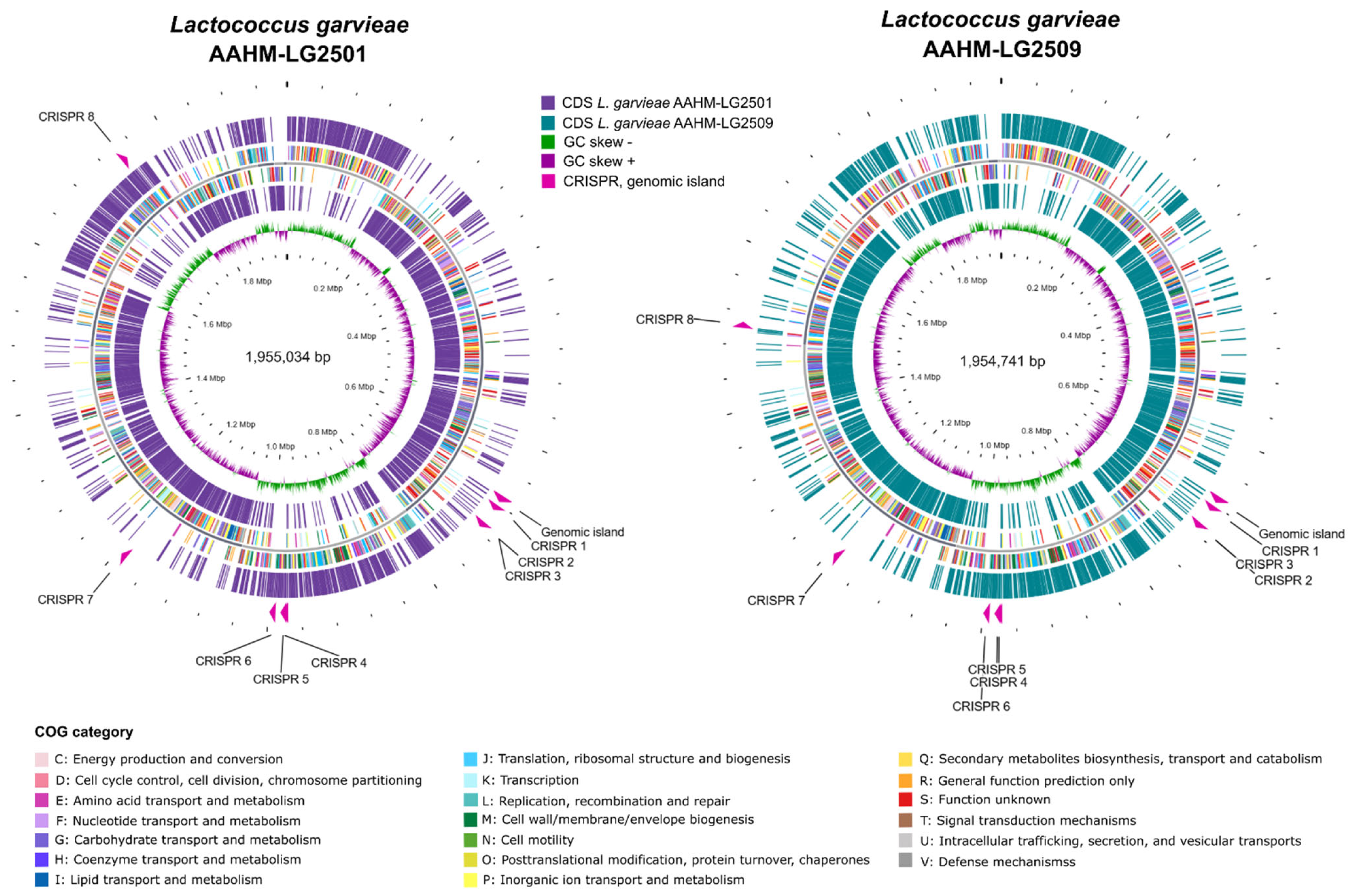
The genome sizes of L. garvieae AAHM-LG2501 and AAHM-LG2509 were 1,955,034 bp and 1,954,741 bp, respectively, with 38.9% G + C content. The genome sequences were deposited to the NCBI database under BioProject: PRJNA1455887 with accession numbers JCAFHE000000000 (AAHM-LG2501) and JCAFHD000000000 (AAHM-LG2509). The L. garvieae AAHM-LG2501 genome encodes for 1866 genes, 7 rRNA, 50 tRNA, 8 CRISPR loci, and one genomic island, whereas the L. garvieae AAHM-LG2509 genome encodes for 1870 genes, 7 rRNA, 50 tRNA, 8 CRISPR loci, and one genomic island (Figure 2).
The numbers of genes that were functionally annotated from all databases of both L. garvieae AAHM-LG2501 and AAHM- LG2509 were identical except the NCBI nr (Table 2). GO, eggNOG, and KEGG databases identified genes ranging from 1845–1847 genes across total CDS while 288 and 171 genes were detected by VFDB and CARD, respectively.
Annotation against the NCBI nr database revealed that the majority of predicted genes from both isolates were assigned to L. garvieae (85.4%) followed by Lactococcus sp. (12.9%) and other Lactococcus species including Lactococcus lactis (0.3%), L. petauri (0.3%), and Lactococcus piscium (0.1%) (Supplementary Table S1).

2.3. Genome-Based Identification and Phylogeny

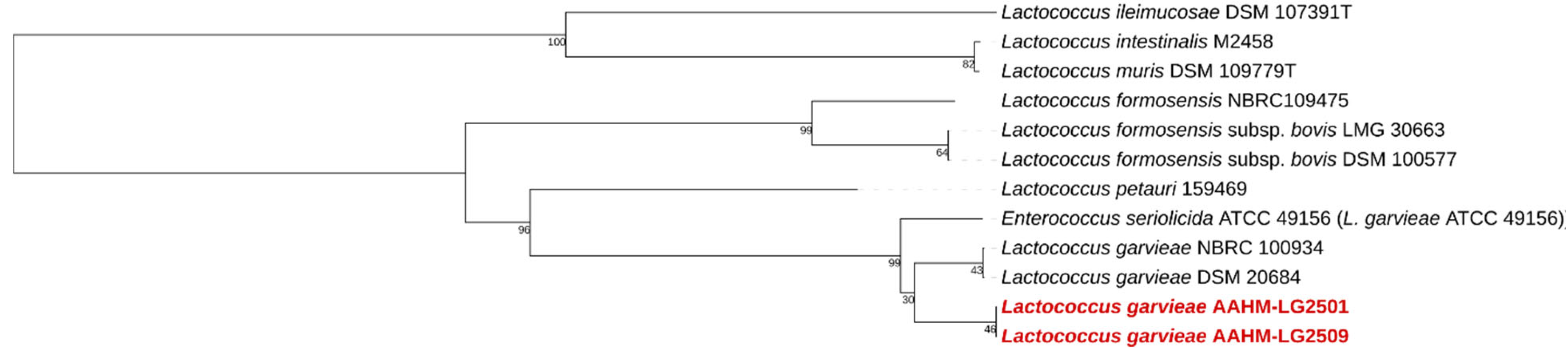
The GBDP-based phylogenetic tree indicated phylogenetic relationship between the two L. garvieae isolates and the reference strain (Figure 3). The L. garvieae AAHM-LG2501 and AAHM-LG2509 are classified into the same clade with type strain L. garvieae NBRC 100943, L. garvieae DSM 20684, and L. garvieae ATCC 49156 (formerly Enterococcus seriolicida).
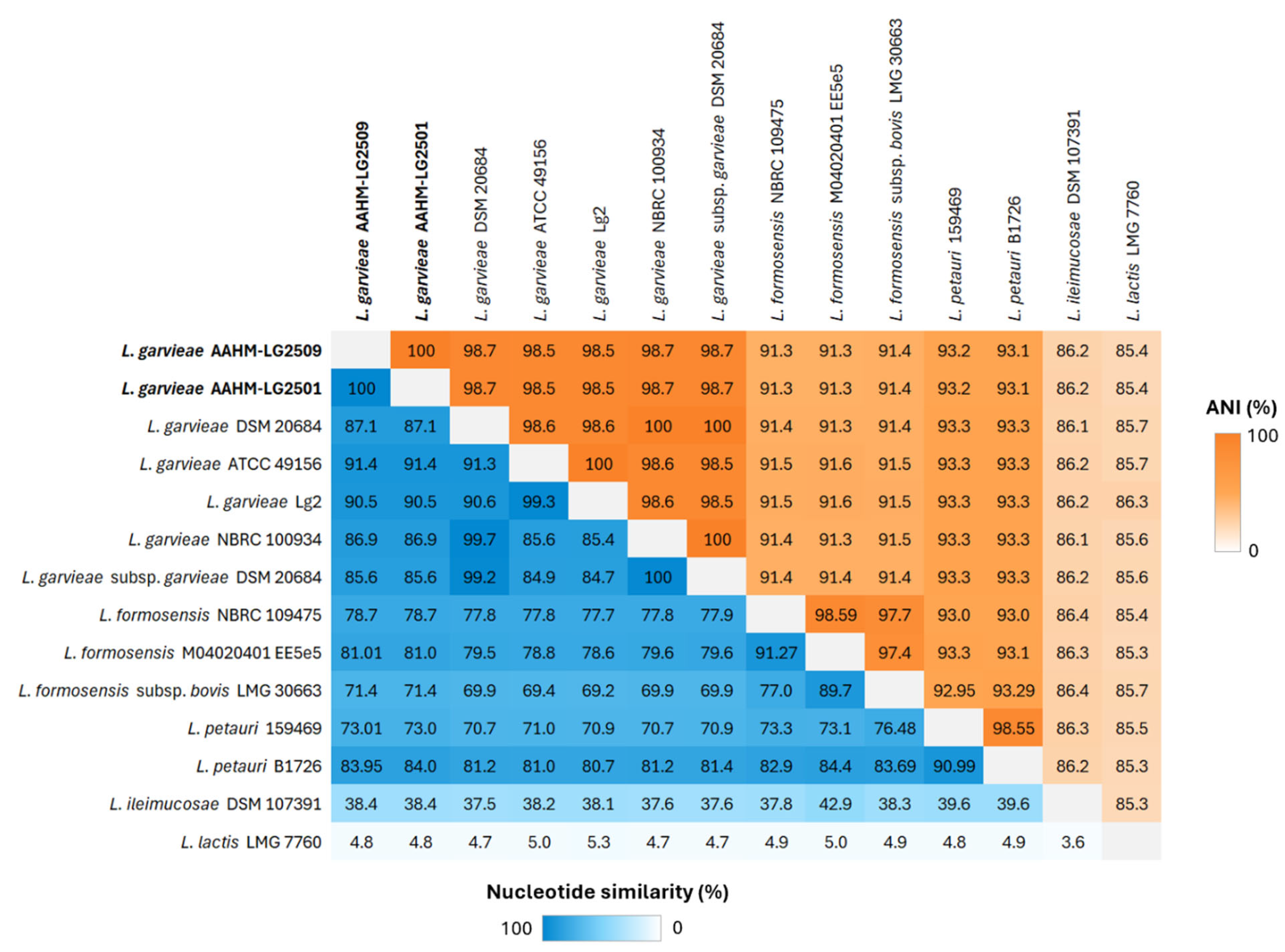
Taxonomic thresholds including dDDH and ANI confirmed that L. garvieae AAHM-LG2501 and AAHM-LG2509 belong to L. garvieae with values exceeding species delineation thresholds (dDDH > 70%, ANI > 95%). The L. garvieae of this study exhibited the highest dDDH and ANI values against L. garvieae and E. seriolicida genomes with 88.6–84.4% and 98.5–98.7%, respectively (Figure 4, Table 3). The G + C difference between the L. garvieae of this study and type strains showing the lowest G + C difference percentage among L. garvieae and L. ileimucosae type strains ranged from 0.04–0.34%, with L. garvieae ATCC 49156 possessing the lowest value. The MLST scheme used to assign the sequence type (ST) of L. garvieae was based on als, atpA, galP, gapC, gyrB, rpoC, and tuf. The results showed that both L. garvieae AAHM-LG2501 and AAHM-LG2509 belong to ST95. Both L. garvieae AAHM-LG2501 and AAHM-LG2509 were assigned to serotype I based on in silico serotyping using serotype-specific PCR primers with predicted amplification products of 285 bp.

2.4. Functional Annotation and Virulence Factors

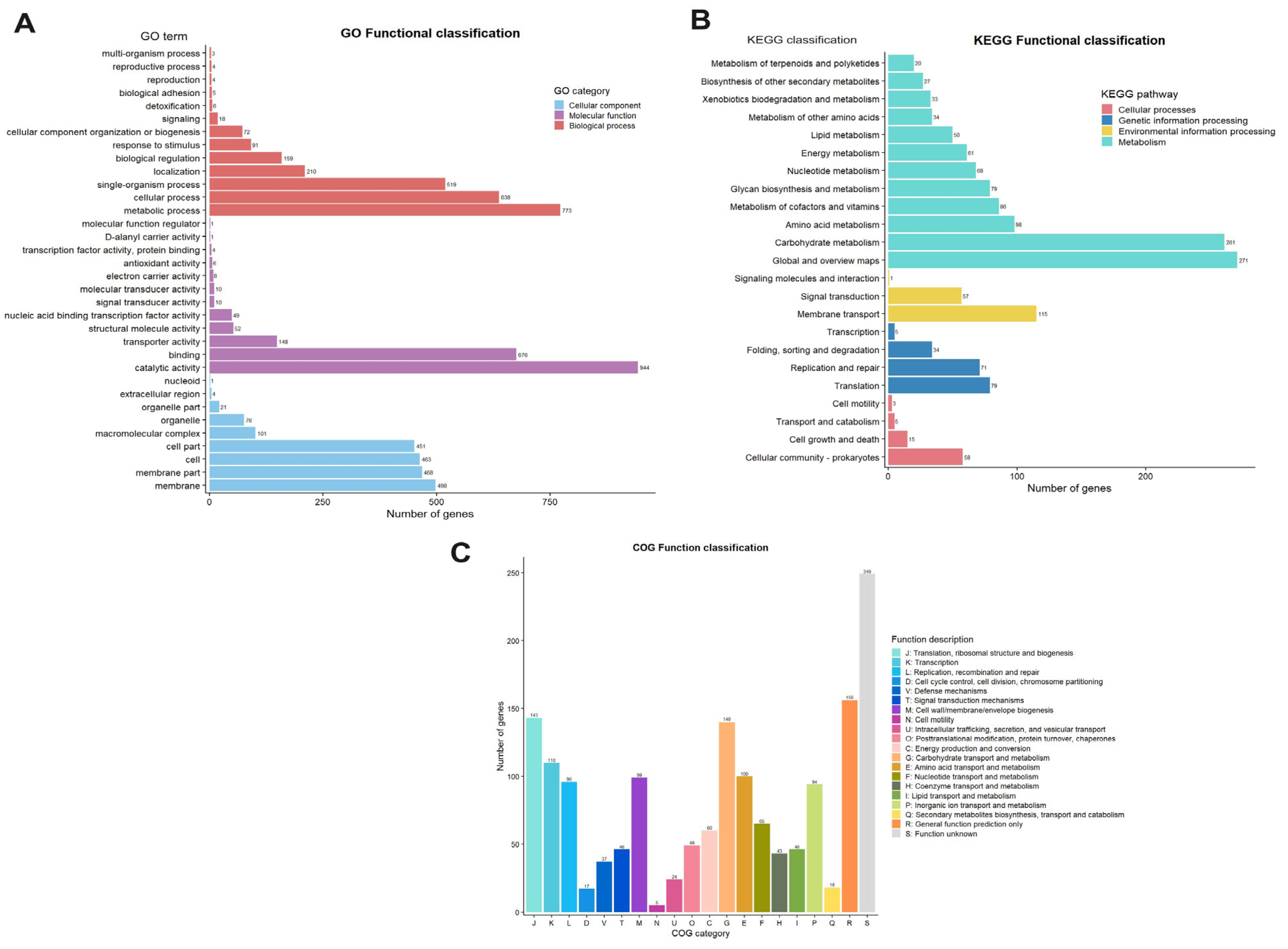
GO functional annotation identified a total of 1537 genes associated with cellular component, molecular function, and biological process (Figure 5A). It should be noted that some genes were assigned to multiple GO categories. Within the cellular component category, most genes were associated with membrane (498 genes), membrane part (468 genes), cell (463 genes), and cell part (451 genes). In the biological process category, metabolic process (773 genes) was the predominant functional group, followed by cellular process (638 genes) and single-organism process (519 genes). In the molecular function category, catalytic activity (944 genes) represented the dominant function followed by binding activity (676 genes).
KEGG functional annotation predicted that 1531 genes were distributed across four KEGG pathway categories, including metabolism (1088 genes), genetic information processing (189 genes), environmental information processing (173 genes), and cellular processes (81 genes) (Figure 5B, Supplementary Table S2). At the KEGG pathways level, the high abundant pathways included biosynthesis of cofactors (84 genes), ABC transporters (65 genes), ribosome (52 genes), biosynthesis of amino acids (49 genes), carbon metabolism (47 genes), two-component system (41 genes), purine metabolism (39 genes), quorum sensing (39 genes), glycolysis/gluconeogenesis (38 genes), and phosphotransferase system (38 genes).
COG classification was performed to classify the predicted protein-coding genes into functional categories. The results showed that 1597 genes were assigned into 20 functional categories (Figure 5C, Supplementary Tables S2 and S3). Apart from the “Function unknown” and “General function prediction only” categories, the five most abundant functional groups were translation, ribosomal structure and biogenesis (143 genes), carbohydrate transport and metabolism (140 genes), transcription (110 genes), amino acid transport and metabolism (100 genes), and cell wall/membrane/envelope biogenesis (99 genes).
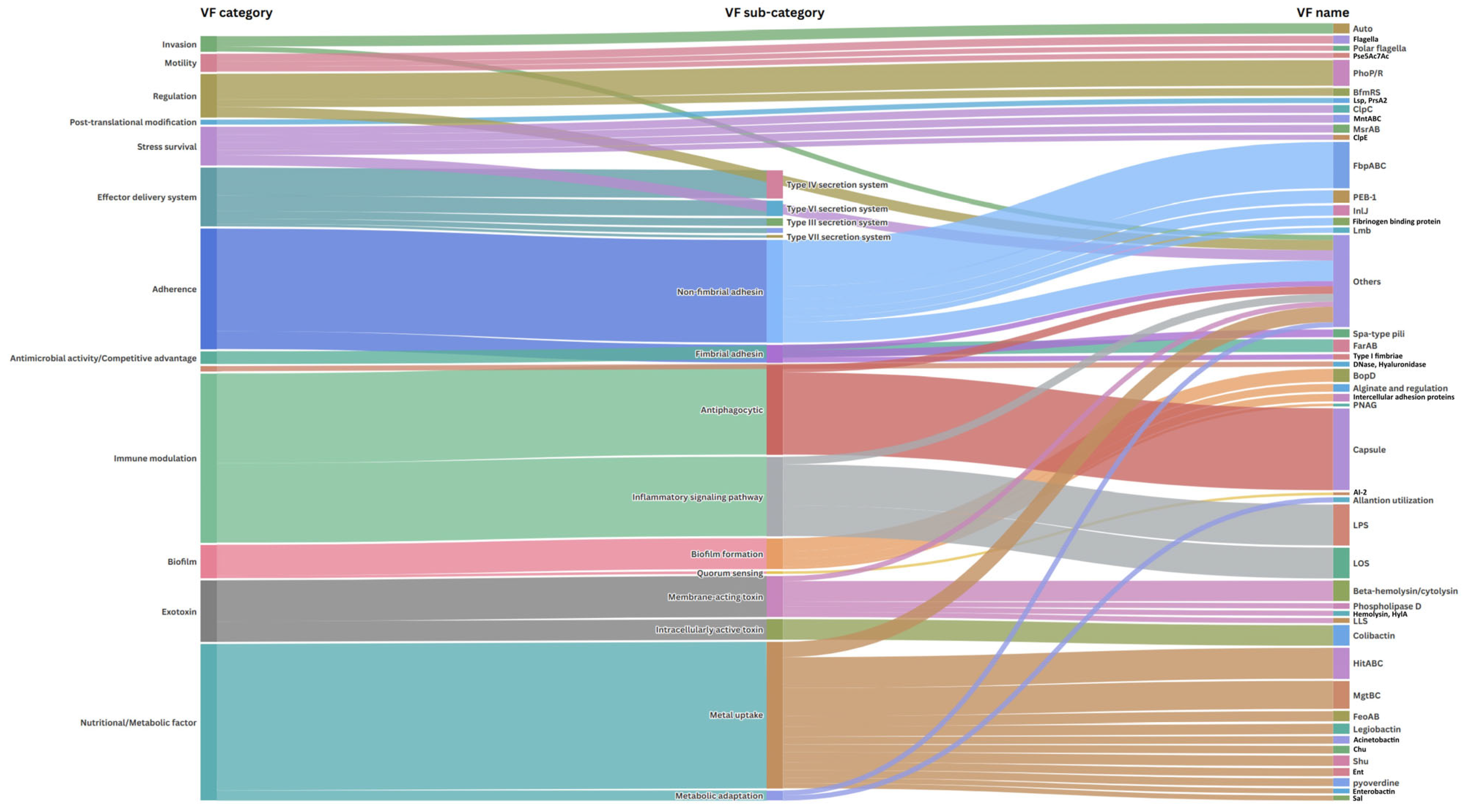
Virulence-associated gene prediction of L. garvieae AAHM-LG2501 and AAHM-LG2509 against VFDB identified a total of 288 virulence-associated genes. These genes were classified into 14 virulence factor categories containing 97 virulence factors. Among the identified categories, genes associated with immune modulation (66 genes) represented the largest proportion followed by nutritional/metabolic (61 genes), adherence (47 genes), exotoxins (24 genes), effector delivery systems (23 genes), regulation (17 genes), biofilm (13 genes), stress survival (15 genes), motility (7 genes), invasion (6 genes), antimicrobial activity/competitive advantage (5 genes), exoenzyme (2 genes), and post-translational modification (2 genes) (Figure 6, Supplementary Tables S2 and S3). Among the identified virulence factors, capsule-associated genes accounted for a notably high proportion (32 genes) compared with other individual virulence factors.

2.5. Antimicrobial Susceptibility and Antibiotic Resistance Properties

The antimicrobial susceptibility profiles of the Lactococcus garvieae isolates were determined using the disk diffusion method against 12 antimicrobial agents, including oxolinic acid (OA, 2 µg), nalidixic acid (NA, 30 µg), enrofloxacin (ENR, 5 µg), sulfamethoxazole-trimethoprim (SXT, 25 µg), tetracycline (TE, 30 µg), oxytetracycline (OT, 30 µg), doxycycline (DO, 30 µg), penicillin G (P, 10 µg), oxacillin (OX, 1 µg), amoxycillin (AML, 10 µg), ampicillin (AMP, 10 µg), and florfenicol (FFC, 30 µg) (Table 4). Notably, no inhibition zones were observed for NA, OA, and OX, indicating resistance of the isolate to these antimicrobial agents. Because interpretive criteria or clinical breakpoints for L. garvieae are currently unavailable, the antimicrobial susceptibility results were interpreted based on the relative diameters of inhibition zones. The isolate exhibited comparatively large inhibition zones against AML, AMP, P, TE, DO, OT, and FFC indicating higher susceptibility to these antimicrobial agents. In contrast, reduced inhibition zones were observed for ENR and SXT, suggesting decreased susceptibility.
The same number of AMR genes were detected from both L. garvieae AAHM-LG2501 and AAHM-LG2509 genomes. A total of 171 ARGs belonging to 29 antimicrobial classes and 117 ARG families were predicted using CARD with strict (3 genes) and loose hits (168 genes) (Supplementary Tables S4 and S5). Among these, genes annotated as being associated with resistance to multiple antimicrobial classes were the most abundant, accounting for 46 genes. Classification of AMR genes according to the antimicrobial classes to which they associated with resistance revealed that macrolide resistance genes were the most prevalent (29 genes), followed by fluoroquinolone (25 genes), peptide antibiotic (18 genes), aminoglycoside (14 genes), tetracycline (12 genes), and glycopeptide (12 genes). Because some ARGs are associated with resistance to multiple antimicrobial classes, individual genes may be represented in more than one resistance category. The ARGs identified as strict hits included lsaD, vanT, and vanY. It should be noted that several mutations were predicted to contribute to antimicrobial resistance through drug target alteration mechanisms. These variants were associated with resistance to multiple antibiotic classes such as fluoroquinolone, cephalosporin, aminoglycoside, etc. Notably, resistance-associated variants were identified in gyrA and gyrB, which encode recognized targets of fluoroquinolone antibiotics. Moreover, antimicrobial resistance phenotype prediction using ResFinder identified an acquired resistance gene, mdtA (identity 89.11%), in L. garvieae AAHM-LG2501 and AAHM-LG2509 genomes. Based on the ResFinder database, mdtA was predicted to confer resistance to macrolides and tetracyclines, including erythromycin, azithromycin, and tetracycline.

3. Discussion

The present study provides the first genome characterization of highly virulent L. garvieae isolates associated with a disease outbreak in cage-cultured Nile tilapia in Thailand. The two L. garvieae isolates were recovered from a disease outbreak in cage-cultured Nile tilapia in Ubon Ratchathani province, Thailand. Both isolates were confirmed as L. garvieae through a combination of conventional biochemical characterization, 16S rRNA gene sequencing, and whole-genome sequencing. Notably, both isolates were assigned to MLST sequence type 95 (ST95), representing the first report of ST95 in Thailand. The isolates were also assigned to serotype I, and their genomes were further characterized for predicted virulence-associated genes and antimicrobial resistance determinants.
Most biochemical characteristics observed in the present study were consistent with those reported previously for L. garvieae. However, variability was observed in several carbohydrate fermentation tests, particularly for sucrose, lactose, mannitol, and sorbitol. Among these, sucrose fermentation has been proposed as a useful phenotypic marker for differentiating L. petauri from L. garvieae and L. formosensis when used in combination with MALDI-TOF MS or 16S rRNA gene sequencing, as L. petauri is typically positive for sucrose utilization whereas L. garvieae and L. formosensis are negative [1]. In contrast, the isolates characterized in this study exhibited positive sucrose fermentation, consistent with previous reports that documented intraspecies variation and positive results in the sucrose test among L. garvieae isolates [6,7,18,19]. The variability observed in carbohydrate utilization patterns suggests that these phenotypic traits may not be universally conserved within L. garvieae. In addition, differences in biochemical testing methods among studies may have contributed to the inconsistent results and some studies may have relied on species identification methods with lower taxonomic resolution, so the possibility of species misidentification cannot be excluded.
Whole-genome sequencing analysis further demonstrated its value as a reliable tool for the taxonomic classification and species identification of lactococcosis-causing bacteria (LCB) in fish. The genome size of the Lactococcus garvieae isolates characterized in this study was consistent with that reported for other L. garvieae strains, which typically range from 1.9 to 2.0 Mb. Although plasmids have been identified in some L. garvieae isolates and have been implicated in the dissemination of virulence- and antimicrobial resistance-associated genes, no plasmids were detected in the present isolates. Based on in silico serotyping using serotype-specific primers, both isolates were assigned to serotype I according to the original serotype classification scheme of L. garvieae. Interestingly, MLST classified the isolates as ST95, which is the same sequence type as L. garvieae MS210922A isolated from farmed greater amberjack (Seriola dumerili) in Japan. This strain was subsequently proposed as a representative of the newly emerged serotype III [20]. The epidemiological study on L. garvieae serotype III reported more than 150 isolates recovered from striped jack (Pseudocaranx dentex) and greater amberjack in Japan during 2021 to 2023 [21]. Although ST95 has previously been associated with serotype III, the isolates in the present study were assigned to serotype I; the available data are insufficient to establish a specific association between ST95 and serotype III as it is unclear whether all serotype III isolates reported in the Japanese study were subjected to MLST. The different discriminatory properties of serotyping and MLST should be considered when selecting a typing approach according to the research objective. Serotyping is based on antigenic properties determined by slide agglutination and therefore provides information on antigenic variation among isolates, which may be relevant to studies of antigenic diversity and vaccine development. In contrast, MLST is based on sequence variation in selected housekeeping genes and is primarily useful for characterizing genetic lineages, population structure, epidemiological relationships, and evolutionary patterns.
In contrast to recent reports describing the distribution of LCB in Nile and red tilapia, in which L. petauri was identified more frequently than L. garvieae, all isolates recovered from the outbreak investigated in the present study were confirmed as L. garvieae [4,8]. A similar observation was reported by [10], where all isolates obtained from diseased red tilapia were identified as L. garvieae. However, species identification in that study was based primarily on 16S rRNA gene sequencing, and phylogenetic analysis revealed overlap between L. garvieae and L. petauri clades, indicating limitations in species discrimination. Therefore, additional investigations involving a larger number of isolates from diverse outbreaks are required to better understand the epidemiology and host association of L. garvieae and L. petauri in tilapia.
The L. garvieae isolates characterized in this study were previously demonstrated to be highly pathogenic to Nile tilapia, causing systemic infection and mortality with an LD50 of 105 CFU/fish following intraperitoneal injection and an LC50 of 106 CFU/fish following immersion challenge [2]. Compared with the L. garvieae isolate reported by [8] which caused a cumulative mortality of 14.82% in Nile tilapia at an infective dose of 107 CFU/fish, the isolates examined in the present study exhibited a higher degree of virulence. The experimental challenge results that demonstrated the high pathogenicity of these isolates provide an important context for interpreting the predicted virulence-associated repertoire. In particular, the extensive capsule-associated gene repertoire and the presence of hemolysin-associated genes may contribute to the ability of these isolates to evade host defenses, colonize host tissues, and cause systemic disease. Similar virulence determinants have been reported in other Gram-positive fish pathogens, including Enterococcus faecalis, Streptococcus agalactiae, and Streptococcus iniae, in which capsule production, adhesion, iron acquisition, and hemolytic activity contribute to host colonization and disease progression [22,23,24]. Although the present genomic analysis cannot establish a causal relationship between individual genes and the observed LD50/LC50 values, these findings provide candidate determinants that warrant further functional investigation.
The cps operon encoding the polysaccharide capsule was composed of ten cps genes (cpsABCFGKO, cps4A, cps4I) that were identified in our L. garvieae isolates. Variations within the capsule-associated gene cluster (CGC) were found between this study and [12] which reported that the virulent L. garvieae strain only carried cpsLW but possessed many eps genes (epsRXABCD) involved in exopolysaccharide (EPS) biosynthesis. The isolates characterized in the present study carried a broader repertoire of cps genes and showed high sequence similarity to capsule-associated genes reported in E. faecalis and Streptococcus spp. These findings suggest that variation exists in the genetic composition of the CGC among virulent L. garvieae strains. Nevertheless, the predominance of capsule-related genes in the present study, together with previous reports demonstrating the importance of capsule formation in virulence, supports the hypothesis that capsule production plays a significant role in the pathogenicity of L. garvieae by facilitating bacterial survival through evasion or interference with host immune responses and reduced susceptibility to phagocytosis.
In most studies, the predominant phenotype for virulent Lactococcus species is hemolysis which is commonly observed in most isolates. Hemolysins and cytolysins are critical to the pathogenesis of hyperacute hemorrhagic multi-organ septicemia in both Lactococcus spp. and Streptococcus spp. [14,22,23]. In the present study, four hemolysin/cytolysin-associated genes, including hlyA (hemolysin A), hemolysin III, cylI (3-ketoacyl-ACP synthase CylI), and cylG (3-ketoacyl-ACP reductase CylG), were identified in all Lactococcus garvieae isolates. Among these, hemolysin A and hemolysin III have previously been reported in both L. garvieae and L. petauri [5,11,18]. However, variation in hemolysin-associated gene profiles was observed among studies. In contrast to the findings of [5], which reported the presence of tlyA and corC/hlyC, the isolates characterized in the present study harbored cylI and cylG, which showed high sequence similarity to homologous genes described in S. agalactiae according to the VFDB. These findings suggest that diversity exists in the repertoire of hemolysin/cytolysin-associated genes among L. garvieae isolates. These exotoxins often co-occur with sortase-anchored proteins (LPxTG) and iron-uptake systems for survival in nutrient-poor host environments [11,15]. Eight genes encoding LPxTG cell wall anchor domain-containing proteins were identified in the L. garvieae isolates in this study. Among these, seven genes showed the highest sequence similarity to LPxTG proteins previously reported in L. garvieae, whereas one gene was most closely related to a homolog identified in Bacillus cereus. LPxTG-containing proteins are surface-associated adhesins that facilitate bacterial attachment to host tissues and have been recognized as important virulence determinants in Lactococcus spp. [11,12,14,15]. However, the LPxTG proteins identified in the present study were annotated based on sequence homology and were not further classified into the LPxTG1–7. Therefore, direct comparison with LPxTG protein and virulence in previous studies was not carried out.
The genes associated with the iron uptake system were also predominant across our L. garvieae isolates. A high number of these genes are associated with HitABC and MgtBC. These genes are related to nutritional/metabolic factors which contribute to bacterial adaptation and persistence within the host environment. The metal transport system plays an important role in metal homeostasis in Streptococcus species. HitABC is an ATP-binding cassette iron uptake system that supports bacterial survival in iron-limited host niches. A HitA lipoprotein was identified among surface-exposed proteins of Streptococcus pyogenes during pharyngitis and reacted with pooled human immune globulin, linking it to host interaction and in vivo fitness [25]. MgtBC is a highly conserved virulence factor found in several major intracellular pathogens such as Mycobacterium tuberculosis and Salmonella enterica, where it helps bacteria to survive within host macrophages [26,27,28]. MgtBC is a membrane-associated protein complex that has been proposed to function as a P-type ATPase involved in Mg2+ transport and other cation transport processes [28]. Beyond its role in ion homeostasis, MgtC has also been implicated in bacterial virulence. A study of Salmonella virulence [27] demonstrated that MgtC activates the PhoR histidine kinase, thereby inducing the Pho regulon and enhancing phosphate uptake which is required for normal Salmonella pathogenesis. Increased phosphate acquisition was associated with hypervirulence in Salmonella and a reduction in the non-replicating bacterial population within macrophages [27]. This observation is consistent with the results of the present study, in which PhoP/PhoR constituted one of the dominant virulence factors within the regulation category. The co-occurrence of MgtBC and the PhoP/PhoR regulatory system may indicate the importance of phosphate sensing and acquisition in the pathogenicity and host adaptation of L. garvieae.
Interpretation of the antimicrobial susceptibility results in Lactococcus spp. remains challenging because no species-specific clinical breakpoints are currently available, specifically bacteria isolated from aquatic animals. Consequently, some previous studies have applied interpretive criteria developed for other bacterial groups, including viridans group streptococci, enterococci, or staphylococci, or have adopted criteria from guidelines established for clinical isolates of human or terrestrial veterinary origin [4,6,14,15]. The use of breakpoints derived from different bacterial species and sources may introduce inconsistencies in the interpretation of inhibition zone diameters and complicate comparisons among studies. Therefore, in the present study, antimicrobial susceptibility results were primarily presented as inhibition-zone diameters rather than assigning categorical susceptible, intermediate, or resistant classifications based on potentially non-equivalent breakpoints. Establishment of standardized, species-appropriate susceptibility criteria based on a sufficiently large collection of Lactococcus isolates would improve the reliability and comparability of antimicrobial resistance surveillance in this genus. The establishment of standardized susceptibility criteria based on a large collection of Lactococcus isolates would therefore improve the consistency and comparability of antimicrobial resistance surveillance.
Resistance to first-generation quinolones including NA and OA was observed in all isolates in this study while ENR, the second generation, showed a quite low inhibition zone. Although we did not assign susceptibility interpretations to the ENR results because appropriate species- and aquatic-animal-specific breakpoints are unavailable, the inhibition-zone diameters observed in the present study would fall within the resistant criteria when compared with the interpretive criteria applied in a previous study [16,29]. This resistance phenotype corresponded to AMR gene profiles, which genes associated with fluoroquinolone resistance were detected. Notably, protein variants predicted to confer resistance to fluoroquinolone were detected in gyrA and gyrB. This mechanism plays an important role in fluoroquinolone resistance by altering the drug target. Substitutions in fluoroquinolone-targeted genes, gyrA (S83R) and parC (S80R), were reported in L. petauri which were associated with a levofloxacin-resistant phenotype [15]. From several studies, lsaD, vanT, and vanY were commonly detected in Lactococcus species and related to phenotype resistance to clindamycin and lincosamide. These genes were found in all isolates in this study which supports the idea of lsaD conferring the intrinsic clindamycin resistance of L. garvieae, and vanT and vanY were related to vancomycin resistance [1,5,11,14,15]. However, antimicrobial susceptibility testing against clindamycin, lincosamides, or vancomycin was not performed in the present study; therefore, the phenotypic relevance of these genes could not be confirmed. Tetracycline resistance in Lactococcus spp. has emerged as a concern because transferable plasmids carrying resistance genes such as tetL and tetS have been reported [1,11,14,15,17]. Although no plasmids were identified in the present isolates, several tetracycline resistance genes, including tetT, tetO, tet38, tet42, tetA(58), and tetA(60), were detected. Despite the presence of these genes, all isolates remained susceptible to the tested tetracycline antibiotics, suggesting that these low-confidence or potentially nonspecific chromosomally encoded determinants may have limited contributions to the observed phenotype, compared with plasmid-mediated tetracycline resistance mechanisms.
An important limitation of the present study is the restricted number and epidemiological origin of the sequenced isolates. Although 16 L. garvieae isolates were recovered during the outbreak investigation, whole-genome sequencing was performed for only two isolates, AAHM-LG2501 and AAHM-LG2509, originating from blood and gill samples, respectively. Moreover, both isolates were recovered within the same outbreak setting and showed highly similar genomic characteristics, including assignment to ST95 and serotype I and comparable virulence- and antimicrobial resistance-associated profiles. Thus, the two genomes should be regarded as representatives of a closely related outbreak-associated lineage rather than independent representatives of the broader L. garvieae population infecting tilapia in Thailand. The present data therefore support characterization of this particular outbreak-associated genotype but do not allow conclusions regarding the prevalence of ST95, genomic diversity, population structure, or geographical dissemination of L. garvieae at the national level. Expanded genomic surveillance incorporating multiple isolates from independent outbreaks, geographical regions, production systems, and sampling periods will be required to determine whether this lineage is locally restricted or more widely distributed in Thai tilapia aquaculture.

4. Materials and Methods

4.1. Field Case Identification and Outbreak Presentation

Two Lactococcus garvieae isolates, namely AAHM-LG2501 and AAHM-LG2509, were isolated from Nile tilapia during a lactococcosis outbreak from May to October 2025 in Ubon Ratchathani province, Thailand. Details of disease investigation and sample collection were provided in our previous study [2]. Briefly, fish with typical signs of lactococcosis, including hemorrhages on external organs, exophthalmia, and erratic swimming, were collected for bacterial isolation. A total of 16 isolates were obtained from blood and gill samples and were previously identified as L. garvieae based on near-full-length 16S rRNA gene sequencing. For the present genome-level characterization, two isolates, AAHM-LG2501 and AAHM-LG2509, recovered from blood and gill samples, respectively, were selected for whole-genome sequencing as representatives of the outbreak-associated isolates obtained from the two sampled tissues. These isolates originated from the same outbreak investigation and were not selected to represent the broader genetic diversity of L. garvieae populations in Thailand. Accordingly, the genomic analyses in the present study were intended to characterize the outbreak-associated isolates rather than to infer population-level diversity or geographical distribution.

4.2. Bacterial Culture and Biochemical Tests

The bacteria were cultured on blood agar (tryptic soy agar (TSA) + 5% sheep blood) and incubated at 28 ± 2 °C for 24 h. Colony morphology, Gram staining and hemolysis activity were observed. Biochemical tests including catalase, oxidase, motile, indole, methyl red and Voges–Proskauer (MR-VP), citrate, urease, carbohydrate fermentation tests (glucose, lactose, sucrose, mannitol, sorbitol), and amino acid decarboxylase tests (ornithine, lysine, arginine) were performed under incubation at 28 ± 2 °C for 24 h. Capsule staining was carried out using nigrosin dye and observed under a light microscope.

4.3. DNA Extraction and Whole-Genome Sequencing

L. garvieae AAHM-LG2501 and AAHM-LG2509 were cultured in tryptic soy broth (TSB) (DifcoTM, San Diego, CA, USA) and incubated at 28 ± 2 °C for 24 h. Genomic DNA was extracted from pure culture using a ZymoBIOMICSTM DNA Miniprep Kit (Zymo Research Corporation, Irvine, CA, USA), following the manufacturer’s instructions. DNA purity and quantity were measured using a NanoDropTM spectrophotometer (Thermo Fisher Scientific, Waltham, MA, USA) prior to use for whole-genome sequencing. DNA libraries were prepared using the Nextera XT kit and sequencing was performed using an Illumina HiSeq platform with paired-end mode (Beijing Biomarker Technologies Co., Ltd., Beijing, China).

4.4. Whole-Genome Sequence Analysis

Quality trimming of raw sequence reads was performed using Trimmomatic v0.39 to exclude low-quality reads (Q < 20) and adapter sequences [30]. The filtered reads were assembled into draft genomes by Spades v3.6.2 [31]. Genome completeness and contamination assessment were determined with checkM [32]. Genome annotation and coding sequence (CDS) prediction were conducted using Prodigal v2.6.3 [33]. The predicted proteins were blasted against NCBI nr (non-redundant) (e-value cut off < 1 × 10⁻⁵) for species identification and genome annotation. Additionally, several databases were used for functional annotation including GO (gene ontology; accessed in December 2025), KEGG (Kyoto Encyclopedia of Genes and Genomes; accessed in December 2025), and eggNOG v6.0 (evolutionary genealogy of genes: Non-supervised Orthologous Groups; accessed in December 2025) [30,34,35,36,37]. The CRISPR system was analyzed using CRISPR Recognition Tool (CRT) v1.2 [38]. Plasmid sequences were screened using MOB-suite v3.1.9 [39].

4.5. Genome-Based Taxonomic Analysis

The Type Strain Genome Server (TYGS) was used to identify closely related type strains based on the Genome Blast Distance Phylogeny approach (GBDP) and to calculate digital DNA–DNA hybridization (dDDH) with the Genome-to-Genome Distance Calculator (GGDC) [40,41]. Average nucleotide identity (ANI) between genomes of this study and other Lactococcus spp. was calculated using pairwise comparison with MUMmer on the JSpeciesWS server [42]. Additionally, the multi-locus sequence typing (MLST) allelic profile was determined using PubMLST (Public databases for molecular typing and microbial genome diversity; accessed in March 2026). Additionally, in silico serotyping was carried out using the NCBI Primer-BLAST tool (https://blast.ncbi.nlm.nih.gov/Blast.cgi, accessed on 31 March 2026) with serotype-specific PCR primers (LGD-F and CGD-R) [43].

4.6. Antibiotic Resistance and Virulence Factors

Antibiotic resistance genes (ARGs) were identified using the online RGI (Resistance Gene Identifier) on CARD (Comprehensive Antibiotic Resistance Database) v4.0.1 with blastp (select criteria: perfect, strict, and loose hits; accessed in March 2026) [44]. ResFinder v4.7.2 was used to identify acquired genes and chromosomal mutations mediating antimicrobial resistance (identity cut off > 80%; accessed in March 2026) [45]. Virulence genes were identified based on the Virulence Factor Database (VFDB) using blastp against the full dataset of protein sequences (e-value cut off < 1 × 10⁻⁵; accessed in March 2026) [46].

4.7. Antimicrobial Susceptibility Test

Antimicrobial susceptibility testing was performed using the disk diffusion method. L. garvieae was cultured on TSA + 5% sheep blood and incubated at 28 °C for 24 h. Subsequently, a single colony was subcultured into TSB and incubated at 28 °C for 24 h with constant shaking. The bacteria were adjusted equal to 0.5 McFarland standard before being streaked onto Mueller–Hinton agar + 5% sheep blood. Antibiotic disks containing oxolinic acid (OA, 2 µg), nalidixic acid (NA, 30 µg), enrofloxacin (ENR, 5 µg), sulfa-trimethoprim (SXT, 265 µg), tetracycline (TE, 30 µg), oxytetracycline (OT, 30 µg), doxycycline (DO, 30 µg), penicillin G (P, 10 µg), oxacillin (OX, 1 µg), amoxicillin (AML, 10 µg), ampicillin (AMP, 10 µg), and florfenicol (FFC, 30 µg) were placed onto the agar. The plates were incubated at 28 °C for 24 h and zones of inhibition were measured using a vernier caliper.

5. Conclusions

In conclusion, the present study characterized two L. garvieae isolates recovered from a disease outbreak in cage-cultured Nile tilapia in Thailand using phenotypic and whole-genome sequencing approaches. Genome-based analyses confirmed the isolates as L. garvieae and demonstrated the utility of whole-genome sequencing for species-level characterization of closely related lactococcosis-causing bacteria. The isolates were assigned to ST95 and serotype I and harbored a diverse repertoire of predicted virulence-associated genes. Antimicrobial susceptibility testing revealed resistance to nalidixic acid, oxolinic acid, and oxacillin, while genomic analysis identified predicted antimicrobial resistance determinants and resistance-associated protein variants, including those associated with fluoroquinolone resistance. These findings provide baseline genomic and phenotypic information on virulence-associated characteristics and antimicrobial resistance in outbreak-associated L. garvieae from Thai Nile tilapia. However, given the limited number of isolates and sampling from a single outbreak, these findings should not be interpreted as evidence of the epidemiological distribution, emergence of resistant L. garvieae populations, or regional transmission patterns. Accordingly, these findings provide baseline genomic and phenotypic information for this outbreak-associated ST95/serotype I lineage but do not establish its prevalence, population structure, or geographical distribution. Broader genomic surveillance involving isolates from independent outbreaks, geographical regions, and sampling periods is required to determine the diversity and dissemination of L. garvieae in Thai aquaculture.

Supplementary Materials

The following supporting information can be downloaded at: https://www.mdpi.com/article/10.3390/ijms27177582/s1.

Author Contributions

Conceptualization, P.C. (Putita Chokmangmeepisarn). and A.U.; methodology, P.C. (Putita Chokmangmeepisarn), Y.A., K.S., N.U. and A.U.; validation, N.U.; formal analysis, P.C. (Putita Chokmangmeepisarn); investigation, P.C. (Putita Chokmangmeepisarn), P.M., B.K., P.C. (Pimrawee Chaemlek), K.S., S.H., N.U. and C.C.; resources, A.U.; data curation, P.C. (Pimrawee Chaemlek) and Y.A.; writing, original draft preparation, P.C. (Putita Chokmangmeepisarn); writing, review and editing, P.S. (Prapansak Srisapoome), K.S., S.H., P.S. (Pichasit Sangmek), C.R. and A.U.; supervision, A.U.; project administration, A.U.; funding acquisition, K.S. and A.U. All authors have read and agreed to the published version of the manuscript.

Funding

This research was funded by The Agricultural Research Development Agency (Public Organization), ARDA, grant number PRP6807030620. The APC was partially funded by the Faculty of Fisheries, Kasetsart University, Thailand.

Institutional Review Board Statement

The experimental procedures involving aquatic animals were conducted in accordance with the Ethical Principles and Guidelines for the Use of Animals outlined by the National Research Council of Thailand, which governs the care and use of animals for research purposes. This protocol received approval from the Animal Ethics Committee at Kasetsart University in Thailand (Approval ID: ACKU68-CSC-004 date of approval: 25 July 2025).

Informed Consent Statement

Not applicable.

Data Availability Statement

The near-full-length 16S rRNA gene sequences generated in this study have been deposited in the NCBI GenBank database under accession numbers PZ007933–PZ007948, corresponding to isolates AAHM-LG2501–AAHM-LG2516, respectively. The whole-genome sequences of isolates AAHM-LG2501 and AAHM-LG2509 have been deposited in GenBank under accession numbers JCAFHE000000000 and JCAFHD000000000, respectively, under BioProject PRJNA1455887. Other data supporting the findings of this study are available from the corresponding authors upon reasonable request.

Acknowledgments

The authors would like to thank the staff and students from this project for technical assistance and support during fish husbandry, sampling, and laboratory analyses. We also acknowledge the cage-culture farmers and local collaborators in Ubon Ratchathani Province for their cooperation and assistance with field sampling during mortality events.

Conflicts of Interest

The authors declare no conflicts of interest.

References

  1. Heckman, T.I.; Yazdi, Z.; Older, C.E.; Griffin, M.J.; Waldbieser, G.C.; Chow, A.M.; Medina Silva, I.; Anenson, K.M.; Garcia, J.C.; LaFrentz, B.R.; et al. Redefining piscine lactococcosis. Appl. Environ. Microbiol. 2024, 90, e0234923. [Google Scholar] [CrossRef] [Scilit]
  2. Adisornprasert, Y.; Kumwan, B.; Meachasompop, P.; Rajitdumrong, C.; Chaemlek, P.; Srisapoome, P.; Buncharoen, W.; Paankhao, N.; Umputhorn, N.; Choppradit, C.; et al. Isolation, Identification, Virulence and Pathogenic Features of Lactococcus garvieae from Cage-Cultured Tilapia (Oreochromis niloticus) in Thailand. Int. J. Mol. Sci. 2026, 27, 3469. [Google Scholar] [CrossRef] [Scilit]
  3. Araki, K.; Mouri, A.; Minami, T.; Nishiki, I.; Yoshida, T. Development of a Discriminating Method for Pathogenic Bacteria that Cause Lactococcal Infections in Marine Fish Farms in Japan. Fish Pathol. 2024, 59, 63–70. [Google Scholar] [CrossRef] [Scilit]
  4. Egger, R.C.; Rosa, J.C.C.; Resende, L.F.L.; de Pádua, S.B.; de Oliveira Barbosa, F.; Zerbini, M.T.; Tavares, G.C.; Figueiredo, H.C.P. Emerging fish pathogens Lactococcus petauri and L. garvieae in Nile tilapia (Oreochromis niloticus) farmed in Brazil. Aquaculture 2023, 565, 739093. [Google Scholar] [CrossRef] [Scilit]
  5. Mahmoud, M.M.; Abdelsalam, M.; Kawato, S.; Harakawa, S.; Kawakami, H.; Hirono, I.; Kondo, H. Comparative genome analyses of three serotypes of Lactococcus bacteria isolated from diseased cultured striped jack (Pseudocaranx dentex). J. Fish Dis. 2023, 46, 829–839. [Google Scholar] [CrossRef] [Scilit]
  6. Sirakov, I.; Strateva, T.V.; Boyanov, V.S.; Orozova, P.; Yordanov, D.; Rusenova, N.; Gergova, R.; Dimov, S.G.; Sirakova, B.; Radosavljevic, V.; et al. Identification, Characterization, and Epidemiological Analysis of Lactococcus garvieae Fish Isolates Obtained in a Period of Eighteen Years. Microorganisms 2025, 13, 436. [Google Scholar] [CrossRef] [Scilit]
  7. Soltani, M.; Naeiji, N.; Zargar, A.; Shohreh, P.; Taherimirghaed, A. Research Article: Biotyping and serotyping of Lactococcus garvieae isolates in affected farmed rainbow trout (Oncorhynchus mykiss) in north Iran. IFRO 2021, 20, 1542. [Google Scholar]
  8. Wongkaew, J.; Chatchaiphan, S.; Taengphu, S.; Dong, H.T.; Senapin, S.; Piyapattanakorn, S. Identification and Pathogenicity of Lactococcus Species in Nile Tilapia (Oreochromis niloticus) and Asian Sea Bass (Lates calcarifer). J. Fish Dis. 2025, 48, e14113. [Google Scholar] [CrossRef] [Scilit]
  9. Vendrell, D.; Balcazar, J.L.; Ruiz-Zarzuela, I.; de Blas, I.; Girones, O.; Muzquiz, J.L. Lactococcus garvieae in fish: A review. Comp. Immunol. Microbiol. Infect. Dis. 2006, 29, 177–198. [Google Scholar] [CrossRef] [Scilit]
  10. Jantrakajorn, S.; Suyapoh, W.; Wongtavatchai, J. Characterization of Lactococcus garvieae and Streptococcus agalactiae in cultured red tilapia Oreochromis sp. in Thailand. J. Aquat. Anim. Health 2024, 36, 192–202. [Google Scholar] [CrossRef] [Scilit]
  11. Blanchard, A.M.; Secker, B.; Atterbury, R.J.; Windle, S.J.; Dong, H.T.; Wongkaew, J.; Dien, L.T.; Huchzermeyer, D.; Hang’ombe, B.M.; Senapin, S. Comparative Genomics of Lactococcus spp. From Global Aquaculture Outbreaks Reveals Virulence Determinants, Antibiotic Resistance, and Phage Defence Mechanisms. Microbiologyopen 2025, 14, e70147. [Google Scholar] [CrossRef] [Scilit]
  12. Morita, H.; Toh, H.; Oshima, K.; Yoshizaki, M.; Kawanishi, M.; Nakaya, K.; Suzuki, T.; Miyauchi, E.; Ishii, Y.; Tanabe, S.; et al. Complete genome sequence and comparative analysis of the fish pathogen Lactococcus garvieae. PLoS ONE 2011, 6, e23184. [Google Scholar] [CrossRef] [Scilit]
  13. Altun, S.; Duman, M.; Ajmi, N.; Tasci, G.; Ozakin, C.; Tuzemen, N.U.; Shahin, K.; Saticioglu, I.B. Comparative proteomic and genomic analysis of Lactococcus garvieae and Lactococcus petauri isolates from Turkey. Aquaculture 2025, 596, 741887. [Google Scholar] [CrossRef] [Scilit]
  14. Ozawa, M.; Kawano, M.; Abo, H.; Kawanishi, M.; Kumakawa, M.; Takahashi, N.; Furushita, M.; Iwamoto, S. Characterization of Lactococcus isolated from diseased yellowtail in Japan using whole-genome sequencing. J. Vet. Med. Sci. 2026, 88, 305–313. [Google Scholar] [CrossRef] [Scilit]
  15. Chan, Y.X.; Cao, H.; Jiang, S.; Li, X.; Fung, K.K.; Lee, C.H.; Sridhar, S.; Chen, J.-K.; Ho, P.L. Genomic investigation of Lactococcus formosensis, Lactococcus garvieae, and Lactococcus petauri reveals differences in species distribution by human and animal sources. Microbiol. Spectr. 2024, 12, e0054124. [Google Scholar] [CrossRef] [Scilit]
  16. Paul, A.; Parida, S.; Mohanty, S.; Biswal, S.; Pillai, B.R.; Panda, D.; Sahoo, P.K. Characteristics and virulence gene profiles of a pathogenic Lactococcus garvieae isolated from diseased Macrobrachium rosenbergii. Braz. J. Microbiol. 2025, 56, 1357–1370. [Google Scholar] [CrossRef] [Scilit]
  17. Maki, T.; Santos Mudjekeewis, D.; Kondo, H.; Hirono, I.; Aoki, T. A Transferable 20-Kilobase Multiple Drug Resistance-Conferring R Plasmid (pKL0018) from a Fish Pathogen (Lactococcus garvieae) Is Highly Homologous to a Conjugative Multiple Drug Resistance-Conferring Enterococcal Plasmid. Appl. Environ. Microbiol. 2009, 75, 3370–3372. [Google Scholar] [CrossRef] [Scilit]
  18. Xu, R.; He, Z.; Deng, Y.; Cen, Y.; Mo, Z.; Dan, X.; Li, Y. Lactococcus garvieae as a Novel Pathogen in Cultured Pufferfish (Takifugu obscurus) in China. Fishes 2024, 9, 406. [Google Scholar] [CrossRef] [Scilit]
  19. Adel, M.; Dehkordi, A.; Yaghoubzadeh, Z.; Khalili Sadrabad, E.; Amiri, A. Isolation and Characterization of Lactococcus garvieae from Diseased Rainbow Trout (Oncorhynchus mykiss, Walbaum) Cultured in Northern Iran Based on the Nucleotide Sequences of the 16s rRNA Gene. Walailak J. Sci. Technol. 2015, 12, 533. [Google Scholar]
  20. Akmal, M.; Yoshida, T.; Nishiki, I. Complete Genome Sequence of Lactococcus garvieae MS210922A, Isolated from Farmed Greater Amberjack (Seriola dumerili) in Japan. Microbiol. Resour. Announc. 2022, 11, e0089022. [Google Scholar] [CrossRef] [Scilit]
  21. Araki, K.; Nishiki, I.; Yoshida, T. Characterization and Epidemiological Study of Newly Emerging Lactococcus garvieae Serotype III in Farmed Fish in Japan. Fish Pathol. 2024, 59, 119–126. [Google Scholar] [CrossRef] [Scilit]
  22. Fyrand, K.; Xu, C.; Zegeye, E.D.; Kjos, M.; Evensen, O. Streptococcus agalactiae 1a capsule (cpsE) and hemolysin (cylE) deletion mutants display attenuated virulence in Nile tilapia (Oreochromis niloticus). Fish Shellfish Immunol. 2026, 170, 111097. [Google Scholar] [CrossRef] [Scilit]
  23. Locke, J.B.; Aziz, R.K.; Vicknair, M.R.; Nizet, V.; Buchanan, J.T. Streptococcus iniae M-like protein contributes to virulence in fish and is a target for live attenuated vaccine development. PLoS ONE 2008, 3, e2824. [Google Scholar] [CrossRef] [Scilit]
  24. Thurlow, L.R.; Thomas, V.C.; Hancock, L.E. Capsular polysaccharide production in Enterococcus faecalis and contribution of CpsF to capsule serospecificity. J. Bacteriol. 2009, 191, 6203–6210. [Google Scholar] [CrossRef] [Scilit]
  25. Zhu, L.; Olsen, R.J.; Beres, S.B.; Saavedra, M.O.; Kubiak, S.L.; Cantu, C.C.; Jenkins, L.; Waller, A.S.; Sun, Z.; Palzkill, T.; et al. Streptococcus pyogenes genes that promote pharyngitis in primates. JCI Insight 2020, 5, e137686. [Google Scholar] [CrossRef] [Scilit]
  26. Akbari, M.S.; Doran, K.S.; Burcham, L.R. Metal Homeostasis in Pathogenic Streptococci. Microorganisms 2022, 10, 1501. [Google Scholar] [CrossRef] [Scilit]
  27. Choi, S.; Choi, E.; Cho, Y.J.; Nam, D.; Lee, J.; Lee, E.J. The Salmonella virulence protein MgtC promotes phosphate uptake inside macrophages. Nat. Commun. 2019, 10, 3326. [Google Scholar] [CrossRef] [Scilit]
  28. Yang, Y.; Labesse, G.; Carrere-Kremer, S.; Esteves, K.; Kremer, L.; Cohen-Gonsaud, M.; Blanc-Potard, A.B. The C-terminal domain of the virulence factor MgtC is a divergent ACT domain. J. Bacteriol. 2012, 194, 6255–6263. [Google Scholar] [CrossRef] [Scilit]
  29. Yılmaz, M.; Sezgin, S.S.; Arslan, T.; Kubilay, A. Current antibiotic sensitivity of Lactococcus garvieae in rainbow trout (Oncorhynchus mykiss) farms from Southwestern Turkey. J. Agric. Sci. 2023, 29, 630–642. [Google Scholar] [CrossRef] [Scilit]
  30. Bolger, A.M.; Lohse, M.; Usadel, B. Trimmomatic: A flexible trimmer for Illumina sequence data. Bioinformatics 2014, 30, 2114–2120. [Google Scholar] [CrossRef] [Scilit] [PubMed]
  31. Prjibelski, A.; Antipov, D.; Meleshko, D.; Lapidus, A.; Korobeynikov, A. Using SPAdes De Novo Assembler. Curr. Protoc. Bioinform. 2020, 70, e102. [Google Scholar] [CrossRef] [Scilit]
  32. Parks, D.H.; Imelfort, M.; Skennerton, C.T.; Hugenholtz, P.; Tyson, G.W. CheckM: Assessing the quality of microbial genomes recovered from isolates, single cells, and metagenomes. Genome Res. 2015, 25, 1043–1055. [Google Scholar] [CrossRef] [Scilit] [PubMed]
  33. Hyatt, D.; Chen, G.L.; Locascio, P.F.; Land, M.L.; Larimer, F.W.; Hauser, L.J. Prodigal: Prokaryotic gene recognition and translation initiation site identification. BMC Bioinform. 2010, 11, 119. [Google Scholar] [CrossRef] [Scilit]
  34. Ashburner, M.; Ball, C.A.; Blake, J.A.; Botstein, D.; Butler, H.; Cherry, J.M.; Davis, A.P.; Dolinski, K.; Dwight, S.S.; Eppig, J.T.; et al. Gene ontology: Tool for the unification of biology. Nat. Genet. 2000, 25, 25–29. [Google Scholar] [CrossRef] [Scilit]
  35. Fu, L.; Niu, B.; Zhu, Z.; Wu, S.; Li, W. CD-HIT: Accelerated for clustering the next-generation sequencing data. Bioinformatics 2012, 28, 3150–3152. [Google Scholar] [CrossRef] [Scilit] [PubMed]
  36. Kanehisa, M.; Goto, S.; Kawashima, S.; Okuno, Y.; Hattori, M. The KEGG resource for deciphering the genome. Nucleic Acids Res. 2004, 32, D277–D280. [Google Scholar] [CrossRef] [Scilit] [PubMed]
  37. Hernández-Plaza, A.A.-O.; Szklarczyk, D.A.-O.; Botas, J.A.-O.; Cantalapiedra, C.A.-O.X.; Giner-Lamia, J.A.-O.; Mende, D.A.-O.; Kirsch, R.; Rattei, T.A.-O.; Letunic, I.A.-O.; Jensen, L.A.-O.X.; et al. eggNOG 6.0: Enabling comparative genomics across 12 535 organisms. Nucleic Acids Res. 2023, 51, D389–D394. [Google Scholar] [CrossRef] [Scilit]
  38. Bland, C.; Ramsey, T.L.; Sabree, F.; Lowe, M.; Brown, K.; Kyrpides, N.C.; Hugenholtz, P. CRISPR recognition tool (CRT): A tool for automatic detection of clustered regularly interspaced palindromic repeats. BMC Bioinform. 2007, 8, 209. [Google Scholar] [CrossRef] [Scilit]
  39. Robertson, J.; Nash, J.H.E. MOB-suite: Software tools for clustering, reconstruction and typing of plasmids from draft assemblies. Microb. Genom. 2018, 4, e000206. [Google Scholar] [CrossRef] [Scilit] [PubMed]
  40. Freese, H.M.; Meier-Kolthoff, J.P.; Sarda Carbasse, J.; Afolayan, A.O.; Goker, M. TYGS and LPSN in 2025: A Global Core Biodata Resource for genome-based classification and nomenclature of prokaryotes within DSMZ Digital Diversity. Nucleic Acids Res. 2026, 54, D884–D891. [Google Scholar] [CrossRef] [Scilit]
  41. Meier-Kolthoff, J.P.; Auch, A.F.; Klenk, H.P.; Goker, M. Genome sequence-based species delimitation with confidence intervals and improved distance functions. BMC Bioinform. 2013, 14, 60. [Google Scholar] [CrossRef] [Scilit] [PubMed]
  42. Richter, M.; Rossello-Mora, R.; Oliver Glockner, F.; Peplies, J. JSpeciesWS: A web server for prokaryotic species circumscription based on pairwise genome comparison. Bioinformatics 2016, 32, 929–931. [Google Scholar] [CrossRef] [Scilit] [PubMed]
  43. Ohbayashi, K.; Oinaka, D.; Hoai, T.D.; Yoshida, T.; Nishiki, I. PCR-mediated Identification of the Newly Emerging Pathogen Lactococcus garvieae Serotype II from Seriola quinqueradiata and S. dumerili. Fish Pathol. 2017, 52, 46–49. [Google Scholar] [CrossRef] [Scilit]
  44. Alcock, B.P.; Huynh, W.; Chalil, R.; Smith, K.W.; Raphenya, A.R.; Wlodarski, M.A.; Edalatmand, A.; Petkau, A.; Syed, S.A.; Tsang, K.K.; et al. CARD 2023: Expanded curation, support for machine learning, and resistome prediction at the Comprehensive Antibiotic Resistance Database. Nucleic Acids Res. 2023, 51, D690–D699. [Google Scholar] [CrossRef] [Scilit] [PubMed]
  45. Florensa, A.F.; Kaas, R.S.; Clausen, P.; Aytan-Aktug, D.; Aarestrup, F.M. ResFinder—An open online resource for identification of antimicrobial resistance genes in next-generation sequencing data and prediction of phenotypes from genotypes. Microb. Genom. 2022, 8, 000748. [Google Scholar] [CrossRef] [Scilit] [PubMed]
  46. Zhou, S.; Liu, B.; Zheng, D.; Chen, L.; Yang, J. VFDB 2025: An integrated resource for exploring anti-virulence compounds. Nucleic Acids Res. 2025, 53, D871–D877. [Google Scholar] [CrossRef] [Scilit]
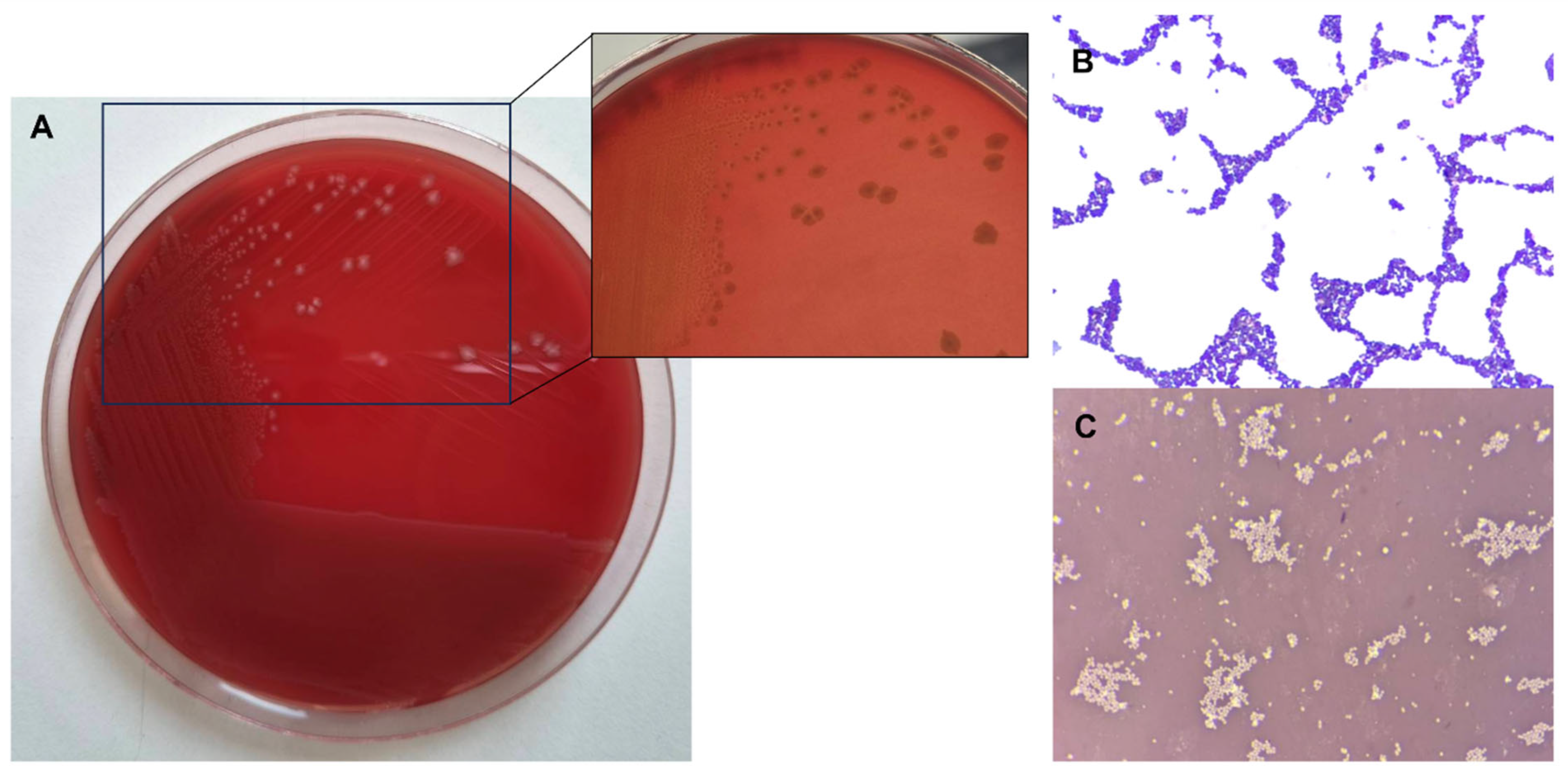
Figure 1. Phenotypic characteristics of L. garvieae AAHM-LG2501 and AAHM-LG2509. (A) Colony morphology and hemolysis appearance on blood agar; (B) bacterial cell morphology with Gram staining; (C) capsule staining under light microscope. Both AAHM-LG2501 and AAHM-LG2509 showed identical morphological characteristics.
Ijms 27 07582 g001
Figure 2. Circular genome maps of L. garvieae AAHM-LG2501 and AAHM-LG2509. The concentric rings display the following features, from the outermost to innermost ring: (1) CRISPR loci and genomic island, with pink arrows indicating the location and direction of the reading frame; (2) forward strand CDS; (3) COG functional categories for forward strand CDS; (4) contig backbone; (5) COG functional categories for reverse strand CDS; (6) reverse strand CDS; and (7) GC skew, where green indicates a positive skew and purple indicates a negative skew.
Ijms 27 07582 g002
Figure 3. Phylogenomic tree showing relationship between L. garvieae of this study (red color) and type strains of Lactococcus spp. The tree was inferred based on the GBDP distance formula d5 and rooted at the midpoint. Number at nodes represent GBDP pseudo-bootstrap support values from 100 replicates. Scale bar represents substitutions per nucleotide position.
Ijms 27 07582 g003
Figure 4. Heat map showing percent average nucleotide identity (ANI) (upper triangle) and percent nucleotide similarity (lower triangle) of L. garvieae of this study (bold) and other Lactococcus spp. Color gradient bar represents percentage of ANI (orange) and nucleotide similarity (blue).
Ijms 27 07582 g004
Figure 5. Functional annotation of L. garvieae AAHM-LG2501 and AAHM-LG2509. (A) GO functional classification, (B) KEGG functional classification, (C) COG functional classification.
Ijms 27 07582 g005
Figure 6. Sankey diagram showing the distribution of predicted virulence-associated genes of L. garvieae AAHM-LG2501 and AAHM-LG2509 identified using the Virulence Factor Database (VFDB). Flows indicate the association between virulence factor (VF) categories, VF sub-categories, and virulence genes. Node width reflects the relative abundance of genes within each VF category.
Ijms 27 07582 g006
Table 1. Biochemical characteristics of L. garvieae AAHM-LG2501 and AAHM-LG2509.
Biochemical TestResult
CatalaseNegative
OxidaseNegative
MotileNegative
IndoleNegative
MRNegative
VPPositive
Carbohydrate fermentation test
   GlucosePositive
   LactosePositive
   SucrosePositive
   MannitolPositive
   SorbitolPositive
Decarboxylase test
   OrnithineNegative
   LysineNegative
   ArgininePositive
Both AAHM-LG2501 and AAHM-LG2509 showed identical biochemical profiles.
Table 2. Assembly statistics and genome features of L. garvieae AAHM-LG2501 and AAHM-LG2509.
L. garvieae AAHM-LG2501L. garvieae AAHM-LG2509
Contig number1413
Contig N50275,740 bp275,743 bp
Mean coverage257.4×298.7×
Completeness100%100%
Genome size1,955,034 bp1,954,741 bp
GC content38.9%38.9%
Number of CDS18661870
Number of rRNA77
Number of tRNA5050
Number of CRISPR88
Total gene length1,730,685 bp1,731,282 bp
Genes assigned to NCBI nr18451847
Genes assigned to GO15371537
Genes assigned to eggNOG15971597
Genes assigned to KEGG15311531
Genes assigned to VFDB288288
Genes assigned to CARD171171
Accession numberJCAFHE000000000JCAFHD000000000
Table 3. dDDH and G + C difference values between L. garvieae of this study and type strains.
Query StrainSubject StraindDDH
(d0, %)
C.I.
(d0, %)
dDDH
(d6, %)
C.I.
(d6, %)
G + C Difference (%)
L. garvieae AAHM-LG2501L. garvieae NBRC 10093484.4[80.6–87.5]87.9[84.9–90.3]0.34
L. garvieae DSM 2068484.5[80.8–87.7]88[85.0–90.4]0.36
E. seriolicida ATCC 4915688.6[85.2–91.3]90.9[88.3–93.0]0.04
L. formosensis subsp. bovis LMG 3066365.8[61.9–69.4]61.5[58.2–64.7]1.33
L. formosensis subsp. bovis DSM 10057766.1[62.2–69.7]61.7[58.4–64.9]1.34
L. formosensis NBRC10947573[69.0–76.7]67.1[63.7–70.3]0.98
L. petauri 15946968.9[65.0–72.6]66.6[63.2–69.8]1.2
L. intestinalis M245830.8[27.4–34.4]28.8[25.9–31.9]0.64
L. muris DSM 109779T32[28.6–35.5]29.7[26.7–32.8]0.3
L. ileimucosae DSM 107391T31.9[28.5–35.5]29.6[26.6–32.7]0.61
L. garvieae AAHM-LG2509L. garvieae NBRC 10093484.4[80.6–87.5]87.9[84.9–90.3]0.34
L. garvieae DSM 2068484.5[80.8–87.7]88[85.0–90.4]0.35
E. seriolicida ATCC 4915688.6[85.2–91.3]90.9[88.3–93.0]0.04
L. formosensis subsp. bovis LMG 3066365.8[61.9–69.4]61.5[58.2–64.7]1.33
L. formosensis subsp. bovis DSM 10057766.1[62.2–69.7]61.7[58.4–64.9]1.34
L. formosensis NBRC10947573[69.1–76.7]67.1[63.7–70.3]0.98
L. petauri 15946968.9[65.0–72.6]66.6[63.2–69.8]1.19
L. intestinalis M245830.8[27.4–34.4]28.8[25.9–31.9]0.65
L. muris DSM 109779T32[28.6–35.5]29.7[26.7–32.8]0.3
L. ileimucosae DSM 107391T31.9[28.5–35.5]29.6[26.6–32.7]0.61
Table 4. Antimicrobial susceptibility profiles and antimicrobial resistance genes.
L. garvieae
Isolates
AAHM-LG2501AAHM-LG2509
Zone of inhibition (mm)
OA (2 µg)6 ± 0 (R)6 ± 0 (R)
NA (30 µg)6 ± 0 (R)6 ± 0 (R)
ENR (5 µg)16 ± 0.714 ± 0.7
SXT (25 µg)18 ± 023 ± 0
AML (10 µg)23 ± 0.725 ± 0.7
AMP (10 µg)29 ± 0.730 ± 0.7
P (10 µg)28 ± 025 ± 0
OX (1 µg)6 ± 0 (R)6 ± 0 (R)
TE (30 µg)28 ± 026 ± 0
DO (30 µg)26 ± 027 ± 0
OT (30 µg)26 ± 0.729 ± 0.7
FFC (30 µg)26 ± 0.725 ± 0.7
Antimicrobial resistance genes
CARDlsaD, vanT, vanYlsaD, vanT, vanY
ResFindermdtAmdtA
R = resistant.
Disclaimer/Publisher’s Note: The statements, opinions and data contained in all publications are solely those of the individual author(s) and contributor(s) and not of MDPI and/or the editor(s). MDPI and/or the editor(s) disclaim responsibility for any injury to people or property resulting from any ideas, methods, instructions or products referred to in the content.