Review

Particulate Matter-Induced Skin Injury: A Dual-Pathway AhR–Nrf2 Framework for Epidermal Homeostasis and Therapeutic Targeting


1
Graduate Institute of Natural Products, College of Pharmacy, Kaohsiung Medical University, Kaohsiung 807378, Taiwan
2
Drug Development and Value Creation Research Center, Kaohsiung Medical University, Kaohsiung 807378, Taiwan
3
Department of Medical Research, Kaohsiung Medical University Hospital, Kaohsiung 807378, Taiwan
4
School of Pharmacy, College of Pharmacy, Kaohsiung Medical University, Kaohsiung 807378, Taiwan
5
Center for Cancer Research, Kaohsiung Medical University, Kaohsiung 807378, Taiwan
6
Department of Fragrance and Cosmetic Science, College of Pharmacy, Kaohsiung Medical University, Kaohsiung 807378, Taiwan
7
Department of Pharmacy, Kaohsiung Municipal Siaogang Hospital, Kaohsiung 812012, Taiwan
8
University Center of Bioscience and Biotechnology, National Cheng Kung University, Tainan 70101, Taiwan
*
Authors to whom correspondence should be addressed.
Int. J. Mol. Sci. 2026, 27(17), 7573; https://doi.org/10.3390/ijms27177573
Submission received: 10 July 2026 / Revised: 14 August 2026 / Accepted: 17 August 2026 / Published: 24 August 2026
(This article belongs to the Section Biochemistry)

Abstract

The aryl hydrocarbon receptor (AhR) is highly expressed in keratinocytes and functions as an environmental sensor regulating xenobiotic metabolism, epidermal differentiation, and inflammatory responses. Particulate matter (PM), a major environmental pollutant containing reactive oxygen species (ROS), transition metals, and polycyclic aromatic hydrocarbons (PAHs), induces oxidative stress and inflammation, leading to skin barrier dysfunction. Transition metals generate ROS via Fenton-type reactions, whereas PAHs undergo AhR-mediated metabolism that further amplifies oxidative stress. Excessive ROS promotes inflammatory cytokine expression and disrupts barrier-related protein expression. In response, activation of the nuclear factor erythroid 2-related factor 2 (Nrf2) pathway induces antioxidant enzymes, including heme oxygenase-1 (HO-1), to counteract oxidative damage. However, sustained PM exposure may overwhelm these defense mechanisms, resulting in impaired cellular homeostasis. Although the roles of AhR and Nrf2 have been extensively investigated individually, their coordinated regulation in PM-induced skin injury remains underexplored. This review summarizes current evidence on the functional interplay between AhR and Nrf2 and discusses how coordinated activation of these pathways integrates xenobiotic metabolism, antioxidant defense, and barrier-associated functions. Overall, the available evidence supports a dual-pathway framework for maintaining epidermal homeostasis under PM-induced environmental stress.

1. Introduction

1.1. Environmental Particulate Matter as an Emerging Public Health Concern

Particulate matter (PM) is a ubiquitous air pollutant with significant adverse health outcomes [1,2]. According to the United States Environmental Protection Agency (US EPA), PM is classified by aerodynamic diameter into coarse particles (PM10, <10 μm), fine particles (PM2.5, <2.5 μm), and ultrafine particles (UFPs, <100 nm) [3,4]. PM originates from multiple anthropogenic and natural sources, including traffic emissions, industrial combustion, fossil fuel burning, wildfire smoke, and secondary atmospheric reactions [5]. The relative contributions of these sources determine particle size distribution and chemical composition, thereby influencing biological reactivity.
Particle size critically affects deposition behavior, cellular interaction, and toxicological outcomes [6]. As aerodynamic diameter decreases, particles exhibit increased surface area relative to mass and enhanced ability to penetrate biological barriers [7]. This amplified surface area-to-mass ratio increases surface reactivity, allowing finer particles to adsorb higher concentrations of toxic trace elements and organic compounds [8,9]. PM2.5 and UFPs exhibit prolonged atmospheric suspension and deeper tissue penetration compared with coarse particles [4,9,10]. Recent evidence suggests a negative association between particle size and in vitro toxicity, with PM2.5 fractions inducing significantly stronger inflammatory reactions in macrophages than coarser particles [11,12].
Beyond its physical dimensions, the toxicity of PM is fundamentally dictated by its highly heterogeneous chemical payload and the resulting physicochemical properties, particularly surface charge and hydrophobicity [13]. PM contains transition metals (e.g., Fe, Mn, Mo, and Cu), polycyclic aromatic hydrocarbons (PAHs), and other redox-active constituents [14,15,16]. Surface-bound reactive species possess redox activity and can induce oxidative reactions in exposed tissues [15]. Water-soluble fractions enriched in bioavailable transition metals catalyze Fenton-type reactions, facilitating the conversion of hydrogen peroxide into hydroxyl radicals [17]. Meanwhile, hydrophobic organic fractions enriched in PAHs, such as benzo[a]pyrene, readily partition into lipid membranes and enter cells [18,19]. Together, these physicochemical characteristics contribute to oxidative stress-related health effects.
Due to their small size and surface reactivity, PM2.5 and UFPs can bypass primary clearance mechanisms and enter the body through inhalation, ingestion, and dermal exposure [6]. Inhaled particles may penetrate the alveolar region and translocate into systemic circulation [9]. Following dermal exposure, particularly when the skin barrier is compromised, nanoscale particles and associated toxicants may penetrate beyond the stratum corneum and affect viable epidermal cells [20]. Epidemiological data from the World Health Organization (WHO) associate long-term PM exposure with respiratory diseases, cardiovascular disorders, metabolic dysfunction, and increased mortality [21,22,23]. Emerging evidence from epidemiological studies and mechanistic investigations further links chronic PM exposure to accelerated skin aging and exacerbation of inflammatory skin diseases, including atopic dermatitis [24,25,26].
Unlike ultraviolet (UV) radiation, which primarily induces photochemical DNA damage [27], or ozone, which reacts predominantly at the skin surface due to limited penetration [28], PM acts as both a particulate carrier and a source of intracellularly active chemical constituents [29]. The combination of physical deposition and metabolic activation distinguishes PM-induced cutaneous injury from other environmental stressors. Understanding the health consequences of PM exposure is therefore essential for developing preventive and therapeutic strategies. However, accurately characterizing PM-induced cellular responses remains technically challenging, partly due to the complex physicochemical properties of PM and the intrinsic autofluorescence of PAHs-enriched organic fractions, which can interfere with fluorescence-based analyses [30,31]. These limitations underscore the need for advanced analytical approaches capable of resolving the spatial and phenotypic heterogeneity of PM-induced skin injury. In this context, a previously established imaging platform provides a useful tool for visualizing PM-induced cellular responses and supporting the mechanistic interpretation of skin barrier dysfunction [32].

1.2. The Skin as the Primary Defense Interface Against Environmental Pollutants

As the primary interface between the human body and the external environment, the skin’s structural integrity is crucial for defense [33,34]. Structurally, the cutaneous barrier relies on two functional layers: the supportive dermis and the highly stratified epidermis [35]. The dermis consists of a collagen- and elastin-rich extracellular matrix that provides tensile strength and mechanical support [36]. The epidermis, the outermost layer of the skin, serves as the primary physical, chemical, and immunological interface with the external environment [37]. It is a stratified, avascular epithelium composed predominantly of keratinocytes, which undergo a tightly regulated differentiation program from the stratum basale to the outermost stratum corneum [38].
At the terminal stage of this differentiation process, the stratum corneum, approximately 10–20 μm thick in humans, is formed by terminally differentiated, anucleated corneocytes embedded within a hydrophobic intercellular lipid matrix [39]. This highly compact organization establishes the principal permeability barrier of the skin. Each corneocyte is surrounded by a highly cross-linked proteinaceous cornified envelope composed of filaggrin, loricrin, involucrin, and small proline-rich proteins, conferring mechanical strength and contributing to hydration homeostasis [40,41]. In the viable epidermis, keratinocytes are interconnected by specialized cell–cell junctions, including tight junctions and adherens junctions, which further restrict paracellular diffusion of water, ions, and xenobiotics. These junctions contain key components such as zonula occludens-1 (ZO-1) and E-cadherin/β-catenin complexes, respectively [42,43,44]. Additionally, integral membrane proteins such as aquaporin-3 (AQP3) facilitate the transport of water and glycerol, serving as a crucial molecular determinant of epidermal hydration and barrier resilience [45,46,47].
Despite its barrier organization, airborne PM continuously interacts with the skin’s surface through adhesion and deposition [48,49]. Although the macroscopic surface area of adult human skin is estimated at approximately 1.8–2.0 m2, the presence of hair follicles and sweat ducts increases the effective interactive surface to approximately 25–30 m2 [34]. In this structural context, PM may traverse the cutaneous barrier via three principal direct pathways: transcellular, intercellular, and trans-appendageal routes [20,50]. The transcellular route involves diffusion across corneocytes and their cornified envelopes and is generally associated with small, relatively hydrophilic molecules. In contrast, the intercellular route involves diffusion through the lipid matrix between corneocytes and is generally associated with lipophilic compounds. The trans-appendageal route utilizes hair follicles and sweat gland ducts, which serve as preferential deposition sites and reservoirs for particles.
Nanoparticles in the ~40 nm size range exhibit enhanced follicular accumulation and may access perifollicular tissue under barrier-disrupted conditions, whereas larger particles predominantly remain confined within the follicular duct [49,51]. Given that UFPs overlap with this size range, the nanoscale fraction of PM may exhibit increased potential for appendageal deposition and penetration, especially in compromised skin. In addition to particle size, the physicochemical properties of PM-associated chemicals critically influence their route of entry. Lipophilic organic constituents, including PAHs, readily partition into the lipophilic intercellular lipid matrix of the stratum corneum, where highly lipophilic congeners tend to form a superficial reservoir rather than efficiently permeating into deeper viable layers [52]. Together, these structural and compositional determinants position the epidermis as a dynamic interface where particulate pollutants may deposit, persist, and interact with viable cellular compartments.
Functionally, the skin is not merely a passive physical shield but also a metabolically active organ [53]. Keratinocytes express xenobiotic-metabolizing enzymes, including extrahepatic cytochrome P450 isoforms, enabling local biotransformation of environmental pollutants [54]. Consequently, PM-associated organic constituents, such as PAHs, may undergo metabolic activation within the epidermis, generating reactive intermediates that amplify oxidative stress and inflammatory signaling. Collectively, the skin’s complex architecture, extensive environmental exposure, and metabolic responsiveness render it highly susceptible to particulate pollutants.

1.3. Mechanistic Overview of PM-Induced Skin Damage

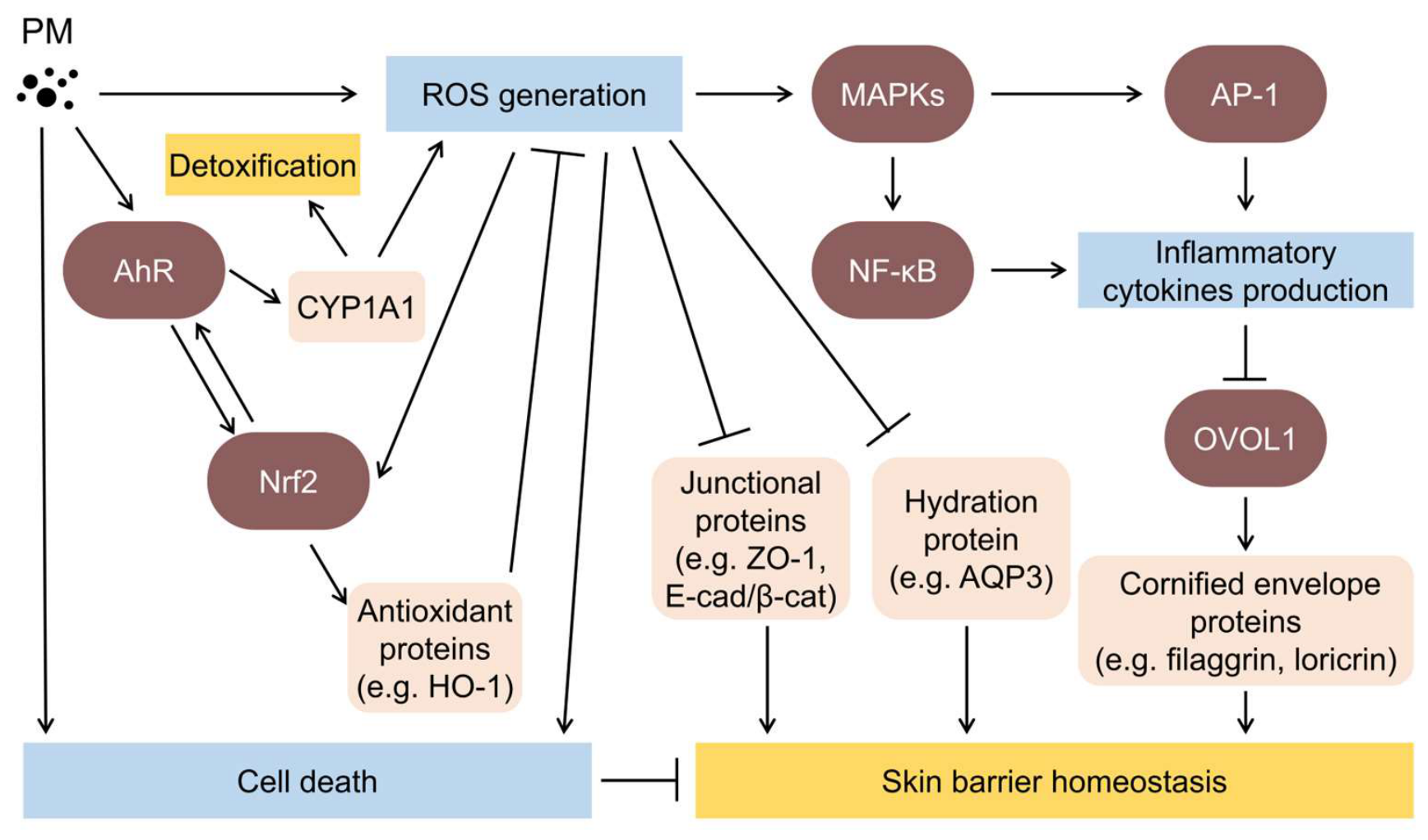
Following cutaneous deposition and epidermal penetration, PM initiates a coordinated cascade of pathogenic events within the viable epidermis [55]. While the physical dimensions of PM determine epidermal penetration depth, its complex chemical payload, comprising transition metals, PAHs, and other redox-active constituents, directly interacts with keratinocytes and drives cytotoxicity [56]. Building upon the physicochemical properties described earlier, these internalized constituents trigger excessive reactive oxygen species (ROS) generation through distinct cellular mechanisms: surface-bound constituents directly induce local oxidative reactions, intracellular transition metals rapidly catalyze Fenton-type reactions, and PAHs strongly activate the cytosolic aryl hydrocarbon receptor (AhR), which transcriptionally induces cytochrome P450 enzymes (e.g., CYP1A1), leading to the formation of reactive electrophilic intermediates and secondary ROS [57]. Collectively, this excessive ROS generation establishes oxidative stress as the central pathogenic driver of PM-induced epidermal injury.
The accumulation of ROS disrupts cellular redox homeostasis, leading to lipid peroxidation, protein oxidation, mitochondrial dysfunction, and DNA damage [58]. Concurrently, oxidative stress activates redox-sensitive signaling pathways, including mitogen-activated protein kinases (MAPKs) and nuclear factor kappa-light-chain-enhancer of activated B cells (NF-κB) cascades, which amplify pro-inflammatory cytokine production and promote immune cell recruitment [59]. In response, endogenous antioxidant defense mechanisms are engaged, primarily through activation of the nuclear factor erythroid 2–related factor 2 (Nrf2) signaling pathway, which coordinates transcriptional cytoprotective programs [60]. However, when ROS generation overwhelms cellular antioxidant defenses, persistent oxidative stress results in keratinocyte cytotoxicity, apoptosis, and amplified inflammatory responses that progressively compromise epidermal integrity, ultimately exacerbating cutaneous inflammatory disorders such as atopic dermatitis [25,56].
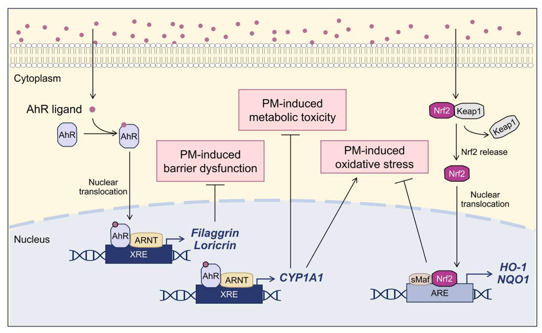
Barrier dysfunction represents a critical pathological hallmark of PM exposure [61]. Mechanistically, in vitro studies using human epithelial models have shown that PM exposure reduces and delocalizes essential structural proteins involved in barrier formation, including filaggrin, loricrin, and involucrin, while simultaneously disrupting tight junction and adherens junction components [62,63,64]. Oxidative damage further impairs hydration-related molecules such as AQP3 and delays barrier recovery [65]. Notably, beyond its role in xenobiotic metabolism and ROS amplification, AhR also functions as a context-dependent regulator of epidermal differentiation and barrier homeostasis [66,67]. Consequently, epidermal barrier integrity under PM exposure depends on the dynamic balance between sustained structural injury and context-dependent regulatory responses mediated by pathways such as AhR. To elucidate these complex interactions, the following sections describe the oxidative mechanisms underlying PM-induced injury, the resulting cellular consequences, the disruption of epidermal barrier integrity, and the regulatory roles of AhR and Nrf2 signaling (Figure 1).

2. Molecular Pathways Regulating Cellular Responses to PM Exposure

2.1. Oxidative Stress-Driven Mechanisms Underlying PM-Induced Skin Injury

At the cellular level, the detrimental effects of PM are largely mediated by excessive ROS production, which disrupts intracellular redox homeostasis and is a central driver of pollution-induced skin injury [55,59]. PM-associated oxidative stress involves multiple ROS with distinct chemical reactivities, including superoxide anion (O2•–), hydrogen peroxide (H2O2), and the highly reactive hydroxyl radical (OH) [17,68]. Initial physicochemical interactions between PM constituents and cellular components commonly generate superoxide anion, which is rapidly converted to hydrogen peroxide by intracellular superoxide dismutase (SOD) [69]. A critical amplification step occurs when hydrogen peroxide encounters bioavailable transition metals derived from PM [70]. Through classical Fenton and Haber–Weiss reactions, these metals catalyze the conversion of hydrogen peroxide into highly reactive hydroxyl radicals, thereby markedly amplifying intracellular oxidative stress [71].
In addition to these direct physicochemical reactions, PM exposure further amplifies oxidative stress through endogenous cellular sources [72]. PM has been reported to impair mitochondrial electron transport chain function, resulting in electron leakage and increased production of mitochondrial ROS [70]. Furthermore, PM constituents can activate innate immune signaling pathways. For example, studies using human keratinocytes and murine skin models have demonstrated that direct interaction between toll-like receptor 5 (TLR5) and NADPH oxidase 4 (NOX4) has been reported to sustain intracellular ROS generation following PM exposure [73]. These combined processes create a persistent oxidative microenvironment that extends beyond the initial physicochemical insult.
Excessive ROS accumulation subsequently induces oxidative damage to multiple cellular macromolecules. Lipid membranes, particularly those enriched in polyunsaturated fatty acids, are highly susceptible to ROS-mediated lipid peroxidation, resulting in the formation of reactive aldehydes such as malondialdehyde (MDA) and 4-hydroxynonenal (4-HNE) [74,75]. Proteins are similarly vulnerable to oxidative modification, including carbonylation and nitration, which impair the function of structural and enzymatic proteins [76]. In addition, ROS-mediated DNA damage, exemplified by oxidative lesions such as 8-oxo-2′-deoxyguanosine (8-oxo-dG), contributes to genomic instability and mutagenesis [77,78,79]. These oxidative modifications collectively compromise cellular integrity and trigger stress responses in epidermal cells.
Beyond direct molecular damage, ROS also function as key signaling mediators that activate multiple redox-sensitive pathways. Elevated oxidative stress stimulates MAPK cascades, including ERK1/2, JNK, and p38, as well as downstream transcription factors such as NF-κB and activator protein-1 (AP-1) [55]. Activation of these pathways promotes the expression of numerous pro-inflammatory mediators, including tumor necrosis factor-α (TNF-α), interleukin-1β (IL-1β), interleukin-6 (IL-6), cyclooxygenase-2 (COX-2), and matrix metalloproteinases (MMPs), thereby linking oxidative stress to inflammatory responses and extracellular matrix degradation in the skin [56].
Nevertheless, when PM-induced ROS generation exceeds the protective capacity of these antioxidant systems, persistent oxidative stress promotes keratinocyte dysfunction, apoptosis, and sustained inflammatory signaling. Collectively, these oxidative processes represent a central mechanistic link between PM exposure and the subsequent development of cellular injury and epidermal barrier disruption.

2.2. Cellular and Molecular Consequences of PM Exposure

Beyond the disruption of cellular redox homeostasis described in Section 2.1, PM-induced oxidative stress exerts direct effects on intracellular organelles within keratinocytes. Following cellular uptake, PM particles have been observed predominantly in secondary lysosomes, autophagy vesicles, and endocytotic vesicles in cultured primary keratinocytes [49]. This intracellular localization may facilitate interactions between PM-associated constituents and cellular components, contributing to oxidative stress, organelle dysfunction, and downstream cytotoxic responses. Mitochondrial dysfunction can further interfere with electron transport chain activity, leading to mitochondrial membrane depolarization and impaired ATP production [80]. This metabolic impairment not only compromises cellular energy metabolism but also sensitizes keratinocytes to subsequent stress signals.
In parallel with organelle impairment, PM exposure also induces substantial genomic damage. PM constituents, particularly PAHs and transition metals, possess genotoxic properties that promote the formation of DNA single- and double-strand breaks [63,81]. In response to such genomic stress, keratinocytes activate cell-cycle checkpoint mechanisms to prevent the propagation of damaged DNA. Several studies have reported that PM exposure induces cell-cycle arrest in keratinocytes, most commonly at the G0/G1 phase [82,83,84]. However, the precise arrest phase may vary depending on the physicochemical characteristics of PM and the cellular context. For example, toxicological studies in respiratory epithelia models, such as BEAS-2B cells, have reported delayed S-phase progression, G2/M arrest, or complete mitotic arrest following PM exposure [85]. These variations highlight the influence of particle composition, oxidative burden, and cell types on the PM-induced cellular stress response.
The accumulation of organelle dysfunction and genomic instability ultimately results in cytotoxicity. Numerous in vitro studies indicate that PM exposure leads to dose- and time-dependent reductions in keratinocyte viability, often accompanied by biochemical indicators of cytotoxicity such as increased lactate dehydrogenase (LDH) release [86,87]. Such cellular deterioration reflects severe impairment of metabolic activity and membrane integrity following PM exposure, ultimately leading to programmed cell death. In HaCaT keratinocytes, PM exposure alters the balance of Bcl-2 family proteins by downregulating the anti-apoptotic protein Bcl-2 and upregulating pro-apoptotic factors such as Bax, thereby promoting mitochondrial outer membrane permeabilization and the subsequent release of cytochrome c into the cytosol [88]. This mitochondrial signaling event activates the caspase cascade, particularly caspase-9, followed by the cleavage of caspase-3, ultimately leading to the cleavage of poly(ADP-ribose) polymerase (PARP) [87,89,90]. Collectively, these events characterize the intrinsic mitochondrial apoptotic pathway triggered by PM exposure.
While apoptosis represents the predominant form of PM-induced cell death, excessive cellular injury may shift the cellular response toward necrotic damage. Cellular energy depletion, damage to membrane lipids, and the loss of function of homeostatic ion pumps/channels can synergistically induce necrosis [91]. Experimental models have reported increased propidium iodide uptake in PM-exposed keratinocytes, suggesting membrane damage consistent with necrotic injury [58]. Crucially, unlike apoptosis, the necrotic rupture of keratinocytes releases intracellular contents and lysosomal hydrolases into the surrounding microenvironment [92]. This uncontrolled exudation acts as a potent pro-inflammatory trigger, exacerbating local tissue damage and playing a pivotal role in the severe epidermal barrier dysfunction and chronic inflammation associated with pollution exposure.
In addition to apoptosis and necrosis, other forms of cell death, such as autophagy-associated cell death, have also been reported following PM exposure [91]. As a lysosome-dependent degradation pathway responsible for removing damaged organelles and protein aggregates, autophagy may initially function as a protective mechanism to mitigate cellular injury. This is experimentally monitored through the lipidation of LC3 (conversion from LC3-I to LC3-II) and the degradation of the cargo receptor p62 [86,93]. However, prolonged or dysregulated autophagy under severe PM exposure can transition from cytoprotective to cytotoxic, highlighting the complex interplay between adaptive and destructive cellular responses.
In addition to acute cytotoxicity and cell death, prolonged PM exposure in cellular models induces persistent ROS-dependent DNA damage, driving keratinocytes and dermal fibroblasts into irreversible cell-cycle arrest and premature cellular senescence [94,95,96]. Persistent oxidative stress activates senescence-related pathways, including p16INK4a, p21, and p53 signaling, leading to the accumulation of senescence-associated β-galactosidase (SA-β-gal) activity and the loss of lamin B1 [97,98]. Consequently, these senescent skin cells develop a senescence-associated secretory phenotype (SASP), which is characterized by the sustained release of pro-inflammatory cytokines, thereby amplifying local epidermal inflammation [99,100]. In parallel, the SASP-driven secretion of MMPs (e.g., MMP-1, MMP-3, and MMP-9) accelerates the degradation of collagen I and elastin, resulting in reduced dermal structural integrity, loss of skin elasticity, and accelerated premature skin aging [101,102].
PM exposure is also closely associated with hyperpigmentation through stress-induced keratinocyte–melanocyte crosstalk [103]. Beyond matrix degradation, the dysfunctional secretory activity of senescent keratinocytes serves as a potent paracrine trigger for melanogenesis [104]. Through the release of SASP mediators, PM-stressed keratinocytes aberrantly stimulate neighboring melanocytes and promote melanin synthesis [103,105]. Consistent with this mechanism, PM exposure has been associated with the upregulation of key melanogenic regulators, including melanocyte-inducing transcription factor (MITF), tyrosinase (TYR), and tyrosinase-related proteins (TRP-1 and TRP-2) [106]. Although the extent of pigmentation changes may vary depending on intrinsic individual susceptibility [107], chronic pollution exposure is commonly associated with hyperpigmentation disorders and uneven skin tone.
Collectively, these cellular and molecular consequences of PM exposure, encompassing organelle dysfunction, genomic instability, cytotoxicity, programmed cell death, cellular senescence, and premature aging-associated alterations, substantially impair overall epidermal homeostasis. The resulting loss of functional keratinocytes, together with extracellular matrix degradation and persistent inflammatory amplification, weakens epidermal structural resilience and promotes the development of pollution-associated skin barrier dysfunction.

2.3. PM-Induced Disruption of Epidermal Barrier Integrity

Keratinocytes constitute the primary cellular component of the epidermis and are responsible for producing structural proteins and maintaining intercellular junctions. Their dysfunction can therefore compromise epidermal cohesion and barrier integrity [56].
PM exposure has been reported to disrupt the expression of key cornified envelope proteins that are essential for epidermal cohesion and differentiation. For example, exposure to the urban particulate standard reference material SRM 1649b significantly reduced the mRNA expression of filaggrin, involucrin, and loricrin in mouse skin [108]. Similarly, PM2.5 exposure suppresses filaggrin gene expression in cultured keratinocytes and induces abnormal protein localization in organotypic skin models, accompanied by increased transepidermal water loss (TEWL), indicating impaired barrier function [109]. These observations suggest that PM interferes with keratinocyte differentiation and cornified envelope formation, thereby weakening epidermal cohesion.
In addition to altering the expression and localization of cornified envelope proteins, PM exposure disrupts intercellular junction complexes that regulate epidermal permeability. Experimental studies in human keratinocytes, corneal epithelial cells, and murine skin indicate that PM exposure and the associated oxidative stress promote the degradation and cytoplasmic redistribution of the tight junction protein ZO-1, leading to compromised junctional assembly and increased epithelial permeability [62,63,108]. Disruption of adherens junction components may further exacerbate epidermal instability. In an ex vivo pig skin model, PM exposure reduced E-cadherin expression and induced its abnormal redistribution toward the lower layers of the epidermis, thereby weakening intercellular adhesion [90]. Desmosomal components may also be affected by PM exposure. In a human three-dimensional skin model, PM exposure inhibited the gene expression of desmocollin-1 and corneodesmosin, which contribute to intercellular adhesion in the epidermis [109].
PM exposure also interferes with mechanisms that regulate epidermal hydration. Experimental evidence indicates that PM-induced oxidative stress can disturb AQP3-mediated water transport in keratinocytes, thereby contributing to epidermal dehydration and impaired barrier repair [65,110,111]. Consistent with the importance of AQP3 in epidermal homeostasis, dysregulation of this protein has been implicated in multiple inflammatory skin disorders, including atopic dermatitis, psoriasis, vitiligo, and chronic pruritus [45,112].
Collectively, PM exposure disrupts epidermal barrier integrity through multiple complementary mechanisms, including impairment of cornified envelope proteins, destabilization of cell–cell junctions, and dysregulation of hydration-regulating pathways. These alterations weaken epidermal cohesion and increase transepidermal permeability, thereby facilitating the development and progression of pollution-associated skin disorders.
Beyond barrier impairment, PM-induced skin injury involves a broader network of interconnected biological responses, ranging from early xenobiotic sensing and oxidative stress to mitochondrial dysfunction, cell fate dysregulation, inflammatory amplification, and tissue-level pathological alterations. These events collectively impair epidermal homeostasis and contribute to pollution-associated skin disorders. A systematic summary of the major biological responses and tissue-level pathological outcomes associated with PM-induced skin damage, together with representative molecular targets and experimental indicators, is presented in Table 1.

2.4. Context-Dependent Roles of the AhR in Skin Barrier Regulation

The AhR is widely expressed in major skin cell types and functions as a ligand-activated transcription factor that senses a wide spectrum of environmental chemicals, dietary metabolites, and endogenous signaling molecules [66]. As an environmental sensor, AhR plays essential roles in xenobiotic metabolism, immune regulation, and epidermal differentiation [113]. In the skin, AhR signaling is increasingly recognized as a key molecular interface linking environmental exposures with epidermal barrier regulation [114,115].
In the canonical AhR signaling pathway, AhR resides in the cytoplasm in its inactive state as part of a multiprotein chaperone complex consisting of 90 kDa heat shock protein (HSP90), AhR-interacting protein (AIP), p23, and the tyrosine kinase c-SRC. Upon ligand binding, AhR undergoes conformational activation and translocates into the nucleus, where it heterodimerizes with the AhR nuclear translocator (ARNT). The resulting AhR–ARNT complex binds xenobiotic response elements (XREs) in target gene promoters and initiates transcription of canonical detoxification genes, most prominently cytochrome P450 enzymes such as CYP1A1 [115,116,117,118]. In addition to cytochrome P450 enzymes, AhR activation also induces the AhR repressor (AhRR), which forms a negative feedback loop that limits excessive AhR signaling by competing with AhR for ARNT binding [119].
Table 1. Major biological responses and pathological outcomes associated with PM-induced skin damage.
Key EventsMolecular Targets/BiomarkersReferences
Upstream stress sensing & xenobiotic response
 ROS generation & stress signaling TLR5 ↑, NOX4 ↑, ROS ↑, MAPKs activation, NF-κB/AP-1 activation[73,120,121,122]
 Xenobiotic metabolismAhR nuclear translocation, CYP1A1 ↑, CYP1B1 ↑, AhRR ↑[32,63,122]
 Keap1–Nrf2 antioxidant responseNrf2 nuclear translocation, Nrf2 ↑, HO-1 ↑[32,123,124,125]
Oxidative stress & molecular damage
 Redox imbalanceGSH ↓, GSH-Px ↓, GSTs ↑, SOD ↓, CAT ↓, GCLC ↓[100,123,124,126]
 Lipid peroxidation & protein oxidationLipid oxidative stress DPPP ↑, protein carbonylation, MDA ↑, 4-HNE ↑[58,75,87,121,124,126,127,128]
 DNA damage responseγH2AX ↑, DNA fragmentation, DNA tail ↑,
8-oxo-dG ↑, p53 activation
[58,63,80,121,126,129]
Organelle dysfunction
 Mitochondrial dysfunctionATP ↓, Δψm collapse, cytochrome c release[80,121,124,126,130]
 Autophagy–lysosomal dysfunctionLC3-II/LC3-I ratio ↑, p62/SQSTM1 ↓[86,93,121,129]
 Plasma membrane damageLDH release[86,87,127]
Cell fate regulation
 Cell-cycle arrestG0/G1 arrest, Sub-G1 ↑, p16 ↑, p21 ↑, p27 ↑, Cyclin D1 ↓, Cyclin E ↓, CDK2 ↓, CDK4 ↓[80,84,121,129]
 Programmed cell deathBax ↑, Bcl-2 ↓, cytochrome c ↑, caspase-9/3 cleavage ↑, PARP cleavage ↑, apoptotic bodies ↑[80,87,90,121,124,126]
 Senescencep16 ↑, SA-β-gal ↑[82,94]
Tissue-level pathological outcomes
 InflammationNF-κB/AP-1 nuclear translocation, NLRP1 inflammasome, IL-1β ↑, IL-6 ↑, TNF-α ↑, COX-2 ↑, iNOS ↑[65,122,127,128,131,132,133]
 Skin barrier dysfunction
  • Tight junctions: ZO-1 ↓, claudin-1 ↓, occludin ↓
  • Adherens junctions: E-cadherin ↓, β-catenin ↓
  • Desmosomal components: desmocollin-1 ↓, corneodesmosin ↓
  • Cornified envelope: FLG ↓, IVL ↓, LOR ↓
[32,63,64,65,90,108,109,134]
 Skin hydration imbalanceTEWL ↑, AQP3 ↓, NMF ↓[65,90,109,135]
 MelanogenesisMITF ↑, TYR ↑, TRP-1 ↑, TRP-2 ↑[103,105]
 Premature aging MMP-1 ↑, MMP-3 ↑, MMP-9 ↑, collagen I ↓, elastin ↓[82,103,128,136]
Abbreviation: AP-1, activator protein-1; ATP, adenosine triphosphate; AQP3, aquaporin-3; AhR, aryl hydrocarbon receptor; AhRR, AhR repressor; CAT, catalase; COX-2, cyclooxygenase-2; CYP1A1, cytochrome P450 1A1; DPPP, diphenyl-1-pyrenylphosphine; FLG, filaggrin; GCLC, glutamate–cysteine ligase catalytic subunit; GSH, glutathione; GSH-Px, glutathione peroxidase; GSTs, glutathione S-transferases; HO-1, heme oxygenase 1; 4-HNE, 4-hydroxynonenal; iNOS, inducible nitric oxide synthase; IL-1β, interleukin-1β; IVL, involucrin; LDH, lactate dehydrogenase; LOR, loricrin; MDA, malondialdehyde; MMP-1, matrix metalloproteinases-1; MITF, melanocyte-inducing transcription factor; LC3-I, cytosolic form of microtubule-associated protein 1 light chain 3; LC3-II, lipidated form of microtubule-associated protein 1 light chain 3; Δψm, mitochondrial membrane potential; MAPKs, mitogen-activated protein kinase; NOX4, NADPH oxidase 4; NMF, natural moisturizing factor; NLRP1, NLR family pyrin domain containing 1; Nrf2, nuclear factor erythroid 2-related factor 2; NF-κB, nuclear factor kappa-light-chain-enhancer of activated B cells; 8-oxo-dG, 8-oxo-2′-deoxyguanosine; γH2AX, phosphorylated histone H2AX; PARP, poly(ADP-ribose) polymerase; ROS, reactive oxygen species; SA-β-gal, senescence-associated β-galactosidase; SOD, superoxide dismutase; TLR5, toll-like receptor 5; TEWL, transepidermal water loss; TNF-α, tumor necrosis factor-α; TYR, tyrosinase; TRP-1, tyrosinase-related protein 1; ZO-1, zonula occludens-1. ↑. Increase; ↓, Decrease.
Beyond this classical pathway, AhR also participates in non-canonical signaling networks through interactions with other transcriptional regulators, including NF-κB, thereby modulating inflammatory gene expression [137,138]. In addition, AhR has been reported to function as an E3 ubiquitin ligase capable of promoting the proteasomal degradation of specific protein targets [139], underscoring its role as a multifunctional signaling protein.
Physiologically, AhR signaling contributes to the maintenance of epidermal barrier integrity [140]. Activation of AhR induces the transcription factor ovo-like 1 (OVOL1), forming the AhR–OVOL1–filaggrin regulatory axis that drives keratinocyte differentiation and terminal epidermal maturation [141,142,143]. In parallel, AhR signaling has been reported to regulate the expression of several junctional proteins involved in epithelial barrier function, including claudin-4, occludin, and the scaffold protein ZO-1, thereby strengthening paracellular barrier integrity [144,145,146]. Collectively, these observations indicate that appropriate AhR activation supports keratinocyte differentiation and maintains structural epidermal barrier stability.
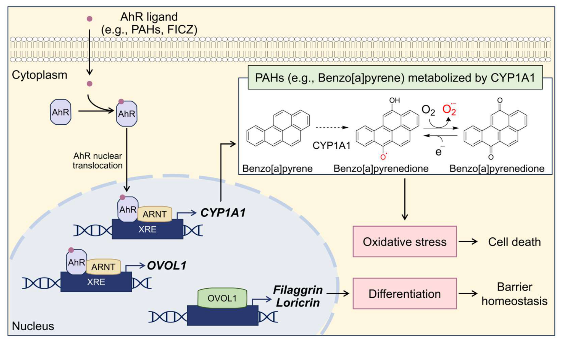
Despite these protective physiological roles, AhR signaling in the skin exhibits a highly context-dependent nature that has been described as “Janus-faced” [147]. The biological outcome of AhR activation is largely determined by the chemical properties, metabolic stability, and persistence of the activating ligand, whether derived from environmental toxins, dietary phytochemicals, microbial metabolites, or pharmaceutical compounds [113]. Many endogenous or dietary ligands, such as the tryptophan photoproduct 6-formylindolo [3,2-b]carbazole (FICZ), are rapidly metabolized by CYP1A1 enzymes induced by AhR activation [148]. This rapid metabolic clearance generates a transient activation cycle that limits prolonged receptor signaling and supports physiological processes such as epithelial barrier maintenance and controlled immune responses [149,150].
In contrast, numerous environmental toxicants, including PAHs associated with PM, act as persistent AhR ligands that resist efficient metabolic clearance [151]. These lipophilic compounds bind AhR with high affinity and drive sustained CYP1A1 expression. During CYP1A1-mediated metabolism, PAHs can be converted into highly reactive intermediates, including redox-active quinones, which undergo repeated redox cycling and generate excessive ROS [54,57]. The persistence of PAHs therefore results in prolonged AhR activation and continuous oxidative stress, shifting AhR signaling away from physiological barrier regulation toward cytotoxic and inflammatory outcomes [152,153].
In the context of environmental pollution, this ligand-dependent divergence is particularly relevant. Persistent exposure to PAHs present in PM can effectively sustain activation of the AhR signaling system, driving sustained CYP1A1 activity, oxidative stress generation, and inflammatory signaling. Consequently, although AhR plays essential physiological roles in maintaining epidermal barrier integrity, its activation by persistent environmental toxicants may contribute to pollution-associated skin damage (Figure 2).

2.5. The Keap1–Nrf2–ARE Pathway: Antioxidant Defense and Cytoprotection

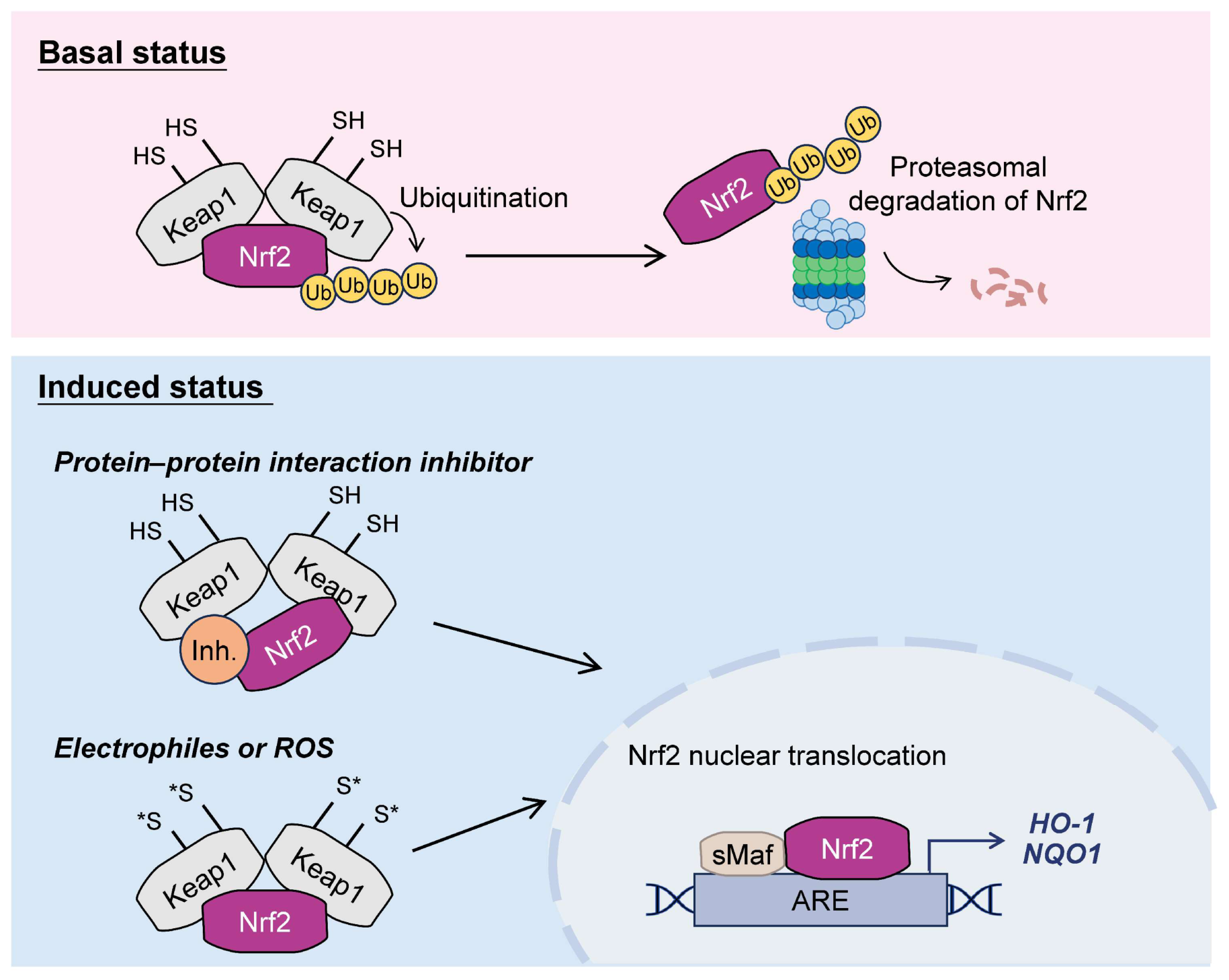
To maintain cutaneous redox homeostasis, the skin relies on the Nrf2 signaling pathway. Nrf2 acts as a master transcriptional regulator that coordinates the expression of numerous cytoprotective genes to defend keratinocytes against PM-induced oxidative injury [125]. Under basal, unstressed conditions, intracellular Nrf2 protein levels remain low due to rapid proteasomal degradation. Nrf2 is a short-lived protein with a half-life of approximately 15–40 min, and about 15 min in HaCaT keratinocytes [154,155].
This strict negative regulation is mediated by Kelch-like ECH-associated protein 1 (Keap1), which contains several functional domains that precisely control Nrf2 stability [156]. The N-terminal Broad complex-Tramtrack-Bric-à-brac (BTB) domain mediates Keap1 homodimerization and recruits Cullin-3 (Cul3) to assemble the E3 ubiquitin ligase complex that targets Nrf2 for continuous ubiquitination and proteasomal degradation [60]. The central intervening region (IVR), containing cysteine residues such as Cys151, Cys273, and Cys288, functions as a sensor of electrophiles and oxidants. The C-terminal Kelch repeat domain directly interacts with the Neh2 regulatory domain of Nrf2 through the widely accepted “hinge-and-latch” mechanism [157]. In this model, Keap1 recognizes a high-affinity ETGE motif (the stable hinge) and a lower-affinity DLG motif (the dynamic latch). Under homeostatic conditions, this dual-binding configuration positions lysine residues on Nrf2 for efficient Cul3-mediated ubiquitination, thereby preventing unnecessary antioxidant activation.
Upon oxidative or electrophilic modification of Keap1 cysteine residues, or disruption of the Keap1–Nrf2 interaction by small-molecule protein–protein interaction (PPI) modulators, a conformational change weakens the interaction with the DLG motif while the ETGE hinge remains intact [158]. For example, LH601A is a direct Keap1–Nrf2 PPI inhibitor that targets the Keap1 Kelch domain and disrupts Nrf2 binding, thereby promoting Nrf2 activation [158]. This partial dissociation alters the spatial alignment of lysine residues between the ETGE and DLG motifs, thereby impairing the Cul3-mediated ubiquitination [159]. Consequently, newly synthesized Nrf2 escapes Keap1-dependent degradation, accumulates in the cytoplasm, and subsequently translocates to the nucleus. In the nucleus, Nrf2 heterodimerizes with small musculoaponeurotic fibrosarcoma (sMaf) transcription factors, and the Nrf2–sMaf complex binds antioxidant response elements (AREs) in the promoter regions of target genes. This transcriptional program induces a wide range of antioxidant and detoxifying enzymes that enhance cellular resistance to oxidative stress [53,160]. Once oxidative stress subsides, Keap1-dependent ubiquitination resumes, leading to Nrf2 degradation and termination of the transcriptional response [161].
Among Nrf2 downstream targets, heme oxygenase-1 (HO-1) and NAD(P)H-quinone oxidoreductase 1 (NQO1) are key components of the antioxidant defense system [162]. HO-1 degrades pro-oxidant heme into biliverdin, carbon monoxide, and free iron; biliverdin is subsequently converted to bilirubin, a potent endogenous antioxidant capable of scavenging ROS [163]. NQO1 protects cells by catalyzing the two-electron reduction in quinones, preventing redox cycling and limiting reactive oxygen species generation [164,165]. Additional Nrf2-regulated genes include enzymes involved in glutathione biosynthesis, such as glutamate–cysteine ligase (GCL), and detoxification enzymes, including glutathione S-transferases (GSTs), which collectively reinforce cellular antioxidant capacity [161]. Through coordinated induction of these cytoprotective genes, Nrf2 plays a central role in maintaining cellular redox equilibrium and protecting keratinocytes from oxidative damage.
Importantly, Keap1 modification in response to thiol-reactive stress follows a dose-dependent continuum rather than a linear relationship. This defines a therapeutic window in which moderate Nrf2 activation is cytoprotective, whereas insufficient or excessive activation may disrupt cellular homeostasis [154]. Consistent with this concept, Nrf2 signaling plays a context-dependent role in epidermal biology [166]. Both loss and constitutive activation of Nrf2 exacerbate disease severity in atopic dermatitis-like mouse models, likely due to impaired keratinocyte differentiation and barrier dysfunction. In contrast, controlled pharmacological activation moderately improves epidermal barrier function, highlighting the need for precise regulation of Nrf2 activation intensity and duration. However, endogenous Nrf2 activation alone may be insufficient to counteract the excessive ROS production induced by PM exposure [125], suggesting that controlled pharmacological enhancement of Nrf2 signaling may help restore protective Nrf2 activity and provide additional protection against PM-induced cell damage.
Collectively, the Keap1–Nrf2–ARE signaling axis represents a central cytoprotective network that safeguards the skin against environmental oxidative stress (Figure 3). Emerging evidence further indicates functional crosstalk between the AhR and Nrf2 pathways. Beyond their downstream signaling interactions, AhR and Nrf2 have been proposed to participate in a shared transcriptional regulatory network involving the AhR–Nrf2 gene battery. This concept has been further extended by the identification of Jun dimerization protein 2 (JDP2) as a chromatin regulator involved in coordinating AhR- and Nrf2-dependent transcriptional responses [167]. In particular, AhR activation may partially contribute to activation of the Nrf2/HO-1 axis by binding to XREs within the Nrf2 gene locus [168,169,170]. Together with AhR-mediated pathways, Nrf2 constitutes a key regulatory node controlling cellular responses to PM exposure. Targeting these interconnected pathways may therefore restore redox homeostasis, reduce oxidative injury, and preserve epidermal barrier integrity, providing a mechanistic framework for developing protective interventions against pollution-associated skin damage.

3. Mechanism-Based Strategies Targeting AhR and Nrf2 Pathways

3.1. Nrf2-Dominant Antioxidant Strategies

Early therapeutic strategies targeting PM-induced skin injury primarily focused on mitigating oxidative stress [171,172]. In response, pharmacological activation of Nrf2 has emerged as a central strategy to restore redox homeostasis. Nrf2 activators can be broadly categorized based on their predominant mechanisms, including covalent modification of Keap1 cysteine residues, disruption of Keap1–Nrf2 protein–protein interactions, and activation of upstream kinase signaling pathways. However, it should be noted that many of these compounds also exhibit secondary effects on other stress-response pathways, including AhR signaling, depending on cellular context.
One major group consists of electrophilic or pro-electrophilic compounds such as artemisitene and sulforaphane that activate Nrf2 through covalent modification of Keap1 cysteine residues (especially Cys 151), thereby disrupting Keap1-mediated ubiquitination and stabilizing Nrf2 [173,174,175]. Artemisitene has been reported to alleviate bleomycin-induced lung injury and inflammation, consistent with its role in Nrf2-mediated cytoprotection [176]. Similarly, sulforaphane has been reported to reduce PM-induced ROS accumulation and oxidative stress in keratinocytes [103].
A second class consists of compounds that interfere with the interaction between the Keap1 Kelch domain and the Nrf2 DLG/ETGE motif. Capsaicin has been reported to disrupt this interaction and reduce oxidative injury in cellular stress models [177]. In addition, several natural flavonoids have been suggested to target the Keap1 Kelch domain and stabilize Nrf2. For example, tiliroside has been reported to bind to the Kelch domain of Keap1 and protect against acetaminophen (APAP)-induced acute liver injury in mice [178]. Similarly, epigallocatechin-3-gallate (EGCG) has been reported to promote Nrf2 nuclear translocation and enhance keratin 16 expression during wound repair, and molecular docking analyses suggest that EGCG may interact with the Keap1 Kelch domain to inhibit Keap1-mediated degradation of Nrf2 [179]. In a 2,4-dinitrochlorobenzene (DNCB)-induced atopic dermatitis mouse model, EGCG also mitigated PM-associated inflammatory responses and preserved keratinocyte differentiation markers, including loricrin and filaggrin [180]. Similarly, a flavonoid-rich fraction from Phyla nodiflora has been reported to activate the Nrf2/HO-1 axis and attenuate PM-induced oxidative stress in keratinocytes, potentially through promotion of Keap1 degradation and interaction with the Keap1 Kelch domain [135].
A third category includes upstream kinase regulators that activate Nrf2 indirectly through signaling cascades such as MAPK, AMP-activated protein kinase α (AMPKα), phosphoinositide 3-kinases (PI3K)/Akt, and protein kinase C (PKC) [181,182]. Activation of these pathways enhances Nrf2 phosphorylation and promotes its nuclear translocation. Several flavonoids have been reported to activate these signaling pathways. For instance, xanthohumol has been reported to stimulate AMPKα signaling and promote Nrf2 activation, thereby strengthening antioxidant defense systems in oxidative stress models [183].
Although antioxidant-based interventions effectively restore redox homeostasis in PM-exposed systems, their ability to fully repair epidermal barrier dysfunction remains limited. Oxidative stress represents only one component of PM-induced skin pathology, whereas structural deterioration of the epidermal barrier arises primarily from impaired keratinocyte differentiation and disruption of intercellular junctions. Therefore, strategies focused predominantly on antioxidant responses may not be sufficient to completely restore epidermal integrity. These observations suggest that additional regulatory pathways involved in epidermal differentiation and barrier formation may also need to be considered when developing therapeutic strategies against PM-induced skin damage.
Importantly, several compounds categorized within Nrf2-dominant strategies have also been reported to modulate AhR signaling. For example, capsaicin and EGCG can inhibit AhR nuclear translocation or transcriptional activity in a context-dependent manner [184,185], while xanthohumol has been reported to inhibit AhR activation under xenobiotic stress conditions [186]. These observations suggest that the functional boundaries between Nrf2- and AhR-regulated pathways are often blurred, supporting the concept that therapeutic strategies should be interpreted based on dominant pathway preference rather than strict mechanistic exclusivity.

3.2. AhR-Dominant Epidermal Regulatory Strategies

Given the central role of the AhR in sensing environmental toxicants and regulating barrier differentiation, pharmacological targeting of AhR has emerged as another potential strategy to mitigate PM-induced skin injury. However, therapeutic manipulation of AhR remains challenging due to its dual biological functions, often described as the “AhR paradox”. Two contrasting approaches have therefore been explored.
On one hand, inhibition of AhR signaling has been proposed as a strategy to prevent pollutant-induced toxicity. By blocking PAH binding to AhR, this approach suppresses downstream xenobiotic metabolism and reduces CYP1A1 induction. Several studies have explored this concept using AhR antagonists. For example, the monoterpenoid S-carvone acts as an allosteric inhibitor of AhR signaling and reduces UV- and benzo[a]pyrene-induced inflammatory responses in mouse skin [187]. Similarly, polyphenol-enriched supplements have been reported to attenuate AhR/ARNT-dependent inflammatory mediators such as COX-2 and MMP-1 induced by PM exposure [188]. While this strategy can reduce pollutant-driven oxidative response and inflammation, sustained AhR inhibition may also interfere with physiological pathways involved in epidermal differentiation, particularly the AhR–OVOL1–filaggrin axis required for barrier maintenance.
Conversely, controlled activation of AhR signaling has been explored as a strategy to promote epidermal differentiation and restore barrier function. Diosmin activates AhR signaling and upregulates epidermal differentiation markers such as filaggrin and loricrin through OVOL1 regulation [189]. Curcumin has also been reported to modulate AhR signaling while suppressing inflammatory responses, including NF-κB activation [190]. Hydroxymetabolites derived from vitamin D3, lumisterol, and tachysterol have been identified as endogenous regulators that can act through AhR and other nuclear receptors, promote keratinocyte differentiation, and enhance protection against oxidative and DNA damage [191,192]. In particular, CYP11A1- and CYP27A1-derived lumisterol metabolites have been shown to interact with AhR and regulate keratinocyte differentiation and cellular responses to oxidative stress [192]. Melatonin and its metabolites may represent another endogenous source of AhR modulation, as they have been reported to activate AhR and induce AhR nuclear translocation in human keratinocytes [193]. Together, these observations indicate that AhR activation contributes not only to xenobiotic sensing but also to epidermal homeostasis and barrier restoration. Notably, these compounds also concurrently influence Nrf2-associated antioxidant responses, highlighting pathway interdependence [194,195,196,197].
To address the potential risks associated with excessive AhR activation, recent studies have introduced the concept of selective AhR modulators (SAhRMs), which exhibit ligand-specific and context-dependent transcriptional outcomes [198,199]. Ligand binding induces distinct conformational changes in AhR, thereby influencing its interaction with cofactors, DNA response elements, and post-translational modifications, ultimately leading to cell- and tissue-specific gene expression profiles. These properties may underlie the differential induction of xenobiotic-metabolizing enzymes such as CYP1A1 and other downstream targets. Accordingly, current research highlights the importance of identifying AhR modulators that favor protective pathways, such as those involved in epidermal differentiation (e.g., filaggrin), while minimizing adverse metabolic responses [200].
Despite these advances, strategies targeting AhR signaling alone primarily regulate inflammatory responses and epithelial differentiation pathways but do not directly address the oxidative burden generated during pollutant exposure. Because PM-induced skin injury arises from both oxidative stress and structural barrier disruption, modulation of AhR signaling alone may be insufficient to fully counteract pollution-associated skin damage. These limitations highlight the need for therapeutic strategies capable of simultaneously regulating oxidative stress and epithelial homeostasis.

3.3. Emerging Perspectives: Coordinated Regulation of AhR and Nrf2

This concept is particularly relevant to PM-induced skin injury, in which oxidative stress and barrier dysfunction occur simultaneously and cannot be fully addressed by single-pathway intervention. The limitations of single-pathway interventions have led to increasing interest in therapeutic strategies capable of simultaneously regulating xenobiotic sensing and antioxidant defense mechanisms. Although these strategies are categorized according to their primary mechanistic emphasis, increasing evidence indicates that AhR and Nrf2 signaling are highly interconnected rather than functionally isolated.
Many electrophilic compounds that stabilize Nrf2 by covalently modifying Keap1 cysteine residues, particularly Cys151, may also influence AhR signaling. Representative examples include sulforaphane [201,202], dimethyl fumarate [203,204,205], and tert-butyl hydroquinone [204,206,207], all of which have been reported to exhibit context-dependent interactions with AhR signaling pathways. Notably, dimethyl fumarate, a clinically used treatment for psoriasis, is metabolized to monomethyl fumarate, which has been reported to exert direct antiproliferative, prodifferentiative, and anti-inflammatory effects on keratinocytes [208,209]. Likewise, several compounds initially characterized as AhR modulators, such as diosmin and curcumin, have been reported to affect oxidative stress responses through secondary activation of Nrf2-related pathways [189,190,194,195]. These studies collectively suggest that the distinction between Nrf2-dominant and AhR-dominant strategies is often not absolute, but rather reflects differences in dominant pathway preference. Therefore, increasing attention has been directed toward compounds that coordinately modulate both AhR and Nrf2 signaling networks.
Early approaches proposed combining AhR inhibition with Nrf2 activation to suppress CYP1A1-derived ROS while enhancing antioxidant capacity [210]. However, as discussed above, antagonizing AhR may interfere with physiological signaling pathways involved in epidermal differentiation. Consequently, coordinated activation of AhR and Nrf2 may represent a potential strategy for counteracting PM-induced skin damage.
Mechanistically, these two pathways perform complementary functions. AhR signaling regulates epidermal barrier integrity by activating the AhR–OVOL1–filaggrin regulatory axis and by modulating the expression of several junctional proteins, while also influencing inflammatory responses in epithelial tissues. In contrast, Nrf2 functions as the principal cellular defense system against oxidative stress, activating a battery of antioxidant and detoxifying enzymes. Importantly, crosstalk between these pathways further strengthens their cooperative effects. Mechanistically, recent studies have revealed that this transcriptional coordination involves the AhR–Nrf2–JDP2 gene battery, in which the chromatin regulator JDP2 forms transcriptional complexes with AhR–ARNT and Nrf2–sMAF, facilitating coordinated recruitment to XREs and AREs within the AhR promoter [167]. These findings suggest that AhR and Nrf2 signaling are not merely parallel pathways but are connected through shared transcriptional machinery. In addition, AhR activation has been reported to contribute to Nrf2 transcription through XREs located within the Nrf2 gene locus [168,169,170]. Meanwhile, Nrf2 upregulates phase II detoxifying enzymes such as NQO1, which catalyzes the two-electron reduction in reactive quinones generated during CYP1A1-mediated metabolism [164,165]. Interestingly, NQO1 expression can also be influenced by AhR signaling, highlighting a shared cytoprotective node between these pathways. Through this coordinated mechanism, Nrf2-mediated antioxidant responses help mitigate the oxidative stress associated with AhR-driven xenobiotic metabolism.
Collectively, dual activation of AhR and Nrf2 may overcome the limitations associated with single-pathway modulation. While AhR activation alone enhances xenobiotic metabolism, it may concurrently increase oxidative burden through CYP1A1 induction. Nrf2 activation alone primarily strengthens antioxidant defenses without directly regulating xenobiotic sensing or epidermal differentiation. Therefore, coordinated activation of both pathways enables a more balanced response, integrating enhanced detoxification capacity with effective antioxidant protection, ultimately reducing oxidative stress while preserving epidermal homeostasis (Figure 4).
Consistent with this concept, accumulating evidence suggests that certain natural compounds simultaneously activate both pathways and confer protective effects across epithelial systems. Prenylated xanthones from mangosteen (Garcinia mangostana) have been reported to co-activate AhR and Nrf2, thereby contributing to protection of intestinal barrier integrity [146]. Similarly, cannabidiol promotes epidermal differentiation and redox balance through AhR-OVOL1-filaggrin and AhR-Nrf2-NQO1 axes [211], while α-tocopherylquinone enhances tight junction formation by modulating claudin expression through both pathways [212]. In addition, environmental stimuli can also trigger coordinated activation of these pathways. For example, Alternaria alternata mycotoxins have been reported to induce nuclear translocation of both AhR and Nrf2 in colon epithelial cells, further illustrating the intrinsic crosstalk between xenobiotic sensing and antioxidant defense systems during epithelial stress responses [213]. Tapinarof is a representative therapeutic example of dual AhR–Nrf2 modulation and has been clinically approved for the treatment of plaque psoriasis and atopic dermatitis. Supported by preclinical models and clinical studies, tapinarof acts as an AhR agonist and promotes Nrf2-associated antioxidant responses. Through these mechanisms, tapinarof enhances epidermal differentiation, restores barrier function, and reduces inflammatory responses in skin disorders [214,215]. Representative compounds reported to modulate AhR and Nrf2 signaling across epithelial injury models are summarized in Table 2. Collectively, the available evidence supports the concept that coordinated modulation of AhR-mediated barrier regulation and Nrf2-dependent antioxidant defense as a promising therapeutic strategy for mitigating PM-induced skin barrier dysfunction.

4. Conceptual Integration of AhR–Nrf2 Interplay in PM-Induced Skin Injury

The interplay between AhR and Nrf2 signaling can be conceptualized as a coordinated regulatory system governing the epidermal response to PM exposure. Although their functions partially overlap, their activation is not necessarily coupled, reflecting ligand-specific and context-dependent transcriptional regulation.
Nrf2 primarily maintains redox homeostasis through antioxidant and cytoprotective gene induction, whereas AhR mediates xenobiotic sensing and regulates epidermal differentiation and barrier organization. Because PM-induced skin injury involves both oxidative stress and structural disruption, effective protection requires coordinated engagement of both pathways. Comparative observations further support the functional complementarity between AhR and Nrf2. Nrf2-dominant activation, exemplified by a flavonoid-rich fraction derived from Phyla nodiflora, attenuates PM-induced oxidative stress and partially restores hydration-related function through AQP3 regulation [135]. However, overall recovery of cellular damage remains incomplete, indicating that redox correction alone is insufficient to fully counteract PM-induced injury, likely due to the lack of direct engagement of AhR-dependent differentiation pathways. In contrast, dual-pathway modulation using tapinarof, a pharmacological AhR agonist with additional Nrf2-activatingcapacity, enables coordinated improvement of PM-induced oxidative stress, cell viability, and barrier-associated functions. This integrated response is associated with more effective restoration of epidermal homeostasis under PM-induced stress conditions [32], highlighting the functional advantage of simultaneous pathway engagement.
Collectively, the available evidence supports a conceptual model in which the balance and integration of AhR-mediated xenobiotic sensing and Nrf2-dependent antioxidant defense determine the overall outcome of PM exposure in the skin. In this model, Nrf2-driven responses primarily buffer oxidative stress, whereas AhR signaling supports xenobiotic metabolism and barrier organization. Coordinated activation of both pathways enables integration of detoxification, antioxidant protection, and structural maintenance, thereby enabling a more comprehensive adaptive response to environmental insults.

5. Conclusions and Perspectives

PM-induced skin damage arises from the convergence of oxidative stress and disruption of epidermal barrier integrity, highlighting the limitations of single-pathway strategies. Modulation of either antioxidant defense or barrier regulation alone is insufficient to fully restore epidermal function. Comparative evidence indicates that Nrf2-dominant activation primarily mitigates oxidative stress, whereas dual activation of AhR and Nrf2 enables coordinated regulation of redox homeostasis, xenobiotic metabolism, and barrier-associated processes. This distinction underscores the functional complementarity of these pathways. Taken together, the coordinated AhR and Nrf2 signaling represents a central regulatory node linking xenobiotic sensing, antioxidant defense, and epidermal homeostasis, providing a mechanistic basis for dual-targeted therapeutic strategies against PM-induced skin barrier dysfunction.
Despite growing evidence supporting the protective potential of coordinated AhR and Nrf2 activation, important knowledge gaps remain in the current understanding of dual-pathway regulation in PM-induced skin injury. Most studies are still based on in vitro systems, with limited integration of in vivo models that capture the complexity of physiological exposure conditions. In particular, how AhR–Nrf2 crosstalk is dynamically regulated under different PM exposure contexts, including variation in exposure duration, dose, and environmental composition, remains poorly understood. Future studies using advanced skin models and in vivo exposure systems will be essential to elucidate the context-dependent regulation of AhR and Nrf2 signaling under physiologically relevant conditions.
An additional unresolved issue concerns the translational generalizability of dual-pathway modulation. Although tapinarof exhibits coordinated modulation of AhR and Nrf2 signaling, it remains unclear whether such activity represents a compound-specific property or a broader class effect across different modulators. The impact of distinct activation profiles on adaptive responses under chronic PM exposure remains incompletely defined. Addressing this gap will require systematic comparative studies of structurally and mechanistically diverse AhR and Nrf2 modulators to establish the robustness of coordinated dual-pathway modulation strategies. Such studies will be essential for defining the translational potential of dual-pathway modulation in environmentally induced skin disorders.
From a translational perspective, future therapeutic development should focus on identifying selective modulators capable of coordinately enhancing epidermal differentiation, barrier integrity, and antioxidant capacity without triggering excessive xenobiotic toxicity. Such approaches may provide a foundation for next-generation topical strategies targeting environmentally induced skin disorders.

Author Contributions

Conceptualization, C.-H.L., C.-H.Y., Y.-T.W., H.-S.C., H.-H.K. and Y.-F.C.; resources, C.-H.Y.; writing—original draft preparation, C.-H.L.; writing—review and editing, Y.-T.W., H.-S.C., H.-H.K. and Y.-F.C.; visualization, C.-H.L., H.-H.K. and Y.-F.C.; funding acquisition, H.-H.K. and Y.-F.C.; project administration, H.-H.K. and Y.-F.C.; supervision, H.-H.K. and Y.-F.C. All authors have read and agreed to the published version of the manuscript.

Funding

This research was funded by the National Science and Technology Council (NSTC, Taiwan), grant numbers NSTC 112-2320-B-037-022-MY3, NSTC 114-2320-B-006-054-, NSTC 115-2320-B-037-016-MY3, and NSTC 115-2320-B-006-046-. The APC was funded by NSTC 112-2320-B-037-022-MY3, NSTC 114-2320-B-006-054-, NSTC 115-2320-B-037-016-MY3, and NSTC 115-2320-B-006-046-.

Data Availability Statement

No new data were created or analyzed in this study. Data sharing is not applicable to this article.

Conflicts of Interest

The authors declare no conflicts of interest.

Abbreviations

The following abbreviations are used in this manuscript:
AhRaryl hydrocarbon receptor
AhRRAhR repressor
AIPAhR-interacting protein
AMPKαAMP-activated protein kinase α
AP-1activator protein-1
APAPacetaminophen
AQP3aquaporin-3
AREsantioxidant response elements
ARNTAhR nuclear translocator
BTBBroad complex-Tramtrack-Bric-à-brac
COX-2cyclooxygenase-2
Cul3Cullin-3
CYP1A1cytochrome P450 1A1
DHEdihydroethidium
ECLenhanced chemiluminescence
EGCGepigallocatechin-3-gallate
FBMNfeature-based molecular networking
FDAU.S. Food and Drug Administration
FICZ6-formylindolo [3,2-b]carbazole
FLGfilaggrin
GCLglutamate–cysteine ligase
GCLCglutamate–cysteine ligase catalytic subunit
GSHglutathione
GSH-Pxglutathione peroxidase
GSSglutathione synthetase
GSTsglutathione S-transferases
γH2AXphosphorylated histone H2AX
H2DCFDA2′,7′-dichlorodihydrofluorescein diacetate
4-HNE4-hydroxynonenal
HO-1heme oxygenase 1
HRNRhornerin
HSP9090 kDa heat shock protein
IFN-γinterferon-γ
IL-1βinterleukin-1β
iNOSinducible nitric oxide synthase
IVLinvolucrin
IVRintervening region
JDP2Jun dimerization protein 2
Keap1Kelch-like ECH-associated protein 1
LC3-Icytosolic form of microtubule-associated protein 1 light chain 3
LC3-IIlipidated form of microtubule-associated protein 1 light chain 3
LDHlactate dehydrogenase
LORloricrin
Δψmmitochondrial membrane potential
MAPKsmitogen-activated protein kinase
MDAmalondialdehyde
MITFmelanocyte inducing transcription factor
MMPsmatrix metalloproteinases
NF-κBnuclear factor kappa-light-chain-enhancer of activated B cells
NLRP1NLR family pyrin domain containing 1
NMFnatural moisturizing factor
NOX4NADPH oxidase 4
NQO1NAD(P)H-quinone oxidoreductase 1
Nrf2nuclear factor erythroid 2–related factor 2
OVOL1ovo-like 1
8-oxo-dG8-oxo-2′-deoxyguanosine
PAHspolycyclic aromatic hydrocarbons
PAI-1plasminogen activator inhibitor-1
PARPpoly(ADP-ribose) polymerase
PI3Kphosphoinositide 3-kinases
PKCprotein kinase C
PMparticulate matter
PPIprotein–protein interaction
ROSreactive oxygen species
SAhRMsselective AhR modulators
SASPsenescence-associated secretory phenotype
SA-β-galsenescence-associated β-galactosidase
sMafsmall musculoaponeurotic fibrosarcoma
SODsuperoxide dismutase
Tapinarof3,5-dihydroxy-4-isopropylstilbene
TBHQtert-butyl hydroquinone
TCDD2,3,7,8-tetrachlorodibenzo-p-dioxin
TERtransepithelial electrical resistance
TEWLtransepidermal water loss
TLR5toll-like receptor 5
TNF-αtumor necrosis factor-α
TRP-1tyrosinase-related protein 1
Trxthioredoxin
TrxRthioredoxin reductase
TYRtyrosinase
UFPsultrafine particles
USEPAUnited States Environmental Protection Agency
UVultraviolet
WHOWorld Health Organization
XREsxenobiotic response elements
ZO-1zonula occludens-1

References

  1. Sangkham, S.; Phairuang, W.; Sherchan, S.P.; Pansakun, N.; Munkong, N.; Sarndhong, K.; Islam, M.A.; Sakunkoo, P. An update on adverse health effects from exposure to PM2.5. Environ. Adv. 2024, 18, 100603. [Google Scholar] [CrossRef] [Scilit]
  2. Li, T.; Yu, Y.; Sun, Z.; Duan, J. A comprehensive understanding of ambient particulate matter and its components on the adverse health effects based from epidemiological and laboratory evidence. Part. Fibre Toxicol. 2022, 19, 67. [Google Scholar] [CrossRef] [Scilit] [PubMed]
  3. U.S. EPA. Particulate Matter (PM) Basics. Available online: https://www.epa.gov/pm-pollution/particulate-matter-pm-basics (accessed on 19 November 2025).
  4. Anderson, J.O.; Thundiyil, J.G.; Stolbach, A. Clearing the air: A review of the effects of particulate matter air pollution on human health. J. Med. Toxicol. 2012, 8, 166–175. [Google Scholar] [CrossRef] [Scilit] [PubMed]
  5. Morawska, L.; Zhang, J.J. Combustion sources of particles. 1. Health relevance and source signatures. Chemosphere 2002, 49, 1045–1058. [Google Scholar] [CrossRef] [Scilit] [PubMed]
  6. Oberdörster, G.; Oberdörster, E.; Oberdörster, J. Nanotoxicology: An emerging discipline evolving from studies of ultrafine particles. Environ. Health Perspect. 2005, 113, 823–839. [Google Scholar] [CrossRef] [Scilit] [PubMed]
  7. Karakoti, A.S.; Hench, L.L.; Seal, S. The potential toxicity of nanomaterials—The role of surfaces. JOM 2006, 58, 77–82. [Google Scholar] [CrossRef] [Scilit]
  8. Chen, R.; Hu, B.; Liu, Y.; Xu, J.; Yang, G.; Xu, D.; Chen, C. Beyond PM2.5: The role of ultrafine particles on adverse health effects of air pollution. Biochim. Biophys. Acta 2016, 1860, 2844–2855. [Google Scholar] [CrossRef] [Scilit] [PubMed]
  9. Moreno-Ríos, A.L.; Tejeda-Benítez, L.P.; Bustillo-Lecompte, C.F. Sources, characteristics, toxicity, and control of ultrafine particles: An overview. Geosci. Front. 2022, 13, 101147. [Google Scholar] [CrossRef] [Scilit]
  10. Liu, L.; Urch, B.; Poon, R.; Szyszkowicz, M.; Speck, M.; Gold, D.R.; Wheeler, A.J.; Scott, J.A.; Brook, J.R.; Thorne, P.S.; et al. Effects of ambient coarse, fine, and ultrafine particles and their biological constituents on systemic biomarkers: A controlled human exposure study. Environ. Health Perspect. 2015, 123, 534–540. [Google Scholar] [CrossRef] [Scilit] [PubMed]
  11. Chen, Y.H.; Nguyen, D.; Brindley, S.; Ma, T.; Xia, T.; Brune, J.; Brown, J.M.; Tsai, C.S. The dependence of particle size on cell toxicity for modern mining dust. Sci. Rep. 2023, 13, 5101. [Google Scholar] [CrossRef] [Scilit] [PubMed]
  12. Li, N.; Sioutas, C.; Cho, A.; Schmitz, D.; Misra, C.; Sempf, J.; Wang, M.; Oberley, T.; Froines, J.; Nel, A. Ultrafine particulate pollutants induce oxidative stress and mitochondrial damage. Environ. Health Perspect. 2003, 111, 455–460. [Google Scholar] [CrossRef] [Scilit] [PubMed]
  13. Kim, S.T.; Saha, K.; Kim, C.; Rotello, V.M. The role of surface functionality in determining nanoparticle cytotoxicity. Acc. Chem. Res. 2013, 46, 681–691. [Google Scholar] [CrossRef] [Scilit] [PubMed]
  14. Pan, S.; Qiu, Y.; Li, M.; Yang, Z.; Liang, D. Recent developments in the determination of PM2.5 chemical composition. Bull. Environ. Contam. Toxicol. 2022, 108, 819–823. [Google Scholar] [CrossRef] [Scilit] [PubMed]
  15. Melzi, G.; Massimi, L.; Frezzini, M.A.; Iulini, M.; Tarallo, N.; Rinaldi, M.; Paglione, M.; Nozza, E.; Crova, F.; Valentini, S.; et al. Redox-activity and in vitro effects of regional atmospheric aerosol pollution: Seasonal differences and correlation between oxidative potential and in vitro toxicity of PM1. Toxicol. Appl. Pharmacol. 2024, 485, 116913. [Google Scholar] [CrossRef] [Scilit] [PubMed]
  16. Kim, K.H.; Kabir, E.; Kabir, S. A review on the human health impact of airborne particulate matter. Environ. Int. 2015, 74, 136–143. [Google Scholar] [CrossRef] [Scilit] [PubMed]
  17. Zhao, S.M.; Liu, W.X.; Miao, Q.Y.; Li, J.M.; Sun, L.M.; Wu, S.P. Emissions characteristics of polycyclic aromatic hydrocarbons, water-soluble heavy metals, and oxidative potential from indoor non-energy combustion sources. Environ. Sci. Pollut. Res. Int. 2025, 32, 15342–15358. [Google Scholar] [CrossRef] [Scilit] [PubMed]
  18. Wei, H.; Feng, Y.; Liang, F.; Cheng, W.; Wu, X.; Zhou, R.; Wang, Y. Role of oxidative stress and DNA hydroxymethylation in the neurotoxicity of fine particulate matter. Toxicology 2017, 380, 94–103. [Google Scholar] [CrossRef] [Scilit] [PubMed]
  19. Zhang, Y.; Hu, Q.; Fu, J.; Li, X.; Mao, H.; Wang, T. Influence of exposure pathways on tissue distribution and health impact of polycyclic aromatic hydrocarbon derivatives. Environ. Health 2023, 1, 150–167. [Google Scholar] [CrossRef] [Scilit] [PubMed]
  20. Menichetti, A.; Mordini, D.; Montalti, M. Penetration of microplastics and nanoparticles through skin: Effects of size, shape, and surface chemistry. J. Xenobiot. 2025, 15, 6. [Google Scholar] [CrossRef] [Scilit] [PubMed]
  21. Fuller, R.; Landrigan, P.J.; Balakrishnan, K.; Bathan, G.; Bose-O’Reilly, S.; Brauer, M.; Caravanos, J.; Chiles, T.; Cohen, A.; Corra, L.; et al. Pollution and health: A progress update. Lancet Planet. Health 2022, 6, e535–e547. [Google Scholar] [CrossRef] [Scilit] [PubMed]
  22. WHO. Air Pollution. Available online: https://www.who.int/health-topics/air-pollution#tab=tab_2 (accessed on 20 November 2025).
  23. Ruckerl, R.; Schneider, A.; Breitner, S.; Cyrys, J.; Peters, A. Health effects of particulate air pollution: A review of epidemiological evidence. Inhal. Toxicol. 2011, 23, 555–592. [Google Scholar] [CrossRef] [Scilit] [PubMed]
  24. Ngoc, L.T.N.; Park, D.; Lee, Y.; Lee, Y.C. Systematic review and meta-analysis of human skin diseases due to particulate matter. Int. J. Environ. Res. Public Health 2017, 14, 1458. [Google Scholar] [CrossRef] [Scilit] [PubMed]
  25. Weslock, N.; Drew, D.; Palanichami, D.; La, A.; Shoctor, H.; Amir, A.; Ramdass, P.; Mitchell, K. Association between fine particulate matter (PM2.5) and atopic dermatitis—A systematic review. Discov. Public Health 2025, 22, 117. [Google Scholar] [CrossRef] [Scilit]
  26. Hartung, F.; Krutmann, J.; Haarmann-Stemmann, T. Evidence that the aryl hydrocarbon receptor orchestrates oxinflammatory responses and contributes to airborne particulate matter-induced skin aging. Free Radic. Biol. Med. 2025, 233, 264–278. [Google Scholar] [CrossRef] [Scilit] [PubMed]
  27. Schuch, A.P.; Moreno, N.C.; Schuch, N.J.; Menck, C.F.M.; Garcia, C.C.M. Sunlight damage to cellular DNA: Focus on oxidatively generated lesions. Free Radic. Biol. Med. 2017, 107, 110–124. [Google Scholar] [CrossRef] [Scilit] [PubMed]
  28. Petracca, B.; Nadaban, A.; Eeman, M.; Gooris, G.S.; Bouwstra, J.A. Effects of ozone on stratum corneum lipid integrity and assembly. Chem. Phys. Lipids 2021, 240, 105121. [Google Scholar] [CrossRef] [Scilit] [PubMed]
  29. Manisalidis, I.; Stavropoulou, E.; Stavropoulos, A.; Bezirtzoglou, E. Environmental and health impacts of air pollution: A review. Front. Public Health 2020, 8, 14. [Google Scholar] [CrossRef] [Scilit] [PubMed]
  30. Plant, A.L.; Benson, D.M.; Smith, L.C. Cellular uptake and intracellular localization of benzo(a)pyrene by digital fluorescence imaging microscopy. J. Cell Biol. 1985, 100, 1295–1308. [Google Scholar] [CrossRef] [Scilit] [PubMed]
  31. Bejaoui, S.; Mercier, X.; Desgroux, P.; Therssen, E. Laser induced fluorescence spectroscopy of aromatic species produced in atmospheric sooting flames using UV and visible excitation wavelengths. Combust. Flame 2014, 161, 2479–2491. [Google Scholar] [CrossRef] [Scilit]
  32. Lin, C.H.; Ko, H.H.; Wu, J.Y.; Chang, H.S.; Yen, C.H.; Chiu, C.C.; Chen, Y.F. Dual activation of AhR and Nrf2 pathways by the natural stilbenoid tapinarof protects against particulate matter-induced skin barrier dysfunction. Toxicol. Appl. Pharmacol. 2025, 505, 117559. [Google Scholar] [CrossRef] [Scilit] [PubMed]
  33. Drakaki, E.; Dessinioti, C.; Antoniou, C.V. Air pollution and the skin. Front. Environ. Sci. 2014, 2, 11. [Google Scholar] [CrossRef] [Scilit]
  34. de Szalay, S.; Wertz, P.W. Protective barriers provided by the epidermis. Int. J. Mol. Sci. 2023, 24, 3145. [Google Scholar] [CrossRef] [Scilit] [PubMed]
  35. Kolarsick, P.A.J.; Kolarsick, M.A.; Goodwin, C. Anatomy and physiology of the skin. J. Dermatol. Nurses’ Assoc. 2011, 3, 203–213. [Google Scholar] [CrossRef] [Scilit]
  36. Edmondson, S.R.; Thumiger, S.P.; Werther, G.A.; Wraight, C.J. Epidermal homeostasis: The role of the growth hormone and insulin-like growth factor systems. Endocr. Rev. 2003, 24, 737–764. [Google Scholar] [CrossRef] [Scilit] [PubMed]
  37. Slominski, R.M.; Raman, C.; Jetten, A.M.; Slominski, A.T. Neuro-immuno-endocrinology of the skin: How environment regulates body homeostasis. Nat. Rev. Endocrinol. 2025, 21, 495–509. [Google Scholar] [CrossRef] [Scilit] [PubMed]
  38. Rawlings, A.V.; Harding, C.R. Moisturization and skin barrier function. Dermatol. Ther. 2004, 17, 43–48. [Google Scholar] [CrossRef] [Scilit] [PubMed]
  39. Bolzinger, M.-A.; Briançon, S.; Pelletier, J.; Chevalier, Y. Penetration of drugs through skin, a complex rate-controlling membrane. Curr. Opin. Colloid Interface Sci. 2012, 17, 156–165. [Google Scholar] [CrossRef] [Scilit]
  40. Fluhr, J.W.; Moore, D.J.; Lane, M.E.; Lachmann, N.; Rawlings, A.V. Epidermal barrier function in dry, flaky and sensitive skin: A narrative review. J. Eur. Acad. Dermatol. Venereol. 2024, 38, 812–820. [Google Scholar] [CrossRef] [Scilit] [PubMed]
  41. Proksch, E.; Brandner, J.M.; Jensen, J.M. The skin: An indispensable barrier. Exp. Dermatol. 2008, 17, 1063–1072. [Google Scholar] [CrossRef] [Scilit] [PubMed]
  42. Turksen, K.; Troy, T.C. Barriers built on claudins. J. Cell Sci. 2004, 117, 2435–2447. [Google Scholar] [CrossRef] [Scilit] [PubMed]
  43. Bäsler, K.; Bergmann, S.; Heisig, M.; Naegel, A.; Zorn-Kruppa, M.; Brandner, J.M. The role of tight junctions in skin barrier function and dermal absorption. J. Control. Release 2016, 242, 105–118. [Google Scholar] [CrossRef] [Scilit] [PubMed]
  44. Lefevre-Utile, A.; Braun, C.; Haftek, M.; Aubin, F. Five functional aspects of the epidermal barrier. Int. J. Mol. Sci. 2021, 22, 11676. [Google Scholar] [CrossRef] [Scilit] [PubMed]
  45. Sougrat, R.; Morand, M.; Gondran, C.; Barre, P.; Gobin, R.; Bonte, F.; Dumas, M.; Verbavatz, J.M. Functional expression of AQP3 in human skin epidermis and reconstructed epidermis. J. Investig. Dermatol. 2002, 118, 678–685. [Google Scholar] [CrossRef] [Scilit] [PubMed]
  46. Patel, R.; Kevin Heard, L.; Chen, X.; Bollag, W.B. Aquaporins in the skin. Adv. Exp. Med. Biol. 2017, 969, 173–191. [Google Scholar] [CrossRef] [Scilit] [PubMed]
  47. Hara-Chikuma, M.; Verkman, A.S. Aquaporin-3 functions as a glycerol transporter in mammalian skin. Biol. Cell 2005, 97, 479–486. [Google Scholar] [CrossRef] [Scilit] [PubMed]
  48. Tham, H.P.; Yip, K.Y.; Aitipamula, S.; Mothe, S.R.; Zhao, W.; Choong, P.S.; Benetti, A.A.; Gan, W.E.; Leong, F.Y.; Thoniyot, P.; et al. Influence of particle parameters on deposition onto healthy and damaged human hair. Int. J. Cosmet. Sci. 2025, 47, 58–72. [Google Scholar] [CrossRef] [Scilit] [PubMed]
  49. Jin, S.P.; Li, Z.; Choi, E.K.; Lee, S.; Kim, Y.K.; Seo, E.Y.; Chung, J.H.; Cho, S. Urban particulate matter in air pollution penetrates into the barrier-disrupted skin and produces ROS-dependent cutaneous inflammatory response in vivo. J. Dermatol. Sci. 2018, 91, 175–183. [Google Scholar] [CrossRef] [Scilit] [PubMed]
  50. Khunger, N.; Dash, A. Impact of air pollution on skin pigmentation: Mechanisms and protective strategies. Int. J. Dermatol. 2025, 64, 1788–1801. [Google Scholar] [CrossRef] [Scilit] [PubMed]
  51. Knorr, F.; Lademann, J.; Patzelt, A.; Sterry, W.; Blume-Peytavi, U.; Vogt, A. Follicular transport route—Research progress and future perspectives. Eur. J. Pharm. Biopharm. 2009, 71, 173–180. [Google Scholar] [CrossRef] [Scilit] [PubMed]
  52. Simon, K.; Bartsch, N.; Schneider, L.; van de Weijgert, V.; Hutzler, C.; Luch, A.; Roloff, A. Polycyclic aromatic hydrocarbon skin permeation efficiency in vitro is lower through human than pigskin and decreases with lipophilicity. Environ. Res. 2024, 255, 119118. [Google Scholar] [CrossRef] [Scilit] [PubMed]
  53. Schalka, S.; Silva, M.S.; Lopes, L.F.; de Freitas, L.M.; Baptista, M.S. The skin redoxome. J. Eur. Acad. Dermatol. Venereol. 2022, 36, 181–195. [Google Scholar] [CrossRef] [Scilit] [PubMed]
  54. Chen, Q.; Wang, T.; Wu, X.; Yuan, H.; Wei, Y.; Xiao, Y. The role of the cytochrome P450 superfamily in the skin. Expert Rev. Mol. Med. 2024, 26, e15. [Google Scholar] [CrossRef] [Scilit] [PubMed]
  55. Paik, K.; Na, J.I.; Huh, C.H.; Shin, J.W. Particulate matter and its molecular effects on skin: Implications for various skin diseases. Int. J. Mol. Sci. 2024, 25, 9888. [Google Scholar] [CrossRef] [Scilit] [PubMed]
  56. Chao, L.; Feng, B.; Liang, H.; Zhao, X.; Song, J. Particulate matter and inflammatory skin diseases: From epidemiological and mechanistic studies. Sci. Total Environ. 2023, 905, 167111. [Google Scholar] [CrossRef] [Scilit] [PubMed]
  57. Burczynski, M.E.; Penning, T.M. Genotoxic polycyclic aromatic hydrocarbon ortho-quinones generated by aldo-keto reductases induce CYP1A1 via nuclear translocation of the aryl hydrocarbon receptor. Cancer Res. 2000, 60, 908–915. [Google Scholar] [PubMed]
  58. Piao, M.J.; Ahn, M.J.; Kang, K.A.; Ryu, Y.S.; Hyun, Y.J.; Shilnikova, K.; Zhen, A.X.; Jeong, J.W.; Choi, Y.H.; Kang, H.K.; et al. Particulate matter 2.5 damages skin cells by inducing oxidative stress, subcellular organelle dysfunction, and apoptosis. Arch. Toxicol. 2018, 92, 2077–2091. [Google Scholar] [CrossRef] [Scilit] [PubMed]
  59. Maunjumpon, P.; Thamsermsang, O.; Panich, U. Mitigating PM2.5-induced skin injury and aging: Botanical strategies targeting redox and inflammatory pathways. Redox Rep. 2026, 31, 2629079. [Google Scholar] [CrossRef] [Scilit] [PubMed]
  60. Dinkova-Kostova, A.T.; Copple, I.M. Advances and challenges in therapeutic targeting of Nrf2. Trends Pharmacol. Sci. 2023, 44, 137–149. [Google Scholar] [CrossRef] [Scilit] [PubMed]
  61. Lee, E.S.; Kim, S.; Lee, S.W.; Jung, J.; Lee, S.H.; Na, H.W.; Kim, H.J.; Hong, Y.D.; Park, W.S.; Lee, T.G.; et al. Molecule-resolved visualization of particulate matter on human skin using multimodal nonlinear optical imaging. Int. J. Mol. Sci. 2021, 22, 5199. [Google Scholar] [CrossRef] [Scilit] [PubMed]
  62. Somayajulu, M.; Wright, R.; Muhammed, F.; McClellan, S.A.; Ibrahim, A.; Hazlett, L.D. PM10 dysregulates epithelial barrier function in human corneal epithelial cells that is restored by antioxidant SKQ1. Toxicol. Appl. Pharmacol. 2024, 492, 117122. [Google Scholar] [CrossRef] [Scilit] [PubMed]
  63. Tang, J.; Diao, P.; Pan, W.; Li, L.; Xiong, L. The cross-linking between DNA damage, oxidative stress and epidermal barrier in keratinocytes after exposure to particulate matters and carbon black. Exp. Dermatol. 2024, 33, e15048. [Google Scholar] [CrossRef] [Scilit] [PubMed]
  64. Hieda, D.S.; Anastacio da Costa Carvalho, L.; Vaz de Mello, B.; Oliveira, E.A.; Romano de Assis, S.; Wu, J.; Du-Thumm, L.; Viana da Silva, C.L.; Roubicek, D.A.; Maria-Engler, S.S.; et al. Air particulate matter induces skin barrier dysfunction and water transport alteration on a reconstructed human epidermis model. J. Investig. Dermatol. 2020, 140, 2343–2352.e3. [Google Scholar] [CrossRef] [Scilit] [PubMed]
  65. Park, S.; Lim, J.; Kim, S.; Jeon, M.; Baek, H.; Park, W.; Park, J.; Kim, S.N.; Kang, N.G.; Park, C.G.; et al. Anti-inflammatory artificial extracellular vesicles with notable inhibition of particulate matter-induced skin inflammation and barrier function impairment. ACS Appl. Mater. Interfaces 2023, 15, 59199–59208. [Google Scholar] [CrossRef] [Scilit] [PubMed]
  66. Esser, C.; Bargen, I.; Weighardt, H.; Haarmann-Stemmann, T.; Krutmann, J. Functions of the aryl hydrocarbon receptor in the skin. Semin. Immunopathol. 2013, 35, 677–691. [Google Scholar] [CrossRef] [Scilit]
  67. Li, Y.; Zeng, Y.; Chen, Z.; Tan, X.; Mei, X.; Wu, Z. The role of aryl hydrocarbon receptor in vitiligo: A review. Front. Immunol. 2024, 15, 1291556. [Google Scholar] [CrossRef] [Scilit] [PubMed]
  68. Lin, M.; Yu, J.Z. Oxidative potential of atmospheric particulate matter: A review of the role of metal-organic interactions, mechanistic insights, and key determinants. Environ. Sci. Technol. 2026, 60, 1546–1559. [Google Scholar] [CrossRef] [Scilit] [PubMed]
  69. Wispriyono, B.; Jalaludin, J.; Kusnoputranto, H.; Pakpahan, S.; Aryati, G.P.; Pratama, S.; Librianty, N.; Rozaliyani, A.; Taufik, F.F.; Novirsa, R. Glutathione (GSH) and superoxide dismutase (SOD) levels among junior high school students induced by indoor particulate matter 2.5 (PM2.5) and nitrogen dioxide (NO2) exposure. J. Public Health Res. 2021, 10, 2372. [Google Scholar] [CrossRef] [Scilit] [PubMed]
  70. Pardo, M.; Qiu, X.; Zimmermann, R.; Rudich, Y. Particulate matter toxicity is Nrf2 and mitochondria dependent: The roles of metals and polycyclic aromatic hydrocarbons. Chem. Res. Toxicol. 2020, 33, 1110–1120. [Google Scholar] [CrossRef] [Scilit] [PubMed]
  71. Rodrigo-Moreno, A.; Poschenrieder, C.; Shabala, S. Transition metals: A double edge sward in ROS generation and signaling. Plant Signal. Behav. 2013, 8, e23425. [Google Scholar] [CrossRef] [Scilit] [PubMed]
  72. Li, N.; Xia, T.; Nel, A.E. The role of oxidative stress in ambient particulate matter-induced lung diseases and its implications in the toxicity of engineered nanoparticles. Free Radic. Biol. Med. 2008, 44, 1689–1699. [Google Scholar] [CrossRef] [Scilit] [PubMed]
  73. Ryu, Y.S.; Kang, K.A.; Piao, M.J.; Ahn, M.J.; Yi, J.M.; Hyun, Y.M.; Kim, S.H.; Ko, M.K.; Park, C.O.; Hyun, J.W. Particulate matter induces inflammatory cytokine production via activation of NF-κB by TLR5-NOX4-ROS signaling in human skin keratinocyte and mouse skin. Redox Biol. 2019, 21, 101080. [Google Scholar] [CrossRef] [Scilit] [PubMed]
  74. Marsal, A.; Sauvain, J.J.; Thomas, A.; Lyon-Caen, S.; Borlaza, L.J.S.; Philippat, C.; Jaffrezo, J.L.; Boudier, A.; Darfeuil, S.; Elazzouzi, R.; et al. Effects of personal exposure to the oxidative potential of PM2.5 on oxidative stress biomarkers in pregnant women. Sci. Total Environ. 2024, 911, 168475. [Google Scholar] [CrossRef] [Scilit] [PubMed]
  75. Pecorelli, A.; Woodby, B.; Prieux, R.; Valacchi, G. Involvement of 4-hydroxy-2-nonenal in pollution-induced skin damage. BioFactors 2019, 45, 536–547. [Google Scholar] [CrossRef] [Scilit] [PubMed]
  76. Chahla, C.; Kovacic, H.; Ferhat, L.; Leloup, L. Pathological impact of redox post-translational modifications. Antioxid. Redox Signal 2024, 41, 152–180. [Google Scholar] [CrossRef] [Scilit] [PubMed]
  77. Hahm, J.Y.; Park, J.; Jang, E.S.; Chi, S.W. 8-Oxoguanine: From oxidative damage to epigenetic and epitranscriptional modification. Exp. Mol. Med. 2022, 54, 1626–1642. [Google Scholar] [CrossRef] [Scilit] [PubMed]
  78. Wei, Y.; Han, I.K.; Shao, M.; Hu, M.; Zhang, O.J.; Tang, X. PM2.5 constituents and oxidative DNA damage in humans. Environ. Sci. Technol. 2009, 43, 4757–4762. [Google Scholar] [CrossRef] [Scilit] [PubMed]
  79. Møller, P.; Folkmann, J.K.; Forchhammer, L.; Brӓuner, E.V.; Danielsen, P.H.; Risom, L.; Loft, S. Air pollution, oxidative damage to DNA, and carcinogenesis. Cancer Lett. 2008, 266, 84–97. [Google Scholar] [CrossRef] [Scilit] [PubMed]
  80. Fernando, P.D.S.M.; Piao, M.J.; Kang, K.A.; Herath, R.M.U.L.; Senavirathna, H.M.M.M.; Hyun, C.; Chae, S.; Park, J.; Hyun, J.W. Apigenin prevents oxidative stress and mitochondrial damage caused by particulate matter 2.5 in skin cells. Environ. Pollut. Bioavailab. 2025, 37, 2544014. [Google Scholar] [CrossRef] [Scilit]
  81. Karlsson, H.L.; Nygren, J.; Moller, L. Genotoxicity of airborne particulate matter: The role of cell-particle interaction and of substances with adduct-forming and oxidizing capacity. Mutat. Res. 2004, 565, 1–10. [Google Scholar] [CrossRef] [Scilit] [PubMed]
  82. Herath, H.; Piao, M.J.; Kang, K.A.; Zhen, A.X.; Fernando, P.; Kang, H.K.; Yi, J.M.; Hyun, J.W. Hesperidin exhibits protective effects against PM2.5-mediated mitochondrial damage, cell cycle arrest, and cellular senescence in human HaCaT keratinocytes. Molecules 2022, 27, 4800. [Google Scholar] [CrossRef] [Scilit] [PubMed]
  83. Zhen, A.X.; Piao, M.J.; Kang, K.A.; Fernando, P.D.; Herath, H.M.; Cho, S.J.; Hyun, J.W. 3-Bromo-4,5-dihydroxybenzaldehyde protects keratinocytes from particulate matter 2.5-induced damages. Antioxidants 2023, 12, 1307. [Google Scholar] [CrossRef] [Scilit] [PubMed]
  84. Qiao, Y.; Li, Q.; Du, H.Y.; Wang, Q.W.; Huang, Y.; Liu, W. Airborne polycyclic aromatic hydrocarbons trigger human skin cells aging through aryl hydrocarbon receptor. Biochem. Biophys. Res. Commun. 2017, 488, 445–452. [Google Scholar] [CrossRef] [Scilit] [PubMed]
  85. Gualtieri, M.; Ovrevik, J.; Mollerup, S.; Asare, N.; Longhin, E.; Dahlman, H.J.; Camatini, M.; Holme, J.A. Airborne urban particles (Milan winter-PM2.5) cause mitotic arrest and cell death: Effects on DNA, mitochondria, AhR binding and spindle organization. Mutat. Res. 2011, 713, 18–31. [Google Scholar] [CrossRef] [Scilit] [PubMed]
  86. Zhu, S.; Li, X.; Dang, B.; Wu, F.; Wang, C.; Lin, C. Lycium barbarum polysaccharide protects HaCaT cells from PM2.5-induced apoptosis via inhibiting oxidative stress, ER stress and autophagy. Redox Rep. 2022, 27, 32–44. [Google Scholar] [CrossRef] [Scilit] [PubMed]
  87. Hu, R.; Xie, X.Y.; Xu, S.K.; Wang, Y.N.; Jiang, M.; Wen, L.R.; Lai, W.; Guan, L. PM2.5 exposure elicits oxidative stress responses and mitochondrial apoptosis pathway activation in HaCaT keratinocytes. Chin. Med. J. 2017, 130, 2205–2214. [Google Scholar] [CrossRef] [Scilit] [PubMed]
  88. Herath, H.; Piao, M.J.; Kang, K.A.; Fernando, P.; Kang, H.K.; Koh, Y.S.; Hyun, J.W. The inhibitory effect of chlorogenic acid on oxidative stress and apoptosis induced by PM2.5 in HaCaT keratinocytes. J. Biochem. Mol. Toxicol. 2024, 38, e23806. [Google Scholar] [CrossRef] [Scilit] [PubMed]
  89. Fernando, P.; Piao, M.J.; Herath, H.; Kang, K.A.; Ha, K.S.; Chae, S.; Hyun, J.W. C-peptide ameliorates particulate matter 2.5-induced skin cell apoptosis by inhibiting NADPH oxidation. Biomol. Ther. 2025, 33, 221–230. [Google Scholar] [CrossRef] [Scilit] [PubMed]
  90. Pan, T.L.; Wang, P.W.; Aljuffali, I.A.; Huang, C.T.; Lee, C.W.; Fang, J.Y. The impact of urban particulate pollution on skin barrier function and the subsequent drug absorption. J. Dermatol. Sci. 2015, 78, 51–60. [Google Scholar] [CrossRef] [Scilit] [PubMed]
  91. Peixoto, M.S.; de Oliveira Galvao, M.F.; Batistuzzo de Medeiros, S.R. Cell death pathways of particulate matter toxicity. Chemosphere 2017, 188, 32–48. [Google Scholar] [CrossRef] [Scilit] [PubMed]
  92. Wang, Y.; Zhong, Y.; Liao, J.; Wang, G. PM2.5-related cell death patterns. Int. J. Med. Sci. 2021, 18, 1024–1029. [Google Scholar] [CrossRef] [Scilit] [PubMed]
  93. Dai, Y.; Wang, Y.; Lu, S.; Deng, X.; Niu, X.; Guo, Z.; Qian, R.; Zhou, M.; Peng, X. Autophagy attenuates particulate matter 2.5-induced damage in HaCaT cells. Ann. Transl. Med. 2021, 9, 978. [Google Scholar] [CrossRef] [Scilit] [PubMed]
  94. Ryu, Y.S.; Kang, K.A.; Piao, M.J.; Ahn, M.J.; Yi, J.M.; Bossis, G.; Hyun, Y.M.; Park, C.O.; Hyun, J.W. Particulate matter-induced senescence of skin keratinocytes involves oxidative stress-dependent epigenetic modifications. Exp. Mol. Med. 2019, 51, 1–14. [Google Scholar] [CrossRef] [Scilit] [PubMed]
  95. Reynolds, W.J.; Hanson, P.S.; Critchley, A.; Griffiths, B.; Chavan, B.; Birch-Machin, M.A. Exposing human primary dermal fibroblasts to particulate matter induces changes associated with skin aging. FASEB J. 2020, 34, 14725–14735. [Google Scholar] [CrossRef] [Scilit] [PubMed]
  96. Martic, I.; Jansen-Durr, P.; Cavinato, M. Effects of air pollution on cellular senescence and skin aging. Cells 2022, 11, 2220. [Google Scholar] [CrossRef] [Scilit] [PubMed]
  97. Lee, Y.I.; Choi, S.; Roh, W.S.; Lee, J.H.; Kim, T.G. Cellular senescence and inflammaging in the skin microenvironment. Int. J. Mol. Sci. 2021, 22, 3849. [Google Scholar] [CrossRef] [Scilit] [PubMed]
  98. Ajoolabady, A.; Pratico, D.; Bahijri, S.; Tuomilehto, J.; Uversky, V.N.; Ren, J. Hallmarks of cellular senescence: Biology, mechanisms, regulations. Exp. Mol. Med. 2025, 57, 1482–1491. [Google Scholar] [CrossRef] [Scilit] [PubMed]
  99. Herranz, N.; Gil, J. Mechanisms and functions of cellular senescence. J. Clin. Investig. 2018, 128, 1238–1246. [Google Scholar] [CrossRef] [Scilit] [PubMed]
  100. Zhen, A.X.; Kang, K.A.; Piao, M.J.; Madushan Fernando, P.D.S.; Lakmini Herath, H.M.U.; Hyun, J.W. Protective effects of astaxanthin on particulate matter 2.5-induced senescence in HaCaT keratinocytes via maintenance of redox homeostasis. Exp. Ther. Med. 2024, 28, 275. [Google Scholar] [CrossRef] [Scilit] [PubMed]
  101. Seong, S.H.; Kim, J.Y.; Kim, S.H.; Lee, J.; Lee, E.J.; Bae, Y.J.; Park, S.; Kwon, I.J.; Yoon, S.M.; Lee, J.; et al. Interleukin-24: A molecular mediator of particulate matter’s impact on skin aging. Ecotoxicol. Environ. Saf. 2024, 282, 116738. [Google Scholar] [CrossRef] [Scilit] [PubMed]
  102. Feng, C.; Chen, X.; Yin, X.; Jiang, Y.; Zhao, C. Matrix metalloproteinases on skin photoaging. J. Cosmet. Dermatol. 2024, 23, 3847–3862. [Google Scholar] [CrossRef] [Scilit] [PubMed]
  103. Ko, H.J.; Kim, J.H.; Lee, G.S.; Shin, T. Sulforaphane controls the release of paracrine factors by keratinocytes and thus mitigates particulate matter-induced premature skin aging by suppressing melanogenesis and maintaining collagen homeostasis. Phytomedicine 2020, 77, 153276. [Google Scholar] [CrossRef] [Scilit] [PubMed]
  104. Kubo, H.; Moriyama, M.; Goto, S.; Miyake, Y.; Nakamura, M.; Ozeki, Y.; Nakamura, Y.; Moriyama, H. Sage extract and ascorbic acid derivative inhibit melanogenesis via downregulating keratinocyte-derived GM-CSF. PLoS ONE 2025, 20, e0325242. [Google Scholar] [CrossRef] [Scilit] [PubMed]
  105. Shi, Y.; Zeng, Z.; Liu, J.; Pi, Z.; Zou, P.; Deng, Q.; Ma, X.; Qiao, F.; Xiong, W.; Zhou, C.; et al. Particulate matter promotes hyperpigmentation via AhR/MAPK signaling activation and by increasing α-MSH paracrine levels in keratinocytes. Environ. Pollut. 2021, 278, 116850. [Google Scholar] [CrossRef] [Scilit] [PubMed]
  106. Peng, F.; Tsuji, G.; Zhang, J.Z.; Chen, Z.; Furue, M. Potential role of PM2.5 in melanogenesis. Environ. Int. 2019, 132, 105063. [Google Scholar] [CrossRef] [Scilit] [PubMed]
  107. Bellei, B.; Picardo, M. Premature cell senescence in human skin: Dual face in chronic acquired pigmentary disorders. Ageing Res. Rev. 2020, 57, 100981. [Google Scholar] [CrossRef] [Scilit] [PubMed]
  108. Bae, Y.J.; Park, K.Y.; Han, H.S.; Kim, Y.S.; Hong, J.Y.; Han, T.Y.; Seo, S.J. Effects of particulate matter in a mouse model of oxazolone-induced atopic dermatitis. Ann. Dermatol. 2020, 32, 496–507. [Google Scholar] [CrossRef] [Scilit] [PubMed]
  109. Kim, B.E.; Kim, J.; Goleva, E.; Berdyshev, E.; Lee, J.; Vang, K.A.; Lee, U.H.; Han, S.; Leung, S.; Hall, C.F.; et al. Particulate matter causes skin barrier dysfunction. JCI Insight 2021, 6, e145185. [Google Scholar] [CrossRef] [Scilit] [PubMed]
  110. Teng, W.L.; Huang, P.H.; Wang, H.C.; Tseng, C.H.; Yen, F.L. Pterostilbene attenuates particulate matter-induced oxidative stress, inflammation and aging in keratinocytes. Antioxidants 2021, 10, 1552. [Google Scholar] [CrossRef] [Scilit] [PubMed]
  111. Yang, C.Y.; Pan, C.C.; Tseng, C.H.; Yen, F.L. Antioxidant, anti-inflammation and antiaging activities of Artocarpus altilis methanolic extract on urban particulate matter-induced HaCaT keratinocytes damage. Antioxidants 2022, 11, 2304. [Google Scholar] [CrossRef] [Scilit] [PubMed]
  112. Choudhary, V.; Olala, L.O.; Kagha, K.; Pan, Z.Q.; Chen, X.; Yang, R.; Cline, A.; Helwa, I.; Marshall, L.; Kaddour-Djebbar, I.; et al. Regulation of the glycerol transporter, aquaporin-3, by histone deacetylase-3 and p53 in keratinocytes. J. Investig. Dermatol. 2017, 137, 1935–1944. [Google Scholar] [CrossRef] [Scilit] [PubMed]
  113. Bock, K.W. From TCDD-mediated toxicity to searches of physiologic AhR functions. Biochem. Pharmacol. 2018, 155, 419–424. [Google Scholar] [CrossRef] [Scilit] [PubMed]
  114. Dawe, H.R.; Di Meglio, P. The aryl hydrocarbon receptor (AhR): Peacekeeper of the skin. Int. J. Mol. Sci. 2025, 26, 1618. [Google Scholar] [CrossRef] [Scilit] [PubMed]
  115. Rothhammer, V.; Quintana, F.J. The aryl hydrocarbon receptor: An environmental sensor integrating immune responses in health and disease. Nat. Rev. Immunol. 2019, 19, 184–197. [Google Scholar] [CrossRef] [Scilit] [PubMed]
  116. Fernandez-Gallego, N.; Sanchez-Madrid, F.; Cibrian, D. Role of AhR ligands in skin homeostasis and cutaneous inflammation. Cells 2021, 10, 3176. [Google Scholar] [CrossRef] [Scilit] [PubMed]
  117. Sondermann, N.C.; Fassbender, S.; Hartung, F.; Hatala, A.M.; Rolfes, K.M.; Vogel, C.F.A.; Haarmann-Stemmann, T. Functions of the aryl hydrocarbon receptor (AhR) beyond the canonical AhR/ARNT signaling pathway. Biochem. Pharmacol. 2023, 208, 115371. [Google Scholar] [CrossRef] [Scilit] [PubMed]
  118. Shang, Q.; Khorasanizadeh, S.; Diao, X.; Rastinejad, F.; Wu, D. Advances in understanding aryl hydrocarbon receptor structure, function, and modulation. Trends Biochem. Sci. 2026, 51, 507–523. [Google Scholar] [CrossRef] [Scilit] [PubMed]
  119. Evans, B.R.; Karchner, S.I.; Allan, L.L.; Pollenz, R.S.; Tanguay, R.L.; Jenny, M.J.; Sherr, D.H.; Hahn, M.E. Repression of aryl hydrocarbon receptor (AhR) signaling by AhR repressor: Role of DNA binding and competition for AhR nuclear translocator. Mol. Pharmacol. 2008, 73, 387–398. [Google Scholar] [CrossRef] [Scilit] [PubMed]
  120. Lee, C.W.; Lin, Z.C.; Hsu, L.F.; Fang, J.Y.; Chiang, Y.C.; Tsai, M.H.; Lee, M.H.; Li, S.Y.; Hu, S.C.; Lee, I.T.; et al. Eupafolin ameliorates COX-2 expression and PGE2 production in particulate pollutants-exposed human keratinocytes through ROS/MAPKs pathways. J. Ethnopharmacol. 2016, 189, 300–309. [Google Scholar] [CrossRef] [Scilit] [PubMed]
  121. Zhen, A.X.; Piao, M.J.; Hyun, Y.J.; Kang, K.A.; Madushan Fernando, P.D.S.; Cho, S.J.; Ahn, M.J.; Hyun, J.W. Diphlorethohydroxycarmalol attenuates fine particulate matter-induced subcellular skin dysfunction. Mar. Drugs 2019, 17, 95. [Google Scholar] [CrossRef] [Scilit] [PubMed]
  122. Choi, S.; Yang, S.; Kim, J.W.; Kwon, K.; Oh, S.W.; Yu, E.; Han, S.B.; Kang, S.H.; Lee, J.H.; Ha, H.; et al. Anti-pollutant effect of oleic acid against urban particulate matter is mediated via regulation of AhR- and TRPV1-mediated signaling in vitro. Environ. Toxicol. 2024, 39, 3500–3511. [Google Scholar] [CrossRef] [Scilit] [PubMed]
  123. Reynolds, W.J.; Bowman, A.; Hanson, P.S.; Critchley, A.; Griffiths, B.; Chavan, B.; Birch-Machin, M.A. Adaptive responses to air pollution in human dermal fibroblasts and their potential roles in aging. FASEB Bioadv. 2021, 3, 855–865. [Google Scholar] [CrossRef] [Scilit] [PubMed]
  124. Suo, D.; Zeng, S.; Zhang, J.; Meng, L.; Weng, L. PM2.5 induces apoptosis, oxidative stress injury and melanin metabolic disorder in human melanocytes. Exp. Ther. Med. 2020, 19, 3227–3238. [Google Scholar] [CrossRef] [Scilit] [PubMed]
  125. Patatian, A.; Delestre-Delacour, C.; Percoco, G.; Ramdani, Y.; Di Giovanni, M.; Peno-Mazzarino, L.; Bader, T.; Benard, M.; Driouich, A.; Lati, E.; et al. Skin biological responses to urban pollution in an ex vivo model. Toxicol. Lett. 2021, 348, 85–96. [Google Scholar] [CrossRef] [Scilit] [PubMed]
  126. Shilnikova, K.; Kang, K.A.; Piao, M.J.; Herath, H.; Fernando, P.; Boo, H.J.; Yoon, S.P.; Hyun, J.W. Shikonin protects skin cells against oxidative stress and cellular dysfunction induced by fine particulate matter. Cell Biol. Int. 2024, 48, 1836–1848. [Google Scholar] [CrossRef] [Scilit] [PubMed]
  127. Magnani, N.D.; Muresan, X.M.; Belmonte, G.; Cervellati, F.; Sticozzi, C.; Pecorelli, A.; Miracco, C.; Marchini, T.; Evelson, P.; Valacchi, G. Skin damage mechanisms related to airborne particulate matter exposure. Toxicol. Sci. 2016, 149, 227–236. [Google Scholar] [CrossRef] [Scilit] [PubMed]
  128. Ferrara, F.; Yan, X.; Pecorelli, A.; Guiotto, A.; Colella, S.; Pasqui, A.; Lynch, S.; Ivarsson, J.; Anderias, S.; Choudhary, H.; et al. Combined exposure to UV and PM affect skin oxinflammatory responses and it is prevented by antioxidant mix topical application: Evidences from clinical study. J. Cosmet. Dermatol. 2024, 23, 2644–2656. [Google Scholar] [CrossRef] [Scilit] [PubMed]
  129. Herath, H.; Piao, M.J.; Kang, K.A.; Fernando, P.; Hyun, J.W. Protective effect of 3-bromo-4,5-dihydroxybenzaldehyde against PM2.5-induced cell cycle arrest and autophagy in keratinocytes. Mol. Cells 2024, 47, 100066. [Google Scholar] [CrossRef] [Scilit] [PubMed]
  130. Piao, M.J.; Kang, K.A.; Zhen, A.X.; Fernando, P.; Ahn, M.J.; Koh, Y.S.; Kang, H.K.; Yi, J.M.; Choi, Y.H.; Hyun, J.W. Particulate matter 2.5 mediates cutaneous cellular injury by inducing mitochondria-associated endoplasmic reticulum stress: Protective effects of ginsenoside Rb1. Antioxidants 2019, 8, 383. [Google Scholar] [CrossRef] [Scilit] [PubMed]
  131. Lee, S.Y.; Lee, G.H.; Maeng, J.; Kim, S.Y.; Yun, H.Y.; Jeong, G.S.; Jeong, H.G. Anti-inflammasome effect of impressic acid on diesel exhaust particulate matter-induced NLRP1 inflammasome via the Keap1/p62/Nrf2-signaling pathway in keratinocytes. Antioxidants 2025, 14, 610. [Google Scholar] [CrossRef] [Scilit] [PubMed]
  132. Choi, H.; Shin, D.W.; Kim, W.; Doh, S.J.; Lee, S.H.; Noh, M. Asian dust storm particles induce a broad toxicological transcriptional program in human epidermal keratinocytes. Toxicol. Lett. 2011, 200, 92–99. [Google Scholar] [CrossRef] [Scilit] [PubMed]
  133. Kirindage, K.; Jayasinghe, A.M.K.; Cho, N.K.; Cho, S.H.; Yoo, H.M.; Jung, K.; Kim, J.S.; Ahn, G. Fucoidan refined from Saccharina japonica ameliorates ambient particulate matter-induced inflammation in keratinocytes, underlying fibroblasts, and 12-O-tetradecanoylphorbol 13-acetate-induced ear edema in mice. Int. J. Biol. Macromol. 2024, 277, 134324. [Google Scholar] [CrossRef] [Scilit] [PubMed]
  134. Choi, J.; Moon, M.Y.; Han, G.Y.; Chang, M.S.; Yang, D.; Cha, J. Phellodendron amurense extract protects human keratinocytes from PM2.5-induced inflammation via PAR-2 signaling. Biomolecules 2021, 11, 23. [Google Scholar] [CrossRef] [Scilit] [PubMed]
  135. Lin, C.H.; Wu, J.Y.; Gu, A.; Wu, H.C.; Chen, Y.F.; Ko, H.H. Flavonoid-rich Phyla nodiflora fraction promotes Keap1 degradation and Nrf2/HO-1 activation to attenuate particulate matter-induced oxidative stress in human keratinocytes. J. Ethnopharmacol. 2026, 360, 121168. [Google Scholar] [CrossRef] [Scilit] [PubMed]
  136. Park, S.Y.; Byun, E.J.; Lee, J.D.; Kim, S.; Kim, H.S. Air pollution, autophagy, and skin aging: Impact of particulate matter (PM10) on human dermal fibroblasts. Int. J. Mol. Sci. 2018, 19, 2727. [Google Scholar] [CrossRef] [Scilit] [PubMed]
  137. Tian, Y.; Rabson, A.B.; Gallo, M.A. Ah receptor and NF-κB interactions: Mechanisms and physiological implications. Chem. Biol. Interact. 2002, 141, 97–115. [Google Scholar] [CrossRef] [Scilit] [PubMed]
  138. Fu, J.; Nogueira, S.V.; Drongelen, V.V.; Coit, P.; Ling, S.; Rosloniec, E.F.; Sawalha, A.H.; Holoshitz, J. Shared epitope-aryl hydrocarbon receptor crosstalk underlies the mechanism of gene-environment interaction in autoimmune arthritis. Proc. Natl. Acad. Sci. USA 2018, 115, 4755–4760. [Google Scholar] [CrossRef] [Scilit] [PubMed]
  139. Ohtake, F.; Baba, A.; Takada, I.; Okada, M.; Iwasaki, K.; Miki, H.; Takahashi, S.; Kouzmenko, A.; Nohara, K.; Chiba, T.; et al. Dioxin receptor is a ligand-dependent E3 ubiquitin ligase. Nature 2007, 446, 562–566. [Google Scholar] [CrossRef] [Scilit] [PubMed]
  140. Haas, K.; Weighardt, H.; Deenen, R.; Kohrer, K.; Clausen, B.; Zahner, S.; Boukamp, P.; Bloch, W.; Krutmann, J.; Esser, C. Aryl hydrocarbon receptor in keratinocytes is essential for murine skin barrier integrity. J. Investig. Dermatol. 2016, 136, 2260–2269. [Google Scholar] [CrossRef] [Scilit] [PubMed]
  141. Chen, Z.; Dragan, M.; Sun, P.; Haensel, D.; Vu, R.; Cui, L.; Zhu, P.; Yang, N.; Shi, Y.; Dai, X. The AhR-OVOL1-ID1 regulatory axis in keratinocytes promotes epidermal and immune homeostasis in atopic dermatitis-like skin inflammation. Cell. Mol. Immunol. 2025, 22, 300–315. [Google Scholar] [CrossRef] [Scilit] [PubMed]
  142. Tsuji, G.; Hashimoto-Hachiya, A.; Kiyomatsu-Oda, M.; Takemura, M.; Ohno, F.; Ito, T.; Morino-Koga, S.; Mitoma, C.; Nakahara, T.; Uchi, H.; et al. Aryl hydrocarbon receptor activation restores filaggrin expression via OVOL1 in atopic dermatitis. Cell Death Dis. 2017, 8, e2931. [Google Scholar] [CrossRef] [Scilit] [PubMed]
  143. Hashimoto-Hachiya, A.; Tsuji, G.; Murai, M.; Yan, X.; Furue, M. Upregulation of FLG, LOR, and IVL expression by Rhodiola crenulata root extract via aryl hydrocarbon receptor: Differential involvement of OVOL1. Int. J. Mol. Sci. 2018, 19, 1654. [Google Scholar] [CrossRef] [Scilit] [PubMed]
  144. Wang, X.; Mao, D.; Jia, J.; Zhang, J. Benvitimod inhibits IL-4- and IL-13-induced tight junction impairment by activating AhR/ARNT pathway and inhibiting STAT6 phosphorylation in human keratinocytes. J. Investig. Dermatol. 2024, 144, 509–519. [Google Scholar] [CrossRef] [Scilit] [PubMed]
  145. Peng, G.; Tsukamoto, S.; Ikutama, R.; Nguyen, H.L.T.; Umehara, Y.; Trujillo-Paez, J.V.; Yue, H.; Takahashi, M.; Ogawa, T.; Kishi, R.; et al. Human β-defensin-3 attenuates atopic dermatitis-like inflammation through autophagy activation and the aryl hydrocarbon receptor signaling pathway. J. Clin. Investig. 2022, 132, e156501. [Google Scholar] [CrossRef] [Scilit] [PubMed]
  146. Tocmo, R.; Le, B.; Heun, A.; van Pijkeren, J.P.; Parkin, K.; Johnson, J.J. Prenylated xanthones from mangosteen (Garcinia mangostana) activate the AhR and Nrf2 pathways and protect intestinal barrier integrity in HT-29 cells. Free Radic. Biol. Med. 2021, 163, 102–115. [Google Scholar] [CrossRef] [Scilit] [PubMed]
  147. Haarmann-Stemmann, T.; Esser, C.; Krutmann, J. The Janus-Faced role of aryl hydrocarbon receptor signaling in the skin: Consequences for prevention and treatment of skin disorders. J. Investig. Dermatol. 2015, 135, 2572–2576. [Google Scholar] [CrossRef] [Scilit] [PubMed]
  148. Kiyomatsu-Oda, M.; Uchi, H.; Morino-Koga, S.; Furue, M. Protective role of 6-formylindolo[3,2-b]carbazole (FICZ), an endogenous ligand for arylhydrocarbon receptor, in chronic mite-induced dermatitis. J. Dermatol. Sci. 2018, 90, 284–294. [Google Scholar] [CrossRef] [Scilit] [PubMed]
  149. Wei, Y.D.; Helleberg, H.; Rannug, U.; Rannug, A. Rapid and transient induction of CYP1A1 gene expression in human cells by the tryptophan photoproduct 6-formylindolo[3,2-b]carbazole. Chem. Biol. Interact. 1998, 110, 39–55. [Google Scholar] [CrossRef] [Scilit] [PubMed]
  150. Rannug, A. How the AhR became important in intestinal homeostasis—A diurnal FICZ/AhR/CYP1A1 feedback controls both immunity and immunopathology. Int. J. Mol. Sci. 2020, 21, 5681. [Google Scholar] [CrossRef] [Scilit] [PubMed]
  151. Farmahin, R.; Crump, D.; O’Brien, J.M.; Jones, S.P.; Kennedy, S.W. Time-dependent transcriptomic and biochemical responses of 6-formylindolo[3,2-b]carbazole (FICZ) and 2,3,7,8-tetrachlorodibenzo-p-dioxin (TCDD) are explained by AhR activation time. Biochem. Pharmacol. 2016, 115, 134–143. [Google Scholar] [CrossRef] [Scilit] [PubMed]
  152. O’Driscoll, C.A.; Gallo, M.E.; Hoffmann, E.J.; Fechner, J.H.; Schauer, J.J.; Bradfield, C.A.; Mezrich, J.D. Polycyclic aromatic hydrocarbons (PAHs) present in ambient urban dust drive proinflammatory T cell and dendritic cell responses via the aryl hydrocarbon receptor (AhR) in vitro. PLoS ONE 2018, 13, e0209690. [Google Scholar] [CrossRef] [Scilit] [PubMed]
  153. Jin, H.; Lin, Z.; Pang, T.; Wu, J.; Zhao, C.; Zhang, Y.; Lei, Y.; Li, Q.; Yao, X.; Zhao, M.; et al. Effects and mechanisms of polycyclic aromatic hydrocarbons in inflammatory skin diseases. Sci. Total Environ. 2024, 925, 171492. [Google Scholar] [CrossRef] [Scilit] [PubMed]
  154. Cuadrado, A.; Rojo, A.I.; Wells, G.; Hayes, J.D.; Cousin, S.P.; Rumsey, W.L.; Attucks, O.C.; Franklin, S.; Levonen, A.L.; Kensler, T.W.; et al. Therapeutic targeting of the Nrf2 and Keap1 partnership in chronic diseases. Nat. Rev. Drug Discov. 2019, 18, 295–317. [Google Scholar] [CrossRef] [Scilit] [PubMed]
  155. Jeong, J.; Wahyudi, L.D.; Keum, Y.S.; Yang, H.; Kim, J.H. E-p-methoxycinnamoyl-α-L-rhamnopyranosyl ester, a phenylpropanoid isolated from Scrophularia buergeriana, increases nuclear factor erythroid-derived 2-related factor 2 stability by inhibiting ubiquitination in human keratinocytes. Molecules 2018, 23, 768. [Google Scholar] [CrossRef] [Scilit] [PubMed]
  156. Magesh, S.; Chen, Y.; Hu, L. Small molecule modulators of Keap1-Nrf2-ARE pathway as potential preventive and therapeutic agents. Med. Res. Rev. 2012, 32, 687–726. [Google Scholar] [CrossRef] [Scilit] [PubMed]
  157. Li, W.; Kong, A.N. Molecular mechanisms of Nrf2-mediated antioxidant response. Mol. Carcinog. 2009, 48, 91–104. [Google Scholar] [CrossRef] [Scilit] [PubMed]
  158. Abed, D.A.; Goldstein, M.; Albanyan, H.; Jin, H.; Hu, L. Discovery of direct inhibitors of Keap1-Nrf2 protein-protein interaction as potential therapeutic and preventive agents. Acta Pharm. Sin. B 2015, 5, 285–299. [Google Scholar] [CrossRef] [Scilit] [PubMed]
  159. Sihvola, V.; Levonen, A.L. Keap1 as the redox sensor of the antioxidant response. Arch. Biochem. Biophys. 2017, 617, 94–100. [Google Scholar] [CrossRef] [Scilit] [PubMed]
  160. Kim, M.J.; Jeon, J.H. Recent advances in understanding Nrf2 agonism and its potential clinical application to metabolic and inflammatory diseases. Int. J. Mol. Sci. 2022, 23, 2846. [Google Scholar] [CrossRef] [Scilit] [PubMed]
  161. Torrente, L.; DeNicola, G.M. Targeting Nrf2 and its downstream processes: Opportunities and challenges. Annu. Rev. Pharmacol. Toxicol. 2022, 62, 279–300. [Google Scholar] [CrossRef] [Scilit] [PubMed]
  162. Singh, S.; Vrishni, S.; Singh, B.K.; Rahman, I.; Kakkar, P. Nrf2-ARE stress response mechanism: A control point in oxidative stress-mediated dysfunctions and chronic inflammatory diseases. Free Radic. Res. 2010, 44, 1267–1288. [Google Scholar] [CrossRef] [Scilit] [PubMed]
  163. Habtemariam, S. The Nrf2/HO-1 axis as targets for flavanones: Neuroprotection by pinocembrin, naringenin, and eriodictyol. Oxid. Med. Cell. Longev. 2019, 2019, 4724920. [Google Scholar] [CrossRef] [Scilit] [PubMed]
  164. Ross, D.; Siegel, D. Functions of NQO1 in cellular protection and CoQ10 metabolism and its potential role as a redox sensitive molecular switch. Front. Physiol. 2017, 8, 595. [Google Scholar] [CrossRef] [Scilit] [PubMed]
  165. Ross, D.; Siegel, D. NQO1 in protection against oxidative stress. Curr. Opin. Toxicol. 2018, 7, 67–72. [Google Scholar] [CrossRef] [Scilit]
  166. Koch, M.; Ferrarese, L.; Ben-Yehuda Greenwald, M.; Werner, S. Dose-dependent effects of Nrf2 on the epidermis in chronic skin inflammation. Dis. Model. Mech. 2025, 18, dmm052126. [Google Scholar] [CrossRef] [Scilit] [PubMed]
  167. Wuputra, K.; Hsu, W.H.; Ku, C.C.; Yang, Y.H.; Kuo, K.K.; Yu, F.J.; Yu, H.S.; Nagata, K.; Wu, D.C.; Kuo, C.H.; et al. The AhR-Nrf2-JDP2 gene battery: Ligand-induced AhR transcriptional activation. Biochem. Pharmacol. 2025, 233, 116761. [Google Scholar] [CrossRef] [Scilit] [PubMed]
  168. Hwang, J.; Newton, E.M.; Hsiao, J.; Shi, V.Y. Aryl hydrocarbon receptor/nuclear factor E2-related factor 2 (AhR/Nrf2) signalling: A novel therapeutic target for atopic dermatitis. Exp. Dermatol. 2022, 31, 485–497. [Google Scholar] [CrossRef] [Scilit] [PubMed]
  169. Yeager, R.L.; Reisman, S.A.; Aleksunes, L.M.; Klaassen, C.D. Introducing the “TCDD-inducible AhR-Nrf2 gene battery”. Toxicol. Sci. 2009, 111, 238–246. [Google Scholar] [CrossRef] [Scilit] [PubMed]
  170. Vogel, C.F.A.; Van Winkle, L.S.; Esser, C.; Haarmann-Stemmann, T. The aryl hydrocarbon receptor as a target of environmental stressors- Implications for pollution mediated stress and inflammatory responses. Redox Biol. 2020, 34, 101530. [Google Scholar] [CrossRef] [Scilit] [PubMed]
  171. Diao, P.; He, H.; Tang, J.; Xiong, L.; Li, L. Natural compounds protect the skin from airborne particulate matter by attenuating oxidative stress. Biomed. Pharmacother. 2021, 138, 111534. [Google Scholar] [CrossRef] [Scilit] [PubMed]
  172. Jeayeng, S.; Kwanthongdee, J.; Jittreeprasert, R.; Runganantchai, K.; Naksavasdi, K.; Rirkkrai, R.; Wongcharoenthavorn, V.; Mahikul, W.; Chatsirisupachai, A. Natural products as promising therapeutics for fine particulate matter-induced skin damage: A review of pre-clinical studies on skin inflammation and barrier dysfunction. PeerJ 2025, 13, e19316. [Google Scholar] [CrossRef] [Scilit] [PubMed]
  173. Rachakonda, G.; Xiong, Y.; Sekhar, K.R.; Stamer, S.L.; Liebler, D.C.; Freeman, M.L. Covalent modification at cys151 dissociates the electrophile sensor Keap1 from the ubiquitin ligase CUL3. Chem. Res. Toxicol. 2008, 21, 705–710. [Google Scholar] [CrossRef] [Scilit] [PubMed]
  174. Liu, S.; Xu, S.; Wei, R.; Cui, Z.; Wu, X.; Wei, R.; Xie, L.; Zhou, Y.; Li, W.; Chen, W. Keap1 cystenine 151 as a potential target for artemisitene-induced Nrf2 activation. Biomed. Res. Int. 2019, 2019, 5198138. [Google Scholar] [CrossRef] [Scilit] [PubMed]
  175. Zhang, D.D.; Lo, S.C.; Cross, J.V.; Templeton, D.J.; Hannink, M. Keap1 is a redox-regulated substrate adaptor protein for a Cul3-dependent ubiquitin ligase complex. Mol. Cell. Biol. 2004, 24, 10941–10953. [Google Scholar] [CrossRef] [Scilit] [PubMed]
  176. Chen, W.; Li, S.; Li, J.; Zhou, W.; Wu, S.; Xu, S.; Cui, K.; Zhang, D.D.; Liu, B. Artemisitene activates the Nrf2-dependent antioxidant response and protects against bleomycin-induced lung injury. FASEB J. 2016, 30, 2500–2510. [Google Scholar] [CrossRef] [Scilit] [PubMed]
  177. Liu, P.; Guo, W.; Gao, X.; Yuwen, M.; Liu, Z.; Tan, R.; Liu, K.; Yang, Z.; Ba, J.; Bai, X.; et al. Capsaicin acts as a novel Nrf2 agonist to suppress ethanol induced gastric mucosa oxidative damage by directly disrupting the Keap1-Nrf2 interaction. eLife 2025, 13, RP97632. [Google Scholar] [CrossRef] [Scilit]
  178. Cai, F.; Zhou, K.; Wang, P.; Zhang, W.; Liu, L.; Yang, Y. A novel Keap1 inhibitor, tiliroside, activates Nrf2 to protect against acetaminophen-induced oxidative stress and acute liver injury. Hepatol. Commun. 2025, 9, e0658. [Google Scholar] [CrossRef] [Scilit] [PubMed]
  179. Ning, Y.; Yuan, Z.; Wang, Q.; He, J.; Zhu, W.; Ren, D.N.; Wo, D. Epigallocatechin-3-gallate promotes wound healing response in diabetic mice by activating keratinocytes and promoting re-epithelialization. Phytother. Res. 2024, 38, 1013–1027. [Google Scholar] [CrossRef] [Scilit] [PubMed]
  180. Kwack, M.H.; Bang, J.S.; Lee, W.J. Preventative effects of antioxidants against PM10 on serum IgE concentration, mast cell counts, inflammatory cytokines, and keratinocyte differentiation markers in DNCB-induced atopic dermatitis mouse model. Antioxidants 2022, 11, 1334. [Google Scholar] [CrossRef] [Scilit] [PubMed]
  181. Kundu, J.; Kim, D.H.; Kundu, J.K.; Chun, K.S. Thymoquinone induces heme oxygenase-1 expression in HaCaT cells via Nrf2/ARE activation: Akt and AMPKα as upstream targets. Food Chem. Toxicol. 2014, 65, 18–26. [Google Scholar] [CrossRef] [Scilit] [PubMed]
  182. Hur, W.; Sun, Z.; Jiang, T.; Mason, D.E.; Peters, E.C.; Zhang, D.D.; Luesch, H.; Schultz, P.G.; Gray, N.S. A small-molecule inducer of the antioxidant response element. Chem. Biol. 2010, 17, 537–547. [Google Scholar] [CrossRef] [Scilit] [PubMed]
  183. Lu, X.; Liu, M.; Dong, H.; Miao, J.; Stagos, D.; Liu, M. Dietary prenylated flavonoid xanthohumol alleviates oxidative damage and accelerates diabetic wound healing via Nrf2 activation. Food Chem. Toxicol. 2022, 160, 112813. [Google Scholar] [CrossRef] [Scilit] [PubMed]
  184. Han, E.H.; Hwang, Y.P.; Kim, H.G.; Choi, J.H.; Park, B.H.; Song, G.Y.; Lee, G.W.; Jeong, T.C.; Jeong, H.G. CCAAT/enhancer-binding protein β activation by capsaicin contributes to the regulation of CYP1A1 expression, mediated by the aryl hydrocarbon receptor. Br. J. Pharmacol. 2011, 164, 1600–1613. [Google Scholar] [CrossRef] [Scilit] [PubMed]
  185. Yin, Z.; Henry, E.C.; Gasiewicz, T.A. (−)-Epigallocatechin-3-gallate is a novel Hsp90 inhibitor. Biochemistry 2009, 48, 336–345. [Google Scholar] [CrossRef] [Scilit] [PubMed]
  186. Leskow, A.; Tarnowska, M.G.; Rosinczuk, J.; Dobrzynski, M.; Kaliszewski, K.; Majda, J.; Zybura-Wszol, A.K.; Sobolewska, S.; Diakowska, D. Xanthohumol effect on 2,3,7,8-tetrachlorodibenzo-p-dioxin-treated Japanese quails in terms of serum lipids, liver enzymes, estradiol, and thyroid hormones. ACS Omega 2020, 5, 24445–24452. [Google Scholar] [CrossRef] [Scilit] [PubMed]
  187. Ondrova, K.; Zuvalova, I.; Vyhlidalova, B.; Krasulova, K.; Mikova, E.; Vrzal, R.; Nadvornik, P.; Nepal, B.; Kortagere, S.; Kopecna, M.; et al. Monoterpenoid aryl hydrocarbon receptor allosteric antagonists protect against ultraviolet skin damage in female mice. Nat. Commun. 2023, 14, 2728. [Google Scholar] [CrossRef] [Scilit] [PubMed]
  188. Sim, W.-J.; Lee, E.; Yun, S.; Lim, W.; Lim, T.-G. Particulate matter-induced skin inflammation is suppressed by polyphenol-enriched dietary supplement via inhibition of the AhR/ARNT signaling pathway. J. Funct. Foods 2023, 106, 105593. [Google Scholar] [CrossRef] [Scilit]
  189. Lee, J.; Song, K.M.; Jung, C.H. Diosmin restores the skin barrier by targeting the aryl hydrocarbon receptor in atopic dermatitis. Phytomedicine 2021, 81, 153418. [Google Scholar] [CrossRef] [Scilit] [PubMed]
  190. Lin, C.H.; Chou, C.C.; Lee, Y.H.; Hung, C.C. Curcumin facilitates aryl hydrocarbon receptor activation to ameliorate inflammatory astrogliosis. Molecules 2022, 27, 2507. [Google Scholar] [CrossRef] [Scilit] [PubMed]
  191. Slominski, A.T.; Kim, T.-K.; Janjetovic, Z.; Slominski, R.M.; Li, W.; Jetten, A.M.; Indra, A.K.; Mason, R.S.; Tuckey, R.C. Biological effects of CYP11A1-derived vitamin D and lumisterol metabolites in the skin. J. Investig. Dermatol. 2024, 144, 2145–2161. [Google Scholar] [CrossRef] [Scilit] [PubMed]
  192. Slominski, A.T.; Kim, T.K.; Janjetovic, Z.; Ravichandran, S.; Song, Y.; Podgorska, E.; Slominski, R.M.; Ewendt, F.; Song, Y.; Szpotan, J.; et al. Metabolic activation of lumisterol to biologically active metabolites and their mechanism of action. Biochem. Pharmacol. 2026, 246, 117711. [Google Scholar] [CrossRef] [Scilit] [PubMed]
  193. Slominski, A.T.; Kim, T.K.; Slominski, R.M.; Song, Y.; Qayyum, S.; Placha, W.; Janjetovic, Z.; Kleszczynski, K.; Atigadda, V.; Song, Y.; et al. Melatonin and its metabolites can serve as agonists on the aryl hydrocarbon receptor and peroxisome proliferator-activated receptor gamma. Int. J. Mol. Sci. 2023, 24, 15496. [Google Scholar] [CrossRef] [Scilit] [PubMed]
  194. Shin, J.W.; Chun, K.S.; Kim, D.H.; Kim, S.J.; Kim, S.H.; Cho, N.C.; Na, H.K.; Surh, Y.J. Curcumin induces stabilization of Nrf2 protein through Keap1 cysteine modification. Biochem. Pharmacol. 2020, 173, 113820. [Google Scholar] [CrossRef] [Scilit] [PubMed]
  195. Salem, M.B.; El-Lakkany, N.M.; Seif El-Din, S.H.; Hammam, O.A.; Samir, S. Diosmin alleviates ulcerative colitis in mice by increasing Akkermansia muciniphila abundance, improving intestinal barrier function, and modulating the NF-κB and Nrf2 pathways. Heliyon 2024, 10, e27527. [Google Scholar] [CrossRef] [Scilit] [PubMed]
  196. Chaiprasongsuk, A.; Janjetovic, Z.; Kim, T.K.; Jarrett, S.G.; D’Orazio, J.A.; Holick, M.F.; Tang, E.K.Y.; Tuckey, R.C.; Panich, U.; Li, W.; et al. Protective effects of novel derivatives of vitamin D3 and lumisterol against UVB-induced damage in human keratinocytes involve activation of Nrf2 and p53 defense mechanisms. Redox Biol. 2019, 24, 101206. [Google Scholar] [CrossRef] [Scilit] [PubMed]
  197. Janjetovic, Z.; Jarrett, S.G.; Lee, E.F.; Duprey, C.; Reiter, R.J.; Slominski, A.T. Melatonin and its metabolites protect human melanocytes against UVB-induced damage: Involvement of Nrf2-mediated pathways. Sci. Rep. 2017, 7, 1274. [Google Scholar] [CrossRef] [Scilit] [PubMed]
  198. Dolciami, D.; Ballarotto, M.; Gargaro, M.; Lopez-Cara, L.C.; Fallarino, F.; Macchiarulo, A. Targeting aryl hydrocarbon receptor for next-generation immunotherapies: Selective modulators (SAhRMs) versus rapidly metabolized ligands (RMAhRLs). Eur. J. Med. Chem. 2020, 185, 111842. [Google Scholar] [CrossRef] [Scilit] [PubMed]
  199. Safe, S.; Jin, U.H.; Park, H.; Chapkin, R.S.; Jayaraman, A. Aryl hydrocarbon receptor (AhR) ligands as selective AhR modulators (SAhRMs). Int. J. Mol. Sci. 2020, 21, 6654. [Google Scholar] [CrossRef] [Scilit] [PubMed]
  200. Sumitomo, A.; Siriwach, R.; Higuchi, M.; Ngo, Q.A.; Tanaka, N.; Prasongtanakij, S.; Thumkeo, D.; Narumiya, S. Phenylalanine amide derivatives promote FLG expression via AhR activation in normal human epidermal keratinocytes. J. Pharmacol. Sci. 2025, 159, 44–53. [Google Scholar] [CrossRef] [Scilit] [PubMed]
  201. Silva-Palacios, A.; Ostolga-Chavarria, M.; Sanchez-Garibay, C.; Rojas-Morales, P.; Galvan-Arzate, S.; Buelna-Chontal, M.; Pavon, N.; Pedraza-Chaverri, J.; Konigsberg, M.; Zazueta, C. Sulforaphane protects from myocardial ischemia-reperfusion damage through the balanced activation of Nrf2/AhR. Free Radic. Biol. Med. 2019, 143, 331–340. [Google Scholar] [CrossRef] [Scilit] [PubMed]
  202. Hu, C.; Eggler, A.L.; Mesecar, A.D.; van Breemen, R.B. Modification of Keap1 cysteine residues by sulforaphane. Chem. Res. Toxicol. 2011, 24, 515–521. [Google Scholar] [CrossRef] [Scilit] [PubMed]
  203. Brennan, M.S.; Matos, M.F.; Li, B.; Hronowski, X.; Gao, B.; Juhasz, P.; Rhodes, K.J.; Scannevin, R.H. Dimethyl fumarate and monoethyl fumarate exhibit differential effects on Keap1, Nrf2 activation, and glutathione depletion in vitro. PLoS ONE 2015, 10, e0120254. [Google Scholar] [CrossRef] [Scilit] [PubMed]
  204. Takaya, K.; Suzuki, T.; Motohashi, H.; Onodera, K.; Satomi, S.; Kensler, T.W.; Yamamoto, M. Validation of the multiple sensor mechanism of the Keap1-Nrf2 system. Free Radic. Biol. Med. 2012, 53, 817–827. [Google Scholar] [CrossRef] [Scilit] [PubMed]
  205. Han, H.; Zhang, G.; Yang, Y.; Li, C.; Li, X.; Zhong, L.; Chen, Z.; Xiong, J.; Cai, T.; Zhang, L.; et al. Therapeutic potential of monomethyl fumarate and aluminum ion combination in alleviating inflammation and oxidative stress in psoriasis. Redox Biol. 2025, 79, 103482. [Google Scholar] [CrossRef] [Scilit] [PubMed]
  206. Gharavi, N.; El-Kadi, A.O.S. tert-Butylhydroquinone is a novel aryl hydrocarbon receptor ligand. Drug Metab. Dispos. 2005, 33, 365–372. [Google Scholar] [CrossRef] [Scilit] [PubMed]
  207. Taguchi, K.; Fujikawa, N.; Komatsu, M.; Ishii, T.; Unno, M.; Akaike, T.; Motohashi, H.; Yamamoto, M. Keap1 degradation by autophagy for the maintenance of redox homeostasis. Proc. Natl. Acad. Sci. USA 2012, 109, 13561–13566. [Google Scholar] [CrossRef] [Scilit] [PubMed]
  208. Landeck, L.; Asadullah, K.; Amasuno, A.; Pau-Charles, I.; Mrowietz, U. Dimethyl fumarate (DMF) vs. monoethyl fumarate (MEF) salts for the treatment of plaque psoriasis: A review of clinical data. Arch. Dermatol. Res. 2018, 310, 475–483. [Google Scholar] [CrossRef] [Scilit] [PubMed]
  209. Helwa, I.; Patel, R.; Karempelis, P.; Kaddour-Djebbar, I.; Choudhary, V.; Bollag, W.B. The antipsoriatic agent monomethylfumarate has antiproliferative, prodifferentiative, and anti-inflammatory effects on keratinocytes. J. Pharmacol. Exp. Ther. 2015, 352, 90–97. [Google Scholar] [CrossRef] [Scilit] [PubMed]
  210. Uchi, H.; Yasumatsu, M.; Morino-Koga, S.; Mitoma, C.; Furue, M. Inhibition of aryl hydrocarbon receptor signaling and induction of Nrf2-mediated antioxidant activity by cinnamaldehyde in human keratinocytes. J. Dermatol. Sci. 2017, 85, 36–43. [Google Scholar] [CrossRef] [Scilit] [PubMed]
  211. Jang, Y.S.; Jeong, S.; Kim, A.R.; Mok, B.R.; Son, S.J.; Ryu, J.S.; Son, W.S.; Yun, S.K.; Kang, S.; Kim, H.J.; et al. Cannabidiol mediates epidermal terminal differentiation and redox homeostasis through aryl hydrocarbon receptor (AhR)-dependent signaling. J. Dermatol. Sci. 2023, 109, 61–70. [Google Scholar] [CrossRef] [Scilit] [PubMed]
  212. Ganapathy, A.S.; Saha, K.; Wang, A.; Arumugam, P.; Dharmaprakash, V.; Yochum, G.; Koltun, W.; Nighot, M.; Perdew, G.; Thompson, T.A.; et al. Alpha-tocopherylquinone differentially modulates claudins to enhance intestinal epithelial tight junction barrier via AhR and Nrf2 pathways. Cell Rep. 2023, 42, 112705. [Google Scholar] [CrossRef] [Scilit] [PubMed]
  213. Groestlinger, J.; Spindler, V.; Pahlke, G.; Rychlik, M.; Del Favero, G.; Marko, D. Alternaria alternata mycotoxins activate the aryl hydrocarbon receptor and Nrf2-ARE pathway to alter the structure and immune response of colon epithelial cells. Chem. Res. Toxicol. 2022, 35, 731–749. [Google Scholar] [CrossRef] [Scilit] [PubMed]
  214. Silverberg, J.I.; Boguniewicz, M.; Quintana, F.J.; Clark, R.A.; Gross, L.; Hirano, I.; Tallman, A.M.; Brown, P.M.; Fredericks, D.; Rubenstein, D.S.; et al. Tapinarof validates the aryl hydrocarbon receptor as a therapeutic target: A clinical review. J. Allergy Clin. Immunol. 2024, 154, 1–10. [Google Scholar] [CrossRef] [Scilit] [PubMed]
  215. Smith, S.H.; Jayawickreme, C.; Rickard, D.J.; Nicodeme, E.; Bui, T.; Simmons, C.; Coquery, C.M.; Neil, J.; Pryor, W.M.; Mayhew, D.; et al. Tapinarof is a natural AhR agonist that resolves skin inflammation in mice and humans. J. Investig. Dermatol. 2017, 137, 2110–2119. [Google Scholar] [CrossRef] [Scilit] [PubMed]
Figure 1. Environmental PM induces epidermal damage. Environmental PM disrupts epidermal homeostasis through multiple interconnected signaling pathways. PM-associated PAHs activate AhR signaling, which exerts dual effects: on one hand, AhR activation enhances xenobiotic metabolism via CYP1A1, leading to increased generation of ROS; on the other hand, it can induce Nrf2 activation and downstream antioxidant responses, including HO-1, as a compensatory mechanism. Excessive ROS further amplifies oxidative stress while activating MAPK signaling, which in turn stimulates transcription factors such as AP-1 and NF-κB, promoting inflammatory cytokine production. These inflammatory pathways suppress OVOL1-mediated epidermal differentiation and impair the formation of cornified envelope proteins. Concurrently, ROS disrupts epidermal barrier integrity by downregulating junctional proteins and reducing hydration-associated proteins such as AQP3. Collectively, these processes lead to keratinocyte damage, impaired barrier homeostasis, and ultimately cell death under PM exposure.
Ijms 27 07573 g001
Figure 2. Dual roles of AhR signaling in pollutant toxicity and skin damage. AhR signaling exhibits a context-dependent dual role in the skin. Under physiological conditions, moderate AhR activation promotes epidermal differentiation and barrier maintenance through the AhR–OVOL1–filaggrin axis. In contrast, excessive activation by environmental pollutants such as PAHs enhances CYP1A1-mediated xenobiotic metabolism, leading to accumulation of reactive intermediates and oxidative stress, thereby contributing to epidermal damage.
Ijms 27 07573 g002
Figure 3. The Keap1–Nrf2 antioxidant defense pathway. The Keap1–Nrf2 signaling pathway regulates cellular antioxidant defense through distinct activation mechanisms. Under basal conditions, Nrf2 is bound to Keap1 in the cytoplasm and undergoes continuous ubiquitination and proteasomal degradation, maintaining low basal activity. Upon oxidative stress or exposure to electrophiles, reactive species modify critical cysteine residues on Keap1, leading to conformational changes that impair Nrf2 ubiquitination. As a result, stabilized Nrf2 accumulates and translocates into the nucleus. In addition, disruption of the Keap1–Nrf2 protein–protein interaction by small molecules prevents Nrf2 binding to Keap1, thereby blocking its degradation and promoting nuclear translocation. In both activation modes, nuclear Nrf2 binds to ARE and induces the expression of cytoprotective genes, including HO-1 and NQO1, thereby enhancing cellular defense against oxidative stress. Inh., inhibitor; SH, reduced cysteine; S*, modified cysteine.
Ijms 27 07573 g003
Figure 4. Conceptual framework of AhR–Nrf2 dual protection against PM-induced epidermal damage. PM induces epidermal injury through three interconnected axes: xenobiotic metabolism, oxidative stress, and barrier dysfunction. Dual activation of AhR and Nrf2 provides coordinated protection by regulating xenobiotic metabolism and promoting epidermal differentiation and barrier integrity via AhR signaling, while simultaneously enhancing antioxidant defense through Nrf2 activation. This integrated mechanism reduces oxidative burden and maintains epidermal homeostasis under PM exposure.
Ijms 27 07573 g004
Table 2. Representative AhR- and/or Nrf2-targeting natural compounds for protection against oxidative stress-induced barrier dysfunction.
CompoundModelMechanismMolecular EffectsOutcomesReferences
Nrf2-dominant strategy
 Capsaicin (capsaicinoid)Ethanol-induced oxidative injury in human gastric mucosal epithelial cells (GES-1) Covalent modification of Keap1 cysteine residues
  • Enhances Nrf2 nuclear translocation
  • Upregulates HO-1, Trx, GSS and NQO1
Preserves mitochondrial function and alleviates oxidative mucosal injury[177]
 EGCG
 (catechin)
High-fat, high-fructose diet-induced metabolic stress in C57BL/6 miceDisruption of Keap1–Nrf2 interaction (supported by docking analysis)
  • Enhances Nrf2 nuclear translocation
  • Upregulates keratin 16
Accelerates re-epithelialization and wound healing[179]
 Xanthohumol
 (prenylated flavonoid)
  • H2O2- and high glucose-induced oxidative injury in human keratinocytes (HaCaT)
  • Streptozotocin-induced diabetes in Sprague-Dawley rats
  • Activation of AMPKα–Nrf2 signaling
  • Covalent modification of Keap1 cysteine residues
  • Enhances Nrf2 nuclear translocation
  • Upregulates HO-1, NQO1, SOD1, and TrxR1
Reduces oxidative stress and promotes diabetic wound healing[183]
AhR-dominant strategy
 S-Carvone
 (monoterpenoid)
UV- and/or BaP-induced skin inflammation in C57BL/6 miceNoncompetitive AhR antagonismSuppresses CYP1A1, CYP1A2, AhRR, and PAI-1 expressionReduces UV- and BaP-induced inflammatory responses[187]
 Diosmin
 (flavone glycoside)
Th2 cytokine-induced barrier dysfunction in normal human epidermal keratinocytesAhR agonismUpregulates OVOL1, FLG, LOR, IVL, HRNR, and NQO1Promotes keratinocyte differentiation and improves epidermal barrier integrity[189]
 FICZ
 (carbazole)
Sodium dodecyl sulfate-induced atopic dermatitis in NC/Nga miceAhR agonism
  • Induces AhR nuclear translocation and CYP1A1 expression
  • Restores FLG expression
  • Suppresses IL-22 and IFN-γ expression
Reduces epidermal and dermal thickness and improves inflammation and TEWL[148]
AhR and Nrf2 dual-targeting strategy
 Altertoxin II
 (mycotoxin)
IL-1β-induced inflammation in noncancerous colonic epithelial cells (HCEC-1CT) Dual activation of AhR and Nrf2
  • Induces nuclear translocation of AhR and Nrf2
  • Suppresses NF-κB/p65 translocation and
    MMP-2 expression
  • Restores claudin-4, ZO-1, and integrin β1 localization
Maintains epithelial barrier integrity and modulates inflammatory responses[213]
 Cannabidiol
 (non-psychoactive cannabinoid)
tert-butyl hydroperoxide-induced oxidative stress in normal human epidermal keratinocytesDual activation of AhR and Nrf2
(AhR–Nrf2 axis)
  • Enhances AhR transcriptional activity and nuclear translocation
  • Promotes Nrf2 nuclear translocation
  • Upregulates CYP1A1, AhRR, OVOL1, FLG, IVL, HO-1, and NQO1
Improves epidermal differentiation and redox balance[211]
 Cinnamaldehyde
 (phenylpropanoid)
BaP-induced oxidative stress in human keratinocytes (HaCaT)AhR inhibition
+ Nrf2 activation
  • Inhibits AhR nuclear translocation and CYP1A1 expression
  • Enhances Nrf2 nuclear translocation and HO-1 expression
Protects keratinocytes from oxidative stress[210]
 Garcinone D
 (prenylated xanthones)
tert-butyl hydroperoxide-induced ROS production in human intestinal cells (HT-29)Dual activation of AhR and Nrf2
  • Enhances nuclear translocation of AhR and Nrf2
  • Upregulates CYP1A1 and HO-1
  • Restores tight junction proteins (ZO-1, occludin, claudin-1)
Improves barrier integrity and reduces oxidative stress[146]
 Tapinarof
 (stilbene)
Imiquimod-induced psoriasis-like dermatitis in Balb/c and C57Bl/6 mice
  • AhR agonism
  • Dual activation of AhR and Nrf2
  • Upregulates CYP1A1, NQO1, FLG, IVL, and HRNR
Enhances epidermal differentiation and reduces skin inflammation[32,215]
 α-Tocopherylquinone
 (quinone)
Dextran sodium sulfate-induced colitis in C57BL/6J mice
  • Dual activation of AhR and Nrf2
  • Upregulates claudin-3
  • Activates AhR-XRE and Nrf2-ARE
Improves TER and attenuates intestinal inflammation[212]
Abbreviation: AhR, aryl hydrocarbon receptor; AhRR, AhR repressor; AMPKα, AMP-activated protein kinase α; ARE, antioxidant response elements; BaP, benzo[a]pyrene; CYP1A1, cytochrome P450 1A1; FLG, filaggrin; GSS, glutathione synthetase; HO-1, heme oxygenase 1; HRNR, hornerin; IFN-γ, interferon-γ; IL-22, interleukin-22; IVL, involucrin; Keap1, Kelch-like ECH-associated protein 1; LOR, loricrin; MMP-2, matrix metalloproteinases-2; NF-κB, nuclear factor kappa-light-chain-enhancer of activated B cells; NQO1, NAD(P)H-quinone oxidoreductase 1; Nrf2, nuclear factor erythroid 2-related factor 2; OVOL1, ovo-like 1; PAI-1, plasminogen activator inhibitor-1; SOD1, superoxide dismutase-1; TER, transepithelial electrical resistance; TEWL, transepidermal water loss; Trx, thioredoxin; TrxR, thioredoxin reductase; UV, ultraviolet; XRE, xenobiotic response element; ZO-1, zonula occludens-1.
Disclaimer/Publisher’s Note: The statements, opinions and data contained in all publications are solely those of the individual author(s) and contributor(s) and not of MDPI and/or the editor(s). MDPI and/or the editor(s) disclaim responsibility for any injury to people or property resulting from any ideas, methods, instructions or products referred to in the content.