Comorbidities and Inflammation: How Chronic Diseases Prime the Host Response in Sepsis

 ,
,  ,
,  ,
,  and
and 
Abstract
1. Introduction
2. Sepsis and MODS
2.1. Innate Immune Dysregulation in Sepsis
2.2. Endothelial Dysfunction in Sepsis
2.3. Metabolic Reprogramming and Mitochondrial Failure in Sepsis
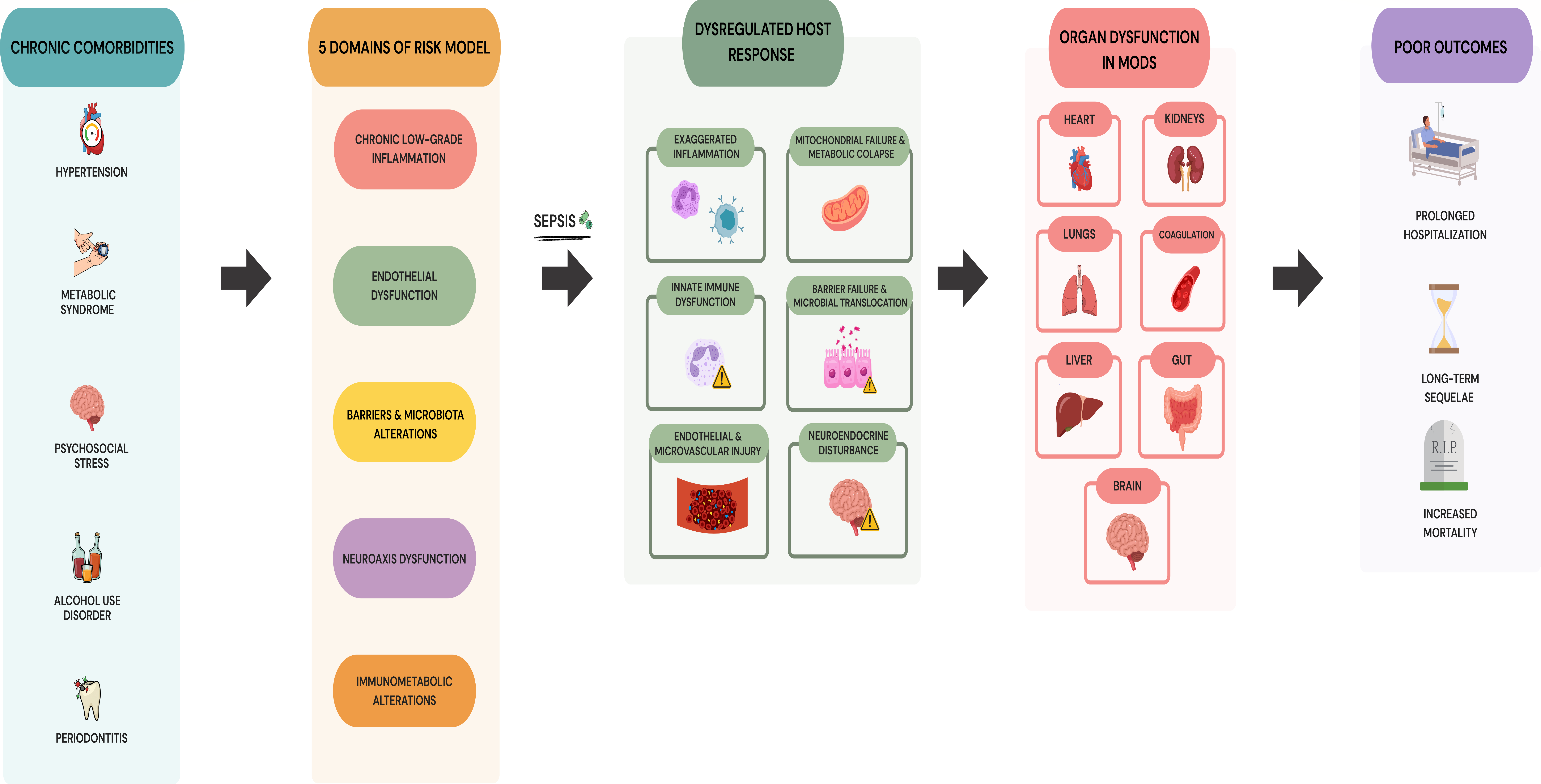
2.4. Multiple-Organ Dysfunction Syndrome
- •
- Pulmonary system: it is often the earliest and most prominent feature, ranging from mild hypoxemia to acute respiratory distress syndrome (ARDS). The clinical hallmark is a marked reduction in lung elasticity, resulting from the combined effects of surfactant dysfunction and alveolar fluid accumulation. These alterations compromise the physiological mechanisms necessary for effective gas exchange [111]. This pathophysiology is further exacerbated by previously described alterations in neutrophil function, such as decreased deformability and excessive NET formation [106,110]. As outlined in earlier sections, while NETs are essential for pathogen containment at sites of infection, their excessive and systemic release in sepsis leads to microvascular occlusion, sustained inflammation, and the propagation of organ injury [106,110,112].
- •
- Cardiovascular System: The heart is the second most frequently affected organ during MODS [113,114]. While excessive NO production is a recognized contributor to septic myocardial dysfunction, the pathophysiology is multifactorial, involving innate immune activation, cytokine storm (e.g., TNF-α, IL-1β, IL-6), mitochondrial dysfunction, and catecholamine-driven β-adrenergic receptor activation [115,116]. TLRs, especially TLR4, are expressed on cardiomyocytes and endothelial cells, and their activation by PAMPs and DAMPs in sepsis triggers NF-κB signaling, cytokine release, and further myocardial depression [117,118]. Mitochondrial dysfunction, characterized by impaired oxidative phosphorylation and excessive ROS generation, also contributes to contractile failure and enhances susceptibility to cell death pathways [102,119]. The convergence of these pathways culminates in severe cardiovascular failure, marked by fluid-refractory hemodynamic instability, reduced vascular tone, endothelial barrier disruption, systemic edema, and impaired tissue oxygen delivery [111,113].
- •
- Hepatic System: Under shock conditions, reduced hepatic perfusion rapidly induces early liver dysfunction, which may progress to sustained hepatic injury if hypoperfusion persists [111,114]. Beyond hemodynamic instability, severe sepsis causes bioenergetic collapse in the liver, marked by a significant decline in the mitochondrial Respiratory Control Ratio (RCR) and impaired ATP synthesis, driven by excessive proton leakage and disrupted Ca2+ homeostasis [70,106]. These mechanisms collectively contribute to hepatic failure and coagulopathy, which are characteristic of advanced organ failure [3,106].
- •
- Renal System: In the renal system, MODS is characterized by a significant reduction in glomerular filtration rate, leading to azotemia, which acts as a systemic metabolic driver of insulin resistance and oxidative stress [3,120]. Beyond waste excretion, impairment of the kidney’s endocrine functions, including abnormal renin production and defective vitamin D activation, further disrupts ionic homeostasis and aggravates systemic metabolic instability [106,113,121]. Pro-inflammatory mediators, such as TNF-α, IL-6, and IL-1β, exacerbate this process by triggering microvascular dysfunction and tubular epithelial cell apoptosis [106].
- •
- Neurological and Neuroendocrine Dysfunction: Sepsis-induced central nervous system dysfunction, often clinically manifested as sepsis-associated encephalopathy (SAE), results from interconnected mechanisms, including blood–brain barrier disruption mediated by matrix metalloproteinases (MMPs) and endothelial injury, microglial and astrocytic activation, neuroinflammation, and impaired cerebral microcirculation [50,122,123]. At the cellular level, mitochondrial oxidative stress, neuronal apoptosis, and neurotransmitter imbalance (cholinergic deficit and glutamate excitotoxicity) significantly impair synaptic plasticity and cognitive function. From a neuroendocrine perspective, severe MODS involves not only dysregulation of the hypothalamic–pituitary–adrenal (HPA) axis and sustained sympathetic overactivity, but also neurohypophyseal vasopressin depletion, which collectively contribute to altered cortisol dynamics, impaired autonomic regulation, vasoplegia, and hemodynamic instability [124,125,126].
- •
- Hematological and Coagulation Systems: In addition to thrombocytopenia, MODS is frequently characterized by profound disturbances in the coagulation cascade, including increased levels of PAI-1, which suppresses fibrinolysis and promotes microvascular thrombosis, contributing to organ ischemia [61,127].
3. Molecular and Pathophysiological Basis of Comorbidities
3.1. Hypertension
3.2. Metabolic Syndrome
3.3. Alcohol Exposure and Alcohol Use Disorder
3.4. Psychosocial Stress
3.5. Periodontitis
4. Comorbidities and the Host Response in Sepsis
4.1. Innate Immune Recognition and Inflammatory Signaling
4.2. Endothelial and Microvascular Dysfunction in Comorbid Hosts
4.3. Metabolic and Mitochondrial Injury in Comorbid Hosts
4.4. Neuroimmune and Autonomic Dysfunction in Comorbid Hosts
4.5. Progression to MODS in Comorbid Hosts
- •
- Pulmonary dysfunction. Sepsis-induced lung injury is aggravated by preexisting comorbidities. Hypertension and diabetes contribute to endothelial dysfunction and microvascular injury [14,15]; obesity promotes chronic low-grade inflammation and impairs respiratory mechanics [13,172]; and chronic alcohol consumption disrupts epithelial barrier integrity and neutrophil antimicrobial function [17,19,250]. Collectively, these alterations enhance susceptibility to acute lung injury, impair gas exchange, and increase the severity of respiratory failure during MODS.
- •
- Cardiovascular dysfunction. Chronic comorbidities further destabilize cardiac function during MODS. Hypertension and diabetes promote myocardial inflammation through increased infiltration of pro-inflammatory immune cells (such as macrophages and T lymphocytes), upregulation of cytokines, including TNF-α and IL-6, enhanced oxidative stress, and activation of TLR signaling pathways, all of which contribute to cardiac dysfunction [14,15]. MetS enhances oxidative stress and impairs mitochondrial energetics [172,218]. Chronic stress and sustained catecholamine exposure promote β-adrenergic receptor desensitization via GRK2 and β-arrestin pathways, reducing cardiac responsiveness and contractile reserve [27,207,300]. There is evidence that NO excess may also induce GRK2 expression, further aggravating β-adrenergic receptor dysfunction [300]. Notably, the upregulation of GRK2 in cardiomyocytes parallels the mechanism previously discussed in this review in septic neutrophils, where NO excess impairs CXCR2-mediated trafficking [240,300,301]. This highlights GRK2 as a shared molecular mediator contributing to organ dysfunction in sepsis. The convergence of these pathways culminates in severe cardiovascular failure, defined by hemodynamic instability that fails to respond to fluid resuscitation, often accompanied by disturbances in cardiac rhythm. This clinical state is exacerbated by reduced vascular tone and disruption of endothelial barriers, which drive systemic edema and reduced oxygen diffusion to the tissues [111,114].
- •
- Central nervous system and neuroendocrine dysfunction. Preexisting comorbidities further compromise neural homeostasis and resilience during critical illness [3,302]. Conditions such as diabetes, hypertension, obesity, AUD, and psychosocial stress, which are associated with baseline neuroimmune and autonomic dysregulation, impair blood–brain barrier integrity and promote microglial priming [3,15,123,200,277,288,295,296,297]. Furthermore, chronic sympathetic overactivity associated with obesity, hypertension, diabetes, and prolonged stress promotes β2-adrenergic receptor desensitization, whereas persistent HPA-axis activation, particularly during chronic psychosocial stress, may induce glucocorticoid receptor resistance, blunting central autonomic control and compromising cholinergic anti-inflammatory regulation [125,180,194,280]. When exposed to acute septic insults, this preconditioned neuroendocrine environment accelerates central neuroinflammation, may contribute to relative vasopressin deficiency or an impaired vasopressinergic response, and disrupts HPA-axis dynamics, predisposing patients to SAE, vasoplegia and hemodynamic instability, and accelerated progression of MODS [279,287,288,303].
- •
- Hepatic dysfunction. Chronic comorbidities substantially lower the threshold for hepatic dysfunction during MODS. In diabetes, persistent hyperglycemia and insulin resistance promote hepatic inflammation and oxidative stress through SAA-mediated NF-κB activation [14,304]. Similarly, chronic alcohol consumption induces baseline oxidative stress and effectively arrests the liver’s regenerative process, preventing hepatocytes from progressing from G1 (prereplicative) to S (replicative) phases necessary for recovery [16,305]. Collectively, these factors exacerbate systemic inflammation and metabolic exhaustion, leading to severe hepatic failure and associated coagulopathy characteristic of MODS [3,106].
- •
- Renal dysfunction. The presence of chronic comorbidities markedly increases renal vulnerability during MODS. Diabetes promotes chronic microvascular damage and low-grade inflammation through SAA-mediated TLR4 signaling, increasing the risk of acute kidney injury during sepsis [14,304]. Simultaneously, obesity and MetS amplify this susceptibility by disrupting adipokine balance and promoting abnormal lipid accumulation within the renal parenchyma. These alterations, together with increased systemic oxidative stress, progressively diminish the kidney’s physiological reserve against septic insults [13,14,119]. Chronic inflammation associated with hypertension induces vascular remodeling and endothelial dysfunction, which reduces the kidney’s ability to maintain stable perfusion under septic stress [15,111,134]. Chronic alcohol consumption impairs host immunity and predisposes to renal failure by promoting accelerated bacterial dissemination to the kidneys and inducing nephromegaly and glomerular swelling [250]. Notably, periodontal disease is a sustained source of systemic inflammation, creating a detrimental reciprocal relationship with the kidneys [120]. This interplay is further influenced by shared genetic predispositions, particularly functional polymorphisms in the IL-1β (IL1B) gene and its receptor antagonist (IL-1RN), which link periodontitis to an elevated risk of both chronic and acute renal dysfunction [120,306]. Finally, chronic psychosocial stress, through sustained activation of the sympathetic nervous system and the HPA axis, alters renal hemodynamics and impairs immune regulation, further predisposing the host to sepsis-associated renal failure [13,121,200]. These comorbidity-driven alterations in immune, vascular, and metabolic pathways not only increase the incidence and severity of renal dysfunction during sepsis and MODS but are also associated with a greater need for renal replacement therapy and poorer patient outcomes [111].
- •
- Gastrointestinal dysfunction. Gastrointestinal manifestations of MODS frequently involve stress-induced mucosal injury, adynamic ileus, and feeding intolerance, which are driven by systemic inflammatory mediators and acute epithelial cell apoptosis [106,299]. These alterations are further aggravated by profound imbalances in autonomic regulation [200,307]. In particular, diminished vagal activity compromises the CAP, eliminating an essential control on cytokine synthesis, whereas concurrent sympathetic hyperactivity induces pronounced mesenteric vasoconstriction and suppresses intestinal motility [111,121,281]. These neuroautonomic disturbances directly contribute to mucosal ischemia and intestinal paralysis, processes that are especially evident in individuals with chronic comorbidities [13,16]. In diabetes and obesity, chronic hyperglycemia and low-grade inflammation further compromise the integrity of the gut barrier [14,106]. Chronic alcohol consumption imposes an additional burden by activating tyrosine kinase pathways that compromise tight junction integrity, markedly increasing paracellular permeability and promoting colonic dysbiosis [174,308]. The resulting barrier dysfunction facilitates the systemic translocation of pathobionts and bacterial products, thereby amplifying inflammation in remote organs [111,114]. This process is further linked to periodontal disease, as oral bacteria such as Klebsiella aerogenes can translocate to the gut, exacerbating intestinal inflammation and contributing to the progression of multi-organ failure [120,309].
- •
- Hematologic dysfunction and coagulopathy. Prothrombotic alterations and microvascular thrombosis are particularly pronounced in patients with hypertension, diabetes, obesity, and MetS, where chronic inflammation and endothelial activation signaling via protease-activated receptors (PARs) sustain a baseline hypercoagulable state [310,311]. PAI-1 expression is upregulated by inflammatory cytokines, such as IL-6, and is closely linked to insulin resistance and central adiposity, thereby increasing the risk of disseminated intravascular coagulation in individuals with comorbidities [127]. Furthermore, metabolic diseases commonly impair the protective functions of the nuclear receptor peroxisome proliferator-activated receptor gamma (PPAR-γ). Reduced activity of this receptor diminishes its ability to suppress the expression of genes that drive inflammation and thrombosis, thereby compromising vascular stability [312]. Chronic alcohol use can further exacerbate coagulopathy by impairing hepatic production of coagulation factors and altering platelet function [19]. The combined effect of these mechanisms leads to a heightened risk of both thrombosis and bleeding complications during MODS in patients with underlying comorbidities.
4.6. Additional Comorbidities Beyond the Scope of the Present Review
5. Translational Framework for Risk Stratification and Precision Therapeutics
6. Conclusions
Author Contributions
Funding
Data Availability Statement
Acknowledgments
Conflicts of Interest
Abbreviations
| AGEs | Advanced Glycation End Products |
| α7nAChR | Alpha-7 Nicotinic Acetylcholine Receptor |
| ARDS | Acute Respiratory Distress Syndrome |
| ATP | Adenosine Triphosphate |
| AUD | Alcohol Use Disorder |
| BAX | BCL-2-associated X Protein |
| BCL-2 | B-cell Lymphoma 2 |
| BNP | B-type Natriuretic Peptide |
| β2AR | β2-Adrenergic Receptors |
| Ca2+ | Calcium |
| CAMs | Cell Adhesion Molecules |
| CAP | Cholinergic Anti-inflammatory Pathway |
| CARS | Compensatory Anti-inflammatory Response Syndrome |
| COX-2 | Cyclooxygenase-2 |
| CXCL | C-X-C Motif Chemokine Ligand |
| CXCR2 | C-X-C Motif Chemokine Receptor 2 |
| DAMPs | Damage-Associated Molecular Patterns |
| DCs | Dendritic Cells |
| DIC | Disseminated Intravascular Coagulation |
| DNA | Deoxyribonucleic Acid |
| eNOS | Endothelial Nitric Oxide Synthase |
| FFA | Free Fatty Acid |
| GRK2 | G-protein-coupled Receptor Kinase 2 |
| HMGB1 | High Mobility Group Box 1 |
| HPA | Hypothalamic–Pituitary–Adrenal Axis |
| ICAM-1 | Intercellular Adhesion Molecule-1 |
| IFN-γ | Interferon-gamma |
| IL | Interleukin |
| IL-1RN | Interleukin-1 Receptor Antagonist |
| iNOS | Inducible Nitric Oxide Synthase |
| IRAK | Interleukin-1 Receptor-Associated Kinase |
| KIM-1 | Kidney Injury Molecule-1 |
| LDL | Low-Density Lipoprotein |
| LPS | Lipopolysaccharide |
| MAPK | Mitogen-Activated Protein Kinase |
| MDSCs | Myeloid-Derived Suppressor Cells |
| MetS | Metabolic Syndrome |
| MIP-1β | Macrophage Inflammatory Protein-1β |
| MMPs | Matrix Metalloproteinases |
| MODS | Multiple Organ Dysfunction Syndrome |
| mtDNA | Mitochondrial DNA |
| NADH | Nicotinamide Adenine Dinucleotide |
| NADPH | Nicotinamide Adenine Dinucleotide Phosphate |
| NDUFS4 | NADH:Ubiquinone Oxidoreductase Subunit S4 |
| NETs | Neutrophil Extracellular Traps |
| NF-κB | Nuclear Factor Kappa B |
| NGAL | Neutrophil Gelatinase-Associated Lipocalin |
| NK | Natural Killer |
| NLR | NOD-like Receptor |
| NLRP3 | NOD-like Receptor Pyrin Domain-containing 3 |
| NO | Nitric Oxide |
| NOD | Nucleotide-binding Oligomerization Domain |
| NRF-1 | Nuclear Respiratory Factor 1 |
| NT-proBNP | N-terminal pro-B-type Natriuretic Peptide |
| O2− | Superoxide Anion |
| ONOO− | Peroxynitrite |
| OPG | Osteoprotegerin |
| OXPHOS | Oxidative Phosphorylation |
| PAI-1 | Plasminogen Activator Inhibitor-1 |
| PAMPs | Pathogen-Associated Molecular Patterns |
| PARS | Protease-Activated Receptors |
| PGC-1α | Proliferator-Activated Receptor Gamma Coactivator 1-α |
| PGE2 | Prostaglandin E2 |
| PPAR-γ | Peroxisome Proliferator-Activated Receptor Gamma |
| PRRs | Pattern Recognition Receptors |
| PVAT | Perivascular Adipose Tissue |
| RAAS | Renin–Angiotensin–Aldosterone System |
| RAGE | Receptor for Advanced Glycation End Products |
| RANKL | Receptor Activator of Nuclear Factor Kappa-B Ligand |
| RCR | Respiratory Control Ratio |
| RNS | Reactive Nitrogen Species |
| ROS | Reactive Oxygen Species |
| SAA | Serum Amyloid A |
| SAE | Sepsis-Associated Encephalopathy |
| SCFAs | Short-Chain Fatty Acids |
| SIRS | Systemic Inflammatory Response Syndrome |
| SNS | Sympathetic Nervous System |
| T2DM | Type 2 Diabetes Mellitus |
| TFAM | Mitochondrial Transcription Factor A |
| Th | T Helper Cells |
| TLR | Toll-like Receptor |
| TMAO | Trimethylamine N-oxide |
| TNF-α | Tumor Necrosis Factor-alpha |
| VCAM-1 | Vascular Cell Adhesion Molecule-1 |
References
- Fleischmann, C.; Scherag, A.; Adhikari, N.K.J.; Hartog, C.S.; Tsaganos, T.; Schlattmann, P.; Angus, D.C.; Reinhart, K. Assessment of Global Incidence and Mortality of Hospital-Treated Sepsis. Current Estimates and Limitations. Am. J. Respir. Crit. Care Med. 2016, 193, 259–272. [Google Scholar] [CrossRef] [Scilit] [PubMed]
- Rudd, K.E.; Johnson, S.C.; Agesa, K.M.; Shackelford, K.A.; Tsoi, D.; Kievlan, D.R.; Colombara, D.V.; Ikuta, K.S.; Kissoon, N.; Finfer, S.; et al. Global, Regional, and National Sepsis Incidence and Mortality, 1990–2017: Analysis for the Global Burden of Disease Study. Lancet 2020, 395, 200–211. [Google Scholar] [CrossRef] [Scilit] [PubMed]
- Singer, M.; Deutschman, C.S.; Seymour, C.; Shankar-Hari, M.; Annane, D.; Bauer, M.; Bellomo, R.; Bernard, G.R.; Chiche, J.D.; Coopersmith, C.M.; et al. The Third International Consensus Definitions for Sepsis and Septic Shock (Sepsis-3). JAMA 2016, 315, 801–810. [Google Scholar] [CrossRef] [Scilit] [PubMed]
- Guarino, M.; Perna, B.; Cesaro, A.E.; Maritati, M.; Spampinato, M.D.; Contini, C.; De Giorgio, R. 2023 Update on Sepsis and Septic Shock in Adult Patients: Management in the Emergency Department. J. Clin. Med. 2023, 12, 3188. [Google Scholar] [CrossRef] [Scilit] [PubMed]
- Chang, Z.; Lu, J.; Zhang, Q.; Wu, H.; Liang, Z.; Pan, X.; Li, B.; Cheng, Z.J.; Sun, B. Clinical Biomarker Profiles Reveals Gender Differences and Mortality Factors in Sepsis. Front. Immunol. 2024, 15, 1413729. [Google Scholar] [CrossRef] [Scilit] [PubMed]
- Gianoli, S.; Tang, J.; Odegard, K.C.; Yuki, K.; Koutsogiannaki, S. Harnessing Adiponectin for Sepsis: Current Knowledge, Clinical Insights and Future Therapies. Crit. Care 2025, 29, 300. [Google Scholar] [CrossRef] [Scilit] [PubMed]
- Schuetz, P.; Castro, P.; Shapiro, N.I. Diabetes and Sepsis: Preclinical Findings and Clinical Relevance. Diabetes Care 2011, 34, 771–778. [Google Scholar] [CrossRef] [Scilit] [PubMed]
- Song, H.; Fall, K.; Fang, F.; Erlendsdóttir, H.; Lu, D.; Mataix-Cols, D.; Fernández De La Cruz, L.; D’Onofrio, B.M.; Lichtenstein, P.; Gottfreosson, M.; et al. Stress Related Disorders and Subsequent Risk of Life Threatening Infections: Population Based Sibling Controlled Cohort Study. BMJ 2019, 367, l5784. [Google Scholar] [CrossRef] [Scilit] [PubMed]
- Simou, E.; Leonardi-Bee, J.; Britton, J. The Effect of Alcohol Consumption on the Risk of ARDS. Chest 2017, 154, 58–68. [Google Scholar] [CrossRef] [Scilit] [PubMed]
- Thomas-Rüddel, D.O.; Fröhlich, H.; Schwarzkopf, D.; Bloos, F.; Riessen, R. Sepsis and Underlying Comorbidities in Intensive Care Unit Patients. Med. Klin. Intensivmed. Notfmed. 2024, 119, 123–128. [Google Scholar] [CrossRef] [Scilit] [PubMed]
- Chawla, A.; Nguyen, K.D.; Goh, Y.P.S. Macrophage-Mediated Inflammation in Metabolic Disease. Nat. Rev. Immunol. 2011, 11, 738. [Google Scholar] [CrossRef] [Scilit] [PubMed]
- Zheng, H.; Yu, F.; Bei, C.; Zhou, Y.; Mo, Z.; Wei, B. Diabetes Mellitus and Mortality in Patients Admitted to ICU with Sepsis: A Meta-Analysis. Front. Med. 2026, 13, 1743706. [Google Scholar] [CrossRef] [Scilit] [PubMed]
- Meydan, C.; Bekenstein, U.; Soreq, H. Molecular Regulatory Pathways Link Sepsis with Metabolic Syndrome: Non-Coding RNA Elements Underlying the Sepsis/Metabolic Cross-Talk. Front. Mol. Neurosci. 2018, 11, 189. [Google Scholar] [CrossRef] [Scilit] [PubMed]
- Esser, N.; Legrand-Poels, S.; Piette, J.; Scheen, A.J.; Paquot, N. Inflammation as a Link between Obesity, Metabolic Syndrome and Type 2 Diabetes. Diabetes Res. Clin. Pract. 2014, 105, 141–150. [Google Scholar] [CrossRef] [Scilit] [PubMed]
- Guzik, T.J.; Nosalski, R.; Maffia, P.; Drummond, G.R. Immune and Inflammatory Mechanisms in Hypertension. Nat. Rev. Cardiol. 2024, 21, 396–416. [Google Scholar] [CrossRef] [Scilit] [PubMed]
- Greiffenstein, P.; Mathis, K.W.; Stouwe, C.V.; Molina, P.E. Alcohol Binge before Trauma/Hemorrhage Impairs Integrity of Host Defense Mechanisms during Recovery. Alcohol. Clin. Exp. Res. 2007, 31, 704–715. [Google Scholar] [CrossRef] [Scilit] [PubMed]
- Ceron, C.S.; do Vale, G.T.; Simplicio, J.A.; Ricci, S.T.; De Martinis, B.S.; de Freitas, A.; Tirapelli, C.R. Chronic Ethanol Consumption Increases Vascular Oxidative Stress and the Mortality Induced by Sub-Lethal Sepsis: Potential Role of INOS. Eur. J. Pharmacol. 2018, 825, 39–47. [Google Scholar] [CrossRef] [Scilit] [PubMed]
- Martínez-García, M.; Hernández-Lemus, E. The Molecular Comorbidity Network of Periodontal Disease. Int. J. Mol. Sci. 2024, 25, 10161. [Google Scholar] [CrossRef] [Scilit] [PubMed]
- Szabo, G.; Bala, S. Alcoholic Liver Disease and the Gut-Liver Axis. World J. Gastroenterol. WJG 2010, 16, 1321. [Google Scholar] [CrossRef] [Scilit] [PubMed]
- Muralidharan, S.; Ambade, A.; Fulham, M.A.; Deshpande, J.; Catalano, D.; Mandrekar, P. Moderate Alcohol Induces Stress Proteins HSF1 and Hsp70 and Inhibits Pro-Inflammatory Cytokines Resulting in Endotoxin Tolerance. J. Immunol. 2014, 193, 1975–1987. [Google Scholar] [CrossRef] [Scilit] [PubMed]
- de Oliveira Malacco, N.L.S.; Souza, J.A.M.; Martins, F.R.B.; Rachid, M.A.; Simplicio, J.A.; Tirapelli, C.R.; de Paula Sabino, A.; Queiroz-Junior, C.M.; Goes, G.R.; Vieira, L.Q.; et al. Chronic Ethanol Consumption Compromises Neutrophil Function in Acute Pulmonary Aspergillus Fumigatus Infection. eLife 2020, 9, e58855. [Google Scholar] [CrossRef] [Scilit] [PubMed]
- Hajishengallis, G. Periodontitis: From Microbial Immune Subversion to Systemic Inflammation. Nat. Rev. Immunol. 2015, 15, 30–44. [Google Scholar] [CrossRef] [Scilit] [PubMed]
- Cao, L.; Hudson, C.A.; Lawrence, D.A. Immune Changes during Acute Cold/Restraint Stress-Induced Inhibition of Host Resistance to Listeria. Toxicol. Sci. 2003, 74, 325–334. [Google Scholar] [CrossRef] [Scilit] [PubMed]
- Avitsur, R.; Hunzeker, J.; Sheridan, J.F. Role of Early Stress in the Individual Differences in Host Response to Viral Infection. Brain Behav. Immun. 2006, 20, 339–348. [Google Scholar] [CrossRef] [Scilit] [PubMed]
- Maydych, V.; Claus, M.; Watz, C.; Kleinsorge, T. Attention to Emotional Information Is Associated with Cytokine Responses to Psychological Stress. Front. Neurosci. 2018, 12, 687. [Google Scholar] [CrossRef] [Scilit] [PubMed]
- Scanzano, A.; Cosentino, M. Adrenergic Regulation of Innate Immunity: A Review. Front. Pharmacol. 2015, 6, 171. [Google Scholar] [CrossRef] [Scilit] [PubMed]
- Marino, F.; Scanzano, A.; Pulze, L.; Pinoli, M.; Rasini, E.; Luini, A.; Bombelli, R.; Legnaro, M.; de Eguileor, M.; Cosentino, M. Β2 -Adrenoceptors Inhibit Neutrophil Extracellular Traps in Human Polymorphonuclear Leukocytes. J. Leukoc. Biol. 2018, 104, 603–614. [Google Scholar] [CrossRef] [Scilit] [PubMed]
- Medzhitov, R. Recognition of Microorganisms and Activation of the Immune Response. Nature 2007, 449, 819–826. [Google Scholar] [CrossRef] [Scilit] [PubMed]
- Akira, S.; Uematsu, S.; Takeuchi, O. Pathogen Recognition and Innate Immunity. Cell 2006, 124, 783–801. [Google Scholar] [CrossRef] [Scilit] [PubMed]
- Takeuchi, O.; Akira, S. Pattern Recognition Receptors and Inflammation. Cell 2010, 140, 805–820. [Google Scholar] [CrossRef] [Scilit] [PubMed]
- Kawai, T.; Akira, S. The Role of Pattern-Recognition Receptors in Innate Immunity: Update on Toll-like Receptors. Nat. Immunol. 2010, 11, 373–384. [Google Scholar] [CrossRef] [Scilit] [PubMed]
- Poltorak, A.; He, X.; Smirnova, I.; Liu, M.Y.; Van Huffel, C.; Du, X.; Birdwell, D.; Alejos, E.; Silva, M.; Galanos, C.; et al. Defective LPS Signaling in C3H/HeJ and C57BL/10ScCr Mice: Mutations in Tlr4 Gene. Science 1998, 282, 2085–2088. [Google Scholar] [CrossRef] [Scilit] [PubMed]
- Kawai, T.; Ikegawa, M.; Ori, D.; Akira, S. Decoding Toll-like Receptors: Recent Insights and Perspectives in Innate Immunity. Immunity 2024, 57, 649–673. [Google Scholar] [CrossRef] [Scilit] [PubMed]
- Kumar, H.; Kawai, T.; Akira, S. Pathogen Recognition by the Innate Immune System. Int. Rev. Immunol. 2011, 30, 16–34. [Google Scholar] [CrossRef] [Scilit] [PubMed]
- Martinon, F.; Mayor, A.; Tschopp, J. The Inflammasomes: Guardians of the Body. Annu. Rev. Immunol. 2009, 27, 229–265. [Google Scholar] [CrossRef] [Scilit] [PubMed]
- Schroder, K.; Tschopp, J. The Inflammasomes. Cell 2010, 140, 821–832. [Google Scholar] [CrossRef] [Scilit] [PubMed]
- Yao, J.; Sterling, K.; Wang, Z.; Zhang, Y.; Song, W. The Role of Inflammasomes in Human Diseases and Their Potential as Therapeutic Targets. Signal Transduct. Target. Ther. 2024, 9, 10. [Google Scholar] [CrossRef] [Scilit] [PubMed]
- Petrasek, J.; Iracheta-Vellve, A.; Saha, B.; Satishchandran, A.; Kodys, K.; Fitzgerald, K.A.; Kurt-Jones, E.A.; Szabo, G. Metabolic Danger Signals, Uric Acid and ATP, Mediate Inflammatory Cross-Talk between Hepatocytes and Immune Cells in Alcoholic Liver Disease. J. Leukoc. Biol. 2015, 98, 249–256. [Google Scholar] [CrossRef] [Scilit] [PubMed]
- Rodriguez-Iturbe, B.; Pons, H.; Johnson, R.J. Role of the Immune System in Hypertension. Physiol. Rev. 2017, 97, 1127–1164. [Google Scholar] [CrossRef] [Scilit] [PubMed]
- Ley, K.; Laudanna, C.; Cybulsky, M.I.; Nourshargh, S. Getting to the Site of Inflammation: The Leukocyte Adhesion Cascade Updated. Nat. Rev. Immunol. 2007, 7, 678–689. [Google Scholar] [CrossRef] [Scilit] [PubMed]
- Kolaczkowska, E.; Kubes, P. Neutrophil Recruitment and Function in Health and Inflammation. Nat. Rev. Immunol. 2013, 13, 159–175. [Google Scholar] [CrossRef] [Scilit] [PubMed]
- Alves-Filho, J.C.; Freitas, A.; Souto, F.O.; Spiller, F.; Paula-Neto, H.; Silva, J.S.; Gazzinelli, R.T.; Teixeira, M.M.; Ferreira, S.H.; Cunha, F.Q. Regulation of Chemokine Receptor by Toll-like Receptor 2 Is Critical to Neutrophil Migration and Resistance to Polymicrobial Sepsis. Proc. Natl. Acad. Sci. USA 2009, 106, 4018–4023. [Google Scholar] [CrossRef] [Scilit] [PubMed]
- Papayannopoulos, V. Neutrophil Extracellular Traps in Immunity and Disease. Nat. Rev. Immunol. 2018, 18, 134–147. [Google Scholar] [CrossRef] [Scilit] [PubMed]
- Ward, N.S.; Casserly, B.; Ayala, A. The Compensatory Anti-Inflammatory Response Syndrome (CARS) in Critically Ill Patients. Clin. Chest Med. 2008, 29, 617–625. [Google Scholar] [CrossRef] [Scilit] [PubMed]
- Hotchkiss, R.S.; Monneret, G.; Payen, D. Sepsis-Induced Immunosuppression: From Cellular Dysfunctions to Immunotherapy. Nat. Rev. Immunol. 2013, 13, 862–874. [Google Scholar] [CrossRef] [Scilit] [PubMed]
- Alves-Filho, J.C.; Sônego, F.; Souto, F.O.; Freitas, A.; Verri, W.A.; Auxiliadora-Martins, M.; Basile-Filho, A.; Mckenzie, A.N.; Xu, D.; Cunha, F.Q.; et al. Interleukin-33 Attenuates Sepsis by Enhancing Neutrophil Influx to the Site of Infection. Nat. Med. 2010, 16, 708–712. [Google Scholar] [CrossRef] [Scilit] [PubMed]
- Van Der Poll, T.; Van De Veerdonk, F.L.; Scicluna, B.P.; Netea, M.G. The Immunopathology of Sepsis and Potential Therapeutic Targets. Nat. Rev. Immunol. 2017, 17, 407–420. [Google Scholar] [CrossRef] [Scilit] [PubMed]
- Pierrakos, C.; Velissaris, D.; Bisdorff, M.; Marshall, J.C.; Vincent, J.L. Biomarkers of Sepsis: Time for a Reappraisal. Crit. Care 2020, 24, 287. [Google Scholar] [CrossRef] [Scilit] [PubMed]
- Candelli, M.; Sacco Fernandez, M.; Rozzi, G.; Sodero, G.; Piccioni, A.; Pignataro, G.; Rigante, D.; Franceschi, F. The Interleukin Network in Sepsis: From Cytokine Storm to Clinical Applications. Diagnostics 2025, 15, 2927. [Google Scholar] [CrossRef] [Scilit] [PubMed]
- Varatharaj, A.; Galea, I. The Blood-Brain Barrier in Systemic Inflammation. Brain Behav. Immun. 2017, 60, 1–12. [Google Scholar] [CrossRef] [Scilit] [PubMed]
- Boisramé-Helms, J.; Kremer, H.; Schini-Kerth, V.; Meziani, F. Endothelial dysfunction in sepsis. Curr. Vasc. Pharmacol. 2013, 11, 150–160. [Google Scholar] [CrossRef] [Scilit]
- Lipowsky, H.H. Protease Activity and the Role of the Endothelial Glycocalyx in Inflammation. Drug Discov. Today Dis. Models 2011, 8, 57–62. [Google Scholar] [CrossRef] [Scilit] [PubMed]
- Ince, C.; Mayeux, P.R.; Nguyen, T.; Gomez, H.; Kellum, J.A.; Ospina-Tascón, G.A.; Hernandez, G.; Murray, P.; De Backer, D. The Endothelium in Sepsis. Shock 2016, 45, 259. [Google Scholar] [CrossRef] [Scilit] [PubMed]
- Joffre, J.; Hellman, J.; Ince, C.; Ait-Oufella, H. Endothelial Responses in Sepsis. Am. J. Respir. Crit. Care Med. 2020, 202, 361–370. [Google Scholar] [CrossRef] [Scilit] [PubMed]
- Vallet, B. Bench-to-Bedside Review: Endothelial Cell Dysfunction in Severe Sepsis: A Role in Organ Dysfunction? Crit. Care 2003, 7, 130–138. [Google Scholar] [CrossRef] [Scilit] [PubMed]
- Xu, J.; Zhang, X.; Pelayo, R.; Monestier, M.; Ammollo, C.T.; Semeraro, F.; Taylor, F.B.; Esmon, N.L.; Lupu, F.; Esmon, C.T. Extracellular Histones Are Major Mediators of Death in Sepsis. Nat. Med. 2009, 15, 1318–1321. [Google Scholar] [CrossRef] [Scilit] [PubMed]
- Luo, M.; Meng, J.; Yan, J.; Shang, F.; Zhang, T.; Lv, D.; Li, C.; Yang, X.; Luo, S. Role of the Nucleotide-Binding Domain-Like Receptor Protein 3 Inflammasome in the Endothelial Dysfunction of Early Sepsis. Inflammation 2020, 43, 1561–1571. [Google Scholar] [CrossRef] [Scilit] [PubMed]
- Flemming, S.; Burkard, N.; Renschler, M.; Vielmuth, F.; Meir, M.; Schick, M.A.; Wunder, C.; Germer, C.T.; Spindler, V.; Waschke, J.; et al. Soluble VE-Cadherin Is Involved in Endothelial Barrier Breakdown in Systemic Inflammation and Sepsis. Cardiovasc. Res. 2015, 107, 32–44. [Google Scholar] [CrossRef] [Scilit] [PubMed]
- Dogné, S.; Flamion, B. Endothelial Glycocalyx Impairment in Disease: Focus on Hyaluronan Shedding. Am. J. Pathol. 2020, 190, 768–780. [Google Scholar] [PubMed]
- Cao, R.N.; Tang, L.; Xia, Z.Y.; Xia, R. Endothelial Glycocalyx as a Potential Theriapeutic Target in Organ Injuries. Chin. Med. J. 2019, 132, 963–975. [Google Scholar] [CrossRef] [Scilit] [PubMed]
- Levi, M.; van der Poll, T. Disseminated Intravascular Coagulation: A Review for the Internist. Intern. Emerg. Med. 2013, 8, 23–32. [Google Scholar] [CrossRef] [Scilit] [PubMed]
- Symeonides, S.; Balk, R.A. Nitric oxide in the pathogenesis of sepsis. Infect. Dis. Clin. N. Am. 1999, 13, 449–463. [Google Scholar] [CrossRef] [Scilit] [PubMed]
- Benjamim, C.F.; Ferreira, S.H.; Cunha, F.D.Q. Role of Nitric Oxide in the Failure of Neutrophil Migration in Sepsis. J. Infect. Dis. 2000, 182, 214–223. [Google Scholar] [CrossRef] [Scilit] [PubMed]
- Farias Benjamim, C.; Santana Silva, J.; Bruno Fortes, Z.; Aparecida Oliveira, M.; Henrique Ferreira, S.; Queiroz Cunha, F. Inhibition of Leukocyte Rolling by Nitric Oxide during Sepsis Leads to Reduced Migration of Active Microbicidal Neutrophils. Infect. Immun. 2002, 70, 3602–3610. [Google Scholar] [CrossRef] [Scilit] [PubMed]
- Gourd, N.M.; Nikitas, N. Multiple Organ Dysfunction Syndrome. J. Intensive Care Med. 2020, 35, 1564–1575. [Google Scholar] [CrossRef] [Scilit] [PubMed]
- Misrani, A.; Tabassum, S.; Yang, L. Mitochondrial Dysfunction and Oxidative Stress in Alzheimer’s Disease. Front. Aging Neurosci. 2021, 13, 617588. [Google Scholar] [CrossRef] [Scilit] [PubMed]
- Nedel, W.; Strogulski, N.R.; Kopczynski, A.; Portela, L.V. Assessment of Mitochondrial Function and Its Prognostic Role in Sepsis: A Literature Review. Intensive Care Med. Exp. 2024, 12, 107. [Google Scholar] [CrossRef] [Scilit] [PubMed]
- Banoth, B.; Cassel, S.L. Mitochondria in Innate Immune Signaling. Transl. Res. 2018, 202, 52–68. [Google Scholar] [CrossRef] [Scilit] [PubMed]
- Jiao, Y.; Yan, Z.; Yang, A. Mitochondria in Innate Immunity Signaling and Its Therapeutic Implications in Autoimmune Diseases. Front. Immunol. 2023, 14, 1160035. [Google Scholar] [CrossRef] [Scilit] [PubMed]
- Freitas, A.; Alves-Filho, J.C.; Trevelin, S.C.; Spiller, F.; Suavinha, M.M.; Nascimento, D.C.; Pestana, C.R.; Dal-Secco, D.; Sônego, F.; Czaikoski, P.G.; et al. Divergent Role of Heme Oxygenase Inhibition in the Pathogenesis of Sepsis. Shock 2011, 35, 550–559. [Google Scholar] [CrossRef] [Scilit] [PubMed]
- Lira Chavez, F.M.; Gartzke, L.P.; van Beuningen, F.E.; Wink, S.E.; Henning, R.H.; Krenning, G.; Bouma, H.R. Restoring the Infected Powerhouse: Mitochondrial Quality Control in Sepsis. Redox Biol. 2023, 68, 102968. [Google Scholar] [CrossRef] [Scilit] [PubMed]
- Kozlov, A.V.; Bahrami, S.; Calzia, E.; Dungel, P.; Gille, L.; Kuznetsov, A.V.; Troppmair, J. Mitochondrial Dysfunction and Biogenesis: Do ICU Patients Die from Mitochondrial Failure? Ann. Intensive Care 2011, 1, 41. [Google Scholar] [CrossRef] [Scilit] [PubMed]
- Calvano, S.E.; Xiao, W.; Richards, D.R.; Felciano, R.M.; Baker, H.V.; Cho, R.J.; Chen, R.O.; Brownstein, B.H.; Cobb, J.P.; Tschoeke, S.K.; et al. A Network-Based Analysis of Systemic Inflammation in Humans. Nature 2005, 437, 1032–1037, Corrigendum in Nature 2005, 438, 696. [Google Scholar] [CrossRef] [Scilit] [PubMed]
- Liu, J.; Zhou, G.; Wang, X.; Liu, D. Metabolic Reprogramming Consequences of Sepsis: Adaptations and Contradictions. Cell. Mol. Life Sci. 2022, 79, 456. [Google Scholar] [CrossRef] [Scilit] [PubMed]
- Paul, S.N.; Nessel, I.; Puthucheary, Z.; Henson, S.M. Sepsis and the immunometabolic inflammatory response. Metab. Health Dis. 2026, 4, 1234567890. [Google Scholar] [CrossRef] [Scilit] [PubMed]
- Brealey, D.; Brand, M.; Hargreaves, I.; Heales, S.; Land, J.; Smolenski, R.; Davies, N.A.; Cooper, C.E.; Singer, M. Association between Mitochondrial Dysfunction and Severity and Outcome of Septic Shock. Lancet 2002, 360, 219–223. [Google Scholar] [CrossRef] [Scilit] [PubMed]
- Rahmel, T.; Marko, B.; Nowak, H.; Bergmann, L.; Thon, P.; Rump, K.; Kreimendahl, S.; Rassow, J.; Peters, J.; Singer, M.; et al. Mitochondrial Dysfunction in Sepsis Is Associated with Diminished Intramitochondrial TFAM despite Its Increased Cellular Expression. Sci. Rep. 2020, 10, 21029. [Google Scholar] [CrossRef] [Scilit] [PubMed]
- Haschemi, A.; Kosma, P.; Gille, L.; Evans, C.R.; Burant, C.F.; Starkl, P.; Knapp, B.; Haas, R.; Schmid, J.A.; Jandl, C.; et al. The Sedoheptulose Kinase CARKL Directs Macrophage Polarization through Control of Glucose Metabolism. Cell Metab. 2012, 15, 813–826. [Google Scholar] [CrossRef] [Scilit] [PubMed]
- Jin, Z.; Wei, W.; Yang, M.; Du, Y.; Wan, Y. Mitochondrial Complex I Activity Suppresses Inflammation and Enhances Bone Resorption by Tipping the Balance of Macrophage-Osteoclast Polarization. Cell Metab. 2014, 20, 483–498. [Google Scholar] [CrossRef] [Scilit] [PubMed]
- Shamriz, O.; Bar-On, Z.; Yosef, O.; Cohen-Daniel, L.; Sheer, A.; Reuven, O.; Salaymeh, W.; Saragovi, A.; Somech, R.; Lev, A.; et al. NDUFS4, a Mitochondrial Complex I Subunit, Is Essential for T-Cell Metabolic Fitness and Immune Function. Front. Immunol. 2026, 16, 1734203. [Google Scholar] [CrossRef] [Scilit] [PubMed]
- Radi, R. Oxygen Radicals, Nitric Oxide, and Peroxynitrite: Redox Pathways in Molecular Medicine. Proc. Natl. Acad. Sci. USA. 2018, 115, 5839–5848. [Google Scholar] [CrossRef] [Scilit] [PubMed]
- Hu, D.; Sheeja Prabhakaran, H.; Zhang, Y.Y.; Luo, G.; He, W.; Liou, Y.C. Mitochondrial Dysfunction in Sepsis: Mechanisms and Therapeutic Perspectives. Crit. Care 2024, 28, 292. [Google Scholar] [CrossRef] [Scilit] [PubMed]
- Gurung, P.; Lukens, J.R.; Kanneganti, T.D. Mitochondria: Diversity in the Regulation of NLRP3 Inflammasome. Trends Mol. Med. 2014, 21, 193–201. [Google Scholar] [CrossRef] [Scilit] [PubMed]
- Murakami, T.; Ockinger, J.; Yu, J.; Byles, V.; McColl, A.; Hofer, A.M.; Horng, T. Critical Role for Calcium Mobilization in Activation of the NLRP3 Inflammasome. Proc. Natl. Acad. Sci. USA 2012, 109, 11282–11287. [Google Scholar] [CrossRef] [Scilit] [PubMed]
- Zhong, Z.; Liang, S.; Sanchez-Lopez, E.; He, F.; Shalapour, S.; Lin, X.J.; Wong, J.; Ding, S.; Seki, E.; Schnabl, B.; et al. New Mitochondrial DNA Synthesis Enables NLRP3 Inflammasome Activation. Nature 2018, 560, 198–203. [Google Scholar] [CrossRef] [Scilit] [PubMed]
- Singer, M. The Role of Mitochondrial Dysfunction in Sepsis-Induced Multi-Organ Failure. Virulence 2014, 5, 66–72. [Google Scholar] [CrossRef] [Scilit] [PubMed]
- Youle, R.J.; Narendra, D.P. Mechanisms of Mitophagy. Nat. Rev. Mol. Cell Biol. 2011, 12, 9–14. [Google Scholar] [CrossRef] [Scilit] [PubMed]
- Zhu, C.L.; Yao, R.Q.; Li, L.X.; Li, P.; Xie, J.; Wang, J.F.; Deng, X.M. Mechanism of Mitophagy and Its Role in Sepsis Induced Organ Dysfunction: A Review. Front. Cell Dev. Biol. 2021, 9, 664896. [Google Scholar] [CrossRef] [Scilit] [PubMed]
- Crouser, E.D.; Julian, M.W.; Huff, J.E.; Struck, J.; Cook, C.H. Carbamoyl Phosphate Synthase-1: A Marker of Mitochondrial Damage and Depletion in the Liver during Sepsis. Crit. Care Med. 2006, 34, 2439–2446. [Google Scholar] [CrossRef] [Scilit] [PubMed]
- Patoli, D.; Mignotte, F.; Deckert, V.; Dusuel, A.; Dumont, A.; Rieu, A.; Jalil, A.; van Dongen, K.; Bourgeois, T.; Gautier, T.; et al. Inhibition of Mitophagy Drives Macrophage Activation and Antibacterial Defense during Sepsis. J. Clin. Investig. 2020, 130, 5858–5874. [Google Scholar] [CrossRef] [Scilit] [PubMed]
- van der Slikke, E.C.; Star, B.S.; van Meurs, M.; Henning, R.H.; Moser, J.; Bouma, H.R. Sepsis Is Associated with Mitochondrial DNA Damage and a Reduced Mitochondrial Mass in the Kidney of Patients with Sepsis-AKI. Crit. Care 2021, 25, 36. [Google Scholar] [CrossRef] [Scilit] [PubMed]
- West, A.P.; Khoury-Hanold, W.; Staron, M.; Tal, M.C.; Pineda, C.M.; Lang, S.M.; Bestwick, M.; Duguay, B.A.; Raimundo, N.; MacDuff, D.A.; et al. Mitochondrial DNA Stress Primes the Antiviral Innate Immune Response. Nature 2015, 520, 553–557. [Google Scholar] [CrossRef] [Scilit] [PubMed]
- Harrington, J.S.; Huh, J.W.; Schenck, E.J.; Nakahira, K.; Siempos, I.I.; Choi, A.M.K. Circulating Mitochondrial DNA as Predictor of Mortality in Critically Ill Patients: A Systematic Review of Clinical Studies. Chest 2019, 156, 1120. [Google Scholar] [CrossRef] [Scilit] [PubMed]
- Rogers, R.S.; Sharma, R.; Shah, H.B.; Skinner, O.S.; Guo, X.A.; Panda, A.; Gupta, R.; Durham, T.J.; Shaughnessy, K.B.; Mayers, J.R.; et al. Circulating N-Lactoyl-Amino Acids and N-Formyl-Methionine Reflect Mitochondrial Dysfunction and Predict Mortality in Septic Shock. Metabolomics 2024, 20, 36. [Google Scholar] [CrossRef] [Scilit] [PubMed]
- Nedel, W.; Deutschendorf, C.; Portela, L.V.C. Sepsis-Induced Mitochondrial Dysfunction: A Narrative Review. World J. Crit. Care Med. 2023, 12, 139–152. [Google Scholar] [CrossRef] [Scilit] [PubMed]
- Vanportfliet, J.J.; Chute, C.; Lei, Y.; Shutt, T.E.; West, A.P. Mitochondrial DNA Release and Sensing in Innate Immune Responses. Hum. Mol. Genet. 2024, 33, R80–R91. [Google Scholar] [CrossRef] [Scilit] [PubMed]
- Vincent, J.L.; Moreno, R.; Takala, J.; Willatts, S.; De Mendonça, A.; Bruining, H.; Reinhart, C.K.; Suter, P.M.; Thijs, L.G. The SOFA (Sepsis-Related Organ Failure Assessment) Score to Describe Organ Dysfunction/Failure. On Behalf of the Working Group on Sepsis-Related Problems of the European Society of Intensive Care Medicine. Intensive Care Med. 1996, 22, 707–710. [Google Scholar] [CrossRef] [PubMed]
- McGhan, L.J.; Jaroszewski, D.E. The Role of Toll-like Receptor-4 in the Development of Multi-Organ Failure Following Traumatic Haemorrhagic Shock and Resuscitation. Injury 2012, 43, 129–136. [Google Scholar] [CrossRef] [Scilit] [PubMed]
- Di Paola, R.; Galuppo, M.; Mazzon, E.; Paterniti, I.; Bramanti, P.; Cuzzocrea, S. PD98059, a Specific MAP Kinase Inhibitor, Attenuates Multiple Organ Dysfunction Syndrome/Failure (MODS) Induced by Zymosan in Mice. Pharmacol. Res. 2010, 61, 175–187. [Google Scholar] [CrossRef] [Scilit] [PubMed]
- Gu, C.; Qiao, W.; Wang, L.; Li, M.; Song, K. Identification of Genes and Pathways Associated with Multiple Organ Dysfunction Syndrome by Microarray Analysis. Mol. Med. Rep. 2018, 18, 31–40. [Google Scholar] [CrossRef] [Scilit] [PubMed]
- Cederbaum, A.I. Alcohol Metabolism. Clin. Liver Dis. 2012, 16, 667–685. [Google Scholar] [CrossRef] [Scilit] [PubMed]
- Frydrych, L.M.; Fattahi, F.; He, K.; Ward, P.A.; Delano, M.J. Diabetes and Sepsis: Risk, Recurrence, and Ruination. Front. Endocrinol. 2017, 8, 271. [Google Scholar] [CrossRef] [Scilit] [PubMed]
- Picard, M.; McEwen, B.S. Psychological Stress and Mitochondria: A Systematic Review. Psychosom. Med. 2018, 80, 141–153. [Google Scholar] [CrossRef] [Scilit] [PubMed]
- Deng, Y.; Xiao, J.; Ma, L.; Wang, C.; Wang, X.; Huang, X.; Cao, Z. Mitochondrial Dysfunction in Periodontitis and Associated Systemic Diseases: Implications for Pathomechanisms and Therapeutic Strategies. Int. J. Mol. Sci. 2024, 25, 1024. [Google Scholar] [CrossRef] [Scilit] [PubMed]
- Souto, F.O.; Alves-Filho, J.C.; Turato, W.M.; Auxiliadora-Martins, M.; Basile-Filho, A.; Cunha, F.Q. Essential Role of CCR2 in Neutrophil Tissue Infiltration and Multiple Organ Dysfunction in Sepsis. Am. J. Respir. Crit. Care Med. 2011, 183, 234–242. [Google Scholar] [CrossRef] [Scilit] [PubMed]
- Liu, Z.; Ting, Y.; Li, M.; Li, Y.; Tan, Y.; Long, Y. From Immune Dysregulation to Organ Dysfunction: Understanding the Enigma of Sepsis. Front. Microbiol. 2024, 15, 1415274. [Google Scholar] [CrossRef] [Scilit] [PubMed]
- Andruszkow, H.; Fischer, J.; Sasse, M.; Brunnemer, U.; Andruszkow, J.H.K.; Gänsslen, A.; Hildebrand, F.; Frink, M. Interleukin-6 as Inflammatory Marker Referring to Multiple Organ Dysfunction Syndrome in Severely Injured Children. Scand. J. Trauma Resusc. Emerg. Med. 2014, 22, 16. [Google Scholar] [CrossRef] [Scilit] [PubMed]
- Jaffer, U.; Wade, R.G.; Gourlay, T. Cytokines in the Systemic Inflammatory Response Syndrome: A Reviwe. HSR Proc. Intensive Care Cardiovasc. Anesth. 2010, 2, 161–175. [Google Scholar] [PubMed] [PubMed Central]
- Manson, J.; Cole, E.; De’Ath, H.D.; Vulliamy, P.; Meier, U.; Pennington, D.; Brohi, K. Early Changes within the Lymphocyte Population Are Associated with the Development of Multiple Organ Dysfunction Syndrome in Trauma Patients. Crit. Care 2016, 20, 176. [Google Scholar] [CrossRef] [Scilit] [PubMed]
- Yuki, K.; Hou, L. Role of 2 Integrins in Neutrophils and Sepsis. Infect. Immun. 2020, 88, 1–10. [Google Scholar] [CrossRef] [Scilit] [PubMed]
- Ziesmann, M.T.; Marshall, J.C. Multiple Organ Dysfunction: The Defining Syndrome of Sepsis. Surg. Infect. 2018, 19, 184–190. [Google Scholar] [CrossRef] [Scilit] [PubMed]
- McDonald, B.; Davis, R.P.; Kim, S.J.; Tse, M.; Esmon, C.T.; Kolaczkowska, E.; Jenne, C.N. Platelets and Neutrophil Extracellular Traps Collaborate to Promote Intravascular Coagulation during Sepsis in Mice. Blood 2017, 129, 1357–1367, Correction in Blood 2022, 139, 952. [Google Scholar] [CrossRef] [Scilit] [PubMed]
- Khadaroo, R.G.; Marshall, J.C. ARDS and the multiple organ dysfunction syndrome. Common mechanisms of a common systemic process. Crit. Care Clin. 2002, 18, 127–141. [Google Scholar] [CrossRef] [Scilit]
- Ramírez, M. Multiple Organ Dysfunction Syndrome. Curr. Probl. Pediatr. Adolesc. Health Care 2013, 43, 273–277. [Google Scholar] [CrossRef] [Scilit] [PubMed]
- Rudiger, A.; Singer, M. Mechanisms of Sepsis-Induced Cardiac Dysfunction. Crit. Care Med. 2007, 35, 1599–1608. [Google Scholar] [CrossRef] [Scilit] [PubMed]
- Smeding, L.; Plötz, F.B.; Groeneveld, A.B.J.; Kneyber, M.C.J. Structural Changes of the Heart during Severe Sepsis or Septic Shock. Shock 2012, 37, 449–456. [Google Scholar] [CrossRef] [Scilit] [PubMed]
- Frantz, S.; Kobzik, L.; Kim, Y.D.; Fukazawa, R.; Medzhitov, R.; Lee, R.T.; Kelly, R.A. Toll4 (TLR4) Expression in Cardiac Myocytes in Normal and Failing Myocardium. J. Clin. Investig. 1999, 104, 271–280. [Google Scholar] [CrossRef] [Scilit] [PubMed]
- Boyd, J.H.; Mathur, S.; Wang, Y.; Bateman, R.M.; Walley, K.R. Toll-like Receptor Stimulation in Cardiomyoctes Decreases Contractility and Initiates an NF-KappaB Dependent Inflammatory Response. Cardiovasc. Res. 2006, 72, 384–393. [Google Scholar] [CrossRef] [Scilit] [PubMed]
- Zhou, Y.; Li, H.; Xia, N. The Interplay Between Adipose Tissue and Vasculature: Role of Oxidative Stress in Obesity. Front. Cardiovasc. Med. 2021, 8, 650214. [Google Scholar] [CrossRef] [Scilit] [PubMed]
- Craig, R.G.; Pernat, A.M.; Pecoits-Filho, R.; Levin, N.W.; Kotanko, P. Periodontal Diseases and Systemic Inflammation. Semin. Dial. 2013, 26, 23–28. [Google Scholar] [CrossRef] [Scilit] [PubMed]
- Selye, H. Stress and the General Adaptation Syndrome. Br. Med. J. 1950, 1, 1383–1392. [Google Scholar] [CrossRef] [Scilit] [PubMed]
- Gofton, T.E.; Young, G.B. Sepsis-Associated Encephalopathy. Nat. Rev. Neurol. 2012, 8, 557–566. [Google Scholar] [CrossRef] [Scilit] [PubMed]
- Heming, N.; Mazeraud, A.; Verdonk, F.; Bozza, F.A.; Chrétien, F.; Sharshar, T. Neuroanatomy of Sepsis-Associated Encephalopathy. Crit. Care 2017, 21, 65. [Google Scholar] [CrossRef] [Scilit] [PubMed]
- Dhabhar, F.S. Effects of Stress on Immune Function: The Good, the Bad, and the Beautiful. Immunol. Res. 2014, 58, 193–210. [Google Scholar] [CrossRef] [Scilit] [PubMed]
- Mueller, B.; Figueroa, A.; Robinson-Papp, J. Structural and Functional Connections between the Autonomic Nervous System, Hypothalamic-Pituitary-Adrenal Axis, and the Immune System: A Context and Time Dependent Stress Response Network. Neurol. Sci. 2022, 43, 951–960. [Google Scholar] [CrossRef] [Scilit] [PubMed]
- Marik, P.E. Critical Illness-Related Corticosteroid Insufficiency. Chest 2009, 135, 181–193. [Google Scholar] [CrossRef] [Scilit] [PubMed]
- Alzahrani, S.H.; Ajjan, R.A. Coagulation and Fibrinolysis in Diabetes. Diab. Vasc. Dis. Res. 2010, 7, 260–273. [Google Scholar] [CrossRef] [Scilit] [PubMed]
- Mills, K.T.; Stefanescu, A.; He, J. The Global Epidemiology of Hypertension. Nat. Rev. Nephrol. 2020, 16, 223–237. [Google Scholar] [CrossRef] [Scilit] [PubMed]
- Dampney, R.A.L. Central Neural Control of the Cardiovascular System: Current Perspectives. Adv. Physiol. Educ. 2016, 40, 283–296. [Google Scholar] [CrossRef] [Scilit] [PubMed]
- Guyton, A.C. Long-Term Arterial Pressure Control: An Analysis from Animal Experiments and Computer and Graphic Models. Am. J. Physiol. 1990, 259, R865–R877. [Google Scholar] [CrossRef] [Scilit] [PubMed]
- Hall, J.E. Renal Dysfunction, Rather Than Nonrenal Vascular Dysfunction, Mediates Salt-Induced Hypertension. Circulation 2016, 133, 894–906. [Google Scholar] [CrossRef] [Scilit] [PubMed]
- Stumpf, C.; Auer, C.; Yilmaz, A.; Lewczuk, P.; Klinghammer, L.; Schneider, M.; Daniel, W.G.; Schmieder, R.E.; Garlichs, C.D. Serum Levels of the Th1 Chemoattractant Interferon-Gamma-Inducible Protein (IP) 10 Are Elevated in Patients with Essential Hypertension. Hypertens. Res. 2011, 34, 484–488. [Google Scholar] [CrossRef] [Scilit] [PubMed]
- Caillon, A.; Schiffrin, E.L. Role of Inflammation and Immunity in Hypertension: Recent Epidemiological, Laboratory, and Clinical Evidence. Curr. Hypertens. Rep. 2016, 18, 21. [Google Scholar] [CrossRef] [Scilit] [PubMed]
- Aboukhater, D.; Morad, B.; Nasrallah, N.; Nasser, S.A.; Sahebkar, A.; Kobeissy, F.; Boudaka, A.; Eid, A.H. Inflammation and Hypertension: Underlying Mechanisms and Emerging Understandings. J. Cell. Physiol. 2023, 238, 1148–1159. [Google Scholar] [CrossRef] [Scilit] [PubMed]
- Zhong, F.; Zhuang, L.; Wang, Y.; Ma, Y. Homocysteine Levels and Risk of Essential Hypertension: A Meta-Analysis of Published Epidemiological Studies. Clin. Exp. Hypertens. 2017, 39, 160–167. [Google Scholar] [CrossRef] [Scilit] [PubMed]
- Satou, R.; Penrose, H.; Navar, L.G. Inflammation as a Regulator of the Renin-Angiotensin System and Blood Pressure. Curr. Hypertens. Rep. 2018, 20, 100. [Google Scholar] [CrossRef] [Scilit] [PubMed]
- Griendling, K.K.; Camargo, L.L.; Rios, F.J.; Alves-Lopes, R.; Montezano, A.C.; Touyz, R.M. Oxidative Stress and Hypertension. Circ. Res. 2021, 128, 993–1020. [Google Scholar] [CrossRef] [Scilit] [PubMed]
- Gallo, G.; Volpe, M.; Savoia, C. Endothelial Dysfunction in Hypertension: Current Concepts and Clinical Implications. Front. Med. 2022, 8, 798958. [Google Scholar] [CrossRef] [Scilit] [PubMed]
- Schramm, A.; Matusik, P.; Osmenda, G.; Guzik, T.J. Targeting NADPH Oxidases in Vascular Pharmacology. Vasc. Pharmacol. 2012, 56, 216–231. [Google Scholar] [CrossRef] [Scilit] [PubMed]
- Bruno, R.M.; Masi, S.; Taddei, M.; Taddei, S.; Virdis, A. Essential Hypertension and Functional Microvascular Ageing. High Blood Press. Cardiovasc. Prev. 2018, 25, 35–40. [Google Scholar] [CrossRef] [Scilit] [PubMed]
- Guzik, T.J.; Hoch, N.E.; Brown, K.A.; McCann, L.A.; Rahman, A.; Dikalov, S.; Goronzy, J.; Weyand, C.; Harrison, D.G. Role of the T Cell in the Genesis of Angiotensin II Induced Hypertension and Vascular Dysfunction. J. Exp. Med. 2007, 204, 2449–2460. [Google Scholar] [CrossRef] [Scilit] [PubMed]
- Mittal, M.; Siddiqui, M.R.; Tran, K.; Reddy, S.P.; Malik, A.B. Reactive Oxygen Species in Inflammation and Tissue Injury. Antioxid. Redox Signal. 2014, 20, 1126–1167. [Google Scholar] [CrossRef] [Scilit] [PubMed]
- Familtseva, A.; Chaturvedi, P.; Kalani, A.; Jeremic, N.; Metreveli, N.; Kunkel, G.H.; Tyagi, S.C. Toll-like Receptor 4 Mutation Suppresses Hyperhomocysteinemia-Induced Hypertension. Am. J. Physiol. Cell Physiol. 2016, 311, C596–C606. [Google Scholar] [CrossRef] [Scilit] [PubMed]
- Zhang, Y.; Zou, C.; Yang, S.; Fu, J. P120 Catenin Attenuates the Angiotensin II-Induced Apoptosis of Human Umbilical Vein Endothelial Cells by Suppressing the Mitochondrial Pathway. Int. J. Mol. Med. 2016, 37, 623–630. [Google Scholar] [CrossRef] [Scilit] [PubMed]
- Harrison, D.G.; Guzik, T.J.; Lob, H.E.; Madhur, M.S.; Marvar, P.J.; Thabet, S.R.; Vinh, A.; Weyand, C.M. Inflammation, Immunity, and Hypertension. Hypertension 2011, 57, 132–140. [Google Scholar] [CrossRef] [Scilit] [PubMed]
- Marvar, P.J.; Thabet, S.R.; Guzik, T.J.; Lob, H.E.; McCann, L.A.; Weyand, C.; Gordon, F.J.; Harrison, D.G. Central and Peripheral Mechanisms of T-Lymphocyte Activation and Vascular Inflammation Produced by Angiotensin II-Induced Hypertension. Circ. Res. 2010, 107, 263–270. [Google Scholar] [CrossRef] [Scilit] [PubMed]
- Wu, J.; Saleh, M.A.; Kirabo, A.; Itani, H.A.; Montaniel, K.R.C.; Xiao, L.; Chen, W.; Mernaugh, R.L.; Cai, H.; Bernstein, K.E.; et al. Immune Activation Caused by Vascular Oxidation Promotes Fibrosis and Hypertension. J. Clin. Investig. 2016, 126, 50–67, Corrigendum in J. Clin. Investig. 2016, 126, 1607. [Google Scholar] [CrossRef] [Scilit] [PubMed]
- McCarthy, C.G.; Goulopoulou, S.; Wenceslau, C.F.; Spitler, K.; Matsumoto, T.; Webb, R.C. Toll-like Receptors and Damage-Associated Molecular Patterns: Novel Links between Inflammation and Hypertension. Am. J. Physiol. Heart Circ. Physiol. 2013, 306, H184–H196. [Google Scholar] [CrossRef] [Scilit] [PubMed]
- Sreejit, G.; Johnson, J.; Jaggers, R.M.; Dahdah, A.; Murphy, A.J.; Hanssen, N.M.J.; Nagareddy, P.R. Neutrophils in Cardiovascular Disease: Warmongers, Peacemakers, or Both? Cardiovasc. Res. 2022, 118, 2596–2609. [Google Scholar] [CrossRef] [Scilit] [PubMed]
- Sulicka-Grodzicka, J.; Guzik, T.J. The ATP Connection: A New Therapeutic Promise of P2X7 Targeting in Hypertension and Vascular Injury. J. Hypertens. 2023, 41, 1696–1698. [Google Scholar] [CrossRef] [Scilit] [PubMed]
- Higaki, A.; Mahmoud, A.U.M.; Paradis, P.; Schiffrin, E.L. Role of Interleukin-23/Interleukin-17 Axis in T-Cell-Mediated Actions in Hypertension. Cardiovasc. Res. 2021, 117, 1274–1283. [Google Scholar] [CrossRef] [Scilit] [PubMed]
- Wang, Y.; Chen, S.; Yao, T.; Li, D.Q.; Wang, Y.X.; Li, Y.Q.; Wu, S.L.; Cai, J. Homocysteine as a Risk Factor for Hypertension: A 2-Year Follow-Up Study. PLoS ONE 2014, 9, e108223. [Google Scholar] [CrossRef] [Scilit] [PubMed]
- Wu, H.; Wang, B.; Ban, Q.; Chen, L.; Yan, D.; Yu, Y.; Song, Y.; Liu, C.; Cao, J.; Zhang, J.; et al. Association of Total Homocysteine with Blood Pressure in a General Population of Chinese Adults: A Cross-Sectional Study in Jiangsu Province, China. BMJ Open 2018, 8, e021103. [Google Scholar] [CrossRef] [Scilit] [PubMed]
- Au-Yeung, K.K.W.; Woo, C.W.H.; Sung, F.L.; Yip, J.C.W.; Siow, Y.L.; Karmin, O. Hyperhomocysteinemia Activates Nuclear Factor-KappaB in Endothelial Cells via Oxidative Stress. Circ. Res. 2004, 94, 28–36. [Google Scholar] [CrossRef] [Scilit] [PubMed]
- Saltiel, A.R.; Olefsky, J.M. Inflammatory Mechanisms Linking Obesity and Metabolic Disease. J. Clin. Investig. 2017, 127, 1–4. [Google Scholar] [CrossRef] [Scilit] [PubMed]
- Alberti, K.G.M.M.; Eckel, R.H.; Grundy, S.M.; Zimmet, P.Z.; Cleeman, J.I.; Donato, K.A.; Fruchart, J.C.; James, W.P.T.; Loria, C.M.; Smith, S.C. Harmonizing the Metabolic Syndrome: A Joint Interim Statement of the International Diabetes Federation Task Force on Epidemiology and Prevention; National Heart, Lung, and Blood Institute; American Heart Association; World Heart Federation; International Atherosclerosis Society; and International Association for the Study of Obesity. Circulation 2009, 120, 1640–1645. [Google Scholar] [CrossRef] [Scilit] [PubMed]
- Hotamisligil, G.S. Inflammation and Metabolic Disorders. Nature 2006, 444, 860–867. [Google Scholar] [CrossRef] [Scilit] [PubMed]
- Shoelson, S.E.; Lee, J.; Goldfine, A.B. Inflammation and Insulin Resistance. J. Clin. Investig. 2006, 116, 1793–1801. [Google Scholar] [CrossRef] [Scilit] [PubMed]
- Cinkajzlová, A.; Mráz, M.; Haluzík, M. Lymphocytes and Macrophages in Adipose Tissue in Obesity: Markers or Makers of Subclinical Inflammation? Protoplasma 2017, 254, 1219–1232. [Google Scholar] [CrossRef] [Scilit] [PubMed]
- Yudkin, J.S.; Stehouwer, C.D.A.; Emeis, J.J.; Coppack, S.W. C-Reactive Protein in Healthy Subjects: Associations with Obesity, Insulin Resistance, and Endothelial Dysfunction: A Potential Role for Cytokines Originating from Adipose Tissue? Arterioscler. Thromb. Vasc. Biol. 1999, 19, 972–978. [Google Scholar] [CrossRef] [Scilit] [PubMed]
- Bastard, J.-P.; Jardel, C.; Bruckert, E.; Blondy, P.; Capeau, J.; Laville, M.; Vidal, H.; Hainque, B. Elevated Levels of Interleukin 6 Are Reduced in Serum and Subcutaneous Adipose Tissue of Obese Women after Weight Loss. J. Clin. Endocrinol. Metab. 2000, 85, 3338–3342. [Google Scholar] [CrossRef] [Scilit] [PubMed]
- The Diabetes Prevention Program Research Group. Intensive Lifestyle Intervention or Metformin on Inflammation and Coagulation in Participants with Impaired Glucose Tolerance. Diabetes 2005, 54, 1566–1572. [Google Scholar] [CrossRef] [Scilit] [PubMed]
- Fujisaka, S.; Usui, I.; Bukhari, A.; Ikutani, M.; Oya, T.; Kanatani, Y.; Tsuneyama, K.; Nagai, Y.; Takatsu, K.; Urakaze, M.; et al. Regulatory Mechanisms for Adipose Tissue M1 and M2 Macrophages in Diet-Induced Obese Mice. Diabetes 2009, 58, 2574–2582. [Google Scholar] [CrossRef] [Scilit] [PubMed]
- Lumeng, C.N.; Bodzin, J.L.; Saltiel, A.R. Obesity Induces a Phenotypic Switch in Adipose Tissue Macrophage Polarization. J. Clin. Investig. 2007, 117, 175–184. [Google Scholar] [CrossRef] [Scilit] [PubMed]
- Shi, H.; Kokoeva, M.V.; Inouye, K.; Tzameli, I.; Yin, H.; Flier, J.S. TLR4 Links Innate Immunity and Fatty Acid-Induced Insulin Resistance. J. Clin. Investig. 2006, 116, 3015–3025. [Google Scholar] [CrossRef] [Scilit] [PubMed]
- Nishimura, S.; Manabe, I.; Nagasaki, M.; Eto, K.; Yamashita, H.; Ohsugi, M.; Otsu, M.; Hara, K.; Ueki, K.; Sugiura, S.; et al. CD8+ Effector T Cells Contribute to Macrophage Recruitment and Adipose Tissue Inflammation in Obesity. Nat. Med. 2009, 15, 914–920. [Google Scholar] [CrossRef] [Scilit] [PubMed]
- Jacks, R.D.; Lumeng, C.N. Macrophage and T Cell Networks in Adipose Tissue. Nat. Rev. Endocrinol. 2024, 20, 50–61. [Google Scholar] [CrossRef] [Scilit] [PubMed]
- Feuerer, M.; Herrero, L.; Cipolletta, D.; Naaz, A.; Wong, J.; Nayer, A.; Lee, J.; Goldfine, A.B.; Benoist, C.; Shoelson, S.; et al. Lean, but Not Obese, Fat Is Enriched for a Unique Population of Regulatory T Cells That Affect Metabolic Parameters. Nat. Med. 2009, 15, 930–939. [Google Scholar] [CrossRef] [Scilit] [PubMed]
- Vandanmagsar, B.; Youm, Y.H.; Ravussin, A.; Galgani, J.E.; Stadler, K.; Mynatt, R.L.; Ravussin, E.; Stephens, J.M.; Dixit, V.D. The NLRP3 Inflammasome Instigates Obesity-Induced Inflammation and Insulin Resistance. Nat. Med. 2011, 17, 179–189. [Google Scholar] [CrossRef] [Scilit] [PubMed]
- Salvi, G.E.; Carollo-Bittel, B.; Lang, N.P. Effects of Diabetes Mellitus Onperiodontal and Peri-Implantconditions: Update Onassociations and Risks. J. Clin. Periodontol. 2008, 35, 389–409. [Google Scholar]
- Swanson, K.V.; Deng, M.; Ting, J.P.Y. The NLRP3 Inflammasome: Molecular Activation and Regulation to Therapeutics. Nat. Rev. Immunol. 2019, 19, 477–489. [Google Scholar] [CrossRef] [Scilit] [PubMed]
- Prieto, D.; Contreras, C.; Sanchez, A. Endothelial Dysfunction, Obesity and Insulin Resistance. Curr. Vasc. Pharmacol. 2014, 12, 412–426. [Google Scholar] [CrossRef] [Scilit] [PubMed]
- Pasala, S.; Barr, T.; Messaoudi, I. Impact of Alcohol Abuse on the Adaptive Immune System. Alcohol Res. 2015, 37, 185–197. [Google Scholar] [CrossRef] [PubMed]
- Barr, T.; Helms, C.; Grant, K.; Messaoudi, I. Opposing Effects of Alcohol on the Immune System. Prog. Neuropsychopharmacol. Biol. Psychiatry 2016, 65, 242–251. [Google Scholar] [CrossRef] [Scilit] [PubMed]
- Passaglia, P.; Ceron, C.S.; Mecawi, A.S.; Antunes-Rodrigues, J.; Coelho, E.B.; Tirapelli, C.R. Angiotensin Type 1 Receptor Mediates Chronic Ethanol Consumption-Induced Hypertension and Vascular Oxidative Stress. Vasc. Pharmacol. 2015, 74, 49–59. [Google Scholar] [CrossRef] [Scilit] [PubMed]
- Aloman, C.; Gehring, S.; Wintermeyer, P.; Kuzushita, N.; Wands, J.R. Chronic Ethanol Consumption Impairs Cellular Immune Responses Against HCV NS5 Protein Due to Dendritic Cell Dysfunction. Gastroenterology 2007, 132, 698–708. [Google Scholar] [CrossRef] [Scilit] [PubMed]
- Boyadjieva, N.I.; Dokur, M.; Advis, J.P.; Meadows, G.G.; Sarkar, D.K. β-Endorphin Modulation of Lymphocyte Proliferation: Effects of Ethanol. Alcohol Clin. Exp. Res. 2002, 26, 1719–1727. [Google Scholar] [CrossRef] [Scilit]
- Greiffenstein, P.; Molina, P.E. Alcohol-Induced Alterations on Host Defense after Traumatic Injury. J. Trauma 2008, 64, 230–240. [Google Scholar] [CrossRef] [Scilit] [PubMed]
- Pruett, S.B.; Fan, R.; Cheng, B.; Glover, M.; Tan, W.; Deng, X. Innate Immunity and Inflammation in Sepsis: Mechanisms of Suppressed Host Resistance in Mice Treated with Ethanol in a Binge-Drinking Model. Toxicol. Sci. 2010, 117, 314–324. [Google Scholar] [CrossRef] [Scilit] [PubMed]
- Yamashina, S.; Wheeler, M.D.; Rusyn, I.; Ikejima, K.; Sato, N.; Thurman, R.G. Tolerance and Sensitization to Endotoxin in Kupffer Cells Caused by Acute Ethanol Involve Interleukin-1 Receptor-Associated Kinase. Biochem. Biophys. Res. Commun. 2000, 277, 686–690. [Google Scholar] [CrossRef] [Scilit] [PubMed]
- Dragoş, D.; Tǎnǎsescu, M.D. The Effect of Stress on the Defense Systems. J. Med. Life 2010, 3, 10. [Google Scholar] [PubMed]
- Segerstrom, S.C.; Miller, G.E. Psychological Stress and the Human Immune System: A Meta-Analytic Study of 30 Years of Inquiry. Psychol. Bull. 2004, 130, 601–630. [Google Scholar] [CrossRef] [Scilit] [PubMed]
- Nicholls, A.J.; Wen, S.W.; Hall, P.; Hickey, M.J.; Wong, C.H.Y. Activation of the Sympathetic Nervous System Modulates Neutrophil Function. J. Leukoc. Biol. 2018, 103, 295–309. [Google Scholar] [CrossRef] [Scilit] [PubMed]
- Kahya, H.F.H.; Mahmood, M.T. The Relationship between Stress and Immune Function: A Review Article. Commagene J. Biol. 2025, 9, 112–122. [Google Scholar] [CrossRef] [Scilit]
- Dhabhar, F.S.; McEwen, B.S. Acute Stress Enhances While Chronic Stress Suppresses Cell-Mediated Immunity in Vivo: A Potential Role for Leukocyte Trafficking. Brain Behav. Immun. 1997, 11, 286–306. [Google Scholar] [CrossRef] [Scilit] [PubMed]
- Sanders, V.M.; Baker, R.A.; Ramer-Quinn, D.S.; Kasprowicz, D.J.; Fuchs, B.A.; Street, N.E. Differential Expression of the Beta2-Adrenergic Receptor by Th1 and Th2 Clones: Implications for Cytokine Production and B Cell Help. J. Immunol. 1997, 158, 4200–4210. [Google Scholar] [CrossRef] [Scilit]
- Sanders, V.M. The Beta2-Adrenergic Receptor on T and B Lymphocytes: Do We Understand It Yet? Brain Behav. Immun. 2012, 26, 195–200. [Google Scholar] [CrossRef] [Scilit] [PubMed]
- Kohm, A.P.; Sanders, V.M. Norepinephrine and Beta 2-Adrenergic Receptor Stimulation Regulate CD4+ T and B Lymphocyte Function in vitro and in vivo. Pharmacol. Rev. 2001, 53, 487–525. [Google Scholar] [CrossRef] [Scilit]
- Chhatar, S.; Lal, G. Role of Adrenergic Receptor Signalling in Neuroimmune Communication. Curr. Res. Immunol. 2021, 2, 202–217. [Google Scholar] [CrossRef] [Scilit] [PubMed]
- Mizuno, K.; Takahashi, H.K.; Iwagaki, H.; Katsuno, G.; Kamurul, H.A.S.M.; Ohtani, S.; Mori, S.; Yoshino, T.; Nishibori, M.; Tanaka, N. Beta2-Adrenergic Receptor Stimulation Inhibits LPS-Induced IL-18 and IL-12 Production in Monocytes. Immunol. Lett. 2005, 101, 168–172. [Google Scholar] [CrossRef] [Scilit] [PubMed]
- Victoni, T.; Salvator, H.; Abrial, C.; Brollo, M.; Porto, L.C.S.; Lagente, V.; Naline, E.; Grassin-Delyle, S.; Devillier, P. Human Lung and Monocyte-Derived Macrophages Differ with Regard to the Effects of Β2-Adrenoceptor Agonists on Cytokine Release. Respir. Res. 2017, 18, 126. [Google Scholar] [CrossRef] [Scilit] [PubMed]
- Kenney, M.J.; Ganta, C.K. Autonomic Nervous System and Immune System Interactions. Compr. Physiol. 2014, 4, 1177–1200. [Google Scholar] [CrossRef] [Scilit] [PubMed]
- Kim, M.; Fisher, D.T.; Bogner, P.N.; Sharma, U.; Yu, H.; Skitzki, J.J.; Repasky, E.A. Manipulating Adrenergic Stress Receptor Signalling to Enhance Immunosuppression and Prolong Survival of Vascularized Composite Tissue Transplants. Clin. Transl. Med. 2022, 12, e996. [Google Scholar] [CrossRef] [Scilit] [PubMed]
- Pongratz, G.; Straub, R.H. The Sympathetic Nervous Response in Inflammation. Arthritis Res. Ther. 2014, 16, 504. [Google Scholar] [CrossRef] [Scilit] [PubMed]
- Sapolsky, R.M.; Romero, L.M.; Munck, A.U. How Do Glucocorticoids Influence Stress Responses? Integrating Permissive, Suppressive, Stimulatory, and Preparative Actions. Endocr. Rev. 2000, 21, 55–89. [Google Scholar] [CrossRef] [Scilit] [PubMed]
- Rhen, T.; Cidlowski, J.A. Antiinflammatory Action of Glucocorticoids—New Mechanisms for Old Drugs. N. Engl. J. Med. 2005, 353, 1711–1723. [Google Scholar] [CrossRef] [Scilit] [PubMed]
- Miller, G.E.; Chen, E.; Sze, J.; Marin, T.; Arevalo, J.M.G.; Doll, R.; Ma, R.; Cole, S.W. A Functional Genomic Fingerprint of Chronic Stress in Humans: Blunted Glucocorticoid and Increased NF-ΚB Signaling. Biol. Psychiatry 2008, 64, 266–272. [Google Scholar] [CrossRef] [Scilit] [PubMed]
- Cohen, S.; Janicki-Deverts, D.; Doyle, W.J.; Miller, G.E.; Frank, E.; Rabin, B.S.; Turner, R.B. Chronic Stress, Glucocorticoid Receptor Resistance, Inflammation, and Disease Risk. Proc. Natl. Acad. Sci. USA 2012, 109, 5995–5999. [Google Scholar] [CrossRef] [Scilit] [PubMed]
- Slavich, G.M.; Irwin, M.R. From Stress to Inflammation and Major Depressive Disorder: A Social Signal Transduction Theory of Depression. Psychol. Bull. 2014, 140, 774–815. [Google Scholar] [CrossRef] [Scilit] [PubMed]
- Zefferino, R.; Di Gioia, S.; Conese, M. Molecular Links between Endocrine, Nervous and Immune System during Chronic Stress. Brain Behav. 2021, 11, e01960. [Google Scholar] [CrossRef] [Scilit] [PubMed]
- Liggett, S.B. Molecular and Genetic Basis of β2-adrenergic Receptor Function. J. Allergy Clin. Immunol. 1999, 104, S42–S46. [Google Scholar] [CrossRef] [Scilit] [PubMed]
- Barnes, P.J. Beta-Adrenergic Receptors and Their Regulation. Am. J. Respir. Crit. Care Med. 1995, 152, 838–860. [Google Scholar] [CrossRef] [Scilit] [PubMed]
- Johnson, M. Molecular Mechanisms of Β2-Adrenergic Receptor Function, Response, and Regulation. J. Allergy Clin. Immunol. 2006, 117, 18–24. [Google Scholar] [CrossRef] [Scilit] [PubMed]
- Walsh, C.P.; Bovbjerg, D.H.; Marsland, A.L. Glucocorticoid Resistance and Β2-Adrenergic Receptor Signaling Pathways Promote Peripheral Pro-Inflammatory Conditions Associated with Chronic Psychological Stress: A Systematic Review Across Species. Neurosci. Biobehav. Rev. 2021, 128, 117. [Google Scholar] [CrossRef] [Scilit] [PubMed]
- Bellinger, D.L.; Lorton, D. Autonomic Regulation of Cellular Immune Function. Auton. Neurosci. 2014, 182, 15–41. [Google Scholar] [CrossRef] [Scilit] [PubMed]
- McEwen, B.S. Protective and Damaging Effects of Stress Mediators. N. Engl. J. Med. 1998, 338, 171–179. [Google Scholar] [CrossRef] [Scilit] [PubMed]
- Miller, G.; Chen, E.; Cole, S.W. Health Psychology: Developing Biologically Plausible Models Linking the Social World and Physical Health. Annu. Rev. Psychol. 2009, 60, 501–524. [Google Scholar] [CrossRef] [Scilit] [PubMed]
- Schaudinn, C.; Gorur, A.; Keller, D.; Sedghizadeh, P.P.; Costerton, J.W. Periodontitis: An Archetypical Biofilm Disease. J. Am. Dent. Assoc. 2009, 140, 978–986. [Google Scholar] [CrossRef] [Scilit] [PubMed]
- Caton, J.G.; Armitage, G.; Berglundh, T.; Chapple, I.L.; Jepsen, S.; Kornman, K.S.; Mealey, B.L.; Papapanou, P.N.; Sanz, M.; Tonetti, M.S. A New Classification Scheme for Periodontal and Peri-Implant Diseases and Conditions—Introduction and Key Changes from the 1999 Classification. J. Clin. Periodontol. 2018, 45, S1–S8. [Google Scholar] [CrossRef] [Scilit] [PubMed]
- Darveau, R.P. Periodontitis: A Polymicrobial Disruption of Host Homeostasis. Nat. Rev. Microbiol. 2010, 8, 481–490. [Google Scholar] [CrossRef] [Scilit] [PubMed]
- Neurath, N.; Kesting, M. Cytokines in Gingivitis and Periodontitis: From Pathogenesis to Therapeutic Targets. Front. Immunol. 2024, 15, 1435054. [Google Scholar] [CrossRef] [Scilit] [PubMed]
- Blackwell, K.A.; Raisz, L.G.; Pilbeam, C.C. Prostaglandins in Bone: Bad Cop, Good Cop? Trends Endocrinol. Metab. 2010, 21, 294–301. [Google Scholar] [CrossRef] [Scilit] [PubMed]
- Page, R.C. The Role of Inflammatory Mediators in the Pathogenesis of Periodontal Disease. J. Periodontal Res. 1991, 26, 230–242. [Google Scholar] [CrossRef] [Scilit] [PubMed]
- Zhang, M.; Liu, Y.; Afzali, H.; Graves, D.T. An Update on Periodontal Inflammation and Bone Loss. Front. Immunol. 2024, 15, 1385436. [Google Scholar] [CrossRef] [Scilit] [PubMed]
- Alghamdi, B.; Jeon, H.H.; Ni, J.; Qiu, D.; Liu, A.; Hong, J.J.; Ali, M.; Wang, A.; Troka, M.; Graves, D.T. Osteoimmunology in Periodontitis and Orthodontic Tooth Movement. Curr. Osteoporos. Rep. 2023, 21, 128–146. [Google Scholar] [CrossRef] [Scilit] [PubMed]
- Cochran, D.L. Inflammation and Bone Loss in Periodontal Disease. J. Periodontol. 2008, 79, 1569–1576. [Google Scholar] [CrossRef] [Scilit] [PubMed]
- Graves, D.T.; Li, J.; Cochran, D.L. Critical Review in Oral Biology & Medicine: Inflammation and Uncoupling as Mechanisms of Periodontal Bone Loss. J. Dent. Res. 2011, 90, 143–153. [Google Scholar] [CrossRef] [Scilit] [PubMed]
- Zhou, M.; Graves, D.T. Impact of the Host Response and Osteoblast Lineage Cells on Periodontal Disease. Front. Immunol. 2022, 13, 998244. [Google Scholar] [CrossRef] [Scilit] [PubMed]
- Tonetti, M.S.; Imboden, M.A.; Lang, N.P. Neutrophil Migration into the Gingival Sulcus Is Associated with Transepithelial Gradients of Interleukin-8 and ICAM-1. J. Periodontol. 1998, 69, 1139–1147. [Google Scholar] [CrossRef] [Scilit] [PubMed]
- Abdulkareem, A.A.; Al-Taweel, F.B.; Al-Sharqi, A.J.B.; Gul, S.S.; Sha, A.; Chapple, I.L.C. Current Concepts in the Pathogenesis of Periodontitis: From Symbiosis to Dysbiosis. J. Oral Microbiol. 2023, 15, 2197779. [Google Scholar] [CrossRef] [Scilit] [PubMed]
- Ohyama, H.; Kato-Kogoe, N.; Kuhara, A.; Nishimura, F.; Nakasho, K.; Yamanegi, K.; Yamada, N.; Hata, M.; Yamane, J.; Terada, N. The Involvement of IL-23 and the Th17 Pathway in Periodontitis. J. Dent. Res. 2009, 88, 633–638. [Google Scholar] [CrossRef] [Scilit] [PubMed]
- Bunte, K.; Beikler, T. Th17 Cells and the IL-23/IL-17 Axis in the Pathogenesis of Periodontitis and Immune-Mediated Inflammatory Diseases. Int. J. Mol. Sci. 2019, 20, 3394. [Google Scholar] [CrossRef] [Scilit] [PubMed]
- Tang, Z.; Jin, L.; Yang, Y. The Dual Role of IL-17 in Periodontitis Regulating Immunity and Bone Homeostasis. Front. Immunol. 2025, 16, 1578635. [Google Scholar] [CrossRef] [Scilit] [PubMed]
- Page, R.C.; Schroeder, H.E. Pathogenesis of Inflammatory Periodontal Disease. A Summary of Current Work—PubMed. Lab. Investig. 1976, 34, 235–249. [Google Scholar] [PubMed]
- Tonetti, M.S.; D’Aiuto, F.; Nibali, L.; Donald, A.; Storry, C.; Parkar, M.; Suvan, J.; Hingorani, A.D.; Vallance, P.; Deanfield, J. Treatment of Periodontitis and Endothelial Function. N. Engl. J. Med. 2007, 356, 353, Correction in N. Engl. J. Med. 2018, 378, 2450. [Google Scholar] [CrossRef] [Scilit] [PubMed]
- Tonetti, M.S.; Jepsen, S.; Jin, L.; Otomo-Corgel, J. Impact of the Global Burden of Periodontal Diseases on Health, Nutrition and Wellbeing of Mankind: A Call for Global Action. J. Clin. Periodontol. 2017, 44, 456–462. [Google Scholar] [CrossRef] [Scilit] [PubMed]
- Peres, M.A.; Macpherson, L.M.D.; Weyant, R.J.; Daly, B.; Venturelli, R.; Mathur, M.R.; Listl, S.; Celeste, R.K.; Guarnizo-Herreño, C.C.; Kearns, C.; et al. Oral Diseases: A Global Public Health Challenge. Lancet 2019, 394, 249–260, Erratum in Lancet 2019, 394, 1010. [Google Scholar] [CrossRef] [Scilit] [PubMed]
- Saavedra-Torres, J.S.; Nati-Castillo, H.A.; Gaibor-Pazmiño, A.; Ortiz Erazo, W.F.; Martínez Castaño, M.A.; Nieto Brandon, C.C.; Parra Ramos, D.C.; Villamil, J.; Sánchez, S.L.; López-Cortés, A.; et al. Sepsis in Multimorbidity: A Domain-Based Framework for Systemic Vulnerability and Precision Care. Front. Med. 2026, 13, 1827818. [Google Scholar] [CrossRef] [Scilit] [PubMed]
- Esper, A.M.; Moss, M.; Lewis, C.A.; Nisbet, R.; Mannino, D.M.; Martin, G.S. The Role of Infection and Comorbidity: Factors That Influence Disparities in Sepsis. Crit. Care Med. 2006, 34, 2576–2582. [Google Scholar] [CrossRef] [Scilit] [PubMed]
- Rhee, C.; Dantes, R.; Epstein, L.; Murphy, D.J.; Seymour, C.W.; Iwashyna, T.J.; Kadri, S.S.; Angus, D.C.; Danner, R.L.; Fiore, A.E.; et al. Incidence and Trends of Sepsis in US Hospitals Using Clinical vs Claims Data, 2009–2014. JAMA 2017, 318, 1241–1249. [Google Scholar] [CrossRef] [Scilit] [PubMed]
- Zador, Z.; Landry, A.; Cusimano, M.D.; Geifman, N. Multimorbidity States Associated with Higher Mortality Rates in Organ Dysfunction and Sepsis: A Data-Driven Analysis in Critical Care. Crit. Care 2019, 23, 247. [Google Scholar] [CrossRef] [Scilit] [PubMed]
- De Wit, M.; Jones, D.G.; Sessler, C.N.; Zilberberg, M.D.; Weaver, M.F. Alcohol-Use Disorders in the Critically Ill Patient. Chest 2010, 138, 994–1003. [Google Scholar] [CrossRef] [Scilit] [PubMed]
- Moss, M.; Burnham, E.L. Chronic alcohol abuse, acute respiratory distress syndrome, and multiple organ dysfunction. Crit. Care Med. 2003, 31, S207–S212. [Google Scholar] [CrossRef] [Scilit]
- Moss, M.; Burnham, E.L. Alcohol Abuse in the Critically Ill Patient. Lancet 2006, 368, 2231–2242. [Google Scholar] [CrossRef] [Scilit] [PubMed]
- Glaser, R.; Kiecolt-Glaser, J.K. Stress-Induced Immune Dysfunction: Implications for Health. Nat. Rev. Immunol. 2005, 5, 243–251. [Google Scholar] [CrossRef] [Scilit] [PubMed]
- Hajishengallis, G.; Chavakis, T. Local and Systemic Mechanisms Linking Periodontal Disease and Inflammatory Comorbidities. Nat. Rev. Immunol. 2021, 21, 426–440. [Google Scholar] [CrossRef] [Scilit] [PubMed]
- Dasu, M.R.; Devaraj, S.; Zhao, L.; Hwang, D.H.; Jialal, I. High Glucose Induces Toll-like Receptor Expression in Human Monocytes: Mechanism of Activation. Diabetes 2008, 57, 3090–3098. [Google Scholar] [CrossRef] [Scilit] [PubMed]
- Nassar, H.; Kantarci, A.; Van Dyke, T.E. Diabetic Periodontitis: A Model for Activated Innate Immunity and Impaired Resolution of Inflammation. Periodontology 2000 2007, 43, 233. [Google Scholar] [CrossRef] [Scilit] [PubMed]
- Carlos, D.; Spiller, F.; Souto, F.O.; Trevelin, S.C.; Borges, V.F.; de Freitas, A.; Alves-Filho, J.C.; Silva, J.S.; Ryffel, B.; Cunha, F.Q. Histamine H2 Receptor Signaling in the Pathogenesis of Sepsis: Studies in a Murine Diabetes Model. J. Immunol. 2013, 191, 1373–1382. [Google Scholar] [CrossRef] [Scilit] [PubMed]
- Spiller, F.; Carlos, D.; Souto, F.O.; De Freitas, A.; Soares, F.S.; Vieira, S.M.; Paula, F.J.A.; Alves-Filho, J.C.; Cunha, F.Q. A1-Acid Glycoprotein Decreases Neutrophil Migration and Increases Susceptibility to Sepsis in Diabetic Mice. Diabetes 2012, 61, 1584–1591. [Google Scholar] [CrossRef] [Scilit] [PubMed]
- Alba-Loureiro, T.C.; Hirabara, S.M.; Mendonça, J.R.; Curi, R.; Pithon-Curi, T.C. Diabetes Causes Marked Changes in Function and Metabolism of Rat Neutrophils. J. Endocrinol. 2006, 188, 295–303. [Google Scholar] [CrossRef] [Scilit] [PubMed]
- Alba-Loureiro, T.C.; Munhoz, C.D.; Martins, J.O.; Cerchiaro, G.A.; Scavone, C.; Curi, R.; Sannomiya, P. Neutrophil Function and Metabolism in Individuals with Diabetes Mellitus. Braz. J. Med. Biol. Res. 2007, 40, 1037–1044. [Google Scholar] [CrossRef] [Scilit] [PubMed]
- Wong, S.L.; Demers, M.; Martinod, K.; Gallant, M.; Wang, Y.; Goldfine, A.B.; Kahn, C.R.; Wagner, D.D. Diabetes Primes Neutrophils to Undergo NETosis, Which Impairs Wound Healing. Nat. Med. 2015, 21, 815–819. [Google Scholar] [CrossRef] [Scilit] [PubMed]
- Moore, S.I.; Huffnagle, G.B.; Chen, G.H.; White, E.S.; Mancuso, P. Leptin Modulates Neutrophil Phagocytosis of Klebsiella Pneumoniae. Infect. Immun. 2003, 71, 4182–4185. [Google Scholar] [CrossRef] [Scilit] [PubMed]
- Mattioli, B.; Straface, E.; Quaranta, M.G.; Giordani, L.; Viora, M. Leptin Promotes Differentiation and Survival of Human Dendritic Cells and Licenses Them for Th1 Priming. J. Immunol. 2005, 174, 6820–6828. [Google Scholar] [CrossRef] [Scilit] [PubMed]
- Mancuso, P. Obesity and Respiratory Infections: Does Excess Adiposity Weigh down Host Defense? Pulm. Pharmacol. Ther. 2013, 26, 412–419. [Google Scholar] [CrossRef] [Scilit] [PubMed]
- Bomfim, G.F.; dos Santos, R.A.; Oliveira, M.A.; Giachini, F.R.; Akamine, E.H.; Tostes, R.C.; Fortes, Z.B.; Webb, R.C.; Carvalho, M.H.C. Toll like Receptor 4 Contributes to Blood Pressure Regulation and Vascular Contraction in Spontaneously Hypertensive Rat. Clin. Sci. 2012, 122, 535. [Google Scholar] [CrossRef] [Scilit] [PubMed]
- Sollinger, D.; Eißler, R.; Lorenz, S.; Strand, S.; Chmielewski, S.; Aoqui, C.; Schmaderer, C.; Bluyssen, H.; Zicha, J.; Witzke, O.; et al. Damage-Associated Molecular Pattern Activated Toll-like Receptor 4 Signalling Modulates Blood Pressure in L-NAME-Induced Hypertension. Cardiovasc. Res. 2014, 101, 464–472. [Google Scholar] [CrossRef] [Scilit] [PubMed]
- Krishnan, S.M.; Sobey, C.G.; Latz, E.; Mansell, A.; Drummond, G.R. IL-1β and IL-18: Inflammatory Markers or Mediators of Hypertension? Br. J. Pharmacol. 2014, 171, 5589–5602. [Google Scholar] [CrossRef] [Scilit] [PubMed]
- Gandhi, J.A.; Ekhar, V.V.; Asplund, M.B.; Abdulkareem, A.F.; Ahmadi, M.; Coelho, C.; Martinez, L.R. Alcohol Enhances Acinetobacter Baumannii-Associated Pneumonia and Systemic Dissemination by Impairing Neutrophil Antimicrobial Activity in a Murine Model of Infection. PLoS ONE 2014, 9, e95707. [Google Scholar] [CrossRef] [Scilit] [PubMed]
- Mandrekar, P.; Bala, S.; Catalano, D.; Kodys, K.; Szabo, G. The Opposite Effects of Acute and Chronic Alcohol on Lipopolysaccharide-Induced Inflammation Are Linked to IRAK-M in Human Monocytes. J. Immunol. 2009, 183, 1320–1327. [Google Scholar] [CrossRef] [Scilit] [PubMed]
- Laukova, M.; Vargovic, P.; Krizanova, O.; Kvetnansky, R. Repeated Stress Down-Regulates Β2- and A2C-Adrenergic Receptors and Up-Regulates Gene Expression of IL-6 in the Rat Spleen. Cell. Mol. Neurobiol. 2010, 30, 1077–1087. [Google Scholar] [CrossRef] [Scilit] [PubMed]
- Rittirsch, D.; Flierl, M.A.; Ward, P.A. Harmful Molecular Mechanisms in Sepsis. Nat. Rev. Immunol. 2008, 8, 776–787. [Google Scholar] [CrossRef] [Scilit] [PubMed]
- Steptoe, A.; Kivimäki, M. Stress and Cardiovascular Disease. Nat. Rev. Cardiol. 2012, 9, 360–370. [Google Scholar] [CrossRef] [Scilit] [PubMed]
- Bermejo-Martin, J.F.; Martín-Fernandez, M.; López-Mestanza, C.; Duque, P.; Almansa, R. Shared Features of Endothelial Dysfunction between Sepsis and Its Preceding Risk Factors (Aging and Chronic Disease). J. Clin. Med. 2018, 7, 400. [Google Scholar] [CrossRef] [Scilit] [PubMed]
- Wang, H.E.; Shapiro, N.I.; Griffin, R.; Safford, M.M.; Judd, S.; Howard, G. Inflammatory and Endothelial Activation Biomarkers and Risk of Sepsis: A Nested Case-Control Study. J. Crit. Care 2013, 28, 549–555. [Google Scholar] [CrossRef] [Scilit] [PubMed]
- Takeda, Y.; Matoba, K.; Sekiguchi, K.; Nagai, Y.; Yokota, T.; Utsunomiya, K.; Nishimura, R. Endothelial Dysfunction in Diabetes. Biomedicines 2020, 8, 182. [Google Scholar] [CrossRef] [Scilit] [PubMed]
- Janaszak-Jasiecka, A.; Płoska, A.; Wierońska, J.M.; Dobrucki, L.W.; Kalinowski, L. Endothelial Dysfunction Due to ENOS Uncoupling: Molecular Mechanisms as Potential Therapeutic Targets. Cell. Mol. Biol. Lett. 2023, 28, 21. [Google Scholar] [CrossRef] [Scilit] [PubMed]
- Pacher, P.; Beckman, J.S.; Liaudet, L. Nitric Oxide and Peroxynitrite in Health and Disease. Physiol. Rev. 2007, 87, 315–424. [Google Scholar] [CrossRef] [Scilit] [PubMed]
- Torres-Dueñas, D.; Freitas, A.; Celes, M.R.N.; Alves-Filho, J.C.; Spiller, F.; Dall-Secco, D.; Dalto, V.F.; Rossi, M.A.; Ferreira, S.H.; Cunha, F.Q. Peroxynitrite Mediates Neutrophil Migration Failure in Severe Polymicrobial Sepsis. Crit. Care 2007, 11, P50. [Google Scholar] [CrossRef] [Scilit]
- Hotamisligil, G.S. Inflammation, Metaflammation and Immunometabolic Disorders. Nature 2017, 542, 177–185. [Google Scholar] [CrossRef] [Scilit] [PubMed]
- Srdić, T.; Đurašević, S.; Lakić, I.; Ružičić, A.; Vujović, P.; Jevđović, T.; Dakić, T.; Đorđević, J.; Tosti, T.; Glumac, S.; et al. From Molecular Mechanisms to Clinical Therapy: Understanding Sepsis-Induced Multiple Organ Dysfunction. Int. J. Mol. Sci. 2024, 25, 7770. [Google Scholar] [CrossRef] [Scilit] [PubMed]
- Wellen, K.E.; Hotamisligil, G.S. Inflammation, Stress, and Diabetes. J. Clin. Investig. 2005, 115, 1111–1119. [Google Scholar] [CrossRef] [Scilit] [PubMed]
- Tiwari, S.; Pratyush, D.D.; Gahlot, A.; Singh, S.K. Sepsis in Diabetes: A Bad Duo. Diabetes Metab. Syndr. Clin. Res. Rev. 2011, 5, 222–227. [Google Scholar] [CrossRef] [Scilit] [PubMed]
- Brownlee, M. Insight Review Articles. Nature 2001, 414, 813–820. [Google Scholar] [PubMed]
- Hameed, I.; Masoodi, S.R.; Mir, S.A.; Nabi, M.; Ghazanfar, K.; Ganai, B.A. Type 2 Diabetes Mellitus: From a Metabolic Disorder to an Inflammatory Condition. World J. Diabetes 2015, 6, 598. [Google Scholar] [CrossRef] [Scilit] [PubMed]
- Bertoni, A.G.; Saydah, S.; Brancati, F.L. Diabetes and the Risk of Infection-Related Mortality in the U.S. Diabetes Care 2001, 24, 1044–1049. [Google Scholar] [CrossRef] [Scilit] [PubMed]
- Boden, G. Free Fatty Acids, Insulin Resistance, and Type 2 Diabetes Mellitus. Proc. Assoc. Am. Physicians 1999, 111, 241–248. [Google Scholar] [CrossRef] [Scilit] [PubMed]
- Matulewicz, N.; Karczewska-Kupczewska, M. Insulin Resistance and Chronic Inflammation. Postep. Hig. Med. Dosw. 2016, 70, 1245–1258. [Google Scholar]
- Rolo, A.P.; Palmeira, C.M. Diabetes and Mitochondrial Function: Role of Hyperglycemia and Oxidative Stress. Toxicol. Appl. Pharmacol. 2006, 212, 167–178. [Google Scholar] [CrossRef] [Scilit] [PubMed]
- Pitocco, D.; Tesauro, M.; Alessandro, R.; Ghirlanda, G.; Cardillo, C. Oxidative Stress in Diabetes: Implications for Vascular and Other Complications. Int. J. Mol. Sci. 2013, 14, 21525–21550. [Google Scholar] [CrossRef] [Scilit] [PubMed]
- Dikalov, S.I.; Ungvari, Z. Role of Mitochondrial Oxidative Stress in Hypertension. Am. J. Physiol. Heart Circ. Physiol. 2013, 305, H1417–H1427. [Google Scholar] [CrossRef] [Scilit] [PubMed]
- Torres, J.S.S.; Tamayo-Giraldo, F.J.; Bejarano-Zuleta, A.; Nati-Castillo, H.A.; Quintero, D.A.; Ospina-Mejía, M.J.; Salazar-Santoliva, C.; Suárez-Sangucho, I.; Ortiz-Prado, E.; Izquierdo-Condoy, J.S. Sepsis and Post-Sepsis Syndrome: A Multisystem Challenge Requiring Comprehensive Care and Management—A Review. Front. Med. 2025, 12, 1560737. [Google Scholar] [CrossRef] [Scilit] [PubMed]
- Ippoliti, F.; Canitano, N.; Businaro, R. Stress and Obesity as Risk Factors in Cardiovascular Diseases: A Neuroimmune Perspective. J. Neuroimmune Pharmacol. 2013, 8, 212–226. [Google Scholar] [CrossRef] [Scilit] [PubMed]
- Leclercq, S.; Matamoros, S.; Cani, P.D.; Neyrinck, A.M.; Jamar, F.; Stärkel, P.; Windey, K.; Tremaroli, V.; Bäckhed, F.; Verbeke, K.; et al. Intestinal Permeability, Gut-Bacterial Dysbiosis, and Behavioral Markers of Alcohol-Dependence Severity. Proc. Natl. Acad. Sci. USA 2014, 111, E4485–E4493. [Google Scholar] [CrossRef] [Scilit] [PubMed]
- Carnagarin, R.; Gregory, C.; Azzam, O.; Hillis, G.S.; Schultz, C.; Watts, G.F.; Bell, D.; Matthews, V.; Schlaich, M.P. The Role of Sympatho-Inhibition in Combination Treatment of Obesity-Related Hypertension. Curr. Hypertens. Rep. 2017, 19, 99. [Google Scholar] [CrossRef] [Scilit] [PubMed]
- Morais, L.H.; Schreiber, H.L.; Mazmanian, S.K. The Gut Microbiota-Brain Axis in Behaviour and Brain Disorders. Nat. Rev. Microbiol. 2021, 19, 241–255. [Google Scholar] [CrossRef] [Scilit] [PubMed]
- Grassi, G.; Dell’Oro, R.; Quarti-Trevano, F.; Vanoli, J.; Oparil, S. Sympathetic Neural Mechanisms in Hypertension: Recent Insights. Curr. Hypertens. Rep. 2023, 25, 263–270. [Google Scholar] [CrossRef] [Scilit] [PubMed]
- Taha, D.H.; Alwahab, D.A.; Obiad, R.M. Physiological Changes in the Autonomic Nervous System Under Chronic Stress: A Systematic Review. J. Sci. Med. Life 2025, 3, 56–62. [Google Scholar]
- Tracey, K.J. The Inflammatory Reflex. Nature 2002, 420, 853–859. [Google Scholar] [CrossRef] [Scilit] [PubMed]
- Pavlov, V.A.; Wang, H.; Czura, C.J.; Friedman, S.G.; Tracey, K.J. The Cholinergic Anti-Inflammatory Pathway: A Missing Link in Neuroimmunomodulation. Mol. Med. 2003, 9, 125–134. [Google Scholar] [CrossRef] [Scilit]
- Guyot, M.; Simon, T.; Panzolini, C.; Ceppo, F.; Daoudlarian, D.; Murris, E.; Macia, E.; Abélanet, S.; Sridhar, A.; Vervoordeldonk, M.J.; et al. Apical Splenic Nerve Electrical Stimulation Discloses an Anti-Inflammatory Pathway Relying on Adrenergic and Nicotinic Receptors in Myeloid Cells. Brain Behav. Immun. 2019, 80, 238–246. [Google Scholar] [CrossRef] [Scilit] [PubMed]
- Gálvez, I.; Martín-Cordero, L.; Hinchado, M.D.; Álvarez-Barrientos, A.; Ortega, E. Anti-Inflammatory Effect of Β2 Adrenergic Stimulation on Circulating Monocytes with a pro-Inflammatory State in High-Fat Diet-Induced Obesity. Brain Behav. Immun. 2019, 80, 564–572. [Google Scholar] [CrossRef] [Scilit] [PubMed]
- Scanzano, A.; Schembri, L.; Rasini, E.; Luini, A.; Dallatorre, J.; Legnaro, M.; Bombelli, R.; Congiu, T.; Cosentino, M.; Marino, F. Adrenergic Modulation of Migration, CD11b and CD18 Expression, ROS and Interleukin-8 Production by Human Polymorphonuclear Leukocytes. Inflamm. Res. 2015, 64, 127–135. [Google Scholar] [CrossRef] [Scilit] [PubMed]
- Silverman, H.J.; Penaranda, R.; Orens, J.B.; Lee, N.H. Impaired β-Adrenergic Receptor Stimulation of Cyclic Adenosine Monophosphate in Human Septic Shock: Association with Myocardial Hyporesponsiveness to Catecholamines. Crit. Care Med. 1993, 21, 31–39. [Google Scholar] [CrossRef] [Scilit] [PubMed]
- Hwang, T.; Lau, Y.; Huang, S.; Chen, M.; Liu, M. Changes of Alpha 1-Adrenergic Receptors in Human Liver during Intraabdominal Sepsis. Hepatology 1994, 20, 638–642. [Google Scholar] [CrossRef] [Scilit]
- Petitjeans, F.; Geloen, A.; Pichot, C.; Leroy, S.; Ghignone, M.; Quintin, L. Is the Sympathetic System Detrimental in the Setting of Septic Shock, with Antihypertensive Agents as a Counterintuitive Approach? A Clinical Proposition. J. Clin. Med. 2021, 10, 4569. [Google Scholar] [CrossRef] [Scilit] [PubMed]
- Carrara, M.; Ferrario, M.; Bollen Pinto, B.; Herpain, A. The Autonomic Nervous System in Septic Shock and Its Role as a Future Therapeutic Target: A Narrative Review. Ann. Intensive Care 2021, 11, 80. [Google Scholar] [CrossRef] [Scilit] [PubMed]
- Mohammad, S.; Thiemermann, C. Role of Metabolic Endotoxemia in Systemic Inflammation and Potential Interventions. Front. Immunol. 2021, 11, 594150. [Google Scholar] [CrossRef] [Scilit] [PubMed]
- Cani, P.D.; Amar, J.; Iglesias, M.A.; Poggi, M.; Knauf, C.; Bastelica, D.; Neyrinck, A.M.; Fava, F.; Tuohy, K.M.; Chabo, C.; et al. Metabolic Endotoxemia Initiates Obesity and Insulin Resistance. Diabetes 2007, 56, 1761–1772. [Google Scholar] [CrossRef] [Scilit] [PubMed]
- Cani, P.D.; Osto, M.; Geurts, L.; Everard, A. Involvement of Gut Microbiota in the Development of Low-Grade Inflammation and Type 2 Diabetes Associated with Obesity. Gut Microbes 2012, 3, 279–288. [Google Scholar] [CrossRef] [Scilit] [PubMed]
- Engen, P.A.; Green, S.J.; Voigt, R.M.; Forsyth, C.B.; Keshavarzian, A. The Gastrointestinal Microbiome: Alcohol Effects on the Composition of Intestinal Microbiota. Alcohol Res. 2015, 37, 223. [Google Scholar] [CrossRef] [Scilit] [PubMed]
- Sansores-España, L.D.; Melgar-Rodríguez, S.; Olivares-Sagredo, K.; Cafferata, E.A.; Martínez-Aguilar, V.M.; Vernal, R.; Paula-Lima, A.C.; Díaz-Zúñiga, J. Oral-Gut-Brain Axis in Experimental Models of Periodontitis: Associating Gut Dysbiosis with Neurodegenerative Diseases. Front. Aging 2021, 2, 781582. [Google Scholar] [CrossRef] [Scilit] [PubMed]
- Gan, G.; Chen, R.; Zheng, P.; Long, K.; Cheng, K.K.Y.; Sulaiman, J.E.; Huang, X. Oral Pathogens Meet the Gut Microbiome: New Mechanistic Insights on Systemic Disease. Front. Cell. Infect. Microbiol. 2026, 15, 1673512. [Google Scholar] [CrossRef] [Scilit] [PubMed]
- Banks, W.A.; Erickson, M.A. The Blood-Brain Barrier and Immune Function and Dysfunction. Neurobiol. Dis. 2010, 37, 26–32. [Google Scholar] [CrossRef] [Scilit] [PubMed]
- Silva, Y.P.; Bernardi, A.; Frozza, R.L. The Role of Short-Chain Fatty Acids From Gut Microbiota in Gut-Brain Communication. Front. Endocrinol. 2020, 11, 25. [Google Scholar] [CrossRef] [Scilit] [PubMed]
- Janeiro, M.H.; Ramírez, M.J.; Milagro, F.I.; Martínez, J.A.; Solas, M. Implication of Trimethylamine N-Oxide (TMAO) in Disease: Potential Biomarker or New Therapeutic Target. Nutrients 2018, 10, 1398. [Google Scholar] [CrossRef] [Scilit] [PubMed]
- Haak, B.W.; Wiersinga, W.J. The Role of the Gut Microbiota in Sepsis. Lancet Gastroenterol. Hepatol. 2017, 2, 135–143. [Google Scholar] [CrossRef] [Scilit] [PubMed]
- Hotchkiss, R.S.; Karl, I.E. The Pathophysiology and Treatment of Sepsis. N. Engl. J. Med. 2003, 348, 138–150. [Google Scholar] [CrossRef] [Scilit] [PubMed]
- Cannavo, A.; Koch, W.J. GRK2 as Negative Modulator of NO Bioavailability: Implications for Cardiovascular Disease. Cell. Signal. 2017, 41, 33–40. [Google Scholar] [CrossRef] [Scilit] [PubMed]
- Rios-Santos, F.; Alves-Filho, J.C.; Souto, F.O.; Spiller, F.; Freitas, A.; Lotufo, C.M.C.; Soares, M.B.P.; Dos Santos, R.R.; Teixeira, M.M.; Cunha, F.D.Q. Down-Regulation of CXCR2 on Neutrophils in Severe Sepsis Is Mediated by Inducible Nitric Oxide Synthase-Derived Nitric Oxide. Am. J. Respir. Crit. Care Med. 2007, 175, 490–497. [Google Scholar] [CrossRef] [Scilit] [PubMed]
- Angus, D.C.; van der Poll, T. Severe Sepsis and Septic Shock. N. Engl. J. Med. 2013, 369, 840–851. [Google Scholar] [CrossRef] [Scilit] [PubMed]
- Giamarellos-Bourboulis, E.J.; Kotsaki, A.; Kotsamidi, I.; Efthymiou, A.; Koutsoukou, V.; Ehler, J.; Paridou, A.; Frantzeskaki, F.; Müller, M.C.A.; Pickkers, P.; et al. Precision Immunotherapy to Improve Sepsis Outcomes: The ImmunoSep Randomized Clinical Trial. JAMA 2025, 335, 775–786. [Google Scholar] [CrossRef] [Scilit] [PubMed]
- Liu, T.; Li, M.; Cui, C.; Zhou, J. Association between Serum Amyloid A Levels and Type 2 Diabetes Mellitus: A Systematic Review and Meta-Analysis. Endocrinol. Metab. 2023, 38, 315–327. [Google Scholar] [CrossRef] [Scilit] [PubMed]
- Yang, S.Q.; Lin, H.Z.; Yin, M.; Albrecht, J.H.; Diehl, A.M. Effects of Chronic Ethanol Consumption on Cytokine Regulation of Liver Regeneration. Am. J. Physiol. 1998, 275, G696–G704. [Google Scholar] [CrossRef] [Scilit] [PubMed]
- Braosi, A.P.R.; de Souza, C.M.; Luczyszyn, S.M.; Dirschnabel, A.J.; Claudino, M.; Olandoski, M.; Probst, C.M.; Garlet, G.P.; Pecoits-Filho, R.; Trevilatto, P.C. Analysis of IL1 Gene Polymorphisms and Transcript Levels in Periodontal and Chronic Kidney Disease. Cytokine 2012, 60, 76–82. [Google Scholar] [CrossRef] [Scilit] [PubMed]
- Aghayan, S.K.; Shabani, A.H.; Badri, T.; Hosseni Nejad, J.; Esmaeili Gouvrchin Ghaleh, H. Relationship between Stress and the Immune System-Related Disorders. Int. J. Med. Rev. 2024, 11, 775–785. [Google Scholar] [CrossRef]
- Mutlu, E.A.; Gillevet, P.M.; Rangwala, H.; Sikaroodi, M.; Naqvi, A.; Engen, P.A.; Kwasny, M.; Lau, C.K.; Keshavarzian, A. Colonic Microbiome Is Altered in Alcoholism. Am. J. Physiol. Gastrointest. Liver Physiol. 2012, 302, G966–G978. [Google Scholar] [CrossRef] [Scilit] [PubMed]
- Kitamoto, S.; Nagao-Kitamoto, H.; Hein, R.; Schmidt, T.M.; Kamada, N. The Bacterial Connection between the Oral Cavity and the Gut Diseases. J. Dent. Res. 2020, 99, 1021–1029. [Google Scholar] [CrossRef] [Scilit] [PubMed]
- Esmon, C. Do-All Receptor Takes on Coagulation, Inflammation. Nat. Med. 2005, 11, 475–477. [Google Scholar] [CrossRef] [Scilit] [PubMed]
- Spronk, H.M.H.; De Jong, A.M.; Crijns, H.J.; Schotten, U.; Van Gelder, I.C.; Ten Cate, H. Pleiotropic Effects of Factor Xa and Thrombin: What to Expect from Novel Anticoagulants. Cardiovasc. Res. 2014, 101, 344–351. [Google Scholar] [CrossRef] [Scilit] [PubMed]
- Tontonoz, P.; Spiegelman, B.M. Fat and beyond: The Diverse Biology of PPARgamma. Annu. Rev. Biochem. 2008, 77, 289–312. [Google Scholar] [CrossRef] [Scilit] [PubMed]
- Groth, C.; Hu, X.; Weber, R.; Fleming, V.; Altevogt, P.; Utikal, J.; Umansky, V. Immunosuppression Mediated by Myeloid-Derived Suppressor Cells (MDSCs) during Tumour Progression. Br. J. Cancer 2019, 120, 16–25. [Google Scholar] [CrossRef] [Scilit] [PubMed]
- Boyko, A.A.; Troyanova, N.I.; Kovalenko, E.I.; Sapozhnikov, A.M. Similarity and Differences in Inflammation-Related Haracteristics of the Peripheral Immune System of Patients with Parkinson’s and Alzheimer’s Diseases. Int. J. Mol. Sci. 2017, 18, 2633. [Google Scholar] [CrossRef] [Scilit] [PubMed]
- Franceschi, C.; Garagnani, P.; Parini, P.; Giuliani, C.; Santoro, A. Inflammaging: A New Immune–Metabolic Viewpoint for Age-Related Diseases. Nature 2018, 14, 576–590. [Google Scholar] [CrossRef] [Scilit] [PubMed]
- Rawat, S.; Shanmugam, H.; Airen, L. Machine Learning and Deep Learning Models for Early Sepsis Prediction: A Scoping Review. Indian J. Crit. Care Med. 2025, 29, 516–524. [Google Scholar] [CrossRef] [Scilit] [PubMed]
- Papathanakos, G.; Andrianopoulos, I.; Xenikakis, M.; Papathanasiou, A.; Koulenti, D.; Blot, S.; Koulouras, V. Clinical Sepsis Phenotypes in Critically Ill Patients. Microorganisms 2023, 11, 2165. [Google Scholar] [CrossRef] [Scilit] [PubMed]
- Vignon, P. Continuous Cardiac Output Assessment or Serial Echocardiography during Septic Shock Resuscitation? Ann. Transl. Med. 2020, 8, 797. [Google Scholar] [CrossRef] [Scilit] [PubMed]
- Seymour, C.W.; Kennedy, J.N.; Wang, S.; Chang, C.-C.H.; Elliott, C.F.; Xu, Z.; Berry, S.; Clermont, G.; Cooper, G.; Gomez, H.; et al. Derivation, Validation, and Potential Treatment Implications of Novel Clinical Phenotypes for Sepsis. JAMA 2019, 321, 2003–2017. [Google Scholar] [CrossRef] [Scilit] [PubMed]
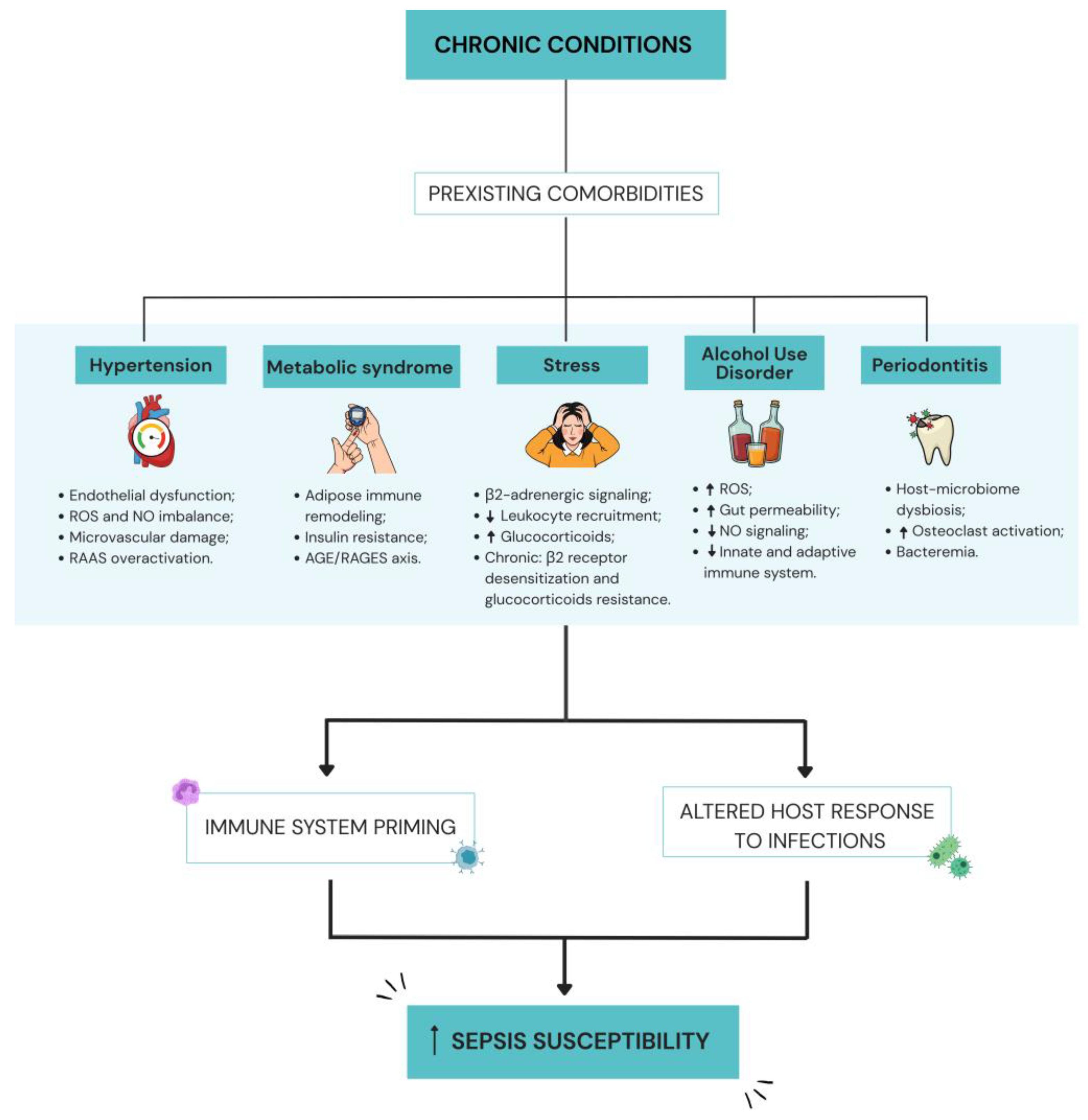
Disclaimer/Publisher’s Note: The statements, opinions and data contained in all publications are solely those of the individual author(s) and contributor(s) and not of MDPI and/or the editor(s). MDPI and/or the editor(s) disclaim responsibility for any injury to people or property resulting from any ideas, methods, instructions or products referred to in the content. |
© 2026 by the authors. Licensee MDPI, Basel, Switzerland. This article is an open access article distributed under the terms and conditions of the Creative Commons Attribution (CC BY) license.
Share and Cite
Miguel, M.V.O.; Santos, R.M.; Oliveira, M.M.d.; Pelosi, G.G.; Freitas, A. Comorbidities and Inflammation: How Chronic Diseases Prime the Host Response in Sepsis. Int. J. Mol. Sci. 2026, 27, 7395. https://doi.org/10.3390/ijms27167395
Miguel MVO, Santos RM, Oliveira MMd, Pelosi GG, Freitas A. Comorbidities and Inflammation: How Chronic Diseases Prime the Host Response in Sepsis. International Journal of Molecular Sciences. 2026; 27(16):7395. https://doi.org/10.3390/ijms27167395
Chicago/Turabian StyleMiguel, Maria Vitória Oliveira, Rayssa Menon Santos, Matheus Marques de Oliveira, Gislaine Garcia Pelosi, and Andressa Freitas. 2026. "Comorbidities and Inflammation: How Chronic Diseases Prime the Host Response in Sepsis" International Journal of Molecular Sciences 27, no. 16: 7395. https://doi.org/10.3390/ijms27167395
APA StyleMiguel, M. V. O., Santos, R. M., Oliveira, M. M. d., Pelosi, G. G., & Freitas, A. (2026). Comorbidities and Inflammation: How Chronic Diseases Prime the Host Response in Sepsis. International Journal of Molecular Sciences, 27(16), 7395. https://doi.org/10.3390/ijms27167395