Molecular and Cellular Mechanisms of Spinal Cord Stimulation: Linking Dorsal Horn Circuits, Glia, and ECAP-Guided Therapy

 , , ,
, , ,  and
and
Abstract
1. Introduction
Literature Search
2. Basic Neurophysiology of Pain and the Spinal Cord
3. Molecular Mechanisms of Pain
4. Technological Advances and Their Mechanistic Implications
4.1. Tonic Spinal Cord Stimulation
4.2. High-Frequency Spinal Cord Stimulation (10 kHz)
4.3. Burst Spinal Cord Stimulation
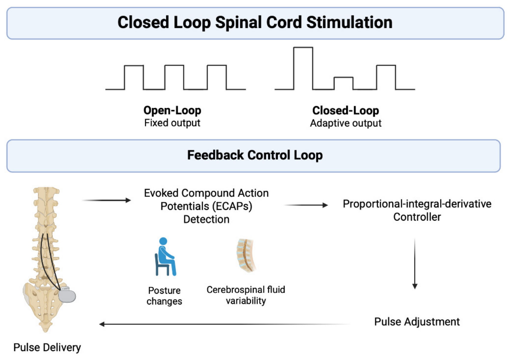
4.4. Closed-Loop Spinal Cord Stimulation
5. Experimental Models and Biomarkers
5.1. Animal Models Used to Explore Spinal Cord Stimulation Mechanisms
5.2. ECAP as a Functional Electrophysiological Biomarker
6. Limitations in Current Understanding
7. Future Directions and Innovations
8. Conclusions
Author Contributions
Funding
Institutional Review Board Statement
Informed Consent Statement
Data Availability Statement
Conflicts of Interest
References
- Melzack, R.; Wall, P.D. Pain mechanisms: A new theory. Science 1965, 150, 971–979. [Google Scholar] [CrossRef] [Scilit] [PubMed]
- Katz, J.; Rosenbloom, B.N. The golden anniversary of Melzack and Wall’s gate control theory of pain: Celebrating 50 years of pain research and management. Pain Res. Manag. 2015, 20, 285–286. [Google Scholar] [CrossRef] [Scilit] [PubMed]
- Wall, P.D.; Sweet, W.H. Temporary abolition of pain in man. Science 1967, 155, 108–109. [Google Scholar] [CrossRef] [Scilit] [PubMed]
- North, R.B.; Wetzel, F.T. Spinal cord stimulation for chronic pain of spinal origin: A valuable long-term solution. Spine 2002, 27, 2584–2591; discussion 2592. [Google Scholar] [CrossRef] [Scilit] [PubMed]
- Shealy, C.N.; Mortimer, J.T.; Reswick, J.B. Electrical inhibition of pain by stimulation of the dorsal columns: Preliminary clinical report. Anesth. Analg. 1967, 46, 489–491. [Google Scholar] [PubMed]
- Ziegler, K.; Folkard, R.; Gonzalez, A.J.; Burghardt, J.; Antharvedi-Goda, S.; Martin-Cortecero, J.; Isaias-Camacho, E.; Kaushalya, S.; Tan, L.L.; Kuner, T.; et al. Primary somatosensory cortex bidirectionally modulates sensory gain and nociceptive behavior in a layer-specific manner. Nat. Commun. 2023, 14, 2999. [Google Scholar] [CrossRef] [Scilit] [PubMed]
- Ho, J.S.; Poon, C.; North, R.; Grubb, W.; Lempka, S.; Bikson, M. A Visual and Narrative Timeline Review of Spinal Cord Stimulation Technology and US Food and Drug Administration Milestones. Neuromodulation 2024, 27, 1020–1025. [Google Scholar] [CrossRef] [Scilit] [PubMed]
- Ali, R.; Schwalb, J.M. History and Future of Spinal Cord Stimulation. Neurosurgery 2024, 94, 20–28. [Google Scholar] [CrossRef] [Scilit] [PubMed]
- Huygen, F.; Soulanis, K.; Rtveladze, K.; Kamra, S.; Schlueter, M. Spinal Cord Stimulation vs. Medical Management for Chronic Back and Leg Pain: A Systematic Review and Network Meta-Analysis. JAMA Netw. Open 2024, 7, e2444608. [Google Scholar] [CrossRef] [Scilit] [PubMed]
- Aman, M.M.; Mahmoud, A.; Deer, T.; Sayed, D.; Hagedorn, J.M.; Brogan, S.E.; Singh, V.; Gulati, A.; Strand, N.; Weisbein, J.; et al. The American Society of Pain and Neuroscience (ASPN) Best Practices and Guidelines for the Interventional Management of Cancer-Associated Pain. J. Pain Res. 2021, 14, 2139–2164. [Google Scholar] [CrossRef] [Scilit] [PubMed]
- Bieze, M.; van Haaps, A.P.; Kapural, L.; Li, S.; Ferguson, K.; de Vries, R.; Schatman, M.E.; Mijatovic, V.; Kallewaard, J.W. Spinal Cord Stimulation for Intractable Visceral Pain Originating from the Pelvic and Abdominal Region: A Narrative Review on a Possible New Indication for Patients with Therapy-Resistant Pain. J. Pain Res. 2024, 17, 691–736. [Google Scholar] [CrossRef] [Scilit] [PubMed]
- Gallone, G.; Baldetti, L.; Tzanis, G.; Gramegna, M.; Latib, A.; Colombo, A.; Henry, T.D.; Giannini, F. Refractory Angina: From Pathophysiology to New Therapeutic Nonpharmacological Technologies. JACC Cardiovasc. Interv. 2020, 13, 1–19. [Google Scholar] [CrossRef] [Scilit] [PubMed]
- Kinfe, T.M.; Pintea, B.; Vatter, H. Is Spinal Cord Stimulation Useful and Safe for the Treatment of Chronic Pain of Ischemic Origin? A Review. Clin. J. Pain 2016, 32, 7–13. [Google Scholar] [CrossRef] [Scilit] [PubMed]
- Shanthanna, H.; Eldabe, S.; Provenzano, D.A.; Bouche, B.; Buchser, E.; Chadwick, R.; Doshi, T.L.; Duarte, R.; Hunt, C.; Huygen, F.; et al. Evidence-based consensus guidelines on patient selection and trial stimulation for spinal cord stimulation therapy for chronic non-cancer pain. Reg. Anesth. Pain Med. 2023, 48, 273–287. [Google Scholar] [CrossRef] [Scilit] [PubMed]
- Sowder, T.; Sayed, D.; Concannon, T.; Pew, S.H.; Strand, N.H.; Abd-Elsayed, A.; Wie, C.S.; Gomez Ramos, D.E.; Raslan, A.M.; Deer, T.R. The American Society of Pain and Neuroscience (ASPN) Guidelines for Radiofrequency Ablative Procedures in Patients with Implanted Devices. J. Pain Res. 2023, 16, 3693–3706. [Google Scholar] [CrossRef] [Scilit] [PubMed]
- Angeli, C.A.; Boakye, M.; Morton, R.A.; Vogt, J.; Benton, K.; Chen, Y.; Ferreira, C.K.; Harkema, S.J. Recovery of Over-Ground Walking After Chronic Motor Complete Spinal Cord Injury. N. Engl. J. Med. 2018, 379, 1244–1250. [Google Scholar] [CrossRef] [Scilit] [PubMed]
- Chalif, J.I.; Chavarro, V.S.; Mensah, E.; Johnston, B.; Fields, D.P.; Chalif, E.J.; Chiang, M.; Sutton, O.; Yong, R.; Trumbower, R.; et al. Epidural Spinal Cord Stimulation for Spinal Cord Injury in Humans: A Systematic Review. J. Clin. Med. 2024, 13, 1090. [Google Scholar] [CrossRef] [Scilit] [PubMed]
- Herrity, A.N.; Aslan, S.C.; Mesbah, S.; Siu, R.; Kalvakuri, K.; Ugiliweneza, B.; Mohamed, A.; Hubscher, C.H.; Harkema, S.J. Targeting bladder function with network-specific epidural stimulation after chronic spinal cord injury. Sci. Rep. 2022, 12, 11179. [Google Scholar] [CrossRef] [Scilit] [PubMed]
- Singh, O.; Carvalho, D.Z.; Espay, A.J.; Benarroch, E.E.; Grewal, S.S.; Pagani-Estevez, G.L. Spinal cord stimulation for gait impairment in Parkinson Disease: Scoping review and mechanistic considerations. Pain Med. 2023, 24, S11–S17. [Google Scholar] [CrossRef] [Scilit] [PubMed]
- Terkelsen, M.H.; Hvingelby, V.S.; Johnsen, E.L.; Moller, M.; Danielsen, E.H.; Henriksen, T.; Glud, A.N.; Tai, Y.; Baun, A.M.; Knudsen, A.L.; et al. Spinal cord stimulation therapy for gait impairment in Parkinson’s disease: A double-blinded, randomised feasibility trial with an open extension. Nat. Commun. 2026, 17, 2168. [Google Scholar] [CrossRef] [Scilit] [PubMed]
- Upadhyay, P.; Tsikopoulos, I.; Hiscock, C.; Houliston, H.; Doherty, S.; Knight, S.; Baxter, D.; Duffell, L. Spinal cord stimulation to manage autonomic dysfunction after spinal cord injury: A systematic review. Front. Hum. Neurosci. 2026, 20, 1763475. [Google Scholar] [CrossRef] [Scilit] [PubMed]
- Chatterjee, N.A.; Singh, J.P. Novel Interventional Therapies to Modulate the Autonomic Tone in Heart Failure. JACC Heart Fail. 2015, 3, 786–802. [Google Scholar] [CrossRef] [Scilit] [PubMed]
- Samotus, O.; Parrent, A.; Jog, M. Spinal Cord Stimulation Therapy for Gait Dysfunction in Advanced Parkinson’s Disease Patients. Mov. Disord. 2018, 33, 783–792. [Google Scholar] [CrossRef] [Scilit] [PubMed]
- Zipes, D.P.; Neuzil, P.; Theres, H.; Caraway, D.; Mann, D.L.; Mannheimer, C.; Van Buren, P.; Linde, C.; Linderoth, B.; Kueffer, F.; et al. Determining the Feasibility of Spinal Cord Neuromodulation for the Treatment of Chronic Systolic Heart Failure: The DEFEAT-HF Study. JACC Heart Fail. 2016, 4, 129–136, Correction in JACC Heart Fail. 2018, 6, 542. https://doi.org/10.1016/j.jchf.2018.05.001. [Google Scholar] [CrossRef] [Scilit] [PubMed]
- Kovermann, P.; Engels, M.; Muller, F.; Fahlke, C. Cellular Physiology and Pathophysiology of EAAT Anion Channels. Front. Cell. Neurosci. 2021, 15, 815279. [Google Scholar] [CrossRef] [Scilit] [PubMed]
- Schaible, H.G. Peripheral and central mechanisms of pain generation. In Analgesia; Handbook of Experimental Pharmacology; Springer: Berlin/Heidelberg, Germany, 2007; pp. 3–28. [Google Scholar] [CrossRef] [Scilit] [PubMed]
- Zhang, Y.; Liu, S.; Zhang, Y.Q.; Goulding, M.; Wang, Y.Q.; Ma, Q. Timing Mechanisms Underlying Gate Control by Feedforward Inhibition. Neuron 2018, 99, 941–955.e944. [Google Scholar] [CrossRef] [Scilit] [PubMed]
- Millan, M.J. Descending control of pain. Prog. Neurobiol. 2002, 66, 355–474. [Google Scholar] [CrossRef] [Scilit] [PubMed]
- Melzack, R. Gate control theory: On the evolution of pain concepts. Pain Forum 1996, 5, 128–138. [Google Scholar] [CrossRef] [Scilit]
- D’Mello, R.; Dickenson, A.H. Spinal cord mechanisms of pain. Br. J. Anaesth. 2008, 101, 8–16. [Google Scholar] [CrossRef] [Scilit] [PubMed]
- Todd, A.J. Anatomy of primary afferents and projection neurones in the rat spinal dorsal horn with particular emphasis on substance P and the neurokinin 1 receptor. Exp. Physiol. 2002, 87, 245–249. [Google Scholar] [CrossRef] [Scilit] [PubMed]
- Tracey, I.; Mantyh, P.W. The cerebral signature for pain perception and its modulation. Neuron 2007, 55, 377–391. [Google Scholar] [CrossRef] [Scilit] [PubMed]
- Benarroch, E.E. Descending monoaminergic pain modulation: Bidirectional control and clinical relevance. Neurology 2008, 71, 217–221. [Google Scholar] [CrossRef] [Scilit] [PubMed]
- Lau, B.K.; Vaughan, C.W. Descending modulation of pain: The GABA disinhibition hypothesis of analgesia. Curr. Opin. Neurobiol. 2014, 29, 159–164. [Google Scholar] [CrossRef] [Scilit] [PubMed]
- Mendell, L.M. Constructing and deconstructing the gate theory of pain. Pain 2014, 155, 210–216. [Google Scholar] [CrossRef] [Scilit] [PubMed]
- Tobaldini, G.; Sardi, N.F.; Guilhen, V.A.; Fischer, L. Pain Inhibits Pain: An Ascending-Descending Pain Modulation Pathway Linking Mesolimbic and Classical Descending Mechanisms. Mol. Neurobiol. 2019, 56, 1000–1013. [Google Scholar] [CrossRef] [Scilit] [PubMed]
- Aby, F.; Lorenzo, L.E.; Grivet, Z.; Bouali-Benazzouz, R.; Martin, H.; Valerio, S.; Whitestone, S.; Isabel, D.; Idi, W.; Bouchatta, O.; et al. Switch of serotonergic descending inhibition into facilitation by a spinal chloride imbalance in neuropathic pain. Sci. Adv. 2022, 8, eabo0689. [Google Scholar] [CrossRef] [Scilit] [PubMed]
- Wei, F.; Guo, W.; Zou, S.; Ren, K.; Dubner, R. Supraspinal glial-neuronal interactions contribute to descending pain facilitation. J. Neurosci. 2008, 28, 10482–10495. [Google Scholar] [CrossRef] [Scilit] [PubMed]
- de Geus, T.J.; Franken, G.; Joosten, E.A.J. Spinal Cord Stimulation Paradigms and Pain Relief: A Preclinical Systematic Review on Modulation of the Central Inflammatory Response in Neuropathic Pain. Neuromodulation 2023, 26, 25–34. [Google Scholar] [CrossRef] [Scilit] [PubMed]
- Stroman, P.W.; Ioachim, G.; Powers, J.M.; Staud, R.; Pukall, C. Pain processing in the human brainstem and spinal cord before, during, and after the application of noxious heat stimuli. Pain 2018, 159, 2012–2020. [Google Scholar] [CrossRef] [Scilit] [PubMed]
- Antal, M. Molecular Anatomy of Synaptic and Extrasynaptic Neurotransmission Between Nociceptive Primary Afferents and Spinal Dorsal Horn Neurons. Int. J. Mol. Sci. 2025, 26, 2356. [Google Scholar] [CrossRef] [Scilit] [PubMed]
- Wang, H.; Xu, C. A Novel Progress: Glial Cells and Inflammatory Pain. ACS Chem. Neurosci. 2022, 13, 288–295. [Google Scholar] [CrossRef] [Scilit] [PubMed]
- Zhao, H.; Alam, A.; Chen, Q.; Eusman, M.A.; Pal, A.; Eguchi, S.; Wu, L.; Ma, D. The role of microglia in the pathobiology of neuropathic pain development: What do we know? Br. J. Anaesth. 2017, 118, 504–516. [Google Scholar] [CrossRef] [Scilit] [PubMed]
- Tanga, F.Y.; Raghavendra, V.; DeLeo, J.A. Quantitative real-time RT-PCR assessment of spinal microglial and astrocytic activation markers in a rat model of neuropathic pain. Neurochem. Int. 2004, 45, 397–407. [Google Scholar] [CrossRef] [PubMed]
- Donnelly, C.R.; Andriessen, A.S.; Chen, G.; Wang, K.; Jiang, C.; Maixner, W.; Ji, R.R. Central Nervous System Targets: Glial Cell Mechanisms in Chronic Pain. Neurotherapeutics 2020, 17, 846–860. [Google Scholar] [CrossRef] [Scilit] [PubMed]
- Latremoliere, A.; Woolf, C.J. Central sensitization: A generator of pain hypersensitivity by central neural plasticity. J. Pain 2009, 10, 895–926. [Google Scholar] [CrossRef] [Scilit] [PubMed]
- Woolf, C.J. Central sensitization: Implications for the diagnosis and treatment of pain. Pain 2011, 152, S2–S15. [Google Scholar] [CrossRef] [Scilit] [PubMed]
- Ji, R.R.; Nackley, A.; Huh, Y.; Terrando, N.; Maixner, W. Neuroinflammation and Central Sensitization in Chronic and Widespread Pain. Anesthesiology 2018, 129, 343–366. [Google Scholar] [CrossRef] [Scilit] [PubMed]
- Bordeleau, M.; Carrondo Cottin, S.; Meier, K.; Prud’Homme, M. Effects of Tonic Spinal Cord Stimulation on Sensory Perception in Chronic Pain Patients: A Systematic Review. Neuromodulation 2019, 22, 149–162. [Google Scholar] [CrossRef] [Scilit] [PubMed]
- Joosten, E.A.; Franken, G. Spinal cord stimulation in chronic neuropathic pain: Mechanisms of action, new locations, new paradigms. Pain 2020, 161, S104–S113. [Google Scholar] [CrossRef] [Scilit] [PubMed]
- Smits, H.; van Kleef, M.; Holsheimer, J.; Joosten, E.A. Experimental spinal cord stimulation and neuropathic pain: Mechanism of action, technical aspects, and effectiveness. Pain Pract. 2013, 13, 154–168. [Google Scholar] [CrossRef] [Scilit] [PubMed]
- Patil, A.S.; Levasseur, B.; Gupta, M. Neuromodulation and Habituation: A Literature Review and Conceptional Analysis of Sustaining Therapeutic Efficacy and Mitigating Habituation. Biomedicines 2024, 12, 930. [Google Scholar] [CrossRef] [Scilit] [PubMed]
- Forouzan, E.J.; Rashid, M.Y.; Nasr, N.F.; Abd-Elsayed, A.; Knezevic, N.N. The Potential of Spinal Cord Stimulation in Treating Spinal Cord Injury. Curr. Pain Headache Rep. 2025, 29, 35. [Google Scholar] [CrossRef] [Scilit] [PubMed]
- Karri, J.; Joshi, M.; Polson, G.; Tang, T.; Lee, M.; Orhurhu, V.; Deer, T.; Abd-Elsayed, A. Spinal Cord Stimulation for Chronic Pain Syndromes: A Review of Considerations in Practice Management. Pain Physician 2020, 23, 599–616. [Google Scholar] [CrossRef] [Scilit]
- Abraham, M.E.; Gold, J.; Dondapati, A.; Sheaffer, K.; Gendreau, J.L.; Mammis, A. High Frequency 10 kHz Spinal Cord Stimulation as a First Line Programming Option for Patients With Chronic Pain: A Retrospective Study and Review of the Current Evidence. Cureus 2021, 13, e17220. [Google Scholar] [CrossRef] [Scilit] [PubMed]
- Peeters, J.B.; Raftopoulos, C. Tonic, Burst, High-Density, and 10-kHz High-Frequency Spinal Cord Stimulation: Efficiency and Patients’ Preferences in a Failed Back Surgery Syndrome Predominant Population. Review of Literature. World Neurosurg. 2020, 144, e331–e340. [Google Scholar] [CrossRef] [Scilit] [PubMed]
- Tieppo Francio, V.; Polston, K.F.; Murphy, M.T.; Hagedorn, J.M.; Sayed, D. Management of Chronic and Neuropathic Pain with 10 kHz Spinal Cord Stimulation Technology: Summary of Findings from Preclinical and Clinical Studies. Biomedicines 2021, 9, 644. [Google Scholar] [CrossRef] [Scilit] [PubMed]
- Arle, J.E.; Mei, L.; Carlson, K.W.; Shils, J.L. High-Frequency Stimulation of Dorsal Column Axons: Potential Underlying Mechanism of Paresthesia-Free Neuropathic Pain Relief. Neuromodulation 2016, 19, 385–397. [Google Scholar] [CrossRef] [Scilit] [PubMed]
- Liao, W.T.; Tseng, C.C.; Wu, C.H.; Lin, C.R. Early high-frequency spinal cord stimulation treatment inhibited the activation of spinal mitogen-activated protein kinases and ameliorated spared nerve injury-induced neuropathic pain in rats. Neurosci. Lett. 2020, 721, 134763. [Google Scholar] [CrossRef] [Scilit] [PubMed]
- Kapural, L.; Yu, C.; Doust, M.W.; Gliner, B.E.; Vallejo, R.; Sitzman, B.T.; Amirdelfan, K.; Morgan, D.M.; Brown, L.L.; Yearwood, T.L.; et al. Novel 10-kHz High-frequency Therapy (HF10 Therapy) Is Superior to Traditional Low-Frequency Spinal Cord Stimulation for the Treatment of Chronic Back and Leg Pain: The SENZA-RCT Randomized Controlled Trial. Anesthesiology 2015, 123, 851–860. [Google Scholar] [CrossRef] [Scilit] [PubMed]
- Kapural, L.; Yu, C.; Doust, M.W.; Gliner, B.E.; Vallejo, R.; Sitzman, B.T.; Amirdelfan, K.; Morgan, D.M.; Yearwood, T.L.; Bundschu, R.; et al. Comparison of 10-kHz High-Frequency and Traditional Low-Frequency Spinal Cord Stimulation for the Treatment of Chronic Back and Leg Pain: 24-Month Results from a Multicenter, Randomized, Controlled Pivotal Trial. Neurosurgery 2016, 79, 667–677. [Google Scholar] [CrossRef] [Scilit] [PubMed]
- Petersen, E.A.; Stauss, T.G.; Scowcroft, J.A.; Brooks, E.S.; White, J.L.; Sills, S.M.; Amirdelfan, K.; Guirguis, M.N.; Xu, J.; Yu, C.; et al. Effect of High-Frequency (10-kHz) Spinal Cord Stimulation in Patients with Painful Diabetic Neuropathy: A Randomized Clinical Trial. JAMA Neurol. 2021, 78, 687–698. [Google Scholar] [CrossRef] [Scilit] [PubMed]
- Petersen, E.A.; Stauss, T.G.; Scowcroft, J.A.; Jaasma, M.J.; Brooks, E.S.; Edgar, D.R.; White, J.L.; Sills, S.M.; Amirdelfan, K.; Guirguis, M.N.; et al. Long-term efficacy of high-frequency (10 kHz) spinal cord stimulation for the treatment of painful diabetic neuropathy: 24-month results of a randomized controlled trial. Diabetes Res. Clin. Pract. 2023, 203, 110865. [Google Scholar] [CrossRef] [Scilit] [PubMed]
- Chakravarthy, K.; Malayil, R.; Kirketeig, T.; Deer, T. Burst Spinal Cord Stimulation: A Systematic Review and Pooled Analysis of Real-World Evidence and Outcomes Data. Pain Med. 2019, 20, S47–S57. [Google Scholar] [CrossRef] [Scilit] [PubMed]
- Swadlow, H.A.; Gusev, A.G. The impact of ‘bursting’ thalamic impulses at a neocortical synapse. Nat. Neurosci. 2001, 4, 402–408. [Google Scholar] [CrossRef] [Scilit] [PubMed]
- Deer, T.; Slavin, K.V.; Amirdelfan, K.; North, R.B.; Burton, A.W.; Yearwood, T.L.; Tavel, E.; Staats, P.; Falowski, S.; Pope, J.; et al. Success Using Neuromodulation with BURST (SUNBURST) Study: Results from a Prospective, Randomized Controlled Trial Using a Novel Burst Waveform. Neuromodulation 2018, 21, 56–66. [Google Scholar] [CrossRef] [Scilit] [PubMed]
- Hou, S.; Kemp, K.; Grabois, M. A Systematic Evaluation of Burst Spinal Cord Stimulation for Chronic Back and Limb Pain. Neuromodulation 2016, 19, 398–405. [Google Scholar] [CrossRef] [Scilit] [PubMed]
- Chakravarthy, K.; Fishman, M.A.; Zuidema, X.; Hunter, C.W.; Levy, R. Mechanism of Action in Burst Spinal Cord Stimulation: Review and Recent Advances. Pain Med. 2019, 20, S13–S22. [Google Scholar] [CrossRef] [Scilit] [PubMed]
- Bocci, T.; De Carolis, G.; Paroli, M.; Barloscio, D.; Parenti, L.; Tollapi, L.; Valeriani, M.; Sartucci, F. Neurophysiological Comparison Among Tonic, High Frequency, and Burst Spinal Cord Stimulation: Novel Insights into Spinal and Brain Mechanisms of Action. Neuromodulation 2018, 21, 480–488. [Google Scholar] [CrossRef] [Scilit] [PubMed]
- Mangano, N.; Torpey, A.; Devitt, C.; Wen, G.A.; Doh, C.; Gupta, A. Closed-Loop Spinal Cord Stimulation in Chronic Pain Management: Mechanisms, Clinical Evidence, and Emerging Perspectives. Biomedicines 2025, 13, 1091. [Google Scholar] [CrossRef] [Scilit] [PubMed]
- Zheng, Y.; Liu, C.W.; Hui Chan, D.X.; Kai Ong, D.W.; Xin Ker, J.R.; Ng, W.H.; Wan, K.R. Neurostimulation for Chronic Pain: A Systematic Review of High-Quality Randomized Controlled Trials With Long-Term Follow-Up. Neuromodulation 2023, 26, 1276–1294. [Google Scholar] [CrossRef] [Scilit] [PubMed]
- Versantvoort, E.M.; Dietz, B.E.; Mugan, D.; Vuong, Q.C.; Luli, S.; Obara, I. Evoked compound action potential (ECAP)-controlled closed-loop spinal cord stimulation in an experimental model of neuropathic pain in rats. Bioelectron. Med. 2024, 10, 2. [Google Scholar] [CrossRef] [Scilit] [PubMed]
- Anaya, C.J.; Zander, H.J.; Graham, R.D.; Sankarasubramanian, V.; Lempka, S.F. Evoked Potentials Recorded from the Spinal Cord During Neurostimulation for Pain: A Computational Modeling Study. Neuromodulation 2020, 23, 64–73. [Google Scholar] [CrossRef] [Scilit] [PubMed]
- Mekhail, N.; Levy, R.M.; Deer, T.R.; Kapural, L.; Li, S.; Amirdelfan, K.; Hunter, C.W.; Rosen, S.M.; Costandi, S.J.; Falowski, S.M.; et al. Long-term safety and efficacy of closed-loop spinal cord stimulation to treat chronic back and leg pain (Evoke): A double-blind, randomised, controlled trial. Lancet Neurol. 2020, 19, 123–134. [Google Scholar] [CrossRef] [Scilit] [PubMed]
- Parker, J.L.; Karantonis, D.M.; Single, P.S.; Obradovic, M.; Cousins, M.J. Compound action potentials recorded in the human spinal cord during neurostimulation for pain relief. Pain 2012, 153, 593–601. [Google Scholar] [CrossRef] [Scilit] [PubMed]
- Mekhail, N.; Levy, R.M.; Deer, T.R.; Kapural, L.; Li, S.; Amirdelfan, K.; Hunter, C.W.; Rosen, S.M.; Costandi, S.J.; Falowski, S.M.; et al. Durability of Clinical and Quality-of-Life Outcomes of Closed-Loop Spinal Cord Stimulation for Chronic Back and Leg Pain: A Secondary Analysis of the Evoke Randomized Clinical Trial. JAMA Neurol. 2022, 79, 251–260, Correction in JAMA Neurol. 2022, 79, 420. https://doi.org/10.1001/jamaneurol.2022.0022. [Google Scholar] [CrossRef] [Scilit] [PubMed]
- Brucker-Hahn, M.K.; Deshmukh, A.; Settell, M.; Chin, J.; Upadhye, A.; Lavrov, I.; Shoffstall, A.J.; Ludwig, K.A.; Zhang, M.; Lempka, S.F. Anatomical Data Driven Modeling of Evoked Compound Action Potentials Recordings During Spinal Cord Stimulation in a Swine Model. Neuromodulation 2026, 29, 40–55. [Google Scholar] [CrossRef] [Scilit] [PubMed]
- Ladner, K.; Versantvoort, E.M.; Mugan, D.; Vuong, Q.C.; Dietz, B.E.; Hu, A.; Thijssen, M.E.G.; Gorman, R.B.; Petersen, E.; Obara, I. Preclinical Insights into the Effects of Frequency and Pulse Width on Evoked Compound Action Potential Morphology During Spinal Cord Stimulation. Neuromodulation 2026, 29, 26–39. [Google Scholar] [CrossRef] [Scilit] [PubMed]
- Zander, H.J.; Graham, R.D.; Anaya, C.J.; Lempka, S.F. Anatomical and technical factors affecting the neural response to epidural spinal cord stimulation. J. Neural Eng. 2020, 17, 036019. [Google Scholar] [CrossRef] [Scilit] [PubMed]
- North, R.B.; Sung, J.H.; Matthews, L.A.; Zander, H.J.; Lempka, S.F. Postural Changes in Spinal Cord Stimulation Thresholds: Current and Voltage Sources. Neuromodulation 2024, 27, 178–182. [Google Scholar] [CrossRef] [Scilit] [PubMed]
- Pilitsis, J.G.; Chakravarthy, K.V.; Will, A.J.; Trutnau, K.C.; Hageman, K.N.; Dinsmoor, D.A.; Litvak, L.M. The Evoked Compound Action Potential as a Predictor for Perception in Chronic Pain Patients: Tools for Automatic Spinal Cord Stimulator Programming and Control. Front. Neurosci. 2021, 15, 673998. [Google Scholar] [CrossRef] [Scilit] [PubMed]
- Brucker-Hahn, M.K.; Zander, H.J.; Will, A.J.; Vallabh, J.C.; Wolff, J.S.; Dinsmoor, D.A.; Lempka, S.F. Evoked compound action potentials during spinal cord stimulation: Effects of posture and pulse width on signal features and neural activation within the spinal cord. J. Neural Eng. 2023, 20, 046028. [Google Scholar] [CrossRef] [Scilit] [PubMed]
- Mugan, D.; Vuong, Q.C.; Dietz, B.E.; Obara, I. Characterization of preclinical models to investigate spinal cord stimulation for neuropathic pain: A systematic review and meta-analysis. Pain Rep. 2025, 10, e1228. [Google Scholar] [CrossRef] [Scilit] [PubMed]
- Sterner, R.C.; Sterner, R.M. Immune response following traumatic spinal cord injury: Pathophysiology and therapies. Front. Immunol. 2022, 13, 1084101. [Google Scholar] [CrossRef] [Scilit] [PubMed]
- Zhang, N.; Fang, M.; Chen, H.; Gou, F.; Ding, M. Evaluation of spinal cord injury animal models. Neural Regen. Res. 2014, 9, 2008–2012. [Google Scholar] [CrossRef] [Scilit] [PubMed]
- Cui, J.G.; O’Connor, W.T.; Ungerstedt, U.; Linderoth, B.; Meyerson, B.A. Spinal cord stimulation attenuates augmented dorsal horn release of excitatory amino acids in mononeuropathy via a GABAergic mechanism. Pain 1997, 73, 87–95. [Google Scholar] [CrossRef] [Scilit] [PubMed]
- Sun, L.; Peng, C.; Joosten, E.; Cheung, C.W.; Tan, F.; Jiang, W.; Shen, X. Spinal Cord Stimulation and Treatment of Peripheral or Central Neuropathic Pain: Mechanisms and Clinical Application. Neural Plast. 2021, 2021, 5607898. [Google Scholar] [CrossRef] [Scilit] [PubMed]
- Song, Z.; Meyerson, B.A.; Linderoth, B. Spinal 5-HT receptors that contribute to the pain-relieving effects of spinal cord stimulation in a rat model of neuropathy. Pain 2011, 152, 1666–1673. [Google Scholar] [CrossRef] [Scilit] [PubMed]
- Song, Z.; Ultenius, C.; Meyerson, B.A.; Linderoth, B. Pain relief by spinal cord stimulation involves serotonergic mechanisms: An experimental study in a rat model of mononeuropathy. Pain 2009, 147, 241–248. [Google Scholar] [CrossRef] [Scilit] [PubMed]
- Li, X.; Wu, Z.; Wang, R.; Bai, J. Spinal Cord Stimulation Alleviates Neuropathic Pain Involves Regulation of the Ca2+/CaN/NFAT4 Pathway in Spinal Dorsal Horn Astrocytes. Cell. Mol. Neurobiol. 2026, 46, 106. [Google Scholar] [CrossRef] [Scilit] [PubMed]
- Sato, K.L.; Johanek, L.M.; Sanada, L.S.; Sluka, K.A. Spinal cord stimulation reduces mechanical hyperalgesia and glial cell activation in animals with neuropathic pain. Anesth. Analg. 2014, 118, 464–472. [Google Scholar] [CrossRef] [Scilit] [PubMed]
- Cedeno, D.L.; Kelley, C.A.; Chakravarthy, K.; Vallejo, R. Modulation of Glia-Mediated Processes by Spinal Cord Stimulation in Animal Models of Neuropathic Pain. Front. Pain Res. 2021, 2, 702906. [Google Scholar] [CrossRef] [Scilit] [PubMed]
- Khadka, N.; Truong, D.Q.; Williams, P.; Martin, J.H.; Bikson, M. The Quasi-Uniform Assumption for Spinal Cord Stimulation Translational Research. J. Neurosci. Methods 2019, 328, 108446. [Google Scholar] [CrossRef] [Scilit] [PubMed]
- Meyerson, B.A.; Linderoth, B. Mode of action of spinal cord stimulation in neuropathic pain. J. Pain Symptom Manag. 2006, 31, S6–S12. [Google Scholar] [CrossRef] [Scilit] [PubMed]
- Muller, L.; Pope, J.; Verrills, P.; Petersen, E.; Kallewaard, J.W.; Gould, I.; Karantonis, D.M. First evidence of a biomarker-based dose-response relationship in chronic pain using physiological closed-loop spinal cord stimulation. Reg. Anesth. Pain Med. 2025, 50, 345–351. [Google Scholar] [CrossRef] [Scilit] [PubMed]
- Chakravarthy, K.; Richter, H.; Christo, P.J.; Williams, K.; Guan, Y. Spinal Cord Stimulation for Treating Chronic Pain: Reviewing Preclinical and Clinical Data on Paresthesia-Free High-Frequency Therapy. Neuromodulation 2018, 21, 10–18. [Google Scholar] [CrossRef] [Scilit] [PubMed]
- Van Buyten, J.P.; Al-Kaisy, A.; Smet, I.; Palmisani, S.; Smith, T. High-frequency spinal cord stimulation for the treatment of chronic back pain patients: Results of a prospective multicenter European clinical study. Neuromodulation 2013, 16, 59–65. [Google Scholar] [CrossRef] [Scilit] [PubMed]
- Wang, D.; Lee, K.Y.; Kagan, Z.B.; Bradley, K.; Lee, D. Frequency-Dependent Neural Modulation of Dorsal Horn Neurons by Kilohertz Spinal Cord Stimulation in Rats. Biomedicines 2024, 12, 1346. [Google Scholar] [CrossRef] [Scilit] [PubMed]
- Todd, A.J. Neuronal circuitry for pain processing in the dorsal horn. Nat. Rev. Neurosci. 2010, 11, 823–836. [Google Scholar] [CrossRef] [Scilit] [PubMed]
- Liu, F.Y.; Qu, X.X.; Cai, J.; Wang, F.T.; Xing, G.G.; Wan, Y. Electrophysiological properties of spinal wide dynamic range neurons in neuropathic pain rats following spinal nerve ligation. Neurosci. Bull. 2011, 27, 1–8. [Google Scholar] [CrossRef] [Scilit] [PubMed]
- Zhang, Z.; Zheng, H.; Yu, Q.; Jing, X. Understanding of Spinal Wide Dynamic Range Neurons and Their Modulation on Pathological Pain. J. Pain Res. 2024, 17, 441–457. [Google Scholar] [CrossRef] [Scilit] [PubMed]
- DiSabato, D.J.; Quan, N.; Godbout, J.P. Neuroinflammation: The devil is in the details. J. Neurochem. 2016, 139, 136–153. [Google Scholar] [CrossRef] [Scilit] [PubMed]
- Hohm, H.L.; Schuster, R.; Buciu, V.B.; Serban, D.M.; Ciurescu, S.; Cornea, A.; Sharma, A.; Nistor, D.; Kundnani, N.R. Glial Cytokine and Metabolic Networks in Progressive Multiple Sclerosis: From Pathophysiology to Biomarkers and Therapeutic Strategies. Int. J. Mol. Sci. 2025, 26, 8817. [Google Scholar] [CrossRef] [Scilit] [PubMed]
- Bilchak, J.N.; Yeakle, K.; Caron, G.; Malloy, D.; Cote, M.P. Enhancing KCC2 activity decreases hyperreflexia and spasticity after chronic spinal cord injury. Exp. Neurol. 2021, 338, 113605. [Google Scholar] [CrossRef] [Scilit] [PubMed]
- Choi, J.E.; Carpena, N.T.; Lee, J.H.; Chang, S.Y.; Lee, M.Y.; Jung, J.Y.; Chung, W.H. Round-window delivery of lithium chloride regenerates cochlear synapses damaged by noise-induced excitotoxic trauma via inhibition of the NMDA receptor in the rat. PLoS ONE 2023, 18, e0284626. [Google Scholar] [CrossRef] [Scilit] [PubMed]
- Kami, K.; Taguchi Ms, S.; Tajima, F.; Senba, E. Improvements in impaired GABA and GAD65/67 production in the spinal dorsal horn contribute to exercise-induced hypoalgesia in a mouse model of neuropathic pain. Mol. Pain 2016, 12, 1744806916629059. [Google Scholar] [CrossRef] [Scilit] [PubMed]
- Rimmele, T.S.; Li, S.; Andersen, J.V.; Westi, E.W.; Rotenberg, A.; Wang, J.; Aldana, B.I.; Selkoe, D.J.; Aoki, C.J.; Dulla, C.G.; et al. Neuronal Loss of the Glutamate Transporter GLT-1 Promotes Excitotoxic Injury in the Hippocampus. Front. Cell. Neurosci. 2021, 15, 788262. [Google Scholar] [CrossRef] [Scilit] [PubMed]
- Messina, G.; Monda, A.; Messina, A.; Di Maio, G.; Monda, V.; Limone, P.; Dipace, A.; Monda, M.; Polito, R.; Moscatelli, F. Relationship Between Non-Invasive Brain Stimulation and Autonomic Nervous System. Biomedicines 2024, 12, 972. [Google Scholar] [CrossRef] [Scilit] [PubMed]
- Guerriero, M.A.; Dipace, A.; Monda, A.; De Maria, A.; Polito, R.; Messina, G.; Monda, M.; di Padova, M.; Basta, A.; Ruberto, M.; et al. Relationship Between Sedentary Lifestyle, Physical Activity and Stress in University Students and Their Life Habits: A Scoping Review with PRISMA Checklist (PRISMA-ScR). Brain Sci. 2025, 15, 78. [Google Scholar] [CrossRef] [Scilit] [PubMed]
- Yousaf, A.; Yamamoto, H.; Fang, J.Y.; Romman, A.; Koutrouvelis, A.P.; Yamamoto, S. Supraspinal Mechanisms of Spinal Cord Stimulation in Pain Mitigation: A Systematic Review. Cureus 2025, 17, e86756. [Google Scholar] [CrossRef] [Scilit] [PubMed]
- Prunskis, J.V.; Masys, T.; Pyles, S.T.; Abd-Elsayed, A.; Deer, T.R.; Beall, D.P.; Gheith, R.; Patel, S.; Sayed, D.; Moten, H.; et al. The Application of Artificial Intelligence to Enhance Spinal Cord Stimulation Efficacy for Chronic Pain Management: Current Evidence and Future Directions. Curr. Pain Headache Rep. 2025, 29, 85. [Google Scholar] [CrossRef] [Scilit] [PubMed]
- Patel, P.M.; Green, M.; Tram, J.; Wang, E.; Murphy, M.Z.; Abd-Elsayed, A.; Chakravarthy, K. Beyond the Pain Management Clinic: The Role of AI-Integrated Remote Patient Monitoring in Chronic Disease Management—A Narrative Review. J. Pain Res. 2024, 17, 4223–4237. [Google Scholar] [CrossRef] [Scilit] [PubMed]
- Abd-Elsayed, A.; Robinson, C.L.; Marshall, Z.; Diwan, S.; Peters, T. Applications of Artificial Intelligence in Pain Medicine. Curr. Pain Headache Rep. 2024, 28, 229–238. [Google Scholar] [CrossRef] [Scilit] [PubMed]
- Dombovy-Johnson, M.L.; Hunt, C.L.; Morrow, M.M.; Lamer, T.J.; Pittelkow, T.P. Current Evidence Lacking to Guide Clinical Practice for Spinal Cord Stimulation in the Treatment of Neuropathic Pain in Spinal Cord Injury: A Review of the Literature and a Proposal for Future Study. Pain Pract. 2020, 20, 325–335. [Google Scholar] [CrossRef] [Scilit] [PubMed]
- Pollard, E.M.; Lamer, T.J.; Moeschler, S.M.; Gazelka, H.M.; Hooten, W.M.; Bendel, M.A.; Warner, N.S.; Murad, M.H. The effect of spinal cord stimulation on pain medication reduction in intractable spine and limb pain: A systematic review of randomized controlled trials and meta-analysis. J. Pain Res. 2019, 12, 1311–1324. [Google Scholar] [CrossRef] [Scilit] [PubMed]
- Bernstein, C.A.; Paicius, R.M.; Barkow, S.H.; Lempert-Cohen, C. Spinal cord stimulation in conjunction with peripheral nerve field stimulation for the treatment of low back and leg pain: A case series. Neuromodulation 2008, 11, 116–123. [Google Scholar] [CrossRef] [Scilit] [PubMed]


| Category | Major Elements | Key Role in Pain |
|---|---|---|
| Glutamatergic Transmission | Glutamate (Aδ, C fibers), VGLUT2, NMDA and AMPA receptors, EAATs | Primary excitatory nociceptive signaling; EAATs prevent excitotoxicity |
| Presynaptic/Extrasynaptic Modulation | mGluRs (inhibitory or excitatory), endocannibinoids (CB1R), inhibitory interneurons (GABA, Glycine), opioids, ATP, descending monoamines (NE, DA, 5-HT) | Fine-tunes glutamate release and postsynaptic responses; balance of inhibition vs. facilitation shapes pain perception |
| Glial and Inflammatory Contribution | Pro-inflammatory cytokines (IL-1β, IL-6) Microglia (early), astrocytes (maintenance), | Drive and sustain neuropathic pain through inflammation and excitability |
| Central Sensitization | Increased glutamatergic transmission, NMDA/AMPA upregulation, ion channel changes, glial activation, LTP | Maladaptive plasticity leads to hyperalgesia and allodynia |
| Primary Target/Cascade | Proposed Mechanistic Role |
|---|---|
| Aβ fibers → inhibitory interneurons/WDR | Segmental gate control: Aβ activation increases inhibitory drive, suppressing WDR neuron firing and nociceptive transmission. |
| GABAergic and glycinergic interneurons | Enhances inhibitory tone in dorsal horn, counteracting hyperexcitability in neuropathic states. |
| KCC2/Cl− gradient | Partial restoration of impaired Cl− homeostasis, improving GABAergic inhibition in injured dorsal horn. |
| Glutamatergic transmission (NMDA/AMPA, NR2B) | Reduces NMDA/AMPA activity and receptor phosphorylation, decreasing central sensitization. |
| Glial activation (microglial p38-MAPK, TLR4/NF-κB) | Attenuates microglial activation and pro-inflammatory signaling pathways in dorsal horn. |
| Cytokine profile (↓ IL-1β/IL-6/TNF-α; ↑ IL-10) | Shifts spinal milieu from pro- to anti-inflammatory, supporting long-term analgesia. |
| EAAT/GLT-1 and glutamate clearance | Improves astrocytic glutamate uptake, reducing excitotoxic drive onto dorsal horn neurons. |
| Primary Target/Cascade | Proposed Mechanistic Role |
|---|---|
| Aβ fibers/medium–small fibers (competing hypotheses) | Either depolarization block of Aβ fibers, preferential inhibition via interneurons, or modulation of nociceptive fibers. |
| Dorsal horn inhibitory interneurons (GABA/Gly) | Increases inhibitory interneuron output without conscious paresthesia, suppressing nociceptive transmission. |
| Kinase signaling (e.g., receptor-associated protein kinases) | Reduces kinase-dependent phosphorylation of NMDA/AMPA (incl. NR2B), dampening central sensitization. |
| Primary Target/Cascade | Proposed Mechanistic Role |
|---|---|
| Aβ-interneurons/WDR (segmental pathways) | Engages classical gate-control mechanisms with distinct burst timing, reducing WDR firing and nociceptive inflow. |
| GABA/Gly, KCC2, NMDA/AMPA (spinal excitability balance) | Modulates inhibitory/excitatory balance and Cl− gradients similar to tonic SCS, but with burst-pattern input. |
| Supraspinal medial pain pathways (thalamo-cingulate, limbic) | Preferentially modulates affective–emotional pain processing via medial thalamus, ACC, and limbic circuits. |
| Primary Target/Cascade | Proposed Mechanistic Role |
|---|---|
| ECAP amplitude as “tissue dose” proxy | ECAP magnitude reflects recruitment of dorsal column Aβ fibers; target ECAP window defines desired neural activation range. |
| Aβ-interneurons/WDR and associated spinal circuits | Same core cellular targets as tonic SCS, but with dynamic control to maintain consistent activation across physiologic changes. |
| Compensation for periprosthetic fibrosis and impedance changes | PID controller adjusts current to maintain ECAP within target window despite fibrosis, encapsulation, and electrode–tissue changes. |
| Modality | Stimulation Parameters | Proposed Mechanism of Action | Clinical Features | Limitations |
|---|---|---|---|---|
| Tonic SCS | 10–500 Hz, 30–500 μs constant pulses | Activates Aβ fibers in dorsal horn → closes gate to nociceptive transmission (Gate Control Theory) | Produces paresthesia (“pins and needles”); well-studied; ~50–70% achieve >50% pain reduction | Limited relief in lower extremities/groin; uncomfortable paresthesia; habituation (loss of effect over time) |
| High Frequency SCS | Continuous 10 kHz stimulation | Paresthesia-free; thought to act via inhibitory interneurons in dorsal horn; possible modulation of medium/small fibers, interference with depolarization, inhibition of protein kinases | Strong efficacy (e.g., RCTs in diabetic neuropathy); patient preference due to no paresthesia | Mechanism unclear; long-term data limited |
| Burst SCS | Delivered in clusters (e.g., 5 pulses at 500 Hz) | Mimics natural neuronal burst firing; engages medial thalamo-cortical pathways (emotional/affective pain) in addition to sensory pathways | Effective for back and limb pain; no paresthesia; improves psychometric outcomes (mood/emotional state) | Mechanism not fully established; long-term comparative data still emerging |
| Closed-Loop SCS | Real-time ECAP-guided output adjustments | Uses feedback (ECAPs) to maintain neural activation within target window; ensures consistent tissue activation | Reduces variability, mitigates habituation; superior to open-loop for back/leg pain | Requires more complex technology; cost/availability concerns |
| Paradigm | Supporting Article(s) | Cellular/Molecular Targets | Supporting Evidence | Level of Evidence |
|---|---|---|---|---|
| Tonic SCS | Bordeleau et al. [49]; Shealy et al. [5] | Dorsal column fibers, large-diameter Aβ afferents, dorsal horn gating circuits | Tonic SCS is supported by clinical sensory studies and early clinical reports showing that dorsal column stimulation can reduce pain through segmental modulation of nociceptive transmission. | Clinical |
| Tonic SCS | Joosten and Franken [50]; Smits et al. [51] | Aβ fibers, inhibitory interneurons, WDR neurons, GABA/glycine signaling, dorsal horn excitability | Mechanistic reviews and experimental SCS studies support the concept that tonic SCS activates non-nociceptive afferents and enhances inhibitory dorsal horn signaling, thereby suppressing WDR neuron activity and nociceptive transmission. | Mixed: preclinical and clinical |
| Tonic SCS | Patil et al. [52] | Neural adaptation pathways, habituation-related mechanisms | Habituation literature suggests that repeated stimulation may lead to reduced therapeutic response over time, supporting the need for alternative waveforms or adaptive stimulation strategies. | Review/mixed clinical-mechanistic evidence |
| High-frequency SCS | Abraham et al. [55]; Peeters and Raftopoulos [56] | Paresthesia-free dorsal horn modulation, possible nontraditional fiber recruitment | Clinical literature suggests that 10 kHz SCS provides analgesia without paresthesia, implying mechanisms beyond classic sensory masking by Aβ fiber activation alone. | Clinical/review |
| High-frequency SCS | Tieppo Francio et al. [57] | Inhibitory interneurons, dorsal horn excitability, small/medium fiber modulation, neuroinflammatory signaling | Summary of preclinical and clinical findings supports multiple possible mechanisms for 10 kHz SCS, including suppression of dorsal horn hyperexcitability and modulation of inflammatory or excitatory signaling. | Mixed: preclinical and clinical |
| High-frequency SCS | Petersen et al. [62] | Clinical pain pathway modulation in painful diabetic neuropathy | Randomized clinical trial evidence showed significant pain reduction with 10 kHz SCS plus conventional medical management compared with conventional medical management alone. This supports clinical efficacy, although it does not directly isolate the molecular mechanism. | Clinical RCT |
| High-frequency SCS | Arle et al. [58] | Dorsal column axons, depolarization-related mechanisms, paresthesia-free neural activation | Mechanistic modeling/theoretical work proposes that high-frequency stimulation may alter dorsal column axonal signaling and produce analgesia without conscious paresthesia. | Computational/mechanistic |
| High-frequency SCS | Liao et al. [59] | MAPK signaling, kinase activation, neuropathic pain-related dorsal horn pathways | Rat spared nerve injury data showed that early high-frequency SCS inhibited spinal MAPK activation and reduced neuropathic pain behavior, supporting a molecular anti-sensitization mechanism. | Preclinical |
| Burst SCS | Chakravarthy et al. [64]; Hou et al. [67] | Burst-pattern neural signaling, segmental and supraspinal pain pathways | Systematic and pooled clinical evidence supports burst SCS efficacy for chronic back and limb pain, suggesting that burst-pattern stimulation can provide analgesia without relying solely on tonic paresthesia-based masking. | Clinical systematic review |
| Burst SCS | Swadlow and Gusev [65] | Thalamocortical burst signaling, cortical activation | Basic neurophysiology evidence shows that burst firing can strongly influence cortical signaling, providing biologic plausibility for burst SCS effects on supraspinal pain processing. | Preclinical/basic neurophysiology |
| Burst SCS | Deer et al. [66] | Burst waveform effects on pain perception and patient-reported outcomes | The SUNBURST randomized controlled trial supports clinical efficacy of burst SCS and suggests benefit beyond traditional tonic stimulation, including paresthesia-free analgesia. | Clinical RCT |
| Burst SCS | Chakravarthy et al. [68]; Bocci et al. [69] | Medial thalamo-cortical pathways, affective-emotional pain networks, cortical plasticity | Mechanistic and neurophysiologic evidence suggests burst SCS may modulate both sensory-discriminative and affective-emotional pain pathways, including supraspinal/cortical mechanisms. | Mixed: clinical neurophysiology and review |
| Closed-loop SCS | Mangano et al. [70]; Zheng et al. [71] | ECAP-guided feedback control, dorsal column activation, adaptive stimulation control | Review-level evidence supports closed-loop SCS as an adaptive control strategy that uses real-time neural feedback to maintain more consistent spinal cord activation than open-loop systems. | Clinical/review |
| Closed-loop SCS | Versantvoort et al. [72] | ECAP-controlled stimulation, neuropathic pain circuitry, dorsal column recruitment | Experimental rat model evidence supports the feasibility of ECAP-controlled closed-loop SCS in neuropathic pain and provides preclinical support for ECAP-based feedback mechanisms. | Preclinical |
| Closed-loop SCS | Anaya et al. [73]; Parker et al. [75] | ECAP generation, dorsal column Aβ fiber recruitment, compound action potentials | Computational and human recording studies show that ECAPs reflect summed neural responses during SCS, supporting their use as a biomarker of dorsal column activation. | Mixed: computational and clinical neurophysiology |
| Closed-loop SCS | Mekhail et al. [74,76] | ECAP-controlled stimulation, neural activation consistency, clinical pain outcomes | EVOKE randomized clinical trial data and follow-up analyses show durable pain relief and improved outcomes with closed-loop SCS compared with open-loop SCS. | Clinical RCT and secondary clinical analysis |
| Closed-loop SCS | Ladner et al. [78]; Brucker-Hahn et al. [77,82]; Zander et al. [79] North et al. [80]; Pilitsis et al. [81] | ECAP amplitude/morphology, pulse width, posture, impedance, electrode-spinal cord distance, fiber recruitment | Preclinical, computational, and clinical neurophysiology studies show that ECAP morphology and neural activation vary with stimulation parameters, posture, and anatomy. These studies support ECAP feedback while also showing that constant ECAP amplitude may not always equal identical fiber recruitment. | Mixed: preclinical, computational, and clinical neurophysiology |
| Model/Target | In Vivo Effect | Confirmations in Humans | Status/Gap |
|---|---|---|---|
| Neuropathic pain models (CRPS-like, PSPS-like, peripheral neuropathy); dorsal horn cytokines (IL-1β, IL-6, TNF-α, IL-10) | SCS reduces IL-1β/IL-6/TNF-α and increases IL-10 in spinal cord/CSF; associated with behavioral analgesia | CSF and sometimes serum samples in CRPS, PSPS, and neuropathic patients show reduced IL-1β/IL-6 under SCS with clinical pain relief | Partial confirmation; small cohorts and heterogeneous sampling; need longitudinal, modality-specific cytokine profiling. |
| Dorsal horn WDR neurons and inhibitory interneurons (GABA/Gly, KCC2/Cl− gradient) | SCS decreases WDR firing, restores inhibitory tone, and partially normalizes Cl− homeostasis in dorsal horn | Indirect evidence via changes in pain thresholds, evoked potentials, and clinical analgesia under SCS | Strong mechanistic support in animals; lack of direct single-unit or KCC2 measurements in humans. |
| Glial activation (microglia/astrocytes; p38-MAPK, TLR4/NF-κB) | SCS reduces microglial activation markers and dampens pro-inflammatory signaling cascades in spinal cord | Limited CSF/serum inflammatory panels and occasional imaging/biopsy data suggesting decreased neuroinflammation | Convergent trend toward anti-inflammatory effects, but very limited human histologic or imaging confirmation. |
| Glutamate handling (NMDA/AMPA subtypes, NR2B; EAAT/GLT-1) | SCS decreases NMDA/AMPA phosphorylation, enhances glutamate uptake, and reduces excitatory drive | Human evidence largely indirect (improved pain and function; occasional MR spectroscopy data in related neuromodulation contexts) | Mechanism well-supported in rodent models; direct receptor or transporter measurements under SCS in humans are lacking. |
| Endocannabinoid system and CB1R signaling | Some preclinical SCS paradigms modulate spinal endocannabinoid tone and CB1R-mediated inhibition | Minimal direct data; endocannabinoid levels under clinical SCS rarely measured | Mechanistic hypothesis based mainly on animal work; human validation essentially absent. |
| SCI models (contusion, compression, traction, photochemical, ischemia-reperfusion) with SCS | SCS improves locomotor function, reduces secondary injury, and modulates inflammation and excitability | Observational data in SCI patients using SCS for pain and motor recovery show functional gains | General functional translation is encouraging, but molecular endpoints are poorly aligned between animal SCI models and clinical SCS cohorts. |
| ECAP-based closed-loop models (Aβ fiber recruitment) | Defined relationship between ECAP amplitude and recruited dorsal column fibers; improved stability of neural activation | Intraoperative and chronic ECAP recordings show that maintaining ECAP within a target window correlates with more stable analgesia | Strong biophysical and signal-level translation; gap remains in linking ECAP control to specific molecular and cellular changes in humans. |
Disclaimer/Publisher’s Note: The statements, opinions and data contained in all publications are solely those of the individual author(s) and contributor(s) and not of MDPI and/or the editor(s). MDPI and/or the editor(s) disclaim responsibility for any injury to people or property resulting from any ideas, methods, instructions or products referred to in the content. |
© 2026 by the authors. Licensee MDPI, Basel, Switzerland. This article is an open access article distributed under the terms and conditions of the Creative Commons Attribution (CC BY) license.
Share and Cite
Patel, M.; Deng, A.; Belamkar, A.; Hasoon, J.; Kaye, A.D.; Abd-Elsayed, A. Molecular and Cellular Mechanisms of Spinal Cord Stimulation: Linking Dorsal Horn Circuits, Glia, and ECAP-Guided Therapy. Int. J. Mol. Sci. 2026, 27, 7373. https://doi.org/10.3390/ijms27167373
Patel M, Deng A, Belamkar A, Hasoon J, Kaye AD, Abd-Elsayed A. Molecular and Cellular Mechanisms of Spinal Cord Stimulation: Linking Dorsal Horn Circuits, Glia, and ECAP-Guided Therapy. International Journal of Molecular Sciences. 2026; 27(16):7373. https://doi.org/10.3390/ijms27167373
Chicago/Turabian StylePatel, Milan, Alison Deng, Ameya Belamkar, Jamal Hasoon, Alan D. Kaye, and Alaa Abd-Elsayed. 2026. "Molecular and Cellular Mechanisms of Spinal Cord Stimulation: Linking Dorsal Horn Circuits, Glia, and ECAP-Guided Therapy" International Journal of Molecular Sciences 27, no. 16: 7373. https://doi.org/10.3390/ijms27167373
APA StylePatel, M., Deng, A., Belamkar, A., Hasoon, J., Kaye, A. D., & Abd-Elsayed, A. (2026). Molecular and Cellular Mechanisms of Spinal Cord Stimulation: Linking Dorsal Horn Circuits, Glia, and ECAP-Guided Therapy. International Journal of Molecular Sciences, 27(16), 7373. https://doi.org/10.3390/ijms27167373