Beyond Oligodendroglioma: An Integrated Diagnostic Approach to CNS Tumors with Oligodendroglioma-like Morphology, with a Focus on Morphological Pitfalls, Immunoprofiles, and Molecular Signatures


 , , ,
, , ,  and
and 
Abstract
1. Introduction
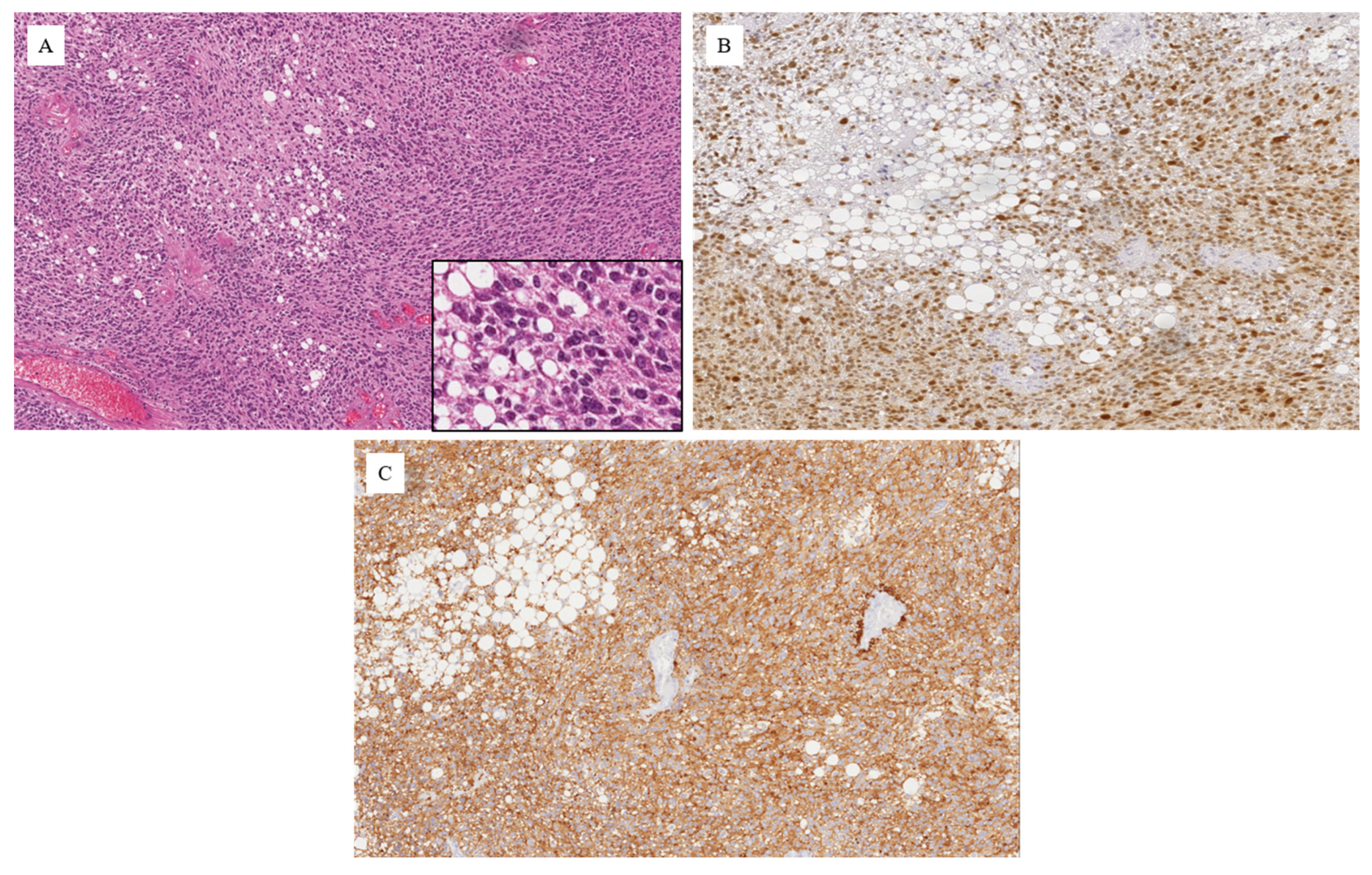
2. Oligodendroglioma-like Morphology: A Pattern, Not a Diagnosis
3. Diagnostic Spectrum
3.1. Adult-Type Diffuse Gliomas
3.2. Ependymal Tumors
3.3. Pediatric-Type Low-Grade and Glioneuronal Tumors
3.4. Neurocytic Tumors
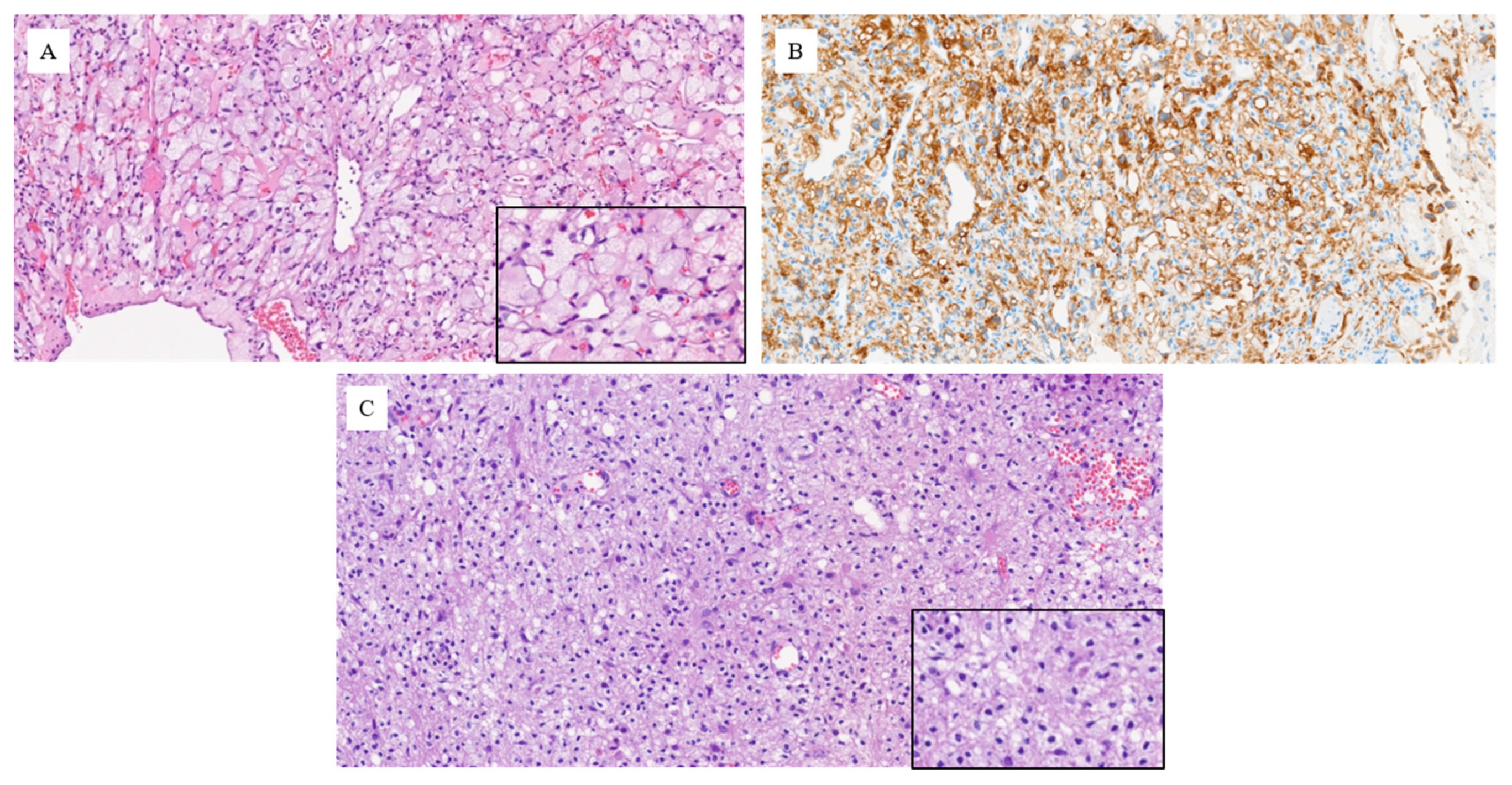
3.5. Other Neoplastic Mimics with Clear-Cell Morphology
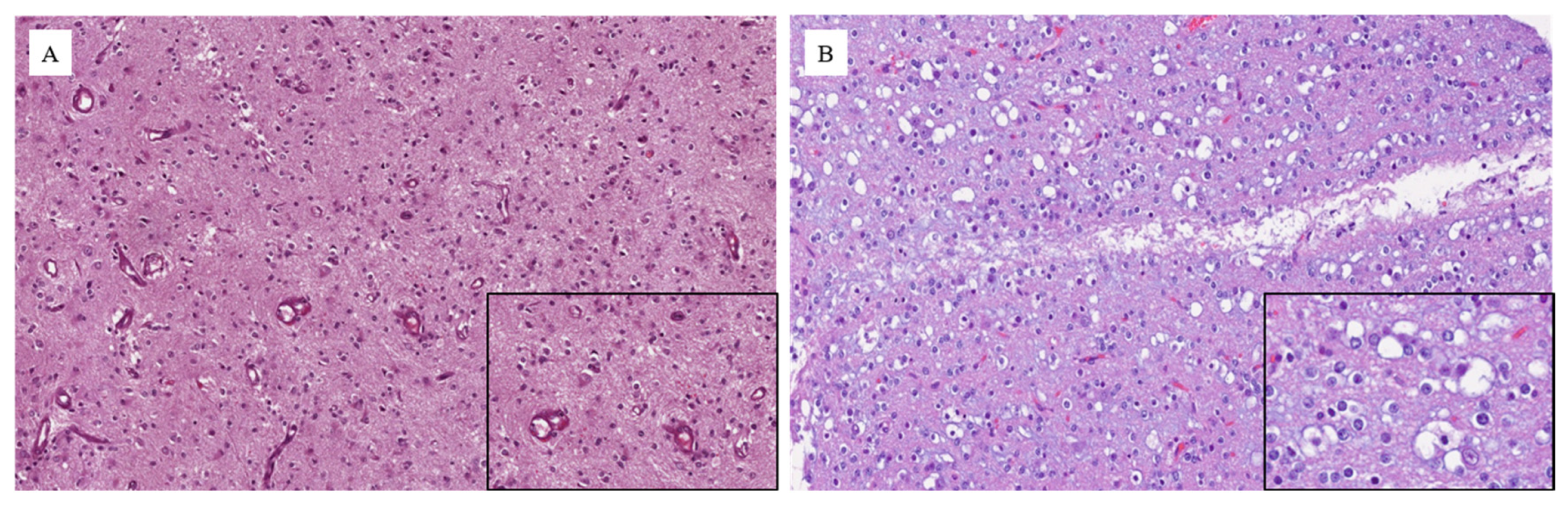
3.6. Non-Neoplastic Mimics
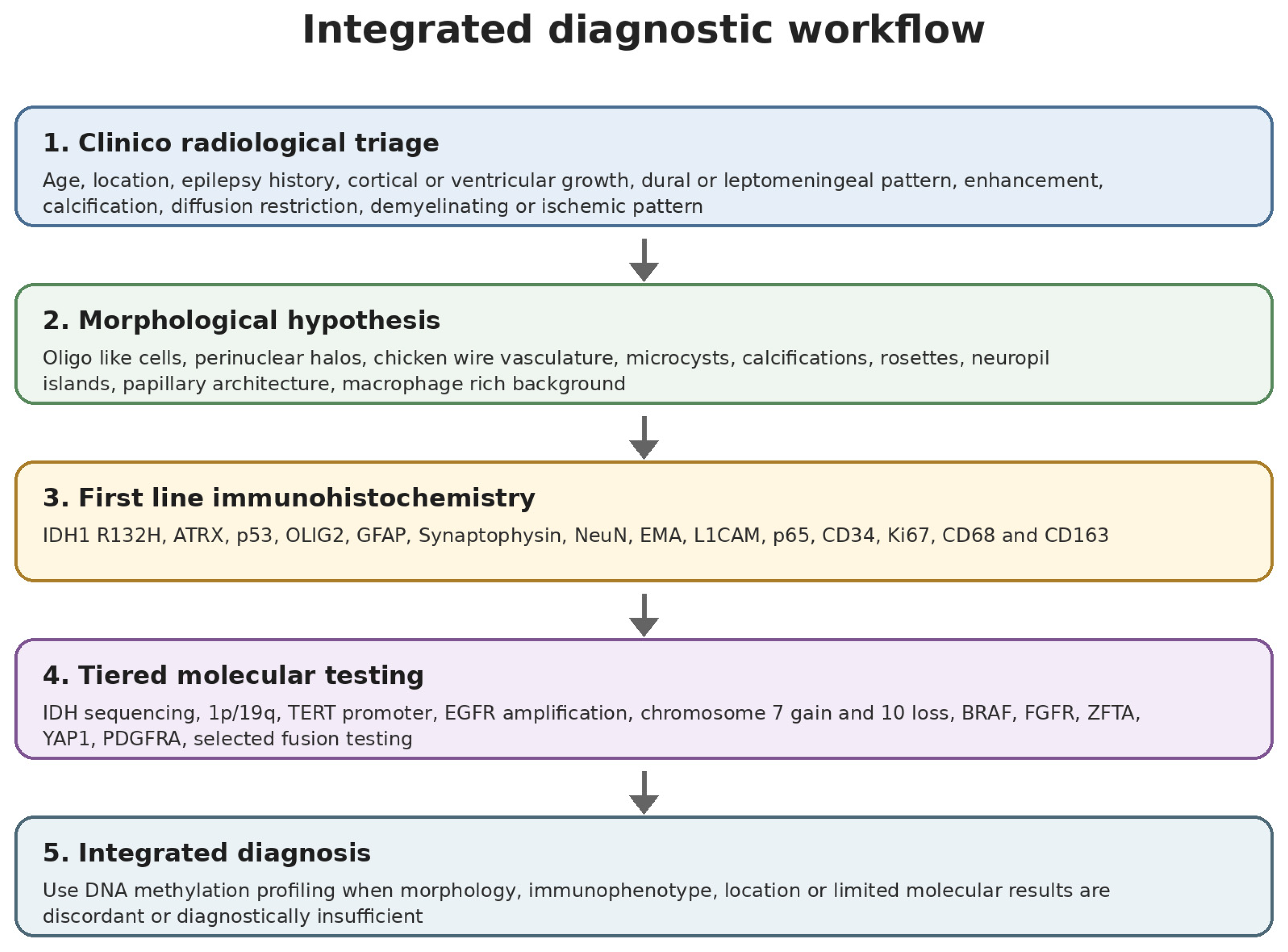
4. Practical Integrated Diagnostic Workflow
4.1. Clinico-Radiological Triage
4.2. First-Line Immunohistochemistry
4.3. Tiered Molecular Testing
4.4. When DNA Methylation Profiling Becomes Essential
5. Discussion
5.1. Why a True Oligodendroglioma May Be Misdiagnosed
5.2. Minimal Diagnostic Panel in Routine Practice
- -
- Pediatric age or very young adult age without the expected adult diffuse glioma context.
- -
- Intraventricular, dural-based, leptomeningeal, sharply circumscribed cortical, or predominantly periventricular location.
- -
- Absence of IDH mutation or absence of whole-arm 1p/19q codeletion.
- -
- ATRX loss or strong diffuse p53 expression, suggesting IDH-mutant astrocytoma.
- -
- EMA dot-like or ring-like staining, L1CAM positivity, or nuclear p65 expression, suggesting ependymal differentiation.
- -
- Diffuse Synaptophysin, NeuN, or MAP2 expression with neuropil islands, suggesting neurocytic or glioneuronal lineage.
- -
- Diffuse CD34 staining in a low-grade epilepsy-associated cortical tumor.
- -
- Prominent CD68- or CD163-positive macrophage population, especially with demyelinating or ischemic radiological features.
- -
- Cytokeratin, PAX8, CAIX, SSTR2A, or CD45 positivity, suggesting metastatic, meningeal, or hematolymphoid disease.
- -
- Any major discordance between morphology, imaging, immunophenotype, and initial molecular results.
5.3. Molecular Diagnostics and Methylation Profiling
5.4. Practical Implications
6. Conclusions
Author Contributions
Funding
Institutional Review Board Statement
Informed Consent Statement
Data Availability Statement
Conflicts of Interest
References
- Louis, D.N.; Perry, A.; Wesseling, P.; Brat, D.J.; Cree, I.A.; Figarella-Branger, D.; Hawkins, C.; Ng, H.K.; Pfister, S.M.; Reifenberger, G.; et al. The 2021 WHO Classification of Tumors of the Central Nervous System: A summary. Neuro-Oncology 2021, 23, 1231–1251. [Google Scholar] [CrossRef] [PubMed]
- Capper, D.; Jones, D.T.W.; Sill, M.; Hovestadt, V.; Schrimpf, D.; Sturm, D.; Koelsche, C.; Sahm, F.; Chavez, L.; Reuss, D.E.; et al. DNA methylation based classification of central nervous system tumours. Nature 2018, 555, 469–474. [Google Scholar] [CrossRef] [PubMed]
- Lebrun, L.; Gilis, N.; Dausort, M.; Gillard, C.; Rusu, S.; Slimani, K.; De Witte, O.; Escande, F.; Lefranc, F.; D’hAene, N.; et al. Diagnostic impact of DNA methylation classification in adult and pediatric CNS tumors. Sci. Rep. 2025, 15, 2857. [Google Scholar] [CrossRef] [PubMed]
- Killela, P.J.; Pirozzi, C.J.; Healy, P.; Reitman, Z.J.; Lipp, E.; Rasheed, B.A.; Yang, R.; Diplas, B.H.; Wang, Z.; Greer, P.K.; et al. Mutations in IDH1, IDH2, and in the TERT promoter define clinically distinct subgroups of adult malignant gliomas. Oncotarget 2014, 5, 1515–1525. [Google Scholar] [CrossRef] [PubMed]
- Bielle, F.; Di Stefano, A.L.; Meyronet, D.; Picca, A.; Villa, C.; Bernier, M.; Schmitt, Y.; Giry, M.; Rousseau, A.; Figarella-Branger, D.; et al. Diffuse gliomas with FGFR3-TACC3 fusion have characteristic histopathological and molecular features. Brain Pathol. 2018, 28, 674–683. [Google Scholar] [CrossRef] [PubMed]
- Yadav, A.K.; Madan, R.; Chatterjee, D.; Dhiman, S.; Goyal, S.; Kumar, N.; Sahoo, S.K. Small cell glioblastoma multiforme: A case series and clinicopathological update. CNS Oncol. 2020, 9, CNS63. [Google Scholar] [CrossRef] [PubMed]
- Appin, C.L.; Gao, J.; Chisolm, C.; Torian, M.; Alexis, D.; Vincentelli, C.; Schniederjan, M.J.; Hadjipanayis, C.; Olson, J.J.; Hunter, S.; et al. Glioblastoma with oligodendroglioma component: Molecular genetic and clinical characteristics. Brain Pathol. 2013, 23, 454–461. [Google Scholar] [CrossRef] [PubMed]
- Das, S.; Ahlawat, S.; Sarangi, J.; Gupta, R.K.; Jain, P.; Kate, U.; Gogi, R.; Patir, R. Oligoastrocytoma: The vanishing entity with true dual genotype, a report, its molecular profiles and review of literature. Int. J. Surg. Pathol. 2025, 33, 1216–1223. [Google Scholar] [CrossRef] [PubMed]
- Palejwala, A.H.; O’Neal, C.M.; Quinton, M.R.; Battiste, J.D.; Peterson, J.E.G.; Dunn, I.F. Polymorphous low grade neuroepithelial tumor of the young, rare tumor and review of the literature. Rare Tumors 2022, 14, 20363613221083360. [Google Scholar] [CrossRef] [PubMed]
- Qaddoumi, I.; Orisme, W.; Wen, J.; Santiago, T.; Gupta, K.; Dalton, J.D.; Tang, B.; Haupfear, K.; Punchihewa, C.; Easton, J.; et al. Genetic alterations in uncommon low grade neuroepithelial tumors: BRAF, FGFR1, and MYB mutations occur at high frequency and align with morphology. Acta Neuropathol. 2016, 131, 833–845. [Google Scholar] [CrossRef] [PubMed]
- Ryall, S.; Zapotocky, M.; Fukuoka, K.; Nobre, L.; Guerreiro Stucklin, A.; Bennett, J.; Siddaway, R.; Li, C.; Pajovic, S.; Arnoldo, A.; et al. Integrated molecular and clinical analysis of 1,000 pediatric low grade gliomas. Cancer Cell 2020, 37, 569–583.e5. [Google Scholar] [CrossRef] [PubMed]
- Daoud, E.V.; Patel, A.; Gagan, J.; Raisanen, J.M.; Snipes, G.J.; Mantilla, E.; Krothapally, R.; Hatanpaa, K.J.; Pan, E. Spinal cord pilocytic astrocytoma with FGFR1-TACC1 fusion and anaplastic transformation. J. Neuropathol. Exp. Neurol. 2021, 80, 283–285. [Google Scholar] [CrossRef] [PubMed]
- Gupta, S.; Rangari, K.V.; Mehrotra, A.; Pal, L.; Jaisawal, A.K.; Kumar, R. Temporal lobe angiocentric glioma with oligodendroglioma-like areas: A rare association of an uncommon tumor, a case report with review of literature. Childs Nerv. Syst. 2020, 36, 641–646. [Google Scholar] [CrossRef] [PubMed]
- Min, K.W.; Scheithauer, B.W. Clear cell ependymoma, a mimic of oligodendroglioma: Clinicopathologic and ultrastructural considerations. Am. J. Surg. Pathol. 1997, 21, 820–826. [Google Scholar] [CrossRef] [PubMed]
- Liang, M.L.; Hsieh, T.H.; Liu, Y.R.; Chen, Y.W.; Lee, Y.Y.; Chang, F.C.; Lin, S.C.; Huang, M.-C.; Ho, D.M.-T.; Wong, T.-T.; et al. Significance of cyclin D1 overexpression in progression and radioresistance of pediatric ependymomas. Oncotarget 2018, 9, 2527–2542. [Google Scholar] [PubMed]
- Gupta, K.; Tripathi, M.; Gupta, N.; Ranganath, S.; Salunke, P. A case of clear cell ependymoma with bizarre aggressiveness: Are we underestimating its ferocity? Neurol. India 2017, 65, 1134–1136. [Google Scholar] [CrossRef] [PubMed]
- Xie, M.; Wang, X.; Duan, Z.; Luan, G. Low grade epilepsy associated neuroepithelial tumors: Tumor spectrum and diagnosis based on genetic alterations. Front. Neurosci. 2023, 16, 1071314. [Google Scholar] [CrossRef] [PubMed]
- Rahim, S.; Ud Din, N.; Abdul Ghafar, J.; Chundriger, Q.; Khan, P.; Ahmad, Z. Clinicopathological features of dysembryoplastic neuroepithelial tumor: A case series. J. Med. Case Rep. 2023, 17, 327. [Google Scholar] [CrossRef] [PubMed]
- Rivera, B.; Gayden, T.; Carrot-Zhang, J.; Nadaf, J.; Boshari, T.; Faury, D.; Zeinieh, M.; Blanc, R.; Burk, D.L.; Fahiminiya, S.; et al. Germline and somatic FGFR1 abnormalities in dysembryoplastic neuroepithelial tumors. Acta Neuropathol. 2016, 131, 847–863. [Google Scholar] [CrossRef] [PubMed]
- Deng, M.Y.; Sill, M.; Sturm, D.; Stichel, D.; Witt, H.; Ecker, J.; Wittmann, A.; Schittenhelm, J.; Ebinger, M.; Schuhmann, M.U.; et al. Diffuse glioneuronal tumour with oligodendroglioma-like features and nuclear clusters (DGONC)—A molecularly defined glioneuronal CNS tumour class displaying recurrent monosomy 14. Neuropathol. Appl. Neurobiol. 2020, 46, 422–430. [Google Scholar] [CrossRef] [PubMed]
- Maurice, W.; Ashkun, N.; Krishnan, I.; Alan, C.; Rosalind, J. A unique case of extracerebral diffuse glioneuronal tumor with oligodendroglioma-like features and nuclear clusters. Radiol. Case Rep. 2025, 20, 3698–3702. [Google Scholar] [CrossRef] [PubMed]
- Torres, F.M.; Denapoli, T.; Chen, P.S.; Mohila, C.; Fisher, K.E.; Aldape, K.; Lee, M.R.; Griffin, T.C. Diffuse glioneuronal tumor with oligodendroglioma-like features and nuclear clusters relapse in a seven year old boy: An unusual case exhibiting near tetraploidy and chromosome 14 diploidy. Cureus 2024, 16, e73157. [Google Scholar] [CrossRef] [PubMed]
- Policicchio, D.; Boccaletti, R.; Cuccu, A.S.; Casu, G.; Di Pellegrini, G.; Doda, A.; Muggianu, G.; Santonio, F.V. Atypical and aggressive diffuse leptomeningeal glioneuronal tumor in a young adult: A case report and review of the literature. Surg. Neurol. Int. 2022, 13, 214. [Google Scholar] [CrossRef] [PubMed]
- Chiang, J.; Moreira, D.C.; Li, X.; Furtado, L.V. Prognostic significance of chromosome arm 1q gain and methylation class in molecularly defined diffuse leptomeningeal glioneuronal tumor. Acta Neuropathol. 2022, 144, 1185–1187. [Google Scholar] [CrossRef] [PubMed]
- Caporalini, C.; Scagnet, M.; Giunti, L.; Cetica, V.; Mei, D.; Conti, V.; Moscardi, S.; Macconi, L.; Giordano, F.; D’Incerti, L.; et al. Myxoid glioneuronal tumor: Histo-pathologic, neuroradiologic, and molecular features in a single center series. Neoplasia 2023, 37, 100885. [Google Scholar] [CrossRef] [PubMed]
- Marastoni, E.; Ammendola, S.; Rossi, S.; Giovannoni, I.; Broggi, G.; Masotto, B.; Feletti, A.; Barresi, V. H3K27M mutation in rosette-forming glioneuronal tumors: A potential diagnostic pitfall. Virchows Arch. 2025, 486, 781–789. [Google Scholar] [CrossRef] [PubMed]
- Hirata, M.; Togao, O.; Yamashita, K.; Kikuchi, K.; Kusunoki, M.; Narutomi, F.; Kuga, D.; Fujioka, Y.; Yoshimoto, K.; Ishigami, K. Papillary glioneuronal tumor located in the subcortical white matter with a purely solid pattern: A case report. Cureus 2025, 17, e82340. [Google Scholar] [CrossRef] [PubMed]
- Thom, M.; Liu, J.; Bongaarts, A.; Reinten, R.J.; Paradiso, B.; Jager, H.R.; Reeves, C.; Somani, A.; An, S.; Marsdon, D.; et al. Multinodular and vacuolating neuronal tumors in epilepsy: Dysplasia or neoplasia? Brain Pathol. 2018, 28, 155–171. [Google Scholar] [CrossRef] [PubMed]
- Sachani, P.; Dhande, R.; Parihar, P.; Saifi, I.; Kasat, P.R. Central neurocytoma: A case report with literature review. Cureus 2024, 16, e60969. [Google Scholar] [CrossRef] [PubMed]
- Sato, D.; Takami, H.; Takayanagi, S.; Ikemura, M.; Matsuura, R.; Tanaka, S.; Saito, N. Intraventricular central neurocytoma molecularly defined as extraventricular neurocytoma: A case representing the discrepancy between clinicopathological and molecular classifications. Brain Tumor Pathol. 2023, 40, 230–234. [Google Scholar] [CrossRef] [PubMed]
- Uro-Coste, E.; Tauziede-Espariat, A.; Dubucs, C.; Chiforeanu, D.C.; Siegfried, A.; Nicaise, Y.; Bauchet, L.; Riffaud, L.; Bielle, F.; Vasiljevic, A.; et al. Two novel tumors with NTRK2 fusion in the methylation class of extraventricular neurocytomas, including one intraventricular. Brain Pathol. 2024, 34, e13223. [Google Scholar] [CrossRef] [PubMed]
- Broggi, G.; Salzano, S.; Mazzucchelli, M.; Rosano, G.N.; Magro, G.; Caltabiano, R.; Barresi, V. Cerebellar liponeurocytoma: An updated comprehensive review of clinicopathologic, immunohistochemical, and molecular features of an unusual but distinct tumor. Clin. Neuropathol. 2025, 44, 26–37. [Google Scholar] [CrossRef] [PubMed]
- Vasiljevic, A. Pineal parenchymal tumors of intermediate differentiation: In need of a stringent definition to avoid confusion. Scientific commentary on ‘Genetical and epigenetical profiling identifies two subgroups of pineal parenchymal tumors of intermediate differentiation (PPTID) with distinct molecular, histological and clinical characteristics’. Acta Neuropathol. 2024, 147, 34. [Google Scholar] [CrossRef] [PubMed]
- Tauziede-Espariat, A.; Uro-Coste, E.; Sievers, P.; Nicaise, Y.; Mariet, C.; Siegfried, A.; Pierron, G.; Guillemot, D.; Benzakoun, J.; Pallud, J.; et al. CNS tumor with EP300::BCOR fusion: Discussing its prevalence in adult population. Acta Neuropathol. Commun. 2023, 11, 26. [Google Scholar] [CrossRef] [PubMed]
- Tathe, S.P.; Jaiswal, K.N. Clear cell meningioma: An uncommon aggressive variant of meningioma. Indian J. Pathol. Microbiol. 2025, 68, 328–332. [Google Scholar] [CrossRef] [PubMed]
- Khan, M.M.; Hall, W.A.; Belkhair, S. Hemangioblastoma. In StatPearls; StatPearls Publishing: Treasure Island, FL, USA, 2025. [Google Scholar]
- Venegas, J.M.; Nawabi, N.L.A.; Miller, A.; Baker, T.; Lindhorst, S.; Zukas, A.; Rivers, C.; Vandergrift, W.; Strickland, B.A. Leptomeningeal hemangioblastoma: Illustrative case. J. Neurosurg. Case Lessons 2025, 9, CASE25204. [Google Scholar] [CrossRef] [PubMed]
- Zheng, W.; Goodman, A.L.; Velazquez-Vega, J.E.; Yin, F.; Fung, K.M.; Osunkoya, A.O. Metastatic renal cell carcinoma to the brain: A contemporary clinicopathologic analysis with comparison of immunohistochemical profiles to selected primary brain tumors with clear cell features. Appl. Immunohistochem. Mol. Morphol. 2020, 28, 395–402. [Google Scholar] [CrossRef] [PubMed]
- Hardy, T.A.; Chataway, J. Tumefactive demyelination: An approach to diagnosis and management. J. Neurol. Neurosurg. Psychiatry 2013, 84, 1047–1053. [Google Scholar] [CrossRef] [PubMed]
- Höftberger, R.; Lassmann, H. Inflammatory demyelinating diseases of the central nervous system. In Handbook of Clinical Neurology; Elsevier: Amsterdam, The Netherlands, 2017; Volume 145, pp. 263–283. [Google Scholar] [CrossRef] [PubMed]
- Stoll, G.; Jander, S. The role of microglia and macrophages in the pathophysiology of the CNS. Prog. Neurobiol. 1999, 58, 233–247. [Google Scholar] [CrossRef] [PubMed]
- Aldape, K.; Capper, D.; von Deimling, A.; Giannini, C.; Gilbert, M.R.; Hawkins, C.; Hench, J.; Jacques, T.S.; Jones, D.; Louis, D.N.; et al. cIMPACT-NOW update 9: Recommendations on utilization of genome-wide DNA methylation profiling for central nervous system tumor diagnostics. Neurooncol. Adv. 2025, 7, vdae228. [Google Scholar] [CrossRef] [PubMed]
- Santosh, V.; Sarkar, C.; Rao, S.; Ng, H.K.; Shibahara, J.; Al-Hussaini, M.; Buckland, M.E.; Park, S.; Tihan, T.; Wesseling, P.; et al. AOSNP-ADAPTR resource level-based recommendations on practical diagnostic strategies for WHO CNS5 adult-type diffuse gliomas. Brain Pathol. 2026, 36, e70046. [Google Scholar] [CrossRef] [PubMed]




| Entity or Group | Main Morphological Clues | Useful Immunohistochemistry | Key Molecular or Diagnostic Features | Practical Pitfall |
|---|---|---|---|---|
| Oligodendroglioma, IDH-mutant and 1p/19q codeleted | Uniform, round nuclei; perinuclear halos; chicken-wire vessels; microcysts; and calcifications. | IDH1 R132H-positive in most cases, ATRX retained, p53 not strong diffuse, and OLIG2-positive. | IDH1/2 mutation and whole-arm 1p/19q codeletion. Frequent TERT-promoter mutation. | Do not diagnose from morphology alone. 1p/19q codeletion is mandatory. |
| Astrocytoma, IDH-mutant | Focal oligodendroglioma-like areas may occur. More irregular nuclei and fibrillary or gemistocytic background may be present. | IDH1 R132H-positive, ATRX loss, strong diffuse p53, and OLIG2-positive. | IDH mutation without 1p/19q codeletion. CDKN2A/B homozygous deletion defines grade 4. | May be mistaken for oligodendroglioma if oligodendrocyte-like cells are diffusely present. |
| Glioblastoma, IDH-wildtype | Small-cell or oligodendroglioma-like morphology, compact growth, and deceptively bland cytology in limited biopsies. | IDH1 R132H-negative, variable p53, high Ki67, and often OLIG2-positive. | TERT-promoter mutation, EGFR amplification, chromosome 7 gain and 10 loss, and possible FGFR3::TACC3 fusion (if present, mutually exclusive with EGFR amplification). | May look lower grade if necrosis or microvascular proliferation is absent in the sample. |
| Ependymoma | Uniform round cells, compact architecture, and subtle or absent pseudorosettes. | EMA dot or ring pattern, L1CAM-positive and nuclear p65 in ZFTA-fused tumors, and usually OLIG2-negative. | ZFTA fusion or YAP1 fusion in supratentorial ependymomas (clear-cell morphology primarily associated with ZFTA-fused tumors, while unusual in YAP1-fused subgroup). | Can closely mimic cortical oligodendroglioma. |
| DNT and other epilepsy-associated glioneuronal tumors | Oligodendrocyte-like cells, mucin-rich matrix, floating neurons, and multinodular cortical architecture. | Synaptophysin highlights neuronal component, OLIG2 variable, and CD34 in selected entities. | Frequent FGFR1 or MAPK pathway alterations; IDH-wildtype. | Clinical epilepsy history and cortical location are often decisive. |
| DGONC | Oligodendroglioma-like areas with nuclear clusters and glioneuronal differentiation. | OLIG2 and neuronal marker co-expression may be present. | Methylation-defined entity; recurrent monosomy 14. | Often requires methylation profiling for confident classification. |
| DLGNT | Oligodendrocyte-like cells with leptomeningeal dissemination. | Glial and neuronal marker expression. | KIAA1549::BRAF fusion, 1p loss, and sometimes 1q gain. | Leptomeningeal pattern should redirect the differential diagnosis. |
| Central or extraventricular neurocytoma | Uniform neurocytic cells, perinuclear halos, and neuropil islands. | Diffuse Synaptophysin and NeuN, MAP2-positive, and GFAP limited to reactive astrocytes. | No single recurrent driver. Methylation profiling may refine classification. | Intraventricular location and neuronal markers argue against oligodendroglioma. |
| Clear-cell meningioma | Glycogen-rich clear cells arranged in sheets, deposition of interstitial and perivascular thick amianthoid-like collagen, and possible dural relationship. | EMA- and SSTR2A-positive, and glial- and neuronal-markers-negative. | SMARCE1 loss in a subset. | May lack obvious dural attachment. |
| Metastatic renal cell carcinoma | Clear cells, vascular network, and epithelial architecture may be subtle. | Cytokeratin-, PAX8- and CAIX-positive. | Metastatic carcinoma diagnosis supported by systemic workup. | Can mimic hemangioblastoma or primary clear-cell CNS tumors. |
| Hemangioblastoma | Clear stromal cells and prominent vascular network. | Inhibin alpha and S100 in stromal cells, and vascular markers in capillary network. | Consider von Hippel–Lindau context. | May resemble metastatic renal cell carcinoma. |
| Tumefactive demyelination or subacute infarct | Macrophage-rich lesion, reactive gliosis, vacuolated cells, or oligodendroglial hyperplasia. | CD68- and CD163-positive macrophages; myelin stains helpful. | Non-neoplastic diagnosis supported by imaging and clinical evolution. | Major source of overdiagnosis as low-grade glioma. |
| Diagnostic Layer | Main Question | Suggested Tools | How the Result Should Be Used |
|---|---|---|---|
| Clinical and radiological triage | Does age, site, or imaging fit conventional oligodendroglioma? | Age, seizure history, lesion site, enhancement pattern, diffusion restriction, calcification, and leptomeningeal or dural pattern. | Defines the initial differential diagnosis and identifies red flags. |
| Histology | Is the oligodendroglioma-like pattern pure or associated with another architecture? | Search for chicken-wire vessels, microcysts, calcifications, rosettes, neuropil islands, papillae, macrophage-rich areas, necrosis, and microvascular proliferation. | Generates the morphological hypothesis and selects the immunohistochemical panel. |
| First-line diffuse glioma panel | Is this an adult-type diffuse glioma? | IDH1 R132H, ATRX, p53, OLIG2, GFAP, and Ki67. | Separates likely oligodendroglioma, astrocytoma, and IDH-wildtype glioma patterns. |
| Lineage expansion panel | Is there ependymal, neuronal, meningeal, metastatic, or lymphoid differentiation? | EMA, L1CAM, p65, Synaptophysin, NeuN, MAP2, CD34, SSTR2A, cytokeratins, PAX8, CAIX, CD45, CD20, CD68, and CD163. | Redirects diagnosis when the oligodendroglioma-like pattern is a mimic. |
| Molecular confirmation | Which molecular alteration defines the entity? | IDH sequencing; 1p/19q testing; TERT promoter; EGFR amplification; chromosome 7 and 10 copy number; and BRAF, FGFR, PDGFRA, ZFTA, and YAP1 fusion testing, if indicated. | Confirms integrated diagnosis and prevents morphology driven misclassification. |
| DNA methylation profiling | Do conventional layers remain discordant or insufficient? | Genome-wide methylation array with copy number profile and classifier score. | Useful for unusual age, atypical site, ambiguous glioneuronal or neurocytic tumors, small biopsies, and suspected methylation-defined entities. |
| Final integrated report | Can the diagnosis be stated with biological coherence? | Synthesis of histology, IHC, molecular data, radiology, and clinical setting. | Reports final entity, grade when applicable, pending tests, and diagnostic limitations. |
Disclaimer/Publisher’s Note: The statements, opinions and data contained in all publications are solely those of the individual author(s) and contributor(s) and not of MDPI and/or the editor(s). MDPI and/or the editor(s) disclaim responsibility for any injury to people or property resulting from any ideas, methods, instructions or products referred to in the content. |
© 2026 by the authors. Licensee MDPI, Basel, Switzerland. This article is an open access article distributed under the terms and conditions of the Creative Commons Attribution (CC BY) license.
Share and Cite
Attanasio, G.; Caltabiano, R.; Amato, F.; Barbagallo, G.M.V.; Certo, F.; Ayan, D.; Barresi, V.; Broggi, G. Beyond Oligodendroglioma: An Integrated Diagnostic Approach to CNS Tumors with Oligodendroglioma-like Morphology, with a Focus on Morphological Pitfalls, Immunoprofiles, and Molecular Signatures. Int. J. Mol. Sci. 2026, 27, 7163. https://doi.org/10.3390/ijms27167163
Attanasio G, Caltabiano R, Amato F, Barbagallo GMV, Certo F, Ayan D, Barresi V, Broggi G. Beyond Oligodendroglioma: An Integrated Diagnostic Approach to CNS Tumors with Oligodendroglioma-like Morphology, with a Focus on Morphological Pitfalls, Immunoprofiles, and Molecular Signatures. International Journal of Molecular Sciences. 2026; 27(16):7163. https://doi.org/10.3390/ijms27167163
Chicago/Turabian StyleAttanasio, Giulio, Rosario Caltabiano, Francesca Amato, Giuseppe Maria Vincenzo Barbagallo, Francesco Certo, Durmus Ayan, Valeria Barresi, and Giuseppe Broggi. 2026. "Beyond Oligodendroglioma: An Integrated Diagnostic Approach to CNS Tumors with Oligodendroglioma-like Morphology, with a Focus on Morphological Pitfalls, Immunoprofiles, and Molecular Signatures" International Journal of Molecular Sciences 27, no. 16: 7163. https://doi.org/10.3390/ijms27167163
APA StyleAttanasio, G., Caltabiano, R., Amato, F., Barbagallo, G. M. V., Certo, F., Ayan, D., Barresi, V., & Broggi, G. (2026). Beyond Oligodendroglioma: An Integrated Diagnostic Approach to CNS Tumors with Oligodendroglioma-like Morphology, with a Focus on Morphological Pitfalls, Immunoprofiles, and Molecular Signatures. International Journal of Molecular Sciences, 27(16), 7163. https://doi.org/10.3390/ijms27167163