Review

Molecular Mechanisms Underlying Antimicrobial Resistance in Mycobacteria

by
Paula López-Roa
1,
Jaime Esteban
2,3 and
María-Carmen Muñoz-Egea
2,3,*
1
Department of Clinical Microbiology, Hospital Universitario 12 de Octubre, 28041 Madrid, Spain
2
Department of Clinical Microbiology, IIS-Fundación Jiménez Díaz, Universidad Autónoma de Madrid (UAM), 28040 Madrid, Spain
3
CIBERINFEC-CIBER de Enfermedades Infecciosas, 28029 Madrid, Spain
*
Author to whom correspondence should be addressed.
Int. J. Mol. Sci. 2026, 27(15), 6893; https://doi.org/10.3390/ijms27156893 (registering DOI)
Submission received: 18 June 2026 / Revised: 28 July 2026 / Accepted: 29 July 2026 / Published: 1 August 2026

Abstract

Antimicrobial resistance in mycobacteria arises from a complex interplay of intrinsic and acquired mechanisms that collectively limit the efficacy of current therapeutic options. Intrinsic resistance is largely driven by the low permeability of the mycobacterial cell envelope, the activity of efflux pumps, and the presence of drug-modifying enzymes, which together restrict intracellular drug accumulation and contribute to broad baseline tolerance. This review integrates resistance mechanisms of both M. tuberculosis and M. abscessus, two clinically relevant mycobacteria that share core molecular pathways while exhibiting species-specific determinants that complicate treatment. Additional intrinsic factors, including biofilm formation and stress-induced adaptive responses, further enhance persistence and reduce susceptibility to multiple drug classes. Acquired resistance predominantly results from chromosomal mutations affecting drug targets or prodrug activation pathways, such as katG, inhA, rpoB, gyrA, and pncA in Mycobacterium tuberculosis, leading to high rates of multidrug-resistant and extensively drug-resistant disease. In nontuberculous mycobacteria, species-specific determinants—including inducible macrolide resistance mediated by erm(41) in M. abscessus, plasmid-mediated erm (55) variants, rrl and rrs mutations, diverse enzymatic inactivation systems, and regulatory alterations in the MarR family that result in inducible resistance to drugs such as ethionamide —generate highly variable resistance profiles that complicate treatment. Recent advances in molecular diagnostics, including PCR-based assays, whole-genome sequencing, CRISPR-based diagnostic platforms, AI-assisted diagnostics, and emerging multi-omics approaches, have improved the detection of resistance-associated mutations and enhanced understanding of mycobacterial pathophysiology. In parallel, new therapeutic agents and optimized regimens offer promising avenues to overcome resistance, although emerging resistance to novel drugs underscores the need for continued surveillance. This review synthesizes current knowledge on the molecular basis of resistance in M. tuberculosis and NTM, highlighting implications for diagnosis, treatment, and future research.

1. Background and Scope of the Review

Mycobacterium tuberculosis remains one of the most significant human pathogens and is responsible for tuberculosis (TB), which continues to be the leading cause of death from a single infectious agent worldwide, with a substantial burden of both latent and active disease [1]. Latent TB infection constitutes a long-term reservoir for future disease, with progression to active TB determined by host immune status and time since initial infection [1]. The clinical importance of M. tuberculosis is further enhanced by its capacity to evade host immunity, persist in a dormant state, and cause a wide spectrum of disease [2].
In parallel, nontuberculous mycobacteria (NTM) have emerged as important opportunistic pathogens. These environmental organisms are increasingly recognized as causes of pulmonary and extrapulmonary infections, particularly in patients with preexisting lung conditions or compromised immunity. The most common etiologic agents of NTM pulmonary disease include Mycobacterium avium complex, M. kansasii, and M. abscessus, and their incidence is rising globally [3,4]. In addition, a history of pulmonary TB may predispose individuals to NTM infection due to residual structural damage, while NTM does not increase the risk of TB but does complicate the differential diagnosis in high-incidence areas. The prevalence of NTM lung disease is increasing globally, with rates ranging from 1 to 6 cases per 100,000 people and annual increases of 8–10% in several countries [3,4].
Antimicrobial resistance represents a major global health threat in mycobacterial infections. In 2022, the WHO updated the classification and previous definitions of TB drug resistance [5]. These changes better reflect current treatment practices. According to the latest definitions, Multidrug-resistant TB (MDR-TB) is resistant to at least isoniazid and rifampicin, pre-extensively drug-resistant TB (pre-XDR-TB) refers to multidrug-resistant or rifampicin-resistant TB (MDR/RR-TB) with additional resistance to any fluoroquinolone, and extensively drug-resistant TB (XDR-TB) has also been redefined and now refers to MDR/RR-TB with resistance to any fluoroquinolone plus at least one additional Group A drug (such as bedaquiline or linezolid). XDR-TB is associated with higher mortality, prolonged and poorly tolerated treatment regimens, and lower cure rates (MDR-TB ~50%, XDR-TB ~30%) [5,6,7]. At the same time, NTMs also exhibit distinct patterns of acquired resistance. In MAC, resistance to macrolides is primarily associated with mutations in rrl and the presence of erm genes, whereas in M. abscessus, erm(41)-mediated inducible resistance coexists with acquired resistance due to mutations in rrl and rrs, resulting in strains with clinically relevant resistance to macrolides and aminoglycosides [3,4]. Inadequate or incomplete treatment regimens, poor adherence, and delayed diagnosis of drug-resistant TB rapidly select M. tuberculosis strains with genetically acquired resistance [8]. Although newer agents such as bedaquiline have improved outcomes, emerging resistance to these drugs has already been reported [9,10]. In NTM infections, the development of drug resistance is also facilitated by prolonged treatment courses, suboptimal drug penetration in structurally damaged lungs, intrinsic species-specific tolerance, and delayed diagnosis, which often leads to extended exposure to ineffective regimens. These factors collectively promote selection of resistant subpopulations and mirror the clinical drivers of resistance observed in M. tuberculosis [3,4].
Similarly, NTM exhibit both intrinsic and acquired resistance mechanisms, particularly among rapidly growing species such as M. abscessus, which pose significant therapeutic challenges and often require individualized, susceptibility-guided treatment regimens due to their broad spectrum of resistance determinants and variable drug response profiles [4]. Overall, the rise of MDR/XDR-TB and resistant NTM underscores the urgent need for rapid diagnostics, optimized treatment regimens, and continuous surveillance to improve patient outcomes.
This review provides a comprehensive overview of the most important molecular mechanisms that underlie antimicrobial resistance in M. tuberculosis and NTM. It first addresses the biological bases of intrinsic resistance, encompassing the cell envelope’s low permeability; biofilm-associated tolerance; the role of drug-modifying enzymes such as β-lactamases, acetyltransferases, and methyltransferases; and stress adaptation responses. It further explores the molecular mechanisms underlying acquired resistance, highlighting critical mutations in drug target genes, efflux pump–mediated drug extrusion, and enzymatic inactivation, with emphasis on species-specific determinants such as erm(41) in M. abscessus. Finally, the review summarizes recent advances in diagnostic and therapeutic strategies, encompassing molecular techniques for resistance detection, such as PCR and sequencing-based assays, and the development of new drugs and advanced treatment regimens designed to enhance efficacy and address emerging resistance.
In addition to summarizing established resistance mechanisms, this review integrates recent conceptual advances—including multi-omics biomarkers, CRISPR-based functional genomics, AI-assisted diagnostics, host-directed therapies, and innovative nanocarrier strategies—that have emerged in the last few years and were not covered in earlier reviews. These elements provide an updated and forward-looking perspective on the molecular basis of resistance in M. tuberculosis and NTM.

2. Biological Basis of Intrinsic Resistance

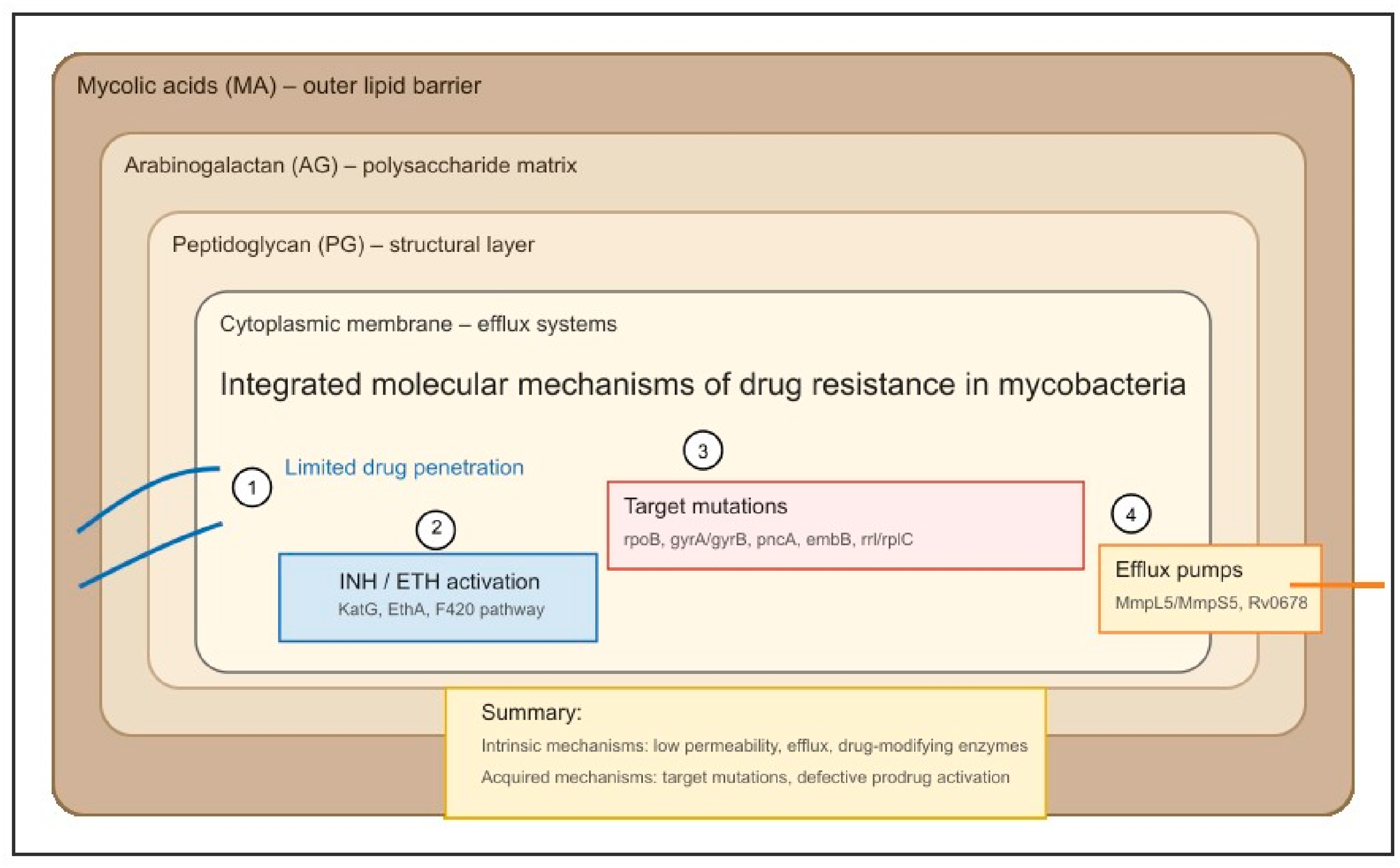
The underlying mechanisms of antimicrobial resistance in mycobacteria are multifactorial and include both intrinsic and acquired processes (Figure 1).
The principal mechanisms of intrinsic antimicrobial resistance in mycobacteria are low permeability of the cell envelope, drug-modifying and drug-target-modifying enzymes, efflux pumps and intrinsic resistance genes. These mechanisms act collectively to confer broad intrinsic resistance to many antibiotic classes, necessitating the use of specialized antimycobacterial agents for effective treatment. These intrinsic mechanisms are mediated by well-defined genetic determinants, including the inhA and kasA pathways involved in mycolic-acid biosynthesis [11,12,13,14,15], the blaC β-lactamase responsible for broad β-lactam hydrolysis [16], rRNA methyltransferases such as ermMT that confer intrinsic macrolide resistance [11], and constitutive efflux systems such as LfrA and MmpL5/MmpS5 that reduce intracellular drug accumulation [12,17,18].

2.1. Low Permeability of the Mycobacterial Cell Envelope

In reference to the low permeability of the mycobacterial cell envelope, it is notable that these organisms possess a unique, thick, lipid-rich cell envelope. The mycobacterial cell envelope follows a four-layer asymmetric architecture composed of an outer capsule, an outer lipid leaflet enriched in trehalose dimycolate and PDIMs, the mAGP core (mycolic-acids–arabinogalactan–peptidoglycan), and the plasma membrane. This multilayered envelope acts as a barrier to the entry of numerous antibiotics, and its low permeability is a key determinant of intrinsic resistance that significantly limits the effectiveness of most conventional antibiotic therapies [11,12,13,14,15].
Inhibition of mycolic acid synthesis represents a major vulnerability of the mycobacterial cell envelope and is the target of several key antimycobacterial drugs. Mycolic acids are essential long-chain fatty acids forming the mycolyl–arabinogalactan–peptidoglycan complex, and their biosynthesis depends on the FAS-I and FAS-II systems, particularly the enoyl-ACP reductase InhA. Isoniazid and ethionamide inhibit this pathway after activation by KatG or EthA, forming adducts that block InhA. Resistance arises mainly through katG S315T mutations or inhA promoter overexpression. Newer nitroimidazoles (delamanid, pretomanid) also inhibit methoxy- and keto-mycolic acid synthesis via F420-dependent activation, with resistance linked to ddn, fgd1, and fbiA–D mutations [19,20,21,22]. Beyond M. tuberculosis, several envelope-associated genes are conserved across NTM species and similarly contribute to low permeability. Rapidly growing mycobacteria such as M. abscessus possess homologues of kasA, kasB, inhA, and FAS-II components involved in mycolic acid biosynthesis, as well as species-specific outer-membrane lipids (e.g., GPLs) that further reduce drug penetration [3,4]. In MAC and M. abscessus, envelope-associated efflux systems (e.g., MmpL, MmpS transporters) also participate in intrinsic tolerance, indicating that cell-envelope-mediated impermeability is a shared resistance determinant across both MTBC and NTM.

2.2. Drug-Modifying and Target-Modifying Enzymes

On the other hand, mycobacteria express a variety of enzymes that can inactivate or modify antibiotics before they reach their targets. Examples include β-lactamases, particularly BlaC in M. tuberculosis, which hydrolyze the β-lactam ring of penicillins and cephalosporins, rendering them inactive and explaining the limited efficacy of β-lactams unless combined with potent inhibitors. Aminoglycoside-modifying enzymes such as the acetyltransferase Eis and the phosphotransferase Aph catalyze acetylation or phosphorylation of drugs like kanamycin, amikacin, and streptomycin, reducing their affinity for the 30S ribosomal subunit and contributing to intrinsic or low-level resistance. Additional enzyme families—including monooxygenases, oxidoreductases, and ADP-ribosyltransferases—further expand the spectrum of antibiotic inactivation across nontuberculous species. A distinct and clinically relevant category is rRNA methyltransferases, which modify specific nucleotides within 23S or 16S rRNA and thereby prevent antibiotic binding. In the M. tuberculosis complex (MTBC), ermMT methylates the macrolide-binding site, conferring intrinsic macrolide resistance and precluding the use of clarithromycin or azithromycin for TB treatment [11,16].
In NTM, particularly rapidly growing species such as M. abscessus, M. bolletii, M. massiliense, M. chelonae, and M. fortuitum, the repertoire of drug-modifying enzymes is broader and clinically more relevant. These organisms encode multiple β-lactamases (e.g., Mabβ, CβlA) with extended hydrolytic activity, diverse aminoglycoside-modifying enzymes including ADP-ribosyltransferases, and inducible rRNA methyltransferases such as erm(41) and erm(55), which mediate inducible macrolide resistance and frequently lead to treatment failure. This expanded enzymatic arsenal constitutes a major pillar of intrinsic resistance in NTM [11,16,23,24].

2.3. Efflux Pump Systems

It is also important to note that mycobacteria encode multiple efflux pump systems that actively export a variety of antibiotics out of the cell, reducing intracellular drug concentrations and contributing to intrinsic resistance. Constitutive efflux—mediated by transporters such as LfrA or MmpL5/MmpS5—operates at baseline levels and contributes to high MICs for drugs including fluoroquinolones and aminoglycosides. Stress-inducible efflux systems (e.g., EfpA, Tap) are transiently upregulated upon antibiotic exposure, providing an adaptive response that enhances short-term survival. In addition, mutations that upregulate efflux pump expression, particularly in regulatory genes such as Rv0678, generate acquired resistance by driving persistent overexpression and increased drug extrusion [12,16,17,18].

2.4. Biofilm-Associated Intrinsic Resistance

Intrinsic resistance in mycobacterial biofilms describes the elevated drug tolerance observed when mycobacteria grow as multicellular communities encased in an extracellular matrix. Biofilm formation leads to several protective adaptations: the matrix physically impedes antibiotic diffusion, the microenvironment within biofilms fosters slow growth and metabolic dormancy, and there is an enrichment of persister cells with heightened stress tolerance [25,26,27,28].
Although biofilm formation is best characterized under experimental conditions, structured biofilm-like aggregates have been described in human TB lesions (caseous necrosis and cavitary disease) and in MAC pulmonary disease, where they correlate with increased drug tolerance and chronic infection. These observations support the clinical relevance of biofilm-associated persistence [29].
In M. tuberculosis and NTM, biofilm-associated cells are significantly less susceptible to antibiotics compared to planktonic cells, often necessitating physical disruption of the biofilm or adjunctive therapies to achieve effective treatment [26,27]. Examples include reduced inhibitory and bactericidal activity of isoniazid and ethambutol in biofilm-associated cells, whereas rifampicin and clofazimine retain higher efficacy under biofilm conditions. Biofilm-associated genetic determinants such as mmpL, pstC and sigE in M. tuberculosis, and GPL biosynthesis genes and whiB7 in NTM, further contribute to biofilm formation and drug tolerance [30,31].

3. Molecular Mechanisms of Acquired Resistance in Mycobacteria

Intrinsic resistance mechanisms have been summarized in Section 2; here we focus exclusively on acquired resistance, which arises from mutations affecting drug targets, activation pathways, and regulatory networks.
Building on earlier reviews such as Miotto et al. [32], which systematically summarized the full spectrum of resistance mutations in M. tuberculosis in 2017, this section integrates post-2020 evidence, including newly validated resistance determinants such as rv2983 (also designated fbiD), updated World Health Organization mutation catalogues, and mechanisms associated with recently introduced drugs.
Acquired resistance in mycobacteria arises primarily through chromosomal mutations, transcriptional regulation, and enzymatic mechanisms, which together reduce the effectiveness of antimicrobial agents. Major contributors to resistance encompass mutations in drug target genes (e.g., katG, rpoB, embB, gyrA) that impair drug binding or activation, inducible efflux systems (e.g., MmpL, Rv1258c) that lower intracellular antibiotic concentrations, and enzymatic mechanisms, including species-specific factors such as erm(41) in M. abscessus that inactivate or modify antimicrobials [33,34]. With a few exceptions—such as plasmid-mediated erm genes in M. abscessus and mobile genetic elements in environmental mycobacteria—mycobacterial resistance is predominantly acquired through chromosomal mutations rather than horizontal gene transfer. Microevolution during infection promotes heteroresistance and the presence of diverse resistant subpopulations, posing significant challenges to both treatment and infection control [35,36,37,38].

3.1. Acquired Resistance in Mycobacterium tuberculosis

Genetic mutations in drug target genes represent the primary mechanism of acquired resistance in M. tuberculosis, affecting both first-line and critical second-line therapies, as described in the World Health Organization Catalogue of mutations in MTBC and their association with drug resistance (2023), which systematically compiles relevant variants and classifies them according to the strength of evidence linking each mutation to drug resistance [39].

3.1.1. Isoniazid

Isoniazid resistance is predominantly mediated by mutations in katG and the inhA promoter. The S315T substitution in katG is the most prevalent chromosomal mutation worldwide, conferring high-level resistance by impairing the catalase–peroxidase activity required for prodrug activation and thereby abolishing drug efficacy [40]. Mutations in the inhA promoter, particularly C-15T, contribute to low-level isoniazid resistance by upregulating expression of the target enoyl-acyl carrier protein reductase and are associated with cross-resistance to ethionamide [41]. Co-occurrence of these mutations in the same strain can lead to enhanced resistance levels, emphasizing their critical contribution to the worldwide prevalence of isoniazid-resistant M. tuberculosis.

3.1.2. Rifampicin

Similarly to the mechanisms underlying isoniazid resistance, high-level rifampicin resistance in M. tuberculosis is predominantly mediated by chromosomal mutations within the 81-base-pair rifampicin-resistance-determining region (RRDR) of the rpoB gene. The S531L substitution is the most prevalent globally, disrupting the RNA polymerase β-subunit binding interface and preventing effective drug interaction while largely preserving enzyme function in the absence of the drug [42]. Other RRDR mutations, including H526Y/D and D516V, are less frequent but may also confer resistance, with variable effects on rifampicin and rifabutin susceptibility [43].
The most clinically relevant resistance-conferring mutations in M. tuberculosis, affecting both first-line and second-line drugs, are summarized in Table 1, together with their molecular mechanisms and diagnostic implications.

3.1.3. Pyrazinamide

Pyrazinamide (PZA) is a prodrug converted by pyrazinamidase into pyrazinoic acid, which disrupts membrane energetics and is particularly active under acidic conditions. Resistance to PZA is primarily mediated by mutations in the pncA gene, which encodes pyrazinamidase, the enzyme responsible for converting PZA into its active metabolite, pyrazinoic acid. These mutations are highly heterogeneous and distributed across the coding sequence and promoter region, resulting in partial or complete loss of enzymatic activity and failure to activate the drug [44]. Additional mechanisms include mutations in rpsA, encoding ribosomal protein S1, which can confer resistance in isolates lacking pncA alterations [45], and mutations in panD, encoding aspartate decarboxylase, representing an alternative resistance pathway in a minority of strains [46]. Low-level resistance can also result from overexpression of efflux pumps, including Rv0191, Rv3756c, Rv3008, and Rv1667c, which increase pyrazinoic acid export, although this mechanism is less common than pncA-mediated resistance [59]. In addition, Mycobacterium bovis exhibits intrinsic resistance to pyrazinamide due to a non-functional pyrazinamidase, resulting in natural drug inactivation failure [60].
Additional recently characterized determinants include pepQ (associated with clofazimine/bedaquiline cross-resistance) and efflux-related genes such as Rv0191, Rv3756c, and Rv3008, which contribute to drug tolerance and may precede fixed mutations [61,62,63,64]. Detection of these genes improves the results of genotypic drug susceptibility testing by capturing low-level resistance variants associated with tolerance.

3.1.4. Ethambutol

Mutations in the embB gene represent the principal mechanism of ethambutol resistance in M. tuberculosis, altering arabinosyl transferase activity, disrupting cell envelope biosynthesis, and diminishing drug susceptibility. The most frequent substitutions occur at codon 306 (Met306Ile/Val/Leu), while additional mutations at codons 406 and 497, within the embB300–500 region, and in the embC–embA intergenic region also contribute to resistance. These embB mutations are present in the majority of ethambutol-resistant isolates and show a strong correlation with phenotypic resistance, although they may occasionally be detected in susceptible strains and can be associated with broader multidrug-resistant profiles [40,47,48].

3.1.5. Fluoroquinolones

Beyond first-line agents, resistance to fluoroquinolones, key second-line drugs, is predominantly mediated by amino acid substitutions in gyrA codons 88–94, which disrupt the quinolone-binding pocket of DNA gyrase A, reduce drug affinity, and elevate minimum inhibitory concentrations (MICs) for ofloxacin, levofloxacin, and moxifloxacin. The most clinically relevant substitutions are A90V, S91P, and D94G/A/N/Y, with D94G being the most prevalent globally; codon 94 mutations generally confer higher-level resistance than A90V or D94A. Structural and functional studies confirm that these alterations directly impair drug–enzyme interactions, and the majority of fluoroquinolone-resistant isolates harbor mutations within this region, allowing sensitive detection via molecular diagnostics [49,50,65,66]. Mutations in gyrB, although less frequent, can further elevate MICs and broaden cross-resistance, particularly when co-occurring with gyrA mutations, underscoring the importance of screening both genes for accurate resistance profiling in MDR and XDR-TB [50,51,67,68]. Mutations at codon 94 (especially D94G) consistently produce higher MICs than A90V or D94A, reflecting their stronger disruption of the quinolone-binding pocket.

3.1.6. Aminoglycosides

Resistance to aminoglycosides, particularly kanamycin and amikacin, is primarily mediated by mutations in the 16S rRNA gene (rrs) and the promoter region of eis, which upregulates aminoglycoside-modifying enzymes. These genetic alterations reduce drug binding to the ribosome, conferring variable levels of resistance. Mutations in rrs are generally associated with high-level resistance, whereas eis promoter mutations often confer low- to moderate-level resistance, particularly to kanamycin.

3.1.7. Bedaquiline and Clofazimine

Following a similar molecular paradigm, resistance to newly introduced anti-tuberculosis agents is primarily mediated by chromosomal mutations disrupting target binding or activation pathways. Bedaquiline resistance is predominantly mediated by mutations in the Rv0678 gene, which derepresses the MmpS5/MmpL5 efflux pump, reducing intracellular drug concentrations and conferring cross-resistance to clofazimine [52,53]. Less frequently, mutations in atpE (the drug target ATP synthase) and pepQ contribute to resistance [69]. Rv0678 mutations often emerge following prior exposure to bedaquiline or clofazimine and may coincide with additional resistance to fluoroquinolones or injectable agents, negatively affecting treatment outcomes and carrying potential for transmission [54].

3.1.8. Nitroimidazoles

Nitroimidazole resistance (delamanid and pretomanid) is driven by mutations in genes involved in prodrug activation and F420 biosynthesis, including ddn, fgd1, and fbiA–D, which impair enzymatic conversion and drug activity [55,56,70]. Additional mechanisms include mutations in rv2983, a guanylyltransferase required for F420 biosynthesis, and specific ddn or fbiA variants that may selectively affect susceptibility to one agent over the other [56,71,72]. As noted in recent studies, rv2983 corresponds to fbiD, and both designations refer to the same gene involved in F420 biosynthesis [56].

3.1.9. Linezolid

Linezolid resistance similarly arises via mutations in target genes that alter the binding site on the 50S ribosomal subunit. Key determinants include mutations in the 23S rRNA gene (rrl), notably G2814T and G2270T, and in the ribosomal protein L3 gene (rplC), particularly T460C, which results in the C154R substitution and elevated minimum inhibitory concentrations [57,58,73]. The rplC T460C mutation (C154R) produces a structural distortion of ribosomal protein L3 that markedly reduces linezolid binding, explaining its strong phenotypic impact.

3.2. Resistance in Non-Tuberculous Mycobacteria

The molecular mechanisms of acquired resistance in NTM are primarily driven by chromosomal mutations, enzymatic modification, transcriptional regulation, and, more recently, plasmid-mediated gene acquisition. This section reviews these major categories of acquired resistance mechanisms in NTM, while also highlighting representative species-specific resistance profiles where relevant. Chromosomal point mutations in drug target genes are a major determinant, with substitutions in the 23S rRNA gene (rrl) conferring high-level macrolide resistance through alteration of the drug-binding site, most commonly at positions A2058 and A2059, while mutations in the 16S rRNA gene (rrs) mediate aminoglycoside resistance. Alterations in other targets, such as gyrA for fluoroquinolones, have also been reported [16,24].
In MAC, macrolide resistance is primarily mediated by rrl mutations and inducible erm genes, whereas M. kansasii exhibits predictable rifampicin susceptibility but variable fluoroquinolone resistance. In M. abscessus and related rapidly growing species, resistance can further arise through inducible or acquired expression of antibiotic-modifying enzymes, including erythromycin ribosome methyltransferases (erm genes, e.g., erm(41)), aminoglycoside acetyltransferases, phosphotransferases, β-lactamases, and rifamycin ADP-ribosyltransferases. These enzymes chemically modify or inactivate antibiotics, producing resistance that develops during or following drug exposure [24,74]. Alterations in transcriptional regulatory genes, particularly within the MarR family of transcriptional repressors, have been implicated in the derepression of downstream resistance determinants, thereby facilitating drug-specific adaptive resistance [75,76], which are discussed in detail in the dedicated subsection below. In addition, emerging evidence indicates that plasmid-mediated resistance contributes to genetic adaptability in NTM, with plasmid-borne genes such as erm(55) associated with transposons and secretion systems that may facilitate horizontal gene transfer and accelerate dissemination of resistance determinants [23,77]. These mechanisms differ fundamentally from intrinsic resistance, mediated by low cell envelope permeability and constitutive efflux pump activity, and are typically selected under antibiotic pressure, often resulting in treatment failure and severely restricted therapeutic options [16,24,74]. Intrinsic resistance arises from low permeability and constitutive efflux, whereas acquired resistance results from target mutations, inducible methyltransferases, and plasmid-borne determinants.

3.2.1. Plasmid-Mediated Macrolide Resistance via erm(55)

The discovery of erm(55) represents the first documented case of plasmid-mediated macrolide resistance in mycobacteria. This gene encodes a novel 23S rRNA methyltransferase that is phylogenetically distinct from previously described RGM erm genes, sharing less than 65% amino acid identity with known homologs. Three variants have been identified: the plasmid-borne erm(55)P, the transposon-associated erm(55)T, and the chromosomal erm(55)C. Screening studies detected erm(55) in 3.8% of M. chelonae isolates, while genomic analyses identified erm(55)P in M. iranicum and M. obuense isolates dating back to 2008, suggesting that plasmid-mediated macrolide resistance has circulated undetected among RGM for more than a decade [78].
Functional characterization of the three erm(55) variants confirmed their capacity to mediate macrolide resistance, providing definitive evidence for the role of erm(55) as a resistance determinant [79].
The epidemiological importance of erm(55) is highlighted by the identification of 30 related plasmids from clinical isolates collected between 1997 and 2024 across at least six RGM species (M. chelonae, M. bacteremicum, M. grossiae, M. iranicum, M. neoaurum, and M. obuense). These plasmids exhibit a conserved backbone and frequently encode heavy-metal resistance operons, indicating that environmental co-selection likely contributes to the sustained maintenance of macrolide resistance in the absence of direct antimicrobial exposure. Together, these findings indicate long-term horizontal dissemination of conjugative erm(55)-carrying plasmids among RGM species [23,77].
The prototype plasmid pMchErm55 encodes both type IV (T4SS) and type VII (T7SS) secretion systems, which cooperate in mycobacterial plasmid conjugation. Although direct conjugative transfer of pMchErm55 has not yet been demonstrated experimentally, its genetic architecture strongly supports transfer competence [80,81].
More broadly, pan-genomic analyses of the M. abscessus complex indicate that most resistance-associated proteins are located within the accessory genome and are linked to mobile genetic elements, including plasmids and prophages. These observations suggest that erm(55) may represent only the first recognized example of a larger reservoir of plasmid-mediated antimicrobial resistance determinants in NTM [23].

3.2.2. MarR-Family Regulators and Adaptive Resistance in M. abscessus

Increasing evidence indicates that MarR-family transcriptional regulators play a central role in the adaptive resistance networks of M. abscessus. These proteins typically act as transcriptional repressors, and their inactivation through mutations or conformational changes leads to derepression of downstream genes involved in antimicrobial resistance and stress adaptation [82].
The contribution of MarR-dependent regulation to ethionamide (ETH) resistance was demonstrated through transposon sequencing and genomic analyses of spontaneous resistant mutants. Loss-of-function mutations in a MarR-family regulator resulted in constitutive expression of downstream targets and selective resistance to ETH, without affecting susceptibility to amikacin or clarithromycin. These findings underscore the specificity of resistance pathways in M. abscessus and are particularly significant given the potent in vitro activity of ETH [76].
Subsequent studies have expanded the role of MarR-like regulators beyond ETH resistance. Mutations in MAB2648c have been associated with reduced nitroxoline susceptibility through deregulation of MmpS5-MmpL5 efflux pumps, whereas alterations in the TetR-family regulator MAB2885 confer resistance to tedizolid and linezolid by upregulating the MAB2302-MAB2303 efflux system. Functional complementation, transcriptomic analyses, and DNA-binding assays confirmed the direct regulatory role of these transcription factors in controlling efflux-mediated resistance [82,83].
Transcriptomic studies further demonstrate that antibiotic exposure triggers both drug-specific and shared adaptive responses. Ribosome-targeting agents, including clarithromycin, amikacin, and tigecycline, induce a common transcriptional program characterized by activation of ribosomal genes and the whiB7 regulon, while simultaneously reducing susceptibility to multiple classes of translation inhibitors, thereby promoting transient cross-resistance phenotypes [75].
ETH resistance is further reinforced by intrinsic mechanisms. The NADH pyrophosphatase NudC (MAB3513c) inactivates the ETH-NAD adduct, limiting drug activity before inhibition of mycolic acid synthesis can occur. Genetic disruption of nudC significantly increases ETH susceptibility, and structural studies have identified unique features of the enzyme that may facilitate future inhibitor development. Together, MarR-mediated transcriptional derepression and NudC-dependent drug inactivation illustrate the multilayered nature of ETH resistance in M. abscessus, combining acquired regulatory mutations with intrinsic protective mechanisms [84,85].
At a broader evolutionary scale, analysis of 5,617 clinical isolates revealed positive selection across multiple resistance-associated genes, indicating ongoing evolutionary pressure on antimicrobial resistance determinants in M. abscessus. Together with the emergence of mobile elements such as erm(55)-carrying plasmids, these findings highlight the remarkable genomic plasticity of RGM and support integrated surveillance strategies combining phenotypic and molecular approaches [86].
Table 2 provides an overview of the major genetic determinants of antimicrobial resistance in NTM, highlighting species-specific mechanisms and their relevance for clinical management and molecular diagnostics.

4. Diagnostic and Therapeutic Advances

Recent advances in the management of mycobacteria, including both M. tuberculosis and NTM, encompass both diagnostics and therapies.
Early detection of emerging drug resistance can be improved by monitoring high-confidence genetic biomarkers, including katG S315T and inhA promoter mutations for isoniazid [41,89], rpoB RRDR substitutions for rifampicin [90], pncA loss-of-function variants for pyrazinamide [91], gyrA codon 90–94 mutations for fluoroquinolones [92], rrs A1401G for aminoglycosides [93], and Rv0678 mutations associated with early bedaquiline and clofazimine resistance [94,95]. Transcriptional indicators such as efflux pump overexpression (e.g., Rv1258c, MmpL5/MmpS5) and eis promoter activation can also precede fixed mutations and signal emerging resistance [96,97].
Diagnostic platforms were condensed into a mechanism-based framework directly linked to Section 2 and Section 3. Xpert MTB/RIF Ultra and Xpert MTB/XDR detect the core resistance mutations described earlier—rpoB (rifampicin), katG/inhA (isoniazid), gyrA/gyrB (fluoroquinolones), and rrs/eis (aminoglycosides)—providing rapid identification of the chromosomal determinants summarized in Table 1. NAAT-based line probe assays similarly target these canonical loci, but remain limited to hotspot regions and may miss variants outside the RRDR, pncA, embB or Rv0678, which are increasingly relevant in clinical practice. Whole-genome sequencing expands detection to emerging determinants such as Rv0678, pepQ, ddn, fgd1 and fbiA–D, directly corresponding to the novel resistance mechanisms detailed in Section 3.1.7, Section 3.1.8 and Section 3.1.9. In contrast, metagenomic NGS provides broad genomic coverage but shows blind spots for plasmid-mediated resistance (e.g., erm(55) in M. abscessus) and transcriptional regulatory mutations (MarR-family), which are central to inducible resistance in NTM (Section 3.2). This integrated mechanism–technology approach replaces previous enumerative descriptions and aligns diagnostic capabilities with the molecular basis of resistance.
AI-assisted design has been proposed as a promising strategy to optimize lipidic and polymeric nanocarriers for antimycobacterial therapy. Although current systems have been developed through conventional formulation approaches, existing evidence shows that nanoparticle-based delivery platforms can enhance intracellular drug accumulation and improve therapeutic efficacy in respiratory infections [98]. In parallel, advances in AI for nanomaterial engineering suggest that machine-learning-guided optimization could accelerate the development of next-generation nanocarriers with improved targeting and performance [99]. Another potential use of AI for tuberculosis diagnosis is image analysis, which has been described for the analysis of acid-fast stains [100] and chest X-rays [101] with excellent negative predictive value, which means that negative results can be considered true negatives, while positive results need to be reviewed by expert technicians. However, these results are likely to improve with the use of new AI tools capable of learning from mistakes through machine learning and deep learning, which will lead to improved diagnosis of these diseases. AI-based resistance prediction remains constrained by training-set bias, limited representation of NTM species, and the absence of curated mutation catalogues comparable to those available for M. tuberculosis [102].
Matrix-assisted laser-desorption-ionization–time-of-flight mass spectrometry (MALDI-TOF MS) has emerged as a valuable tool for rapid, cost-effective species-level identification of NTM from cultured isolates, substantially shortening turnaround times and enhancing laboratory efficiency. Recent refinements in sample preparation protocols and continuous expansion of spectral reference databases have improved the discriminatory capacity of MALDI-TOF MS, particularly for closely related or newly described mycobacterial species, although performance remains dependent on database completeness and culture quality [103,104,105].
Whole-genome sequencing (WGS) now forms the foundational layer of molecular diagnostics in mycobacteriology, providing high-resolution data for phylogenetic reconstruction, outbreak investigation, transmission mapping, and prediction of antimicrobial resistance profiles. Building on this genomic baseline, integrated multi-omics approaches—including transcriptomics, proteomics, lipidomics, and metabolomics—are being explored to identify novel microbial and host-derived biomarkers and to refine early diagnostic strategies. Complementary artificial intelligence (AI) and machine learning frameworks aim to integrate these heterogeneous molecular, microbiological, radiological, and clinical datasets to improve diagnostic accuracy and clinical decision-making. Despite their promise, all three layers face shared limitations, including variable standardization across platforms, incomplete validation for NTM species, and the need for robust clinical datasets to ensure reproducibility and real-world applicability [103].
Despite these substantial advances, important limitations continue to challenge the implementation and clinical impact of modern mycobacterial diagnostics. Molecular assays may show reduced sensitivity in paucibacillary specimens, extrapulmonary tuberculosis, or pediatric populations, and they are unable to discriminate between viable and non-viable bacilli, limiting their value for treatment monitoring and assessment of microbiological cure [3,4]. Furthermore, many commercial NAATs target a restricted set of predefined resistance-conferring mutations, potentially overlooking rare, lineage-specific, or novel resistance mechanisms, particularly in NTM species with complex resistance profiles [36].
Although MALDI-TOF MS has significantly accelerated NTM identification, it generally requires prior culture growth, which remains time-consuming for slow-growing mycobacteria, and its accuracy may be compromised for uncommon species or mixed infections. WGS and multi-omics platforms, while highly informative, remain constrained by high costs, long turnaround times in routine settings, substantial bioinformatic demands, and the need for specialized infrastructure and expertise, limiting their widespread adoption, particularly in low- and middle-income countries. Additionally, the clinical validation, standardization, regulatory approval, and ethical oversight of AI-driven diagnostic tools are still evolving, posing barriers to their integration into routine diagnostic algorithms. Addressing these limitations through technological innovation, harmonized standards, and equitable implementation strategies will be essential to fully realize the potential of advanced diagnostics in improving the global management of mycobacterial diseases.
Therapeutically, the introduction of new and repurposed drugs has transformed the treatment landscape. Therapeutic advances focus on improving efficacy and tolerability.
Short-course all-oral regimens such as BPaLM/BPaL represent a major therapeutic advance because they directly target the molecular pathways described in this review. Their efficacy depends on the absence of resistance to bedaquiline (Rv0678, atpE), pretomanid (F420-dependent activation via ddn, fgd1, fbiA–D), linezolid (rrl, rplC), and moxifloxacin (gyrA/gyrB). These regimens illustrate how modern treatment strategies increasingly rely on precise molecular characterization of resistance determinants to guide individualized therapy.
Regarding NTM, for pulmonary disease caused by Mycobacterium avium complex (MAC), the recommended treatment continues to be a combination of a macrolide (either azithromycin or clarithromycin), ethambutol, and rifampicin, administered for a minimum of 12 months after cultures become negative, in accordance with guidelines from the American Thoracic Society, the Infectious Diseases Society of America, and the European Respiratory Society [33,106].
Inhaled liposomal amikacin is approved for refractory Mycobacterium avium complex (MAC) pulmonary disease, and ongoing trials are evaluating the use of bedaquiline, linezolid, and novel agents [107,108,109].
For pulmonary disease caused by M. abscessus, treatment remains particularly challenging due to intrinsic and inducible macrolide resistance mediated by the erm(41) gene. Current regimens typically include a multidrug combination of macrolides (when active); intravenous agents such as amikacin, imipenem or cefoxitin; and oral agents including clofazimine or linezolid, although outcomes remain suboptimal and prolonged therapy is often required [4].
In contrast, M. kansasii pulmonary disease is generally more susceptible to standard antimycobacterial therapy, with recommended regimens including rifampicin, ethambutol, and either isoniazid or a macrolide, typically administered for at least 12 months after culture conversion, achieving high treatment success rates [4].
Adjunctive strategies under investigation include host-directed therapies, immunotherapies, and bacteriophage therapy, particularly for drug-resistant and refractory cases [109,110,111]. The integration of molecular diagnostics with individualized therapy is a key focus for future management [103].
These diagnostic advances are particularly relevant because they enable detection of resistance-associated variants before phenotypic expression, improving early therapeutic decision-making. In parallel, emerging host-directed therapies and AI-assisted nanocarrier design offer complementary strategies to enhance drug delivery and mitigate resistance development.

CRISPR-Based Tools

Recent advances in genome-editing technologies, particularly CRISPR-based systems, have provided powerful tools to dissect the genetic basis of drug resistance in mycobacteria.
CRISPR-based technologies have transformed the study of drug resistance in mycobacteria by enabling precise genetic manipulation, high-throughput functional genomics, and targeted gene silencing. CRISPR interference (CRISPRi), based on catalytically inactive dCas9, allows tunable repression of essential genes, facilitating the identification of vulnerabilities involved in cell envelope synthesis, efflux, and redox metabolism. This approach has been particularly valuable for dissecting resistance mechanisms linked to katG, inhA, ddn, and F420-dependent pathways. CRISPRi libraries have also enabled genome-wide chemical–genetic profiling to predict drug synergies and resistance liabilities [112,113]. CRISPR-based base editing and CRISPR-Cas systems are currently being explored in M. tuberculosis but remain in an early developmental stage. These platforms are well established in M. smegmatis and are only being gradually optimized for M. tuberculosis, whose low homologous recombination efficiency and slow growth rate hinder precise gene editing. CRISPR-based base editing and CRISPRi platforms are used to dissect drug-resistance mechanisms through intracellular genetic manipulation. In contrast, CRISPR-Cas12 and Cas14 detection systems function as cell-free diagnostic tools for highly sensitive identification of drug-resistant strains [114,115]. The CRISPR-Cas12 systems, and the more recently developed Cas14a platform, have been applied for the detection of resistance in M. tuberculosis. An example of this is the direct detection in sputum of rifampicin- and isoniazid-resistant M. tuberculosis by identifying mutations in rpoB, katG, and inhA, with high sensitivity and specificity [116].
Given that IS6110 is widely recognized as a molecular marker in the diagnosis of tuberculosis, a recently presented study used a cascade strategy combining DNA enzymes with the CRISPR-Cas12a system to accurately identify the IS6110 sequence of M. tuberculosis. This approach exhibits high specificity and sensitivity, differentiating it from non-tuberculous mycobacteria and other respiratory pathogens [117].

5. Discussion

Antimicrobial resistance in mycobacteria represents one of the most pressing challenges in modern infectious disease medicine [1,2].
Genotype–phenotype discordance remains a major challenge in mycobacterial drug resistance. Mutations in embB, pncA and gyrB frequently show variable MICs across strains and species, complicating interpretation of molecular results and limiting the predictive value of genotypic assays [44,47,50].
The fitness cost associated with resistance mutations is highly heterogeneous. Variants such as katG S315T and rpoB S531L preserve enzymatic function and favour transmission, whereas several gyrA D94 substitutions impose substantial metabolic burden, reducing competitive fitness in the absence of drug pressure [40,42,49].
Heteroresistance and within-host microevolution generate mixed bacterial populations during treatment. These dynamic subpopulations complicate molecular detection, contribute to discordant phenotypic results, and may facilitate stepwise acquisition of multidrug resistance [35,36,37,38].
A central question is why drug resistance in the MTBC arises almost exclusively through chromosomal mutations, whereas plasmid-mediated resistance is increasingly recognized in NTM, particularly RGM [74,75,76]. This divergence reflects fundamental ecological and evolutionary differences: MTBC is an obligate human pathogen with limited opportunities for horizontal gene transfer, a clonal population structure, and strong purifying selection, all of which favor mutation-driven adaptation [32,39]. In contrast, NTMs inhabit diverse environmental niches rich in mobile genetic elements, conjugative plasmids, and phage-derived cargo genes, creating a genomic landscape that facilitates acquisition and dissemination of plasmid-encoded resistance determinants such as erm(55). These contrasting evolutionary contexts shape not only how resistance emerges but also how rapidly it spreads across species [74].
These observations indicate that resistance evolution in mycobacteria cannot be interpreted through a single mechanistic framework. MTBC and NTM follow distinct evolutionary logics shaped by ecological niche, genome architecture, and horizontal-gene-transfer potential [32,74]. Recognizing these differences is essential for designing species-tailored diagnostic algorithms and treatment strategies.
Future work should integrate WGS-based mutation catalogues, plasmid surveillance, accessory-genome analysis, and AI-driven predictive models to capture both mutation-driven and plasmid-mediated resistance trajectories [39,56]. This integrative approach will be essential for improving diagnostic accuracy, anticipating emerging resistance reservoirs, and guiding the development of next-generation therapeutics.
In conclusion, understanding the molecular basis of antimicrobial resistance in mycobacteria is essential for guiding both diagnostic and therapeutic strategies [1,2]. Continued research into the genetic, biochemical, and regulatory mechanisms of resistance will be critical for the development of new drugs, the optimization of treatment regimens, and the design of effective public health interventions [5,6,7].
By integrating post-2020 molecular insights, CRISPR-based functional genomics, multi-omics biomarkers, and AI-driven therapeutic innovations, this review provides an updated synthesis that reflects the rapidly evolving landscape of mycobacterial resistance [17,18,19,20,56].

6. Conclusions

A comprehensive understanding of the molecular mechanisms underlying antimicrobial resistance in MTBC and NTM is essential for improving diagnostic accuracy, guiding individualized therapy, and anticipating emerging resistance. Continued integration of genomic, functional, and AI-driven approaches will be critical for developing next-generation diagnostic and therapeutic strategies.

Author Contributions

Conceptualization, M.-C.M.-E.; literature review, P.L.-R., J.E. and M.-C.M.-E.; writing—review and editing, P.L.-R., J.E. and M.-C.M.-E.; supervision, M.-C.M.-E.; project administration, M.-C.M.-E. All authors have participated in all stages of the review. All authors have read and agreed to the published version of the manuscript.

Funding

This research received no external funding.

Informed Consent Statement

Not applicable.

Data Availability Statement

No new data were created or analyzed in this study. Data sharing is not applicable to this article.

Conflicts of Interest

The authors declare no conflicts of interest.

Abbreviations

The following abbreviations are used in this manuscript:
AIArtificial Intelligence
ATPAdenosine Triphosphate
ATSAmerican Thoracic Society
BPaLBedaquiline Pretomanid Linezolid
BPaLMBedaquiline Pretomanid Linezolid Moxifloxacin
CRISPRiCRISPR interference
EMBEthambutol
ERSEuropean Respiratory Society
ETHEthionamide
HIVHuman Immunodeficiency Virus
INHIsoniazid
LPALine Probe Assay
MACMycobacterium avium Complex
MALDI-TOF MSMatrix-Assisted Laser Desorption Ionization–Time of Flight Mass Spectrometry
MDRMultidrug Resistant
MDR-TBMultidrug-Resistant Tuberculosis
MICMinimum Inhibitory Concentration
mNGSmetagenomic Next-Generation Sequencing
MTBCMycobacterium tuberculosis Complex
NAATNucleic Acid Amplification Test
NGSNext-Generation Sequencing
NTMNontuberculous Mycobacteria
PCRPolymerase Chain Reaction
RGMRapidly growing mycobacteria
PZAPyrazinamide
RIFRifampicin
RRRifampicin Resistant
RRDRRifampicin Resistance-Determining Region
TBTuberculosis
WHOWorld Health Organization
WGSWhole-Genome Sequencing
XDRExtensively Drug Resistant
XDR-TBExtensively Drug-Resistant Tuberculosis

References

  1. Shah, M.; Dorman, S.E. Latent Tuberculosis Infection. N. Engl. J. Med. 2021, 385, 2271–2280. [Google Scholar] [CrossRef] [PubMed]
  2. Koch, A.; Mizrahi, V. Mycobacterium tuberculosis. Trends Microbiol. 2018, 26, 555–556. [Google Scholar] [CrossRef] [PubMed]
  3. Schiff, H.F.; Jones, S.; Achaiah, A.; Pereira, A.; Stait, G.; Green, B. Clinical Relevance of Non-Tuberculous Mycobacteria Isolated from Respiratory Specimens: Seven Year Experience in a UK Hospital. Sci. Rep. 2019, 9, 1730. [Google Scholar] [CrossRef] [PubMed]
  4. Daley, C.L.; Iaccarino, J.M.; Lange, C.; Cambau, E.; Wallace, R.J.; Andrejak, C.; Böttger, E.C.; Brozek, J.; Griffith, D.E.; Guglielmetti, L.; et al. Treatment of Nontuberculous Mycobacterial Pulmonary Disease: An Official ATS/ERS/ESCMID/IDSA Clinical Practice Guideline. Clin. Infect. Dis. 2020, 71, e1–e36. [Google Scholar] [CrossRef] [PubMed]
  5. World Health Organization. WHO Consolidated Guidelines on Tuberculosis. Module 4: Treatment-Drug-Resistant Tuberculosis Treatment, 2022 Update, 1st ed.; World Health Organization: Geneva, Switzerland, 2022; ISBN 978-92-4-006312-9. [Google Scholar]
  6. Domínguez, J.; Boeree, M.J.; Cambau, E.; Chesov, D.; Conradie, F.; Cox, V.; Dheda, K.; Dudnyk, A.; Farhat, M.R.; Gagneux, S.; et al. Clinical Implications of Molecular Drug Resistance Testing for Mycobacterium tuberculosis: A 2023 TBnet/RESIST-TB Consensus Statement. Lancet Infect. Dis. 2023, 23, e122–e137. [Google Scholar] [CrossRef] [PubMed]
  7. Farhat, M.; Cox, H.; Ghanem, M.; Denkinger, C.M.; Rodrigues, C.; Abd El Aziz, M.S.; Enkh-Amgalan, H.; Vambe, D.; Ugarte-Gil, C.; Furin, J.; et al. Drug-Resistant Tuberculosis: A Persistent Global Health Concern. Nat. Rev. Microbiol. 2024, 22, 617–635. [Google Scholar] [CrossRef] [PubMed]
  8. Dookie, N.; Ngema, S.L.; Perumal, R.; Naicker, N.; Padayatchi, N.; Naidoo, K. The Changing Paradigm of Drug-Resistant Tuberculosis Treatment: Successes, Pitfalls, and Future Perspectives. Clin. Microbiol. Rev. 2022, 35, e00180-19. [Google Scholar] [CrossRef] [PubMed]
  9. Brown, T.S.; Tang, L.; Omar, S.V.; Joseph, L.; Meintjes, G.; Maartens, G.; Wasserman, S.; Shah, N.S.; Farhat, M.R.; Gandhi, N.R.; et al. Genotype–Phenotype Characterization of Serial Mycobacterium tuberculosis Isolates in Bedaquiline-Resistant Tuberculosis. Clin. Infect. Dis. 2024, 78, 269–276. [Google Scholar] [CrossRef] [PubMed]
  10. Nguyen, T.V.A.; Anthony, R.M.; Bañuls, A.-L.; Nguyen, T.V.A.; Vu, D.H.; Alffenaar, J.-W.C. Bedaquiline Resistance: Its Emergence, Mechanism, and Prevention. Clin. Infect. Dis. 2018, 66, 1625–1630. [Google Scholar] [CrossRef] [PubMed]
  11. Buriánková, K.; Doucet-Populaire, F.; Dorson, O.; Gondran, A.; Ghnassia, J.-C.; Weiser, J.; Pernodet, J.-L. Molecular Basis of Intrinsic Macrolide Resistance in the Mycobacterium tuberculosis Complex. Antimicrob. Agents Chemother. 2004, 48, 143–150. [Google Scholar] [CrossRef] [PubMed]
  12. Nasiri, M.J.; Haeili, M.; Ghazi, M.; Goudarzi, H.; Pormohammad, A.; Imani Fooladi, A.A.; Feizabadi, M.M. New Insights in to the Intrinsic and Acquired Drug Resistance Mechanisms in Mycobacteria. Front. Microbiol. 2017, 8, 681. [Google Scholar] [CrossRef] [PubMed]
  13. Xu, W.; DeJesus, M.A.; Rücker, N.; Engelhart, C.A.; Wright, M.G.; Healy, C.; Lin, K.; Wang, R.; Park, S.W.; Ioerger, T.R.; et al. Chemical Genetic Interaction Profiling Reveals Determinants of Intrinsic Antibiotic Resistance in Mycobacterium tuberculosis. Antimicrob. Agents Chemother. 2017, 61, e01334-17. [Google Scholar] [CrossRef] [PubMed]
  14. Gygli, S.M.; Borrell, S.; Trauner, A.; Gagneux, S. Antimicrobial Resistance in Mycobacterium tuberculosis: Mechanistic and Evolutionary Perspectives. FEMS Microbiol. Rev. 2017, 41, 354–373. [Google Scholar] [CrossRef] [PubMed]
  15. Nguyen, L.; Thompson, C.J. Foundations of Antibiotic Resistance in Bacterial Physiology: The Mycobacterial Paradigm. Trends Microbiol. 2006, 14, 304–312. [Google Scholar] [CrossRef] [PubMed]
  16. Luthra, S.; Rominski, A.; Sander, P. The Role of Antibiotic-Target-Modifying and Antibiotic-Modifying Enzymes in Mycobacterium abscessus Drug Resistance. Front. Microbiol. 2018, 9, 2179. [Google Scholar] [CrossRef] [PubMed]
  17. Sgaragli, G.; Frosini, M. Human Tuberculosis II. M. Tuberculosis Mechanisms of Genetic and Phenotypic Resistance to Anti-Tuberculosis Drugs. Curr. Med. Chem. 2016, 23, 1186–1216. [Google Scholar] [CrossRef] [PubMed]
  18. Datta, D.; Jamwal, S.; Jyoti, N.; Patnaik, S.; Kumar, D. Actionable Mechanisms of Drug Tolerance and Resistance in Mycobacterium tuberculosis. FEBS J. 2024, 291, 4433–4452. [Google Scholar] [CrossRef] [PubMed]
  19. Bastolla, U.; Rotkevich, M.; Arenas, M.; Arrayás, M.; Dogonadze, M.; Lavrova, A.; Molina-Sejas, J.; Tadesse, M.; Xulvi-Brunet, R.; Cox, J.A.G.; et al. Fitness Effect of the Isoniazid Resistance Mutation S315T of the Catalase-Peroxidase Enzyme KatG of Mycobacterium tuberculosis. Genome Biol. Evol. 2025, 17, evaf120. [Google Scholar] [CrossRef] [PubMed]
  20. Vilchèze, C.; Jacobs, W.R. The Mechanism of Isoniazid Killing: Clarity through the Scope of Genetics. Annu. Rev. Microbiol. 2007, 61, 35–50. [Google Scholar] [CrossRef] [PubMed]
  21. DeBarber, A.E.; Mdluli, K.; Bosman, M.; Bekker, L.G.; Barry, C.E. Ethionamide Activation and Sensitivity in Multidrug-Resistant Mycobacterium tuberculosis. Proc. Natl. Acad. Sci. USA 2000, 97, 9677–9682. [Google Scholar] [CrossRef] [PubMed]
  22. Singh, R.; Manjunatha, U.; Boshoff, H.I.M.; Ha, Y.H.; Niyomrattanakit, P.; Ledwidge, R.; Dowd, C.S.; Lee, I.Y.; Kim, P.; Zhang, L.; et al. PA-824 Kills Nonreplicating Mycobacterium tuberculosis by Intracellular NO Release. Science 2008, 322, 1392–1395. [Google Scholar] [CrossRef] [PubMed]
  23. Graham, C.I.; Brown-Elliott, B.A.; Gonzalez-Ramirez, B.; Somayaji, K.; Melton, S.; Wuzinski, M.; Wengenack, N.L.; Wallace, R.J.; Alexander, D.C. Widespread Macrolide Resistance among Rapidly Growing Mycobacteria Due to Plasmids Containing Erm (55). Sci. Rep. 2026, 16, 2358. [Google Scholar] [CrossRef] [PubMed]
  24. Rubio, M.; March, F.; Garrigó, M.; Moreno, C.; Español, M.; Coll, P. Inducible and Acquired Clarithromycin Resistance in the Mycobacterium abscessus Complex. PLoS ONE 2015, 10, e0140166. [Google Scholar] [CrossRef] [PubMed]
  25. Richards, J.P.; Cai, W.; Zill, N.A.; Zhang, W.; Ojha, A.K. Adaptation of Mycobacterium tuberculosis to Biofilm Growth Is Genetically Linked to Drug Tolerance. Antimicrob. Agents Chemother. 2019, 63, e01213-19. [Google Scholar] [CrossRef] [PubMed]
  26. Esteban, J.; García-Coca, M. Mycobacterium Biofilms. Front. Microbiol. 2017, 8, 2651. [Google Scholar] [CrossRef] [PubMed]
  27. Chakraborty, P.; Bajeli, S.; Kaushal, D.; Radotra, B.D.; Kumar, A. Biofilm Formation in the Lung Contributes to Virulence and Drug Tolerance of Mycobacterium tuberculosis. Nat. Commun. 2021, 12, 1606. [Google Scholar] [CrossRef] [PubMed]
  28. Patel, R.R.; Arun, P.P.; Singh, S.K.; Singh, M. Mycobacterial Biofilms: Understanding the Genetic Factors Playing Significant Role in Pathogenesis, Resistance and Diagnosis. Life Sci. 2024, 351, 122778. [Google Scholar] [CrossRef] [PubMed]
  29. Nasrollahian, S.; Pourmoshtagh, H.; Sabour, S.; Hadi, N.; Azimi, T.; Soleiman-Meigooni, S. Biofilm Formation in Mycobacterial Genus; Mechanism of Biofilm Formation and Anti-Mycobacterial Biofilm Agents. Curr. Pharm. Biotechnol. 2025, 26, 982–991. [Google Scholar] [CrossRef] [PubMed]
  30. Ojha, A.K.; Baughn, A.D.; Sambandan, D.; Hsu, T.; Trivelli, X.; Guerardel, Y.; Alahari, A.; Kremer, L.; Jacobs, W.R.; Hatfull, G.F. Growth of Mycobacterium tuberculosis Biofilms Containing Free Mycolic Acids and Harbouring Drug-Tolerant Bacteria. Mol. Microbiol. 2008, 69, 164–174. [Google Scholar] [CrossRef] [PubMed]
  31. Howard, S.T.; Rhoades, E.; Recht, J.; Pang, X.; Alsup, A.; Kolter, R.; Lyons, C.R.; Byrd, T.F. Spontaneous Reversion of Mycobacterium abscessus from a Smooth to a Rough Morphotype Is Associated with Reduced Expression of Glycopeptidolipid and Reacquisition of an Invasive Phenotype. Microbiology 2006, 152, 1581–1590. [Google Scholar] [CrossRef] [PubMed]
  32. Miotto, P.; Zhang, Y.; Cirillo, D.M.; Yam, W.C. Drug Resistance Mechanisms and Drug Susceptibility Testing for Tuberculosis. Respirology 2018, 23, 1098–1113. [Google Scholar] [CrossRef] [PubMed]
  33. Brown-Elliott, B.A.; Nash, K.A.; Wallace, R.J. Antimicrobial Susceptibility Testing, Drug Resistance Mechanisms, and Therapy of Infections with Nontuberculous Mycobacteria. Clin. Microbiol. Rev. 2012, 25, 545–582. [Google Scholar] [CrossRef] [PubMed]
  34. Musser, J.M. Antimicrobial Agent Resistance in Mycobacteria: Molecular Genetic Insights. Clin. Microbiol. Rev. 1995, 8, 496–514. [Google Scholar] [CrossRef] [PubMed]
  35. Lang, A.S.; Buchan, A.; Burrus, V. Interactions and Evolutionary Relationships among Bacterial Mobile Genetic Elements. Nat. Rev. Microbiol. 2025, 23, 423–438. [Google Scholar] [CrossRef] [PubMed]
  36. Culviner, P.H.; Frey, A.M.; Liu, Q.; Ha, D.T.M.; Thai, P.V.K.; Thu, D.D.A.; Quang, N.L.; Calderon, R.; Lecca, L.; Caws, M.; et al. Evolution of Mycobacterium tuberculosis Transcription Regulation Is Associated with Increased Transmission and Drug Resistance. Cell 2025, 188, 6620–6635.e14. [Google Scholar] [CrossRef] [PubMed]
  37. Sonnenkalb, L.; Strohe, G.; Dreyer, V.; Andres, S.; Hillemann, D.; Maurer, F.P.; Niemann, S.; Merker, M. Microevolution of Mycobacterium tuberculosis Subpopulations and Heteroresistance in a Patient Receiving 27 Years of Tuberculosis Treatment in Germany. Antimicrob. Agents Chemother. 2021, 65, e02520-20. [Google Scholar] [CrossRef] [PubMed]
  38. Hjort, K.; Jurén, P.; Toro, J.C.; Hoffner, S.; Andersson, D.I.; Sandegren, L. Dynamics of Extensive Drug Resistance Evolution of Mycobacterium tuberculosis in a Single Patient During 9 Years of Disease and Treatment. J. Infect. Dis. 2022, 225, 1011–1020. [Google Scholar] [CrossRef] [PubMed]
  39. World Health Organization. Catalogue of Mutations in Mycobacterium tuberculosis Complex and Their Association with Drug Resistance, 2nd ed.; World Health Organization: Geneva, Switzerland; Available online: https://www.who.int/publications/i/item/9789240082410 (accessed on 20 April 2026).
  40. Brossier, F.; Boudinet, M.; Jarlier, V.; Petrella, S.; Sougakoff, W. Comparative Study of Enzymatic Activities of New KatG Mutants from Low- and High-Level Isoniazid-Resistant Clinical Isolates of Mycobacterium tuberculosis. Tuberculosis 2016, 100, 15–24. [Google Scholar] [CrossRef] [PubMed]
  41. Vilchèze, C.; Jacobs, W.R. Resistance to Isoniazid and Ethionamide in Mycobacterium tuberculosis: Genes, Mutations, and Causalities. Microbiol. Spectr. 2014, 2, MGM2-0014–2013. [Google Scholar] [CrossRef] [PubMed]
  42. Xu, G.; Liu, H.; Jia, X.; Wang, X.; Xu, P. Mechanisms and Detection Methods of Mycobacterium tuberculosis Rifampicin Resistance: The Phenomenon of Drug Resistance Is Complex. Tuberculosis 2021, 128, 102083. [Google Scholar] [CrossRef] [PubMed]
  43. Li, M.-C.; Wang, X.-Y.; Xiao, T.-Y.; Lin, S.-Q.; Liu, H.-C.; Qian, C.; Xu, D.; Li, G.-L.; Zhao, X.-Q.; Liu, Z.-G.; et al. rpoB Mutations Are Associated with Variable Levels of Rifampin and Rifabutin Resistance in Mycobacterium tuberculosis. Infect. Drug Resist. 2022, 15, 6853–6861. [Google Scholar] [CrossRef] [PubMed]
  44. Rajendran, A.; Palaniyandi, K. Mutations Associated with Pyrazinamide Resistance in Mycobacterium tuberculosis: A Review and Update. Curr. Microbiol. 2022, 79, 348. [Google Scholar] [CrossRef] [PubMed]
  45. Khan, M.T.; Khan, A.; Rehman, A.U.; Wang, Y.; Akhtar, K.; Malik, S.I.; Wei, D.-Q. Structural and Free Energy Landscape of Novel Mutations in Ribosomal Protein S1 (rpsA) Associated with Pyrazinamide Resistance. Sci. Rep. 2019, 9, 7482. [Google Scholar] [CrossRef] [PubMed]
  46. Nusrath Unissa, A.; Hanna, L.E. Molecular Mechanisms of Action, Resistance, Detection to the First-Line Anti Tuberculosis Drugs: Rifampicin and Pyrazinamide in the Post Whole Genome Sequencing Era. Tuberculosis 2017, 105, 96–107. [Google Scholar] [CrossRef] [PubMed]
  47. Zhao, L.; Sun, Q.; Liu, H.; Wu, X.; Xiao, T.; Zhao, X.; Li, G.; Jiang, Y.; Zeng, C.; Wan, K. Analysis of embCAB Mutations Associated with Ethambutol Resistance in Multidrug-Resistant Mycobacterium tuberculosis Isolates from China. Antimicrob. Agents Chemother. 2015, 59, 2045–2050. [Google Scholar] [CrossRef] [PubMed]
  48. Starks, A.M.; Gumusboga, A.; Plikaytis, B.B.; Shinnick, T.M.; Posey, J.E. Mutations at embB Codon 306 Are an Important Molecular Indicator of Ethambutol Resistance in Mycobacterium tuberculosis. Antimicrob. Agents Chemother. 2009, 53, 1061–1066. [Google Scholar] [CrossRef] [PubMed]
  49. Avalos, E.; Catanzaro, D.; Catanzaro, A.; Ganiats, T.; Brodine, S.; Alcaraz, J.; Rodwell, T. Frequency and Geographic Distribution of gyrA and gyrB Mutations Associated with Fluoroquinolone Resistance in Clinical Mycobacterium tuberculosis Isolates: A Systematic Review. PLoS ONE 2015, 10, e0120470. [Google Scholar] [CrossRef] [PubMed]
  50. Chien, J.-Y.; Chiu, W.-Y.; Chien, S.-T.; Chiang, C.-J.; Yu, C.-J.; Hsueh, P.-R. Mutations in gyrA and gyrB among Fluoroquinolone- and Multidrug-Resistant Mycobacterium tuberculosis Isolates. Antimicrob. Agents Chemother. 2016, 60, 2090–2096. [Google Scholar] [CrossRef] [PubMed]
  51. Bernard, C.; Veziris, N.; Brossier, F.; Sougakoff, W.; Jarlier, V.; Robert, J.; Aubry, A. Molecular Diagnosis of Fluoroquinolone Resistance in Mycobacterium tuberculosis. Antimicrob. Agents Chemother. 2015, 59, 1519–1524. [Google Scholar] [CrossRef] [PubMed]
  52. Xu, J.; Li, D.; Shi, J.; Wang, B.; Ge, F.; Guo, Z.; Mu, X.; Nuermberger, E.; Lu, Y. Bedquiline Resistance Mutations: Correlations with Drug Exposures and Impact on the Proteome in M. Tuberculosis. Antimicrob. Agents Chemother. 2023, 67, e01532-22. [Google Scholar] [CrossRef] [PubMed]
  53. Andries, K.; Villellas, C.; Coeck, N.; Thys, K.; Gevers, T.; Vranckx, L.; Lounis, N.; De Jong, B.C.; Koul, A. Acquired Resistance of Mycobacterium tuberculosis to Bedaquiline. PLoS ONE 2014, 9, e102135. [Google Scholar] [CrossRef] [PubMed]
  54. Ismail, N.A.; Omar, S.V.; Moultrie, H.; Bhyat, Z.; Conradie, F.; Enwerem, M.; Ferreira, H.; Hughes, J.; Joseph, L.; Kock, Y.; et al. Assessment of Epidemiological and Genetic Characteristics and Clinical Outcomes of Resistance to Bedaquiline in Patients Treated for Rifampicin-Resistant Tuberculosis: A Cross-Sectional and Longitudinal Study. Lancet Infect. Dis. 2022, 22, 496–506. [Google Scholar] [CrossRef] [PubMed]
  55. Liu, Y.; Shi, J.; Li, L.; Wu, T.; Chu, P.; Pang, Y.; Guo, Y.; Gao, M.; Lu, J. Spontaneous Mutational Patterns and Novel Mutations for Delamanid Resistance in Mycobacterium tuberculosis. Antimicrob. Agents Chemother. 2022, 66, e00531-22. [Google Scholar] [CrossRef] [PubMed]
  56. Rifat, D.; Li, S.-Y.; Ioerger, T.; Shah, K.; Lanoix, J.-P.; Lee, J.; Bashiri, G.; Sacchettini, J.; Nuermberger, E. Mutations in fbiD (Rv2983) as a Novel Determinant of Resistance to Pretomanid and Delamanid in Mycobacterium tuberculosis. Antimicrob. Agents Chemother. 2020, 65, e01948-20. [Google Scholar] [CrossRef] [PubMed]
  57. Zhang, S.; Chen, J.; Cui, P.; Shi, W.; Shi, X.; Niu, H.; Chan, D.; Yew, W.W.; Zhang, W.; Zhang, Y. Mycobacterium tuberculosis Mutations Associated with Reduced Susceptibility to Linezolid. Antimicrob. Agents Chemother. 2016, 60, 2542–2544. [Google Scholar] [CrossRef] [PubMed]
  58. Kim, Y.; Lee, D.-G.; Bae, J.; Jung, J.; Jeon, H.; Chung, M.; Park, J.-A.; Ahn, K.; Mun, J.-W.; Jang, Y.; et al. Characteristic Exploration of Mycobacterium tuberculosis Isolated from Patients with Linezolid-Treated Multidrug-Resistant Tuberculosis. Eur. J. Clin. Microbiol. Infect. Dis. 2026, 45, 429–440. [Google Scholar] [CrossRef] [PubMed]
  59. Zhang, Y.; Zhang, J.; Cui, P.; Zhang, Y.; Zhang, W. Identification of Novel Efflux Proteins Rv0191, Rv3756c, Rv3008, and Rv1667c Involved in Pyrazinamide Resistance in Mycobacterium tuberculosis. Antimicrob. Agents Chemother. 2017, 61, e00940-17. [Google Scholar] [CrossRef] [PubMed]
  60. Scorpio, A.; Zhang, Y. Mutations in pncA, a Gene Encoding Pyrazinamidase/Nicotinamidase, Cause Resistance to the Antituberculous Drug Pyrazinamide in Tubercle Bacillus. Nat. Med. 1996, 2, 662–667. [Google Scholar] [CrossRef] [PubMed]
  61. Almeida, D.; Ioerger, T.; Tyagi, S.; Li, S.-Y.; Mdluli, K.; Andries, K.; Grosset, J.; Sacchettini, J.; Nuermberger, E. Mutations in pepQ Confer Low-Level Resistance to Bedaquiline and Clofazimine in Mycobacterium tuberculosis. Antimicrob. Agents Chemother. 2016, 60, 4590–4599. [Google Scholar] [CrossRef] [PubMed]
  62. Zhang, S.; Chen, J.; Shi, W.; Cui, P.; Zhang, J.; Cho, S.; Zhang, W.; Zhang, Y. Mutation in clpC1 Encoding an ATP-Dependent ATPase Involved in Protein Degradation Is Associated with Pyrazinamide Resistance in Mycobacterium tuberculosis. Emerg. Microbes Infect. 2017, 6, e8. [Google Scholar] [CrossRef] [PubMed]
  63. Richard-Greenblatt, M.; Bagga, R.; Duncan, C.; Billick, M.J.; Song, H.; Sabur, N.F.; Escuyer, V.; Lam, K.; Brode, S.K. Acquisition of Bedaquiline and Clofazimine Resistance in Association with a Novel Loss-of-Function Mutation in the pepQ Gene during Treatment of Multidrug-Resistant Tuberculosis. ASM Case Rep. 2026, 2, e00126-25. [Google Scholar] [CrossRef] [PubMed]
  64. Hameed, H.M.A.; Fang, C.; Liu, Z.; Gao, Y.; Wang, S.; Chen, X.; Zhong, N.; Aung, H.L.; Hu, J.; Zhang, T. The Identification of Novel Mutations in ATP-Dependent Protease ClpC1 Assists in the Molecular Diagnosis of Obscured Pyrazinamide-Resistant Tuberculosis Clinical Isolates. Microorganisms 2025, 13, 1401. [Google Scholar] [CrossRef] [PubMed]
  65. Willby, M.; Sikes, R.D.; Malik, S.; Metchock, B.; Posey, J.E. Correlation between GyrA Substitutions and Ofloxacin, Levofloxacin, and Moxifloxacin Cross-Resistance in Mycobacterium tuberculosis. Antimicrob. Agents Chemother. 2015, 59, 5427–5434. [Google Scholar] [CrossRef] [PubMed]
  66. Aubry, A.; Veziris, N.; Cambau, E.; Truffot-Pernot, C.; Jarlier, V.; Fisher, L.M. Novel Gyrase Mutations in Quinolone-Resistant and -Hypersusceptible Clinical Isolates of Mycobacterium tuberculosis: Functional Analysis of Mutant Enzymes. Antimicrob. Agents Chemother. 2006, 50, 104–112. [Google Scholar] [CrossRef] [PubMed]
  67. Pantel, A.; Petrella, S.; Veziris, N.; Brossier, F.; Bastian, S.; Jarlier, V.; Mayer, C.; Aubry, A. Extending the Definition of the GyrB Quinolone Resistance-Determining Region in Mycobacterium tuberculosis DNA Gyrase for Assessing Fluoroquinolone Resistance in M. Tuberculosis. Antimicrob. Agents Chemother. 2012, 56, 1990–1996. [Google Scholar] [CrossRef] [PubMed]
  68. Farhat, M.R.; Jacobson, K.R.; Franke, M.F.; Kaur, D.; Sloutsky, A.; Mitnick, C.D.; Murray, M. Gyrase Mutations Are Associated with Variable Levels of Fluoroquinolone Resistance in Mycobacterium tuberculosis. J. Clin. Microbiol. 2016, 54, 727–733. [Google Scholar] [CrossRef] [PubMed]
  69. Peretokina, I.V.; Krylova, L.Y.; Antonova, O.V.; Kholina, M.S.; Kulagina, E.V.; Nosova, E.Y.; Safonova, S.G.; Borisov, S.E.; Zimenkov, D.V. Reduced Susceptibility and Resistance to Bedaquiline in Clinical M. tuberculosis Isolates. J. Infect. 2020, 80, 527–535. [Google Scholar] [CrossRef] [PubMed]
  70. Nguyen, T.V.A.; Nguyen, Q.H.; Nguyen, T.N.T.; Anthony, R.M.; Vu, D.H.; Alffenaar, J.-W.C. Pretomanid Resistance: An Update on Emergence, Mechanisms and Relevance for Clinical Practice. Int. J. Antimicrob. Agents 2023, 62, 106953. [Google Scholar] [CrossRef] [PubMed]
  71. Lee, B.M.; Harold, L.K.; Almeida, D.V.; Afriat-Jurnou, L.; Aung, H.L.; Forde, B.M.; Hards, K.; Pidot, S.J.; Ahmed, F.H.; Mohamed, A.E.; et al. Predicting Nitroimidazole Antibiotic Resistance Mutations in Mycobacterium tuberculosis with Protein Engineering. PLoS Pathog. 2020, 16, e1008287. [Google Scholar] [CrossRef] [PubMed]
  72. Mudde, S.E.; Upton, A.M.; Lenaerts, A.; Bax, H.I.; De Steenwinkel, J.E.M. Delamanid or Pretomanid? A Solomonic Judgement! J. Antimicrob. Chemother. 2022, 77, 880–902. [Google Scholar] [CrossRef] [PubMed]
  73. Hajizadeh, M.; Hekmatpour, N.; Rahimlou, B.; Eskandari, A.; Zamani, M.S.; Ferdosnejad, K.; Sakhaee, F.; Masoumi, M.; Marjani, M.; Tarashi, S.; et al. Prevalence and Genetic Insights of Linezolid Resistance in Tuberculosis: A Study of Sensitive and Resistant Clinical Isolates. Diagn. Microbiol. Infect. Dis. 2025, 111, 116719. [Google Scholar] [CrossRef] [PubMed]
  74. Nguyen, T.Q.; Heo, B.E.; Jeon, S.; Ash, A.; Lee, H.; Moon, C.; Jang, J. Exploring Antibiotic Resistance Mechanisms in Mycobacterium abscessus for Enhanced Therapeutic Approaches. Front. Microbiol. 2024, 15, 1331508. [Google Scholar] [CrossRef] [PubMed]
  75. Schildkraut, J.A.; Coolen, J.P.M.; Burbaud, S.; Sangen, J.J.N.; Kwint, M.P.; Floto, R.A.; Op Den Camp, H.J.M.; Te Brake, L.H.M.; Wertheim, H.F.L.; Neveling, K.; et al. RNA Sequencing Elucidates Drug-Specific Mechanisms of Antibiotic Tolerance and Resistance in Mycobacterium abscessus. Antimicrob. Agents Chemother. 2022, 66, e01509-21. [Google Scholar] [CrossRef] [PubMed]
  76. Rodriguez, R.; Campbell-Kruger, N.; Gonzalez Camba, J.; Berude, J.; Fetterman, R.; Stanley, S. MarR-Dependent Transcriptional Regulation of mmpSL5 Induces Ethionamide Resistance in Mycobacterium abscessus. Antimicrob. Agents Chemother. 2023, 67, e01350-22. [Google Scholar] [CrossRef] [PubMed]
  77. Komine, T.; Sathianpitayakul, P.; Sakagami, N.; Yoshida, M.; Suzuki, M.; Hoshino, Y.; Ratthawongjirakul, P.; Ato, M.; Fukano, H. Plasmid-Mediated Macrolide Resistance among Rapidly Growing Mycobacteria in Japan. JAC-Antimicrob. Resist. 2026, 8, dlag048. [Google Scholar] [CrossRef] [PubMed]
  78. Brown-Elliott, B.A.; Wallace, R.J.; Wengenack, N.L.; Workman, S.D.; Cameron, A.D.S.; Bush, G.; Hughes, M.D.; Melton, S.; Gonzalez-Ramirez, B.; Rodriguez, E.; et al. Emergence of Inducible Macrolide Resistance in Mycobacterium Chelonae Due to Broad-Host-Range Plasmid and Chromosomal Variants of the Novel 23S rRNA Methylase Gene, Erm (55). J. Clin. Microbiol. 2023, 61, e00428-23. [Google Scholar] [CrossRef] [PubMed]
  79. Alexander, D.C.; Farquhar, T.; Adams, J.M.E.; Suchan, D.M.; Workman, S.D.; Wallace., R.J., Jr.; Brown-Elliott, B.A.; El-Halfawy, O.M.; Cameron, A.D.S. Macrolide Resistance Due to Erm (55). Microbiol. Spectr. 2025, 13, e02397-24. [Google Scholar] [CrossRef] [PubMed]
  80. Mortimer, T.D.; Weber, A.M.; Pepperell, C.S. Evolutionary Thrift: Mycobacteria Repurpose Plasmid Diversity during Adaptation of Type VII Secretion Systems. Genome Biol. Evol. 2017, 9, 398–413. [Google Scholar] [CrossRef] [PubMed]
  81. Ummels, R.; Abdallah, A.M.; Kuiper, V.; Aâjoud, A.; Sparrius, M.; Naeem, R.; Spaink, H.P.; Van Soolingen, D.; Pain, A.; Bitter, W. Identification of a Novel Conjugative Plasmid in Mycobacteria That Requires Both Type IV and Type VII Secretion. mBio 2014, 5, e01744-14. [Google Scholar] [CrossRef] [PubMed]
  82. Zhang, H.; Wang, S.; Zhang, Y.; Hua, W.; Lin, S.; Chen, X.; Xu, T.; Chen, J.; Zhang, W. Mutations in the Transcriptional Regulator MAB_2885 Confer Tedizolid and Linezolid Resistance through the MmpS-MmpL Efflux Pump MAB_2302-MAB_2303 in Mycobacterium abscessus. PLoS Pathog. 2025, 21, e1013190. [Google Scholar] [CrossRef] [PubMed]
  83. Xu, P.; Li, Y.; Xu, B.; Cao, D.; Dong, X.; Xiang, Y.; Jiang, X.; Yuan, X.; Qiu, Y.; Zhang, Y. Novel Mutations in the marR Gene (MAB_2648c) Modify Nitroxoline Activity in Mycobacterium abscessus. Antimicrob. Agents Chemother. 2025, 69, e01744-24. [Google Scholar] [CrossRef] [PubMed]
  84. Meng, L.; Sun, Z.; Zhang, Y.; Dong, Y.; Du, X.; Wu, Y.; Yuan, Y.; Sun, Y.; Xu, Y.; Ding, H.; et al. Structural Studies on Mycobacterial NudC Reveal a Class of Zinc Independent NADH Pyrophosphatase. J. Mol. Biol. 2024, 436, 168864. [Google Scholar] [CrossRef] [PubMed]
  85. Wang, S.; Zhang, X.; Fang, X.; Hameed, H.M.A.; Malik, A.; Long, L.; Gao, Y.; Fang, C.; Tian, X.; Hu, J.; et al. The MAB_3513c Gene Plays a Key Role in Intrinsic Resistance of Mycobacterium abscessus to Isoniazid and Ethionamide. Microbiol. Spectr. 2026, 14, e0288925. [Google Scholar] [CrossRef] [PubMed]
  86. Yang, T.; Beach, K.E.; Zhu, C.; Gan, M.; Wang, W.; Zhou, H.; Peng, L.; Wang, S.; Cai, L.; Li, W.; et al. Genomic Analysis of Global Mycobacterium abscessus Isolates Reveals Ongoing Evolution of Drug Resistance-Associated Genes. J. Infect. Dis. 2025, 231, 1150–1154. [Google Scholar] [CrossRef] [PubMed]
  87. Falkinham, J.O. Nontuberculous Mycobacteria in the Environment. Tuberculosis 2022, 137, 102267. [Google Scholar] [CrossRef] [PubMed]
  88. Brennan, P.J.; Nikaido, H. The Envelope of Mycobacteria. Annu. Rev. Biochem. 1995, 64, 29–63. [Google Scholar] [CrossRef] [PubMed]
  89. Zhang, Y.; Yew, W.-W. Mechanisms of Drug Resistance in Mycobacterium tuberculosis: Update 2015. Int. J. Tuberc. Lung Dis. 2015, 19, 1276–1289. [Google Scholar] [CrossRef] [PubMed]
  90. Campbell, E.A.; Korzheva, N.; Mustaev, A.; Murakami, K.; Nair, S.; Goldfarb, A.; Darst, S.A. Structural Mechanism for Rifampicin Inhibition of Bacterial Rna Polymerase. Cell 2001, 104, 901–912. [Google Scholar] [CrossRef] [PubMed]
  91. Carter, J.J.; Walker, T.M.; Walker, A.S.; Whitfield, M.G.; Morlock, G.P.; Lynch, C.I.; Adlard, D.; Peto, T.E.A.; Posey, J.E.; Crook, D.W.; et al. Prediction of Pyrazinamide Resistance in Mycobacterium tuberculosis Using Structure-Based Machine-Learning Approaches. JAC Antimicrob. Resist. 2024, 6, dlae037. [Google Scholar] [CrossRef] [PubMed]
  92. Maruri, F.; Sterling, T.R.; Kaiga, A.W.; Blackman, A.; van der Heijden, Y.F.; Mayer, C.; Cambau, E.; Aubry, A. A Systematic Review of Gyrase Mutations Associated with Fluoroquinolone-Resistant Mycobacterium tuberculosis and a Proposed Gyrase Numbering System. J. Antimicrob. Chemother. 2012, 67, 819–831. [Google Scholar] [CrossRef] [PubMed]
  93. Maus, C.E.; Plikaytis, B.B.; Shinnick, T.M. Molecular Analysis of Cross-Resistance to Capreomycin, Kanamycin, Amikacin, and Viomycin in Mycobacterium tuberculosis. Antimicrob. Agents Chemother. 2005, 49, 3192–3197. [Google Scholar] [CrossRef] [PubMed]
  94. Hartkoorn, R.C.; Uplekar, S.; Cole, S.T. Cross-Resistance between Clofazimine and Bedaquiline through Upregulation of MmpL5 in Mycobacterium tuberculosis. Antimicrob. Agents Chemother. 2014, 58, 2979–2981. [Google Scholar] [CrossRef] [PubMed]
  95. Villellas, C.; Coeck, N.; Meehan, C.J.; Lounis, N.; de Jong, B.; Rigouts, L.; Andries, K. Unexpected High Prevalence of Resistance-Associated Rv0678 Variants in MDR-TB Patients without Documented Prior Use of Clofazimine or Bedaquiline. J. Antimicrob. Chemother. 2017, 72, 684–690. [Google Scholar] [CrossRef] [PubMed]
  96. Li, G.; Zhang, J.; Guo, Q.; Jiang, Y.; Wei, J.; Zhao, L.; Zhao, X.; Lu, J.; Wan, K. Efflux Pump Gene Expression in Multidrug-Resistant Mycobacterium tuberculosis Clinical Isolates. PLoS ONE 2015, 10, e0119013. [Google Scholar] [CrossRef] [PubMed]
  97. Zaunbrecher, M.A.; Sikes, R.D.; Metchock, B.; Shinnick, T.M.; Posey, J.E. Overexpression of the Chromosomally Encoded Aminoglycoside Acetyltransferase Eis Confers Kanamycin Resistance in Mycobacterium tuberculosis. Proc. Natl. Acad. Sci. USA 2009, 106, 20004–20009. [Google Scholar] [CrossRef] [PubMed]
  98. Cinar, O.; Kimiz-Gebologlu, I.; Oncel, S.S. Nanoparticle-Based Therapeutic Strategies in Respiratory Diseases: Current Approaches and Future Perspectives. Thorac. Res. Pract. 2025, 26, 31–33. [Google Scholar] [CrossRef] [PubMed]
  99. Luo, G.; Jiang, X.; Hu, C.; Li, L.; Yan, L.; Xiao, G.; Duo, Y.; Zhang, X. Artificial Intelligence-Powered Nanomedicine. Chem. Soc. Rev. 2026, 55, 2070–2119. [Google Scholar] [CrossRef] [PubMed]
  100. Gupta, P.; Khare, V.; Srivastava, A.; Agarwal, J.; Mittal, V.; Sonkar, V.; Saxena, S.; Agarwal, A.; Jain, A. A Prospective Observational Multicentric Clinical Trial to Evaluate Microscopic Examination of Acid-Fast Bacilli in Sputum by Artificial Intelligence-Based Microscopy System. J. Investig. Med. 2023, 71, 716–721. [Google Scholar] [CrossRef] [PubMed]
  101. Moonan, P.K.; Holtz, T.H. Research Overview-2025: Integrating Scientific Progress to Accelerate Global Tuberculosis Elimination. Front. Tuberc. 2026, 4, 1805962. [Google Scholar] [CrossRef] [PubMed]
  102. Lakhani, P.; Sundaram, B. Deep Learning at Chest Radiography: Automated Classification of Pulmonary Tuberculosis by Using Convolutional Neural Networks. Radiology 2017, 284, 574–582. [Google Scholar] [CrossRef] [PubMed]
  103. Saminathan, G.; Kumar, V.; Shanmugam, S.; Rajendran, P. Revolutionizing Non-Tuberculous Mycobacteria Research: Advances in Molecular Diagnostics, Phylogenetics, and Clinical Management. J. Microbiol. Methods 2025, 237, 107226. [Google Scholar] [CrossRef] [PubMed]
  104. Rindi, L.; Puglisi, V.; Franconi, I.; Fais, R.; Lupetti, A. Rapid and Accurate Identification of Nontuberculous Mycobacteria Directly from Positive Primary MGIT Cultures by MALDI-TOF MS. Microorganisms 2022, 10, 1447. [Google Scholar] [CrossRef] [PubMed]
  105. DeCurtis, E.K.; Machado, I.; Kuss-Duerkop, S.K.; Wang, Y.; Khare, R. MALDI-TOF Mass Spectrometry from Nucleic Acid: Development and Evaluation of a Novel Platform for Identification of Mycobacteria and Detection of Genetic Markers of Resistance. Microbiol. Spectr. 2024, 12, e0163824. [Google Scholar] [CrossRef] [PubMed]
  106. Hendrix, C.; McCrary, M.; Hou, R.; Abate, G. Diagnosis and Management of Pulmonary NTM with a Focus on Mycobacterium Avium Complex and Mycobacterium abscessus: Challenges and Prospects. Microorganisms 2022, 11, 47. [Google Scholar] [CrossRef] [PubMed]
  107. Ying, C.; Zhang, L.; Jin, X.; Zhu, D.; Wu, W. Advances in Diagnosis and Treatment of Non-Tuberculous Mycobacterial Lung Disease. Diagn. Microbiol. Infect. Dis. 2024, 109, 116254. [Google Scholar] [CrossRef] [PubMed]
  108. Gill, L.I.; Dominic, C.; Tiberi, S. Atypical Mycobacterial Infections-Management and When to Treat. Curr. Opin. Pulm. Med. 2021, 27, 216–223. [Google Scholar] [CrossRef] [PubMed]
  109. Conyers, L.E.; Saunders, B.M. Treatment for Non-Tuberculous Mycobacteria: Challenges and Prospects. Front. Microbiol. 2024, 15, 1394220. [Google Scholar] [CrossRef] [PubMed]
  110. Motta, I.; Boeree, M.; Chesov, D.; Dheda, K.; Günther, G.; Horsburgh, C.R.; Kherabi, Y.; Lange, C.; Lienhardt, C.; McIlleron, H.M.; et al. Recent Advances in the Treatment of Tuberculosis. Clin. Microbiol. Infect. 2024, 30, 1107–1114. [Google Scholar] [CrossRef] [PubMed]
  111. Mahmoud, M.; Tan, Y. New Advances in the Treatments of Drug-Resistant Tuberculosis. Expert Rev. Anti Infect. Ther. 2023, 21, 863–870. [Google Scholar] [CrossRef] [PubMed]
  112. Choudhary, E.; Thakur, P.; Pareek, M.; Agarwal, N. Gene Silencing by CRISPR Interference in Mycobacteria. Nat. Commun. 2015, 6, 6267. [Google Scholar] [CrossRef] [PubMed]
  113. Rock, J.M.; Hopkins, F.F.; Chavez, A.; Diallo, M.; Chase, M.R.; Gerrick, E.R.; Pritchard, J.R.; Church, G.M.; Rubin, E.J.; Sassetti, C.M.; et al. Programmable Transcriptional Repression in Mycobacteria Using an Orthogonal CRISPR Interference Platform. Nat. Microbiol. 2017, 2, 16274. [Google Scholar] [CrossRef] [PubMed]
  114. Huang, Z.; Song, Z.; Zeng, J.; Liu, X.; Fang, M.; Wu, Z.; Zhao, Y.; Chen, Y.; Li, D.; Huang, H.; et al. Sensitive Pathogen DNA Detection by a Multi-Guide RNA Cas12a Assay Favoring Trans- versus Cis-Cleavage. Nat. Commun. 2025, 16, 8257. [Google Scholar] [CrossRef] [PubMed]
  115. Zein-Eddine, R.; Le Meur, A.; Skouloubris, S.; Jelsbak, L.; Refrégier, G.; Myllykallio, H. Genome Wide Analyses Reveal the Role of Mutator Phenotypes in Mycobacterium tuberculosis Drug Resistance Emergence. npj Antimicrob. Resist. 2025, 3, 35. [Google Scholar] [CrossRef] [PubMed]
  116. Xiao, G.; Liu, H.; Xu, H.; Shi, H.; Liu, D.; Ou, M.; Liu, P.; Zhang, G. Direct Detection from Sputum for Drug-Resistant Mycobacterium tuberculosis Using a CRISPR-Cas14a-Based Approach. BMC Microbiol. 2025, 25, 188. [Google Scholar] [CrossRef] [PubMed]
  117. Yu, Y.; Zhang, J.; Xie, G.; Lin, Y.; Huang, Y.; Rao, C.; Hou, Y.; Chen, H. A DNAzyme-CRISPR Cascade Strategy for Preamplification-Free Detection of Mycobacterium tuberculosis. Biosens. Bioelectron. 2026, 298, 118428. [Google Scholar] [CrossRef] [PubMed]
Figure 1. Integrated molecular mechanisms of drug resistance in mycobacteria. This original figure summarizes the key intrinsic and acquired mechanisms contributing to antimicrobial resistance in Mycobacterium tuberculosis and nontuberculous mycobacteria. Created with Inkscape 1.3.2.
Ijms 27 06893 g001
Table 1. Major genetic determinants of antimicrobial resistance in Mycobacterium tuberculosis.
Drug Gene(s)Gene FunctionKey Mutations/RegionsMolecular Mechanism of ResistanceClinical and Diagnostic Relevance Reference
Isoniazid (INH)katGCatalase-peroxidase
(INH activation)
S315T (most prevalent), S315NLoss of catalase-peroxidase activity; failure of prodrug activationHigh-level INH resistance; routinely detected by LPAs and WGS[40]
inhA promoterEnoyl-ACP reductaseC-15T, A-16GOverexpression of InhA target enzyme; low-level resistance and ethionamide cross-resistanceImportant for regimen adjustment[41]
Rifampicin (RIF)rpoBRNA polymerase β subunitS531L, H526Y/D, D516V (RRDR)Reduced binding of rifampicin to RNA polymerase β-subunitSurrogate marker of MDR-TB; cornerstone of rapid diagnostics[42,43]
Pyrazinamide (PZA)pncAPyrazinamidaseHighly heterogeneous mutations (coding/promoter)Loss of pyrazinamidase activity; lack of drug activationChallenging molecular detection; best assessed by WGS[44]
rpsA, panDRibosomal protein S1/Aspartate decarboxylaseRare substitutionsImpaired drug–target interactionAdjunctive resistance mechanisms[45,46]
Ethambutol (EMB)embBArabinosyl transferaseM306I/V/L; mutations at codons 406, 497Altered arabinosyl transferase; defective cell envelope synthesis inhibitionModerate diagnostic value; may appear in susceptible strains[47,48]
FluoroquinolonesgyrADNA gyrase AA90V, S91P, D94G/A/N/YReduced binding to DNA gyrase ACore determinant of XDR-TB[49]
gyrBDNA gyrase BN538D, E540VAdditional impairment of DNA gyrase functionIncreases resistance level when combined with gyrA[50]
Aminoglycosides (amikacin, kanamycin)rrs16S rRNAA1401G, C1402TAltered 16S rRNA; reduced ribosomal bindingHigh-level injectable resistance[16,24]
eis promoterAcetyltransferase−10G→A, −14C→TOverexpression of acetyltransferaseLow- to moderate-level resistance[16,24]
Bedaquiline Rv0678Regulator of MmpS5/MmpL5 effluxFrameshift, nonsense, missense mutationsDerepression of MmpS5–MmpL5 efflux pumpCross-resistance; increasingly monitored by WGS[51,52]
atpEATP synthase subunit cA63P (rare)Reduced drug binding to ATP synthaseRare but high-impact mutations[53]
pepQPeptidaseFrameshift, missense mutationsEfflux-linked toleranceLow-level resistance marker[53]
ClofazimineRv0678Efflux regulatorFrameshift, missenseDerepression of efflux pumpCross-resistance[51,52]
Nitroimidazoles (delamanid, pretomanid)ddn, fgd1, rv2983 (fbiD)F420-dependent prodrug activationLoss-of-function mutationsImpaired F420-dependent prodrug activationEmerging relevance in short-course regimens[54,55,56]
Linezolidrrl (23S rRNA)23S rRNAG2814T, G2270TReduced binding to 50S ribosomal subunitIncreasingly reported in XDR-TB[56,57,58]
rplCRibosomal protein L3T460C (C154R)Structural alteration of ribosomal protein L3Important WGS target[56,57,58]
clpC1Clp proteaseN-terminal domainAltered clp protease activityImportant WGS target; documented association with PZA resistance[56,57,58]
Table 2. Major genetic determinants of antimicrobial resistance in non-tuberculous mycobacteria (NTM).
Drug Species (Examples)Gene(s)Gene FunctionKey Mutations/MechanismsClinical and Diagnostic RelevanceReference
MacrolidesM. abscessus, MACrrl (23S rRNA)Target of macrolides; mutations confer high-level macrolide resistanceA2058, A2059High-level macrolide resistance; critical for therapy selection[4,87]
M. abscessuserm(41)Inducible methyltransferase causing macrolide resistance in M. abscessus subsp. AbscessusFunctional vs. truncated variantsInducible macrolide resistance; must be assessed before treatment[88]
AminoglycosidesM. abscessus, MACrrs (16S rRNA)Aminoglycoside binding site; mutations confer resistanceA1408G (species-dependent)High-level resistance to amikacin[88]
FluoroquinolonesMAC, M. kansasiigyrADNA gyrase; mutations confer fluoroquinolone resistanceVariable substitutionsReduced DNA gyrase binding; variable clinical impact[17,18]
β-lactamsM. abscessusblaMabβ-lactamase; hydrolyzes β-lactamsHigh basal expression; structural variantsIntrinsic and acquired β-lactam resistance[16,88]
RifamycinsM. abscessusarrADP-ribosyltransferase; inactivates rifamycinsADP-ribosyltransferase activityInactivation of rifamycins[16]
TetracyclinesM. abscessus
M. chelonae
M. fortuitum
tet(M)
efflux systems
Ribosomal protection protein; tetracycline resistance
Active efflux of multiple antibiotics
Ribosomal protection/effluxEmerging resistance[12,17,18,33]
EthionamideM. abscessusMarR-like regulatorsTranscriptional repressors controlling efflux and stress responsesDerepression of resistance genesAdaptive resistance under drug pressure[76,83]
Multidrug resistanceM. abscessus
M. chelonae
M. fortuitum
Efflux pumps (MmpL, ABC)/erm(55)Lipid transporters and ABC pumps mediating multidrug efflux
Inducible macrolide resistance in M. chelonae
OverexpressionReduced intracellular drug accumulation[12,17,23,78,79]
Plasmid-mediated resistanceM. avium
M. intracellulare
M. kansasii
transposon-associated genesMobile elements carrying resistance determinantsHorizontal gene transferEmerging epidemiological concern[35,77]
Disclaimer/Publisher’s Note: The statements, opinions and data contained in all publications are solely those of the individual author(s) and contributor(s) and not of MDPI and/or the editor(s). MDPI and/or the editor(s) disclaim responsibility for any injury to people or property resulting from any ideas, methods, instructions or products referred to in the content.