Communication

Short Tandem Repeat 3D Structure Database (STR3SD): A Resource for Structural Biology Research of Short Tandem Repeats in Neurodegenerative Disorders


1
College of Pharmaceutical Science, Zhejiang University of Technology, Hangzhou 310014, China
2
Hangzhou Institute of Medicine (HIM), Chinese Academy of Sciences, Hangzhou 310022, China
3
College of Materials Science and Engineering, Zhejiang University of Technology, Hangzhou 310014, China
4
Zhejiang Lab, Hangzhou 311100, China
*
Authors to whom correspondence should be addressed.
Int. J. Mol. Sci. 2026, 27(15), 6872; https://doi.org/10.3390/ijms27156872
Submission received: 16 June 2026 / Revised: 27 July 2026 / Accepted: 28 July 2026 / Published: 31 July 2026
(This article belongs to the Special Issue DNA, Chromatin and Genome Structure)

Abstract

The Short Tandem Repeat 3D Structure Database (STR3SD) is a web-based database that provides a comprehensive resource for structural biology research of short tandem repeats (STRs) in cancers and neurodegenerative diseases. STR3SD contains three-dimensional (3D) structures of STRs surveyed from literature. The data are organized into three main categories, including 3D structures of nucleic acids only, nucleic acids–protein complexes, and nucleic acids–ligand complexes. Under these categories, each entry is annotated with repeat type, molecular type (DNA or RNA), sequence, PDB ID, structural component, ligand name, structural determination method, experimental conditions (temperature, pH, and ion), PubMed ID, and interactive 3D structure view. The database is built on direct literature investigation by human experts and serves as a crucial tool for studying structures and functions of STRs in cancers and neurodegenerative diseases, supporting research on structural polymorphisms and pathogenic mechanisms of STRs, and facilitating drug design targeting STRs for disease therapy.

1. Introduction

Short tandem repeats (STRs) are repetitive DNA elements composed of tandem arrays of 1–6 base pairs [1]. STRs are widely distributed in exons, introns and regulatory regions, comprising nearly 7% of the human genome [2]. STRs exhibit a high propensity for genetic instability, whereby the number of repeats increases or decreases dynamically, namely repeat expansions or contractions. Such genetic instability arises from error-prone processes during DNA replication, repair, recombination, and transcription [3,4], and is often mediated by unusual structures of STRs that promote repeat-length changes [5]. In particular, STR expansions have been reported to associate with more than 50 human neurodegenerative diseases [6], such as Huntington’s disease (HD), spinocerebellar ataxias (SCAs), myotonic dystrophies (DMs), frontotemporal dementia (FTD) and amyotrophic lateral sclerosis (ALS). The threshold number of repeats that cause disease onset varies among different types of STRs in different diseases. For instance, the ranges of CAG repeats in the HTT gene of normal individuals and HD patients are 9–29 and 36–121, respectively [7], whereas the ranges of GGGGCC repeats in the C9orf72 gene of normal individuals and ALS/FTD patients are 2–19 and 250–1600, respectively [8,9]. Recently, Hujoel et al. have analyzed DNA sequencing data from more than 900,000 biobank participants and revealed widespread germline variation and somatic repeat instability [10], and Erwin et al. analyzed 2622 cancer genomes from 2509 patients and identified recurrent repeat expansions in human cancer genomes [11]. These findings position STRs as both common sources of heritable and somatic variation and as consequential contributors to human diseases, particularly in neurodegenerative disorders and cancers.
The pathogenicity of STRs is shaped not only by repeat length but also by their secondary and three-dimensional (3D) structures. While the canonical right-handed double-helical DNA structure (B-DNA) is well known to be responsible for maintaining genomic and hereditary stability [12], STRs display significant structural diversity due to the unique nature of their sequences [13]. STRs with distinct sequence compositions and varying numbers of repeat units can fold into a wide variety of non-canonical DNA and RNA structures, such as hairpins formed by CAG and CUG repeats [14,15], Z-DNA by CG repeats [16], triplexes or H-DNA by GAA repeats [17], G-quadruplexes and hairpins by GGGGCC repeats [18,19], and i-motifs by TCCCCC repeats [20]. The non-B DNA structures formed by STRs affect cellular processes of DNA replication, transcription, repair and recombination [21,22]. They not only serve as structural intermediates that drive repeat expansions [23], but also act as roadblocks that impede polymerase progression and inhibit gene expression [24]. At the RNA molecular level, expanded RNA repeats exhibit toxic gain-of-function. They adopt unusual structures that sequester various RNA-binding proteins (RBPs) to form nuclear RNA foci [25,26]. Sequestration of RBPs that usually function in RNA splicing and metabolic processes will lead to splicing errors and subsequent perturbations of cellular functions.
As STRs are associated with various diseases, therapeutic strategies targeting their 3D DNA and RNA structures have garnered increasing interest. For example, a naphthyridine-azaquinolone ligand was designed to selectively bind A·A mismatches within CAG repeats [27], and triggered contractions of CAG/CTG repeats in cells derived from HD patients [28]. In addition, a conjugate molecule that combines an RNA-binding domain (triaminotriazine-acridine) with a synthetic mimic of ribonuclease A was capable of binding and cleaving pathogenic expanded CUG repeats in a Drosophila model of DM1 [29]. Despite these advances, general principles for designing small molecules that target 3D structures of STRs remain elusive [30,31], with current knowledge largely restricted to a few case-specific insights derived from structure-guided studies. Hence, there is an urgent need to compile a comprehensive 3D structural database of STRs, which would enable computational and artificial intelligence-based approaches for the design of therapeutics targeting these repetitive elements.
Given the biological significance of STRs, several databases focusing on STRs have been developed, such as the STRBase [32], ATCC STR Database [33], and STRipy [34]. STRBase provides forensic STR marker information for law enforcement researchers, ATCC STR Database offers cell line authentication for cell biology researchers, and STRipy enables pathogenic STR detection for clinical geneticists. Despite the genetic and clinical insights provided by these databases, there is currently no database that specifically collects high-resolution 3D structural information of STRs, encompassing both experimentally determined structures by X-ray crystallography, nuclear magnetic resonance (NMR) spectroscopy, and cryogenic electron microscopy (cryo-EM), as well as computational models by molecular dynamics (MD) simulations. Furthermore, the 3D structures of STRs deposited in the Protein Data Bank (PDB) lack complete documentation of experimental conditions (e.g., ion concentration, temperature, and pH). In addition, MD simulations of STR structures and MD simulation-based virtual screening for STR-targeting drugs have not been systematically curated or subjected to integrated analysis [35].
Herein, we report a Short Tandem Repeat 3D Structure Database (STR3SD) that comprehensively archives 3D structural data for STRs, STR–protein, and STR–ligand complexes. This database allows data retrieval based on sequence, structural method, and other metadata, offers flexible data download options, and provides operable 3D molecular visualization. STR3SD will facilitate researchers from various fields, including structural biology, neuroscience, chemistry and pharmaceutics, in their studies on STRs and the relevant human diseases.

2. Results

2.1. Overview of STR3SD

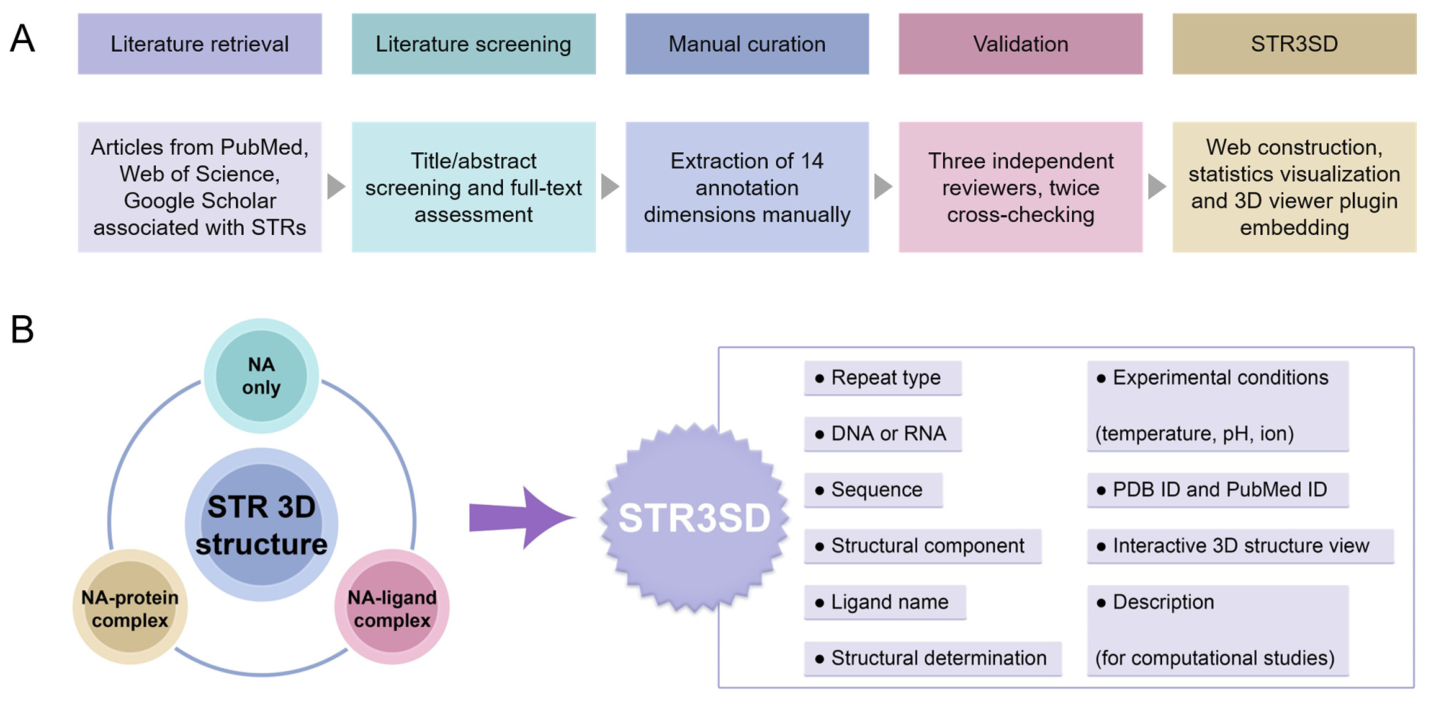
To establish STR3SD, we systematically searched PubMed, Web of Science, and Google Scholar for publications pertaining to STRs and 3D structures by human experts. This search retrieved 5483 articles published up to 1 June 2026. Through successive rounds of title/abstract screening, full-text assessment, and manual curation against predefined inclusion criteria that required the presence of experimentally determined or computationally modeled 3D structures of STRs, 162 qualified entries were ultimately retained. All the retained data underwent independent cross-checking by three trained reviewers to ensure accuracy and consistency. The workflow for data extraction and curation is illustrated in Figure 1A.
Each entry in STR3SD represents a single curated 3D structure record. For experimental studies, one entry corresponds to one PDB record. For computational studies, one entry corresponds to one MD simulation structure or simulation-based virtual screening. When an article reports multiple PDB records or multiple independent simulations, each of them is registered as an independent entry, and these entries share the same PubMed ID. Entries in STR3SD are annotated across the following fields. The field of “Repeats” means the sequence of the repetitive unit of STRs. The field of “DNA or RNA” means the type of nucleic acid. The field of “Sequence” means the nucleotide sequence in single-letter format with a 5′-to-3′ direction. The field of “PDB ID” means the accession code of the structure in the PDB. The field of “Ligand name” means the name of the bound molecule. The field of “Method” means the approach used to obtain the structure. The field of “PubMed ID” means the identifier of the source article in PubMed. The field of “Temperature” means the temperature of the crystallization for X-ray structures or the sample temperature during NMR spectroscopy. The field of “Ion” means the ionic composition of the crystallization or NMR buffer. The field of “pH” means the pH value of the crystallization buffer or NMR sample. The field of “3D structure” directs to an integrated 3D structure viewer [36,37] of the determined structure in NMR and X-ray studies, or the initial structure in MD studies. The field of “Description” shows a professional summary of the main conclusion for MD simulations and virtual screenings. The symbol “-“ indicates that the corresponding information was not reported in the original article.
To distinguish the type of ligand bound to the structure of STRs, we introduced a “Data subset” dimension to categorize whether the STRs are bound to a ligand or protein. Three categories are presented, including structures of nucleic acids only (NA only), nucleic acid–ligand complexes (NA–ligand), and nucleic acid–protein complexes (NA–protein). An additional “Component” dimension was introduced to further refine the molecular composition within each category, including DNA only, RNA only, DNA–ligand complex, RNA–ligand complex, DNA–protein complex, RNA–protein complex, RNA-PNA complex and DNA/RNA hybrid. In other words, the “Data subset” dimension classifies each entry according to the presence and type of the bound partner, whereas the “Component” dimension describes the detailed composition of the structure itself. Figure 1B shows the attributes of STR3SD.

2.2. Data Curation Reveals the Structural Diversity and Research Focus of STRs

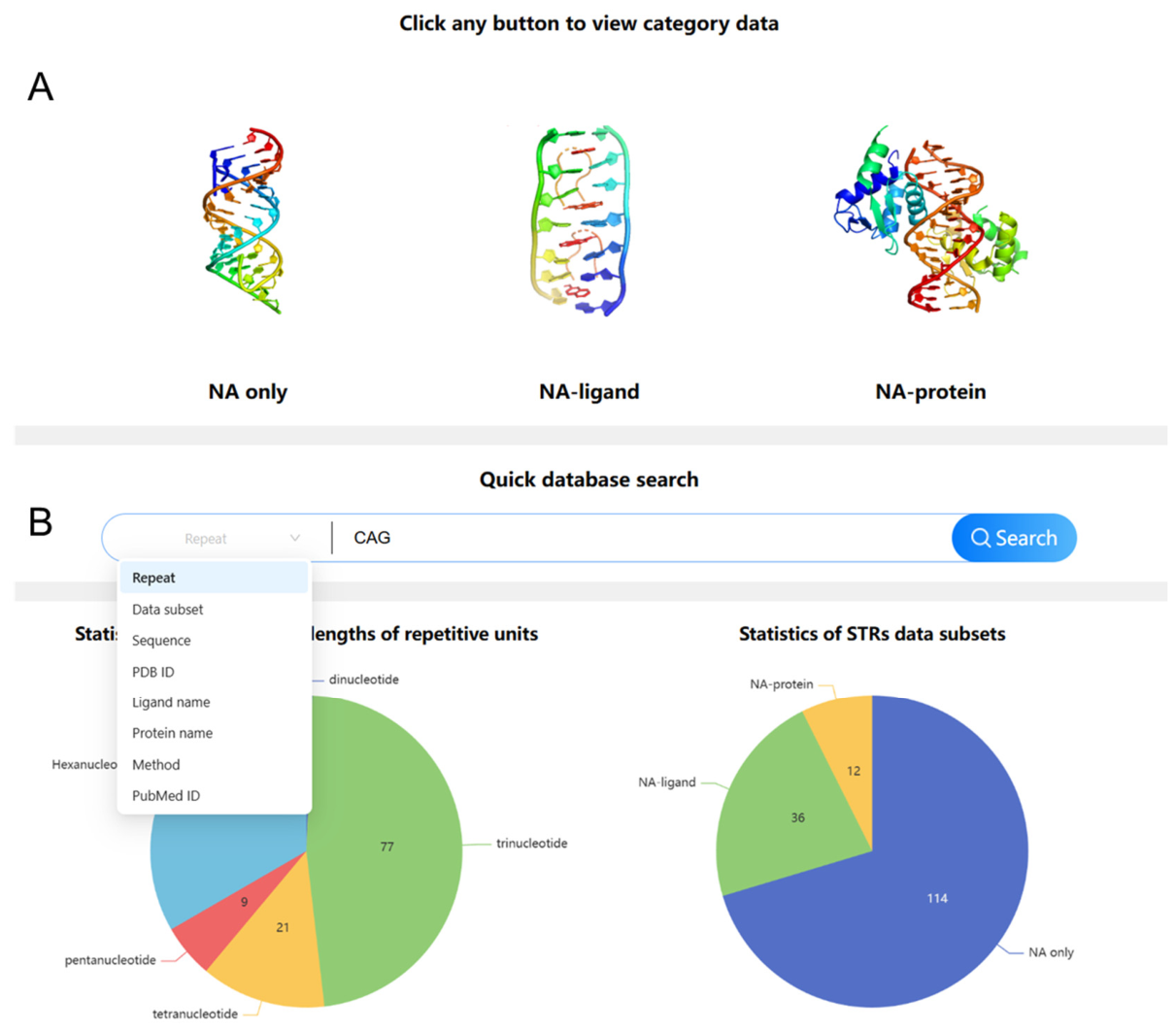
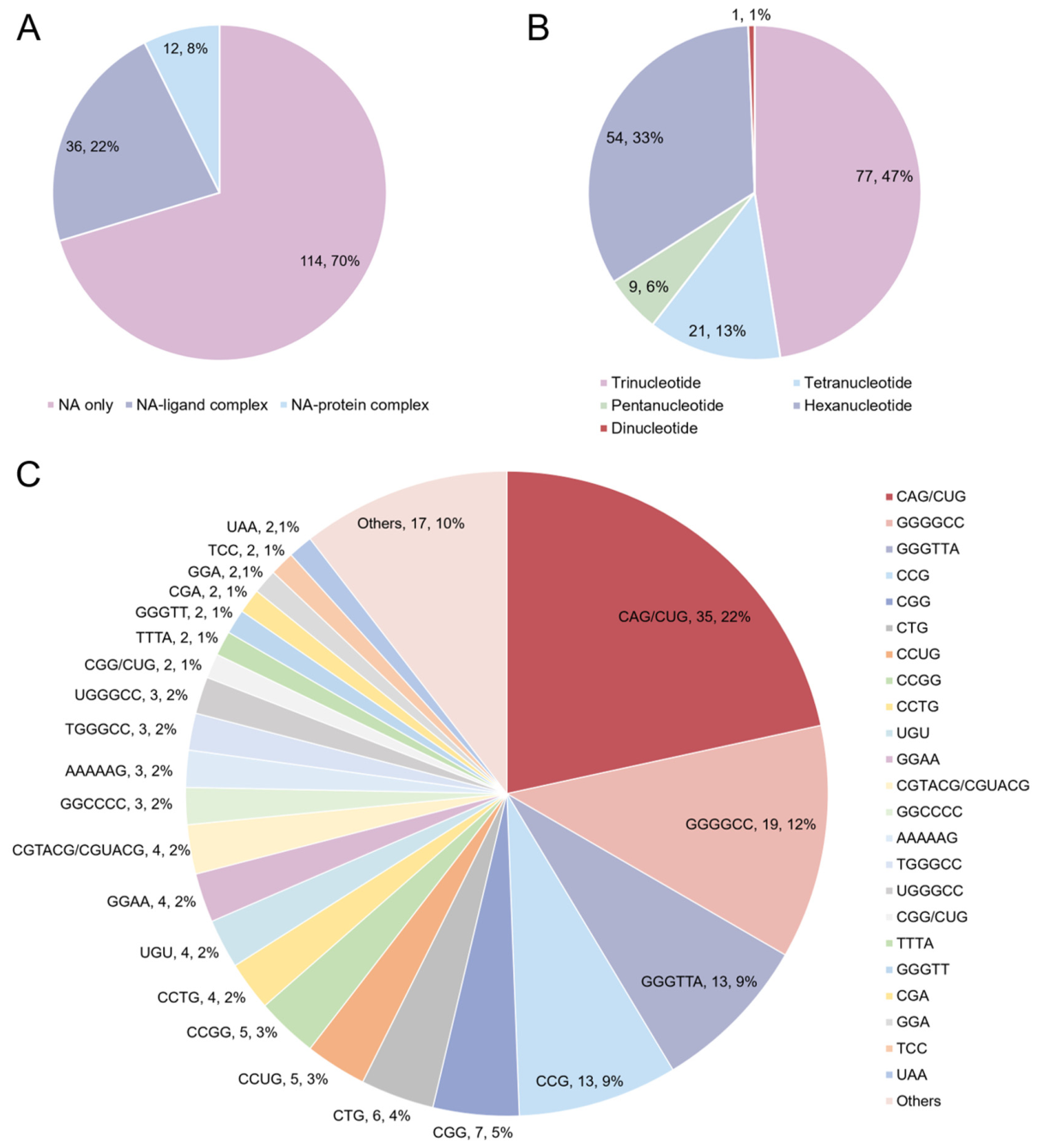
The statistics of data subsets and repeat lengths are displayed in Figure 2A,B. Research on 3D structures predominantly focuses on nucleic acids without bound ligands or proteins (NA only, 70%). Among these, trinucleotide repeats (47%) and hexanucleotide repeats (33%) are the most extensively studied. These statistical charts are automatically generated on the website homepage. Figure 2C illustrates the distribution of different repetitive units, with CAG/CUG (22%) and GGGGCC (12%) repeats being the most frequently studied, reflecting the predominant trends in current research. All the percentages are calculated based on the number of entries.

2.3. Interactive Web Interface for Data Retrieval, Visualization, and Export

2.3.1. Web Interface

STR3SD is a freely accessible, web-based online database available at https://str3sd.fnadb.com. It features a user-friendly graphical interface, offering multi-button search functionalities that present results in categorized order. The homepage is organized into five main sections: database introduction, general search, advanced search, data statistics, and update log.

2.3.2. Search Section

STR3SD provides two search sections for user access: general search and advanced search. In the general search module, three main categories including “NA only”, “NA–ligand” and “NA–protein” are presented (Figure 3A). The “NA only” category collects 3D structures of STRs without any bound partner, the “NA–ligand” category collects structures of STRs in complex with small molecule ligands, and the “NA–protein” category collects structures of STRs bound to proteins. Clicking on the category icon displays search results for a broad class of STRs, and thus the general search is designed for one-click browsing of an entire main category. In the advanced search module, the dropdown menu allows users to search for results by typing keywords for eight domains including Repeat, Data subset, Sequence, PDB ID, Ligand name, Protein name, Method and PubMed ID (Figure 3B), which enables precise, field-specific retrieval. Switching between any advanced search fields dynamically displays sample entries. These flexible search modules assist researchers in efficiently exploring the database and achieving an optimized search experience.

2.3.3. Result Section

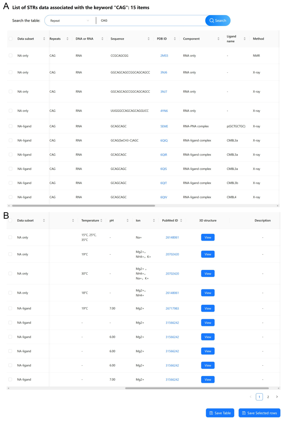
The results are presented on a results page in a tabular format with one record per row and multiple data columns (Figure 4). As an example, searching “CAG” in the Repeat field of the advanced search module returns 15 entries (Figure 4). These entries cover RNA-only structures as well as RNA–ligand, RNA-PNA and DNA–ligand complexes, encompassing annotated sequences, structural determination methods, ligand names, experimental conditions (temperature, pH, and ion composition), and hyperlinked PDB IDs, PubMed IDs, and interactive 3D structure views. For example, the first entry (PDB ID: 2MS5) corresponds to an RNA containing one CAG repeat as determined by NMR spectroscopy at 15, 25, and 35 °C in the presence of Na+. The Ligand name of “-“ means the absence of a bound partner. The pH field is marked as “-“, indicating that the pH condition was not reported in the original publication. The database provides multiple interactive functions. The hyperlinked PDB ID (in blue) directs users to the corresponding PDB entry. The hyperlinked PubMed accession number (in blue) links to the PubMed abstract page. The “View” button under the “3D structure” column launches the online 3D structure viewer. The Description field is marked as “-“ for this entry, as it is an experimentally determined structure.
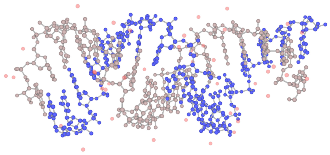
Notably, the LiteMol plugin is embedded to enable online viewing and interactive manipulation of 3D structures [36,37]. Users can click the “View” button to visualize a 3D structure and switch the rendering mode between ball-and-stick, surface and van der Waals sphere models (Figure 5). All or selected data can be exported directly from the search results page as an .xlsx file. Using various search combinations, users can sort the literature and obtain statistical summaries at the database level.

2.4. Meta-Analysis of Structural Polymorphisms Across Disease-Associated STRs

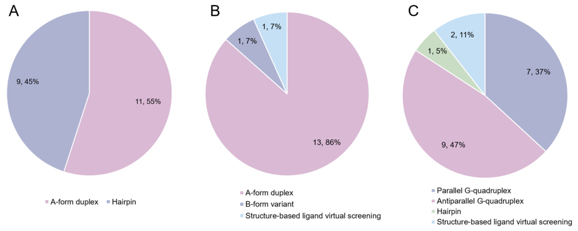
STR3SD enables systematic meta-analysis of the structural polymorphism of STRs efficiently. As shown in Figure 6, we utilized STR3SD to integrate and visually summarize the three most common types of STR structural polymorphisms. For CUG repeats associated with DM1 [39], nearly all the existing studies focus on their A-form RNA duplex structure, with approximately 45% of these studies further investigating their hairpin structure. Regarding CAG repeats associated with HD and many SCAs [40], the vast majority of studies (86%) concentrate on the A-form RNA duplex, while only 7% of structural research involves the B-form variant. In addition, structure-based virtual screening has identified the small molecule somniferine, which exhibits high-affinity binding to CAG repeats. In the case of GGGGCC repeats associated with ALS/FTD [41], most studies focus on the antiparallel G-quadruplex (47%), followed by the parallel G-quadruplex (37%). A small proportion of studies (5%) report that this sequence can also form a hairpin structure. In terms of drug discovery, virtual screening results integrated by STR3SD suggest that the small molecules Au(TMX)2 and orotate are two reported potential ligands targeting the 3D structures of GGGGCC repeats [42,43]. All the percentages are calculated based on the number of entries.
In sum, the powerful data mining and pattern recognition capabilities of STR3SD allow it to not only present individual data points but also greatly facilitate researchers in performing cross-study comparisons and meta-analyses. This helps reveal structural preferences, research trends, and even underexplored areas associated with specific STR sequences, such as GGGGCC repeats, across different studies.

2.5. Advantages and the Expanding Scope of STR3SD

To the best of our knowledge, currently there is no other database specifically dedicated to the study of 3D structures of STRs. While researchers can query structural data through databases such as PDB, the information retrieved may be incomplete or even contain errors. STR3SD offers meta-analysis capabilities for homologous STRs, providing more accurate experimental conditions, an interactive interface for 3D structure visualization, and multi-level data categorization. This demonstrates that STR3SD delivers a more precise, goal-oriented, and user-friendly experience, and enables seamless comparisons across different studies. STR3SD is updated every 3 months to incorporate the latest research, thereby surpassing the limitations of static reviews. Additionally, to facilitate access to original articles and structural data, PubMed IDs and PDB IDs are clearly listed.

3. Discussion

STRs play important biological roles, with their sequences tuning gene expression [13] and their expansions driving more than 50 human diseases [6]. Because the pathogenicity of STRs is intimately linked to the non-canonical 3D structures they adopt, a database dedicated to the 3D structures of STRs is of great significance for elucidating disease mechanisms and developing STR-targeting drugs. Over the past two decades, a number of STR-related databases have been developed (Table 1). For disease and phenotype-related databases, STRipy [34] enables the detection of pathogenic STRs from sequencing data. In forensic and human identity testing, STRBase [32] provides STR marker information, and STRidER [44] offers quality-controlled STR allele frequencies. In the field of cell line authentication, ATCC STR Database [33] supports the verification of cell identity. For genome-wide STR identification and population variation analysis, TRDB [45], WebSTR [46], SNPSTR [47], UgMicroSat db [48] and Microsatellites Explorer [49] catalog the sequences, genomic coordinates and population variation in STRs identified by computational mining of genomes. Despite the wealth of genomic, forensic and clinical information and dedicated search algorithms, none of them provide high-resolution structure information of STRs.
In terms of nucleic acid structural databases, NAKB [50] archives all experimentally determined nucleic acid 3D structures held by the NDB and PDB, and ONQUADRO [36] specializes in experimentally determined quadruplex structures with detailed geometric annotations. These databases are not specific to STRs, and they do not document experimental conditions (e.g., ion concentration, temperature, pH) or computational studies such as MD simulations and MD simulation-based virtual screening.
To facilitate the development of small-molecule drugs targeting 3D structures of pathogenic STRs, our STR3SD represents the first database collecting the experimentally determined high-resolution 3D structures of STRs. Because STR3SD focuses on well-defined, disease-associated STRs for which 3D structures have been experimentally determined or computationally modeled, no STR searching algorithm is required. The unique contribution of STR3SD lies in the systematic manual integration, cross-validation and functional annotation of dispersed structural data, which transforms isolated literature results into a unified and accessible resource that facilitates structure-based drug design targeting STRs.
By providing standardized 3D structural data and comprehensive annotations, STR3SD lays a foundation for investigating the “sequence-structure-function” relationships in STRs. It facilitates the exploration of structural polymorphisms in STRs, aids in identifying disease-associated conformations, and enables computational drug design and modeling targeting STR ligands. The database supports searches based on sequence, structural attributes, and other metadata, offers flexible data export options, and includes an interactive 3D molecular visualization interface. STR3SD represents a much-needed and valuable resource in the fields of nucleic acid structural biology and precision medicine.
STR3SD meticulously documents nearly all the available STR 3D structures, supports large-scale data analysis, assists researchers in optimizing experimental design, and provides pharmaceutical companies with targetable sequences to accelerate drug discovery. For academic and research institutions, it offers a new perspective on STR research progress. The author team is committed to maintaining and updating STR3SD regularly, addressing issues, and expanding features to enhance user experience. We cordially invite users to submit error reports, seek assistance, or suggest new functionalities via email or other contact channels.

4. Materials and Methods

4.1. Data Collection

We first systematically collected, reviewed, and categorized a vast amount of information related to various attributes of STRs. By searching articles from PubMed, Web of Science and Google Scholar published up to 1 June 2026 using keywords for “short tandem repeats” and “3D structure”, 5483 articles were obtained for further screening. Subsequently, refined STR-related keywords such as disease names of “Huntington’s Disease” or “Fragile X Syndrome”, and STR types of “CAG repeats” or “GGGGCC repeats” in titles/abstracts were used to further identify literature specifically related to 3D structures of STRs, followed by full-text assessment to confirm the presence of experimentally determined or computationally modeled 3D structures of STRs, which were then subjected to the next stage of data extraction.
The extracted data were systematically organized into the following fields, with definitions and sources specified. The field of “Data subset” means the main category classified by the binding partner, which is annotated by the curator based on the original article. The field of “Repeats” means the sequences of the repetitive unit, which is sourced from the original article. The field of “DNA or RNA” means the type of nucleic acid, which is sourced from the original article. The field of “Sequence” means the nucleotide sequence in single-letter format with a 5′-to-3′ direction, which is sourced from the original article. The field of “PDB ID” means the accession code of the structure in the PDB, which is sourced from the original article and cross-validated against the PDB. The field of “Component” means the fine classification of the molecular composition, which is annotated by the curator based on the original article. The field of “Ligand name” means the name of the bound molecule, which is sourced from the original article. The field of “Method” means the approach used to obtain the structure, which is sourced from the original article. The field of “Temperature” means the temperature of crystallization for X-ray structures or the sample temperature during NMR spectroscopy, which is sourced from the original article. The field of “pH” means the pH value of the crystallization or NMR buffer, which is sourced from the original article. The field of “Ion” means the ionic composition of the crystallization or NMR buffer, which is sourced from the original article. The field of “PubMed ID” means the identifier of the source article with a hyperlink to PubMed, which is annotated by the curator based on PubMed. The field of “3D structure” directs to the interactive structure view, which is sourced from the PDB and the original article. The field of “Description” displays the professional summary of the study for MD simulations and virtual screenings, which is extracted from the original article with curator interpretation.
When a single article reported multiple PDB records or multiple independent computational models, each PDB record or computational model was registered as an independent entry, with all the entries from the same article sharing the same PubMed ID.

4.2. Data Validation

All the data extractors received standardized procedural training to ensure consistency in their operations. The data from each article were independently reviewed and consolidated by three well-trained reviewers. After all the data entries were completed, random sampling was performed twice for verification against the original sources to ensure the accuracy and reliability of the final dataset. All the 3D structures accessible in STR3SD were subjected to online rendering tests to ensure accurate and rapid interactive visualization.

4.3. Database Architecture and Implementation

STR3SD is built on a modern full-stack architecture that emphasizes modularity, maintainability, and a seamless user experience. The system strictly separates the backend service layer from the frontend presentation layer.
The server-side application is developed using the RuoYi framework (v3.8.1, RuoYi Open Source Project, Zhanjiang, Guangdong, China), a rapid development platform based on Spring Boot (v2.5.8, Broadcom Inc., San Jose, CA, USA), Spring Security (v5.5.4, Broadcom Inc., San Jose, CA, USA), and MyBatis-Plus (v3.4.2, baomidou Open Source Project, China). It employs a microservice architecture managed by Spring Cloud Alibaba for scalability and resilience. Core services are registered and configured through Nacos (v2.2.1, Alibaba Group, Hangzhou, Zhejiang, China). Data are persisted in a MySQL database (v5.7.40, Oracle Corporation, Redwood City, CA, USA), with Redis (v6.1.5, Redis Ltd., Mountain View, CA, USA) utilized for high-performance caching. This backend stack provides a robust, secure, and efficient foundation for data management, API services, and business logic. The interactive web interface is constructed as a single-page application (SPA) using React (v19.1.0, Meta Platforms, Inc., Menlo Park, CA, USA), with Ant Design (v6.2.0, Ant Group, Hangzhou, Zhejiang, China) as the core user interface (UI) component library. This combination enables the development of a responsive, dynamic, and visually consistent user interface that works flawlessly across desktop and mobile platforms. Specialized functionalities are integrated into this core architecture. The interactive 3D molecular viewer is implemented by embedding the LiteMol plugin (v2, CEITEC-Masaryk University, Brno, Czech Republic) within the React application. Data exploration is enhanced through dynamic table components and advanced search filters. Users can conveniently export query results as .xlsx files for offline analysis.

Author Contributions

Conceptualization, J.Q. and P.G.; methodology, K.J.; software, K.J.; validation, K.J., A.S., Y.W., Y.L. (Yu Liu) and Y.L. (Yang Liu); formal analysis, K.J.; investigation, K.J., M.G. and Z.Z.; resources, J.Q. and P.G.; data curation, K.J., Y.W., Y.L. (Yu Liu) and Y.L. (Yang Liu); writing—original draft preparation, K.J.; writing—review and editing, N.X., Y.T., L.W., J.Q. and P.G.; visualization, K.J.; supervision, J.Q. and P.G.; project administration, J.Q. and P.G.; funding acquisition, J.Q. and P.G. All authors have read and agreed to the published version of the manuscript.

Funding

This research was funded by the National Natural Science Foundation of China, grant numbers 22374132, 22507123, and 62306290; Leading Innovative and Entrepreneur Team Introduction Program of Zhejiang Province, grant number 2024R01005; and Natural Science Foundation of Zhejiang Province, grant number QKHM25B0501.

Institutional Review Board Statement

Not applicable.

Informed Consent Statement

Not applicable.

Data Availability Statement

The STR3SD database will be updated regularly (every 3 months). It is now publicly and freely available at https://str3sd.fnadb.com/. Related data will be available upon request from the authors.

Conflicts of Interest

The authors declare no conflicts of interest.

Abbreviations

The following abbreviations are used in this manuscript:
3DThree-Dimensional
ALSAmyotrophic Lateral Sclerosis
Cryo-EMCryogenic Electron Microscopy
DMMyotonic Dystrophy
FTDFrontotemporal Dementia
HDHuntington’s Disease
MDMolecular Dynamics
NANucleic Acid
NMRNuclear Magnetic Resonance
PDBProtein Data Bank
RBPRNA-Binding Protein
SCASpinocerebellar Ataxia
SPASingle-Page Application
STR3SDShort Tandem Repeat 3D Structure Database
STRShort Tandem Repeat
UIUser Interface

References

  1. Verbiest, M.; Maksimov, M.; Jin, Y.; Anisimova, M.; Gymrek, M.; Bilgin Sonay, T. Mutation and selection processes regulating short tandem repeats give rise to genetic and phenotypic diversity across species. J. Evol. Biol. 2023, 36, 321–336. [Google Scholar] [CrossRef] [PubMed]
  2. Rajan-Babu, I.S.; Dolzhenko, E.; Eberle, M.A.; Friedman, J.M. Sequence composition changes in short tandem repeats: Heterogeneity, detection, mechanisms and clinical implications. Nat. Rev. Genet. 2024, 25, 476–499. [Google Scholar] [CrossRef] [PubMed]
  3. McGinty, R.J.; Balick, D.J.; Mirkin, S.M.; Sunyaev, S.R. Inherent instability of simple DNA repeats shapes an evolutionarily stable distribution of repeat lengths. Nat. Commun. 2025, 17, 93. [Google Scholar] [CrossRef] [PubMed]
  4. Li, L.; Scott, W.S.; Mirkin, S.M. Emerging drivers of DNA repeat expansions. Biochem. Soc. Trans. 2025, 53, 995–1010. [Google Scholar] [CrossRef] [PubMed]
  5. Simone, R.; Balendra, R.; Moens, T.G.; Preza, E.; Wilson, K.M.; Heslegrave, A.; Woodling, N.S.; Niccoli, T.; Gilbert-Jaramillo, J.; Abdelkarim, S.; et al. G-quadruplex-binding small molecules ameliorate C9orf72 FTD/ALS pathology in vitro and in vivo. EMBO Mol. Med. 2018, 10, 22–31. [Google Scholar] [CrossRef] [PubMed]
  6. Wu, Y.; Song, T.; Xu, Q. R-LOOPs on Short Tandem Repeat Expansion Disorders in Neurodegenerative Diseases. Mol. Neurobiol. 2023, 60, 7185–7195. [Google Scholar] [CrossRef] [PubMed]
  7. MacDonald, M.E.; Ambrose, C.M.; Duyao, M.P.; Myers, R.H.; Lin, C.; Srinidhi, L.; Barnes, G.; Taylor, S.A.; James, M.; Groot, N.; et al. A novel gene containing a trinucleotide repeat that is expanded and unstable on Huntington’s disease chromosomes. Cell 1993, 72, 971–983. [Google Scholar] [CrossRef] [PubMed]
  8. Renton, A.E.; Majounie, E.; Waite, A.; Simón-Sánchez, J.; Rollinson, S.; Gibbs, J.R.; Schymick, J.C.; Laaksovirta, H.; van Swieten, J.C.; Myllykangas, L.; et al. A Hexanucleotide Repeat Expansion in C9ORF72 Is the Cause of Chromosome 9p21-Linked ALS-FTD. Neuron 2011, 72, 257–268. [Google Scholar] [CrossRef] [PubMed]
  9. DeJesus-Hernandez, M.; Mackenzie, I.R.; Boeve, B.F.; Boxer, A.L.; Baker, M.; Rutherford, N.J.; Nicholson, A.M.; Finch, N.A.; Flynn, H.; Adamson, J.; et al. Expanded GGGGCC Hexanucleotide Repeat in Noncoding Region of C9ORF72 Causes Chromosome 9p-Linked FTD and ALS. Neuron 2011, 72, 245–256. [Google Scholar] [CrossRef] [PubMed]
  10. Hujoel, M.L.A.; Handsaker, R.E.; Tang, D.; Kamitaki, N.; Mukamel, R.E.; Rubinacci, S.; Palamara, P.F.; McCarroll, S.A.; Loh, P.R. Insights into DNA repeat expansions among 900,000 biobank participants. Nature 2026, 650, 920–929. [Google Scholar] [CrossRef] [PubMed]
  11. Erwin, G.S.; Gürsoy, G.; Al-Abri, R.; Suriyaprakash, A.; Dolzhenko, E.; Zhu, K.; Hoerner, C.R.; White, S.M.; Ramirez, L.; Vadlakonda, A.; et al. Recurrent repeat expansions in human cancer genomes. Nature 2023, 613, 96–102. [Google Scholar] [CrossRef] [PubMed]
  12. Watson, J.D.; Crick, F.H.C. Molecular Structure of Nucleic Acids: A Structure for Deoxyribose Nucleic Acid. Nature 1953, 171, 737–738. [Google Scholar] [CrossRef] [PubMed]
  13. Horton, C.A.; Alexandari, A.M.; Hayes, M.G.B.; Marklund, E.; Schaepe, J.M.; Aditham, A.K.; Shah, N.; Suzuki, P.H.; Shrikumar, A.; Afek, A.; et al. Short tandem repeats bind transcription factors to tune eukaryotic gene expression. Science 2023, 381, eadd1250. [Google Scholar] [CrossRef] [PubMed]
  14. Nalavade, R.; Griesche, N.; Ryan, D.P.; Hildebrand, S.; Krauss, S. Mechanisms of RNA-induced toxicity in CAG repeat disorders. Cell Death Dis. 2013, 4, e752. [Google Scholar] [CrossRef] [PubMed]
  15. Chen, J.L.; VanEtten, D.M.; Fountain, M.A.; Yildirim, I.; Disney, M.D. Structure and Dynamics of RNA Repeat Expansions That Cause Huntington’s Disease and Myotonic Dystrophy Type 1. Biochemistry 2017, 56, 3463–3474. [Google Scholar] [CrossRef] [PubMed]
  16. Luo, Z.; Dauter, Z.; Gilski, M. Four highly pseudosymmetric and/or twinned structures of d(CGCGCG)2 extend the repertoire of crystal structures of Z-DNA. Acta Crystallogr. D Struct. Biol. 2017, 73, 940–951. [Google Scholar] [CrossRef] [PubMed]
  17. Mariappan, S.V.S.; Catasti, P.; Silks, L.A.; Bradbury, E.M.; Gupta, G. The high-resolution structure of the triplex formed by the GAA/TTC triplet repeat associated with Friedreich’s ataxia. J. Mol. Biol. 1999, 285, 2035–2052. [Google Scholar] [CrossRef] [PubMed]
  18. Liu, Y.; Yang, Y.; Zhang, Y.; Yi, J.; Wan, L.; Wang, Y.; Gao, Q.; Xu, D.; Guo, P.; Han, D. Structural Basis for the GGGGCC Repeat RNA Binding to SRSF2 Protein. Chin. J. Chem. 2025, 43, 1348–1356. [Google Scholar] [CrossRef]
  19. Geng, Y.; Liu, C.; Miao, H.; Suen, M.C.; Xie, Y.; Zhang, B.; Han, W.; Wu, C.; Ren, H.; Chen, X.; et al. Crystal structures of distinct parallel and antiparallel DNA G-quadruplexes reveal structural polymorphism in C9orf72 G4C2 repeats. Nucleic Acids Res. 2025, 53, gkaf879. [Google Scholar] [CrossRef] [PubMed]
  20. Abou Assi, H.; Garavís, M.; González, C.; Damha, M.J. i-Motif DNA: Structural features and significance to cell biology. Nucleic Acids Res. 2018, 46, 8038–8056. [Google Scholar] [CrossRef] [PubMed]
  21. Mirkin, S.M. Expandable DNA repeats and human disease. Nature 2007, 447, 932–940. [Google Scholar] [CrossRef] [PubMed]
  22. Schmidt, M.H.M.; Pearson, C.E. Disease-associated repeat instability and mismatch repair. DNA Repair 2016, 38, 117–126. [Google Scholar] [CrossRef] [PubMed]
  23. Zhao, J.; Bacolla, A.; Wang, G.; Vasquez, K.M. Non-B DNA structure-induced genetic instability and evolution. Cell. Mol. Life Sci. 2010, 67, 43–62. [Google Scholar] [CrossRef] [PubMed]
  24. Chen, J.-M.; Cooper, D.N.; Chuzhanova, N.; Férec, C.; Patrinos, G.P. Gene conversion: Mechanisms, evolution and human disease. Nat. Rev. Genet. 2007, 8, 762–775. [Google Scholar] [CrossRef] [PubMed]
  25. Swinnen, B.; Robberecht, W.; Van Den Bosch, L. RNA toxicity in non-coding repeat expansion disorders. EMBO J. 2020, 39, e101112. [Google Scholar] [CrossRef] [PubMed]
  26. Wojciechowska, M.; Krzyzosiak, W.J. Cellular toxicity of expanded RNA repeats: Focus on RNA foci. Hum. Mol. Genet. 2011, 20, 3811–3821. [Google Scholar] [CrossRef] [PubMed]
  27. Nakatani, K.; Hagihara, S.; Goto, Y.; Kobori, A.; Hagihara, M.; Hayashi, G.; Kyo, M.; Nomura, M.; Mishima, M.; Kojima, C. Small-molecule ligand induces nucleotide flipping in (CAG)n trinucleotide repeats. Nat. Chem. Biol. 2005, 1, 39–43. [Google Scholar] [CrossRef] [PubMed]
  28. Nakamori, M.; Panigrahi, G.B.; Lanni, S.; Gall-Duncan, T.; Hayakawa, H.; Tanaka, H.; Luo, J.; Otabe, T.; Li, J.; Sakata, A.; et al. A slipped-CAG DNA-binding small molecule induces trinucleotide-repeat contractions in vivo. Nat. Genet. 2020, 52, 146–159. [Google Scholar] [CrossRef] [PubMed]
  29. Nguyen, L.; Luu, L.M.; Peng, S.; Serrano, J.F.; Chan, H.Y.; Zimmerman, S.C. Rationally designed small molecules that target both the DNA and RNA causing myotonic dystrophy type 1. J. Am. Chem. Soc. 2015, 137, 14180–14189. [Google Scholar] [CrossRef] [PubMed]
  30. Di Giorgio, A.; Duca, M. Synthetic small-molecule RNA ligands: Future prospects as therapeutic agents. Med. Chem. Comm. 2019, 10, 1242–1255. [Google Scholar] [CrossRef] [PubMed]
  31. Granzhan, A.; Kotera, N.; Teulade-Fichou, M.P. Finding needles in a basestack: Recognition of mismatched base pairs in DNA by small molecules. Chem. Soc. Rev. 2014, 43, 3630–3665. [Google Scholar] [CrossRef] [PubMed]
  32. Ruitberg, C.M.; Reeder, D.J.; Butler, J.M. STRBase: A short tandem repeat DNA database for the human identity testing community. Nucleic Acids Res. 2001, 29, 320–322. [Google Scholar] [CrossRef] [PubMed]
  33. ATCC STR Database. Available online: https://www.atcc.org/search-str-database (accessed on 27 July 2026).
  34. Halman, A.; Dolzhenko, E.; Oshlack, A. STRipy: A graphical application for enhanced genotyping of pathogenic short tandem repeats in sequencing data. Hum. Mutat. 2022, 43, 859–868. [Google Scholar] [CrossRef] [PubMed]
  35. Singh, S.; Singh, S.; Joshi, D.; Mohanty, C.; Singh, R. In Silico Prediction of Potential Inhibitors for Targeting RNA CAG Repeats via Molecular Docking and Dynamics Simulation: A Drug Discovery Approach. J. Cell. Biochem. 2024, 125, e30611. [Google Scholar] [CrossRef] [PubMed]
  36. Zok, T.; Kraszewska, N.; Miskiewicz, J.; Pielacinska, P.; Zurkowski, M.; Szachniuk, M. ONQUADRO: A database of experimentally determined quadruplex structures. Nucleic Acids Res. 2022, 50, D253–D258. [Google Scholar] [CrossRef] [PubMed]
  37. Sehnal, D.; Deshpande, M.; Vařeková, R.S.; Mir, S.; Berka, K.; Midlik, A.; Pravda, L.; Velankar, S.; Koča, J. LiteMol suite: Interactive web-based visualization of large-scale macromolecular structure data. Nat. Methods 2017, 14, 1121–1122. [Google Scholar] [CrossRef] [PubMed]
  38. Mooers, B.H.; Logue, J.S.; Berglund, J.A. The structural basis of myotonic dystrophy from the crystal structure of CUG repeats. Proc. Natl. Acad. Sci. USA 2005, 102, 16626–16631. [Google Scholar] [CrossRef] [PubMed]
  39. Kumar, A.; Park, H.; Fang, P.; Parkesh, R.; Guo, M.; Nettles, K.W.; Disney, M.D. Myotonic dystrophy type 1 RNA crystal structures reveal heterogeneous 1 × 1 nucleotide UU internal loop conformations. Biochemistry 2011, 50, 9928–9935. [Google Scholar] [CrossRef] [PubMed]
  40. Tawani, A.; Kumar, A. Structural Insights Reveal the Dynamics of the Repeating r(CAG) Transcript Found in Huntington’s Disease (HD) and Spinocerebellar Ataxias (SCAs). PLoS ONE 2015, 10, e0131788. [Google Scholar] [CrossRef] [PubMed]
  41. Brčić, J.; Plavec, J. Solution structure of a DNA quadruplex containing ALS and FTD related GGGGCC repeat stabilized by 8-bromodeoxyguanosine substitution. Nucleic Acids Res. 2015, 43, 8590–8600. [Google Scholar] [CrossRef] [PubMed]
  42. D’Anna, L.; Wragg, D.; Mauro, D.; Rubino, S.; Terenzi, A.; Barone, G.; Thomas, S.R.; Casini, A.; Bonsignore, R.; Spinello, A. Unraveling the Molecular Basis for G-Quadruplex-Binders to ALS/FTD-Associated G4C2 Repeats of the C9orf72 Gene. ChemBioChem 2025, 26, e202400974. [Google Scholar] [CrossRef] [PubMed]
  43. Falanga, A.P.; Piccialli, I.; Greco, F.; D’Errico, S.; Nolli, M.G.; Borbone, N.; Oliviero, G.; Roviello, G.N. Nanostructural Modulation of G-Quadruplex DNA in Neurodegeneration: Orotate Interaction Revealed Through Experimental and Computational Approaches. J. Neurochem. 2025, 169, e16296. [Google Scholar] [CrossRef] [PubMed]
  44. Bodner, M.; Bastisch, I.; Butler, J.M.; Fimmers, R.; Gill, P.; Gusmão, L.; Morling, N.; Phillips, C.; Prinz, M.; Schneider, P.M.; et al. Recommendations of the DNA Commission of the International Society for Forensic Genetics (ISFG) on quality control of autosomal Short Tandem Repeat allele frequency databasing (STRidER). Forensic Sci. Int. Genet. 2016, 24, 97–102. [Google Scholar] [CrossRef] [PubMed]
  45. Gelfand, Y.; Rodriguez, A.; Benson, G. TRDB—The Tandem Repeats Database. Nucleic Acids Res. 2007, 35, D80–D87. [Google Scholar] [CrossRef] [PubMed]
  46. Lundström, O.S.; Adriaan Verbiest, M.; Xia, F.; Jam, H.Z.; Zlobec, I.; Anisimova, M.; Gymrek, M. WebSTR: A Population-wide Database of Short Tandem Repeat Variation in Humans. J. Mol. Biol. 2023, 435, 168260. [Google Scholar] [CrossRef] [PubMed]
  47. Agrafioti, I.; Stumpf, M.P. SNPSTR: A database of compound microsatellite-SNP markers. Nucleic Acids Res. 2007, 35, D71–D75. [Google Scholar] [CrossRef] [PubMed]
  48. Aishwarya, V.; Sharma, P.C. UgMicroSatdb: Database for mining microsatellites from unigenes. Nucleic Acids Res. 2008, 36, D53–D56. [Google Scholar] [CrossRef] [PubMed]
  49. Provatas, K.; Chantzi, N.; Patsakis, M.; Nayak, A.; Mouratidis, I.; Georgakopoulos-Soares, I. Microsatellites explorer: A database of short tandem repeats across genomes. Comput. Struct. Biotechnol. J. 2024, 23, 3817–3826. [Google Scholar] [CrossRef] [PubMed]
  50. Lawson, C.L.; Berman, H.M.; Chen, L.; Vallat, B.; Zirbel, C.L. The Nucleic Acid Knowledgebase: A new portal for 3D structural information about nucleic acids. Nucleic Acids Res. 2024, 52, D245–D254. [Google Scholar] [CrossRef] [PubMed]
Figure 1. Construction and attributes of STR3SD: (A) Workflow of database construction. (B) Attributes of STR3SD. The central circle represents the 3D structures of STRs, which are classified into three data subsets shown as colored satellites: NA only (green), NA–ligand complexes (magenta) and NA–protein complexes (olive). The right panel lists the annotation dimensions of each entry, including Repeat type, DNA or RNA, Sequence, Structural component, Ligand name, Structural determination, Experimental conditions (temperature, pH, and ion), PDB ID and PubMed ID, Interactive 3D structure view and Description for computational studies. NA, nucleic acid.
Ijms 27 06872 g001
Figure 2. The statistics of STR3SD: (A) Three data subsets including NA only, NA–ligand complexes and NA–protein complexes. (B) Repetitive units with different lengths. (C) Different repetitive units.
Ijms 27 06872 g002
Figure 3. The search module of STR3SD. (A) General search. Three category icons are provided, representing “NA only” (STRs without bound partners), “NA–ligand” (nucleic acid–ligand complexes) and “NA–protein” (nucleic acid–protein complexes). Clicking an icon directly returns all entries of the corresponding category. (B) Advanced search. Eight query domains (Repeat, Data subset, Sequence, PDB ID, Ligand name, Protein name, Method and PubMed ID) are available in the dropdown menu for precise retrieval.
Ijms 27 06872 g003
Figure 4. Screenshot of the STR3SD database search results using “CAG” as the query keyword (15 entries with CAG as the repeat unit are retrieved): (A) Structural feature and method columns, including Data subset, Repeats, DNA or RNA, Sequence, PDB ID, Component, Ligand name and Method. (B) Experimental condition and interactive function columns, including Temperature, pH, Ion, PubMed ID, 3D structure and Description. Hyperlinked PDB IDs and PubMed accession numbers (in blue) direct users to the corresponding PDB and PubMed webpages, respectively, and the blue “View” buttons launch the integrated 3D structure visualization tool. For experimentally determined structures, the Description field is designated as “-”.
Ijms 27 06872 g004
Figure 5. The online 3D visualization function of STR3SD by the embedded LiteMol viewer, as exemplified by the CUG-repeat RNA structure (PDB ID: 1ZEV) [38]. The structure is shown as a ball-and-stick model, with the two RNA strands depicted as gray and blue spheres, respectively, and ordered water molecules in the crystal lattice shown as pink spheres.
Ijms 27 06872 g005
Figure 6. The structural polymorphisms of the top 3 STRs: (A) CUG repeats. (B) CAG RNA repeats. (C) GGGGCC DNA and RNA repeats.
Ijms 27 06872 g006
Table 1. Comparison of STR-related databases.
DatabaseDescriptionReference
STRipyGenotyping of pathogenic STRs from sequencing data for clinical diagnosis[34]
STRBaseSTR marker information for forensic human identity testing[32]
STRidERQuality-controlled autosomal STR allele frequency database for forensic genetics[44]
ATCC STR DatabaseSTR profiles for human cell line authentication[33]
TRDBTandem repeat repository with analysis tools for genomic DNA[45]
WebSTRPopulation-wide STR variation in humans[46]
SNPSTRCompound microsatellite-SNP markers in human and model organisms[47]
UgMicroSat dbMicrosatellites mined from UniGene sequences of 80 genomes[48]
Microsatellites ExplorerSTRs across 117,253 organisms, T2T genomes and human pangenome haplotypes[49]
STR3SD3D structures of all STRs, with experimental conditions, MD simulations and virtual screening recordsThis work
Disclaimer/Publisher’s Note: The statements, opinions and data contained in all publications are solely those of the individual author(s) and contributor(s) and not of MDPI and/or the editor(s). MDPI and/or the editor(s) disclaim responsibility for any injury to people or property resulting from any ideas, methods, instructions or products referred to in the content.