Cyanobacteria and Microalgae as Promising Sustainable Sources of Bioactive Phytochemicals for Nutraceutical and Pharmaceutical Applications

 ,
,  , , , , ,
Abstract
1. Introduction
2. Methods
2.1. Data Sources and Search Strategy
2.1.1. ScienceDirect
2.1.2. Scopus
2.1.3. PubMed
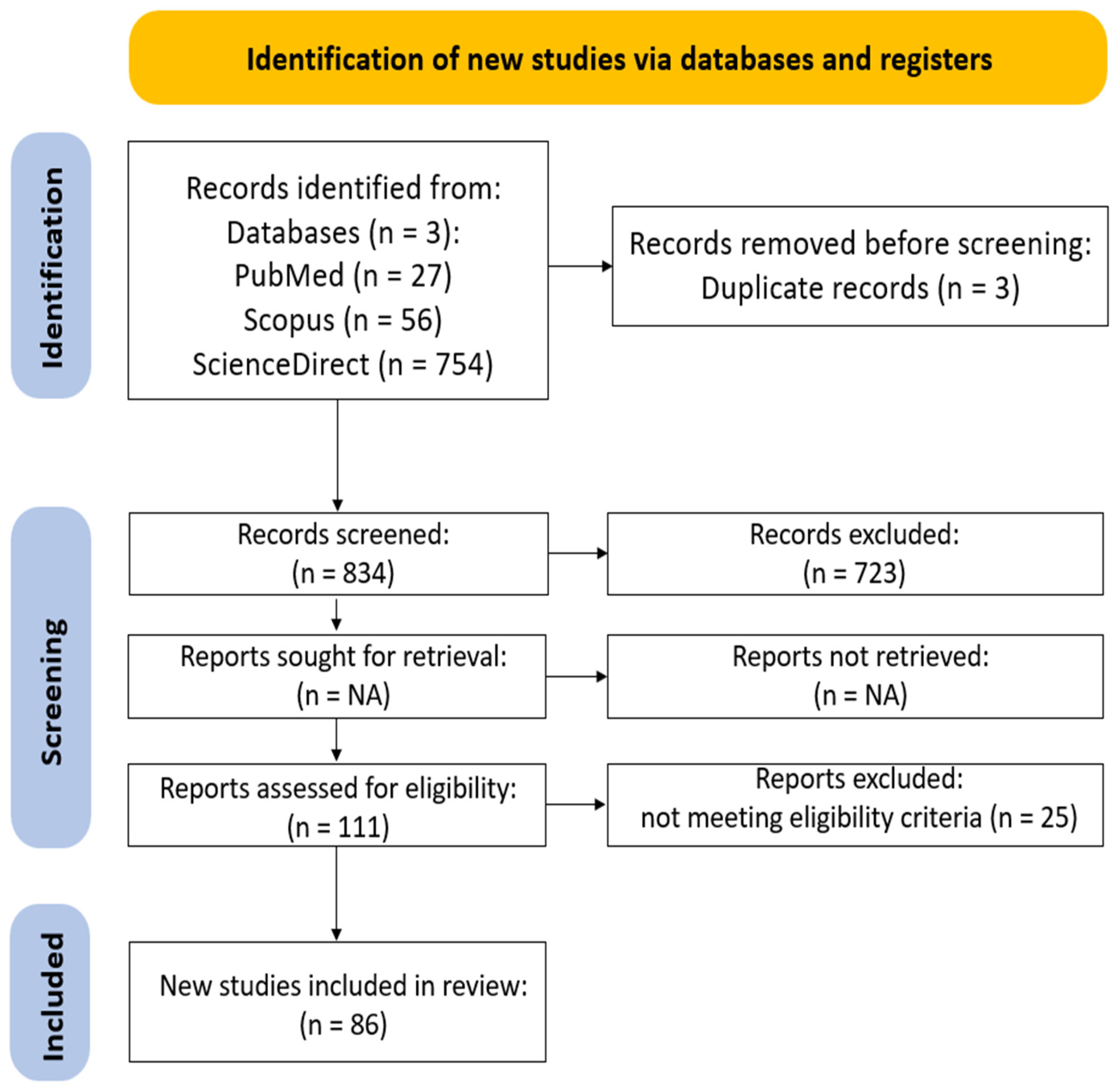
2.2. Inclusion/Exclusion Criteria and Selection Process
2.3. Analysis of Co-Occurrence of the Selected Keywords
2.4. Extraction and Definition of Data
3. Results
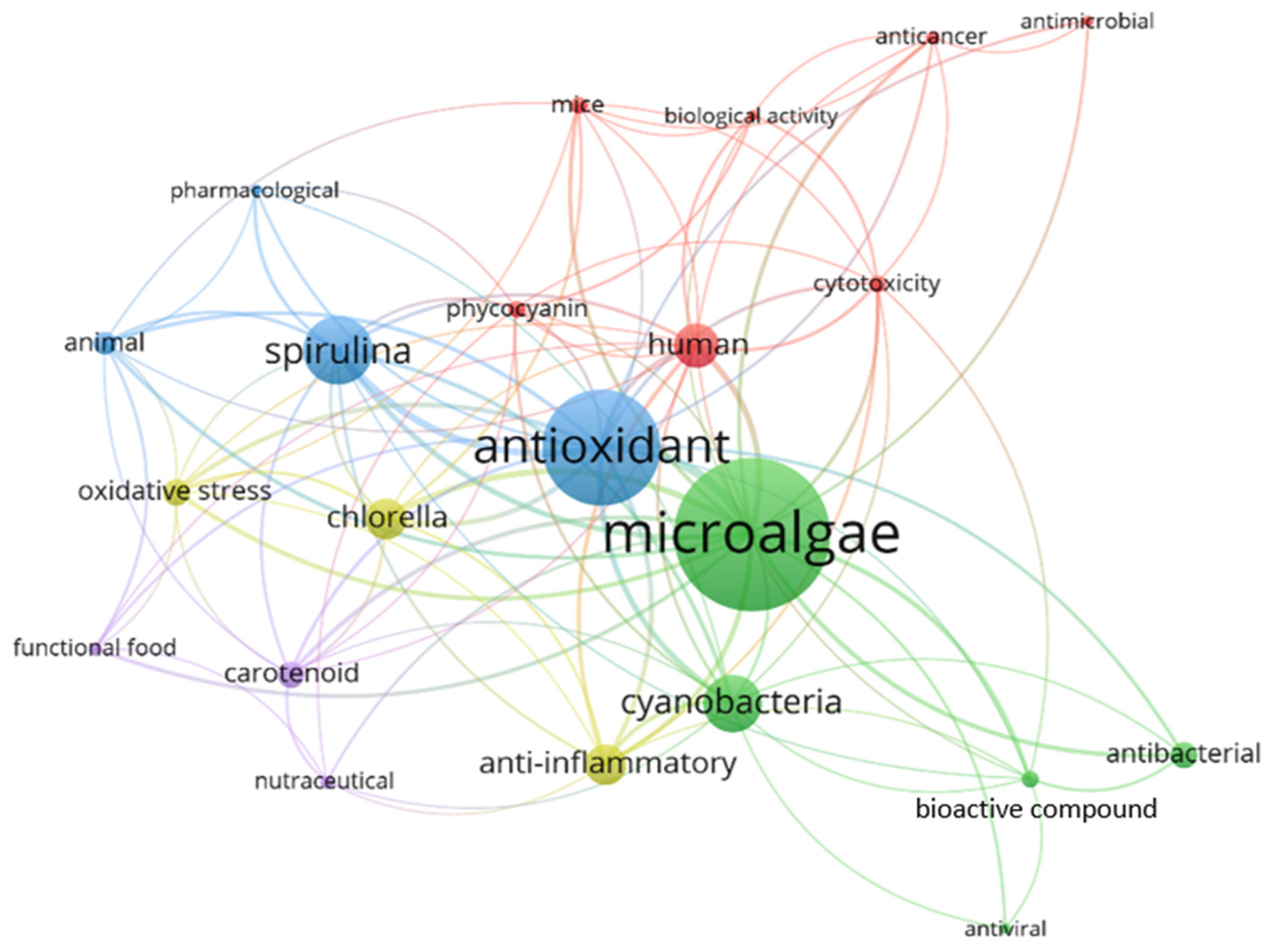
3.1. Keyword Frequency and Co-Occurrence Mapping
3.2. Study Types
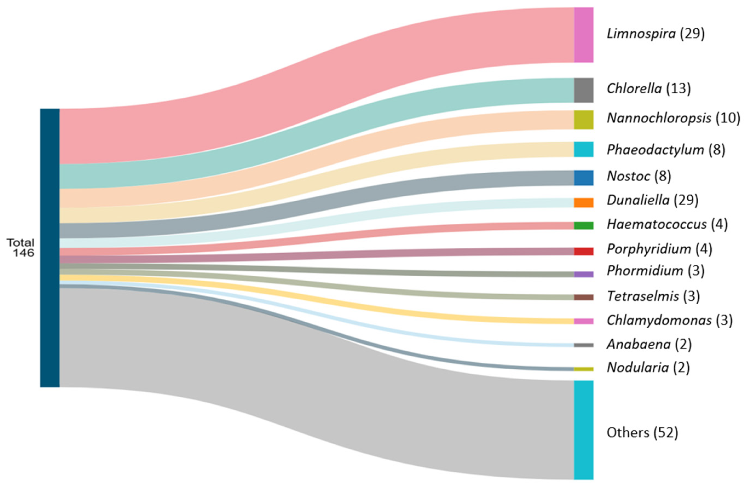
3.3. Genera Studied
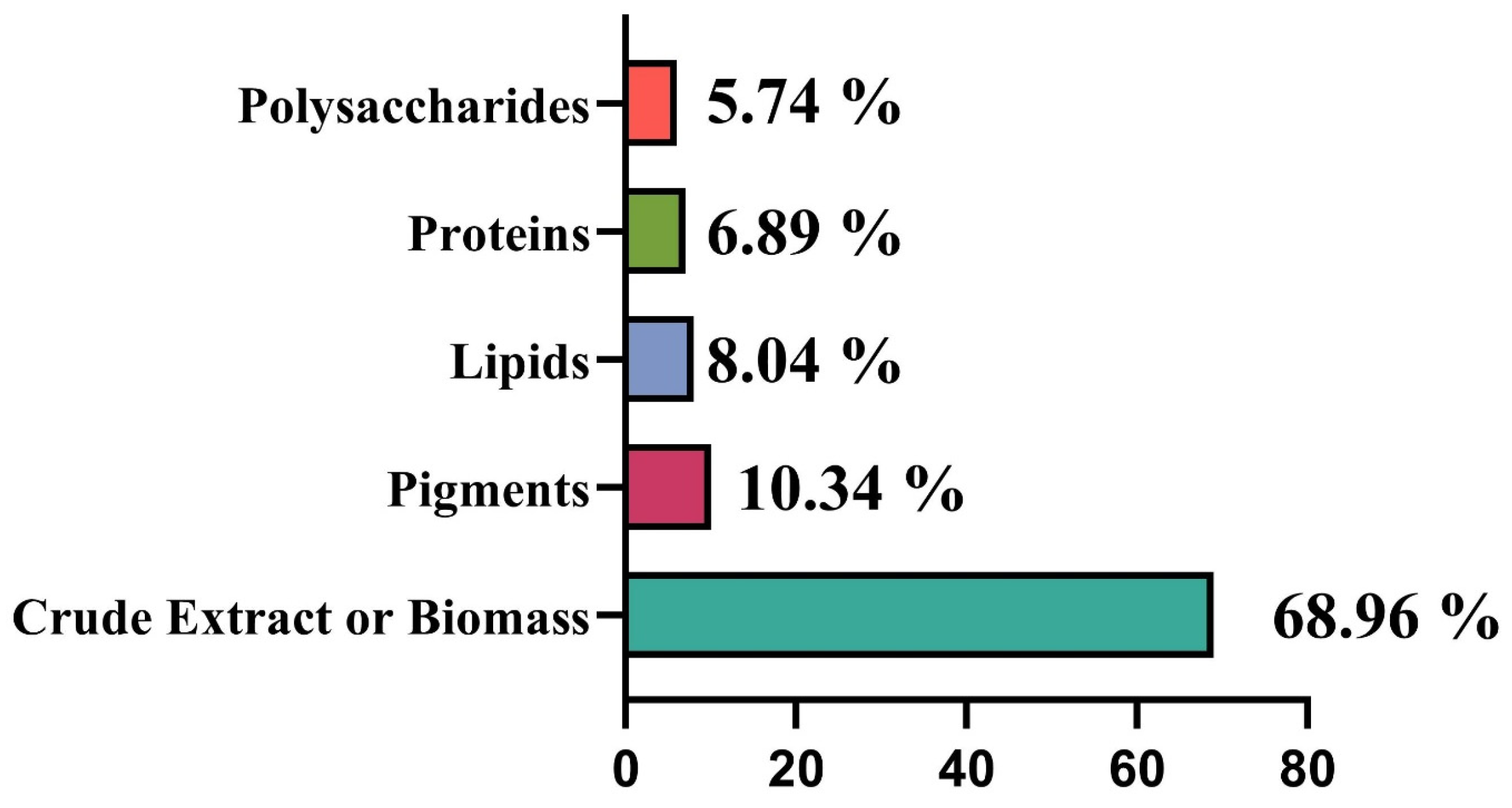
3.4. Cyanobacteria- and Microalgae-Based Products
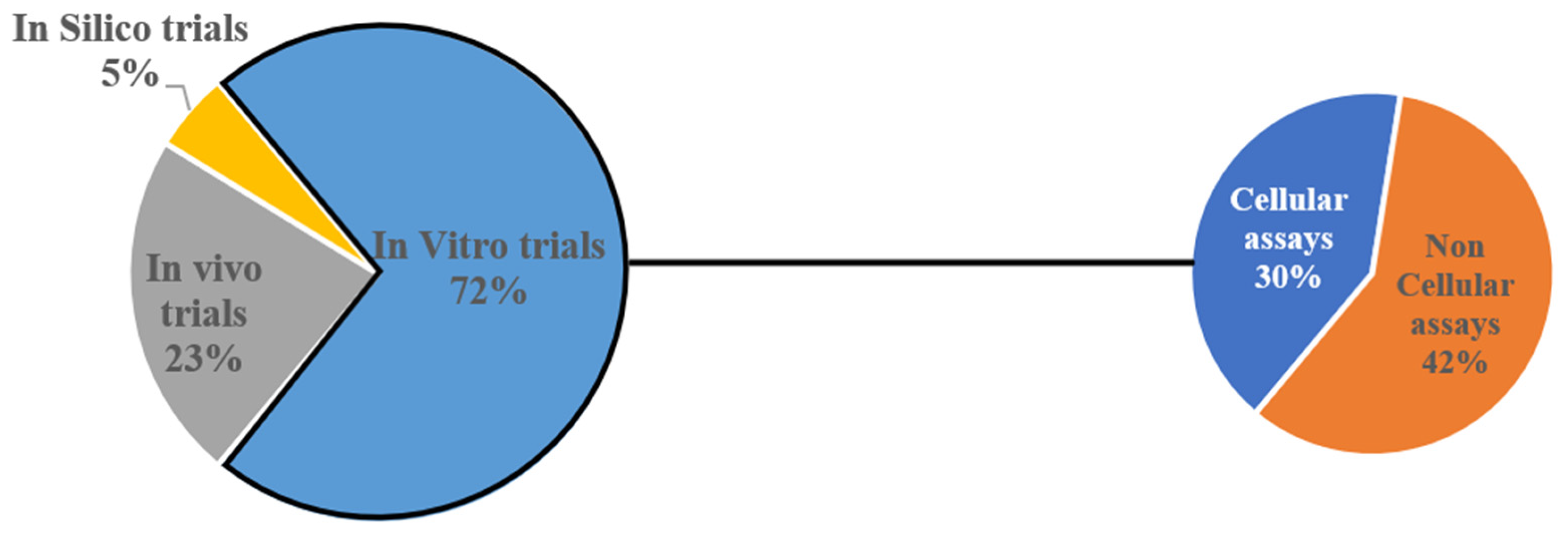
3.5. Study Models
4. Discussion
4.1. Genera
4.2. Microalgae- and Cyanobacteria-Based Products
4.2.1. Crude Extract or Biomass
4.2.2. Pigments
4.2.3. Lipids
4.2.4. Polysaccharides
4.2.5. Proteins
4.3. Antioxidant Potential
4.4. Anti-Inflammatory Properties
4.5. Antimicrobial Activity
4.6. Other Pharmacological Potentials
4.7. Nutritional and Dietary Applications
5. Conclusions and Future Perspectives
Author Contributions
Funding
Institutional Review Board Statement
Informed Consent Statement
Data Availability Statement
Acknowledgments
Conflicts of Interest
References
- Prasetiya, F.S.; Destiarani, W.; Nuwarda, R.F.; Rohmatulloh, F.G.; Natalia, W.; Novianti, M.T.; Ramdani, T.; Agung, M.U.K.; Arsad, S.; Sari, L.A.; et al. The Nanomolar Affinity of C-Phycocyanin from Virtual Screening of Microalgal Bioactive as Potential ACE2 Inhibitor for COVID-19 Therapy. J. King Saud Univ.—Sci. 2023, 35, 102533. [Google Scholar] [CrossRef] [Scilit] [PubMed]
- Mohammed, D.M.; El-Messery, T.M.; Baranenko, D.A.; Hashim, M.A.; Tyutkov, N.; Marrez, D.A.; Elmessery, W.M.; El-Said, M.M. Effect of Spirulina maxima Microcapsules to Mitigate Testicular Toxicity Induced by Cadmium in Rats: Optimization of in Vitro Release Behavior in the Milk Beverage. J. Funct. Foods 2024, 112, 105938. [Google Scholar] [CrossRef] [Scilit]
- Yan, B.; Chen, X.; Wang, Y.; Yuan, M.; Xian, J.; Lu, D.; Shao, Z.; Qiu, M.; Fu, T.; Zheng, X. Chlorella pyrenoidosa Ameliorates Ulcerative Colitis by Tuning Intestinal Microecology: Butyric Acid Is a Crucial Player. J. Funct. Foods 2024, 121, 106414. [Google Scholar] [CrossRef] [Scilit]
- Bhattacharjya, R.; Bansal, H.; Santoshi, S.; Rastogi, S.; Tiwari, A. Characterization of Natural Compounds Derived from Diatom C. gracilis as Potential Therapeutic Agents: An in-Silico Networking and Docking Study. Algal Res. 2024, 83, 103712. [Google Scholar] [CrossRef] [Scilit]
- Lobus, N.V.; Kulikovskiy, M.S. The Co-Evolution Aspects of the Biogeochemical Role of Phytoplankton in Aquatic Ecosystems: A Review. Biology 2023, 12, 92. [Google Scholar] [CrossRef] [Scilit] [PubMed]
- da Costa, E.; Amaro, H.M.; Melo, T.; Guedes, A.C.; Domingues, M.R. Screening for Polar Lipids, Antioxidant, and Anti-Inflammatory Activities of Gloeothece Sp. Lipid Extracts Pursuing New Phytochemicals from Cyanobacteria. J. Appl. Phycol. 2020, 32, 3015–3030. [Google Scholar] [CrossRef] [Scilit]
- Rizwan, M.; Mujtaba, G.; Memon, S.; Lee, K.; Rashid, N. Exploring the Potential of Microalgae for New Biotechnology Applications and beyond: A Review. Renew. Sustain. Energy Rev. 2018, 92, 394–404. [Google Scholar] [CrossRef] [Scilit]
- Dolganyuk, V.; Belova, D.; Babich, O.; Prosekov, A.; Ivanova, S.; Katserov, D.; Patyukov, N.; Sukhikh, S. Microalgae: A Promising Source of Valuable Bioproducts. Biomolecules 2020, 10, 1153. [Google Scholar] [CrossRef] [Scilit] [PubMed]
- Microalgae Market Size, Share, Growth and Industry Forecast 2030|Data Library Research. Available online: https://www.datalibraryresearch.com/reports/microalgaes-market-2882 (accessed on 26 August 2024).
- Research and Markets. Microalgae Global Market Report 2023: Sector to Reach $3.08 Billion by 2030 at a 9.4% CAGR. Available online: https://www.globenewswire.com/news-release/2023/02/14/2607336/28124/en/Microalgae-Global-Market-Report-2023-Sector-to-Reach-3-08-Billion-by-2030-at-a-9-4-CAGR.html (accessed on 26 August 2024).
- Vidyesh, S. Microalgae Market Size, Share, and Trends 2026 to 2035. Precedence Research. Available online: https://www.precedenceresearch.com/microalgae-market (accessed on 1 June 2026).
- Garcia, M.P.; Regueiras, A.; Lopes, G.; Matos, G.; Silva, L.P.d.; Cerqueira, M.T.; Cardoso, H.; Correia, N.; Saraiva, J.A.; Silva, J.L.; et al. Nonthermal High-Pressure Microalgae Extracts: A New Source of Natural Ingredients for Cosmetics. Algal Res. 2024, 81, 103591. [Google Scholar] [CrossRef] [Scilit]
- Office of the Commissioner. Classification of Products as Drugs and Devices and Additional Product Classification Issues. Available online: https://www.fda.gov/regulatory-information/search-fda-guidance-documents/classification-products-drugs-and-devices-and-additional-product-classification-issues (accessed on 22 June 2026).
- Puri, V.; Nagpal, M.; Singh, I.; Singh, M.; Dhingra, G.A.; Huanbutta, K.; Dheer, D.; Sharma, A.; Sangnim, T. A Comprehensive Review on Nutraceuticals: Therapy Support and Formulation Challenges. Nutrients 2022, 14, 4637. [Google Scholar] [CrossRef] [Scilit] [PubMed]
- Vignesh, A.; Amal, T.C.; Sarvalingam, A.; Vasanth, K. A Review on the Influence of Nutraceuticals and Functional Foods on Health. Food Chem. Adv. 2024, 5, 100749. [Google Scholar] [CrossRef] [Scilit]
- U.S. Food and Drug Administration. Questions and Answers on Dietary Supplements. Available online: https://www.fda.gov/food/information-consumers-using-dietary-supplements/questions-and-answers-dietary-supplements (accessed on 20 June 2026).
- Grande, T.; Vornoli, A.; Lubrano, V.; Vizzarri, F.; Raffaelli, A.; Gabriele, M.; Novoa, J.; Sandoval, C.; Longo, V.; Echeverria, M.C.; et al. Chlamydomonas agloeformis from the Ecuadorian Highlands: Nutrients and Bioactive Compounds Profiling and In Vitro Antioxidant Activity. Foods 2023, 12, 3147. [Google Scholar] [CrossRef] [Scilit] [PubMed]
- Nowruzi, B.; Zakerfirouzabad, M. Anti-Inflammatory Activities of Phycoerythrin and Phycocyanin on Human Fibroblast Cells. Phytomed. Plus 2024, 4, 100604. [Google Scholar] [CrossRef] [Scilit]
- Lin, M.-W.; Chiu, W.-H.; Lin, C.-H.; Liu, D.-H.; Wu, P.-C.; Lin, C.-S. Macular Pigments Produced from Microalga Chlorella Sp. and Applied to Alleviate the Pathogenic Process in Diabetic Mice. Algal Res. 2024, 78, 103414. [Google Scholar] [CrossRef] [Scilit]
- El-Baz, F.K.; Aly, H.F.; Abd-Alla, H.I. The Ameliorating Effect of Carotenoid Rich Fraction Extracted from Dunaliella salina Microalga against Inflammation- Associated Cardiac Dysfunction in Obese Rats. Toxicol. Rep. 2020, 7, 118–124. [Google Scholar] [CrossRef] [Scilit] [PubMed]
- Soliman, T.N.; El-Dein, A.N.; Al-Daim, S.A.; Allayeh, A.; Awad, H.; Flefil, N.S. Characterization of C-Phycocyanin Antioxidant, Anti-Inflammatory, Anti-Tumour, and Anti-HCoV-229E Activities and Encapsulation for Implementation in an Innovative Functional Yogurt. Heliyon 2024, 10, e31642. [Google Scholar] [CrossRef] [Scilit] [PubMed]
- Saraei, Z.P.; Nowruzi, B.; Morowvat, M.H. Studying the Effect of Magnetic Fields on Biological Activity of Phycocyanin Extracted from Desmonostoc alborzicum. Microbe 2024, 5, 100207. [Google Scholar] [CrossRef] [Scilit]
- Capek, P.; Matulová, M.; Šutovská, M.; Barboríková, J.; Molitorisová, M.; Kazimierová, I. Chlorella Vulgaris α-L-Arabino-α-L-Rhamno-α,β-D-Galactan Structure and Mechanisms of Its Anti-Inflammatory and Anti-Remodelling Effects. Int. J. Biol. Macromol. 2020, 162, 188–198. [Google Scholar] [CrossRef] [Scilit] [PubMed]
- Vijayakumar, S.; Menakha, M. Pharmaceutical Applications of Cyanobacteria—A Review. J. Acute Med. 2015, 5, 15–23. [Google Scholar] [CrossRef] [Scilit]
- Vaz, B.d.S.; Moreira, J.B.; Morais, M.G.d.; Costa, J.A.V. Microalgae as a New Source of Bioactive Compounds in Food Supplements. Curr. Opin. Food Sci. 2016, 7, 73–77. [Google Scholar] [CrossRef] [Scilit]
- Gómez, P.I.; Mayorga, J.; Flaig, D.; Castro-Varela, P.; Jaupi, A.; Ulloa, P.A.; Soto-Bartierra, J.; Henríquez, V.; Rojas, V. Looking beyond Arthrospira: Comparison of Antioxidant and Anti-Inflammatory Properties of Ten Cyanobacteria Strains. Algal Res. 2023, 74, 103182. [Google Scholar] [CrossRef] [Scilit]
- Silva, M.; Kamberovic, F.; Uota, S.T.; Kovan, I.-M.; Viegas, C.S.B.; Simes, D.C.; Gangadhar, K.N.; Varela, J.; Barreira, L. Microalgae as Potential Sources of Bioactive Compounds for Functional Foods and Pharmaceuticals. Appl. Sci. 2022, 12, 5877. [Google Scholar] [CrossRef] [Scilit]
- Noore, S.; Tiwari, B.K.; Jambrak, A.R.; Dukić, J.; Wanigasekara, J.; Curtin, J.F.; Fuentes-Grunewald, C.; O’Donnell, C. Extraction Yield and Biological Activity of Phycobiliproteins from Porphyridium purpureum Using Atmospheric Cold Plasma Discharge and Jet Systems. LWT 2023, 187, 115204. [Google Scholar] [CrossRef] [Scilit]
- Calleja-Gómez, M.; Abi-Khattar, A.-M.; Debs, E.; Rajha, H.N.; Maroun, R.; Martínez-Culebras, P.V.; Louka, N. Role of Infrared-Assisted Extraction of Antioxidant Phytochemicals in Enhancing Antimicrobial Activity of Phaeodactylum tricornutum. LWT 2024, 198, 116072. [Google Scholar] [CrossRef] [Scilit]
- Vahdati, S.N.; Lashkari, A.; Navasatli, S.A.; Ardestani, S.K.; Safavi, M. Butylated Hydroxyl-Toluene, 2,4-Di-Tert-Butylphenol, and Phytol of Chlorella sp. Protect the PC12 Cell Line against H2O2-Induced Neurotoxicity. Biomed. Pharmacother. 2022, 145, 112415. [Google Scholar] [CrossRef] [Scilit] [PubMed]
- Paramanya, A.; Abiodun, A.O.; Ola, M.S.; Ali, A. Enhancing the Quality and Antioxidant Capacity of Phycocyanin Extracted from Spirulina platensis PCC 7345: A Quality-by-Design Approach. Arab. J. Chem. 2024, 17, 105653. [Google Scholar] [CrossRef] [Scilit]
- Silva, R.; Gonçalves, T.; Morone, J.; Moreira, G.A.; Morais, J.; Hentschke, G.S.; Álvarez-Gutiérrez, P.E.; Batista-García, R.A.; Vasconcelos, V.; Lopes, G. Pigments Profile and Antioxidant Potential of Extremophile Cyanobacteria Isolated from the Mexican Volcanic Lake Chichonal. Algal Res. 2024, 81, 103578. [Google Scholar] [CrossRef] [Scilit]
- Feller, R.; Matos, Â.P.; Mazzutti, S.; Moecke, E.H.S.; Tres, M.V.; Derner, R.B.; Oliveira, J.V.; Junior, A.F. Polyunsaturated ω-3 and ω-6 Fatty Acids, Total Carotenoids and Antioxidant Activity of Three Marine Microalgae Extracts Obtained by Supercritical CO2 and Subcritical n-Butane. J. Supercrit. Fluids 2018, 133, 437–443. [Google Scholar] [CrossRef] [Scilit]
- Tiwari, O.N.; Mondal, A.; Bhunia, B.; Bandyopadhyay, T.k.; Jaladi, P.; Oinam, G.; Indrama, T. Purification, Characterization and Biotechnological Potential of New Exopolysaccharide Polymers Produced by Cyanobacterium Anabaena sp. CCC 745. Polymer 2019, 178, 121695. [Google Scholar] [CrossRef] [Scilit]
- Bulut, O.; Köse, I.E.; Sönmez, Ç.; Öktem, H.A. Antioxidant Activity of Micractinium sp. (Chlorophyta) Extracts against H2O2 Induced Oxidative Stress in Human Breast Adenocarcinoma Cells. Sci. Rep. 2024, 14, 27593, Erratum in Sci. Rep. 2025, 15, 4513. https://doi.org/10.1038/s41598-025-87734-w. [Google Scholar] [CrossRef] [Scilit] [PubMed]
- Phinyo, K.; Ruangrit, K.; Pekkoh, J.; Tragoolpua, Y.; Kaewkod, T.; Duangjan, K.; Pumas, C.; Suwannarach, N.; Kumla, J.; Pathom-Aree, W.; et al. Naturally Occurring Functional Ingredient from Filamentous Thermophilic Cyanobacterium Leptolyngbya sp. KC45: Phytochemical Characterizations and Their Multiple Bioactivities. Antioxidants 2022, 11, 2437. [Google Scholar] [CrossRef] [Scilit] [PubMed]
- Parkes, R.; Barone, M.E.; Herbert, H.; Gillespie, E.; Touzet, N. Antioxidant Activity and Carotenoid Content Responses of Three Haematococcus sp. (Chlorophyta) Strains Exposed to Multiple Stressors. Appl. Biochem. Biotechnol. 2022, 194, 4492–4510. [Google Scholar] [CrossRef] [Scilit] [PubMed]
- Melo, T.; Figueiredo, A.R.P.; da Costa, E.; Couto, D.; Silva, J.; Domingues, M.R.; Domingues, P. Ethanol Extraction of Polar Lipids from Nannochloropsis oceanica for Food, Feed, and Biotechnology Applications Evaluated Using Lipidomic Approaches. Mar. Drugs 2021, 19, 593. [Google Scholar] [CrossRef] [Scilit] [PubMed]
- Foo, S.C.; Lee, Z.S.; Yap, M.K.K.; Tan, J.W. The Antioxidant, Wound Healing Properties and Proteomic Analysis of Water Extracts from the Tropical Cyanobacteria, Nostoc NIES-2111_MUM004. 3 Biotech 2023, 13, 71. [Google Scholar] [CrossRef] [Scilit] [PubMed]
- Kumar, R.S.; Shakambari, G.; Ashokkumar, B.; Varalakshmi, P. Inhibition of Advanced Glycation End Products Formation and Inflammation in C. Elegans: Studies of Potential of Lyngbya sp. against Expression of Stress Related Genes and Live Cell Imaging. Biocatal. Agric. Biotechnol. 2019, 17, 233–241. [Google Scholar] [CrossRef] [Scilit]
- Concórdio-Reis, P.; Cardeira, M.; Macedo, A.C.; Ferreira, S.S.; Serra, A.T.; Coimbra, M.A.; Amorim, A.; Reis, M.A.M.; Freitas, F. Novel Exopolysaccharide Produced by the Marine Dinoflagellate Heterocapsa AC210: Production, Characterization, and Biological Properties. Algal Res. 2023, 70, 103014. [Google Scholar] [CrossRef] [Scilit]
- Casero, M.C.; Velázquez, D.; Pereira, A.; Tejedor, M.d.M.; García, L.; Quesada, A.; Cirés, S. Hidden inside Desert Rocks: Salinity Triggers an Increase in Exopolysaccharides from Endolithic Cyanobacteria with Anti-Inflammatory Potential. Algal Res. 2024, 84, 103817. [Google Scholar] [CrossRef] [Scilit]
- Guleria, S.; Simsek, H.; Chawla, P.; Relhan, A.; Bhasin, A. Evaluation of Cladophora and Chlamydomonas Microalgae for Environmental Sustainability: A Comparative Study of Antimicrobial and Photocatalytic Dye Degradation. Environ. Pollut. 2024, 340, 122806. [Google Scholar] [CrossRef] [Scilit] [PubMed]
- Fihri, R.F.; Ez-Zoubi, A.; Mbarkiou, L.; Amar, A.; Farah, A.; Bouchamma, E.O. Antibacterial and Antioxidant Activities of Chlorella vulgaris and Scenedesmus incrassatulus Using Natural Deep Eutectic Solvent under Microwave Assisted by Ultrasound. Heliyon 2024, 10, e35071. [Google Scholar] [CrossRef] [Scilit] [PubMed]
- Wang, F.; Cao, Y.; Guo, Y.; Zhu, Z.; Zhang, C. Evaluation of Antioxidant and Antibacterial Activities of Lipid Extracts from Eustigmatos cf. Polyphem (Eustigmatophyceae) and Preliminary Identification of Bioactive Compound. Algal Res. 2021, 59, 102446. [Google Scholar] [CrossRef] [Scilit]
- Wilkinson, I.V.L.; Castro-Falcón, G.; Roda-Serrat, M.C.; Purdy, T.N.; Straetener, J.; Brauny, M.M.; Maier, L.; Brötz-Oesterhelt, H.; Christensen, L.P.; Sieber, S.A.; et al. The Cyanobacterial “Nutraceutical” Phycocyanobilin Inhibits Cysteine Protease Legumain. Chembiochem Eur. J. Chem. Biol. 2023, 24, e202200455. [Google Scholar] [CrossRef] [Scilit] [PubMed]
- Mendonça, I.; Silva, D.; Conde, T.; Maurício, T.; Cardoso, H.; Pereira, H.; Bartolomeu, M.; Vieira, C.; Domingues, M.R.; Almeida, A. Insight into the Efficiency of Microalgae’ Lipidic Extracts as Photosensitizers for Antimicrobial Photodynamic Therapy against Staphylococcus aureus. J. Photochem. Photobiol. B 2024, 259, 112997. [Google Scholar] [CrossRef] [Scilit] [PubMed]
- Wali, A.F.; Al Dhaheri, Y.; Ramakrishna Pillai, J.; Mushtaq, A.; Rao, P.G.M.; Rabbani, S.A.; Firdous, A.; Elshikh, M.S.; Farraj, D.A.A. LC-MS Phytochemical Screening, In Vitro Antioxidant, Antimicrobial and Anticancer Activity of Microalgae Nannochloropsis oculata Extract. Separations 2020, 7, 54. [Google Scholar] [CrossRef] [Scilit]
- Borah, D.; Das, N.; Sarmah, P.; Ghosh, K.; Chandel, M.; Rout, J.; Pandey, P.; Ghosh, N.N.; Bhattacharjee, C.R. A Facile Green Synthesis Route to Silver Nanoparticles Using Cyanobacterium Nostoc carneum and Its Photocatalytic, Antibacterial and Anticoagulative Activity. Mater. Today Commun. 2023, 34, 105110. [Google Scholar] [CrossRef] [Scilit]
- İnan, B.; Özçimen, D. Preparation and Characterization of Microalgal Oil Loaded Alginate/Poly (Vinyl Alcohol) Electrosprayed Nanoparticles. Food Bioprod. Process. 2021, 129, 105–114. [Google Scholar] [CrossRef] [Scilit]
- Grubišić, M.; Šantek, B.; Zorić, Z.; Čošić, Z.; Vrana, I.; Gašparović, B.; Čož-Rakovac, R.; Šantek, M.I. Bioprospecting of Microalgae Isolated from the Adriatic Sea: Characterization of Biomass, Pigment, Lipid and Fatty Acid Composition, and Antioxidant and Antimicrobial Activity. Molecules 2022, 27, 1248. [Google Scholar] [CrossRef] [Scilit] [PubMed]
- Teneva, I.; Batsalova, T.; Bardarov, K.; Moten, D.; Dzhambazov, B. A Novel Approach for Fast Screening of a Complex Cyanobacterial Extract for Immunomodulatory Properties and Antibacterial Activity. Appl. Sci. 2022, 12, 2847. [Google Scholar] [CrossRef] [Scilit]
- Chatterjee, S.; Raval, I.; Raval, K.; Tapader, R.; Bhojani, G.; Pal, A.; Mishra, S. Influence of Microalgal Lipids from Chlorella variabilis (ATCC PTA 12198) in Reducing the Virulence Factors of Multidrug-Resistant Vibrio cholerae Variant Strains. LWT 2021, 135, 110047. [Google Scholar] [CrossRef] [Scilit]
- Marrez, D.A.; Badr, A.N.; El-Bahrawy, A.; Naeem, M.A. Algal Extracts Evaluation as an Antitoxicity Sustainable Solution against Aflatoxin B1 Toxicity in Rat Tissues. Toxicon 2024, 250, 108098. [Google Scholar] [CrossRef] [Scilit] [PubMed]
- Munawaroh, H.S.H.; Hazmatulhaq, F.; Gumilar, G.G.; Pratiwi, R.N.; Kurniawan, I.; Ningrum, A.; Hidayati, N.A.; Koyande, A.K.; Kumar, P.S.; Show, P.-L. Microalgae as a Potential Sustainable Solution to Environment Health. Chemosphere 2022, 295, 133740. [Google Scholar] [CrossRef] [Scilit] [PubMed]
- Xie, Z.; Meng, X.; Yu, S.; Jiang, L.; Pei, H. Continuous Extraction and Application Potential of Value-Added Products from a Promising Microalga Coelastrella sp. SDEC-28 for Green Microalgae-Based Industry. J. Clean. Prod. 2023, 428, 139364. [Google Scholar] [CrossRef] [Scilit]
- Xavier, G.; Lima Farias de Sousa, A.C.; Queiroz dos Santos, L.; Aguiar, D.; Gonçalves, E.; Santos Siqueira, A. Structural and Functional Analysis of Cyanovirin-N Homologs: Carbohydrate Binding Affinities and Antiviral Potential of Cyanobacterial Peptides. J. Mol. Graph. Model. 2024, 129, 108718, Erratum in J. Mol. Graph. Model. 2024, 130, 108788. https://doi.org/10.1016/j.jmgm.2024.108788. [Google Scholar] [CrossRef] [Scilit] [PubMed]
- Ribeiro, M.C.M.; Salles, T.S.; Moreira, M.F.; Barbarino, E.; Valle, A.F.d.; Couto, M.A.P.G. Antiviral Activity of Microalgae Extracts against Mayaro Virus. Algal Res. 2022, 61, 102577. [Google Scholar] [CrossRef] [Scilit]
- Selvaraju, K.; Raguraman, V.; Yadav, H.N.; Hariprasad, P.; Malik, A. Spectral Characterization and Binding Dynamics of Bioactive Compounds from Chlorella minutissima against α-Glucosidase: An in Vitro and in Silico Approach. Algal Res. 2023, 75, 103281. [Google Scholar] [CrossRef] [Scilit]
- Ghallab, D.S.; Shawky, E.; Khalifa, A.A.; Ibrahim, R.S. Insights into the Molecular Mechanisms of Spirulina platensis against Rheumatoid Arthritis through Integrative Serum Pharmacochemistry and Network Pharmacology Analysis. Food Biosci. 2024, 59, 103902. [Google Scholar] [CrossRef] [Scilit]
- Amjad, M.; Iqbal, M.; Faisal, A.; Junjua, A.M.; Hussain, I.; Hussain, S.Z.; Ghramh, H.A.; Khan, K.A.; Janjua, H.A. Hydrothermal Synthesis of Carbon Nanodots from Bovine Gelatin and PHM3 Microalgae Strain for Anticancer and Bioimaging applications. Nanoscale Adv. 2019, 1, 2924–2936. [Google Scholar] [CrossRef] [Scilit] [PubMed]
- Miceli, M.; Cutignano, A.; Conte, M.; Ummarino, R.; Romanelli, A.; Ruvo, M.; Leone, M.; Mercurio, F.A.; Doti, N.; Manzo, E.; et al. Monoacylglycerides from the Diatom Skeletonema marinoi Induce Selective Cell Death in Cancer Cells. Mar. Drugs 2019, 17, 625. [Google Scholar] [CrossRef] [Scilit] [PubMed]
- Konkel, R.; Milewska, A.; Do, N.D.T.; Duran, E.B.; Szczepanski, A.; Plewka, J.; Wieczerzak, E.; Iliakopoulou, S.; Kaloudis, T.; Jochmans, D.; et al. Anti-SARS-CoV-2 Activity of Cyanopeptolins Produced by Nostoc edaphicum CCNP1411. Antivir. Res. 2023, 219, 105731. [Google Scholar] [CrossRef] [Scilit] [PubMed]
- Sasaki, K.; Linh, T.N.; Hirano, A.; Tominaga, K.; Nukaga, S.; Nozaki, H.; Arimura, T.; Isoda, H. Microalgae Extract Induces Antidepressant-like Activity via Neuroinflammation Regulation and Enhances the Neurotransmitter System. Food Chem. Toxicol. 2022, 170, 113508. [Google Scholar] [CrossRef] [Scilit] [PubMed]
- Serwotka-Suszczak, A.M.; Marcinkowska, K.A.; Smieszek, A.; Michalak, I.M.; Grzebyk, M.; Wiśniewski, M.; Marycz, K.M. The Haematococcus pluvialis Extract Enriched by Bioaccumulation Process with Mg(II) Ions Improves Insulin Resistance in Equine Adipose-Derived Stromal Cells (EqASCs). Biomed. Pharmacother. 2019, 116, 108972. [Google Scholar] [CrossRef] [Scilit] [PubMed]
- Fagundes, M.B.; Alvarez-Rivera, G.; Mendiola, J.A.; Bueno, M.; Sánchez-Martínez, J.D.; Wagner, R.; Jacob-Lopes, E.; Zepka, L.Q.; Ibañez, E.; Cifuentes, A. Phytosterol-Rich Compressed Fluids Extracts from Phormidium autumnale Cyanobacteria with Neuroprotective Potential. Algal Res. 2021, 55, 102264. [Google Scholar] [CrossRef] [Scilit]
- Yu, H.Y.; Cho, D.-H.; Seo, D.; Yoo, C.; Park, S.-B.; Jung, W.K.; Jung, J.E.; Kim, H.-S.; Kim, J. Microalga Chlorella Sp. Biomass Containing High Lutein Prevents Light-Induced Photooxidation and Retinal Degeneration in Mice. Algal Res. 2024, 82, 103620. [Google Scholar] [CrossRef] [Scilit]
- Halaj, M.; Matulová, M.; Šutovská, M.; Barboríková, J.; Kazimierová, I.; Fraňová, S.; Přibyl, P.; Cepák, V.; Lukavský, J.; Capek, P. Chemico-Physical and Pharmacodynamic Properties of Extracellular Dictyosphaerium chlorelloides Biopolymer. Carbohydr. Polym. 2018, 198, 215–224. [Google Scholar] [CrossRef] [Scilit] [PubMed]
- Amato, A.; Terzo, S.; Marchesa, P.; Maffongelli, A.; Martorana, M.; Scoglio, S.; Mulè, F. Spasmolytic Effects of Aphanizomenon Flos Aquae (AFA) Extract on the Human Colon Contractility. Nutrients 2021, 13, 3445. [Google Scholar] [CrossRef] [Scilit] [PubMed]
- Khalil, S.R.; Elhady, W.M.; Elewa, Y.H.A.; El-Hameed, N.E.A.; Ali, S.A. Possible Role of Arthrospira platensis in Reversing Oxidative Stress-Mediated Liver Damage in Rats Exposed to Lead. Biomed. Pharmacother. 2018, 97, 1259–1268. [Google Scholar] [CrossRef] [Scilit] [PubMed]
- Castro-Ferreira, C.; Gomes-Dias, J.S.; Ferreira-Santos, P.; Pereira, R.N.; Vicente, A.A.; Rocha, C.M.R. Phaeodactylum tricornutum Extracts as Structuring Agents for Food Applications: Physicochemical and Functional Properties. Food Hydrocoll. 2022, 124, 107276. [Google Scholar] [CrossRef] [Scilit]
- Davani, L.; Terenzi, C.; Tumiatti, V.; De Simone, A.; Andrisano, V.; Montanari, S. Integrated Analytical Approaches for the Characterization of Spirulina and Chlorella Microalgae. J. Pharm. Biomed. Anal. 2022, 219, 114943. [Google Scholar] [CrossRef] [Scilit] [PubMed]
- Couto, D.; Conde, T.A.; Melo, T.; Neves, B.; Costa, M.; Cunha, P.; Guerra, I.; Correia, N.; Silva, J.T.; Pereira, H.; et al. Effects of Outdoor and Indoor Cultivation on the Polar Lipid Composition and Antioxidant Activity of Nannochloropsis oceanica and Nannochloropsis limnetica: A Lipidomics Perspective. Algal Res. 2022, 64, 102718. [Google Scholar] [CrossRef] [Scilit]
- Moreira, A.S.P.; Gonçalves, J.; Conde, T.A.; Couto, D.; Melo, T.; Maia, I.B.; Pereira, H.; Silva, J.; Domingues, M.R.; Nunes, C. Chrysotila pseudoroscoffensis as a Source of High-Value Polar Lipids with Antioxidant Activity: A Lipidomic Approach. Algal Res. 2022, 66, 102756. [Google Scholar] [CrossRef] [Scilit]
- Tsai, S.-C.; Huang, Y.-W.; Wu, C.-C.; Wang, J.-J.; Chen, Y.-T.; Singhania, R.R.; Chen, C.-W.; Dong, C.-D.; Hsieh, S.-L. Anti-Obesity Effect of Nostoc commune Ethanol Extract In Vitro and In Vivo. Nutrients 2022, 14, 968. [Google Scholar] [CrossRef] [Scilit] [PubMed]
- Fields, F.J.; Lejzerowicz, F.; Schroeder, D.; Ngoi, S.M.; Tran, M.; McDonald, D.; Jiang, L.; Chang, J.T.; Knight, R.; Mayfield, S. Effects of the Microalgae Chlamydomonas on Gastrointestinal Health. J. Funct. Foods 2020, 65, 103738. [Google Scholar] [CrossRef] [Scilit]
- D’Ambrosio, M.; Bigagli, E.; Cinci, L.; Cipriani, G.; Niccolai, A.; Biondi, N.; Rodolfi, L.; Zambelli, F.; Gencarelli, M.; Laurino, A.; et al. Arthrospira platensis F&M-C265 Reduces Cardiometabolic Risk Factors in Rats Fed a High Fat Diet. J. Funct. Foods 2024, 116, 106150. [Google Scholar] [CrossRef] [Scilit]
- Tranchida, N.; Inferrera, F.; Impellizzeri, D.; D’Amico, R.; Siracusa, R.; Fusco, R.; Cuzzocrea, S.; Cordaro, M.; Paola, R.D. Investigation of Neurochemical and Behavioral Alterations in the Brain during Fibromyalgia with Supplementation of Spirulina platensis. J. Funct. Foods 2024, 121, 106387. [Google Scholar] [CrossRef] [Scilit]
- Wang, M.; Morón-Ortiz, Á.; Zhou, J.; Benítez-González, A.; Mapelli-Brahm, P.; Meléndez-Martínez, A.J.; Barba, F.J. Effects of Pressurized Liquid Extraction with Dimethyl Sulfoxide on the Recovery of Carotenoids and Other Dietary Valuable Compounds from the Microalgae Spirulina, Chlorella and Phaeodactylum tricornutum. Food Chem. 2023, 405, 134885. [Google Scholar] [CrossRef] [Scilit] [PubMed]
- Silva, M.E.T.d.; Correa, K.d.P.; Martins, M.A.; da Matta, S.L.P.; Martino, H.S.D.; Coimbra, J.S.d.R. Food Safety, Hypolipidemic and Hypoglycemic Activities, and in Vivo Protein Quality of Microalga Scenedesmus obliquus in Wistar Rats. J. Funct. Foods 2020, 65, 103711. [Google Scholar] [CrossRef] [Scilit]
- Fernandes, F.; Martins, R.; Barbosa, M.; Valentão, P. Algae-Based Supplements Claiming Weight Loss Properties: Authenticity Control and Scientific-Based Evidence on Their Effectiveness. Mar. Drugs 2024, 22, 123. [Google Scholar] [CrossRef] [Scilit] [PubMed]
- Sikiru, A.B.; Arangasamy, A.; Alemede, I.C.; Guvvala, P.R.; Egena, S.S.A.; Ippala, J.R.; Bhatta, R. Chlorella vulgaris Supplementation Effects on Performances, Oxidative Stress and Antioxidant Genes Expression in Liver and Ovaries of New Zealand White Rabbits. Heliyon 2019, 5, e02470. [Google Scholar] [CrossRef] [Scilit] [PubMed]
- Minic, S.; Stanic-Vucinic, D.; Radomirovic, M.; Radibratovic, M.; Milcic, M.; Nikolic, M.; Velickovic, T.C. Characterization and Effects of Binding of Food-Derived Bioactive Phycocyanobilin to Bovine Serum Albumin. Food Chem. 2018, 239, 1090–1099. [Google Scholar] [CrossRef] [Scilit] [PubMed]
- Diniz, A.F.A.; Claudino, B.F.d.O.; Francelino, D.M.C.; Silva, J.M.A.d.; Barros, B.C.; Arruda, R.R.A.; Melchiades, M.K.d.N.; Ferreira, P.B.; Júnior, F.F.L.; Abreu, L.S.; et al. Arthrospira platensis Prevents Contractile Reactivity Damage in Obese Rats Fed a Hypercaloric Diet by Positive Modulating the Rho-A/Rho-Kinase Pathway, Inflammation and Oxidative Stress. J. Funct. Foods 2024, 115, 106116. [Google Scholar] [CrossRef] [Scilit]
- Machado, A.R.; Pinheiro, A.C.; Vicente, A.A.; Souza-Soares, L.A.; Cerqueira, M.A. Liposomes Loaded with Phenolic Extracts of Spirulina LEB-18: Physicochemical Characterization and Behavior under Simulated Gastrointestinal Conditions. Food Res. Int. 2019, 120, 656–667. [Google Scholar] [CrossRef] [Scilit] [PubMed]
- Gharbi, K.; Fathalli, A.; Fassatoui, C.; Romdhane, M.S.; Jenhani, A.B.R. Biochemical Analysis of Cyanobacterial Strains Isolated from Different Tunisian Inland Waters: Therapeutical and Nutritional Potential. Int. J. Biol. Chem. Sci. 2019, 13, 2286–2300. [Google Scholar] [CrossRef] [Scilit]
- Kopp, L.; Seethaler, B.; Neumann, U.; Bischoff, S.C. Oral Intake of the Microalgae Nannochloropsis oceanica, Chlorella vulgaris, or Phaeodactylum tricornutum Improves Metabolic Conditions in Hypercaloric-Fed Mice. J. Funct. Foods 2024, 121, 106429. [Google Scholar] [CrossRef] [Scilit]
- Bigagli, E.; Cinci, L.; Niccolai, A.; Biondi, N.; Rodolfi, L.; D’Ottavio, M.; D’Ambrosio, M.; Lodovici, M.; Tredici, M.R.; Luceri, C. Preliminary Data on the Dietary Safety, Tolerability and Effects on Lipid Metabolism of the Marine Microalga Tisochrysis lutea. Algal Res. 2018, 34, 244–249. [Google Scholar] [CrossRef] [Scilit]
- Magpusao, J.; Oey, I.; Kebede, B. Evaluation of the Bioprotective Potential of Gastrointestinal Digest Fractions of Arthrospira sp. and Nannochloropsis sp. Suspensions Treated with High Pressure homogenisation. Sustain. Food Technol. 2024, 2, 222–231. [Google Scholar] [CrossRef] [Scilit]
- de Marco Castro, E.; Shannon, E.; Abu-Ghannam, N. Effect of Fermentation on Enhancing the Nutraceutical Properties of Arthrospira platensis (Spirulina). Fermentation 2019, 5, 28. [Google Scholar] [CrossRef] [Scilit]
- Chen, H.; Zeng, F.; Li, S.; Liu, Y.; Gong, S.; Lv, X.; Zhang, J.; Liu, B. Spirulina Active Substance Mediated Gut Microbes Improve Lipid Metabolism in High-Fat Diet Fed Rats. J. Funct. Foods 2019, 59, 215–222. [Google Scholar] [CrossRef] [Scilit]
- Ekeuku, S.O.; Chong, P.N.; Chan, H.K.; Mohamed, N.; Froemming, G.R.A.; Okechukwu, P.N. Spirulina Supplementation Improves Bone Structural Strength and Stiffness in Streptozocin-Induced Diabetic Rats. J. Tradit. Complement. Med. 2022, 12, 225–234. [Google Scholar] [CrossRef] [Scilit] [PubMed]
- Santiago-Díaz, P.; Rico, M.; Rivero, A.; Santana-Casiano, M. Bioactive Metabolites of Microalgae from Canary Islands for Functional Food and Feed Uses. Chem. Biodivers. 2022, 19, e202200230. [Google Scholar] [CrossRef] [Scilit] [PubMed]
- Braga, A.R.C.; Nunes, M.C.; Raymundo, A. The Experimental Development of Emulsions Enriched and Stabilized by Recovering Matter from Spirulina Biomass: Valorization of Residue into a Sustainable Protein Source. Molecules 2023, 28, 6179. [Google Scholar] [CrossRef] [Scilit] [PubMed]
- Martínez, R.; García Beltrán, A.; Kapravelou, G.; Guzmán, A.; Lozano, A.; Gómez-Villegas, P.; León, R.; Vigara, J.; Galisteo, M.; Aranda, P.; et al. Nutritional and Functional Assessment of Haloarchaea and Microalgae from the Andalusian Shoreline: Promising Functional Foods with a High Nutritional Value. J. Funct. Foods 2024, 116, 106194. [Google Scholar] [CrossRef] [Scilit]
- Araj-Shirvani, M.; Honarvar, M.; Jahadi, M.; Mizani, M. Biochemical Profile of Dunaliella Isolates from Different Regions of Iran with a Focus on Pharmaceutical and Nutraceutical Potential Applications. Food Sci. Nutr. 2024, 12, 4914–4926. [Google Scholar] [CrossRef] [Scilit] [PubMed]
- Garg, D.; Dar, R.A.; Phutela, U.G. Characterization of Novel Euryhaline Microalgal Cultures from Punjab, India for Bioactive Compounds. Arch. Microbiol. 2022, 204, 370. [Google Scholar] [CrossRef] [Scilit] [PubMed]
- Tamala, J.K.; Maramag, E.I.; Simeon, K.A.; Ignacio, J.J. A Bibliometric Analysis of Sustainable Oil and Gas Production Research Using VOSviewer. Clean. Eng. Technol. 2022, 7, 100437. [Google Scholar] [CrossRef] [Scilit]
- Araújo, R.; Vázquez Calderón, F.; Sánchez López, J.; Azevedo, I.C.; Bruhn, A.; Fluch, S.; Garcia Tasende, M.; Ghaderiardakani, F.; Ilmjärv, T.; Laurans, M.; et al. Current Status of the Algae Production Industry in Europe: An Emerging Sector of the Blue Bioeconomy. Front. Mar. Sci. 2021, 7, 626389. [Google Scholar] [CrossRef] [Scilit]
- Aizpuru, A.; González-Sánchez, A. Traditional and New Trend Strategies to Enhance Pigment Contents in Microalgae. World J. Microbiol. Biotechnol. 2024, 40, 272. [Google Scholar] [CrossRef] [Scilit] [PubMed]
- Singh, S.; Shukla, M.; Verma, A.; Siddique, G.; Singh, D.; Dwivedi, S. Cyanobacteria and Microalgae as Promising Sources of Novel Bioactive Compounds: A Review. J. Adv. Microbiol. Res. 2025, 6, 255–263. [Google Scholar] [CrossRef] [Scilit]
- Conde, T.A.; Neves, B.F.; Couto, D.; Melo, T.; Neves, B.; Costa, M.; Silva, J.; Domingues, P.; Domingues, M.R. Microalgae as Sustainable Bio-Factories of Healthy Lipids: Evaluating Fatty Acid Content and Antioxidant Activity. Mar. Drugs 2021, 19, 357. [Google Scholar] [CrossRef] [Scilit] [PubMed]
- Soares, C.L.R.; Wilairatana, P.; Silva, L.R.; Moreira, P.S.; Vilar Barbosa, N.M.M.; da Silva, P.R.; Coutinho, H.D.M.; de Menezes, I.R.A.; Felipe, C.F.B. Biochemical Aspects of the Inflammatory Process: A Narrative Review. Biomed. Pharmacother. 2023, 168, 115764. [Google Scholar] [CrossRef] [Scilit] [PubMed]




| Phylum | Family | Species | Type of Extract and Method of Extraction | Activity | Main Finding | Possible Pharmaceutical/Therapeutic Application | References |
|---|---|---|---|---|---|---|---|
| Cyanobacteria | Microcystaceae | Gloeothece sp. | Polar lipids: Solvent-based (Methanol/Chloroform, Biphasic Extraction) | Antioxidant/anti-inflammatory | IC50 (μg mL−1): (O2·−): 939.8 ± 15.6 (·NO): 1033.7 ± 17.9. (COX-2): 58% inhibition of PG2. | NR | [6] |
| Cyanobacteria | Nodulariaceae, Prochlorococcaceae, Oscillatoriaceae, Microcoleaceae, Spirulinaceae, Nostocaceae, Nostocaceae, Nostocaceae, Desertifilaceae | Nodularia sp. (CCM-UdeC 012), Cyanobium sp. (CCM-UdeC 013), Phormidium sp. (CCM-UdeC 014), Nodularia sp. (CCM-UdeC 015), Limnospira maxima (formerly Arthrospira maxima) (CCM-UdeC 040), Spirulina subsalsa (CCM-UdeC 050), Nostoc sp. (CCM-UdeC 087), Nostoc linckia (CCM-UdeC 088), Anabaena iyengarii (CCM-UdeC 089), Desertifilum sp. (CCM-UdeC 135). | Crude extract or biomass: Aqueous extraction (water-based)—cold maceration (stirring at room temperature for 24 h) | Antioxidant/anti-inflammatory | DPPH: CCM-UdeC 012 and CCM-UdeC 015 strains (8.12 and 8.83 μmol TE g−1). ABTS and FRAP: CCM-UdeC 050 strain (33.26 ± 1.71, 40.97 ± 4.35 μmol TE/g−1) and CCM-UdeC 135 strain (33.69 ± 2.36, 36.93 ± 4.95 μmol TE/g−1). Percentage of hemolysis: CCM-UdeC 135 (56.7%)-CCM-UdeC 014 (52.8%) | NR | [26] |
| Chlorophyta, Rhodophyta, Heterokontophyta, Haptophyta, Heterokontophyta, Heterokontophyta, Cyanobacteria, Chlorophyta, Chlorophyta | Chlorodendraceae, Porphyridiaceae, Monodopsidaceae, Isochrysidaceae, Phaeodactylaceae, Skeletonemataceae, Spirulinaceae, Haematococcaceae, Chlorodendraceae | Tetraselmis striata CTP4, Porphyridium sp., Nannochloropsis sp., Tisochrysis lutea, Phaeodactylum tricornutum, Skeletonema sp., Limnospira (formerly Spirulina), Haematococcus lacustris (formerly Haematococcus pluvialis), Tetraselmis chui | Crude extract or biomass: solvent extraction (Ethanol; ethyl acetate; water) | Antioxidant/anti-inflammatory | P. tricornutum (ethyl acetate extract) IC50: 0.452 ± 0.002 mg/mL Skeletonema sp. (ethyl acetate extract) IC50: 0.447 ± 0.060 mg/mL H. pluvialis (ethanol extract) IC50: 0.408 ± 0.007 mg/mL. TNF-α Inhibition: significant downregulation of TNF-α production (p < 0.001) | Anticancer | [27] |
| Rhodophyta | Porphyridiaceae | Porphyridium purpureum | Crude extract or biomass: cold plasma-assisted extraction | Antioxidant | DPPH: (69.44 ± 0.10%). FRAP: 207.29 ± 12.96 μmol Fe2+/g | Anticancer | [28] |
| Bacillariophyta | Phaeodactylaceae | Phaeodactylum tricornutum | Crude extract or biomass: Infrared-assisted extraction, Water-based solvent | Antioxidant/Antimicrobial | DPPH: 870 µg TE/mL. S. aureus inhibition zones (~2 mm) | Antimicrobial | [29] |
| Cyanobacteria | Nostocaceae | Desmonostoc alborzicum | Pigments: cell disruption, Freeze–thaw extraction | Antioxidant/Antimicrobial | DPPH: 4.02 ± 0.10. Inhibition zone against: Citrobacter freundii (7.00 ± 0.00) Escherichia coli k12 (4.16 ± 1.44). Antifungal effect against Saprolegnia parasitica (6.66 ± 0.47). | Antimicrobial | [22] |
| Chlorophyta | Chlorellaceae | Chlorella sp. | Crude extract or biomass: chloroform, methanol, ethyl acetate and ethanol. | Antioxidant | ABTS (methanol extract): 36.7% at 0.5 mg/mL DPPH (methanol extract): 34.10% at 1 mg/mL FRAP (methanol extract): 237.08 μM Fe2+ at 1 mg/mL. | Anticancer/neuroprotectivity | [30] |
| Cyanobacteria | Spirulinaceae | Limnospira platensis (formerly Spirulina platensis) PCC 7345 | Pigments: phycocyanin, freeze–thaw method with sodium phosphate buffer (1 M, pH 7.0) | Antioxidant | DPPH- IC50: 40.70 µg/mL ABTS- IC50: 23.25 µg/mL NO- IC50: 17.74 µg/mL | Antidiabetic | [31] |
| Cyanobacteria | Tolypothrichaceae, Nostocaceae, Scytonemataceae, Oculatellaceae | Tolypothrix sp. LEGE 221228, Nostoc sp. LEGE 221229, Scytonema sp. LEGE 221230, Drouetiella sp. LEGE 221231 | Crude extract or biomass: organic solvent-based extraction with acetone—Aqueous extraction (water-based solubilization) | Antioxidant | (O2·−): Tolypothrix sp. LEGE 221,228 IC50 (53.75 μg/mL). (·NO): Tolypothrix sp. LEGE 221,228 IC50 (1220.00 ± 39.60). | NR | [32] |
| Heterokontophyta, Rhodophyta | Phaeodactylaceae, Monodopsidaceae, Porphyridiaceae | Phaeodactylum tricornutum, Nannochloropsis oculata, and Porphyridium purpureum (formerly Porphyridium cruentum) | Lipids: supercritical CO2 extraction (SC-CO2)/subcritical n-Butane extraction | Antioxidant | DPPH: (44.6%) | NR | [33] |
| Chlorophyta | Chlamydomonadaceae | Chlamydomonas agloeformis | Crude extract or biomass: solvent extraction using 80% methanol/water solution, cold maceration/solid–liquid extraction. | Antioxidant | ORAC (µmolTE/100 g DW): 4827.66 ± 1.33 FRAP (µmolTE/100 g DW): 45.53 ± 5.53 DPPH (µmolTE/100 g DW): 375.51 ± 3.6 | NR | [17] |
| Cyanobacteria | Aphanizomenonaceae | Anabaena sp. CCC 745 | Polysaccharides: Salt/EDTA-assisted extraction + dialysis + precipitation (for EPS purification) | Antioxidant | RPS scavenging activity: 71.77% | NR | [34] |
| Chlorophyta | Chlorellaceae | Micractinium sp. ME05 | Crude extract or biomass: Ultrasound-assisted solvent extraction. | Antioxidant | DPPH: 7.72 ± 0.95%/93.80 ± 6.28 µmol TE/g DW | Cytoprotective | [35] |
| Cyanobacteria | Leptolyngbyaceae | Leptolyngbya sp. KC45 | Crude extract or biomass: microwave-assisted solvent extraction using ethanol | Antioxidant | DPPH: IC50 = 35.51 mg/mL ABTS: IC50 = 3.31 mg/mL PFRAP: 12.51 mg GAE/g | Anticancer—enzyme related disease inhibition | [36] |
| Chlorophyta | Haematococcaceae | Haematococcus sp. CCAP 34/7, LAFW15, RPFW01 | Pigment (astaxanthin): 100% acetone extract, saponification using methanolic NaOH | Antioxidant | Strain RPFW01: highest FC activity (62.9–155.0 µmoL g−1 of DW) | NR | [37] |
| Heterokontophyta | Monodopsidaceae | Nannochloropsis oceanica | Lipids: Solvent Extraction: Chloroform/methanol (2:1, v/v) Folch method-based, Dichloromethane/methanol (2:1, v/v), Dichloromethane/ethanol (2:1, v/v)—Lipid Purification. | Antioxidant | ABTS (all extracts): 90% of inhibition under 500 µg/mL DPPH (E + USP extract): 55–60% of inhibition under 500 µg/mL | NR | [38] |
| Cyanobacteria | Nostocaceae | Nostoc NIES 2111_MUM004 | Crude extract or biomass: ultrasound-assisted aqueous extraction | Antioxidant | ABTS: 10.65 ± 0.34 mg FRAP: 110.71 mg β-carotene bleaching assay: 537.98 ± 58.15 mg | wound healing properties | [39] |
| Cyanobacteria | Nostocaceae, Spirulinaceae | Nostoc commune, Limnospira platensis (formerly Spirulina platensis) | Pigments: freeze–thaw aqueous buffer extraction | Anti-inflammatory | PE and PC extracts at: 1.95 ppm and 62.5 ppm for IL-6 release 7.81 ppm and 31.25 ppm for TNF-α release | Anticancer | [18] |
| Chlorophyta | Chlorellaceae | Chlorella vulgaris (BEIJ. 1890, strain P13/1998) | Polysaccharides/Method of extraction: NR | Anti-inflammatory | EPS50 elevated IL-12 levels: 13 pg.mL−1, p ≤ 0.05. IFN-γ levels were significantly increased in the EPS50 group: 33 pg.mL−1. | Anti-remodelling, anti-asthmatic | [23] |
| Cyanobacteria | Oscillatoriaceae | Lyngbya sp. | Crude extract or biomass: mechanical Lysis/cryogenic grinding with Liquid nitrogen, solvent extraction (Methanol/Chloroform 1:2), (Hexane:Ethanol:Methanol 2:1:1). | Anti-inflammatory/antioxidant | COX-2 Inhibition: IC50 = 117.98 ± 0.41 µg/mL DPPH: IC50 = 25.89 ± 0.21 µg/mL | NR | [40] |
| Cyanobacteria | Spirulinaceae | Limnospira platensis (formerly Spirulina platensis) | Pigments: C-phycocyanin = buffer-based mechanical disruption, freeze–thaw cycles | Anti-inflammatory/antioxidant/antiviral/anti-tumour | Inflammatory activity: 98.76 ± 0.065% DPPH: 99.12 ± 0.027%. Reduced viral infectivity by 56%, SI up to 36.76%. Anticancer activity: inhibiting up to 70% of liver (HepG-2), 74% of colon (HCT-116) and 62% of breast (MCF-7) cancer. | Anticancer | [21] |
| Dinoflagellata | Heterocapsaceae | Heterocapsa strain AC210 | Polysaccharides: dialysis-based purification of EPS, Initial separation of the culture broth at 11,000× g for 45 min. | Anti-inflammatory/antioxidant | IL-8 Reduction: 79.3%. IL-6 Suppression: 46.2%. ROS inhibition: 18.3% at the highest concentration. | NR | [41] |
| Chlorophyta | Chlorellaceae | Auxenochlorella pyrenoidosa (formerly Chlorella pyrenoidosa) | Crude extract or biomass: Ultrasound-assisted extraction = 0.5 g of processed CP → 50 mL of ultrapure water → Ultrasonication, 250 W/40 kHz/30 min | Anti-inflammatory | Reduced inflammatory cytokines (IL-6, IL-1β, TNF-α) Increased protective markers (SIgA, IL-22). Reduced pro-inflammatory cytokines (IFN-γ, IL-17, IL-23). | Ulcerative colitis therapy | [3] |
| Cyanobacteria | Gloeocapsaceae, Chroococcidiopsidaceae, Gloeocapsaceae, Chroococcidiopsidaceae, Acaryochloridaceae | Gloeocapsa sp. UAM572, Chroococcidiopsis sp. UAM574, Gloeocapsopsis sp. UAM575, Chroococcidiopsis sp. UAM577, Chroococcidiopsis sp. UAM579, Chroococcidiopsis sp. UAM584, Pseudoacaryochloris sp. UAM587. | Polysaccharides: The dry biomass was resuspended in 1 mL of Milli-Q water → EPS quantification. | Anti-inflammatory | Best inhibitor against neutrophil elastase: Chroococcidiopsis sp. UAM579—IC50 = 78 μg EPS/mL. | Antielastase | [42] |
| Chlorophyta | Cladophoraceae, Chlamydomonadaceae | Cladophora, Chlamydomonas | Crude extract or biomass: hot aqueous extraction method | Antimicrobial | Inhibition zones: 17.7 mm to 18.3 mm. MIC: 10.0 MBC: 12.0 mg/mL in the case of S. aureus bacteria. | NR | [43] |
| Chlorophyta | Chlorellaceae, Scenedesmaceae | Chlorella vulgaris, Scenedesmus incrassatulus | Crude extract or biomass: sonication-assisted microwave extraction method | Antimicrobial/antioxidant | MICs: 16 mg/mL against P. aeruginosa VOC extracts MICs: 1–2 mg/mL DPPH: SIDES2 (S. incrassatulus, IC50 = 3.98 mg/g) and CVDES1 (C. vulgaris, IC50 = 5.58 mg/g). | NR | [44] |
| Heterokontophyta | Eustigmataceae | Eustigmatos cf. Vischeria polyphem (formerly Eustigmatos polyphem) | Lipids: Freeze-dried biomass was suspended in 95% ethanol, ultrasound treatment at 40 °C for 60 min. | Antimicrobial/antioxidant | ORAOs: inhibition zones of 9.21 ± 0.51 mm and 9.68 ± 0.38 mm, against V. alginolyticus and V. parahaemolyticus, respectively. ABTS: ORAOs achieved 68.27% scavenging activity | NR | [45] |
| Cyanobacteria | Spirulinaceae | Limnospira platensis (formerly Spirulina platensis) PCC 7345 | Pigments: 20 g of phycocyanobilin-based colorant Linablue G1 suspended in 250 mL of 96% ethanol. Heated at 95 °C in an oil bath. Cooled, filtered through a Büchner funnel. | Antimicrobial | MICs > 64 μg mL−1 | NR | [46] |
| Cyanobacteria, Chlorophyta, Heterokontophyta | Microcoleaceae, Chlorococcaceae, Monodopsidaceae, Phaeodactylaceae, Scenedesmaceae, Chlorodendraceae | Limnospira platensis (formerly Arthrospira platensis), Oophila amblystomatis (formerly Chlorococcum amblystomatis), Nannochloropsis oceanica, Phaeodactylum tricornutum, Tetradesmus obliquus (formerly Scenedesmus obliquus), Tetraselmis chui | Lipids: Folch extraction method: Biomass (25 mg) was homogenized with a 2:1 (v/v) dichloromethane:methanol solution, drying and re-extraction, Centrifugation at 2000 rpm/10 min. | Antimicrobial | Lipid extracts from P. tricornutum, S. costatum, T. lutea, and P. gyrans: ≥6 log10 CFU mL−1 bacteria reduction. Residual activity was noted for P. gyrans, T. lutea, S. costatum, and D. salina, causing reductions of 0.6–3.6 log10 CFU mL−1 | NR | [47] |
| Heterokontophyta | Monodopsidaceae | Nannochloropsis oculata | Crude extract or biomass: ultrasound-assisted extraction with methanol | Antimicrobial/antioxidant | Strongest inhibition against S. aureus: MIC = 15.62 µg/mL, MBC = 31.25 µg/mL. DPPH assay: IC50 = 52.10 ± 0.85 µg/mL H2O2 assay: IC50 = 122.84 ± 2.32 µg/mL ABTS assay: IC50 = 96.95 ± 1.23 µg/mL | Anticancer | [48] |
| Cyanobacteria | Nostocaceae | Nostoc carneum | Crude extract or biomass Method of extraction: NR | Antimicrobial | Inhibition zone: 15 mm at the lowest concentration of AgNPs (5 µg) against Pseudomonas aeruginosa | Anticoagulative | [49] |
| Chlorophyta, Cyanobacteria | Botryococcaceae, Microcystaceae | Botryococcus braunii, Microcystis aeruginosa | Crude extract or biomass: Soxhlet extraction with ethanol | Antimicrobial | Inhibition zones: 20.00 ± 0.30 mm (BB oil) and 25.00 ± 0.20 mm (MA oil) against E. coli ATCC 8739. | NR | [50] |
| Heterokontophyta, Heterokontophyta, Chlorophyta, Chlorophyta, Chlorophyta, Cyanobacteria | Bacillariaceae, Staurosiraceae, Chlorellales, Chlorodendraceae, Chlorodendraceae, Halothecacae | Nitzschia sp. S5, Nanofrustulum shiloi D1, Picochlorum sp. D3, Tetraselmis sp. Z3, Tetraselmis sp. C6, Euhalothece sp. C1 | Crude extract or biomass: Ultrasound-assisted extraction using 100% methanol, 25–50 °C, 1 h. | Antimicrobial/antioxidant | Tetraselmis sp. C6 showed the strongest antibacterial activity against E. coli: 26.05 ± 0.07 mm TEAC > 40 µmol TE/g Tetraselmis sp. C6 had strong free radical scavenging capacity (0.87 ± 0.23 mg/mL). | NR | [51] |
| Cyanobacteria | Oscillatoriaceae | Phormidium papyraceum PACC 8600 | Crude extract or biomass: Solvent extraction with ultrasound-assisted extraction. | Antimicrobial | 20.28 mm inhibition zone against E. coli. | Immunomodulatory | [52] |
| Chlorophyta | Chlorellaceae | Chlorella variabilis (ATCC PTA 12198) | Lipids: Ultrasonication-assisted solvent extraction (Chloroform:Methanol—2:1) | Antimicrobial | Inhibition of Cholera Toxin (CT) Production: highest inhibition (95%) was observed with 0.2% TLCV. | Anti-virulence | [53] |
| Cyanobacteria, Heterokontophyta | Spirulinaceae, Catenulaceae | Limnospira platensis (formerly Spirulina platensis), Amphora coffeaeformis | Crude extract or biomass: aqueous extraction assisted by sonication, followed by centrifugation and lyophilization | Antitoxicity/antioxidant | Extracts markedly mitigated AFB1 induced toxic effects. Red blood cells 106/mm3: 6.02 ± 0.11 DPPH Assay: Limnospira extract: 179.56 ± 3.05 μmol TE/g DW Amphora extract: 141.66 ± 2.51 μmol TE/g DW ABTS Assay: Limnospira extract: 194.18 ± 3.21 μmol TE/g DW Amphora extract: 136.52 ± 1.14 μmol TE/g DW | NR | [54] |
| Cyanobacteria | Spirulinaceae | Limnospira platensis (formerly Spirulina platensis) | NR | Antidiabetic type-2 | α-Amylase Binding affinity: Pheophytin 0.4 kcal/mol Phycocyanobilin 2.6 kcal/mol β-carotene 2.0 kcal/mol Zeaxanthin 1.4 kcal/mol α-Glucosidase: Pheophytin 2.1 kcal/mol. | NR | [55] |
| Chlorophyta | Scenedesmaceae | Coelastrella sp. SDEC-28 | Crude extract or biomass: ultrasound-assisted solvent extraction using methanol, 1:20 (m/v) for crude polysaccharides, chloroform/methanol (2:1, v/v) mixture for lipids. | Anti-tumour/antioxidant | Methanol Extract: 76.61% inhibition of A375 cell at 1000 μg/mL IC50 of ME before purification: 357.81 µg/mL IC50 of ME after purification: 73.69 µg/mL DPPH· scavenging (crude polysaccharides): 91.18% at 2 mg/mL ABTS· scavenging (crude polysaccharides): 96.17% at 2 mg/mL | Anticancer | [56] |
| Cyanobacteria | Microcystaceae, Nostocaceae, Microcoleaceae | Microcystis aeruginosa NIES-298/PCC 9807, Nostoc calcicola, Nostoc sp., Planktothrix rubescens | NR | Antiviral | Nostoc calcicola with high carbohydrate affinity and favorable binding dynamics. Binding Free Energies: Site A: (Mannose Binding): 10–28 kcal/mol Site B: 2–26 kcal/mol | NR | [57] |
| Cyanobacteria, chlorophyta | Microcoleaceae, Chlorellaceae, Dunaliellaceae, Haematococcaceae | Limnospira maxima (formerly Arthrospira maxima) (LEAF046), Chlorella vulgaris (LEAF749), Dunaliella salina (LEAF754), Haematococcus lacusrtis (formerly Haematococcus pluvialis) | Crude extract or biomass: solvent extraction using DMSO | Antiviral | Viral Inhibition (IC50 values) against MAYV: H. pluvialis: 17.53 µg/mL D. salina: 16.00 µg/mL C. vulgaris: 12.19 µg/mL A. maxima: 36.47 µg/m | NR | [58] |
| Chlorophyta | Chlorellaceae | Mychonastes homosphaera (formerly Chlorella minutissima) | Crude extract or biomass: Soxhlet extraction with sequential solvent polarity (Hexane, Chloroform, Ethyl acetate, Acetone, Methanol, Water) | Antidiabetic type-2 | C. minutissima acetone extract exhibited α-Glucosidase inhibition potential with an IC50 value of 1.61 mg·mL−1. | NR | [59] |
| Chlorophyta | Chlorellaceae | Chlorella sp. AT1 | Pigments: Two-stage solvent extraction (Methanol + Diethyl ether/60% KOH). | Antidiabetic type-2 | Reduced glucose intolerance by 25% (p < 0.01) Reduced insulin resistance by 41% compared to diabetic mice (p < 0.05). | Antidiabetic | [19] |
| Heterokontophyta | Chaetocerotaceae | Chaetoceros neogracilis (formerly Chaetoceros gracilis) | Crude extract or biomass: solvent extraction followed by sonication-assisted extraction/methanol:chloroform:water mixture in a ratio of 2:1:2 | Anticancer/antidegenerative/anti-inflammatory/cardioprotective | Cancer-related pathways: 130 significant metabolic pathways (p ≤ 0.05) and gene counts >20. Neuroactive ligand-receptor interaction: 37 gene targets. Cardioprotective pathways: ligands binding with MTOR ranged from 6.981 to 5.149 kcal/mol. | NR | [4] |
| Cyanobacteria | Spirulinaceae | Limnospira platensis (formerly Spirulina platensis) | Crude extract or biomass: Ultrasound-assisted hydroalcoholic extraction (70% ethanol, 35 °C + 2 h). | Anti-rheumatoid arthritis/anti-inflammatory | SP significantly reduced: IL-6 by 46% MCP-1 by 46% MMP-9 by 41% TNF-α by 33% IL-1β by 49% IL-17 by 37% TLR4 (26%), NLRP3 (34%), ASC (37%) Pro-caspase-1 (32%), Caspase-1 (28%), NF-κB p65 (32%) | Anti-inflammatory reactions of RA | [60] |
| Chlorophyta | Scenedesmaceae | Pectinodesmus sp. PHM3 | Crude extract or biomass: Hydrothermal carbonization | Anticancer | On HCC 1954 (breast cancer) cells: CA nanodots: GI50 = 0.316–0.447 ng/mL CG nanodots: GI50 = 8.156–6.596 ng/mL On HCT 116 (colorectal cancer) cells: CA nanodots: GI50 = 0.542–0.715 ng/mL CG nanodots: GI50 = 23.860–14.524 ng/mL | Anticancer | [61] |
| Heterokontophyta | Skeletonemataceae | Skeletonema marinoi | Crude extract or biomass: Ultrasound-assisted extraction with methanol | Anticancer | 100% blockage of cells in the sub-G1 phase of the cell cycle after 48 h at 500 µg/mL | Anticancer | [62] |
| Cyanobacteria | Microcoleaceae | Limnospira platensis (formerly Arthrospira platensis) | Pigments: Maceration extraction assisted by vortexing and centrifugation | Antiviral | C-PC binds strongly to the ACE2: 9.7 kcal/mol. C-PC binds to ACE2 with high affinity: KD = 3.37 nM. | Anti-SARS-CoV-2 | [1] |
| Cyanobacteria | Nostocaceae | Nostoc edaphicum CCNP1411 | Crude extract or biomass: 150 g of biomass was homogenised by grinding and extracted five times with 75% methanol → vortexing (15 min) → sonication in ultrasonic bath (10 min) → centrifugation | Antiviral | Strongest antiviral activity by CCNP1411, IC50 = 80 ng/mL | Anti-SARS-CoV | [63] |
| Chlorophyta, Heterokontophyta, Chlorophyta, Chlorophyta, Heterokontophyta | Chlorellaceae, Monodopsidaceae, Chlorodendraceae, Chlorococcaceae, Phaeodactylaceae | Chlorella vulgaris, Nannochloropsis oceanica, Tetraselmis chui, Oophila amblystomatis (formerly Chlorococcum amblystomatis), Phaeodactylum tricornutum | Crude extract or biomass: High-pressure extraction = 25 mg of dried microalgae → Water, ethanol (48% or 96%), or acetone | Anticytotoxic | Low cytotoxicity (IC50 > 500 μg/mL) except for P. tricornutum in fibroblasts (IC50 < 150 μg/mL). Higher sensitivity (IC50 < 500 μg/mL) was observed in keratinocytes and melanocytes for most extracts. | Cosmetics | [12] |
| Chlorophyta | Botryococcaceae | Botryococcus terribilis | Crude extract or biomass: cold maceration extraction = 70% ethanol, room temperature, dark, 2 weeks | Antidepressant-like | Significant decrease in immobility time from day 2 onward (65.5 ± 6.1 s, 62.0 ± 9.8 s, 56.8 ± 13.3 s, 68.8 ± 8.3 s, 73.5 ± 4.6 s, 70.3 ± 11.9 s, and 71.7 ± 8.4 s on days 1–7, respectively) (p < 0.01). Significantly decreased (p < 0.01) expressions of BCL2, CD14, CD40LG, CXCL17, ICAM1, LY96, PLAU, and TRAF3 (74.1 ± 1.1%, 63.6 ± 1.6%, 62.7 ± 9.6%, 55.1 ± 4.0%, 51.7 ± 4.3%, 76.7 ± 5.8%, 60.4 ± 1.0%, and 63.0 ± 3.2%, respectively). | Anti-neurodegenerative | [64] |
| Chlorophyta | Haematococcaceae | Haematococcus lacustris (formerly Haematococcus pluvialis) | Crude extract or biomass: Ultrasound-assisted extraction with solvents (water and methanol) | Insulin resistance | Significant enhancement of EqASCs metabolic activity: 10%, 15%, and 20% (v/v) of the water extract. Decreased expression of mRNA for (IRS1): 0.3–0.4 Ct [target gene/GAPDH]. | Insulin sensitivity | [65] |
| Cyanobacteria | Oscillatoriaceae | Microcoleus autumnalis (formerly Phormidium autumnale) | Crude extract or biomass: T1 = Solvent extraction with heptane for 24 h. T2 = Chemical saponification with KOH (1 mL; 6% w/v) + chloroform (0.5 mL) and hexane (4 mL). | Neuroprotective | Acetylcholinesterase inhibition: IC50 = 65.80 μg/mL Lipoxygenase inhibition: IC50 = 58.20 μg/mL Antioxidant activity: IC50 = 7.40 μg/mL | Anticholinergic | [66] |
| Chlorophyta | Chlorellaceae, Monodopsidaceae, Chlorodendraceae, Chlorococcaceae, Phaeodactylaceae | Chlorella sp. HS5 | Crude extract or biomass: ethanolic extraction using high-pressure stirred extraction = Lyophilized Chlorella (400 kg) → 95% ethanol (1:10 biomass-to-solvent) | Retinoprotective | Reduced formation of oxidized A2E: IC50 = 30.28 ± 4.77 µg/ML. CEE restored cell viability dose-dependently by 98%. DCFH assay: Reduced oxidative stress. TUNEL assay: Decreased apoptosis. ONL thickness dropped after light exposure (from ~51 µm to ~16 µm). | Retinal degeneration | [67] |
| Chlorophyta | Chlorellaceae | Brachionococcus chlorelloides (formerly Dictyosphaerium chlorelloides) | Polysaccharides: algal biomass was removed by centrifugation at 2260× g → 96% ethanol precipitation in a ratio of 1:4 (v/v) → centrifugation at 3230× g → Freeze drying | Antitussive | The Dch biopolymer at a dose of 50 mg/kg significantly suppressed cough reflex to 2/300 min compared to control (8/300 min). | Antitussive | [68] |
| Cyanobacteria | Aphanizomenonaceae | Aphanizomenon flos-aquae | NR | Antispasmodic | Klamin (5–35 mg/mL) caused a dose-dependent decrease in the amplitude of the contractions (up to ~52% at 30 mg/mL). | Spasmolytic | [69] |
| Chlorophyta | Dunaliellaceae | Dunaliella salina | Crude extract or biomass: Solvent extraction by maceration (solvent: ethyl acetate:n-hexane 20:80, v/v) | Antiobesity | Improved adiponectin: +20.26%. Glucagon decreased: −41.43% in obese rats. Improved troponin I and PAI-1 levels: 46.06% and 52.15%. ICAM: +163.29%, VCAM: +35.38%, CRP: +84.48%. Col II: +112.52%, Col 3A1: +127.83%, LOX: +98.00% crf-DS amelioration: Col II: −83.30%/Col 3A1: −103.42%/LOX: −76.00% | Obesity-associated cardiac dysfunction | [20] |
| Cyanobacteria | Microcoleaceae | Limnospira platensis (formerly Arthrospira platensis) | NR | Antioxidative stress-mediated liver damage | liver/body weight ratio reduced to 121.2% SP/LA group: Blood lead decreased to +187.5% Liver lead decreased to +2666.6% TAC increased by 68% SOD & GSH improved CAT returned to normal MDA decreased to 256% | Antioxidative stress caused by metal exposure | [70] |
| Phylum | Family | Species | Method of Extraction | Biochemical Composition | Application | Main Findings | References |
|---|---|---|---|---|---|---|---|
| Heterokontophyta | Phaeodactylaceae | Phaeodactylum tricornutum | Heat-assisted extraction = Water/ethanol mixture (75/25, v/v) → 40 °C/80 °C, 1 h. Biomass concentration: 2% dried P. tricornutum in 100 mL solvent | 42% of crude protein, 18% carbohydrates, 12% lipids, and 33% of ash. Monosaccharides = Glucose (E40: 6.3 ± 0.8%)/Man, Gal, Xyl (E80: 4.3 ± 0.2%)/Fucose (E80: 2.9 ± 0.3%). | Emulsifying properties | Emulsifying index = 85–90%. | [71] |
| Chlorophyta, Cyanobacteria | Chlorellaceae, Microcoleaceae | Chlorella vulgaris, Limnospira platensis (formerly Arthrospira Platensis) | Sequential solvent extraction | Total lipids + pigments (% w/w): Chlorella: 11.44 ± 0.131% Limnospira: 11.24 ± 0.210% Total carotenoids + chlorophylls: Limnospira: 123.93 ± 6.13 mg/100 g Chlorella: 99.10 ± 5.45 mg/100 g Protein content: Limnospira: 65.38% ± 5.59% Chlorella: 53.78% ± 13.51% | Antioxidant | S3: IC50 = 90 ± 19.0 μg/mL S2: IC50 = 160.2 ± 0.0 μg/mL S5: IC50 = 180 ± 10.0 μg/mL C4: IC50 = 518 ± 79.0 μg/mL (lowest activity) C3: IC50 = 363 ± 73.0 μg/mL C1: IC50 = 303 ± 60.0 μg/mL | [72] |
| Heterokontophyta | Monodopsidaceae | Nannochloropsis oceanica 0011NN, Nannochloropsis limnetica 0065NA | Chloroform–methanol-based biphasic lipid extraction | Total Lipid Content: 23.6 ± 2.6% to 35.3 ± 0.8%. Phospholipids (μg/mg lipid extract): 57.6 ± 8.0 (NL-In) to 104.8 ± 3.3 (NO-Out). Glycolipids (μg/mg lipid extract): 233.2 ± 10.0 (NL-Out) to 274.8 ± 11.0 (NO-In). Neutral lipids + pigments: (22.9 ± 0.8%). | Antioxidant | IC15: ranging from 30.4 ±1.8 to 45.7 ± 1.6 μmol | [73] |
| Haptophyta | Isochrysidaceae | Chrysotila pseudoroscoffensis | Folch extraction method: Dichloromethane:Methanol (2:1, v/v) → Vortexing for 2 min → Repeated centrifugation (626× g) → aqueous washing → re-extraction → purification → Drying & quantification. | Moisture: 6.5 ± 0.4%. Ash: 45.5 ± 1.0%. Total Sugars: 11.0 ± 3.2%. Dominant sugars: uronic acids (53.8 mol%), glucose, and galactose. Proteins: 11.6 ± 0.4%. Lipids: 6.4 ± 0.0%. | Antioxidant | IC50 (ABTS): 111.9 μg/mL IC35 (DPPH): 234.8 μg/mL | [74] |
| Cyanobacteria | Nostocaceae | Nostoc commune | Solvent extraction technique using ethanol: 100 g powder with 300 mL ethanol 1:3 (w/v) → Room temperature, 1 h → Filtration → Concentration via rotary evaporation → Freeze-drying → Sonication | Total polyphenols: 25.89 ± 1.18 µg Total flavonoids: 19.32 ± 0.45 µg Total terpenoids: 926.53 ± 0.03 µg | Anti-obesity | NEE reduced the body weight (13.5%), fat tissue weight (13.3%), and the serum FFA (19.4%), TG (14.2%), TC (11.8%), and LDL-C (16.4%) of rats. | [75] |
| Chlorophyta | Chlamydomonadaceae | Chlamydomonas reinhardtii (THN6) | NR | NR | Gastrointestinal health | Mice given C. reinhardtii biomass: Lost less weight (12%) → weight gain by 2% Humans participants: LowGIS_1g: 85% Rarely Frequency of GIS LowGIS_3g: 73% Rarely Frequency of GIS HighGIS_1g: 57% Often Frequency of GIS HighGIS_3g: 44% Often Frequency of GIS | [76] |
| Cyanobacteria | Microcoleaceae | Limnospira platensis (formerly Arthrospira platensis) F&M-C265 | NR | NR | Cardiometabolic | Reduced liver weight: 32 gr (p < 0.05). Reduced triglycerides: 155 ± 12.62 mg/dL (p < 0.05). Reduced total cholesterol: 107 ± 3.45 mg/dL (p < 0.01). Reduced systolic and diastolic blood pressure: 136 ± 4.28 and 73 ± 3.92 mm Hg. | [77] |
| Cyanobacteria | Spirulinaceae | Limnospira platensis (formerly Spirulina platensis) | NR | Diadinoxanthin (28.01 ± 0.11 μg/g), Alloxanthin/Canthaxanthin (22.76 ± 0.04 μg/g) Diatoxanthin (100.11 ± 0.22 μg/g), Zeaxanthin (113.76 ± 0.15 μg/g), Antheraxanthin (27.20 ± 0.02 μg/g), Echinenone (24.95 ± 0.16 μg/g), β-carotene (1226.99 ± 7.67 μg/g). | Fibromyalgia | Reduced neuroinflammation (astrocyte and microglial activation). Limited oxidative stress (lower MDA, H2O2, NO; restored SOD, GSH, CAT). Restored biogenic amine levels (NE, DA, 5-HT). Improved behavioral symptoms (pain sensitivity, depression-like behaviors, and locomotion) | [78] |
| Cyanobacteria, chlorophyta, Heterokontophyta | Microcoleaceae, Chlorellaceae, Phaeodactylaceae | Limnospira platensis (formerly Arthrospira platensis), Chlorella vulgaris, and Phaeodactylum tricornutum | Pressurized liquid extraction | Limnospira: Magnesium: 383.5 mg, Phosphorus: 752.5 mg, Calcium: 798 mg, Iron: 96.8 mg, Zinc: 2.73 mg, Selenium: 0.11 mg Chlorella: Magnesium: 344.3 mg, Phosphorus: 1761.5 mg, Calcium: 593.7 mg, Iron: 259.1 mg, Zinc: 1.19 mg, Selenium: 0.07 mg P. tricornutum: Magnesium: 555 mg, Phosphorus: 269 mg, Calcium: 1910 mg, Zinc: 373 mg. | Dietary sup plements | PLE + 100% DMSO, the highest yields in Limnospira: Protein: 45 mg/g Polyphenols: 12.5 mg/g Chlorophyll a: 8.0 mg/g Chlorophyll b: 3.0 mg/g Carotenoids: 1.97 mg/g 33 mg/L of Fucoxanthin in P. tricornutum 26.50 mg/L of (all-E) β-carotene in Limnospira 3.31 mg/L of lutein in Chlorella | [79] |
| Chlorophyta | Scenedesmaceae | Tetradesmus obliquus (formerly Scenedesmus obliquus) | Ultrasound-assisted cell disruption | Protein content: 40.42%. Insoluble fibers: 16.23% Soluble fibers: 3.14% Phenolic compounds: 1.96% Carotenoids: 1.10% Fatty acids: Oleic acid (C18:1): 1.38% Linoleic acid (C18:2): 0.95% Linolenic acid (C18:3): 0.28% | Hypolipidemic and hypoglycemic | All diets showed > 75% digestibility. Decreased glucose Levels (mg/dL): M100 diet → 128.75 Decreased triglyceride Levels (mg/dL): M50 diet → 75.87 mg/dL | [80] |
| Cyanobacteria, Heterokontophyta | Microcoleaceae, Fucaceae | Limnospira platensis (formerly Arthrospira platensis), Fucus vesiculosus | Ethanolic extraction using ultrasound-assisted extraction followed by maceration | Total pigment content: 139.91 mg/g Chlorophyll a content: ~28% of total pigments. Highest TPC: F. vesiculosus from aquaculture: 11.34 µg GAE/mg F. vesiculosus based supplement E: 12.33 µg GAE/mg | Weight Loss | Metabolism of carbohydrates → inhibitory effects: F. vesiculosus + Limnospira supplement F: IC25 = 4.54 ± 0.81 µg/mL Limnospira-Supplement B: IC25= 10.17 ± 0.95 µg/mL F. vesiculosus (Wild): IC25= 30.59 ± 0.08 µg/mL Maximum aldose reductase activity: F. vesiculosus supplement E: IC25= 72.39 ± 9.88 µg/mL | [81] |
| Chlorophyta | Chlorellaceae, Microcoleaceae | Chlorella vulgaris | NR | NR | Oxidative stress and antioxidant genes expression in liver and ovaries | ▸In liver tissue: Mean MDA = 6.61 ± 1.54 nmol/mg (p < 0.001) Mean SOD = 169.70 ± 18.3 U/mg (p < 0.002) Mean CAT = 8.70 ± 0.30 U/mg (p < 0.010) Mean GSH = 9.71 ± 0.49 μmol/g (p < 0.001) ▸In uterus tissue: Mean MDA = 1.70 ± 0.46 nmol/mg (p < 0.001) Mean TAC = 244.00 ± 18.00 μmol/mg (p < 0.012) Mean SOD = 144.40 ± 18.4 U/mg (p < 0.002) Mean CAT = 3.87 ± 0.02 U/mg (p < 0.001) Mean GSH = 9.52 ± 0.48 μmol/g (p < 0.001) ▸Gene expression: In liver: sod1: 2.02 ± 0.35 fold change (p < 0.009) gpx1: 5.89 ± 0.76 fold change (p < 0.004) ▸In ovaries: sod1: 1.76 ± 0.61 fold change (not significant, p < 0.42) gpx1: 2.07 ± 0.39 fold change (p < 0.030) | [82] |
| Cyanobacteria | Spirulinaceae | Limnospira platensis (formerly Spirulina platensis) | Solvent extraction using DMSO | NR | Functional food additive | ▸High-affinity Binding: Binding constant (Ka) = 2 × 10⁶ M−1 ▸Improved Thermal Stability: The BSA–PCB complex shows increased thermal stability compared to free BSA or PCB. | [83] |
| Cyanobacteria | Microcoleaceae | Limnospira platensis (formerly Arthrospira platensis) | Ultrasound-assisted extraction using chloroform | NR | Obesity and intestinal diseases | Hypercaloric diet: Emax dropped to 32.7%, pEC50: increased to 6.6. A. platensis (25 mg/kg) in HCD: improved Emax = 63.9% and pEC50 = 6.2. With ROCK inhibitor (Y27632): HCD + A. platensis: Emax = 49.1%, pEC50 = 5.6. With NOS inhibitor (L-NAME): HCD + A. platensis: Emax = 34.1%, pEC50 = 4.6. With COX inhibitor (Indomethacin): HCD + A. platensis: Emax = 54.4% With NOX inhibitor (Apocynin): HCD + A. platensis: Emax = 40.9%, pEC50 = 4.8. | [84] |
| Cyanobacteria | Spirulinaceae | Limnospira (formerly Spirulina LEB-18) | Methanol-based solvent extraction | Caffeic acid: 47.02 µg/g extract Hydroxybenzoic acid: 54.66 µg/g extract Chlorogenic acid: 19.27 µg/g extract Gallic acid: 17.74 µg/g extract Protocatechuic acid: 11.06 µg/g extract | Gastrointestinal conditions | Initial size of lactoferrin-loaded liposomes: ▸After 1 min in simulated intestinal fluid (SIF): 2389 ± 215 nm (PDI = 0.69 ± 0.11) ▸After digestion in gastric fluid without pepsin: reduced to ~500 nm Initial ζ-potential (LF-loaded liposomes): +13.0 ± 0.4 mV In SGF: ▸Without pepsin: +9.2 ± 1.0 mV ▸With pepsin: 0 mV In SIF: ▸Without pancreatic lipase: –44.2 ± 0.4 mV | [85] |
| Cyanobacteria | Chroococcaceae, Microcoleaceae, Leptolyngbyaceae, Pseudanabaenaceae, Microcoleaceae, Spirulinaceae, Leptolyngbyaceae, Nodulariaceae, Aphanizomenonaceae | Chroococcus Chroo-CH, Limnospira platensis (formerly Arthrospira platensis) Arthro-KB, Leptolyngbya Osci-NB-01, Leptolyngbya Lepto-CH, Limnothrix Osci-BM-01, Planktothrix agardhii Plank-SS-01, Limnospira (formerly Spirulina Spir-ML), Lyngbya Lyng-ML, Anabaenopsis circularis Pseud-01, Raphidiopsis raciborskii (formerly Cylindrospermopsis raciborskii) Cyl-NB-05. | Fatty acids: solvent mixture of dichloromethane: methanol:0.7% NaCl (1:2:0.8, v/v/v). Proteins: Manual grinding of 50 mg of freeze-dried sample + 4 mL of distilled water → centrifugation. Carbohydrate: extraction/hydrolysis method: Acid hydrolysis of 100 mg dry biomass using 2.5 N HCl at 100 °C for 1 h. Phycobiliproteins: Tris-Cl buffer (pH 8.1) → Freezing/thawing cycles and sonication → Centrifugation at 12,000 rpm. | Protein Content (% DW) → Osci-BM-01: ~30%-Cyl-NB-05: ~30%-Chroo-CH: <5%. High polysaccharide → Lepto-CH: 46.6% DW-Cyl-NB-05: 33.3% DW-Chroo-CH: 30.6% DW-Other strains: <30% DW. Total phycobiliproteins max value: Cyl-NB-05: >3150 µg/mL PC: 2873 µg/mL APC + PE: 277 µg/mL PC across strains: 11 to 671 µg/mL | NR | ▸linolenic acid, (C18:3): 25%. ▸Plank-SS-01, Lepto-CH and Osci-NB 01, contained a high percentage of oleic acid (25%, 28%, and 27%, respectively). ▸Cylind-NB was the most productive of total phycobiliproteins = 3000 µg/mL. | [86] |
| Heterokontophyta, Chlorophyta, Heterokontophyta | Monodopsidaceae, Chlorellaceae, Phaeodactylaceae | Nannochloropsis oceanica, Chlorella vulgaris, Phaeodactylum tricornutum | NR | Crude protein [g/kg dry mass]: N. oceanica = 387/P. tricornutum = 429/C. vulgaris = 542. Crude lipids [g/kg dry mass]: N. oceanica = 167/P. tricornutum = 99/C. vulgaris = 104. Crude ash [g/kg dry mass]: N. oceanica = 237/P. tricornutum = 177/C. vulgaris = 71. Calcium [g/kg dry mass]: N. oceanica = 2.6/P. tricornutum = 24.1/C. vulgaris = 3.4. Phosphorus [g/kg dry mass]: N. oceanica = 11.9/P. tricornutum = 27.9/C. vulgaris = 14.0. Caloric value [MJ/kg dry mass]: N. oceanica = 22.0/P. tricornutum = 19.2/C. vulgaris = 22.6. | Metabolic conditions | Energy content in WSD group: 18.6 MJ/kg (4.4 kcal/g) feed. Caloric intake in the WSD groups: 13.6% (p < 0.01). N. oceanica: significant reduced liver steatosis in mice fed a WSD (from 17% to 4.7%, p < 0.002). | [87] |
| Haptophyta | Isochrysidaceae | Tisochrysis lutea | Fucoxanthin: Freeze-dried biomass treated with methanol, diethyl ether/petroleum ether, and NaCl aqueous solution (liquid–liquid extraction) → Evaporation, then resuspension in methanol/MTBE (4:1). | Proteins: 42.4% Fibers: 18.2% Ash: 13.1% Total Lipids: 7.9% (MUFAS: 3.8%/PUFAS: 4.1%) Carotenoids: 0.6% | Dietary safety and tolerability | Digestibility: lower than AIN-76 diet (−4%). Daily water intake: Doubled compared to controls (NaCl content: 1.5% vs. 0.3%). Urinary sodium excretion: 5.7 ± 0.45 mEq/24 h Relative heart weight: Increased (p < 0.05) Uric acid excretion: 0.15 ± 0.01 mg/24 h Liver weight: reduced (p < 0.05). Total cholesterol: Increased HDL cholesterol: +112% (p < 0.05) Fecal lipid excretion: +75% Plasma triglycerides: −73% (p = 0.06) Fucoxanthin in diet: 0.12% DHA intake (rats): ~30 mg/day ω-6/ω-3 ratio: 0.58 (favorable) | [88] |
| Cyanobacteria, Heterokontophyta | Microcoleaceae, Monodopsidaceae | Limnospira sp. (formerly Arthrospira sp.), Nannochloropsis sp. | High-pressure homogenisation: Biomass is rehydrated in distilled water at 8% (w/v) → Mechanical Pre-treatment: The suspension is mixed at 5000 rpm for 5 min → high-pressure homogenisation at 300, 600, and 900 bar. | Chlorophyll a (mg/mL): Limnospira = 0.12–0.36/Nannochloropsis = 0.11–0.24. Carotenoides (mg/mL): Limnospira = 0.19–0.29/Nannochloropsis = 0.10–0.17. Total phenolic content (mg GAE/g DM): Limnospira = 240.13–247.78 Nannochloropsis = 195.63–225.82 Protein content: Limnospira = 29–31% Nannochloropsis = 3.8–8.7% | Bioprotective potential | Digestibility: Limnospira and Nannochloropsis → Enhanced post-pancreatin FRAP: Limnospira → Unaffected by HPH Nannochloropsis → Improved at 900 bar only DPPH (%): Limnospira → ~46–48% Nannochloropsis → ~50–56% Bioprotective effect: Limnospira → best at 600 bar Cytotoxicity (>48 h): Limnospira and Nannochloropsis → Observed at ≥6 mg/mL | [89] |
| Cyanobacteria | Spirulinaceae | Limnospira maxima (formerly Spirulina maxima) UTEX LB2342 | Total fat was extracted using the Soxhlet method. | Proteins: 59.0% Carbohydrates: 19.11% Lipids: 3.12% Crude fiber: 3.05% Moisture: 7.42% Ash: 8.22% | Microcapsules for nutritional enrichment preventing toxicity | Significant improvement in: Testosterone: 3.69 ± 0.21 a ng/mL LH: 1.62 ± 0.54 ng/L FSH: 2.58 ± 0.51 ng/L TNF-α reduction: 59.32 ± 1.41 pg/mL Decrease in: MDA: 2.74 ± 0.41 nmol/mg ACP: 12.03 ± 0.84 U/g LDH: 93.11 ± 1.53 U/g | [2] |
| Cyanobacteria | Microcoleaceae | Limnospira platensis (formerly Arthrospira platensis) | NR | Fermentation Time TPC (mg GAE/g dw) 36 h 17.87 ± 0.77 Fermentation Time C-Phycocyanin (mg/g dw) 36 h 187.00 ± 3.79 | functional ingredient in nutraceuticals | Total phenolic content increased by 112% after 36 h. FRAP: improved by 85% at 36 h. ORAC: rose by 36% at 36 h. DPPH: 60% increase after 24 h. FMC increased by 94% after 72 h. | [90] |
| Cyanobacteria | Spirulinaceae | Limnospira platensis (formerly Spirulina platensis) | Sequential solvent extraction: Extraction with 95% ethanol 1:10 (w/v), 45 °C, 0.5 h → Filtrate 1 →Extraction with 55% ethanol → 1:10 (w/v), 45 °C, 0.5 h → Filtrate 2 →Extraction with water → 1:10 (w/v), 45 °C, 0.5 h → Filtrate 3 | NR | Lipid metabolism | Improved lipid profiles in HFD-fed rats over 8 weeks by: Lowering TG (up to 33.33%) Reducing TC (up to 27.62%) Elevating HDL-C (by 24.44%) Decreasing LDL-C significantly (p < 0.01) | [91] |
| Cyanobacteria | Spirulinaceae | Limnospira platensis (formerly Spirulina platensis) | NR | Crude protein, ash, lipid, carbohydrate, sugar, fiber, chlorophyll, phycocyanin, calcium, chromium, sodium, magnesium, gamma-linolenic acid, vitamin B12, vitamin D, total carotenoids (β-carotene). | Supplementation for an improved bone strength and stiffness. | Improved Tb.N (1/mm): 3.39 ± 0.69 Increased number of expressed OCN genes: 2.00 of relative expression. | [92] |
| Charophyta | Desmidiaceae, Spirogyraceae | Cosmarium blytii BEA0204, Cosmarium sp. BEA0208, Spyrogyra sp. BEA0666B | Ultrasound-assisted methanolic extraction | Carbohydrate content (% DW): Cosmarium sp. → 43.2% Cosmarium blytii → 41.8% Spirogyra sp → 35.9% Glutamic Acid (mg/g): Cosmarium sp. → 12 Cosmarium blytii → < 12 Spirogyra sp → < Cosmarium sp. | Functional Food and Feed Uses | Carbohydrate content: 35.8% to 43.3%. Glutamic acid: 3.63 to 12.2 mg/g. Highest in Cosmarium blytii: 24.02 mg/g. DPPH Assay: 21.2% | [93] |
| Cyanobacteria | Microcoleaceae | Limnospira platensis (formerly Arthrospira platensis) | Cold aqueous extraction | NR | Vegan emulsion | DPPH: 15.4 ± 0.16 to 43.0 ± 1.5 µmol TE/g FRAP: 169.38 ± 5.75 to 343.91 ± 21.12 µmol Fe2+/g | [94] |
| Chlorophyta | Dunaliellaceae, Chlorellales incertae sedis | Dunaliella salina C5, Dunaliella salina C13, Picochlorum spp HM1 | Treatment 1 (T1): Cold Ethanolic Extraction Treatment 2 (T2): Forced Cold Ethanolic Extraction Treatment 3 (T3): Forced Aqueous Extraction Treatment 4 (T4): Forced Aqueous Extraction | Protein (g/100 g DM) D. salina C13: 57.0 Fat (g/100 g DM) D. salina C13: 6.96 Carbohydrates (g/100 g DM) Picochlorum spp. HM1: 32.6 Ash (g/100 g DM) D. salina C13: 22.6 Total-P (mg/g ash) Picochlorum spp. HM1: 70.4 Energy (Kcal/100 g) Picochlorum spp. HM1: 366.3 Energy (Kjul/100 g) Picochlorum spp. HM1: 1531.0 | Nutrition and diet | ABTS (T4) extracts (highest in Picochlorum spp. HM1): 6928.4 ± 138.4 µg GAE/g | [95] |
| Chlorophyta | Dunaliellaceae | Dunaliella salina (IBRC-M 50030), Dunaliella viridis (IBRC-M 50069), Dunaliella spp. (IBRC-M 50065). | Protein: solubilization in phosphate buffer with polyvinylpyrrolidone Carbohydrate: Phenol-sulfuric acid method Dietary Fiber: Van Soest method Total Fat Content: Soxhlet extraction using petroleum ether Fatty Acid Profile: Bligh and Dyer method Pigment Content (Chlorophylls & Carotenoids): Biomass centrifuged and extracted with acetone Total Phenolic Compounds: 80% methanol extraction at 25 °C for 10 min → Mixed with Folin–Ciocalteu reagent and sodium carbonate | Protein: D. salina: 22.50 ± 0.22 Dunaliella sp.: 17.68 ± 0.11 D. viridis: 19.67 ± 0.15 Carbohydrate (mono- and disaccharide): D. salina: 12.37 ± 0.26 Dunaliella sp.: 7.59 ± 0.39 D. viridis: 10.26 ± 0.13 Lipid: D. salina: 13.19 ± 0.26 Dunaliella sp.: 25.02 ± 0.15 D. viridis: 20.80 ± 0.45 Dietary fiber: D. salina: 48.82 ± 0.08 Dunaliella sp.: 48.16 ± 0.13 D. viridis: 42.10 ± 0.14 Total phenolics (mg/100 g): D. salina: 1.87 ± 0.01 Dunaliella sp.: 1.68 ± 0.09 D. viridis: 2.42 ± 0.02 | Nutraceuticals and food | High pigment content in D. salina (~11.5%). DPPH: 55.63% of inhibition. High levels of polyunsaturated fatty acids: EPA (11.26%) and DHA (6.15%). | [96] |
| Chlorophyta | Scenedesmaceae | BGLR8, BGLR16 | Carbohydrates: Solvent-assisted thermal extraction Proteins: Alkaline thermal extraction Lipids: Solvent + ultrasonic extraction Carotenoids: Cold solvent extraction Flavonoids: Solvent extraction with complexing agents Astaxanthin: Acid hydrolysis + solvent extraction Phycocyanin: Inorganic acid extraction β-Carotene: Organic solvent extraction Total Phenols: Solvent extraction + redox assay | Carbohydrates BGLR16: 191 mg/g DW BGLR8: 124 mg/g DW Proteins BGLR16: 538 mg/g DW (53.8%) BGLR8: 511 mg/g DW (51.1%) Lipids BGLR16: >2% DW BGLR8: 86 mg/g DW (8.6%) Phenolic Compounds BGLR16: 3.62 mg/g DW BGLR8: 4.46 mg/g DW | Nutraceuticals and bioactive components | BGLR8 outperformed BGLR16 in terms of: Lipids: 86 mg/g Total chlorophyll: 29.42 mg/g Carotenoids: 28.82 mg/g Phenols: 4.46 mg/g Phycocyanin: 52 mg/g Astaxanthin: 19.27 mg/g β-Carotene: 5.6 mg/g Antioxidant activity: 31.73% | [97] |
Disclaimer/Publisher’s Note: The statements, opinions and data contained in all publications are solely those of the individual author(s) and contributor(s) and not of MDPI and/or the editor(s). MDPI and/or the editor(s) disclaim responsibility for any injury to people or property resulting from any ideas, methods, instructions or products referred to in the content. |
© 2026 by the authors. Licensee MDPI, Basel, Switzerland. This article is an open access article distributed under the terms and conditions of the Creative Commons Attribution (CC BY) license.
Share and Cite
Essahli, Z.; Sabour, T.; Boutkida, Y.; Lyamlouli, K.; Oukarroum, A.; Aanniz, T.; Merghoub, N.; Bouyahya, A.; Chamkhi, I. Cyanobacteria and Microalgae as Promising Sustainable Sources of Bioactive Phytochemicals for Nutraceutical and Pharmaceutical Applications. Int. J. Mol. Sci. 2026, 27, 6764. https://doi.org/10.3390/ijms27156764
Essahli Z, Sabour T, Boutkida Y, Lyamlouli K, Oukarroum A, Aanniz T, Merghoub N, Bouyahya A, Chamkhi I. Cyanobacteria and Microalgae as Promising Sustainable Sources of Bioactive Phytochemicals for Nutraceutical and Pharmaceutical Applications. International Journal of Molecular Sciences. 2026; 27(15):6764. https://doi.org/10.3390/ijms27156764
Chicago/Turabian StyleEssahli, Zouhair, Tarik Sabour, Yassine Boutkida, Karim Lyamlouli, Abdellah Oukarroum, Tarik Aanniz, Nawal Merghoub, Abdelhakim Bouyahya, and Imane Chamkhi. 2026. "Cyanobacteria and Microalgae as Promising Sustainable Sources of Bioactive Phytochemicals for Nutraceutical and Pharmaceutical Applications" International Journal of Molecular Sciences 27, no. 15: 6764. https://doi.org/10.3390/ijms27156764
APA StyleEssahli, Z., Sabour, T., Boutkida, Y., Lyamlouli, K., Oukarroum, A., Aanniz, T., Merghoub, N., Bouyahya, A., & Chamkhi, I. (2026). Cyanobacteria and Microalgae as Promising Sustainable Sources of Bioactive Phytochemicals for Nutraceutical and Pharmaceutical Applications. International Journal of Molecular Sciences, 27(15), 6764. https://doi.org/10.3390/ijms27156764